

Submitted:
08 August 2026
Posted:
11 August 2026
You are already at the latest version
Abstract
Metabolic dysfunction – Associated Steatotic Liver Disease (MASLD) represents the emerging leading cause of Chronic Liver Disease (CLD) worldwide, with a global prevalence of approx. 30% in the general population that parallels global rates of obesity and Type 2 Diabetes (T2D). At present no validated therapy is available to block or slow down disease progression to Metabolic dysfunction – Associated SteatoHepatitis (MASH), liver fibrosis and cirrhosis and hepatocellular carcinoma (HCC). At present there is a lack of reliable biomarkers able to identify MASH patients at risk of disease progression and/or HCC development. According to the knowledge that pro-inflammatory cytokines play a key role in MASLD/MASH progression and HCC development, in this review we will discuss the role in this disease and other CLD of Oncostatin M (OSM), a cytokine belonging to the IL-6 family, and of pathways involving OSM and its receptor β (OSM/OSMRβ axis). OSM and related pathways are emerging as selective in sustaining disease progression by promoting chronic inflammation and fibrogenesis. Moreover, OSM/OSMRβ axis is critically involved in MASH-related HCC development by affecting proliferation, angiogenesis, invasiveness and metastasis as well as by reshaping MASH-related tumour immune microenvironment. OSM/OSMRβ axis is then emerging as a selective MASH-related putative therapeutic target.
Keywords:
MASH
; OSM/OSMRβ axis
; chronic inflammation
; liver fibrosis
; hepatocellular carcinoma
1. Metabolic Dysfunction - Associated Steatotic Liver Disease (MASLD): A Worldwide Relevant Disease
Metabolic dysfunction – Associated Steatotic Liver Disease (MASLD) represents the emerging leading cause of Chronic Liver Disease (CLD) worldwide, with epidemiological studies indicating that MASLD affects globally approx. 1 billion individuals (including adults, children and adolescents) in the general population [1]. A recent meta-analysis performed by pooling MASLD prevalence estimates and ultrasound-defined MASLD, revealed an overall global prevalence of approx. 30.0 % [1]; moreover, in USA, Europe and Southeast Asia, the MASLD prevalence is estimated to further raise by 2030 by affecting in these areas more than 400 million of individuals [2]. Such a high global prevalence of MASLD has been proposed to parallel the worldwide rising rates of obesity and type 2 diabetes (T2D), with approx 65% of T2D patients having MASLD. This metabolic disease is currently viewed as the hepatic manifestation of the metabolic syndrome and then associated with additional metabolic risk factors [3]. MASLD can be actually diagnosed on the basis of the following major evidence: i) hepatic steatosis, as for histopathological analysis of liver biopsies or imaging techniques indicating the presence of more than 5% of hepatocytes with accumulation of triglycerides (TG) in association with at least one or more cardiometabolic risk factors (i.e., including altered levels of body mass index or BMI, fasting glucose, blood pressure, plasma triglycerides, plasma HDL/cholesterol). Diagnosis of MASLD also requires that these patients consume little or no alcohol beverages and the possibility to exclude the involvement of other metabolic, ereditary, toxic- or drug-induced causes of fatty liver [4].
1.1. MASLD as a Progressive Chronic Liver Disease: From Simple Steatosis to Cirrhosis and Hepatocellular Carcinoma (HCC) Development
MASLD, according to histopathological analyses, is currently defined as a spectrum of conditions ranging from simple steatosis to the progressive form of Metabolic dysfunction – Associated SteatoHepatitis (MASH) [5]. Approx 75-80% of MASLD patients present only simple steatosis, which is widely considered as a non-progressive or slowly progressive condition. The diagnosis of MASH implies morphological evidence of steatosis associated with aspects of hepatocyte injury and death (including ballooning, apoptosis and other kind of cell death such as necroptosis, pyroptosis and ferroptosis) as well as lobular and/or portal inflammatory infiltrate and a variable degree of fibrosis. MASH is then diagnosed in the remaining MASLD patients and MASH patients are at consistent risk to undergo fibrogenic progression towards a more advanced stage of CLD. Accordingly, about 15–20% of MASH patients can develop cirrhosis usually over 3-4 decades, with MASH-associated liver fibrosis representing the strongest predictor for disease-specific progression and mortality [6]. Finally, cirrhotic MASH patients carry a 1.3–2% per year risk of developing hepatocellular carcinoma or HCC and overall MASH is currently indicated as the most rapidly growing cause of HCC in liver transplant candidates [7,8].
HCC accounts for approx. 85-90% of all primary liver cancers, representing the sixth most common cancer and the third leading cause of cancer-related death worldwide. HCC is often diagnosed at an advanced stage in most patients (50-60%), with a survival at five years from diagnosis usually less than 30% due to the insufficient results obtained with conventional therapeutic approaches (i.e., surgical resection or, for unresectable tumours, transarterial chemoembolization or TACE, or systemic treatments), with some improvement more recently related to the use of immune-checkpoint inhibitors (ICI) in viral patients [1,5,7,8,9,10,11]. Unfortunately, the overall scenario is even more unsatisfying for MASLD/MASH patients. In addition to the very high prevalence of MASLD in the general population, one should consider that: i) differently from other etiologies, in which HCC mainly develops in cirrhotic patients, MASH-related HCC is now increasingly reported to develop also in non-cirrhotic patients [8,9]; ii) at present there is a lack of validated therapeutic strategies to block or slow down disease progression and of reliable biomarkers able to identify MASH patients at risk of disease progression and/or HCC development; iii) ICIs are less efficient in MASLD/MASH-related HCC, likely as a consequence of the distinctive disease-related metabolic and immune microenvironment [1,5,10].
1.2. The Metabolic and Pathophysiological Basis of MASLD/MASH: Of Fatty Acid, Carbohydrate and Liver X Receptors (LXR) / Cholesterol Metabolism
MASLD is closely linked to obesity and T2D [3,6,10,12] and the resulting metabolic changes reflect an imbalance in liver energy metabolism, due to an excess delivery of lipids and other substrates. Excess nutrients overcome the ability of the liver to oxidize these substrates or to export them incorporated into very low-density lipoproteins (VLDL). The major metabolic and pathophysiological determinants leading to MASLD can be summarized as follows: i) increased intake of food containing high levels of lipids, particularly saturated fatty acids (FA), and/or carbohydrates such mono- and di-saccharides, particularly fructose and sucrose, which in turn can activate de novo lipogenesis (DNL); ii) the development of insulin resistance (IR) in the expanded and inflamed white adipose tissue (WAT), resulting in increased lipolysis and increased fatty acid delivery to the liver. iii) the development of hepatic IR, strictly related to peripheral IR and more severe during disease progression. This results in an insufficient suppression of hepatic gluconeogenesis, decreased glycogen synthesis and redirection of glucose into lipogenic pathways; iv) development of lipid deposition in skeletal muscle, with the consequent development of local IR [3,6,10,12].
The following interconnected mechanisms represent the major contributors to steatosis development: i) increased DNL, resulting mainly from increased delivery to the liver of glucose and other carbohydrates like fructose (closely associated with hyperinsulinemia). This, in turn, results in ii) the up-regulation of key genes encoding for lipogenic enzymes regulating DNL, mainly through the involvement of sterol regulatory element binding-protein 1c (SREBP1c), carbohydrate response element binding-protein (ChREBP), LXR or peroxisome proliferator-activated receptor γ (PPARγ); iii) increased delivery of FA to the liver results in an increased esterification of fatty acids into TG, involving increased formation of diacylglycerol (DG) and increased activity of diacylglycerol O-acyltransferase 2 (DGAT2) [10,12,13].
In response to the increased FA load and TG formation, hepatocytes face several metabolic limitations including: i) decreased intrahepatic lipolysis, ii) decreased or insufficient TG export via VLDL, and iii) insufficient FA metabolism via mitochondrial and/or peroxisomal oxidation [10,12,13]. Some of these events can be exacerbated by variants of genes involved in lipid metabolism. In addition, increased mitochondrial β-oxidation of free fatty acids (FFA) may be potentially deleterious. Coenzyme A - linked FFA are shuttled into the mitochondrial matrix through carnitine O-palmitoyltransferase (CPT)1 and CPT2 but this leads to an excess of acetyl-CoA into the tricarboxylic acid cycle and of NADH into the mitochondrial electron transport chain. The resulting increased mitochondrial oxidative metabolism can lead to an exhaustion of superoxide dismutase (SOD)-2 and glutathione-peroxidase activity, overall resulting in an increased production of reactive oxygen species (ROS) and then of oxidative stress [10,13].
Disruption of lipid metabolism (as well as of glucose metabolism) represents not only a key driver for MASLD progression but also for MASH-related HCC development [11]. In fact, the above, interconnected, metabolic abnormalities (IR, increased DNL and gluconeogenesis, increased hepatic delivery and synthesis of FA, impaired β-oxidation, increased oxidative stress, and persistent inflammatory responses) synergize to promote hepatic fat accumulation, inflammation, and fibrosis, creating a pro-carcinogenic microenvironment. As an example, abnormal glucose and lipid metabolism can favor HCC development promoting cell proliferation, inhibiting apoptosis and inducing genetic mutations [11].
A relevant additional role in MASLD progression is played by LXR/Cholesterol pathways. LXRs are members of the nuclear receptor superfamily and pivotal regulators of lipid metabolism [14]. Elevated intracellular cholesterol concentrations trigger the synthesis of oxysterols, which in turn activate LXR-dependent transcriptional programs. These programs facilitate cholesterol efflux, suppress de novo synthesis and influx, and coordinate the metabolism of phospholipids and FA [15,16]. LXRs play a central, yet paradoxical, role in MASLD and MASH [13]. Human MASLD/MASH patients have significant increased hepatic expression of LXRα and its target genes which correlated with the severity of steatosis, inflammation, and fibrosis [13]. While LXR activation may be beneficial in reducing hepatic cholesterol levels and decreasing inflammation, it simultaneously promotes FA accumulation by increasing DNL that drives and worsen liver steatosis. Studies on mice lacking LXR and fed lipid-enriched diets report resistance to obesity, decreased lipogenic gene expression and decreased steatosis but still accumulate cholesterol that fuels steatohepatitis [13].
2. The Drivers of MASLD Progression to MASH, Liver Fibrosis, Cirrhosis and HCC
The matter analyzed in this section would require just by itself a dedicated review but since the present one is dedicated to the role of OSM/OSMRβ axis, here only a selection of major issues will be synthetically proposed as a sort of guide to the actual comprehension of how MASLD can progress. The interested reader can find more detailed description of the proposed issues in a number of excellent and authoritative reviews [3,6,10,11,12,13]. According to current literature data MASLD progression is a very complex matter of several mediators, cell types and mechanisms involving critical events like hepatocyte injury and death, chronic inflammatory response, fibrogenesis, genetic variants, lipotoxicity, oxidative stress and much more, including gut microbiome and dysbiosis. Along these lines, it is widely accepted that simple liver steatosis, detected in 75-80% of patients with a diagnosis of MASLD, usually obese and/or T2D patients, should be considered as a liver benign and essentially non-progressive condition (but note that these patients remain at significant risk to develop cardiovascular diseases and extrahepatic cancers) [3,10,11,12,13].
2.1. Hepatocyte Injury and Death: A Matter of Altered Metabolism, Lipotoxicity, Oxidative Stress and Mitochondrial Dysfunction
From a histopathological point of view, MASH is characterized by aspects of sublethal hepatocyte injury (often referred to as ballooning degeneration) [17] as well as of evidence of cell death. According to current literature cell death can be ascribed to different variants of regulated cell death, including mainly apoptosis, necroptosis, pyroptosis and ferroptosis as extensively reviewed elsewhere [18,19,20,21]. Although it is unclear which is the relative quantitative role of these different types of cell death to disease progression, damage-associated molecular patterns (DAMPs) released by dead hepatocytes are without any doubt critical for chronic induction of inflammatory response in MASH conditions [10,12,18,19,20,21].
Hepatocyte injury and death in conditions of MASH can be triggered by different stimuli, factors and conditions but with lipotoxicity, oxidative stress and mitochondrial dysfunction playing the most relevant role [10,12,18,19,20,21]. Lipotoxicity deserve a brief mention here since it is the consequence of the previously described excess hepatic delivery of lipid substrates to as well as of the strictly related impairment of lipid-related metabolic pathways. This particular scenario is believed to generate lipotoxic lipids that, by inducing critical events like mitochondrial dysfunction, endoplasmic reticulum (ER) stress and oxidative stress, in the end triggers hepatocyte injury and death and then genomic instability, persistent inflammation, and chronic activation of fibrogenesis [10,12,18,19,22]. Monounsaturated FA (in particular palmitate and stearate), ceramides, lysophosphatidyl choline, lysophosphatidic acid and diacylglycerols are the major directly cytotoxic lipids: these toxic lipids have been reported to induce apoptosis via activation of c-Jun-N-terminal kinases (JNK) and mitochondrial death pathways, as well as by inducing ER stress and inflammasome activation [19,22]. In addition, lipotoxicity can induce the release from fat laden hepatocytes of extracellular vesicles (EV) that can exert pro-inflammatory, pro-fibrogenic and pro-angiogenic effects on surrounding cells [23,24,25,26]. Cell injury can be exacerbated by free cholesterol [13,14] and its accumulation in hepatocytes, Kupffer cells (KC) and hepatic stellate cells (HSC) resulting in oxidative stress, mitochondrial dysfunction and ATP depletion, leading to either apoptosis or necrotic-like cell death [10,19].
2.2. Genetic Variants
Apart from mechanisms that can lead to hepatocyte injury and death, it now clear that MASLD progression to a more advance stage of the disease can occur mostly in patients carrying one or more genetic variants of genes encoding for proteins involved in lipid metabolism (patatin-like phospholipase domain containing 3 or PNPLA3, transmembrane 6 superfamily member 2 or TM6SF2, membrane bound O-acyltransferase domain-containing 7 or MBOAT7), glucose uptake (glucokinase regulator or GCKR), VLDL secretion (Apolipoprotein B or APOB, Microsomal triglycerides transfer protein or MTTP), mitochondrial lipid metabolism (Uncoupling protein 2) or in the control of oxidative stress, inflammation and fibrogenesis (Collagen type XIII alpha 1 chain or COL13A1, EF-Hand Calcium Binding Domain 4B or EFCAB4B, Farnesyl-diphosphate farnesyltransferase 1 or FDFT1, Interferon Lambda 4 or IFNL4) [19,27,28,29]. These gene variants have been reported to be strongly associated with the susceptibility not only to develop MASLD but also MASH and fibrosis (such as PNPLA3, TM6SF2, APOB, MBOAT7, IFNL4) or even to the development of HCC (PNPLA3 and APOB). This means that the presence of these variants represents for the single individual an additive and critical driver of progression in addition to the metabolic and dietary ones. Moreover, it has been proposed that MASLD itself should be considered as an inheritable disease on the basis of epidemiological, familial aggregation and twin studies [27,28,29]. The interested readers can find more extensive and detailed analysis of the relationships between the presence of genetic variants, the susceptibility to MASLD and disease progression in a number of authoritative reviews, [28,29,30,31].
2.3. Chronic Inflammatory Response: A Focus on the Role of Macrophages in MASLD Progression
MASLD progression in the previously described scenario of dysregulated metabolism and persistent hepatocyte injury and death is driven by the two essential and interconnected processes of chronic inflammatory response and chronic activation of repair mechanisms (i.e., liver fibrogenesis, see next section) [10,12,21,32].
Persistent inflammatory response is a process that fuels progression and fibrogenesis and involves primarily several innate and adaptive immune cells and the use of novel single cell multiomic technologies as well as of spatial trancriptomics, imaging mass cytometry and others advanced techniques has started to reveal some major issues: i) the hepatic scenario of innate and adaptive response in either patients or murine models is significantly reshaped during steatohepatitis, an issue that deeply affects disease progression and HCC development [33,34,35,36]; ii) MASLD-associated inflammation is reflecting systemic changes and is sustained and/or modulated by multiple intrahepatic and extrahepatic factors, with inflammatory signals coming from adipose tissue, gut, skeletal muscle, and bone marrow; iii) the disease progression relies on an extremely complex scenario of cell-to-cell interactions between innate immunity cells like KC, monocyte/macrophages and neutrophils recruited from peripheral blood, dendritic cells and cells of adaptive immunity cells (B and T lymphocytes, including cluster designation (CD) 4+ and CD8+ T lymphocytes, TH17 cells), innate lymphoid cells like NK and NKT cells, regulatory T cells (Treg), hepatic myofibroblasts and their precursor cells (mainly HSC and portal fibroblasts), with the additional contribution of injured hepatocytes, activated platelets and sinusoidal endothelial cells (SEC). This very complex scenario is synthetically summarized in Figure 1 [10,12,19,21,22,32,33,34,35,36]. In this section we will focus essentially on the prominent role of macrophages but the interested reader may find more details on the role of other innate or adaptive immunity cells in MASLD progression in a number of recent and authoritative reviews [10,12,33,34,37]. The role of adaptive immunity will be recalled later in relation to the development of MASH-related HCC.
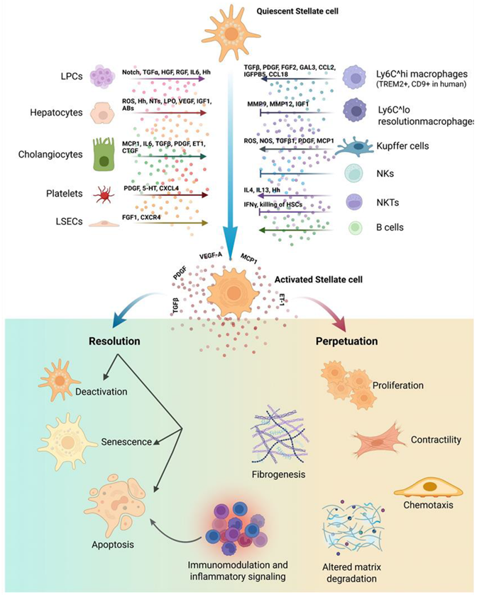
Figure Cell-to-cell signaling network regulating hepatic stellate cell (HSC) activation, resolution and perpetuation of the fibrogenic response. Quiescent HSCs are exposed to signals from parenchymal and non-parenchymal liver cells: liver progenitor cells (LPCs, via Notch, TGFα, HGF, RGF, IL6, Hedgehog (Hh)), hepatocytes (ROS, Hh, nucleotides (NTs), lipid peroxidation (LPO) products, VEGF, IGF1, apoptotic bodies (ABs)), cholangiocytes (MCP1, IL6, TGFβ, PDGF, ET1, CTGF), platelets (PDGF, serotonin (5-HT), CXCL4) and liver sinusoidal endothelial cells (LSECs, via FGF1, CXCR4), together with signals from innate and adaptive immune cells like Ly6C^high and Ly6C^low macrophages (TREM2+, CD9+ in humans), Kupffer cells, NK cells, NKT cells and B cells, releasing TGFβ, PDGF, FGF2, GAL3, CCL2, IGFBP5, CCL18, MMP9, MMP12, IGF1, ROS, NOS, TGFβ1, MCP1, IL4, IL13, Hh and IFNγ. Upon activation, HSCs release VEGF-A, PDGF, TGFβ, MCP1 and ET-1 (acting both in a autocrine and paracrine manner), driving either Resolution (through deactivation, senescence and/or apoptosis) or Perpetuation of the fibrogenic response (characterized by HSC proliferation, contractility, chemotaxis, fibrogenesis, altered matrix degradation and immunomodulatory/inflammatory signalling). Created in BioRender. Cannito, S. (2026) https://BioRender.com/nhturqw.
In this scenario a major role is unequivocally played by the activation of KC, relevant in the early phases of the disease, and the activation of macrophages recruited from circulating monocytes (MoMs) becoming predominant afterwards [33,34,38]. Literature data indicate that macrophages indeed play a dual role in the initiation and progression of MASLD and MASH and the following major issues can be recalled concerning MASLD progression: i) KC, that in normal conditions fulfil several functions as resident macrophages and represent a heterogeneous cell population in both human and murine liver [38], in early MASLD conditions are activated via pattern recognition receptors (PRR) in a pro-inflammatory phenotype by DAMPs (ADP, free cholesterol, mitochondrial DNA, etc) released by injured and fat-laden hepatocytes as well as by pathogen-associated molecular pattern (PAMPs) reaching the liver as a consequence of intestinal barrier dysfunction and microbial dysbiosis; ii) in conditions of persisting injury KC diminish in number and the inflammatory response is then sustained by macrophage derived from infiltrated Ly6Chigh monocytes (MoMs) which replenish KC niche to form the so called TREM2+ lipid-associated macrophages (LAMs) [38,39]; iii) TREM2+ LAMs form the so-called hepatic crown-like structures (hCLS), histopathologically evident structures in which macrophages accumulate around fat-laden and degenerating hepatocytes; in this early phase TREM2+ LAMs have an efficient efferocytic capacity that clear apoptotic cells and limit the detrimental accumulation of free lipids, thereby limiting the release of DAMPs and of inflammatory response (Wang et al. 2023); iv) under conditions of persistent lipotoxic injury LAMs shed TREM2 (released as soluble TREM2) and progressively loose efferocytic capacity and then LAMs or TREM2+ macrophages are reshaped into fully activated pro-inflammatory cells releasing IL-1β, TNFα and other mediators (see Figure 2) that drive both inflammation and fibrogenesis.
Before concluding this section, we would like to mention the putative role of the “liver-spleen axis” in the development of CLD, which refers to as a bidirectional interaction between the spleen and the liver through vascular, immunological and metabolic pathways [40]. The concept of the liver-spleen axis is suggested by clinical observations demonstrating a close relationship between the two organs during CLD progression. Splenomegaly and hypersplenism are well-recognized manifestations of liver cirrhosis [41], whereas in MASLD patients spleen enlargement has been reported to correlate with the severity of hepatic fat accumulation, further suggesting the existence of a functional crosstalk between the liver and the spleen [42,43]. Since, to the best of our knowledge, no evidence currently links OSM or OSM-related pathways directly to the liver-spleen axis in MASLD, we will only briefly summarize some major concepts emerging from recent studies, particularly those related to inflammatory and immune mechanisms [40,44,45]. As it is well known, the spleen serves as a major immunologic organ, reservoir and modulator of myeloid and lymphoid cells. Recent evidence suggests that obesity and MASLD are associated with a splenic immune remodeling that may contribute to the maintenance of systemic low-grade inflammation and to the amplification of hepatic inflammatory responses [40,46]. In particular, we would like to focus on the following issues: i) the liver-spleen axis has been proposed to sustain obesity-induced systemic inflammation and fatty liver disease through the enrichment and activation of myeloid-derived suppressor cells (MDSCs) and NK/NKT cell populations, which subsequently modulate hepatic immune responses [47]; ii) experimental evidence suggests that CD11b+ CD43hi Ly6Clo splenocyte-derived macrophages may contribute to the expansion of the hepatic macrophage compartment during MASLD progression to promote chronic inflammation and fibrogenesis [48]; iii) a very recent study, that confirmed enlarged spleens in patients with MASLD, identifed induced TRNP1hi CD8+ T cells in the spleens of MASLD/MASH mouse models and patients; these cells exhibited pro-fibrogenic properties through secretion of INSR-α that in the end contribute to activate HSC [49]. Overall, although the potential interaction between the OSM/OSMRβ axis and the liver-spleen axis has not yet been investigated, these studies suggest that the spleen may operate as an active contributor of liver disease progression, an issue that deserves further investigation. The interested reader may find more details and issues on the role of spleen-liver axis in a very recently published review [40].
2.4. Fibrogenesis
Liver fibrogenesis, the biological process leading to fibrosis (i.e., excess deposition of extracellular matrix or ECM), is mainly sustained by a heterogeneous population of hepatic α-smooth muscle actin (αSMA) positive cells defined as myofibroblasts (MF). MF originate from different precursor cells of mesenchymal origin through a process defined as of activation/transdifferentiation [10,50,51,52,53]. Hepatic MF, in addition to be activated by a plethora of mediators (growth factors, cytokines, chemokines, ROS, adipokines, proangiogenic factors, released by KC and macrophages, damaged hepatocytes, SEC, platelets, cholangiocytes, NK, NKT and other cells of adaptive immunity (i.e., as in any form of CLD), can also respond to metabolic signals as well as signals related to gut dysbiosis or delivered from adipose tissue [10,50,51,52,53]. The following major issues regarding the role of liver MF in the scenario of MASLD/MASH (see also Figure 1) can be summarized as follows [10,50,51,52]: i) MF originate in progressive MASLD mainly from activation/transdifferentiation of quiescent HSC and only marginally from other precursors like portal fibroblasts; ii) as described for other CLD of different aetiology MF operate the classic phenotypic responses including a) active proliferation in response mainly to platelet-derived growth factor (PDGF), transforming growth factor α (TGFα), connective tissue growth factor (CTGF), basic fibroblast growth factor (bFGF) and leptin, b) excess deposition of ECM as induced by transforming growth factor β1 (TGFβ1), bFGF, CTGF); c) reprogramming of ECM remodeling due to overexpression of tissue inhibitors of metalloprotease (TIMP) and of matrix-metalloproteases (MMP) not efficient to remove fibrillar collagen), d) pro-angiogenic role responding to hypoxic conditions by releasing vascular endothelial growth factor (VEGF), Angiopoietin-1 and -2, Hedgehog ligands); e) pro-inflammatory role by releasing CC chemokine ligand-2 or -21 (CCL2, CCL21), interleukin 1β (IL1β) and response to several inflammatory mediators, ability to migrate (as induced mainly by PDGF, CCL2, VEGF, Angiotensin I); iii) the removal of the etiological cause of CLD, is
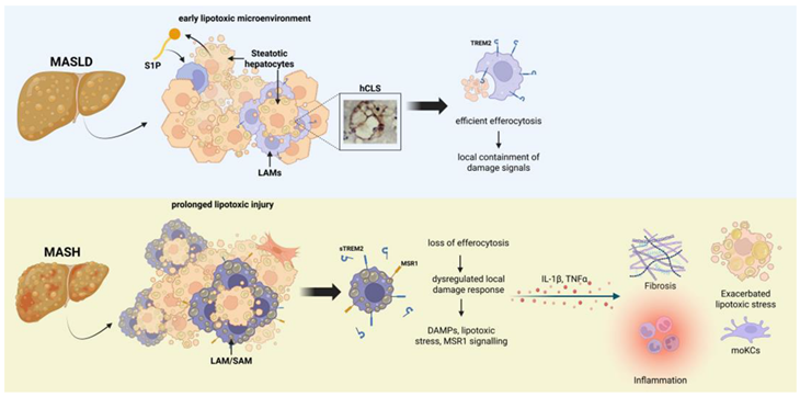
Figure Efferocytosis capacity distinguishes early MASLD from established MASH. In early MASLD, steatotic hepatocytes release sphingosine-1-phosphate (S1P), promoting the formation of hepatic crown-like structures (hCLS), in which lipid-associated macrophages (LAMs) expressing TREM2 (Triggering Receptor Expressed on Myeloid cells 2) efficiently clear damaged/dying hepatocytes (efferocytosis), locally containing damage-associated signals. Upon prolonged lipotoxic injury in MASH, this protective mechanism is lost: macrophages showing a LAM/scar-associated macrophage (SAM) phenotype release soluble TREM2 (sTREM2) and engage MSR1 (Macrophage Scavenger Receptor 1) signaling, resulting in a dysregulated local damage response with accumulation of damage-associated molecular patterns (DAMPs) and persistent lipotoxic stress. The consequent release of IL-1β and TNFα sustains fibrosis, exacerbated lipotoxic stress and expansion of monocyte-derived Kupffer cells (moKCs), fueling chronic inflammation. Created in BioRender. Cannito, S. (2026) https://BioRender.com/nhturqw.
predominant event allowing fibrosis arrest or even regression to occur; regression of fibrosis is accompanied by a number of relevant issues including the decrease in pro-inflammatory and pro-fibrogenic mediators, the decrease of synthesis of ECM components and the predominance of MMPs action vs those of TIMPs. When this ideal condition happens approx. 50% of HSC-derived MFs can undergo apoptosis, an event reported to be induced by NKT and resolution macrophages. HSC-derived MFs escaped to apoptosis can either become inactivated or de-differentiated or become senescent to then still undergo apoptosis possibly with the active contribution of activated NK cells.
It should be noted that recent data using single-cell transcriptomics have revealed that HSCs should be considered as a more heterogeneous cell population of what previously believed [54]. In addition, HSC have been proposed as cells contributing to regulate carbohydrate, mitochondrial and lipid homeostasis, suggesting that in order to undergo activation/transdifferentiation into MF HSC need to rapidly adapt from a metabolic point of view [53]. Accordingly, adaptations should include reprogramming of carbon metabolism and enhanced mitochondrial number and activity, as well as ER stress, and release of FFA through autophagy-dependent hydrolysis of retinyl esters stored in cytoplasmic droplets [53].
What is clear from literature data is that HSC as well as HSC-derived MF during the course of any CLD, including then progressive MASLD, are cells able to establish an extensive cross-talk with practically all the other cell types involved in disease progression. This means that HSCs and MFs are cells able to sense a plethora of signals/mediators as well as cells to respond to these signals/mediators by releasing, in a autocrine/paracrine manner, several mediators as unequivocally shown for TGFβ1, PDGF, VEGF, CCL2 and other mediators [10,50,51,52,53,55], a scenario summarized in Figure 1.
It should be emphasized that all these cell-to-cell interactions are critical not only to MASLD progression but also for HCC development, with several ligand/receptor axis indicated as putative biomarkers of progression or suitable molecular targets for novel therapeutic strategies [3,6,7,11,12,32,34,37]. In the next sections of this review we will focus on the emerging role of OSM/OSMRβ axis in the progression of CLD and HCC development, with a focus on MASLD and MASH.
3. Oncostatin M: Introducing A Peculiar and Enigmatic Cytokine
3.1. Oncostatin M: Introductory Remarks
Oncostatin-M (OSM) is a 22-26 kDa (depending on the species) pleiotropic and multifunctional cytokine belonging to the interleukin-6 (IL-6) family of cytokines [56] that was originally identified as a protein produced by U937 cells (a cell line from human histiocytic lymphoma) [57]. The IL-6 family of cytokines includes also leukemia inhibitory factor (LIF), a number of interleukins (IL-11, IL-27, IL-30 and IL-31) cardiotrophin-1, ciliary neurotrophic factor and cardiotrophin-like cytokine factor 1 [58,59,60,61]. In particular, OSM share a high level of homology (structural, functional and genetic) with LIF [56,59], possibly due to an ancestral event of gene duplication [62]. The name “Oncostatin M” reflects the initial finding that identified this cytokine as a cytostatic one for melanoma cells [57]. However, literature data indicate that OSM can modulate several processes, either in physiological or pathological conditions, including cell proliferation, extracellular matrix remodeling, hematopoiesis, differentiation, inflammatory response, angiogenesis, acquisition of cancer stem cell markers and of a metastatic phenotype [58,59,60,61].
OSM is produced and released by different type of cells, including cells of innate (mainly monocytes/macrophages, dendritic cells and neutrophils) and adaptive (T lymphocytes) immunity as well as hematopoietic cells, mesenchymal cells [58,59,60,61,63] and even cancer cells, as recently shown for liver cancer cells in MASH-related HCC [64].
3.2. OSM: Transcription, Biosynthesis and Secretion
Different laboratories have cloned in the last two decades the orthologues of human, murine and rat OSM, as reviewed in ref. [63]. Interestingly, the exon–intron structure of the OSM encoding gene has been reported to be identical between the mentioned species, resulting of three exons separated by two introns. Moreover, the gene is always located in direct proximity to the gene that encodes LIF, suggesting that both genes may have originated from a gene duplication [65,66]. Concerning human (hOSM) and murine (mOSM) genes, the promoters have been characterized and available evidence indicate that they are controlled by CCAAT-enhancer-binding proteins (C/EBP) and GC-rich elements for basal activity. However, signal transducer and activator of transcription (STAT)5 responsive elements are believed to be critical for stimuli-induced OSM transcription, with transcriptional silencer(s) (located between nt-194 and 109) in the human OSM promoter being apparently able to ensure cell-specific control of gene expression [67,68]. For example, in haemopoietic cells OSM transcription is strongly up-regulated by cytokines operating through STAT5, like IL-2, IL-6, erythropoietin and granulocyte-monocyte colony stimulating factor. OSM transcription has been reported to be also upregulated by prostaglandin E2 via cAMP signaling pathway [69,70]. Of interest OSM is seen as an amplifier of cytokine production/release [63] and both the human and murine OSM mRNAs contain several AU-rich sequences known to contribute to the upregulation of mRNA stability of several cytokines, including IL-6 [71].
OSM full-length proteins, after translation, have been reported to contain from 239 to 263 amino acids with a 3D structure fold into a long-chain four helix-bundle protein with an up-up-down-down topology closely similar to those of other IL-6 family cytokines [63]. OSM proteins are then post-translationally modified by N-glycosylation that seems to differ depending on the specific cell type [72]. Concerning hOSM (murine and rat data being at present less clear), after post translational cleavage of C-terminal pro-domain and of N-terminal signal peptide, the protein results of 195 amino acids and 22 kDa which has been reported to elicit the highest bioactivity although the 24 kDa isoform seems to be also active at similar levels in competition assays [72]. Both protein form can bind to the receptor complex (see next section) with high affinity but have different growth inhibitory activity: the 24 kDa isoform is from 5 to 60-fold less active in growth inhibition assays employing A375 melanoma cells [72].
Once secreted, OSM has been reported to bind, in a bioactive form, to various ECM components including different collagen types (I, III, IV, VI and XI) as well as to laminin and fibronectin [73,74]. This is a unique property of OSM since other cytokines belonging to IL-6 family do not bind to ECM components and it has been proposed that OSM, in an inflammatory microenvironment, may act as a strong inducer of ECM deposition [75,76].
3.3. OSM, Related Receptors and OSM-Dependent Signaling Pathways
OSM operates by binding to two different heterodimeric receptors which have in common the glycoprotein 130 (gp130), a membrane receptor protein shared by all members of the IL-6 family of cytokines. OSM has been reported to first interact with the extracellular cytokine-binding domain of gp130 to then recruit either the LIF receptor β, (LIFRβ) to form the OSM type I receptor or to the OSMRβ to form the OSM type II receptor. The two heterodimeric receptors are differentially engaged in human and mouse: human OSM can bind to either gp130/OSMRβ or gp130/LIFRβ, whereas murine OSM binds only to the type II receptor [77,78,79,80]. Interestingly, the OSMRβ/LIFRβ ratio and their different expression in various cell type is the proposed mechanism by which OSM and LIF may exert common biological effects on particular tissues but distinct and rather unique in other tissues [78]. It should be noted that OSMRβ is believed to mediate the majority of OSM effects [58] by mainly activating the Janus kinase (JAK)/STAT) pathway [58], as well as the Ras–mitogen-activated protein kinase (MAPK), extracellular-regulated kinases (ERK) 1/2 and p38 pathways [79,80].
Concerning the activation of JAK/STAT pathway it is known that the interaction of OSM and gp130 leads first to receptor heterodimerization and then to the recruitment and activation of JAK1, JAK2 and tyrosine kinase 2 (TYK2). In particular, the C-terminal domain of both OSM receptor type I and II contains tyrosine motifs which once phosphorylated by JAK1 and JAK2 acts as docking sites for STAT1 and STATOSM has been reported to also lead to the activation of STAT5 and STAT6 [58,59,78].
Apart from activation of the JAK/STAT pathways, OSM can also recruit on the conserved Tyr861 residue of the OSMRβ adapter sarcoma (Src) homology and collagen (Shc) protein: this can lead to the activation of a number of downstream kinases such as ERK1/2), p38, or JNKs [58,63,78,80], as well as, in same cells, the phosphatidyl inositol 3 kinase (PI3K)/Akt pathway [81] and the protein kinase C δ [82]. Finally, it has been reported that OSM can also negatively modulate mitogen-activated protein (MAP) kinase signaling cascades through the recruitment of the tyrosine phosphatase SHP-2 (specifically on Tyr759 and Tyr974 residues of the type I receptor) or of the suppressors of cytokine signaling (SOCS, in particular SOCS3) on Tyr759, or through the direct action of JAK1/2 [58,63,78].
4. Biological Activities of Oncostatin M: A Complex Scenario
As mentioned in the previous section, OSM should be regarded as a pleiotropic and somewhat enigmatic cytokine, produced and released by several cell types. OSM has been reported to induce or modulate various biological processes, particularly in pathological conditions. According to current literature data on pathological conditions OSM can affect, in several different tissues and organs, inflammatory response, cell proliferation, angiogenesis, epithelial to mesenchymal transition (EMT), invasiveness, metastasis and behavior of cancer stem cells (CSC) [58,59,60,61] as well as, more recently, to have a role in reshaping tumor immune microenvironment [83].
4.1. OSM in Inflammatory Diseases
OSM is known to play a major role in inflammation and it has been reported to be involved either in the acute phase response as well as in chronic inflammatory conditions, resulting eventually in tissue or organ fibrosis and cancer [58,59,60,61,84,85]. As already mentioned, OSM can be synthetized and released by several immune cell type in response to a variety of soluble mediators. OSM can exert either pro-inflammatory or anti-inflammatory effects according to the cellular context and the intracellular signaling elicited. OSM can act as a pro-inflammatory cytokine by stimulating both receptors (with a prevalence of OSMRβ or type II receptor) in target cells to release several cytokines and chemokines (particularly CCL2, CCL5, CCL20 and CXC chemokine ligand [CXCL] 3), the latter contributing to further recruitment of neutrophils and monocytes [58,59,60,61,84,85]. Interestingly, the anti-inflammatory action of OSM operates essentially on type I receptor (LIFR). Moreover, in chronic inflammatory conditions OSM and OSMRβ are often overexpressed, with OSM generally envisaged as a cytokine able to sustain inflammation and to promote fibrosis.
In particular, the role of OSM has been investigated in several chronic inflammatory diseases, including lung diseases, inflammatory bowel disease, cardiovascular diseases, skin diseases, arthritis, COVID-19 and many other conditions, as reviewed in ref. [61]. It should be noted from the beginning that OSM has been reported to exert either positive or negative effects on different diseases, with strategies designed to inhibit OSM-related signaling being beneficial in some diseases but detrimental in others [61]. Here we will briefly focus only on some relevant example of diseases in which OSM and OSM-related signaling has been proposed as relevant. The interested reader can refer to a more comprehensive review [61].
Concerning lung diseases, for example, OSM has been reported to be up-regulated and to play a critical role in patients affected by pulmonary fibrosis and asthma. In both diseases, as it will be a relatively common finding also for diseases in other tissues, OSM up-regulation paralleled the levels of lung chronic inflammation, the accumulation of pro-fibrotic macrophages and excess deposition (particularly in lung fibrosis) of ECM components, suggesting that OSM and related pathways may sustain development and progression of these lung diseases [86,87,88].
Another interesting example is represented by inflammatory bowel diseases (IBD) like Chron’s disease and ulcerative cholitis in which OSM and OSMR are found overexpressed in many IBD lesions [89]. Moreover, a retrospective study has shown that patients with increased levels of OSM and treated with anti-TNFα therapies had, one year later, a lower chance of remaining in remission [90]. Similarly, IBD patients having high levels of OSM and OSMR expression before of infliximab therapy showed a significantly lower response to the therapy [91]. In the latter study, the use of mice knockout for OSM in a murine IBD experimental model resulted in a very significant decrease in disease severity, confirming that OSM signaling not only represent a key feature of IBD pathogenesis but can also interfere negatively with anti-TNFα therapies [92]. Along these lines, it is interesting to note that JAK/STAT inhibitors like tofacinib (inhibitor of JAK1 and JAK3) and filgotinib (selective inhibitor of JAK1) are currently under evaluation in clinical trials for IBD, as reviewed in ref. [61].
A closely similar scenario to IBD is emerged from studies on rheumatoid arthritis in which, once again OSM, and here also IL1β, have a critical role in disease pathogenesis and progression [61]. In in vitro studies OSM has been reported to increase the deleterious effects of TNFα through activation of STAT3 signaling and that the use of tofacinib exerted significant positive effects [91,92]. In an in vivo study on models of murine arthritis, the administration of antibodies anti-OSM resulted in a significant improvement of the experimental disease [93], leading to the design of dedicated clinical trials employing anti OSM therapeutics (GSK315234 and GSK2330811) [61].
Differently from IBD and arthritis, in cardiovascular disease the role of OSM is more controversial. OSM-dependent persistent STAT3 activation has been reported in some studies to exert detrimental effects on arterial vessels and to favor atherogenesis [94,95]. However, another study indicates that chronic OSM administration to mice results in reduction of atherosclerosis development; moreover, patients with higher serum levels of OSM showed improved survival to coronary heart disease [96]. Moreover, experimental in vivo studies indicate that OSM increase cardiac function following myocardial infarction [97,98].
4.2. OSM in Cancer Biology
The multifaceted role of OSM is emphasized by literature data investigating its role in relation to cancer biology. There is a general agreement that OSM (and/or OSM/OSMRβ axis) play essentially a deleterious role in cancers of several different tissues and organs. Available literature data (see for a detailed review ref. [61]) concerning solid human cancers indicate that the involvement of OSM is sometime more relevant in specific cancer types like, for example, in ductal carcinoma of the breast, ductal adenocarcinoma of the prostate and of pancreas, adenocarcinoma of the lung and of colon, squamous cell carcinoma of the cervix and of the skin. Whether brain cancers are concerned, OSM has been involved practically in all the major types (i.e., astrocytoma, astroglioma, glioblastoma, medullo blastoma etc). A detailed analysis of the involvement of OSM in human cancer is out of the scope of this review and we will just emphasize some of the most common findings see ref. [59,60,61]: i) in most of human solid tumors OSM is usually overexpressed, particularly in the tumor mass, and the expression has been reported for some tumor to correlate with poor prognosis; ii) in different tumors it has been outlined that OSM and OSM/OSMRβ axis (often through involvement of STAT3) are strictly related to increased epithelial-to-mesenchymal transition (EMT) and increased invasiveness and metastasis; iii) another common finding is that OSM/OSMRβ axis results in an increased pathological angiogenesis, with a direct correlation between OSM and VEGF expression; iv) in some cancer type, but not all, OSM is able to stimulate proliferation of cancer cells; v) OSM is emerging as a cytokine potentially able to reshape the tumor immunosuppressive microenvironment (TIME). Within this scenario are also reported few isolated examples of positive effects played by OSM in very particular cancers like chondrosarcoma, in which OSM apparently induced in cancer cell lines cell cycle arrest and enhanced apoptosis [61,99] or in melanoma, where OSM has been reported to inhibit cancer cell proliferation [61,100].
5. The Role of OSM in Chronic Liver Diseases and in MASLD/MASH
According to already mentioned literature data, OSM and the related OSMRβ/JAK/STAT signaling axis, have been reported to regulate critical biological processes in normal and pathological conditions, including cell proliferation, extracellular matrix remodeling, hematopoiesis, differentiation, inflammatory response, angiogenesis, acquisition of cancer stem cell markers and of a metastatic phenotype, as well as, concerning the liver, also hepatic development and regeneration [,56,58,60,61,84,101]. All these critical processes are known to be deeply involved in the progression of CLDs of different etiology towards more advanced pathological stages, including cirrhosis and liver failure as well as HCC development [38,50,51,53,54]. In the following sections we will analyse literature data reporting the roles of OSM in CLDs, with a focus on progressive MASLD and MASH-related HCC. For the interested readers, we just mention here (not reviewed in this manuscript) the fact that OSM is emerging also as a mediator involved in the development of intrahepatic cholangiocarcinoma [60].
5.1. OSM in Chronic Liver Diseases: A Pro-Inflammatory and Pro-Fibrogenic Mediator
One of the first in vivo observations involving OSM in CLD was reported in an immunohistochemistry study performed on a limited number of human specimens obtained from either cirrhotic livers (n=6) and donor livers (n=4) not used for liver transplantation [102]. This rather preliminary morphological analysis concluded that OSM was mainly expressed in CD68 positive cells, likely Kupffer cells, with the level of expression being low and variable in control livers but more significantly elevated in cirrhotic livers. Authors of this pioneeristic study also suggested that the expression of OSMRβ, very low in normal hepatocytes, was apparently unchanged in cirrhotic livers, with LIFRβ being weakly expressed in normal livers, but more intensely in colangiocytes of reactive biliary ductules in cirrhotic livers [102]. Although the Authors analysed only few cirrhotic livers of unreported etiology, these data were in some way giving more emphasis on previous studies from the same research group indicating that OSM was able to up-regulate the expression collagen type I and of TIMP1 by liver MF derived from activated HSC [103]. In our knowledge, these two studies were the first to suggest OSM as a putative profibrogenic mediator. In those years, another study reported that knock out mice for OSMRβ treated with the hepatotoxin carbon tetrachloride (CCl4) or undergoing patial hepatectomy showed impaired liver regeneration vs control mice. Moreover, in these ko mice a reduction of TIMP1 and TIMP2 was also reported, suggesting then OSM as a mediator involved in hepatocyte proliferation and tissue remodeling [104]. Along these lines, a study indicated that OSM, in addition to KC, is expressed also in oval cells in the rat model of 2-acetylaminofluorene/partial hepatectomy and regeneration. Moreover, the OSM receptor was found mainly in oval cells suggesting that OSM/OSMR system may be critical in pushing oval cell differentiation into hepatocytes [105]. In addition, in a rat experimental model of liver inury by repeated injections of dimethylnitrosamine it was shown that OSM operated as a key mediator for proliferation and anti-apoptosis of hepatocytes [106]. A subsequent study added evidence suggesting for the fist time that OSM may contribute to liver angiogenesis, a critical process in both physiological and pathological conditions, by up-regulating in HepG2 cells as well as in the PH5CH8 human hepatocyte cell line transcription and synthesis of hypoxia inducible factor (HIF) 1α by involving OSM-dependent JAK/STAT3 and ERK1/2 signaling pathways. Accordingly, this HIF1α up-regulation also resulted in increased expression of HIF1-downstream genes coding for VEGF and plasminogen activator inhibitor 1 [107]. The interest for the role of OSM in CLD was renewed by an in vitro study reporting that administration of OSM to cultured human HepG2 cells resulted in a strong up-regulation of GP73 [108], a glycoprotein proposed as a biomarker of cirrhosis and HCC [109,110]. Interestingly, the response of HepG2 cells to OSM was by far more impressive than the response of the same cells to IL-6.
The involvement of OSM in CLD progression was proposed relatively early [92,93,94], including the preliminary data indicating that OSM was able to up-regulate collagen type I and TIMP1 expression in activated HSC or liver MF [103,111] as well as the notion that in mice carrying the genetic deletion of OSM receptor hepatocyte proliferation and tissue remodeling were impaired following persistent liver injury [112]. However, it was only in 2017 that was published the first convincing paper that correlated mechanistically OSM to liver fibrogenesis [113], with OSM proposed to act as a pro-fibrogenic mediator by regulating the activation of macrophages during chronic liver injury. Authors of this study employed mice with genetic deletion of OSM that were then chronically treated (12 weeks) with the hepatotoxin thioacetamide (TAA). OSM deletion resulted in a significant decrease in liver fibrosis and in the expression of both collagen 1a1 and TIMP1 versus wild type animals, without affecting parameters of hepatocyte injury. Moreover, the exposure of co-cultures of HSC and liver macrophages to recombinant OSM resulted in a significant increased expression of critical pro-fibrogenic mediators like PDGF and TGFβ in parallel with up-regulation of TIMP1 by HSC. In addition, the continuous expression of OSM, obtained by employing the procedure of hydrodynamic tail vein injection of OSM, was sufficient to induce liver fibrosis. Finally, by over expressing OSM and using fluorescence-activated cell sorting, these Authors proposed that macrophages recruited from peripheral blood were responsible for OSM-dependent pro-fibrogenic activation of HSC [113]. This study, although in part performed using an experimental model of chronic liver injury by TAA that is not really representative of human conditions, it has been the first one to show unequivocally the pro-fibrogenic action of OSM [113].
A study from our laboratory offered an additional contribution to the concept that OSM can operate as a pro-fibrogenic mediator by showing that human recombinant OSM (hrOSM) was able to stimulate the oriented migration (i.e. chemotaxis) of human MF-like LX2 cells, originally obtained from activated HSC [114]. In particular, OSM was reported to stimulate MF migration by eliciting a raise in intracellular ROS and through activation of STA1/STAT3 (with activation of Ras/Erk, JNK1/2 and PI3/Akt signaling pathways also detected in LX2 cells). In addition, OSM also induced recruitment and stabilization of HIF1α, confirming what previously described by other Authors [107]. This issue is relevant since in the same study evidence was provided that OSM-dependent migration relied on a biphasic mechanisms requiring early intracellular generation of ROS and a later and HIF1α-dependent expression and relase of VEGF, the latter being a mediator contributing in part to oriented migration of LX2 cells stimulated by OSM. hrOSM was also able to up-regulate in LX2 cells an increased transcription of pro-inflammatory mediators like the chemokine CCL2 and the cytokines IL-6 and TNFα [114].
OSM was also proposed by a clinical study as an essential component of an immune gene signature able to predict advanced fibrosis in CLD of mixed etiology by analysing snap-frozen liver tissues from patients at different stage of fibrosis. The other component of the immune signature were all chronic injury and inflammation-related factors including, in addition to OSM, also VEGFA, Chitinase 1, Fc fragment of IgE - high affinity I receptor for gamma polypeptide and zeta chain of T-cell receptor associated protein kinase This signature was reported to discriminate patients with low grade of fibrosis (i.e., F1, F2) from those with more advanced stages of fibrosis (F3,F4). What is interesting is that the signature was reported to be independent on pathological and clinical features, with OSM and other factors reported to decrease in more advanced stage [115]. In a more recent study performed by employing the rat model of liver fibrosis requiring chronic administration of CCl4, OSM was found to be the main cytokine (together with IL1β) up-regulated. Moreover, OSM was reported to be produced and released by KC, macrophages and endothelial cells. Interestingly, according to this study, the target cells were hepatocytes, endothelial cells and HSC, that all expressed the necessary receptors [116].
A final mention in relation to liver fibrogenesis is dedicated to a recent study investigating early events in a murine model of alcohol-associated liver disease (ALD) which has revealed an interesting correlation between OSM and Yes-associated protein (YAP) [117]. YAP is a transcriptional coactivator, representing a downstream effector of the Hippo signalling pathway, that a number of studies demonstrated to be involved in the pathogenesis of various liver diseases [118,119]. YAP-/- mice, mice injected with an adeno-associated virus 9-delivered saCas9/sgYAP system or YAPΔHep mice were fed the Gao-binge diet to mimick early ALD events. YAP deficiency was found to exacerbate early aspects of ALD, including steatosis and inflammatory response whereas overexpression of YAP in hepatocytes significantly reversed thes aspects. In particular, hepatocellular YAP was reported as a negative regulator of OSM/STAT3 dependent up-regulation of CD36 [117], a well known critical membrane multifunctional protein that regulates the uptake of fatty acids in several cells, including hepatocytes, that has a key role in lipid metabolism, inflammation and immune response.
5.2. OSM in the Progression of MASLD/MASH
The involvement of OSM and related signaling pathways in MASLD/MASH (or in NAFLD/NASH, as for the previous definition of the disease) is surprisingly limited to few specific studies, with somewhat contradictory results. According to the actual definition of MASLD as a spectrum of conditions, including steatosis, steatohepatitis plus/minus fibrosis, fibrosis and then cirrhosis, we can synthetise the few data actually available in the literature. At present, at least in our knowledge, one single study has analysed OSM serum levels and the liver expression of OSM in MASLD/MASH patients stratified according to progression of the disease [64]. OSM was reported to be almost undetectable in patients with either a diagnosis of simple steatosis or of steatohepatitis. However, OSM serum levels were significantly increased in cirrhotic patients to be then dramatically raised in patients with MASH-related HCC (see later), suggesting that the relevance of OSM might be related to the progression of the disease rather than the genesis of fatty liver and/or early conditions of MASH [64].
Along these lines, the first report involving OSM in relation to MASLD/MASH was performed almost fifteen years ago in order to investigate the contribution of this cytokine released by KC to hepatic insulin resistance and steatosis. This experimental study, performed either in vitro or using two different dietary protocol to induce MASLD/MASH proposed that prostaglandin E2 (PGE2) produced by activated KC attenuated insulin-dependent glucose utilization by interrupting the intracellular signal chain downstream of the insulin receptor in hepatocytes as well as by affecting insulin resistance by modulating cytokine production in non-parenchymal cells. The Authors of this study showed that OSM produced by KC exposed to PGE2 attenuated insulin-dependent Akt activation and, glucokinase induction in hepatocytes through the induction of SOCSIn addition, OSM inhibited the expression of key enzymes of hepatic lipid metabolism, overall suggesting that OSM may contribute to both the raise of IR and the development of steatosis and then possibly MASH [120]. Few years later, another study analyzed the roles of OSM signaling in obesity and related metabolic disorders. The Authors employed mice genetically manipulated in order to carry global deletion of OSMRβ gene (OSMRβ-/- mice) and fed these mice on a high fat diet (HFD). Data from this study reported that OSMRβ-/- mice, when compared to WT littermates, showed a severe increase in body weight and food intake as well as an increase in peripheral adipose tissue inflammation, insulin resistance and liver steatosis [121]. This scenario was quite unexpected since several studies on chronic inflammatory diseases, showed that OSM inhibition was effective in preventing or negatively modulating the disease [56,60,61]. In particular, it was reported that OSMRβ-/- mice displayed a higher expression of liver genes related to de novo lipogenesis. A major limitation of this study was represented by the fact that Authors used mice carrying global deletion of OSMRβ gene and then its expression from all involved cells, including cells of innate (mainly monocytes/macrophages, dendritic cells and neutrophils) and adaptive (T lymphocytes) immunity as well as hematopoietic cells, mesenchymal cells as well as hepatocytes [58,59,60,61,63,64].
A more appropriated approach was reported in a study that, in addition to mice carrying global deletion of the receptor, also employed mice lacking hepatocellular OSMRβ fed on a HFD. In these mice the dietary protocol exacerbated IR, hepatic steatosis and inflammation, effects that were markedly attenuated by hepatocyte-specific overexpression of OSMRβ [122].
Overall, the results of the latter two studies should be interpreted with caution, taking in mind that HFD protocol is useful to reproduce steatosis but the degree of inflammation is, in our experience, modest with very low or negligible fibrosis. The murine HFD protocol can then reproduce reliably just the early stage of the disease. Moreover, it should be noted that none of the two mentioned studies analysed either serum or tissue levels of OSM in wild type and genetically manipulated mice [121,122]. This is relevant since when employing dietary protocols leading to significant inflammation and fibrosis, like high fat / high fructose diet or choline-deficient L-amino acid–defined (CDAA) diet, the hepatic expression of both OSM and OSMRβ was significantly up-regulated at a relatively later stage and in parallel with increased collagen deposition and recruitment/activation of MF derived from HSC [114], a scenario more compatible with the one mentioned in relation to OSM for MASLD patients stratified according to disease progression [64].
More recently OSM has been reported to represent a possible factor mediating the protective effects versus the development of MASH exerted by osteopontin (OPN) released by macrophages [123]. OPN (the product of secreted phosphoprotein 1 or SPP1 gene) is a mediator whose expression correlate with TG accumulation in MASLD patients [124] and proposed as a putative profibrogenic mediator (see for a review ref. [125]. In this study, OPN/SSP1 expression in macrophages increases during MASH progression in humans and in mice fed on a very potent dietary protocol (high fat/high fructose/high cholesterol diet), although in mice the expression was up-regulated also in hepatocytes. This very elegant and complex study, using properly designed genetically modified mice, show that a specific cluster of macrophages (i.e., of five identified), referred to as SPP1high, is formed by cells having maximal expression of OPN which are also expressing TREM2 and CD9 antigens. This subset of macrophages has no pro-inflammatory or profibrogenic profile but rather a metabolic one and, according to the study, these macrophages are those responsible for MASH protection which, in turn, is mediated by up-regulation of OSM that act on hepatocytes through STAT3 signalling resulting in up-regulation of arginase expression. There are some comments that should be taken in mind: i) the conclusions are mainly based on the use of genetically modified mice in which SSP1 gene was either overexpressed (i.e., using SPP1 knockin mice) or deleted in myeloid cells; ii) as suggested by Authors in their discussion the SPP1high TREM2 and CD9 positive macrophages may behave differently during the disease; iii) data of this group [123] may fit into the current view that TREM2 CD9 positive cells (previously defined as LAM or scar-associated macrophages) should be interpreted as cells that in the early stages of MASH may act as protective cells (i.e., by aggregating and surrounding lipid-laden hepatocytes to form hepatic crown-like structures, displaying efferocytic capacity and then containing local injury and inflammation) to become more pro-inflammatory and then profibrogenic in conditions of prolonged lipotoxic injury (i.e., following shedding TREM2 and loosing efferocytic capacity) [38].
5.3. OSM and HCC: Experimental and Clinical Data not Directly Referred to MASLD/MASH
As mentioned in a previous section, OSM is currently believed to represent a pro-carcinogenic cytokine, as observed in several human solid tumors (see section 4.2), then progressively replacing the initial idea of OSM as a cytokine able to inhibit proliferation that was essentially based on in vitro studies in different cancer cell lines (reviewed in ref. [58,59,60,61]). Literature data specifically concerning OSM and HCC are somewhat relatively limited and only recently OSM and its related signaling axis have been investigated in human patients and in mechanistic experimental models.
One should note that, curiously, for several years literature data have investigated OSM-related effects, some potentially related to a role in liver cancer, mostly by performing in vitro studies employing the widely used HepG2 human cell line, an immortalized line of human liver cancer cells originally isolated from a liver biopsy of a 15-year-old boy with a well-differentiated hepatoblastoma, or other hepatoma cells. Indeed, in our knowledge, the first study published on OSM and HepG2 cells is back to 1991, when a biochemical article proposed that OSM was able to strongly increase the uptake of low density lipoproteins (LDL) by HepG2 cells through a tyrosine kinase – related mechanism resulting in an increased transcription of the LDL receptor and its increased presence on the plasma membrane [126]. These findings, that were confirmed by other subsequent studies [127,128], were initially unnoticed although a relevant message was quite clear: a cytokine secreted by macrophages had effects on hepatocyte lipoprotein and cholesterol metabolism, indicating a possible connection between the immune system and cholesterol homeostasis and that HepG2 (i.e., liver cancer cells) expressed the receptors for OSM. The relevance of this message is clear now since, as we mentioned previously and will briefly recall in the next paragraph, changes in fatty acid metabolism and cholesterol pathways are particularly relevant in the pathogenesis of MASLD/MASH and MASH-related HCC and, more generally, for liver carcinogenesis [6,10,11,12,13]. Other in vitro studies conducted on HepG2 cells revealed a role of OSM in the regulation of the synthesis of liver acute phase proteins related to coagulation, as for example fibrinogen [129] and secreted phospholipase A2 [130] as well as a role in the up-regulation of conventional as well as soluble IL-6 receptor, the latter a protein that lacks the transmembrane domain and has been suggested to be a potent immunomodulator of IL-6 biologic activity, but once again this finding was somewhat neglected for its putative relation to liver cancer progression [131].
The most relevant in vitro study is, in our view, the one indicating that, differently from control hepatocytes that express both OSM related receptors, human hepatoma cells were characterized by a reduced expression of LIFR and reduced responsiveness to LIF. This was ascribed to epigenetic changes in hepatoma cells, likely due to methylation-dependent inactivation of the LIFR-gene. Reduced responsivess to LIF by hepatoma cells was remarkably similar to what observed also for other cancer cell lines like colon HCT8 cells, breast MCF7 cells and lung carcinoma cells [132]. If we limit the observation to hepatoma cells, the message was that liver cancer cells do not respond or minimally respond to LIF but maintain the capacity to respond to OSM through OSMRβ, suggesting that OSM can act on liver cancer cells.
Data more directly related to HCC were published later in an attempt to clarify the role of OSM in mediating the activation of acute-phase protein (APP) gene expression [133]. By employing mice ko for amphiregulin (AR), a ligand for epidermal growth factor receptor (EGFR) and related wild type mice, Authors of this study found that APP genes were overexpressed in the livers of AR knockout mice during inflammation and hepatocyte proliferation. Moreover, AR-null hepatocytes in culture and human HCC cells after AR knockdown, APP gene expression is enhanced, suggesting that AR counteracts OSM-triggered STAT3 signalling in hepatocytes and attenuates APP gene transcription, supporting the relevance of epithelial growth factor receptor (EGFR)-mediated signalling in the modulation of cytokine-activated pathways. This is potentially relevant since AR, not expressed in normal liver, is up-regulated during chronic liver injury and particularly in human HCC tissues and cell lines, behaving as a mitogen and antiapoptotic growth factor for HCC cells [134]. In a very interesting study OSM has been investigated in relation to liver cancer stem cells (CSCs) [135] based on the knowledge that IL-6 related cytokines can induce the differentiation of hepatoblasts into hepatocytes [136]. In this study OSM receptor expression was found in the majority of epithelial cell adhesion molecule (EpCAM)-positive HCC cells with stem/progenitor cell features. Of interest, OSM induced hepatocyte differentiation of these cells through STAT3 signaling. These EpCAM positive cells treated with OSM showed increased cell proliferation and expansion of the EpCAM-negative non-CSC population.
It was only in 2012 that OSM levels were reported to be elevated 6–7 fold in sera from cirrhotic patients (carrying or not HCC) vs healthy controls, with cirrhotic patients displaying a significant association between serum levels of OSM and GP73, a Golgi phosphoprotein protein considered at the time a putative biomarker for HCC [108]. In our knowledge this study was indeed the first to report a very significant increase of serum OSM in cirrhotic patients (carrying or not HCC). Unfortunately, in this study no information was provided concerning the specific etiology of affected cirrhotic patients [108].
In another study performed on different liver cancer cell lines (HepG2, Hep3B and Huh7 cells) OSM as well as IL-6, were found to inhibit IL-27 induced IFNγ-like, STAT1-dependent transcriptional response through a SOCS3-mediated mechanism [137]. The relevance of the study relies in the fact that IL-27 is upregulated in patients with HCC as compared to healthy controls or patients with hepatitis and/or liver cirrhosis [138,139]. It should be mentioned here that HCC is an immunosensitive tumor and that TH1 cytokine detection in patients is usually associated with a good prognosis, with type I interferons, IFNγ and IL12 being crucial for immune rejection of HCC [140]. Accordingly, IL-27 has been proposed as a putative antitumor molecule with an effect mediated by increased antigen presentation but just combining the treatment with blocking antibodies against PD-L1 or/and IL6-type cytokines [141]. The overall message is that IL-6 related cytokines, like OSM, can affect the efficacy of immune responses towards liver cancer cells, a concept that will be emphasized by studies published afterwards [140,141].
Finally, it seems relevant to mention an experimental study performed by using the N-diethylnitrosamine (DEN)-induced HCC in rats revealing that the overexpression of OSM significantly increased the number of tumor nodules and shortened rat survival [142]. Moreover, OSM was reported to promote activation in vivo of hepatic progenitor cells and to induce TNFα secretion as well as the accumulation of CD68 positive macrophages. Accordingly, depletion of either TNFα or of macrophages resulted in an inhibition of the procarcinogenic role of OSM [142].
5.4. OSM and OSM/OSMRβ in MASH-Related HCC
The specific interest in the role of OSM and OSM/OSMRβ axis in MASH-related HCC is relatively recent and, in our knowledge, the first clinical and experimental study involving OSM also in HCC raised in MASH patients was published just in 2020 [143]. In this study Authors analysed 81 cirrhotic and non-cirrhotic patients carrying HCC having either a HBV (n=52) or MASLD/MASH (n=29) etiology. This study revealed a novel concept, with data that focused on the role of OSM released by neutrophils infiltrating the tumor mass [143]. This is in principle relevant since we know that neutrophils, although less characterized that tumor-associated macrophages (TAMs) and tumor-infiltrating lymphocytes (TILs), are a significant cellular component in the HCC mass with a putative contributing role [144,145]. In synthesis, the study outlined a correlation between the metabolic switch versus aerobic glycolysis of macrophages resulting in their release of chemokines CXCL2 and CXCL8, which are responsible for the recruitment of neutrophils in the HCC. In addition, activated macrophages release TNFα that in turn is responsible for OSM production by neutrophils which correlates with HCC metastasis. then suggesting for the first time OSM as a pro-metastatic agent in HCC progression [143].
The first clinical and experimental study entirely dedicated to investigate the role of OSM in the development and progression of MASH-related HCC was published in 2022 [64]. This study was performed by analysing selected cohorts of MASLD/MASH-related HCC patients, liver cancer cell lines exposed to human recombinant OSM or stably transfected to overexpress human OSM, murine HCC xenografts, and the DEN-CDAA murine MASH-related protocol to induce experimental liver carcinogenesis. From a clinical point of view, OSM was selectively overexpressed in HCC cells of MASH patients; moreover, the analysis of serum levels of OSM in patients stratified accordingly to the different stages of the disease revealed that OSM serum levels, barely detectable in patients with steatosis or early steatohepatitis, started to increase in cirrhotics to be then strongly and significantly increased in patients carrying HCC. OSM overexpression is a selective feature of MASH-related HCC: comparing circulating OSM levels in MASH-related HCC patients with those in 51 patients with HCC developed on a different background (HBV, HCV and alcoholic liver disease), serum OSM levels were significantly higher in MASH patients than in those with other etiologies [64]. When MASH-related patients carrying HCC were divided according to the Barcelona Clinic Liver Cancer (BCLC) staging system [146], patients in the intermediate/advanced HCC stages (classes B/C/D, respectively) had OSM serum levels significantly higher than those at early stages of HCC with preserved liver function (classes 0/A), with higher levels of OSM serum levels correlating to decreased survival. A series of in vitro experiments on liver cancer cell lines indicated then the overexpression of OSM or the exposure to hrOSM were resulting in increased angiogenesis and in the induction of EMT and invasiveness, indicating that OSM up-regulation in liver cancer cells can contribute to HCC progression. This concept was reinforced by xenograft experiments in which the inoculation of cells overexpressing OSM was associated with a slower tumor growth but a markedly increased and diffuse vascularization of the tumor graft resulting in an increased rate of lung metastasis. OSM overexpression and its positive correlation with the angiogenic switch were confirmed in the DEN-CDAA model of liver carcinogenesis and the observation that in human MASH-related HCC with vascular invasion OSM was overexpressed in liver cancer cells invading hepatic vessels. This study also outlined that liver cancer cells can at the same time respond to OSM as well as to prodeuce and release OSM and overall suggested that OSM/OSMRβ axis might support HCC progression in MASH patients [64].
In a subsequent study the role of OSM and OSM/OSMRβ axis in driving the progression of MASH-related HCC was more mechanistically investigated by employing mice carrying hepatocyte specifc deletion of OSMRβ (hOSMRβ−/− mice) and related wild type mice undergoing the DEN-CDAA murine protocol of MASH-related liver carcinogenesis in association to in vitro experiments on HCC cell line and immune cell lines and analyses of cohorts of MASH patients carrying or not HCC [83]. The use of genetically manipulated mice revealed that the hepatocyte specific deletion of the receptor did not modify OSM production by HCC but significantly reduced the size and weight of hepatic tumors vs those detected in wild type animals. Moreover, confirming data obtained in a previous study [64], the reduction of size and weight of tumors in hOSMRβ−/− mice was associated with a decrease in markers of proliferation (Ki67, proliferating cell nuclear antigen or PCNA) as well as with a decrease of markers of angiogenesis (VEGFA, VEGF receptor 2 and CD105), indicating that autocrine/paracrine OSM/OSMRβ signaling contributes to MASH-related HCC growth and angiogenesis.
The same study then outlined that OSM expression was associated with a tumor immunosuppressive microenvironment (TIME) [83]. The hypothesis that OSM may have a role in sustaining the TIME in HCC was first introduced by a clinical study investigating a large number (n=397) of HCC patients of mixed etiology that received surgical resection [146]. Data reported in this study indicated that the majority of patients (n=248) had a viral etiology (HBV or HCV) whereas for the remaining patients (n=149) unfortunately no specific indication of the etiology was reported [136]. In any case, Authors focused their attention on vessels encapsulating tumor cluster (VETC) positive HCC (n=62) and VETC negative by analyzing OSM positive cells and inflammatory/immune cells, including CD4, CD8, CD163 and forkhead box P3 (FOXP3) positive cells by immunohistochemistry. Data provided indicate that tumor-infiltrating OSM-positive cells were significantly low in VETC-positive HCC. Of interest, in VETC-positive HCC, characterized by a lower grade of hepatocyte differentiation, the number of OSM-positive cells was not associated with vascular invasion, whereas in VETC-negative HCC, an increase in the number of OSM-positive cells was associated with vascular invasion [146]. In a subsequent study by the same group Authors, working on the same liver HCC liver specimens used in the previous article reported a direct correlation between OSM, tumor CD147 expression and TILs in HCC as a mechanism by which CD147 may be able to facilitate HCC immune evasion [147] since CD147 over expression has been proposed as a predictor of high malignant potential and advanced clinical stage [148,149]. In this second study Authors investigated the expression of CD147 in liver tumor cells to quantify OSM-positive cells and TILs by using a microarray and immunohistochemistry approach [147]. Interestingly, most of HCC investigated had a high CD147 expression (n=332) and in these tumors the density of OSM positive cells was associated Cd8, CD4, FOXP3 and CD20 positive cells, whereas in HCC showing a low CD147 expression OSM psotivity was limited to FOXP3 positive cells.
The concept of OSM as a mediator involved in the reshaping of TIME was confirmed by an analysis of data from the TCGA-LIHC cohort that include HCC patients of different etiology [83] (Figure 3). This analysis identified a significant positive correlation between OSM expression and the one of several genes that literature associate with TIME such as TAMs and regulatory immune cells markers (CD206, CD163, PD-L1, TGF-β1, CCL22, CCL2, FOXP3, IL2RA, CXCL12, CD33, CD11b, and cyclooxygenase-2). An almost overlapping pattern was detected in HCC obtained in the liver of mice submitted to the DEN-CDAA protocol of MASH-related carcinogenesis, with OSM expression correlating with either CCL2, TGF-β1, and CXCL12 (i.e., involved in TAMs recruitment), or PD-L1, prostaglandin E synthase 2 (PTGSE2), and CD206 (TAM-associated markers) as well as FOXP3, IL2 receptor antagonist (IL2-RA), CCL22 and CC chemokine receptor (CCR) 4 (markers of regulatory T cells or Treg). As a matter of fact, all these cytokines, chemokines and markers were significantly down-regulated in HCC from hOSMRβ−/− mice and this scenario was associated with a decrease in OSM-related signaling (i.e., involving STAT3 phosphorylation) [83]. Finally, a single-cell RNA-seq analysis of human HCCs identified liver cancer as the source of the chemokine CCL15 which has been reported to be a specific HCC biomarker associated with immunosuppression in HCCs. Serum levels of CCL15 were strongly up-regulated in both humans and mice carrying MASH-related HCC and Kaplan-Meier analysis showed that high CCL15 serum levels were correlated with a significant worst survival of human patients [83]. Moreover, CCL15 serum levels were significantly higher in MASH patients carrying HCC than in patients carrying HCC but of different etiology. These data are consistent with those previously reported by another research group that suggested CCL15 as specific HCC biomarker whose expression significantly correlated with malignant HCC behavior and poor patient survival [150]. In vitro studies confirmed that by blocking autocrine OSM signaling in HepG2 or Huh7 cells overexpressing OSM reduced STAT3 phosphorylation, CCL15 production, and prevented TIME markers expression by co-cultured macrophage-derived THP-1 cells, indicating a direct relationship between OSM and up-regulation of CCL15.
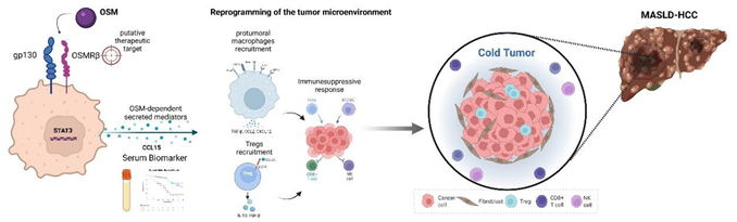
Figure Oncostatin M (OSM)/OSMRβ signaling drives immunosuppressive reprogramming of the tumor microenvironment in MASLD-related HCC. OSM binds the heterodimeric receptor complex composed of glycoprotein 130 (gp130) and Oncostatin M receptor β (OSMRβ) - a putative therapeutic target - activating intracellular STAT3 signaling and the release of OSM-dependent secreted mediators, including CCL15, a candidate serum biomarker associated with patient survival. These mediators promote recruitment of pro-tumoral macrophages expressing CD206, CD163, CD33, CD11b and PD-L1, which release TGF-β, CCL2 and CXCL12, together with recruitment of regulatory T cells (Tregs) via the CCL22/CCR4 axis, leading to local release of IL-10 and TGF-β. The resulting immunosuppressive response favours the establishment of an immune-excluded, poorly infiltrated "cold tumor" phenotype within the MASLD-HCC nodule. Created in BioRender. Cannito, S. (2026) https://BioRender.com/nhturqw.
6. Conclusions
Although also involved in CLD progression and HCC development of different etiology, OSM and OSM/OSMRβ axis have an emerging and apparently selective role in the progression of MASH related HCC, with OSM increased expression being a specific feature of HCC of metabolic etiology that correlates with clinical parameters and disease outcome. Along these lines, available data indicate that OSM and its signaling have a pro-angiogenic role and can efficiently sustain proliferation, EMT and increased inveasiveness of liver cancer cells. Moreover, OSM is now reported to have a critical role in reshaping TIME and then favor immune evasion of cancer cells in HCC. Overall, OSM can be proposed as a specific biomarker and a putative therapeutic target for MASH-related HCC.
Author Contributions
All Authors contributed to conceptualization, writing—review and editing of the manuscript. All authors have read and agreed to the published version of the manuscript. .
Funding
This research was funded by AIRC (Italian Association for Cancer Research, Italy), grant number IG 2022 ID 27667, to MP.
Informed Consent Statement
Not applicable
Acknowledgments
Figure included in this manuscript were created in BioRender. Cannito, S. (2026) https://BioRender.com/nhturqw
Conflicts of Interest
The authors declare no conflicts of interest
Abbreviations
The following abbreviations are used in this manuscript:
| ALD | Alcohol-related Liver Disease |
| αSMA | α-Smooth Muscle Actin |
| APOB | Apolipoprotein B |
| APP | Acute Phase Protein |
| AR | Amphiregulin |
| BCLC | Barcelona Clinic Liver Cancer |
| bFGF | basic Fibroblast Growth Factor |
| BMI | Body Mass Index |
| CCL | CC-chemokine Ligand |
| CCL4 | Carbon tetrachloride |
| CCR | CC-chemokine Receptor |
| CD | Cluster Designation |
| CDAA | choline-deficient L-amino acid–defined |
| C/EBP | CCAAT-enhancer-binding proteins |
| ChREBP | Carbohydrate Response Element Binding-Protein |
| CLD | Chronic Liver Disease |
| COL13A1 | Collagen type XIII Alpha 1 chain |
| CPT1 | Carnitine O-PalmitoylTransferase |
| CSCs | Cancer Stem Cells |
| CTGF | Connective Tissue Growth Factor |
| CXCL | CXC-chemokine Ligand |
| DAMPs | Damage Associated Molecular Patterns |
| DEN | N-diethyl-nitrosamine |
| DG | DiacylGlycerol |
| DGAT2 | DiacylGlycerol O-AcylTransferase 2 |
| DNL | De Novo Lipogenesis |
| ECM | ExtraCellular Matrix |
| EFCAB4B | EF-Hand Calcium Binding Domain 4B |
| EGFR | Epithelial Growth Factor Receptor |
| EMT | Epithelial-to-Mesenchymal Transition |
| EpCAM | Epithelial Cell Adhesion Molecule |
| ER | Endoplasmic Reticulum |
| ERK | Extracellular Regulated Kinase |
| EV | Extracellular Vesicles |
| FA | Fatty Acids |
| FDFT1 | Farnesyl-Diphosphate FarnesylTransferase 1 |
| FFA | Free Fatty Acids |
| FOXP3 | Forkhead box P3 |
| GCKR | Glucokinase Receptor |
| GP73 | GlycoProtein 73 |
| gp130 | glycoprotein 130 |
| HCC | Hepatocellular carcinoma |
| HDL | High Density Lipoproteins |
| HSC | Hepatic Stellate Cells |
| HFD | High Fat Diet |
| HIF | Hypoxia-Inducible Factor |
| IBD | Inflammatory Bowel Disease |
| ICI | Immune-Checkpoint Inhibitors |
| IFNγ | Interferon γ |
| IFNL4 | Interferon Lambda 4 |
| IL | Interleukin |
| IL1β | Interleukin 1β |
| IL2RA | IL-2 receptor antagonist |
| IR | Insulin Resistance |
| JAK | Janus Kinases |
| JNK | c-Jun NH2 aminoterminal Kinase |
| KC | Kupffer Cells |
| LAMs | Lipid-Associated Macrophages |
| LDL | Low Density Lipoprotein |
| LIF | Leukemia Inhibitory Factor |
| LIFRβ | LIF Receptor β |
| LXR | Liver X Receptors |
| MAPK | Mitogen-Associated Protein Kinase |
| MASH | Metaolic dysfunction – Associated SteatoHepatitis |
| MASLD | Metabolic dysfunction – Associated Steatotic Liver Disease |
| MBOAT7 | Membrane Bound O-AcylTransferase domain-containing 7 |
| MFs | MyoFibroblasts |
| MMPs | Matrix MetalloProteases |
| MoMs | Monocyte-derived macrophages |
| MTTP | Microsomal Triglycerides Transfer Protein |
| NK | Natural Killer cells |
| NKT | Natural Killer T cells |
| OPN | Osteopontin |
| OSM | Oncostatin M |
| OSMRβ | Oncostatin M Receptor β |
| PAMPs | Pathogen-Associated Molecular Patterns |
| PCNA | Proliferating Cell Nuclear Antigen |
| PDGF | Platelet-Derived Growth Factor |
| PD-L1 | Programmed Death-Ligand 1 |
| PGE2 | Prostaglandin E2 |
| PTGSE2 | Prostaglandin E synthase 2 |
| PI3K | Phosphatidyl Inositol 3 Kinase |
| PNPLA3 | Patatin-like PhosphoLipase domain containing 3 |
| PPARγ | Peroxisome Proliferator-Activated Receptor γ |
| PRR | Pattern Recognition Receptors |
| ROS | Reactive Oxygen Species |
| SEC | Sinusoidal Endothelial Cells |
| SREBP1c | Sterol Regulatory Element Binding-Protein 1c |
| SOCS | Suppressors Of Cytokine Signaling |
| SOD | SuperOxide Dismutase |
| SPP1 | Secreted PhosphoProtein 1 gene |
| STAT | Signal Transducer and Activator of Transcription |
| TAA | Thioacetamide |
| TACE | TransArterial ChemoEmbolization |
| TAMs | Tumor Associated Macrophages |
| TG | Triglycerides |
| TGFα | Transforming Growth Factor α |
| TGFβ1 | Transforming Growth Factor β1 |
| TH1 | T Helper 1 |
| TILs | Tumor Infiltrating Lymphocytes |
| TIME | Tumor Immunosuppressive MicroEnvironment |
| TIMPs | Tissue Inhibitors of MetalloProteases |
| TM6SF2 | TransMembrane 6 SuperFamily member 2 |
| TNFα | Tumor Necrosis Factor α |
| T2D | Type 2 Diabetes |
| Treg | regulatory T cells |
| TREM2 | Triggering Receptor Expressed on Myeloid cells 2 |
| TYK2 | Tyrosine Kinase 2 |
| UCP2 | Uncoupling Protein 2 |
| VETC | Vessels Encapsulating Tumor Cluster |
| VEGF | Vascular Endothelial Growth Factor |
| VLDL | Very Low Density Liporoteins |
| WAT | White Adipose Tissue |
| YAP | Yes Associated Protein |
References
- Golabi, P.; Isakov, V.; Younossi, Z.M. Nonalcoholic fatty liver disease: disease burden and disease awareness. Clin. Liver Dis. 2023, 27, 173–186. [Google Scholar] [CrossRef]
- Estes, C.; Anstee, Q.M.; Arias-Loste, M.T.; Bantel, H.; Bellentani, S.; Caballeria, J.; Colombo, M.; Craxi, A.; Crespo, J.; Day, C.P.; Eguchi, Y.; Geier, A.; Kondili, L.A.; Kroy, D.C.; Lazarus, J.V.; Loomba, R.; Manns, M.P.; Marchesini, G.; Nakajima, A.; Negro, F.; Petta, S.; Ratziu, V.; Romero-Gomez, M.; Sanyal, A.; Schattenberg, J.M.; Tacke, F.; Tanaka, J.; Trautwein, C.; Wei, L.; Zeuzem, S.; Razavi, H. Modeling NAFLD disease burden in China, France, Germany, Italy, Japan, Spain, United Kingdom, and United States for the period 2016-J. Hepatol. 2018, 69, 896–904. [Google Scholar] [CrossRef] [PubMed]
- Byrne, C.D.; Armandi, A.; Pellegrinelli, V.; Vidal-Puig, A.; Bugianesi, E. Μetabolic dysfunction-associated steatotic liver disease: a condition of heterogeneous metabolic risk factors, mechanisms and comorbidities requiring holistic treatment. Nat. Rev. Gastroenterol. Hepatol. 2025, 22, 314–328. [Google Scholar] [CrossRef] [PubMed]
- Rinella, M.E.; Lazarus, J.V.; Ratziu, V.; Francque, S.M.; Sanyal, A.J.; Kanwal, F.; Romero, D.; Abdelmalek, M.F.; Anstee, Q.M.; Arab, J.P.; Arrese, M.; Bataller, R.; Beuers, U.; Boursier, J.; Bugianesi, E.; Byrne, C.; Castro Narro, G.E.; Chowdhury, A.; Cortez-Pinto, H.; Cryer, D.; Cusi, K.; El-Kassas, M.; Klein, S.; Eskridge, W.; Fan, J.; Gawrieh, S.; Guy, C.D.; Harrison, S.A.; Kim, S.U.; Koot, B.; Korenjak, M.; Kowdley, K.; Lacaille, F.; Loomba, R.; Mitchell-Thain, R.; Morgan, T.R.; Powell, E.; Roden, M.; Romero-Gomez, M.; Silva, M.; Singh, S.P.; Sookoian, S.C.; Spearman, C.W.; Tiniakos, D.; Valenti, L.; Vos, M.B.; Wai-Sun Wong, V.; Xanthakos, S.; Yilmaz, Y.; Younossi, Z.; Hobbs, A.; Villota-Rivas, M.; Newsome, P.N. NAFLD Nomenclature consensus group. A multi-society Delphi consensus statement on new fatty liver disease nomenclature. J. Hepatol. 2023, 79, 1542–1556. [Google Scholar] [CrossRef] [PubMed]
- Chalasani, N.; Younossi, Z.; Lavine, J.E.; Charlton, M.; Cusi, K.; Rinella, M.; Harrison, S.A.; Brunt, E.M.; Sanyal, A.J. The diagnosis and management of nonalcoholic fatty liver disease: practice guidance from the American Association for the Study of Liver Diseases. Hepatology 2018, 67, 328–357. [Google Scholar] [CrossRef] [PubMed]
- Powell, E.E.; Wong, V.W.; Rinella, M. Non-alcoholic fatty liver disease. Lancet 2021, 397, 2212–2224. [Google Scholar] [CrossRef] [PubMed]
- Huang, D.Q.; El-Serag, H.B.; Loomba, R. Global epidemiology of NAFLD-related HCC: trends, predictions, risk factors and prevention. Nat. Rev. Gastroenterol. Hepatol. 2021, 18, 223–238. [Google Scholar] [CrossRef] [PubMed]
- Singal, A.G.; Kanwal, F.; Llovet, J.M. Global trends in hepatocellular carcinoma epidemiology: implications for screening, prevention and therapy. Nat. Rev. Clin. Oncol. 2023, 20, 864–884. [Google Scholar] [CrossRef] [PubMed]
- Mauro, E.; de Castro, T.; Zeitlhofer, M.; Sung, M.W.; Villanueva, A.; Mazzaferro, V.; Llovet, J.M. Hepatocellular carcinoma: epidemiology, diagnosis and treatment. JHEP Rep. 2025, 7, 10571. [Google Scholar] [CrossRef]
- Parola, M.; Pinzani, M. Liver fibrosis in NAFLD/NASH: from pathophysiology towards diagnostic and therapeutic strategies. Mol. Asp. Med. 2024, 95, 101231. [Google Scholar] [CrossRef] [PubMed]
- Geng, Y.; Liu, L.; Sun, Y.; Guo, L.; Wu, Y.; Jia, Z. Metabolic dysregulation in MASLD-associated HCC: diagnostic biomarkers and therapeutic opportunities. Front Med. 2025, 12, 1705723. [Google Scholar] [CrossRef] [PubMed]
- Loomba, R.; Friedman, S.L.; Shulman, G.I. Mechanisms and disease consequences of nonalcoholic fatty liver disease. Cell 2021. [Google Scholar] [CrossRef] [PubMed]
- 184, 2537–2564. [CrossRef] [PubMed]
- Piccinin, E.; Villani, G.; Moschetta, A. Cholesterol-LXR axis in metabolic regulation of liver fibrosis and hepatocarcinogenesis. Trends Endocrinol. Metab. 2026, S1043-2760(26), 00090–1. [Google Scholar] [CrossRef] [PubMed]
- Wang, B.; Tontonoz, P. Liver X receptors in lipid signalling and membrane homeostasis. Nat. Rev. Endocrinol. 2018, 14, 452–463. [Google Scholar] [CrossRef] [PubMed]
- Bovenga, F.; Sabbà, C.; Moschetta, A. Uncoupling nuclear receptor LXR and cholesterol metabolism in cancer. Cell Metab. 2015, 21, 517–526. [Google Scholar] [CrossRef] [PubMed]
- Schultz, J.R.; Tu, H.; Luk, A.; Repa, J.J.; Medina, J.C.; Li, L.; Schwendner, S.; Wang, S.; Thoolen, M.; Mangelsdorf, D.J.; Lustig, K.D.; Shan, B. Role of LXRs in control of lipogenesis. Genes Dev. 2000, 14, 2831–2838. [Google Scholar] [CrossRef] [PubMed]
- Ibrahim, S.H.; Hirsova, P.; Gores, G.J. Non-alcoholic steatohepatitis pathogenesis: sublethal hepatocyte injury as a driver of liver inflammation. Gut 2018, 67, 963–972. [Google Scholar] [CrossRef] [PubMed]
- Hirsova, P.; Gores, G.J. Death receptor-mediated cell death and proinflammatory signaling in nonalcoholic steatohepatitis. Cell. Mol. Gastroenterol. Hepatol. 2015, 1, 17–27. [Google Scholar] [CrossRef] [PubMed]
- Schuster, S.; Cabrera, D.; Arrese, M.; Feldstein, A.E. Triggering and resolution of inflammation in NASH. Nat. Rev. Gastroenterol. Hepatol. 2018, 15, 349–364. [Google Scholar] [CrossRef]
- Knorr, J.; Wree, A.; Feldstein, A.E. Pyroptosis in steatohepatitis and liver diseases. J. Mol. Biol. 434, 167271. [CrossRef] [PubMed]
- Horn, P.; Tacke, F. Metabolic reprogramming in liver fibrosis. Cell Metab. 2024, 36, 1439–1455. [Google Scholar] [CrossRef] [PubMed]
- Marra, F.; Svegliati-Baroni, G. Lipotoxicity and the gut-liver axis in NASH pathogenesis. J. Hepatol. 2018, 68, 280–295. [Google Scholar] [CrossRef] [PubMed]
- Hirsova, P.; Ibrabim, S.H.; Gores, G.J.; Malhi, H. Lipotoxic lethal and sublethal stress signaling in hepatocytes: relevance to NASH pathogenesis. J. Lipid Res. 2016, 57, 1758–1770. [Google Scholar] [CrossRef] [PubMed]
- Povero, D.; Eguchi, A.; Niesman, I.R.; Andronikou, N.; de Mollerat du Jeu, X.; Mulya, A.; Berk, M.; Lazic, M.; Thapaliya, S.; Parola, M.; Patel, H.H.; Feldstein, A.E. Lipid-induced toxicity stimulates hepatocytes to release angiogenic microparticles that require Vanin-1 for uptake by endothelial cells. Sci. Signal. 2013, 6(296), ra88. [Google Scholar] [CrossRef] [PubMed]
- Povero, D.; Panera, N.; Eguchi, A.; Johnson, C.D.; Papouchado, B.G.; de Araujo Horcel, L.; Pinatel, E.M.; Alisi, A.; Nobili, V.; Feldstein, A.E. Lipid-induced hepatocyte-derived extracellular vesicles regulate hepatic stellate cell via microRNAs targeting PPAR-γ. Cell. Mol. Gastroenterol. Hepatol. 2015, 1, 646–663. [Google Scholar] [CrossRef] [PubMed]
- Cannito, S.; Morello, E.; Bocca, C.; Foglia, B.; Benetti, E.; Novo, E.; Chiazza, F.; Rogazzo, M.; Fantozzi, R.; Povero, D.; Sutti, S.; Bugianesi, E.; Feldstein, A.E.; Albano, E.; Collino, M.; Parola, M. Microvesicles released from fat-laden cells promote activation of hepatocellular NLRP3 inflammasome: a pro-inflammatory link between lipotoxicity and non-alcoholic steatohepatitis. PLoS ONE 2017, 12(3), e0172575. [Google Scholar] [CrossRef] [PubMed]
- Anstee, Q.M.; Seth, D.; Day, C.P. Genetic factors that affect risk of alcoholic and nonalcoholic fatty liver disease. Gastroenterology 2016, 150, 1728–1744. [Google Scholar] [CrossRef] [PubMed]
- Eslam, M.; George, J. Genetic contributions to NAFLD: leveraging shared genetics to uncover systems biology. Nat. Rev. Gastroenterol. Hepatol. 2020, 17, 40–52. [Google Scholar] [CrossRef]
- Eslam, M.; Valenti, L.; Romeo, S. Genetics and epigenetics of NAFLD and NASH: clinical impact. J. Hepatol. 2018, 68, 268–279. [Google Scholar] [CrossRef]
- Trépo, E.; Valenti, L. Update on NAFLD genetics: from new variants to the clinic. J. Hepatol. 2020, 72, 1196–1209. [Google Scholar] [CrossRef] [PubMed]
- Sookoian, S.; Rotman, Y.; Valenti, L. Genetics of Metabolic Dysfunction-associated Steatotic Liver Disease: the state of the art update. Clin. Gastroenterol. Hepatol. 2024, 22, 2177–2187.e3. [Google Scholar] [CrossRef] [PubMed]
- Schwabe, R.F.; Tabas, I.; Pajvani, U.B. Mechanisms of fibrosis development in nonalcoholic steatohepatitis. Gastroenterology 2020, 158, 1913–1928. [Google Scholar] [CrossRef] [PubMed]
- Peiseler, M.; Schwabe, R.; Hampe, J.; Kubes, P.; Heikenwalder, M.; Tacke, F. Immune mechanisms linking metabolic injury to inflammation and fibrosis in fatty liver disease - novel insights into cellular communication circuits. J. Hepatol. 2022, 77, 1136–1160. [Google Scholar] [CrossRef] [PubMed]
- Wallace, S.J.; Tacke, F.; Schwabe, R.F.; Henderson, N.C. Understanding the cellular interactome of non-alcoholic fatty liver disease. JHEP Rep. 2022, 4, 100524. [Google Scholar] [CrossRef] [PubMed]
- Ramachandran, P.; Matchett, K.P.; Dobie, R.; Wilson-Kanamori, J.R.; Henderson, N.C. Single-cell technologies in hepatology: new insights into liver biology and disease pathogenesis. Nat. Rev. Gastroenterol. Hepatol. 2020, 17, 457–472. [Google Scholar] [CrossRef]
- Saviano, A.; Henderson, N.C.; Baumert, T.F. Single-cell genomics and spatial transcriptomics: discovery of novel cell states and cellular interactions in liver physiology and disease biology. J. Hepatol. 2020, 73, 1219–1230. [Google Scholar] [CrossRef] [PubMed]
- Sutti, S.; Albano, E. Adaptive immunity: an emerging player in the progression of NAFLD. Nat. Rev. Gastroenterol. Hepatol. 2020, 17, 81–92. [Google Scholar] [CrossRef]
- Horn, P.; Tacke, F. Liver Macrophage Diversity in Health and Disease. In Monocytes and Macrophages in Development, Regeneration, and Disease. Results and Problems in Cell Differentiation; Kloc, M., Kubiak, J.Z., Halasa, M., Eds.; Springer: Cham; vol 74, pp. 175–209. [CrossRef] [PubMed]
- Daemen, S.; Gainullina, A.; Kalugotla, G.; He, L.; Chan, M.M.; Beals, J.W.; Liss, K.H.; Klein, S.; Feldstein, A.E.; Finck, B.N.; Artyomov, M.N.; Schilling, J.D. Dynamic shifts in the composition of resident and recruited macrophages influence tissue re- modeling in NASH. Cell Rep. 2021, 34, 108626. [Google Scholar] [CrossRef] [PubMed]
- Tarantino, G.; Citro, V. Liver-spleen axis: Deciphering a crucial crosstalk in non-alcoholic fatty liver disease progression and therapeutic implications. World J. Gastroenterol. 2026. [Google Scholar] [CrossRef]
- Lv, Y.; Lau, W.Y.; Li, Y.; Deng, J.; Han, X.; Gong, X.; Liu, N.; Wu, H. Hypersplenism: History and current status. Exp. Ther. Med. 2016, 12, 2377–2382. [Google Scholar] [CrossRef] [PubMed]
- Tsushima, Y.; Endo, K. Spleen enlargement in patients with nonalcoholic fatty liver: correlation between degree of fatty infiltration in liver and size of spleen. Dig. Dis. Sci. 2000, 45, 196–200. [Google Scholar] [CrossRef] [PubMed]
- Keramida, G.; Dunford, A.; Kaya, G.; Anagnostopoulos, C.D.; Peters, A.M. Hepato-splenic axis: hepatic and splenic metabolic activities are linked. Am. J. Nucl. Med. Mol. Imaging 2018, 8, 228–238. [Google Scholar] [PubMed]
- Tarantino, G.; Citro, V.; Balsano, C. Liver-spleen axis in nonalcoholic fatty liver disease. Expert Rev. Gastroenterol. Hepatol. 2021, 15, 759–769. [Google Scholar] [CrossRef] [PubMed]
- Savastano, S.; Di Somma, C.; Pizza, G.; De Rosa, A.; Nedi, V.; Rossi, A.; Orio, F.; Lombardi, G.; Colao, A.; Tarantino, G. Liver- spleen axis, insulin-like growth factor-(IGF)-I axis and fat mass in overweight/obese females. J. Transl. Med. 2011, 9, 136. [Google Scholar] [CrossRef] [PubMed]
- Barrea, L.; Di Somma, C.; Muscogiuri, G.; Tarantino, G.; Tenore, G.C.; Orio, F.; Colao, A.; Savastano, S. Nutrition, inflammation and liver-spleen axis. Crit. Rev. Food. Sci. Nutr. 2018, 58, 3141–3158. [Google Scholar] [CrossRef] [PubMed]
- Brummer, C.; Singer, K.; Renner, K.; Brussa, C.; Hellerbrand, C.; Dorne, C.; Reichelt-Wurmf, S.; Gronwaldg, W.; Pukropa, T.; Herra, W.; Banasf, M.; Kreutza, M. The spleen-liver axis supports obesity-induced systemic and fatty liver inflammation via MDSC and NKT cell enrichment. Mol. Cell. Endocrinol. 2025, 601, 112518. [Google Scholar] [CrossRef] [PubMed]
- Zhang, S.; Wan, D.; Zhu, M.; Wang, G.; Zhang, X.; Huang, N.; Zhang, J.; Zhang, C.; Shang, Q.; Zhang, C.; Liu, X.; Liang, F.; Zhang, C.; Kong, G.; Geng, J.; Yao, L.; Lu, S.; Chen, Y.; Li, Z. CD11b + CD43 hi Ly6C lo splenocyte-derived macrophages exacerbate liver fibrosis via spleen- liver axis. Hepatology 2023, 77, 1612–1629. [Google Scholar] [CrossRef] [PubMed]
- Zhang, L.; Wang, Y.; Wei, K.; Nie, W.; Feng, Y.; Shi, Z.; Xiao, H.; Xie, W.; Lin, Y.; Zeng, X.; Shi, Y.; Tang, W.; Li, T.; Yang, F.; Zhou, Y.; Wang, M.; Liu, Y.; Liu, S.; Hou, J. Metabolically and epigenetically reprogrammed splenic TRNP1hiCD8+ T cells exacerbate liver fibrosis. Nat. Genet. Online ahead of print. 2026. [Google Scholar] [CrossRef] [PubMed]
- Lee, Y.A.; Wallace, M.C.; Friedman, S.L. Pathobiology of liver fibrosis: a translational success story. Gut 2015, 64, 830–841. [Google Scholar] [CrossRef] [PubMed]
- Parola, M.; Pinzani, M. Liver fibrosis: pathophysiology, pathogenetic targets and clinical issues. Mol. Asp. Med. 2019, 65, 37–55. [Google Scholar] [CrossRef] [PubMed]
- Schwabe, R.F.; Tabas, I.; Pajvani, U.B. Mechanisms of fibrosis development in nonalcoholic steatohepatitis. Gastro-Enterol. 2020, 158, 1913–1928. [Google Scholar] [CrossRef] [PubMed]
- Trivedi, P.; Wang, S.; Friedman, S.L. The power of plasticity-metabolic regulation of hepatic stellate cells. Cell Metabol. 2021, 33, 242–257. [Google Scholar] [CrossRef] [PubMed]
- Krenkel, O.; Hundertmark, J.; Ritz, T.P.; Weiskirchen, R.; Tacke, F. Single cell RNA sequencing identifies subs ets of hepatic stellate cells and myofibroblasts in liver fibrosis. Cells 2019, 8, 503. [Google Scholar] [CrossRef] [PubMed]
- Xiong, X.; Kuang, H.; Ansari, S.; Liu, T.; Gong, J.; Wang, S.; Zhao, X.Y.; Ji, Y.; Li, C.; Guo, L.; Zhou, L.; Chen, Z.; Leon- Mimila, P.; Chung, M.T.; Kurabayashi, K.; Opp, J.; Campos-Perez, F.; Villamil-Ramírez, H.; Canizales-Quinteros, S.; Lyons, R.; Lumeng, C.N.; Zhou, B.; Qi, L.; Huertas-Vazquez, A.; Lusis, A.J.; Xu, X.Z.S.; Li, S.; Yu, Y.; Li, J.Z.; Lin, J. Landscape of intercellular crosstalk in healthy and NASH liver revealed by single-cell secretome gene analysis. Mol. Cell 2019, 75, 644–660. [Google Scholar] [CrossRef] [PubMed]
- Tanaka, M.; Miyajima, A. Oncostatin M, a multifunctional cytokine. Rev. Physiol. Biochem. Pharmacol. 2003, 149, 39–52. [Google Scholar] [CrossRef] [PubMed]
- Zarling, J.M.; Shoyab, M.; Marquardt, H.; Hanson, M.B.; Lioubin, M.N.; Todaro, G.J. Oncostatin M: a growth regulator produced by differentiated histiocytic lymphoma cells. Proc. Natl. Acad. Sci. 1986, 83, 9739–9743. [Google Scholar] [CrossRef] [PubMed]
- Stephens, J.M.; Elks, C.M. Oncostatin M: Potential Implications for Malignancy and Metabolism. Curr. Pharm. Des. 2017, 23, 3645–3657. [Google Scholar] [CrossRef]
- Masjedi, A.; Hajizadeh, F.; Beigi Dargani, F.; Beyzai, B.; Aksoun, M.; Hojjat-Farsangi, M.; Zekiy, A.; Jadidi-Niaragh, F. Oncostatin M: A mysterious cytokine in cancers. Int. Immunopharmacol. 2021, 90, 107158. [Google Scholar] [CrossRef]
- Caligiuri, A.; Gitto, S.; Lori, G.; Marra, F.; Parola, M.; Cannito, S.; Gentilini, A. Oncostatin M: from intracellular signaling to therapeutic targets in liver cancer. Cancers 2022, 14, 4211. [Google Scholar] [CrossRef] [PubMed]
- Wolf, C.L.; Pruett, C.; Lighter, D.; Jorcyk, C.L. The clinical relevance of OSM in inflammatory diseases: a compre- hensive review. Front. Immunol. 2023, 14, 1239732. [Google Scholar] [CrossRef] [PubMed]
- Rose, T.M.; Bruce, A.G. Oncostatin M is a member of a cytokine family that includes leukemia-inhibitory factor, granulocyte colony-stimulating factor, and interleukin. Proc. Natl. Acad. Sci. U S A. 1991, 88, 8641–8645. [Google Scholar] [CrossRef] [PubMed]
- Hermanns, H.M. Oncostatin M and interleukin-31: Cytokines, receptors, signal transduction and physiology. Cytokine Growth Factor Rev. 2015, 26, 545–558. [Google Scholar] [CrossRef] [PubMed]
- Di Maira, G.; Foglia, B.; Napione, L.; Turato, C.; Maggiora, M.; Sutti, S.; Novo, E.; Alvaro, M.; Autelli, R.; Colombatto, S.; Bussolino, F.; Carucci, P.; Gaia, S.; Rosso, C.; Biasiolo, A.; Pontisso, P.; Bugianesi, E.; Albano, E.; Marra, F.; Parola, M.; Cannito, S. Oncostatin M is overexpressed in NASH-related hepatocellular carcinoma and promotes cancer cell invasiveness and angiogenesis. J. Pathol. 2022, 257, 82–95. [Google Scholar] [CrossRef] [PubMed]
- Giovannini, M.; Djabali, M.; McElligott, D.; Selleri, L.; G.A. Evans, G.A. Tandem linkage of genes coding for leukemia inhibitory factor (LIF) and oncostatin M (OSM) on human chromosome. Cytogenet. Cell Genet. 1993, 64, 240–244. [Google Scholar] [CrossRef] [PubMed]
- Jeffery, E.; Price, V.; Gearing, D.A. Close proximity of the genes for leukemia inhibitory factor and oncostatin M. Cytokine 1993, 5, 107–111. [Google Scholar] [CrossRef] [PubMed]
- Yoshimura, A.; Ichihara, M.; Kinjyo, I.; Moriyama, M.; Copeland, N.G.; Gilbert, D.J.; Jenkins, N.A.; Hara, T.; Miyajima, A. Mouse oncostatin M: an immediate early gene induced by multiple cytokines through the JAK-STAT5 pathway. EMBO J. 1996, 15, 1055–1063. [Google Scholar] [CrossRef]
- Ma, Y.; Streiff, R.J.; Liu, J.; Spence, M.J.; Vestal, R.E. Cloning and characterization of the human oncostatin M pro- moter. Nucleic Acid. Res. 1999, 27, 4649–4657. [Google Scholar] [CrossRef] [PubMed]
- Repovic, P.; Benveniste, E.N. Prostaglandin E2 is a novel inducer of Oncostatin M expression in macrophages and microglia. J. Neurosci. 2002, 22, 5334–5343. [Google Scholar] [CrossRef] [PubMed]
- Henkel, J.; Gärtner, D.; Dorn, C.; Hellerbrand, C.; Schanze, N.; Elz, S.R.; Püschel, G.P. Oncostatin M produced in Kupffer cells in response to PGE2: possible contributor to hepatic insulin resistance and steatosis. Lab. Invest. 2011, 91, 1107–1117. [Google Scholar] [CrossRef] [PubMed]
- Neininger, A.; Kontoyiannis, D.; Kotlyarov, A.; Winzen, R.; Eckert, R.; Volk, H.-D.; Holtmann, H.; Kollias, G.; Gaestel, M. MK2 targets AU-rich elements and regulates niosynthesis of tumor necrosis factor and Interleukin-6 independently at different post-transcriptional Levels. J. Biol. Chem. 2002, 277, 3065–3068. [Google Scholar] [CrossRef] [PubMed]
- Linsley, P.S.; Kallestad, J.; Ochs, V.; Neubauer, M. Cleavage of a hydrophilic C-terminal domain increases growth- inhibitory activity of oncostatin M. Mol. Cell. Biol. 1990, 10, 1882–1890. [Google Scholar] [CrossRef]
- Somasundaram, R.; Ruehl, M.; Schaefer, B.; Schmid, M.; Ackermann, R.; Riecken, E.O.; Zeitz, M.; Schuppan, D. Interstitial Collagens I, III, and VI Sequester and Modulate the Multifunctional Cytokine Oncostatin M. J. Biol. Chem. 2002, 277, 3242–3246. [Google Scholar] [CrossRef] [PubMed]
- Ryan, R.E.; Martin, B.; Mellor, L.; Jacob, R.B.; Tawara, K.; McDougal, O.M.; Oxford, J.T.; Jorcyk, C.L. Oncostatin M binds to extracellular matrix in a bioactive conformation: Implications for inflammation and metastasis. Cytokine 2015, 72, 71–85. [Google Scholar] [CrossRef] [PubMed]
- Bamber, B.; Reife, R.A.; Haugen, H.; Clegg, C. Oncostatin M stimulates excessive extracellular matrix accumulation in a transgenic mouse model of connective tissue disease. J. Mol. Med. 1998, 76, 61–69. [Google Scholar] [CrossRef]
- Wong, S.; Botelho, F.M.; Rodrigues, R.M.; Richards, C.D. Oncostatin M overexpression induces matrix deposition, STAT3 activation, and SMAD1 Dysregulation in lungs of fibrosis-resistant BALB/c mice. Lab. Invest. 2014, 94, 1003–1016. [Google Scholar] [CrossRef] [PubMed]
- Heinrich, P.C.; Behrmann, I.; Müller-Newen, G.; Schaper, F.; Graeve, L. Interleukin-6-type cytokine signalling through the gp130/Jak/STAT pathway. Biochem. J. 1998, 334, 297–314. [Google Scholar] [CrossRef] [PubMed]
- Grant, S.L.; Begley, C.G. The oncostatin M signalling pathway: Reversing the neoplastic phenotype? Mol. Med. Today 1999, 5, 406–412. [Google Scholar] [CrossRef]
- Heinrich, P.C.; Behrmann, I.; Haan, S.; Hermanns, H.M.; Schaper, F. Principles of interleukin (IL)-6-type cytokine signalling and its regulation. Biochem. J. 2003, 374, 1–20. [Google Scholar] [CrossRef] [PubMed]
- Böing, I.; Stross, C.; Radtke, S.; Lippok, B.E.; Heinrich, P.C.; Hermanns, H.M. Oncostatin M-induced activation of stress-activated MAP kinases depends on tyrosine 861 in the OSM receptor and requires Jak1 but not Src kinases. Cell Signal 2006, 18, 50–61. [Google Scholar] [CrossRef] [PubMed]
- Wang, Y.; Robledo, O.; Kinzie, E.; Blanchard, F.; Richards, C.; Miyajima, A.; Baumann, H. Receptor subunit-specific action of oncostatin M in hepatic cells and its modulation by leukemia inhibitory factor. J. Biol. Chem. 2000, 275, 25273–25285. [Google Scholar] [CrossRef] [PubMed]
- Smyth, D.C.; Kerr, C.; Richards, C.D. Oncostatin M-induced IL-6 expression in murine fibroblasts requires the activ ation of protein kinase C delta. J. Immunol. 2006, 177, 8740–8747. [Google Scholar] [CrossRef] [PubMed]
- Nurcis, J.; Foglia, B.; Rosso, C.; Provera, A.; Vecchio, C.; Maggiora, M.; Gambella, A.; Chianese, U.; Bocca, C.; Caviglia, G.P.; Benedetti, R.; Novo, E.; Bossi, F.; Doto, F.; Kowalik, M.A.; Caddeo, A.; Carucci, P.; Gaia, S.; Romagnoli, R.; Menconi, A.; Tusa, I.; Rovida, E.; Perra, A.; Bugianesi, E.; Altucci, L.; Albano, E.; Parola, M.; Sutti, S.; Cannito, S. The role of OSM/OSMRβ axis in shaping the tumor microenvironment favoring MASLD-related HCC immune evasion. Hepatology. online ahead of print. 2026. [CrossRef]
- Wallace, P.M.; MacMaster, J.F.; Rouleau, K.A.; Brown, T.J.; Loy, J.K.; Donaldson, K.L.; Wahl, A.F. Regulation of inflammatory responses by oncostatin M. J. Immunol. 1999, 162, 5547–5555. [Google Scholar] [CrossRef]
- Wahl, A.F.; Wallace, P.M. Oncostatin M in the anti-inflammatory response. Ann. Rheum. Dis. 2001, 60, iii75–iii80. [Google Scholar] [CrossRef] [PubMed]
- Mozaffarian, A.; Brewer, A.W.; Trueblood, E.S.; Luzina, I.G.; Todd, N.W.; Atamas, S.P.; Arnett, H.A. Mechanisms of oncostatin M-induced pulmonary inflammation and fibrosis. J. Immunol. 2008, 181, 7243–7253. [Google Scholar] [CrossRef] [PubMed]
- Ayaub, E.A.; Dubey, A.; Imani, J.; Botelho, F.; Kolb, M.R.J.; Richards, C.D.; Ask, K. Overexpression of OSM and IL-6 impacts the polarization of pro-fibrotic macrophages and the development of bleomycin-induced lung fibro sis. Sci. Rep. 2017, 7, 13281. [Google Scholar] [CrossRef] [PubMed]
- Simpson, J.L.; Baines, K.J.; Boyle, M.J.; Scott, R.J.; Gibson, P.G. Oncostatin m (osm) is increased in asthma with incompletely reversible airflow obstruction. Exp. Lung. Res. 2009, 35, 781–794. [Google Scholar] [CrossRef] [PubMed]
- West, N.R.; Hegazy, A.N.; Owens, B.M.J.; Bullers, S.J.; Linggi, B.; Buonocore, S.; Coccia, M.; Görtz, D.; This, S.; Stoc kenhuber, K.; Pott, J.; Friedrich, M.; Ryzhakov, G.; Baribaud, F.; Brodmerkel, C.; Cieluch, C.; Rahman, N.; Müller- Newen, G.; Owens, R.J.; Kühl, A.A.; Maloy, K.J.; Plevy, S.E.; Oxford IBD Cohort Investigators; Keshav, S.; Powrie, F. Oncostatin M drives intestinal inflammation and predicts response to tumor necrosis factor–neutralizing the rapy in patients with inflammatory bowel disease. Nat. Med. 2017, 23, 579–589. [Google Scholar] [CrossRef] [PubMed]
- Guo, A.; Ross, C.; Chande, N.; Gregor, J.; Ponich, T.; Khanna, R.; Sey, M.; Beaton, M.; Yan, B.; Kim, R.B.; Wilson, A. High oncostatin M predicts lack of clinical remission for patients with inflammatory bowel disease on tumor necrosis factor α antagonists. Sci. Rep. 2022, 12, 1185. [Google Scholar] [CrossRef] [PubMed]
- Hanlon, M.M.; Rakovich, T.; Cunningham, C.C.; Ansboro, S.; Veale, D.J.; Fearon, U.; McGarry, T. STAT3 mediates the differential effects of oncostatinMand TNFa on RA synovial fibroblast and endothelial cell function. Front. Immunol. 2019, 10, 2056. [Google Scholar] [CrossRef] [PubMed]
- McGarry, T.; Orr, C.; Wade, S.; Biniecka, M.; Wade, S.; Gallagher, L.; Low, C.; Veale, D.J.; Fearon, U. JAK/STAT blockade alters synovial bioenergetics, mitochondrial function, and proinflammatory mediators in rheumatoid arthritis. Arthritis Rheumatol. 2018, 70, 1959–1970. [Google Scholar] [CrossRef] [PubMed]
- Plater-Zyberk, C.; Buckton, J.; Thompson, S.; Spaull, J.; Zanders, E.; Papworth, J.; Life, P.F. Amelioration of arthritis in two murine models using antibodies to oncostatin M. Arthritis Rheumatol. 2001, 44, 2697–2702. [Google Scholar] [CrossRef]
- Zhang, X.; Li, J.; Qin, J.J.; Cheng, W.L.; Zhu, X.; Gong, F.H.; She, Z.; Huang, Z.; Xia, H.; Li, H. Oncostatin M receptor β deficiency attenuates atherogenesis by inhibiting JAK2/STAT3 signaling in macrophages. J. Lipid Res. 2017, 58, 895–906. [Google Scholar] [CrossRef] [PubMed]
- Albasanz-Puig, A.; Murray, J.; Preusch, M.; Coan, D.; Namekata, M.; Patel, Y.; Dong, Z.M.; Rosenfeld, M.E.; Wijelath, E.S. Oncostatin M is expressed in atherosclerotic lesions: A role for Oncostatin M in the pathogenesis of athero- sclerosis. Atherosclerosis 2011, 216, 292–298. [Google Scholar] [CrossRef] [PubMed]
- van Keulen, D.; Pouwer, M.G.; Emilsson, V.; Matic, L.P.; Pieterman, E.J.; Hedin, U.; Gudnason, V.; Jennings, L.L.; Holmstrøm, K.; Nielsen, B.S.; Pasterkamp, G.; Lindeman, J.H.N.; van Gool, A.J.; Sollewijn Gelpke, M.D.; Princen, H.M.G.; Tempel, D. Oncostatin Mreduces atherosclerosis development in APOE3Leiden.CETP mice and is associated with increased survival probability in humans. PLoS ONE 2019, 14, e0221477. [Google Scholar] [CrossRef] [PubMed]
- Zhang, X.; Zhu, D.; Wei, L.; Zhao, Z.; Qi, X.; Li, Z.; Sun, D. OSM enhances angiogenesis and improves cardiac fun ction after myocardial infarction. BioMed. Res. Int. 2015, 317905. [Google Scholar] [CrossRef] [PubMed]
- Hu, J.; Zhang, L.; Zhao, Z.; Zhang, M.; Lin, J.; Wang, J.; Yu, W.; Man, W.; Li, C.; Zhang, R.; Gao, E.; Wang, H.; Sun, D. OSM mitigates postinfarction cardiac remodeling and dysfunction by up-regulating autophagy through Mst1 suppression. Biochim. Biophys. Acta Mol. Basis. Dis. 2017, 1863, 1951–1961. [Google Scholar] [CrossRef] [PubMed]
- David, E.; Guihard, P.; Brounais, B.; Riet, A.; Charrier, C.; Battaglia, S.; Gouin, F.; Ponsolle, S.; Bot, R.L.; Richards, C.D.; Heymann, D.; Rédini, F.; Blanchard, F. Direct anti-cancer effect of oncostatin M on chondrosarcoma. Int. J. Cancer 2011, 128, 1822–1835. [Google Scholar] [CrossRef] [PubMed]
- Lázár-Molnár, E.; Hegyesi, H.; Tóth, S.; Falus, A. Autocrine and paracrine regulation by cytokines and growth Factors in melanoma. Cytokine 2000, 12, 547–554. [Google Scholar] [CrossRef] [PubMed]
- Miyajima, A.; Kinoshita, T.; Tanaka, M.; Kamiya, A.; Mukouyama, Y.; Hara, T. Role of oncostatin M in hematopoiesis and liver development. Cytokine Growth Factor Rev. 2000, 11, 177–183. [Google Scholar] [CrossRef] [PubMed]
- Znoyko, I.; Sohara, N.; Spicer, S.S.; Trojanowska, M.; Reuben, A. Expression of oncostatin M and its receptors in normal and cirrhotic human liver. J. Hepatol. 2005, 43, 893–900. [Google Scholar] [CrossRef] [PubMed]
- Levy, M.T.; Trojanowska, M.; Reuben, A. Oncostatin M: A cytokine upregulated in human cirrhosis, increases collagen production by human hepatic stellate cells. J. Hepatol. 2000, 32, 218–226. [Google Scholar] [CrossRef] [PubMed]
- Nakamura, K.; Nonaka, H.; Saito, H.; Tanaka, M.; Miyajima, A. Hepatocyte proliferation and tissue remodeling is impaired after liver injury in oncostatin M receptor knockout mice. Hepatology 2004, 39, 635–644. [Google Scholar] [CrossRef] [PubMed]
- Okaya, A.; Kitanaka, J.; Kitanaka, N.; Satake, M.; Kim, Y.; Terada, K.; Sugiyama, T.; Takemura, M.; Fujimoto, J.; Ter ada, N.; Miyajima, A.; Tsujimura, T. Oncostatin M inhibits proliferation of rat oval cells, OC15-5, inducing differen- tiation into hepatocytes. Am. J. Pathol. 2005, 166, 709–719. [Google Scholar] [CrossRef] [PubMed]
- Hamada, T.; Sato, A.; Hirano, T.; Yamamoto, T.; Son, G.; Onodera, M.; Torii, I.; Nishigami, T.; Tanaka, M.; Miyajima, A.; Nishiguchi, S.; Fujimoto, J.; Tsujimura, T. Oncostatin M gene therapy attenuates liver damage induced by dime thylnitrosamine in rats. Am. J. Pathol. 2007, 171, 872–881. [Google Scholar] [CrossRef] [PubMed]
- Vollmer, S.; Kappler, V.; Kaczor, J.; Flügel, D.; Rolvering, C.; Kato, N.; Kietzmann, T.; Behrmann, I.; Haan, C. Hypoxia-inducible factor 1α is up-regulated by oncostatin M and participates in oncostatin M signaling. Hepa-Tology 2009, 50, 253–260. [Google Scholar] [CrossRef] [PubMed]
- Liang, H.; Block, T.M.; Wang, M.; Nefsky, B.; Long, R.; Hafner, J.; Mehta, A.S.; Marreroc, J.; Gishd, R.; Norton, P.A. Interleukin-6 and oncostatin M are elevated in liver disease in conjunction with candidate hepatocellular carci- noma biomarker. GPCancer Biomark. 2012, 11, 161–171. [Google Scholar] [CrossRef] [PubMed]
- Iftikhar, R.; Kladney, R.D.; Havlioglu, N.; Schmitt-Gräff, A.; Gusmirovic, I.; Solomon, H.; Luxon, B.A.; Bacon, B.R.; Fimmel, C.J. Disease- and cell-specific expression of GP73 in human liver disease. Am. J. Gastroenterol. 2004, 99, 1087–1095. [Google Scholar] [CrossRef] [PubMed]
- Reiner, M.O.; Stenner, F.; Liewen, H.; Soll, C.; Breitenstein, S.; Pestalozzi, B.C.; Samaras, P.; Probst-Hensch, N.; Hellerbrand, C.; Müllhaupt, B.; Clavien, P.A.; Bahra, M.; Neuhaus, P.; Wild, P.; Fritzsche, F.; Moch, H.; Jochum, W.; Kristiansen, G. Golgi phosphoprotein 2 (GOLPH2) expression in liver tumors and its value as a serum mar ker in hepatocellular carcinoma. Hepatology 2009, 49, 1602–1609. [Google Scholar] [CrossRef] [PubMed]
- Sohara, N.; Trojanowska, M.; Reuben, A. Oncostatin M stimulates tissue inhibitor of metalloproteinase-1 via a MEK- sensitive mechanism in human myofibroblasts. J. Hepatol. 2002, 36, 191–199. [Google Scholar] [CrossRef] [PubMed]
- Nakamura, K.; Nonaka, H.; Saito, H.; Tanaka, M.; Miyajima, A. Hepatocyte proliferation and tissue remodeling is impaired after liver injury in oncostatin M receptor knockout mice. Hepatology 2004, 39, 635–644. [Google Scholar] [CrossRef] [PubMed]
- Matsuda, M.; Tsurusaki, S.; Miyata, N.; Saijou, E.; Okochi, H.; Miyajima, A.; Tanaka, M. Oncostatin M causes liver fibrosis by regulating cooperation between hepatic stellate cells and macrophages in mice. Hepatology 2017, 67, 296–312. [Google Scholar] [CrossRef] [PubMed]
- Foglia, B.; Sutti, S.; Pedicini, D.; Cannito, S.; Bocca, C.; Maggiora, M.; Bevacqua, M.R.; Rosso, C.; Bugianesi, E.; Albano, E.; Novo, E.; Parola, M. Oncostatin M, A Profibrogenic Mediator Overexpressed in Non-Alcoholic Fatty Liver Disease, Stimulates Migration of Hepatic Myofibroblasts. Cells 2019, 9, 28. [Google Scholar] [CrossRef] [PubMed]
- Sung, P.S.; Kim, C.M.; Cha, J.H.; Park, J.Y.; Yu, Y.S.; Wang, H.J.; Kim, J.K.; Bae, S.H. A Unique Immune-Related Gene Signature Represents Advanced Liver Fibrosis and Reveals Potential Therapeutic Targets. Biomedicines 2022, 10, 180. [Google Scholar] [CrossRef] [PubMed]
- Melis, M.; Marino, R.; Tian, J.; Johnson, C.; Sethi, R.; Oertel, M.; Fox, I.J.; Locker, J. Mechanism and Effect of HNF4alpha Decrease in a Rat Model of Cirrhosis and Liver Failure. Cell. Mol. Gastroenterol. Hepatol. 2024, 17, 453–479. [Google Scholar] [CrossRef] [PubMed]
- Zhang, X.; Yang, X.; Sun, X.; Qin, Q.; Hou, Y.; Jia, M.; Su, X.; Chen, Y. Yes-associated protein in hepatocytes protects against early alcohol-associated liver disease. Liver Int. 2025, 45, e70372. [Google Scholar] [CrossRef] [PubMed]
- Russell, J.O.; Camargo, F.D. Hippo Signalling in the Liver: Rolein Development, Regeneration and Disease. Nat. Rev. Gastroenterol. Hepatol. 2022, 19, 297–312. [Google Scholar] [CrossRef] [PubMed]
- Chen, D.; Zhang, H.; Zhang, X.; Xia, S.; Qin, Q.; Hou, Y.; Jia, M.; Chen, Y. Roles of Yes-associated protein and transcriptional coactivator with PDZ-binding motif in non-neoplastic liver liseases. Biomed. Pharm.-Ther. 2022, 151, 113166. [Google Scholar] [CrossRef] [PubMed]
- Henkel, J.; Gärtner, D.; Dorn, C.; Hellerbrand, C.; Schanze, N.; Elz, S.R.; Püschel, G.P. Oncostatin M produced in Kupffer cells in response to PGE2: possible contributor to hepatic insulin resistance and steatosis. Lab Invest. 2011, 91, 1107–1117. [Google Scholar] [CrossRef] [PubMed]
- Komori, T.; Tanaka, M.; Senba, E.; Miyajima, A.; Morikawa, Y. Deficiency of oncostatin M receptor β (OSMRβ) exacerbates high-fat diet-induced obesity and related metabolic disorders in mice. J. Biol. Chem. 2014, 289, 13821–13837. [Google Scholar] [CrossRef] [PubMed]
- Luo, P.; Wang, P.-X.; Li, Z.-Z.; Zhang, Xiao-Jing; Jiang, X.-J.; Gong, X.; Qin, J.; Guo, J.-J.; Zhu, J.; Yang, X.; Hong -liang, S.L. Hepatic oncostatin M receptor β regulates obesity-induced steatosis and insulin resistance. Am. J. Pathol. 2016, 186, 1278–1292. [Google Scholar] [CrossRef] [PubMed]
- Han, H.; Ge, X.; Komakula, S.S.B.; Desert, R.; Das, S.; Song, Z.; Chen, W.; Athavale, D.; Gaskell, H.; Lantvit, D.; Guzman, G.; Nieto, N. Macrophage-derived Osteopontin (SPP1) protects from nonalcoholic steatohepatitis. Gastroenterology 2023, 165, 201–217. [Google Scholar] [CrossRef] [PubMed]
- Nunez-Garcia, M.; Gomez-Santos, B.; Buque, X.; Garcìa-Rodrigues, J.L.; Romero, M.R.; Marin, J.J.G.; Arteta, B.; Garcìa-Monzon, C.; Castaño, L.; Syn, W.; Fresnedo, O.; Aspichueta, P. Osteopontin regulates the cross-talk between phosphatidylcholine and cholesterol metabolism in mouse liver. J. Lipid Res. 2017, 58, 1903–1915. [Google Scholar] [CrossRef] [PubMed]
- Song, Z.; Chen, W.; Athavale, D.; Ge, X.; Desert, R.; Das, S.; Han, H.; Nieto, N. Osteopontin takes center stage in chronic liver disease. Hepatology 2021, 73, 1594–1608. [Google Scholar] [CrossRef] [PubMed]
- Grove, R.I.; Mazzucco, C.E.; Radka, S.F.; Shoyab, M.; Kiener, P.A. Oncostatin M up-regulates low density lipoprotein re- ceptors in HepG2 cells by a novel mechanism. J. Biol. Chem. 1991, 266, 18194–18199. [Google Scholar] [CrossRef]
- Liu, J.; Shoyab, M.; Grove, R.I. Induction of Egr-1 by oncostatin M precedes up-regulation of low density lipoprotein receptors in HepG2 cells. Cell Growth Differ. 1993, 4, 611–616. [Google Scholar] [PubMed]
- Liu, J.; Grove, R.I.; Vestal, R.E. Oncostatin M activates low density lipoprotein receptor gene transcription in sterol- repressed liver cells. Cell Growth Differ. 1994, 5, 1333–1338. [Google Scholar] [PubMed]
- Vasse, M.; Paysant, J.; Soria, J.; Collet, J.P.; Vannier, J.P.; Soria, C. Regulation of fibrinogen biosynthesis by cytokines, consequences on the vascular risk. Cytokine 1996, 26, 331–339, Haselmann, J.; Goppelt-Struebe, M. Glucocorticoids inhibit oncostatin M-induced phospholipase A2 gene expres sion in human hepatoma cells. Cytokine 1997, 9, 199-205. https://doi.org/10.1006/cyto.1996.0154. [Google Scholar]
- Cichy, J.; Rose-John, S.; Potempa, J.; Pryjma, J.; Travis, J. Oncostatin M stimulates the expression and release of the IL-6 receptor in human hepatoma HepG2 cells. J. Immunol. 1997, 159, 5648–5653. [Google Scholar] [CrossRef]
- Blanchard, F.; Tracy, E.; Smith, J.; Chattopadhyay, S.; Wang, Y.; Held, W.A.; Baumann, H. DNA methylation controls the responsiveness of hepatoma cells to leukemia inhibitory factor. Hepatology 2003, 38, 1516–1528. [Google Scholar] [CrossRef] [PubMed]
- Pardo-Saganta, A.; Latasa, M.U.; Castillo, J.; Alvarez-Asiain, L.; Perugorría, M.J.; Sarobe, P.; Rodriguez-Ortigosa, C.M.; Prieto, J.; Berasain, C.; Santamaría, M.; Avila, M.A. The epidermal growth factor receptor ligand amphire- gulin is a negative regulator of hepatic acute-phase gene expression. J. Hepatol. 2009, 51, 1010–1020. [Google Scholar] [CrossRef] [PubMed]
- Castillo, J.; Erroba, E.; Perugorría, M.J.; Santamaría, M.; Lee, D.C.; Prieto, J.; Avila, M.A.; Berasain, C. Amphiregulin contributes to the transformed phenotype of human hepatocellular carcinoma cells. Cancer Res. 2006, 66, 6129–6138. [Google Scholar] [CrossRef] [PubMed]
- Yamashita, T.; Honda, M.; Nio, K.; Nakamoto, Y.; Yamashita, T.; Takamura, H.; Tani, T.; Zen, Y.; Kaneko, S. Oncostatin m renders epithelial cell adhesion molecule-positive liver cancer stem cells sensitive to 5-Fluorouracil by inducing hepatocytic differentiation. Cancer Res. 2010, 70, 4687–4697. [Google Scholar] [CrossRef] [PubMed]
- Kamiya, A.; Kinoshita, T.; Ito, Y.; Matsui, T.; Morikawa, Y.; Senba, E.; Nakashima, K.; Taga, T.; Yoshida, K.; Kishi moto, T.; Miyajima, A. Fetal liver development requires a paracrine action of oncostatin M through the gp130 signal transducer. EMBO J. 1999, 18, 2127–2136. [Google Scholar] [CrossRef] [PubMed]
- Rolvering, C.; Zimmer, A.D.; Kozar, I.; Hermanns, H.M.; Letellier, E.; Vallar, L.; Nazarov, P.V.; Nicot, N.; Ginolhac, A.; Haan, S.; Behrmann, I.; Haan, C. Crosstalk between different family members: IL27 recapitulates IFNγ responses in HCC cells, but is inhibited by IL6-type cytokines. Biochim. Biophys. Acta Mol. Cell. Res. 2017, 1864, 516–526. [Google Scholar] [CrossRef] [PubMed]
- Zhu, C.; Zhang, R.; Liu, L.; Rasool, S.T.; Mu, Y.; Sun, W.; Hao, Q.; Liu, F.; Zhu, Y.; Wu, J. Hepatitis B virus enhances interleukin-27 expression both in vivo and in vitro. Clin. Immunol. 2009, 131, 92–97. [Google Scholar] [CrossRef] [PubMed]
- Kao, J.T.; Feng, C.L.; Yu, C.J.; Tsai, S.M.; Hsu, P.N.; Chen, Y.L.; Wu, Y.Y. IL-6, through p-STAT3 rather than p- STAT1, activates hepatocarcinogenesis and affects survival of hepatocellular carcinoma patients: a cohort study. BMC Gastroenterol. 2015, 15, 50. [Google Scholar] [CrossRef] [PubMed]
- Prieto, J.; Melero, I.; Sangro, B. Immunological landscape and immunotherapy of hepatocellular carcinoma. Nat. Rev. Gastroenterol. Hepatol. 2015, 12, 681–700. [Google Scholar] [CrossRef] [PubMed]
- Rolvering, C.; Zimmer, A.D.; Ginolhac, A.; Margue, C.; Kirchmeyer, M.; Servais, F.; Hermanns, H.M.; Hergovits, S.; Nazarov, P.V.; Nicot, N.-; Kreis, S.; Haan, S.; Behrmann, I.; Haan, C. The PD-L1- and IL6-mediated dampening of the IL27/STAT1 anticancer responses are prevented by α-PD-L1 or α-IL6 antibodies. J. Leukoc. Biol. 2018, 104, 969–985. [Google Scholar] [CrossRef] [PubMed]
- Yang, X.; Shao, C.; Duan, L.; Hou, X.; Huang, Y.; Gao, L.; Zong, C.; Liu, W.; Jiang, J.; Ye, F.; Shi, J.; Zhao, Q.; Wu, D.; Wei, L. Oncostatin M promotes hepatic progenitor cell activation and hepatocarcinogenesis via macrophage-deri- ved tumor necrosis factor-α. Cancer Lett. 2021, 517, 46–54. [Google Scholar] [CrossRef] [PubMed]
- Peng, Z.P.; Jiang, Z.Z.; Guo, H.F.; Zhou, M.M.; Huang, Y.F.; Ning, W.R.; Huang, J.H.; Zheng, L.; Wu, Y. Glycolytic activation of monocytes regulates the accumulation and function of neutrophils in human hepatocellular carcinoma. J. Hepatol. 2020, 73, 906–917. [Google Scholar] [CrossRef] [PubMed]
- Kuang, D.M.; Zhao, Q.; Wu, Y.; Peng, C.; Wang, J.; Xu, Z.; Yin, X.-Y.; Zheng, L. Peritumoral neutrophils link inflam- matory response to disease progression by fostering angiogenesis in hepatocellular carcinoma. J. Hepatol. 2011, 54, 948–955. [Google Scholar] [CrossRef] [PubMed]
- Coffelt, S.B.; Wellenstein, M.D.; de Visser, K.E. Neutrophils in cancer: neutral no more. Nat. Rev. Cancer 2016, 16, 431–446. [Google Scholar] [CrossRef] [PubMed]
- Shigematsu, Y.; Tanaka, K.; Amori, G.; Kanda, H.; Takahashi, Y.; Takazawa, Y.; Takeuchi, K.; Inamura, K. Potential involvement of oncostatin M in the immunosuppressive tumor immune microenvironment in hepatocellular carcinoma with vessels encapsulating tumor clusters. Hepatol. Res. 2024, 54, 368–381. [Google Scholar] [CrossRef] [PubMed]
- Shigematsu, Y.; Kanda, H.; Takahashi, Y.; Takeuchi, K.; Inamura, K. Relationships between tumor CD147 expres sion, tumor-infiltrating lymphocytes, and oncostatin M in hepatocellular carcinoma. Virchows Arch. 2025, 487, 117–125. [Google Scholar] [CrossRef] [PubMed]
- Peng, F.; Li, H.; You, Q.; Li, H.; Wu, D.; Jiang, C.; Deng, G.; Li, Y.; Li, Y.; Wu, Y. CD147 as a novel prognostic biomarker for hepatocellular carcinoma: a meta-analysis. Biomed. Res. Int. 2017, 5019367. [Google Scholar] [CrossRef] [PubMed]
- Zhang, Q.; Zhou, J.; Ku, X.M.; Chen, X.G.; Zhang, L.; Xu, J.; Chen, G.S.; Li, Q.; Qian, F.; Tian, R.; Wen, N.; Chen, Z.N. Expression of. [CrossRef]
- CD147 as a significantly unfavorable prognostic factor in hepatocellular carcinoma. Eur. J. Cancer. Prev. 2007, 16, 196–202. [CrossRef] [PubMed]
- Li, Y.; Wu, J.; Zhang, P. CCL15/CCR1 axis is involved in hepatocellular carcinoma cells migration and invasion. Tumor Biol. 2016, 37, 4501–4507. [Google Scholar] [CrossRef] [PubMed]
Disclaimer/Publisher’s Note: The statements, opinions and data contained in all publications are solely those of the individual author(s) and contributor(s) and not of MDPI and/or the editor(s). MDPI and/or the editor(s) disclaim responsibility for any injury to people or property resulting from any ideas, methods, instructions or products referred to in the content. |
© 2026 by the authors. Licensee MDPI, Basel, Switzerland. This article is an open access article distributed under the terms and conditions of the Creative Commons Attribution (CC BY) license (http://creativecommons.org/licenses/by/4.0/).
Copyright: This open access article is published under a Creative Commons CC BY 4.0 license, which permit the free download, distribution, and reuse, provided that the author and preprint are cited in any reuse.