

Submitted:
09 August 2026
Posted:
10 August 2026
You are already at the latest version
Abstract
Significance: Chronic wounds are a growing public-health and economic burden. In the United States, about one-sixth of Medicare beneficiaries (~10.5 million people) carry a chronic wound, with Medicare spending an estimated $22.5 billion annually and global wound-care expenditure reached $148.65 billion in 2022. This review, written for clinicians and biomedical engineers, is organized from basic to advanced.Recent Advances: It defines normal skin architecture and regional variation, classifies the full spectrum of skin injury, and describes the body’s coupled tissue repair and immune response. It quantifies the epidemiology, risk, and mortality of the principal chronic wounds: arterial/ischemic, venous leg, and diabetic foot ulcers, pressure injury, and non-healing surgical wounds, and summarizes the clinical evidence and biological mechanisms of current chronic wound management strategies including revascularization, pressure redistribution, hyperbaric oxygen, debridement, infection control, and moisture-balancing, negative-pressure, growth factor, and skin substitute treatments, and it points to the databases, guidelines, and registries that anchor evidence-based practice.Critical Issues: The normal acute trajectory, which recovers only to about 70-80% of tensile strength, is contrasted with chronic wounds that stall in a self-sustaining inflammatory state marked by sustained neutrophils, cytokines, and M1 macrophages, bacterial biofilm, elevated matrix metalloproteinases, fibroblast senescence, and stalled keratinocytes, that recur or resist standard care despite management.Future Directions: Urgent unmet needs, from durable infection control to true regeneration and personalized care, are increasingly addressed through medical devices and engineered biomaterials.
Keywords:
skin biology
; chronic wounds
; arterial/ischemic ulcer
; venous leg ulcer
; diabetic foot ulcer
; pressure injury
; non-healing surgical wounds
; tissue repair and regeneration
; immune response
; clinical studies
; chronic wound management
; wound bed microenvironment
; biological mechanism
; unmet needs
; wound healing resources
Introduction
The skin is the largest body organ and its repair after injury is among the most tightly orchestrated processes in human physiology [1,2]. A wound becomes chronic when repair stalls, defined as failure to achieve substantial closure (typically ~40-50% area reduction) within 4 weeks or complete closure within 3 months [3,4]. Chronic wounds are common, costly, and lethal. In the United States, they affect about 1 in 6 Medicare beneficiaries, with substantial associated Medicare and global expenditure [5,6]. Certain principal chronic wounds, particularly diabetic foot ulcers (DFUs), carry 5-year mortality rates that rival or exceed those of many prevalent cancers (e.g., breast, prostate, colorectal) [7,8]. Despite this burden, wound care remains fragmented across specialties and no single therapy reliably restores durable skin integrity. For clinicians, this review consolidates the quantitative epidemiology and the current evidence base for each therapy. For researchers, including scientists and biomedical engineers, it exposes the biological and biophysical mechanisms that any new device, drug, or material must engage, and it identifies where present approaches fail.
Methods: This is a structured narrative review. PubMed/MEDLINE, Scopus, Web of Science, the Cochrane Library, and ClinicalTrials.gov were searched using Medical Subject Headings (MeSH) and free-text terms for skin anatomy, wound classification, wound healing, and each chronic wound type and therapy. Priority was given to human clinical evidence and to quantitative data published primarily between January 2015 and January 2026, with retention of foundational works.
1. The Skin and Its Underlying Structures
The skin covers ~1.5-2 m2 in adults and accounts for about 15% of body weight [1,9]. Together with hair, nails, glands, and associated nerves and blood vessels, they form the integumentary system to protect the body’s internal structures from the external environment. The skin is organized into layers (Table 1 and Figure 1): epidermis, dermis, and hypodermis, over the fascia, skeletal muscle, and bone that a deep wound may expose.
1.1. Tissue Layers
Epidermis: is an avascular, stratified squamous keratinized epithelium. Keratinocytes are the main cells, comprising 90-95%. In the stratum basale, epidermal stem cells (EpSCs) give rise to basal keratinocytes (transit-amplifying (TA) progenitor cells) anchored to the 2D, sheet-like basement membrane extracellular matrix (ECM). TA cells differentiate upward through the epidermal strata over ~40-56 days [1]. The stratum corneum has 15-20 anucleate corneocyte layers embedded in a lipid matrix that forms the permeability barrier [2]. Interspersed cells include melanocytes (about 1 per 4-10 basal keratinocytes varying by anatomical site) 10, Langerhans, and Merkel cells. The dermoepidermal junction (DEJ; basement membrane zone) interdigitates as rete ridges that resist shear. Barrier integrity is quantified by transepidermal water loss (TEWL), which rises sharply when the stratum corneum is breached and may remain elevated even after a chronic wound appears closed [1,9].
Dermis: provides mechanical strength, vascular supply, working compartment for immune response, and the dermal fibroblast population central to repair within the 3D ECM (interstitial matrix). It has a superficial papillary and a deep reticular layer. The dermis houses the cutaneous vasculature, nerves, lymphatics, and epidermal appendages (hair follicles, and sebaceous and eccrine sweat glands) whose epithelial reservoirs re-epithelialize partial-thickness wounds [9,11].
Hypodermis (subcutaneous adipose): insulates, stores energy, and cushions mechanical load [9]. Its fibrous septa contain fibroblasts, and the adipose stromal-vascular fraction is a rich reservoir of adipose-derived mesenchymal stromal/stem cells (MSCs) that contribute to regeneration and are widely harvested for investigational cell-based wound therapies.
Underlying tissues: The deep fascia is the plane along which necrotizing infection spreads [9,12]. Skeletal muscle is metabolically active and highly vascular. Its exposure or necrosis in a deep wound (such as in stage 4 pressure injury or a 4th-degree burn) signals a repair burden the skin cannot meet alone [13,14]. Bone is the deepest structure a wound may reach. Exposed or infected bone (osteomyelitis) complicates advanced DFUs and pressure injuries and significantly increases the risk of amputation [7]. This vertical anatomy underlies the partial- versus full-thickness distinction and explains why deeper wounds heal more slowly and scar more.
1.2. Regional Skin Variation
Skin is not uniform: thickness (Figure 2), vascular density, mechanical capacity, and appendage count vary several-fold across the body, and this variation can predict where chronic wounds form [9,15]. Full-thickness skin (epidermis plus entire dermis) measures ~2.5 mm at the suprascapular region, 2.0 mm at the deltoid, 1.9 mm at the waist, and 1.5 mm at the thigh 15, while the palm and plantar foot sole carry the thickest epidermis (stratum corneum up to ~0.6-1.5 mm) to withstand pressure and abrasion [1,9]. Four sites dominate chronic wound epidemiology (Figure 2): 1) sacrum and ischium, 2) lower leg, 3) plantar foot, and 4) heel.
The lower leg (gaiter region) drains against gravity through a low-pressure venous system. When calf muscle-pump failure or valvular incompetence prevents ambulatory venous pressure from falling below approximately 30 mmHg (~4 kPa) during exercise, sustained venous hypertension produces the edema, capillary damage, and dermal fibrosis (lipodermatosclerosis) that precede venous leg ulcers (VLUs) [16]. The plantar foot bears peak pressures that reach several hundred kilopascals (kPa) during gait. In diabetic peripheral neuropathy the protective pain reflex is lost, and repetitive loading over bony prominences (including metatarsal heads and hallux) produces callus, subkeratotic hemorrhage, and ulceration [7,17]. The sacrum, ischium, and heel are sites where bone lies close to the surface, concentrating external pressure on a thin tissue envelope. Sustained loading above the capillary closing pressure for a critical duration causes ischemia and pressure injury at these prominences [14,18]. The heel combines a thin cover, end-arterial supply, and high pressure in the recumbent patient, making it a leading site of deep tissue injury [18,19]. Acral and lower leg skin sit at the distal end of the arterial tree, where peripheral arterial disease (PAD) first reduces perfusion. A transcutaneous oxygen tension below ~30-40 mmHg (~4-5.3 kPa) predicts impaired healing at these sites [20,21].
2. Skin and Deeper Tissue Injury Types
Damage to normal intact skin can be classified by the mechanism of delivered energy or insult. A comprehensive taxonomy (Table 2 and Figure 3) spans physical (force and energy), chemical, and biological injuries. Two descriptors determine whether a wound heals acutely or becomes chronic: 1) depth and 2) microbial load [1,3,22].
Mechanical: trauma disrupts tissue by tensile, shear, compressive, or frictional force. Crush and blast injuries combine tissue disruption with ischemia and contamination [2,23]. Bites add a polymicrobial infectious component.
Pressure: injuries arise when sustained pressure (stress = normal force per area), often combined with shear, over a bony prominence occludes perfusion. They are classified by depth from Stage 1 (non-blanchable erythema) through Stage 4 (exposed fascia, muscle, or bone), plus unstageable and deep tissue injury categories [14,24]. Arterial and ischemic ulcers, a related ischemic injury, result from PAD that starves distal skin of oxygen [20]. Capillary closing pressure is ~32 mmHg (~4.3 kPa; tissue tolerates this pressure only briefly), and the pressure-time threshold falls as load rises (e.g., ~60 mmHg (~8 kPa) for ~1-2 hours in animal models or lower pressures over longer periods can initiate injury) [14].
Thermal: burns and cold are classified by depth and by % total body surface area (TBSA), estimated with the Rule of Nines (head 9%, each arm 9%, each leg 18%, anterior and posterior trunk 18% each, genitalia 1%) or the more precise Lund-Browder chart; superficial (1st-degree) burns are excluded from %TBSA [13]. 2nd-degree (partial-thickness) burns may heal or require grafting, full-thickness (3rd-degree) burns need excision and grafting, and 4th-degree burns reach fascia, muscle, or bone and may require amputation. Severity and mortality rise with depth and %TBSA, and loss of the epidermal barrier makes infection the leading cause of burn death [13]. Fluid resuscitation (Parkland formula: 4 mL × kg × %TBSA, used as an initial guideline; though the modified Brooke formula (2 mL × kg × %TBSA) is now advocated [25]) is typically triggered at ≥ 20% TBSA in adults, and burn-center referral is advised for partial-thickness burns > 10% TBSA. Frostbite produces an analogous depth spectrum through freezing and reperfusion injury, developing as tissue cools below ~0 °C (freezing point ~ -0.55 °C), with irreversible ice-crystal injury below about -4 to -10 °C.
Radiation: (ionizing) damages basal keratinocytes (TA and EpSCs), microvasculature, and fibroblasts, producing acute dermatitis and, months to years later, chronic wounds with fibrosis and poor healing from endarteritis obliterans [2]. UV radiation is the principal driver of skin cancer.
Electrical: current injures by direct membrane electroporation and by resistive (Joule) heating along its path. Small cutaneous entry and exit wounds frequently overlie extensive deep-tissue injury [13]. Injury is graded by voltage: high-voltage (> 1000 V) exposure drives deep tissue and muscle necrosis, rhabdomyolysis, and compartment syndrome (fasciotomy indicated at compartment pressures > ~30 mmHg (~4 kPa)), so the visible wound underestimates the true injury far more than in thermal burns [13].
Chemical: burns result from acids, which cause coagulative necrosis that tends to be self-limiting, or alkalis which cause liquefactive necrosis that penetrates and progresses. Hydrofluoric acid chelates calcium and causes systemic toxicity. Injury severity depends on agent, concentration, and contact time. Irrigation at least 15-30 minutes with running water > ~1-2 L (or until pH normalizes toward 7) is the shared first step, and alkalis penetrate deeper than acids of comparable strength [13].
Infections: injure skin directly, ranging from superficial impetigo and cellulitis (spreading erythema, warmth, and edema) to abscesses and necrotizing soft tissue. The latter is rare (incidence approximately 1-4 per 100,000 person-years) but destroys skin, subcutis, fascia, and muscle along fascial planes and carries historical mortality up to roughly 30-40%. Survival depends on emergent surgical debridement and broad-spectrum antibiotics [12]. Chronic infection also converts otherwise healing wounds into chronic ones through biofilm formation and maintenance [22].
Neoplastic and surgery-related: Skin cancers are themselves ulcerating wounds and a source of surgical wounds. Basal cell carcinoma (BCC), squamous cell carcinoma (SCC), and melanoma can erode the epidermis and dermis, and chronic non-healing wounds can undergo malignant transformation (Marjolin ulcer). Excisional treatment (including Mohs surgery) creates surgical defects that heal by primary closure, grafts, flaps, or secondary intention [11,26]. Any “ulcer” that fails standard therapy must be biopsied to exclude carcinoma [27].
Immune-mediated and inflammatory (atypical) ulcers: Roughly 20% of chronic leg ulcers had a non-vascular etiology, including vasculitis, pyoderma gangrenosum, and other autoimmune or coagulopathic diseases (although diagnostic rigor varied) [28]. Pyoderma gangrenosum, a neutrophilic dermatosis, is a diagnosis of exclusion that exhibits pathergy and responds to immunosuppression rather than surgery [27,28]. The differential diagnosis of a leg ulcer thus extends beyond venous, arterial, and diabetic causes to vasculitis, livedoid vasculopathy, calciphylaxis, necrobiosis lipoidica, ecthyma, and SCC. These change management entirely and are a common reason wounds appear treatment-resistant [27].
Iatrogenic: Medical care itself injures skin: surgical incisions, skin graft donor sites, medical-device-related injuries, medical-adhesive-related skin injury, extravasation of vesicant infusions, and radiation dermatitis [11]. Surgical site infections (SSIs) are the archetypal iatrogenic wound complication.
Systemic and metabolic contributors: Many injuries become chronic because of systemic disease rather than the wound itself. Diabetes, PAD, chronic venous insufficiency, chronic kidney disease, malnutrition, obesity, immunosuppression, and advanced age each impair one or more phases of repair [4,23].
Combination: Real-world wounds frequently combine mechanisms: a pressure injury that becomes infected, a DFU with both neuropathy and ischemia (a neuroischemic ulcer), a burn with inhalation and crush components, or a traumatic wound with tissue loss, contamination, and vascular compromise. Combination injuries impose overlapping repair burdens, heal least predictably, and account for a disproportionate share of chronic, limb-threatening wounds [7,29].
3. The Body’s Response to Injury
Every breach of the skin triggers 2 coupled programs (Figure 4): 1) repair and regeneration that restores the tissue, and 2) immune response, specifically antimicrobial and foreign body response (FBR) that eliminates the microbes and debris introduced at the moment of injury [30,31].
3.1. Tissue Repair
Phases: Skin repair proceeds through 4 overlapping phases (Figure 5) [2,23,31]. 1) Hemostasis (minutes): Vascular injury causes platelets to adhere, aggregate, and degranulate, and the coagulation cascade generates a fibrin clot that stops bleeding and forms a provisional matrix, while activated platelets release platelet-derived growth factor (PDGF), transforming growth factor-β1 (TGF-β1), and vascular endothelial growth factor (VEGF) as the initial chemotactic and mitogenic signals [2,32]. 2) Inflammation (hours-~3-5 days): Neutrophils arrive (peak at ~24-48 hours) and kill bacteria by phagocytosis, reactive oxygen species (ROS), and proteases. Monocytes follow and differentiate into macrophages (~48-72 hour peak) that clear debris and apoptotic neutrophils and then switch from a pro-inflammatory (M1) to a regenerative (M2) phenotype, secreting PDGF, TGF-β1, VEGF, and fibroblast growth factor (FGF) to launch proliferation [4,30]. 3) Proliferation (~3 days-2-3 weeks): Granulation tissue (a provisional matrix of fibroblasts, new collagen, and capillaries) forms as fibroblasts migrate into the clot and deposit type III-rich collagen. Resident MSCs (largely perivascular) may supply additional fibroblasts and progenitors and contribute paracrine signals that support repair. Endothelial cells form new capillaries (angiogenesis driven by VEGF and FGF under the hypoxic gradient). Keratinocytes, specifically basal keratinocytes together with their epidermal stem cells (EpSCs) and hair follicle stem cells (HFSCs), proliferate and migrate from the wound edges and appendages, under epidermal growth factor (EGF), to re-epithelialize the surface and restore the barrier [2,31]. 4) Remodeling (weeks to > 1 year): Type III collagen is gradually replaced by type I to restore the ~4:1 ratio. Matrix metalloproteinases (MMPs) and their tissue inhibitors (TIMPs) balance degradation against synthesis. The scar recovers to ~20% of normal tensile strength at 3 weeks and plateaus at approximately 70-80% of unwounded skin, never regaining full strength: adult skin heals by repair rather than regeneration, laying down collagen in dense parallel bundles rather than the original basketweave and not reconstituting elastin (fiber network that gives skin its elasticity and recoil), the dermal microarchitecture, or appendages [2,23].
Myofibroblast and contraction: Fibroblasts synthesize and remodel the ECM, and their activation-driven differentiation into contractile myofibroblasts is a defining event of the proliferation-remodeling transition. TGF-β1 signaling through Smad2/3, together with mechanical tension sensed through the Rho and Rho-associated protein kinase (ROCK) pathway and integrins, induces α-smooth-muscle actin (α-SMA), which assembles into contractile stress fibers [33,34]. Myofibroblasts contract the wound and deposit collagen. When they undergo apoptosis at closure, an anatomically-appropriate scar results. Persistent mechanical load and inflammation prevent this apoptosis and drive excess collagen deposition, producing hypertrophic scars (confined to the wound boundary) and keloids (extending beyond it with a genetic predisposition) and linking normal contraction to pathological fibrosis. They reflect excessive rather than deficient healing and do not produce chronic, non-healing wounds [34,35].
3.2. Immune Response
Injury introduces microbes and foreign material. The innate response consists of neutrophils (acute), macrophages (chronic), their secretory products including antimicrobial peptides (such as defensins and cathelicidin), and complement. A foreign body response or reaction is mounted to particulate or implanted material, walling it off with macrophages and foreign-body giant cells and, if the stimulus persists, a fibrous capsule [22,30]. Bacterial burden lies on a continuum from contamination through colonization (replicating without host injury) to critical colonization (local infection) and invasive infection, at which point host repair is diverted and arrested. Because the repair and immune programs share cells and mediators, unresolved infection directly suppresses healing [22].
3.3. The Normal Trajectory and Its Failure
Acute wounds progress through the 4 healing phases in an orderly, timely sequence and achieve durable closure, typically within ~4 weeks for uncomplicated wounds [3,4]. Chronic wounds fail to properly proceed through the repair processes and do not restore anatomic and functional integrity. Wounds that remain open beyond 4 weeks to 3 months are commonly classified as chronic or hard-to-heal. An early-trajectory marker: failure to reduce in area by ~40-50% within 4 weeks, predicts nonhealing; but this threshold is validated specifically for VLUs 36 and DFUs 37 (rather than being uniformly established across all chronic wound types) [3,4]. In practice, healing is monitored mainly by serial visual inspection and manual wound-area measurement, an inherently subjective assessment. Closure is not synonymous with healing: many wounds resurface without restoring the epidermal barrier (TEWL remains elevated), predisposing to reopening [5].
Stalled-wound microenvironment: Chronic wounds are arrested in a self-perpetuating inflammatory and hostile state (Figure 6). With sustained neutrophil influx, pro-inflammatory cytokines (tumor necrosis factor-α (TNF-α), interleukin-1β (IL-1β), and interleukin-6 (IL-6)) are elevated. ROS and oxidative damage are in excess. The macrophage M1-M2 switch fails due to impaired efferocytosis (inability to clear apoptotic neutrophils) and persistent stimuli (biofilm, hyperglycemia, ischemia) [4,30]. The wound-bed pH also shifts: whereas intact skin maintains an acidic (“acid mantle” ~pH 4-6) and acute healing wounds trend acidic-to-neutral, chronic wounds are characteristically alkaline (~pH 7.2-8.9), as urease-producing bacteria, and neutral serum exudate raise the pH [38]. A protease-inhibitor imbalance develops, with elevated MMPs (notably MMP-9 and MMP-8, released by neutrophil and macrophage infiltrate) and reduced TIMPs that degrade and reduce growth factors (GFs), their receptors, and provisional ECM as fast as they form [30,39].
Principal causes of failed healing: Distinct mechanisms produce and sustain this stalled microenvironment; relieving each one shifts the wound back toward an acute, healing trajectory. 1) Metabolic and vascular disease: Diabetes, venous hypertension, and arterial insufficiency each impair perfusion, oxygen delivery, leukocyte function, and matrix synthesis, with tissue hypoxia as a common final pathway; despite this hypoxia, new-vessel formation is blunted because VEGF is proteolytically degraded and hypoxia-inducible factor 1α (HIF-1α) signaling is impaired; in longstanding venous disease, peri-wound dermal fibrosis (lipodermatosclerosis) stiffens the bed and further impedes healing [4,16,20,23,40]. 2) Persistent infection and biofilm: Biofilm consisting of structured, matrix-encased polymicrobial communities up to ~100-1000-fold, are more tolerant of antibiotics and host defenses than planktonic (individual) bacteria, because the extracellular polymeric matrix shields them from phagocytosis and antimicrobial peptides while frustrated neutrophils discharge proteases and ROS that instead damage host tissue. This sustains inflammation and protease activity and reforms within days of debridement [22,41,42]. 3) Hostile wound-bed chemistry: The proteolytic, alkaline, and oxidative milieu is also a self-sustaining driver, continually degrading the growth factors and matrix that repair requires [4,30]. 4) Dysfunctional repair cells: Under chronic ROS and inflammatory cytokines, chronic wound fibroblasts undergo stress-induced senescence (proliferation-arrested, senescence-associated secretory phenotype (SASP)) and become unresponsive to GFs through receptor downregulation and proteolytic cleavage of ligands and receptors, while wound-edge keratinocytes proliferate but fail to switch to the migratory phenotype (c-Myc overexpression, mislocalized β-catenin), so the edge thickens and rolls under (epibole) without advancing, compounded with aging [4,31].
3.4. Major Chronic Wounds
Five wound types: arterial/ischemic ulcers, VLUs, DFUs, pressure injuries, and non-healing surgical wounds, account for the overwhelming majority of chronic wounds (common sites in Figure 2; statistics in Table 3 and representative images in Figure 7).
Arterial or ischemic ulcers: arise from PAD and critical limb ischemia (CLI). They occur distally (toes, foot, lateral malleolus), are painful, and will not heal without restoration of perfusion [16,20]. They frequently coexist with diabetic neuropathy as neuroischemic ulcers (a large fraction of DFUs), and CLI carries high rates of amputation and 1-year mortality [7,20]. Venous leg ulcers: are the most common leg ulcer and result from chronic venous hypertension [16]. They are recalcitrant and recurrent without correction of the underlying venous disease, imposing quality-of-life loss through pain, exudate, immobility, and social isolation [6,16]. Diabetic foot ulcers: More than 500 million adults worldwide have diabetes. DFUs precede most diabetes-related lower-extremity amputations [21]. Recurrence is high [7,8,43]. Screening the at-risk (neuropathic, ischemic) foot is valuable [7]. Pressure injuries: affect hospitalized and immobile patients, especially in intensive care [19]. The sacrum and heel are the predominant sites [14,18]. They independently increase mortality and length of stay and are a benchmarked indicator of care quality, and Stage 3-4 and unstageable injuries carry high risk of infection and osteomyelitis [14,24]. In the United States and United Kingdom they are treated as largely preventable events, concentrating cost, liability, and prevention effort. Non-healing surgical wounds: including surgical site infections (SSIs) are among the most common healthcare-associated infections, especially after contaminated or colorectal surgery [44]. Each infection adds cost and length of stay and can convert a clean incision into a chronic, dehisced wound [44]. Prevention is a major quality target.
4. Clinical Management and Mechanisms of Action
Effective chronic wound care rests on 2 pillars: 1) treating the (systemic) cause and 2) optimizing/managing the local wound bed (microenvironment). The former extends beyond the wound to the host: glycemic control in diabetes, nutritional repletion (adequate protein, calories, and micronutrients), correction of anemia and tissue hypoxia, and control of edema and venous hypertension together set the ceiling on what any local therapy can achieve [23,45]. The latter is codified in the standard of care (SOC) TIME/Wound Bed Preparation framework (Figure 8): Tissue debridement, Infection and inflammation control, Moisture balance, and Edge advancement [38,45]. Advanced therapies are added when a well-managed wound fails to reduce in area by ~40-50% at 4 weeks [37,46]. Wound depth also stratifies management: partial-thickness wounds re-epithelialize from epidermal appendages, whereas full-thickness wounds require debridement, granulation, and often grafting or flap coverage. Mechanistically, the available modalities group into 4 functional strategies: 1) restoring perfusion and oxygenation, 2) removing barriers and controlling infection, 3) optimizing the wound environment, and 4) stimulating cells and rebuilding tissue, each countering specific defects of the stalled chronic wound. Evidence-based options and their success metrics are summarized in Table 4, with each modality engaging a defined biological or biophysical mechanism (Figure 9).
4.1. Restoration of Perfusion and Oxygenation (Including Pressure Management)
Revascularization (arterial/ischemic ulcers): Ischemic ulcers require restoration of perfusion (endovascular angioplasty or stenting, or surgical bypass), without which local wound care fails. Restoring arterial inflow re-establishes the oxygen and nutrient delivery and immune-cell access that the energy-intensive proliferative phase requires for granulation and epithelialization to proceed. Revascularization is the determinant of healing and limb salvage in CLI [7,20].
Compression therapy (VLUs): Graduated compression (multicomponent bandages or stockings delivering 40 mmHg (5.3 kPa) at the ankle) is the cornerstone of VLU care. It roughly halves recurrence, and correcting superficial venous reflux (endovenous ablation or surgery) added to compression further accelerates healing and reduces recurrence [16]. Compression narrows dilated superficial veins, restores valve competence, and augments the calf muscle pump, lowering ambulatory venous pressure and reducing edema, fibrin accumulation, and inflammatory mediators in the gaiter skin [16].
Offloading (DFUs) and pressure redistribution (pressure injury): Pressure offloading is the single most important intervention for plantar DFUs. Nonremovable total contact casting (TCC) is the reference standard. It redistributes plantar pressure below the threshold for repetitive microtrauma, letting granulation and epithelialization proceed, and outperforms removable devices largely by enforcing adherence. Removable cast walkers are alternatives when casting is contraindicated [17]. For pressure injuries, scheduled repositioning and pressure-redistributing support surfaces (reactive foam or active alternating-pressure mattresses, with heel elevation for highest-risk site) spread load to hold the tissue-interface stress over bony prominences below the capillary closing pressure, interrupting the sustained ischemia-reperfusion injury that drive deep-tissue necrosis [24].
Hyperbaric oxygen therapy: (HBOT; breathing 100% oxygen at ~2-2.5 atm (~203-253 kPa) absolute) raises dissolved plasma and tissue oxygen and is used as an adjunct for selected hypoxic DFUs Wagner 3 and 4, and radiation wounds. Patient selection is critical to benefit [29]. Raising dissolved oxygen (Henry’s law) supports oxidative bacterial killing, oxygen-dependent collagen cross-linking, and VEGF-driven angiogenesis [29,47].
4.2. Removal of Barriers (Anti-Regenerative Structures and Foreign Matter) and Infection Control
Debridement: removes necrotic tissue, slough, senescent cells, and biofilm to shift a chronic wound bed toward an acute-healing phenotype. Methods include sharp/surgical (fastest and first-line for extensive necrosis), enzymatic (topically applied exogenous enzymes that selectively digest devitalized tissue, most commonly clostridial collagenase ointment, and useful when sharp debridement is contraindicated or between sharp debridements), autolytic (moisture-retentive dressings), and biosurgical (larval) debridement. Serial sharp debridement is standard for DFUs and pressure injuries [38,45]. This lowers bacterial burden, excises senescent GF-unresponsive cells, reduces protease and inflammatory load, and exposes viable tissue, resetting the wound toward the proliferative phase [41,45].
Antiseptic and antibiotic: A more acidic wound bed favors healing: it suppresses bacterial and biofilm proliferation, increases tissue oxygen availability, and lowers the activity of pH-sensitive MMPs, so the persistent alkalinity of chronic wounds reinforces the issues that infection control aims to reverse [42]. Common topical antiseptics (povidone-iodine, cadexomer iodine, chlorhexidine, and ionic-silver dressings, hypochlorous acid, hypochlorite solutions, and acetic acid) reduce bioburden with low resistance potential and are used for critically colonized wounds, while systemic antibiotics are reserved for spreading or systemic infection guided by culture [38,41]. Antimicrobial (silver, iodine, and polyhexamethylene biguanide (PHMB)) dressings serve as adjuncts [38,44]. Ionic silver is broad-spectrum, binding thiol groups, disrupting the respiratory chain, and interfering with DNA replication to kill organisms and disrupt biofilm with low resistance. Iodine and PHMB act through complementary oxidative and membrane mechanisms [38,41]. Notably, several of these agents (acetic acid (pH ~2-3) and hypochlorous acid) act partly by acidifying the wound bed that disfavors bacteria and biofilm. Hypochlorous acid can be effective with reduced cytotoxicity [48].
4.3. Wound Environment Optimization Treatments
Moisture-balancing dressing: (hydrocolloids, foams, meshes, hydrogels, and hydrofibers) address the chronic wound by re-establishing a moist, protected microenvironment. This enables keratinocyte migration and re-epithelialization, preserves GFs and viable cells in the wound bed, promotes autolytic debridement of slough, and reduces pain and dressing-related trauma. By managing exudate, they also prevent the peri-wound maceration and protease-rich fluid that perpetuate chronic-wound breakdown. Because each product balances these functions differently (hydrogels donate moisture to dry wounds, while foams and hydrofibers absorb heavy exudate), dressing choice is matched to the wound phase and exudate level rather than any single product being universally superior [30,38]. These dressings are also combined with antimicrobial agents. Antimicrobial meshes such as methylglyoxal-rich Manuka honey add bactericidal and biofilm-disrupting activity, drawing fluid osmotically and acidifying the wound bed to lower bioburden while sustaining a moist environment [49].
Negative-pressure wound therapy: (NPWT) applies subatmospheric pressure, typically -80 to -125 mmHg (~-10.7 to -16.7 kPa), through a sealed foam or gauze interface. In diabetic foot amputation wounds it reduced re-amputation versus standard moist care, and it accelerates granulation across complex and surgical wounds. NPWT acts through 4 mechanisms [29,32]. 1) Macrodeformation: draws the wound edges together via the foam. 2) Microdeformation: causes surface undulation that stretches cells and activates integrin/Rho mechanotransduction promoting proliferation and angiogenesis. 3) Exudate removal: through continuous suction also eliminates excess MMPs and inflammatory mediators. 4) Edema reduction: improves local perfusion and stimulates granulation tissue [29,32].
4.4. Regeneration Treatments (Stimulation of Cells and Tissue Rebuilding)
Becaplermin GF: 0.01% recombinant human PDGF-BB gel, the only FDA-approved GF for DFUs, is applied to clean, granulating, well-perfused ulcers [50]. A 2008 FDA boxed warning notes a possible increased cancer mortality with ≥ 3 tubes and the clinical significance of this remains debated. PDGF-BB binds PDGF receptor-β on fibroblasts, triggering phosphoinositide 3-kinase (PI3K)/Akt and mitogen-activated protein kinase (MAPK) signaling that drive chemotaxis, proliferation, and matrix synthesis. PDGF also promotes angiogenesis and myofibroblast differentiation [32,50].
Platelet-rich plasma: (PRP) Autologous platelet concentrate, prepared at the point-of-care (POC) and applied as a gel or injected, delivers a cocktail of α-granule GFs (PDGF, TGF-β, VEGF, EGF) that recruit keratinocytes, fibroblasts, and endothelial cells, and drive angiogenesis and re-epithelialization. PRP is autologous and low-cost but, unlike becaplermin, is not FDA-approved as a wound therapy [51].
Skin substitute: (cellular, acellular, and tissue-based products) deliver living cells and/or ECM with GFs that stimulate the patient’s own cells [32,52]. A bilayered tissue-engineered construct of keratinocytes and fibroblasts in bovine type I collagen (Apligraf) is used for noninfected neuropathic DFUs [52,53]. Acellular matrices (such as Integra’s Dermal Regeneration Template), instead provide an ECM template that the patient’s own fibroblasts and endothelial cells repopulate to regenerate a neodermis before a thin epidermal autograft is applied, broadening the options for full-thickness DFUs, pressure injuries, and non-healing surgical wounds [54]. The Centers for Medicare & Medicaid Services now permit up to 8 applications over a 16-week window (this coverage policy continues to evolve) [5,52].
No single therapy suits every patient; the most effective approach is condition-specific and, increasingly, phenotype- or biomarker-guided: DFUs require offloading with debridement and infection control, adding becaplermin or a cellular/tissue-based product when recalcitrant. Pressure injuries require pressure redistribution with debridement and moisture balance. Surgical wounds require prevention bundles, then debridement and, when indicated, NPWT [7,16,20,24,44].

Table 4.
Representative clinical studies of chronic wound management and statistically-significant (p < 0.05) effectiveness.
Table 4.
Representative clinical studies of chronic wound management and statistically-significant (p < 0.05) effectiveness.
| Modality | Primary indication | Key clinical result (vs. negative control) | Ref. |
|---|---|---|---|
| Revascularization (endovascular) | Arterial/ischemic ulcer (including neuroischemic) | Increased healing in DFU with PAD: 53.7% vs. 20.7% (failed revascularization); decreased major amputation: 3.9% vs. 24.1% over 105 w (n = 314) | [55] |
| Compression therapy (4-layer bandaging) | VLU | Increased healing in VLU: 54% vs. 34% (usual care) at 12 w (n = 200) | [56] |
| Offloading (total contact casting) | DFU (plantar, neuropathic) | Increased healing in plantar DFU: 90% vs. 32% (no casting) in 6 & 9 w, respectively (n = 40) | [57] |
| Pressure redistribution (air-fluidized support surface) | Pressure injury | Decreased area in pressure injury: -1.2 vs. 0.5 cm2 (alternating air-mattress); increased healing appearance: 71% vs. 47% over ~1.9 w (n = 65) | [58] |
| HBOT | DFU (hypoxic, Wagner 3-4) | Increased healing in DFU: 52% vs. 29% (hyperbaric air) at 52 w (n = 94) | [59] |
| Debridement (serial sharp, weekly or more frequent) | All (with slough/necrotic tissue) | Increased relative healing rate in chronic wounds: 4.3 vs. 1 (every 2 w or less frequent) over follow-up to > 14 w (n = 312,744) | [60] |
| Debridement (collagenase (SANTYL) with serial sharp) | All (with slough/necrotic tissue) | Decreased area in DFU: -44.9% vs. 0.8% (saline with serial sharp) at 4 w (n = 48) | [61] |
| Antiseptic (cadexomer iodine) | All (critically-colonized/exuding) | Decreased area in VLU: -71% vs. -54% (standard dressing) at 6 w (n = 67) | [62] |
| Moisture-balancing dressing (hydrogel: collagen & chitosan) | All | Increased healing in neuropathic DFU: 60% vs. 35.5% (saline gauze) at 20 w; decreased area: -54.5% vs. -38.8% at 4 w (n = 61) | [63] |
| Moisture-balancing dressing (mesh: cellulose with methylglyoxal (Manuka honey)) | All (critically-colonized) | Decreased healing time in neuropathic DFU: 4.4 vs. 6.1 w (saline gauze) (n = 63) | [49] |
| NPWT | DFU, pressure injury, non-healing surgical wound (complex/dehisced) | Increased healing in post-amputation DFU: 56% vs. 39% (standard moist wound care) at 16 w (n = 162) | [64] |
| Becaplermin GF (PDGF-BB) | DFU (neuropathic) | Increased complete closure in neuropathic DFU: 50% vs. 35% (placebo) by 20 w; decreased closure time: 12 vs. 18 w (n = 382) | [50] |
| PRP (gel) | DFU, VLU, pressure injury (adjunct) | Decreased dimension in DFU: -43.2% vs. -4.1% (saline) at 20 w; decreased max healing time: 6.3 vs. 10.4 w (n = 24) | [65] |
| Skin substitute (tissue: bilayered keratinocytes and fibroblasts in collagen (Apligraf)) | DFU, VLU | Increased healing in neuropathic DFU: 56% vs. 38% (saline gauze) at 12 w (n = 208) | [53] |
| Skin substitute (acellular: bilayered collagen and chondroitin sulfate (Dermal Regeneration Template)) | DFU, pressure injury, non-healing surgical wound (full-thickness) | Increased complete closure in DFU: 51% vs. 32% (saline gauze) at 16 w; decreased closure time: 6 vs. 11 w (n = 307) | [54] |
5. Urgent Unmet Needs
Outcomes remain inadequate, and the gaps define where new technology is needed (Figure 10). 1) Biofilm and antimicrobial resistance: Biofilm persists in most chronic wounds and combined with rising antimicrobial resistance, makes durable infection control a major unmet need [38,41,42]. 2) Incomplete and non-durable healing: Senescent, GF-unresponsive SASP fibroblasts and a non-advancing keratinocyte edge limit the efficacy of exogenous growth factors and cellular/tissue-based products (becaplermin’s ~15% closure benefit, Table 4, reflects this ceiling), so strategies to clear or rejuvenate senescent cells and restore keratinocyte migration are needed [4,50]. Even when closure is achieved it is often neither durable nor complete: recurrence is high (DFU and VLU, Table 3), the epidermal barrier frequently remains defective, and repaired skin plateaus at ~70-80% of normal tensile strength [5,16,43]. True regeneration, restoring the barrier and mechanical integrity rather than mere surface closure, is the appropriate endpoint; achieving it will require next-generation technologies that not only close the wound but regenerate skin appendages (hair follicles and glands), restore the elastin network, and re-establish the native basketweave collagen architecture, recapitulating development rather than scarring [4]. 3) Weak evidence base and lack of standardization: Many wound care products reach the market on limited randomized evidence, and heterogeneity in endpoints, wound definitions, and follow-up limits comparison while guideline uptake remains inconsistent. Higher-quality standardized randomized trials with barrier-based endpoints, and stronger implementation science are required [24,38,45]. 4) Diagnostic and monitoring gaps and the role of AI: Wound assessment remains largely subjective (visual inspection and manual measurement), with few POC tools to quantify bioburden, perfusion, or healing trajectory. Artificial intelligence (AI) and machine learning are being applied to wound imaging for automated measurement, tissue classification, and prediction of healing trajectory, flagging wounds that need escalation, and AI-assisted assessment with telehealth has reduced healing time, pain, and amputation in recent analyses [46,66]. Rigorous validation, data standardization, and clear reporting norms for AI in scientific and clinical work remain prerequisites [66]. 5) Personalization and access inequity: No single therapy suits all patients. Multiomic biomarkers and phenotype-guided selection are early-stage, and access to advanced therapies is inequitable. Any scalable, low-cost technology should aim to close this gap [5,29].
Medical-device and biomaterials-based solutions: can potentially address the gaps (Figure 10): antimicrobial, biofilm-disrupting dressings for durable infection control; GF- and cell-instructive scaffolds to counter senescence and rebuild the barrier and mechanical integrity; standardized, well-characterized products that enable reproducible trials with barrier-based endpoints; sensor-enabled dressings and AI-guided imaging for objective, POC monitoring; and low-cost, scalable, sustainable materials for equitable, personalized delivery. Bioactive biomaterials: wound dressings (hydrogels, foams, and scaffolds) and drug delivery matrices, including sustainable human hair keratin biomaterials, are designed to engage these mechanisms. Human hair keratin is especially attractive: structurally similar to keratinocyte intermediate filaments, it retains moisture and can serve as a drug-delivery matrix, and as an allograft it is biocompatible, abundant, and inexpensive to produce, with growing preclinical and clinical data and FDA-cleared predicate-device products. A detailed, dedicated analysis of keratins and other biomaterials is warranted to address the unmet needs above [29,32,52,67].
6. Wound Healing Resources
Progress in wound care depends on shared evidence, guidelines, and community. Databases and evidence resources: Primary literature is indexed in PubMed/MEDLINE, Scopus, Web of Science, Embase, and CINAHL [68]. Registered trials and their results are in ClinicalTrials.gov, systematic reviews in the Cochrane Library and JBI, and wound registries for benchmarking [69]. Professional organizations and guideline bodies: Key societies and guideline bodies are summarized in Table 5. These bodies standardize terminology, staging, and care pathways, and implementation science links their guidelines to practice [17,24]. Support for patients, clinicians, and bioengineers: Multidisciplinary wound centers integrate the specialties (vascular surgery, endocrinology, infectious disease, plastic surgery, podiatry, nursing, and biomedical engineering) required for complex wounds. For patients, condition-specific support addresses the substantial quality-of-life and psychosocial burden of chronic wounds [6]. For engineers, these resources and the mechanistic targets and unmet needs above define where new devices, drugs, and materials can improve outcomes [29]. Concretely, wound registries and standardized guidelines address the evidence-and-standardization gap; guideline bodies narrow practice heterogeneity and speed adoption; multidisciplinary wound centers and patient-support networks extend access and enable phenotype-guided, personalized care; and research foundations together with the bioengineering community fund and translate the regenerative, biomaterials-based solutions the unmet needs demand.
Conclusion
Chronic wounds are a large, costly, and lethal burden that arises when the skin’s layered architecture and ordered repair program (quantified here from molecular signaling through tissue mechanics) fails under the weight of metabolic and vascular disease, persistent infection and biofilm, hostile wound-bed chemistry, and dysfunctional repair cells. This review traced that arc from skin structure and the mechanisms of injury, through the coupled repair and immune responses, to the epidemiology and evidence-based management of the major chronic wounds, and the databases, guidelines, and registries that anchor evidence-based practice. Current therapy succeeds by treating the cause and preparing the wound bed, with hyperbaric oxygen, negative-pressure, growth factors, and cellular/tissue-based products as adjuncts of defined but limited effect. As a narrative structured synthesis, this article offers selective rather than exhaustive coverage of the literature and applies neither formal evidence grading nor meta-analysis, so the reported therapeutic effect sizes should be interpreted with that caveat. Closing the remaining gaps: from durable infection control to true regeneration rather than surface closure, defines an engineering agenda that medical devices and engineered biomaterials, including sustainable human hair keratin, are well positioned to address.
Acknowledgments
The authors thank the members of the Bioengineering Materials Laboratory (https://sites.hofstra.edu/roche-deguzman/bioengineering-materials-lab) including Muskan Maniani, Jash Mody, Zara Mahmood, Naatram Jotis, Olivia Yagnisis, and 2026 summer high school students. Image credits for Figure 7: A) Image by Jonathan Moore, sourced from https://commons.wikimedia.org/wiki/File:Arterial_ulcer_peripheral_vascular_disease.jpg, licensed under CC BY 3.0 (https://creativecommons.org/licenses/by/3.0/). Cropped from the original. B) Image by Ahmed Shawky Mohammedin, sourced from https://en.wikipedia.org/wiki/Venous_ulcer#/media/File:Chronic_venous_insufficiency_&_Venous_ulcer.jpg, licensed under CC BY-SA 4.0 (https://creativecommons.org/licenses/by-sa/4.0/). Cropped from the original. C) Image by Mark Dreyer, sourced from https://commons.wikimedia.org/wiki/File:Diabetic_Foot_Ulcer.jpg, licensed under CC BY 4.0 (https://creativecommons.org/licenses/by/4.0/). Cropped from the original. D) Image sourced from https://en.wikipedia.org/wiki/File:Pressure_ulcer.JPG. Public domain. Cropped and brightness increased by 20%. E) Image cropped from Figure 2 of FACTSHEET 7: Surgical Site Infections - Signs, Symptoms and Management, by Viviana Gonçalves. Source: EWMA, available at https://ewma.org/wp-content/uploads/2025/11/Wound_Infection_FactSheet7_SSI.pdf. Cropped from the original. Used under fair scholarly use; no license specified in the source document [70].
Author Contributions
R.C. de Guzman: conceptualization, supervision, resources, investigation (literature analysis), writing - original draft, writing - review and editing., visualization. B. Arora: investigation (literature analysis), writing - original draft. S.B. Kim: investigation (literature analysis), writing - original draft. M.A. Shivji: investigation (literature analysis), writing - original draft. H. Consunji de Guzman: investigation (literature analysis), writing - review and editing. A.R. Oropallo: investigation (literature analysis), writing - review and editing. All authors read and approved the final manuscript.
Funding
This research received no specific grant from any funding agency in the public, commercial, or not-for-profit sectors.
Author Disclosure Statement
R.C. de Guzman is the founder of Hair Life Regeneration LLC, which intends to develop wound care products. H. Consunji de Guzman is affiliated with Hair Life Regeneration LLC. These relationships constitute a financial and commercial interest related to the subject matter of this work. The remaining authors declare no conflict of interest. Illustrations (Figure 1, Figure 2, Figure 3, Figure 4, Figure 5 and Figure 6 and 10) were created using Figure Labs and all figures were finalized and edited in Microsoft PowerPoint. They are conceptual illustrations prepared for this review and do not depict original experimental data.
Abbreviations and Acronyms
AAWC, Association for the Advancement of Wound Care
ABWM, American Board of Wound Management
AI, artificial intelligence
APWCA, American Professional Wound Care Association
BCC, basal cell carcinoma
CINAHL, Cumulative Index to Nursing and Allied Health Literature
CKD, chronic kidney disease
CLI, critical limb ischemia
DEJ, dermoepidermal junction
DFSG, Diabetic Foot Study Group
DFU, diabetic foot ulcer
DTI, deep tissue injury
EGF, epidermal growth factor
EpSC, epidermal stem cell
EPUAP, European Pressure Ulcer Advisory Panel
EWMA, European Wound Management Association
FDA, U.S. Food and Drug Administration
FGF, fibroblast growth factor
FBR, foreign body response or reaction
GAG, glycosaminoglycan
GF, growth factor
HBOT, hyperbaric oxygen therapy
HFSC, hair follicle stem cell
HIF-1α, hypoxia-inducible factor 1α
ICU, intensive care unit
IDF, International Diabetes Federation
IL-1β, interleukin-1β
IL-6, interleukin-6
IWGDF, International Working Group on the Diabetic Foot
IWII, International Wound Infection Institute
JBI, Joanna Briggs Institute
M1, classically activated (pro-inflammatory) macrophage
M2, alternatively activated (pro-regenerative) macrophage
MAPK, mitogen-activated protein kinase
MeSH, Medical Subject Headings
MMP, matrix metalloproteinase
NICE, National Institute for Health and Care Excellence
NPIAP, National Pressure Injury Advisory Panel
NPWT, negative-pressure wound therapy
PAD, peripheral arterial disease
PDGF, platelet-derived growth factor
PDGF-BB, platelet-derived growth factor-BB
PHMB, polyhexamethylene biguanide
PI3K, phosphoinositide 3-kinase
POC, point-of-care
PPPIA, Pan Pacific Pressure Injury Alliance
QoL, quality of life
rhPDGF-BB, recombinant human platelet-derived growth factor-BB
RNAO, Registered Nurses’ Association of Ontario
ROCK, Rho-associated protein kinase
ROS, reactive oxygen species
SASP, senescence-associated secretory phenotype
SCC, squamous cell carcinoma
SIGN, Scottish Intercollegiate Guidelines Network
SOC, standard of care
SSI, surgical site infection
TA, transit-amplifying
TBSA, total body surface area
TCC, total contact casting
TEWL, transepidermal water loss
TGF-β1, transforming growth factor-β1
TIME, tissue, infection/inflammation, moisture, edge (wound bed preparation framework)
TIMP, tissue inhibitor of metalloproteinases
TNF-α, tumor necrosis factor-α
UHMS, Undersea and Hyperbaric Medical Society
UV, ultraviolet
VEGF, vascular endothelial growth factor
VLU, venous leg ulcer
WHS, Wound Healing Society
WOCN, Wound, Ostomy and Continence Nurses Society
WUWHS, World Union of Wound Healing Societies
α-SMA, α-smooth muscle actin
References
- Yousef H, Alhajj M, Fakoya AO, et al. Anatomy, Skin (Integument), Epidermis. In: StatPearls. Treasure Island (FL); 2026.
- Wang PH, Huang BS, Horng HC, et al. Wound healing. J Chin Med Assoc 2018;81(2):94–101. [CrossRef]
- Lazarus GS, Cooper DM, Knighton DR, et al. Definitions and guidelines for assessment of wounds and evaluation of healing. Arch Dermatol 1994;130(4):489–93.
- Falanga V, Isseroff RR, Soulika AM, et al. Chronic wounds. Nat Rev Dis Primers 2022;8(1):50. [CrossRef]
- Sen CK. Human Wound and Its Burden: Updated 2025 Compendium of Estimates. Adv Wound Care (New Rochelle) 2025;14(9):429–438. [CrossRef]
- Olsson M, Jarbrink K, Divakar U, et al. The humanistic and economic burden of chronic wounds: A systematic review. Wound Repair Regen 2019;27(1):114–125. [CrossRef]
- Armstrong DG, Tan TW, Boulton AJM, et al. Diabetic Foot Ulcers: A Review. JAMA 2023;330(1):62–75. [CrossRef]
- McDermott K, Fang M, Boulton AJM, et al. Etiology, Epidemiology, and Disparities in the Burden of Diabetic Foot Ulcers. Diabetes Care 2023;46(1):209–221. [CrossRef]
- Lopez-Ojeda W, Pandey A, Alhajj M, et al. Anatomy, Skin (Integument). In: StatPearls. Treasure Island (FL); 2026.
- Cichorek M, Wachulska M, Stasiewicz A, et al. Skin melanocytes: biology and development. Postepy Dermatol Alergol 2013;30(1):30–41. [CrossRef]
- Braza ME, Marietta M, Fahrenkopf MP. Split-Thickness Skin Grafts. In: StatPearls. Treasure Island (FL); 2026.
- Stevens DL, Bryant AE. Necrotizing Soft-Tissue Infections. N Engl J Med 2017;377(23):2253–2265. [CrossRef]
- Warby R, Maani CV. Burn Classification. In: StatPearls. Treasure Island (FL); 2026.
- Zaidi SRH, Sharma S. Pressure Ulcer. In: StatPearls. Treasure Island (FL); 2026.
- Laurent A, Mistretta F, Bottigioli D, et al. Echographic measurement of skin thickness in adults by high frequency ultrasound to assess the appropriate microneedle length for intradermal delivery of vaccines. Vaccine 2007;25(34):6423–30. [CrossRef]
- Raffetto JD, Ligi D, Maniscalco R, et al. Why Venous Leg Ulcers Have Difficulty Healing: Overview on Pathophysiology, Clinical Consequences, and Treatment. J Clin Med 2020;10(1). [CrossRef]
- Bus SA, Armstrong DG, Crews RT, et al. Guidelines on offloading foot ulcers in persons with diabetes (IWGDF 2023 update). Diabetes/metabolism research and reviews 2024;40(3):e3647. [CrossRef]
- Mervis JS, Phillips TJ. Pressure ulcers: Pathophysiology, epidemiology, risk factors, and presentation. J Am Acad Dermatol 2019;81(4):881–890. [CrossRef]
- Li Z, Lin F, Thalib L, et al. Global prevalence and incidence of pressure injuries in hospitalised adult patients: A systematic review and meta-analysis. Int J Nurs Stud 2020;105(103546. [CrossRef]
- Li WW, Carter MJ, Mashiach E, et al. Vascular assessment of wound healing: a clinical review. Int Wound J 2017;14(3):460–469. [CrossRef]
- Pecoraro RE, Reiber GE, Burgess EM. Pathways to diabetic limb amputation. Basis for prevention. Diabetes Care 1990;13(5):513–21. [CrossRef]
- Bowler PG, Duerden BI, Armstrong DG. Wound microbiology and associated approaches to wound management. Clin Microbiol Rev 2001;14(2):244–69. [CrossRef]
- Guo S, Dipietro LA. Factors affecting wound healing. Journal of dental research 2010;89(3):219–29. [CrossRef]
- Gould LJ, Alderden J, Aslam R, et al. WHS guidelines for the treatment of pressure ulcers-2023 update. Wound Repair Regen 2024;32(1):6–33. [CrossRef]
- Chung KK, Wolf SE, Cancio LC, et al. Resuscitation of severely burned military casualties: fluid begets more fluid. J Trauma 2009;67(2):231–7; discussion 237. [CrossRef]
- Antoury L, Chavez AE. Mohs Micrographic Surgery, Optimizing Wound Healing. In: StatPearls. Treasure Island (FL); 2026.
- Dissemond J, Placke JM, Moelleken M, et al. The Differential Diagnosis of Leg Ulcers. Dtsch Arztebl Int 2024;121(22):733–739. [CrossRef]
- Shanmugam VK, Angra D, Rahimi H, et al. Vasculitic and autoimmune wounds. J Vasc Surg Venous Lymphat Disord 2017;5(2):280–292. [CrossRef]
- Wang C, Shirzaei Sani E, Shih CD, et al. Wound management materials and technologies from bench to bedside and beyond. Nat Rev Mater 2024;9(8):550–566. [CrossRef]
- Raziyeva K, Kim Y, Zharkinbekov Z, et al. Immunology of Acute and Chronic Wound Healing. Biomolecules 2021;11(5). [CrossRef]
- Martin P, Nunan R. Cellular and molecular mechanisms of repair in acute and chronic wound healing. Br J Dermatol 2015;173(2):370–8. [CrossRef]
- Saghazadeh S, Rinoldi C, Schot M, et al. Drug delivery systems and materials for wound healing applications. Advanced drug delivery reviews 2018;127(138–166. [CrossRef]
- Hinz B, Celetta G, Tomasek JJ, et al. Alpha-smooth muscle actin expression upregulates fibroblast contractile activity. Mol Biol Cell 2001;12(9):2730–41. [CrossRef]
- Ogawa R. Keloid and Hypertrophic Scars Are the Result of Chronic Inflammation in the Reticular Dermis. Int J Mol Sci 2017;18(3). [CrossRef]
- Aarabi S, Bhatt KA, Shi Y, et al. Mechanical load initiates hypertrophic scar formation through decreased cellular apoptosis. FASEB J 2007;21(12):3250–61. [CrossRef]
- Kantor J, Margolis DJ. A multicentre study of percentage change in venous leg ulcer area as a prognostic index of healing at 24 weeks. Br J Dermatol 2000;142(5):960–4. [CrossRef]
- Sheehan P, Jones P, Caselli A, et al. Percent change in wound area of diabetic foot ulcers over a 4-week period is a robust predictor of complete healing in a 12-week prospective trial. Diabetes Care 2003;26(6):1879–82. [CrossRef]
- Sood A, Granick MS, Tomaselli NL. Wound Dressings and Comparative Effectiveness Data. Adv Wound Care (New Rochelle) 2014;3(8):511–529. [CrossRef]
- McCarty SM, Percival SL. Proteases and Delayed Wound Healing. Adv Wound Care (New Rochelle) 2013;2(8):438–447. [CrossRef]
- Zhang P, Lu J, Jing Y, et al. Global epidemiology of diabetic foot ulceration: a systematic review and meta-analysis (dagger). Annals of medicine 2017;49(2):106–116. [CrossRef]
- Sen CK, Roy S, Mathew-Steiner SS, et al. Biofilm Management in Wound Care. Plastic and reconstructive surgery 2021;148(2):275e–288e. [CrossRef]
- Shen AZ, Taha M, Ghannoum M, et al. Biofilms and Chronic Wounds: Pathogenesis and Treatment Options. J Clin Med 2025;14(21). [CrossRef]
- Armstrong DG, Boulton AJM, Bus SA. Diabetic Foot Ulcers and Their Recurrence. N Engl J Med 2017;376(24):2367–2375. [CrossRef]
- Seidelman JL, Mantyh CR, Anderson DJ. Surgical Site Infection Prevention: A Review. JAMA 2023;329(3):244–252. [CrossRef]
- Sibbald RG, Elliott JA, Persaud-Jaimangal R, et al. Wound Bed Preparation 2021. Adv Skin Wound Care 2021;34(4):183–195. [CrossRef]
- Dallmann AC, Sheridan M, Mattke S, et al. Prediction of Healing Trajectory of Chronic Wounds Using a Machine Learning Approach. Adv Wound Care (New Rochelle) 2025;14(12):645–654. [CrossRef]
- Abdelhakim M, Ogawa R. Emerging Therapies in Chronic Wound Healing: Advances in Stem Cell Therapy, Growth Factor Modulation, Mechanical Strategies and Adjuvant Interventions. Dermatol Ther (Heidelb) 2025;15(12):3533–3545. [CrossRef]
- Oropallo A, Rao AS, Del Pin C, et al. An objective comparative study of non-surgical cleansing techniques and cleanser types in bacterial burden management. Int Wound J 2024;21(2):e14730. [CrossRef]
- Kamaratos AV, Tzirogiannis KN, Iraklianou SA, et al. Manuka honey-impregnated dressings in the treatment of neuropathic diabetic foot ulcers. Int Wound J 2014;11(3):259–63. [CrossRef]
- Wieman TJ, Smiell JM, Su Y. Efficacy and safety of a topical gel formulation of recombinant human platelet-derived growth factor-BB (becaplermin) in patients with chronic neuropathic diabetic ulcers. A phase III randomized placebo-controlled double-blind study. Diabetes Care 1998;21(5):822–7. [CrossRef]
- Ullah A, Jawaid SI, Qureshi P, et al. Effectiveness of Injected Platelet-Rich Plasma in the Treatment of Diabetic Foot Ulcer Disease. Cureus 2022;14(8):e28292. [CrossRef]
- Vecin NM, Kirsner RS. Skin substitutes as treatment for chronic wounds: current and future directions. Front Med (Lausanne) 2023;10(1154567. [CrossRef]
- Veves A, Falanga V, Armstrong DG, et al. Graftskin, a human skin equivalent, is effective in the management of noninfected neuropathic diabetic foot ulcers: a prospective randomized multicenter clinical trial. Diabetes Care 2001;24(2):290–5. [CrossRef]
- Driver VR, Lavery LA, Reyzelman AM, et al. A clinical trial of Integra Template for diabetic foot ulcer treatment. Wound Repair Regen 2015;23(6):891–900. [CrossRef]
- Caetano AP, Conde Vasco I, Veloso Gomes F, et al. Successful Revascularization has a Significant Impact on Limb Salvage Rate and Wound Healing for Patients with Diabetic Foot Ulcers: Single-Centre Retrospective Analysis with a Multidisciplinary Approach. Cardiovasc Intervent Radiol 2020;43(10):1449–1459. [CrossRef]
- O’Brien JF, Grace PA, Perry IJ, et al. Randomized clinical trial and economic analysis of four-layer compression bandaging for venous ulcers. Br J Surg 2003;90(7):794–8. [CrossRef]
- Mueller MJ, Diamond JE, Sinacore DR, et al. Total contact casting in treatment of diabetic plantar ulcers. Controlled clinical trial. Diabetes Care 1989;12(6):384–8. [CrossRef]
- Allman RM, Walker JM, Hart MK, et al. Air-fluidized beds or conventional therapy for pressure sores. A randomized trial. Ann Intern Med 1987;107(5):641–8. [CrossRef]
- Londahl M, Katzman P, Nilsson A, et al. Hyperbaric oxygen therapy facilitates healing of chronic foot ulcers in patients with diabetes. Diabetes Care 2010;33(5):998–1003. [CrossRef]
- Wilcox JR, Carter MJ, Covington S. Frequency of debridements and time to heal: a retrospective cohort study of 312 744 wounds. JAMA Dermatol 2013;149(9):1050–8. [CrossRef]
- Tallis A, Motley TA, Wunderlich RP, et al. Clinical and economic assessment of diabetic foot ulcer debridement with collagenase: results of a randomized controlled study. Clin Ther 2013;35(11):1805–20. [CrossRef]
- Laudanska H, Gustavson B. In-patient treatment of chronic varicose venous ulcers. A randomized trial of cadexomer iodine versus standard dressings. J Int Med Res 1988;16(6):428–35. [CrossRef]
- Djavid GE, Tabaie SM, Tajali SB, et al. Application of a collagen matrix dressing on a neuropathic diabetic foot ulcer: a randomised control trial. J Wound Care 2020;29(Sup3):S13–S18. [CrossRef]
- Armstrong DG, Lavery LA, Diabetic Foot Study C. Negative pressure wound therapy after partial diabetic foot amputation: a multicentre, randomised controlled trial. Lancet 2005;366(9498):1704–10. [CrossRef]
- Elsaid A, El-Said M, Emile S, et al. Randomized Controlled Trial on Autologous Platelet-Rich Plasma Versus Saline Dressing in Treatment of Non-healing Diabetic Foot Ulcers. World J Surg 2020;44(4):1294–1301. [CrossRef]
- Rippon MG, Fleming L, Chen T, et al. Artificial intelligence in wound care: diagnosis, assessment and treatment of hard-to-heal wounds: a narrative review. J Wound Care 2024;33(4):229–242. [CrossRef]
- Konop M, Rybka M, Drapala A. Keratin Biomaterials in Skin Wound Healing, an Old Player in Modern Medicine: A Mini Review. Pharmaceutics 2021;13(12). [CrossRef]
- Falagas ME, Pitsouni EI, Malietzis GA, et al. Comparison of PubMed, Scopus, Web of Science, and Google Scholar: strengths and weaknesses. FASEB J 2008;22(2):338–42. [CrossRef]
- Hollander JE, Singer AJ, Valentine S, et al. Wound registry: development and validation. Ann Emerg Med 1995;25(5):675–85. [CrossRef]
- Gonçalves V. FACTSHEET 7: Surgical Site Infections – Signs, Symptoms and Management. European Wound Management Association (EWMA): 2025.
Figure 1.
Cross-section of the skin and underlying tissues, highlighting mediators of chronic wound maintenance and management.
Figure 1.
Cross-section of the skin and underlying tissues, highlighting mediators of chronic wound maintenance and management.
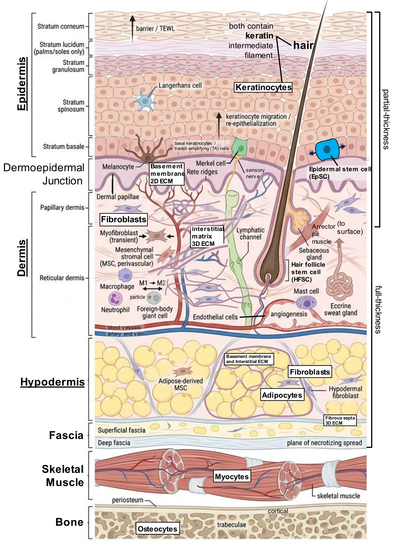
Figure 2.
Regional skin thickness variation and concentration of chronic wound location.
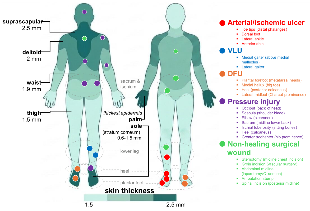
Figure 3.
Taxonomy of skin injury.
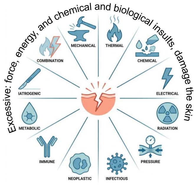
Figure 4.
Biological responses to injury: tissue repair and immune response.
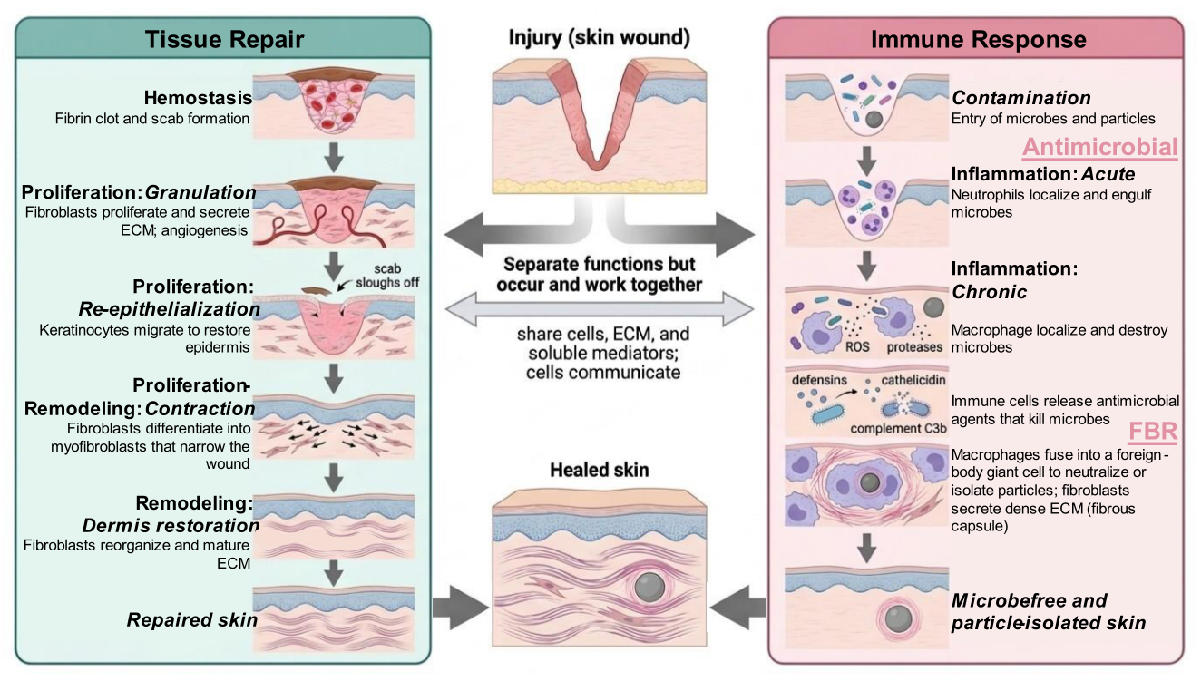
Figure 5.
Phases of skin repair including immune cells over time and the recovery of wound tensile strength.
Figure 5.
Phases of skin repair including immune cells over time and the recovery of wound tensile strength.
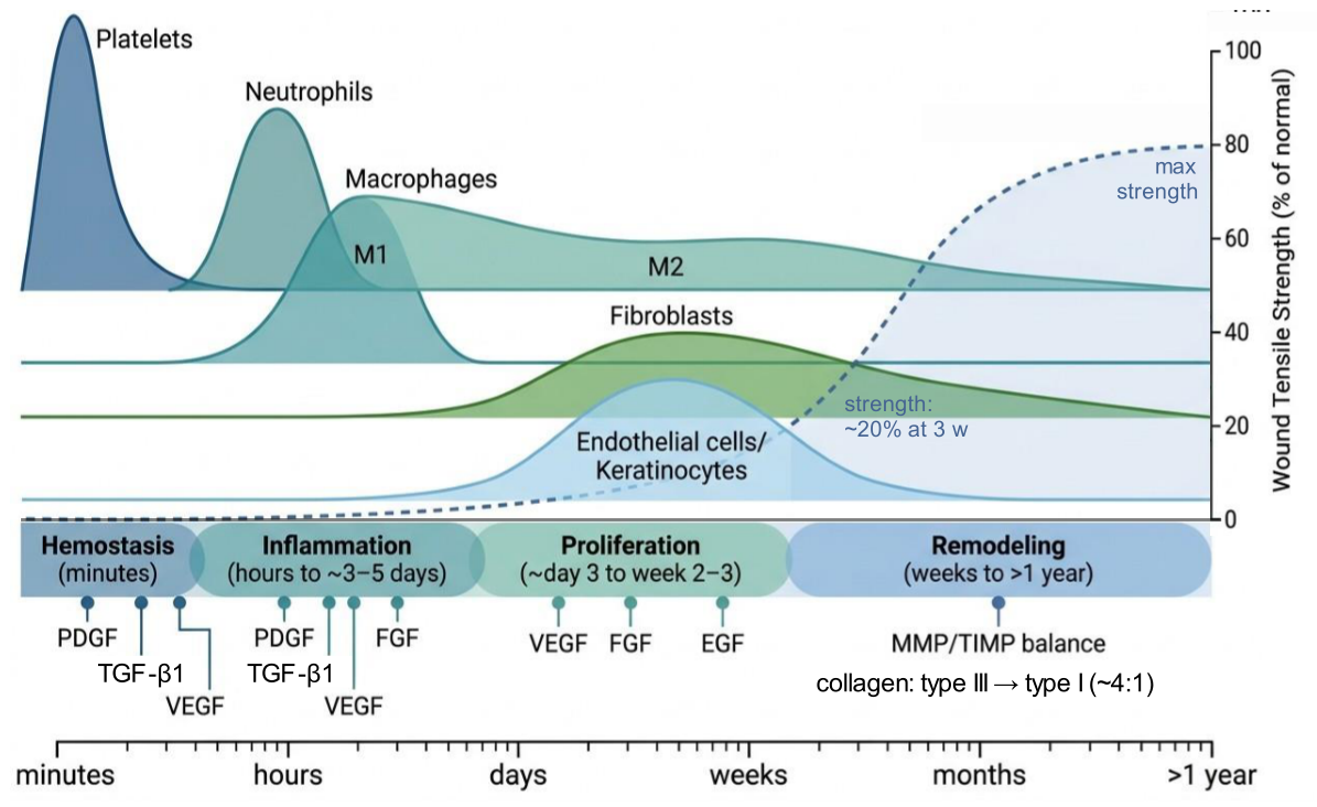
Figure 6.
Acute versus chronic wound local microenvironment.
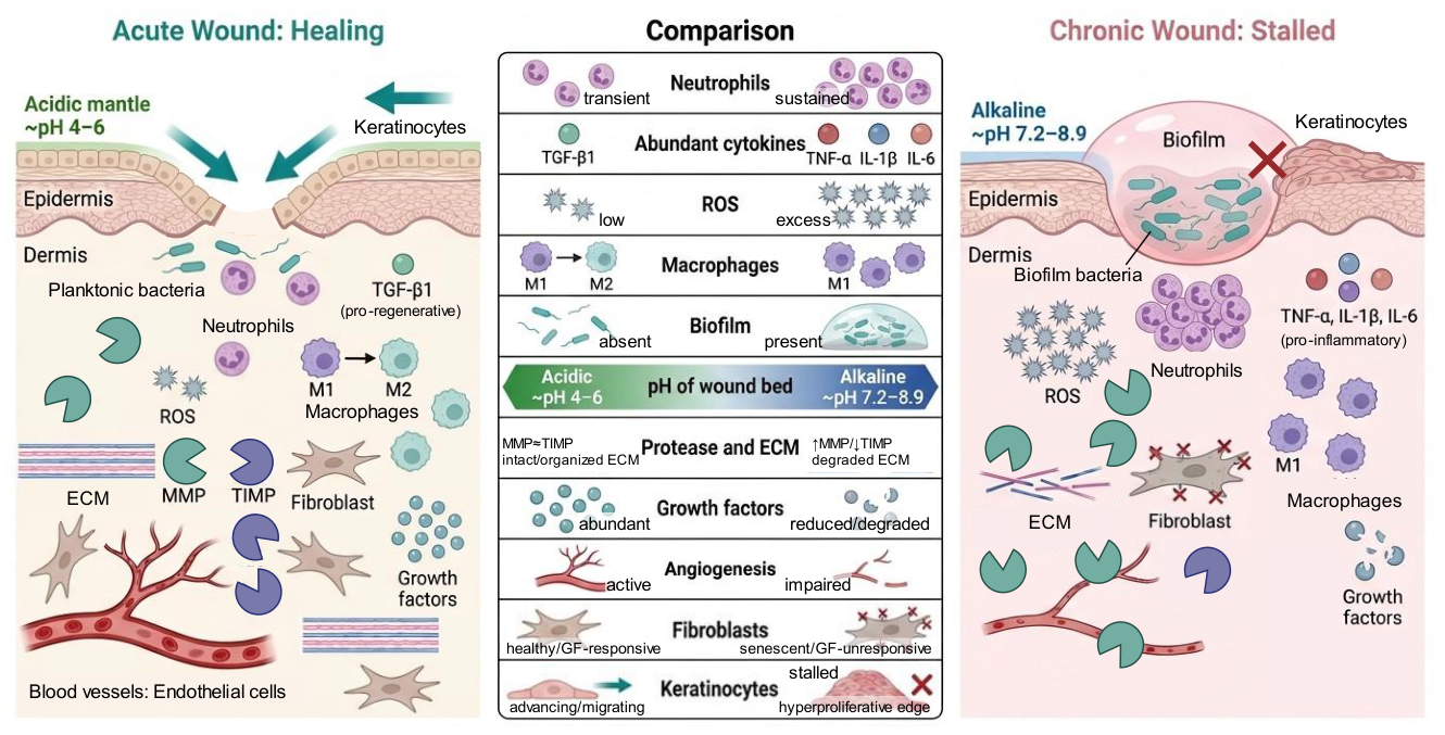
Figure 7.
The major chronic wounds: A) arterial/ischemic ulcer, B) VLU, C) DFU, D) pressure injury, and E) non-healing surgical wound (representative clinical images). Full image credits are provided in the Acknowledgments section.
Figure 7.
The major chronic wounds: A) arterial/ischemic ulcer, B) VLU, C) DFU, D) pressure injury, and E) non-healing surgical wound (representative clinical images). Full image credits are provided in the Acknowledgments section.
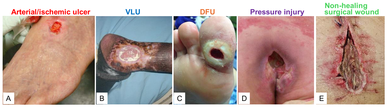
Figure 8.
Condition-specific management of chronic wounds. Management follows the TIME/Wound Bed Preparation framework: cause-specific therapy precedes the shared core (tissue debridement (T), infection and inflammation control (I), and moisture balance (M)) with edge advancement (E) adjuncts matched to wound type. In arterial/ischemic ulcers, standard wound-bed care (the shared debridement, infection-control, and moisture-balance preparation) resumes only after revascularization, with debridement of dry, stable eschar deferred until perfusion is restored. For non-healing surgical wounds, prevention bundles (perioperative skin antisepsis, correctly timed prophylactic antibiotics, normothermia, and glycemic control) reduce SSI.
Figure 8.
Condition-specific management of chronic wounds. Management follows the TIME/Wound Bed Preparation framework: cause-specific therapy precedes the shared core (tissue debridement (T), infection and inflammation control (I), and moisture balance (M)) with edge advancement (E) adjuncts matched to wound type. In arterial/ischemic ulcers, standard wound-bed care (the shared debridement, infection-control, and moisture-balance preparation) resumes only after revascularization, with debridement of dry, stable eschar deferred until perfusion is restored. For non-healing surgical wounds, prevention bundles (perioperative skin antisepsis, correctly timed prophylactic antibiotics, normothermia, and glycemic control) reduce SSI.
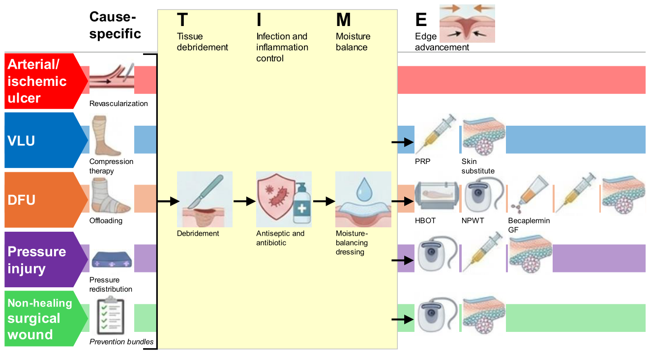
Figure 9.
Principal causes of chronic wound and mechanisms of action of established management strategies. Each numbered cause (left) drives a set of chronic-wound issues (center) that the correspondingly numbered strategy (right) counters (⊣, inhibition), with metabolic and vascular disease as the systemic cause and solution; see Section 3.3 and Section 4 for details.
Figure 9.
Principal causes of chronic wound and mechanisms of action of established management strategies. Each numbered cause (left) drives a set of chronic-wound issues (center) that the correspondingly numbered strategy (right) counters (⊣, inhibition), with metabolic and vascular disease as the systemic cause and solution; see Section 3.3 and Section 4 for details.
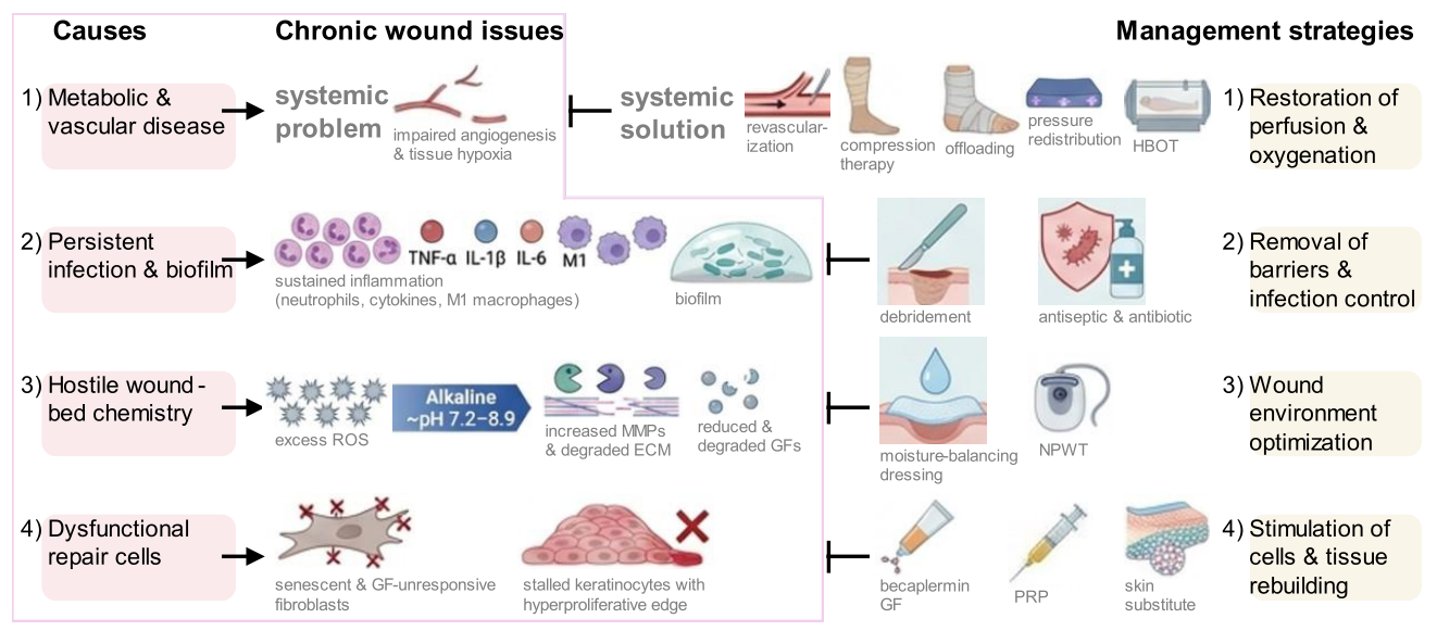
Figure 10.
Unmet needs and role of biomaterials including hair keratins.
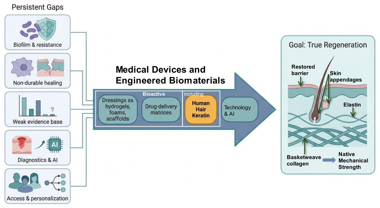
Table 1.
Specific function of skin layers and underlying tissues.
| Tissue layer | Thickness | Cells | ECM & features | Role in repair |
|---|---|---|---|---|
| Epidermis | 0.05-0.10 mm, 0.6-1.5 mm in palms/soles | Keratinocytes (90-95%), basal keratinocytes, EpSCs, HFSCs, melanocytes, Langerhans, Merkel | Basement membrane, keratin (cytoskeleton), stratum corneum lipid barrier, five strata | Re-epithelialization, barrier (TEWL) |
| Dermoepidermal junction (basement membrane zone) | < 1 µm | - | Type IV collagen, laminin-332, type VII collagen (anchoring fibrils) | Anchors epidermis, resists shear |
| Dermis (papillary & reticular) | 1-4 mm | Dermal fibroblasts, MSCs, myofibroblasts (transient), mast cells, endothelial cells, recruited immune cells: neutrophils, macrophages, foreign-body giant cells | Interstitial matrix: collagen (70-80% dry weight, type I:III ~ 4:1), elastin (2-4%), GAGs, vessels, nerves, appendages | Granulation, contraction, matrix, vascular supply, angiogenesis, immune defense, FBR |
| Hypodermis or subcutis | mm-cm | Adipocytes, MSCs, hypodermal fibroblasts | Basement membrane, interstitial matrix, fibrous septa, adipose lobules | Cushioning, insulation, energy |
| Fascia: superficial & deep | < 1-few mm | Fibroblasts, adipocytes | Interstitial matrix, areolar fat (superficial), dense type I collagen (deep) | Compartmentalization, plane of necrotizing spread |
| Skeletal muscle | variable | Myocytes | Fascicles (endo-, peri-, & epimysium), highly vascular | Deep-wound bed, exposure signals severe injury |
| Bone | variable | Osteocytes, periosteal cells | Cortical & trabecular, periosteal supply | Deepest structure, osteomyelitis risk |
Table 2.
Skin injury taxonomy by mechanism.
| Mechanism | Examples | Usual depth | Defining feature |
|---|---|---|---|
| Mechanical | Abrasion, laceration, incision, puncture, avulsion/degloving, crush, bite | Partial-full | Force with contamination |
| Pressure | Pressure injury (Stage 1-4, DTI, unstageable), arterial/ischemic ulcer | Partial-full to bone | Sustained pressure and ischemia > capillary closing pressure |
| Thermal | Flame, scald, contact burns, frostbite | Partial-full (by depth/TBSA) | Protein denaturation, barrier loss |
| Radiation | Radiotherapy, accidental exposure, UV | Dermal & microvascular | Endarteritis obliterans, delayed chronic wound |
| Electrical | Low and high voltage injury | Deep ≫ surface | Joule heating + electroporation, hidden deep necrosis |
| Chemical | Acid (coagulative), alkali (liquefactive), hydrofluoric acid | Variable & progressive | Agent × concentration × time |
| Infections | Cellulitis, abscess, necrotizing fasciitis | Superficial-deep | Direct microbial tissue destruction |
| Neoplastic | BCC/SCC/melanoma, Marjolin ulcer, Mohs/excision defect | Variable | Malignant tissue, biopsy required |
| Immune & inflammatory | Pyoderma gangrenosum, vasculitis, calciphylaxis | Dermal-full | Pathergy, needs immunosuppression (not surgery) |
| Iatrogenic | Surgical incision, donor site, device & adhesive injury, extravasation | Variable | Care-related, SSI archetype |
| Systemic & metabolic | Diabetes, venous & arterial disease, CKD, malnutrition | Modifier | Impairs repair phases leading to chronicity |
| Combination | Neuroischemic DFU, infected pressure injury, polytrauma | Mixed | Overlapping burdens, least predictable |
Table 3.
Statistics of the major chronic wounds. Aggregate United States burden: about 10.5 million Medicare beneficiaries and roughly $22.5 billion in annual Medicare spending (all-payer $28.1-96.8 billion). Global wound-care expenditure $148.65 billion in 2022 [5,6].
Table 3.
Statistics of the major chronic wounds. Aggregate United States burden: about 10.5 million Medicare beneficiaries and roughly $22.5 billion in annual Medicare spending (all-payer $28.1-96.8 billion). Global wound-care expenditure $148.65 billion in 2022 [5,6].
| Wound type | Prevalence & incidence | Cost & amputation risk | Recurrence | Mortality |
|---|---|---|---|---|
| Arterial/ischemic ulcer | With PAD & critical limb ischemia, often neuroischemic | High amputation rate in CLI | Perfusion-dependent | High 1-y mortality in CLI |
| VLU | ~70% of leg ulcers, prevalence 0.1-0.3% (1-3% in elderly) | Dominant cost of leg-ulcer care, major QoL loss | ~55-70% without reflux correction | Low direct, high morbidity |
| DFU | Lifetime risk 19-34% in diabetes, global prevalence ~6.3% | Precedes ~80% of diabetes-related amputations | ~40% at 1 y, ~65% at 5 y | 5-y ~30%, ~50-70% after major amputation |
| Pressure injury | Hospital point prevalence ~12.8%, higher in ICU | Largely preventable, osteomyelitis in Stage 3-4 | Site-dependent | Independently increases mortality |
| Non-healing surgical wound | SSI ~0.5-3% of procedures | Thousands of dollars and inpatient days per case | - | Contributes to sepsis risk |
Table 5.
Important wound care resources.
| Category | Resource | Primary Use |
|---|---|---|
| Literature database | PubMed / MEDLINE (Medical Subject Headings) | Primary-literature retrieval |
| Literature database | Scopus, Web of Science | Citation coverage and breadth |
| Literature database | Embase, CINAHL | Biomedical and nursing/allied-health literature |
| Evidence synthesis | Cochrane Library | Systematic reviews |
| Evidence synthesis | JBI (Joanna Briggs Institute) | Systematic reviews and evidence implementation |
| Clinical studies | ClinicalTrials.gov | Trial registration and results |
| Guideline body | NPIAP, EPUAP, PPPIA | Pressure injury staging and prevention |
| Guideline body | IWGDF | Diabetic foot and offloading guidelines |
| Guideline body | WUWHS | Global consensus documents (e.g., TIME/wound bed prep) |
| Guideline body | NICE, SIGN, RNAO | National clinical guidelines (wound/ulcer care) |
| Guideline body | IWII | Wound-infection guidance |
| Society | WHS | Wound-treatment guidelines |
| Society | AAWC, EWMA | Standards, education, advocacy |
| Society | WOCN, APWCA, ABWM | Certification, standards, and education |
| Society | Wounds UK, Wounds Canada, Wounds Australia | Regional guidance, journals, and congresses |
| Society | UHMS | Hyperbaric-oxygen therapy guidance |
| Foundation / diabetic foot | Wound Healing Foundation, IDF, DFSG | Research funding and diabetic-foot standards |
| Registry | Wound registries | Real-world outcomes and benchmarking |
Disclaimer/Publisher’s Note: The statements, opinions and data contained in all publications are solely those of the individual author(s) and contributor(s) and not of MDPI and/or the editor(s). MDPI and/or the editor(s) disclaim responsibility for any injury to people or property resulting from any ideas, methods, instructions or products referred to in the content. |
© 2026 by the authors. Licensee MDPI, Basel, Switzerland. This article is an open access article distributed under the terms and conditions of the Creative Commons Attribution (CC BY) license (http://creativecommons.org/licenses/by/4.0/).
Copyright: This open access article is published under a Creative Commons CC BY 4.0 license, which permit the free download, distribution, and reuse, provided that the author and preprint are cited in any reuse.