

Submitted:
06 August 2026
Posted:
07 August 2026
You are already at the latest version
Abstract
A 4-year-old nonverbal boy with level 3 autism spectrum disorder and Duchenne muscular dystrophy (DMD) who was receiving chronic prednisone therapy presented to the emergency room with acute appendicitis. The diagnosis was significantly delayed due to atypical pain presentation, a potentially blunted inflammatory response from chronic steroid use, and the absence of classic abdominal examination findings due to DMD. The patient presented with five days of fever, poor oral intake, decreased stool output and behavioral changes. Initial physical examination and evaluation over two emergency department visits failed to establish the diagnosis. By the third visit, the patient exhibited abdominal distension, hypoactive bowel sounds, and bandemia. Computed tomography demonstrated perforated appendicitis with a walled-off abscess. Nonoperative management was unsuccessful, resulting in the development of multiple intra-abdominal abscesses that required percutaneous drainage. Following a 10-day hospitalization, the patient improved and was discharged. He subsequently underwent a scheduled laparoscopic appendectomy two months later, with pathology demonstrating persistent inflammation. This case raises awareness of the atypical presentation of pain in children with autism and the potential for DMD and chronic steroid therapy to mask expected examination findings. These factors should prompt clinicians to maintain a low threshold for advanced imaging when clinical concern is present. It also illustrates that non operative management of perforated appendicitis in immunosuppressed children may have an increased risk of failure.
Keywords:
autism spectrum disorder
; Duchenne muscular dystrophy
; acute appendicitis
1. Introduction
Acute appendicitis is one of the most common pediatric emergencies and timely diagnosis is essential to prevent complications such as perforation and abscess formation [1]. Diagnosis typically relies on history and physical exam findings, with the classic presentation including migration of pain from the periumbilical region to the right lower quadrant, vomiting, and fever [2]. In younger children, however, these classic features may be absent, contributing to delayed diagnosis [3,4,5].
Diagnostic challenges are amplified in children with autism spectrum disorder (ASD), particularly those who are nonverbal. Rather than verbalizing discomfort or localizing pain, these children may express distress through deviations from their baseline behavior, including increased irritability, aggression, self-injurious behaviors, or altered facial expressions [6,7]. Consequently, their behavioral manifestations are often misunderstood, leading to higher rates of complications from acute appendicitis [8,9]. Furthermore, commonly used visual self-report pain assessment tools like the Wong-Baker FACES Pain Rating Scale may have limited utility in pediatric populations with ASD or cognitive delays. This inaccuracy stems from developmental barriers that limit their ability to map their internal sensory experiences onto abstract pictorial representations of emotion [6,10].
Diagnosing appendicitis in children with Duchenne muscular dystrophy (DMD) is complicated by the fact that classic peritoneal signs, such as abdominal rigidity and guarding, may be attenuated due to characteristic progressive muscle weakness, limiting abdominal wall contraction [11]. Moreover, the chronic use of steroids in patients with DMD may both increase susceptibility to infection and blunt inflammatory responses, including fever and leukocytosis, potentially masking appendicitis [12].
Herein, we present the case of a 4-year-old nonverbal boy with the coexistence of level 3 ASD and DMD on chronic steroid therapy whose atypical presentation resulted in a delayed diagnosis of acute appendicitis leading to perforation and abscess formation. This case highlights the importance of maintaining a high index of suspicion for appendicitis in medically complex children who present with behavioral changes or nonspecific symptoms despite initially reassuring evaluations.
2. Case Presentation
A 4-year-old nonverbal boy with level 3 ASD and DMD (deletion of introns 53-60), managed with prednisone 10 mg daily, presented to his pediatrician with a five-day history of fever, poor oral intake, decreased stool output, frequent crying and poor sleep. Serial vital signs, laboratory results, imaging findings, and key clinical events are summarized in Table 1.
On initial evaluation, he was irritable, combative, and unable to cooperate with the physical examination. His temperature was 38.3°C, with otherwise normal vital signs. He was referred to the emergency department for further evaluation, where repeat temperature was 36.4°C. Abdominal examination was reportedly benign, with normal bowel sounds and no tenderness, guarding or rigidity. His Wong-Baker FACES Pain Rating Scale was 0. Chest radiograph was normal. Given the absence of concerning physical examination findings, he was diagnosed with presumed gastroenteritis and discharged with supportive care.
The following day, he returned to the emergency department due to persistent fever, irritability, poor oral intake, vomiting and four days without a bowel movement. His temperature was 38.9°C and his Wong-Baker FACES Pain Rating Scale was 2. Repeat abdominal examination remained benign, demonstrating a soft, nondistended, nontender abdomen with normal bowel sounds and no organomegaly or palpable masses. Abdominal radiograph indicated a nonspecific bowel gas pattern with a mild stool burden and no free air or appendicolith. He was treated for constipation and discharged with instructions to continue supportive care.
Two days later, he presented for a third time with persistent symptoms and worsening fever to 39.4°C. Physical examination revealed abdominal distension and hypoactive bowel sounds. Computed tomography (CT) demonstrated perforated appendicitis with a walled-off abscess (Figure 1). He was admitted for nonoperative management with broad-spectrum antibiotics and supportive care. Four days after admission, progressive abdominal distension prompted abdominal ultrasonography, revealing multiple intra-abdominal abscesses. He subsequently underwent percutaneous drainage with catheter placement by interventional radiology and continued antibiotic therapy. The drains were removed four days later, and he was discharged after a 10-day hospitalization with an additional seven-day course of oral antibiotics. Two months later, he underwent an uncomplicated laparoscopic appendectomy, which showed persistent appendicitis with perforation (Figure 2).
Figure 1.
Axial contrast-enhanced Computed Tomography (CT) findings. (a) Inflamed appendix with surrounding periappendiceal inflammatory changes. (b) Periappendiceal abscess measuring 38.8 × 24.2 mm.
Figure 1.
Axial contrast-enhanced Computed Tomography (CT) findings. (a) Inflamed appendix with surrounding periappendiceal inflammatory changes. (b) Periappendiceal abscess measuring 38.8 × 24.2 mm.
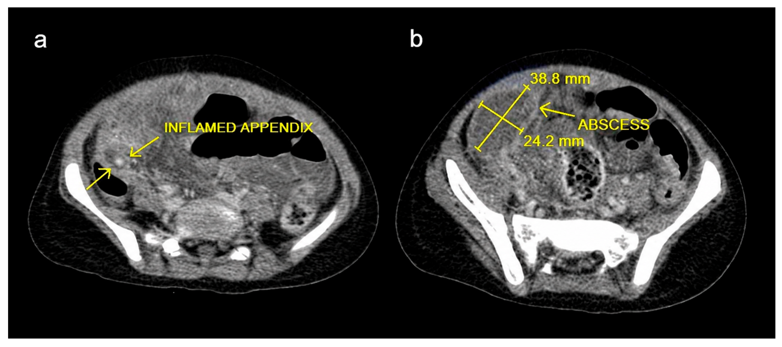
Figure 2.
Cross Section of Appendix. (a) H&E stain at 2x. The entire surface mucosa has been replaced by inflammation; inflammation is seen spreading relatively close to the outer surface of the appendiceal wall; no residual appendiceal mucosa is present in this section. Nodules of residual normal lymphoid aggregates are present. (b) H&E stain at 20x demonstrating occasional multinucleate giant cells (*).
Figure 2.
Cross Section of Appendix. (a) H&E stain at 2x. The entire surface mucosa has been replaced by inflammation; inflammation is seen spreading relatively close to the outer surface of the appendiceal wall; no residual appendiceal mucosa is present in this section. Nodules of residual normal lymphoid aggregates are present. (b) H&E stain at 20x demonstrating occasional multinucleate giant cells (*).
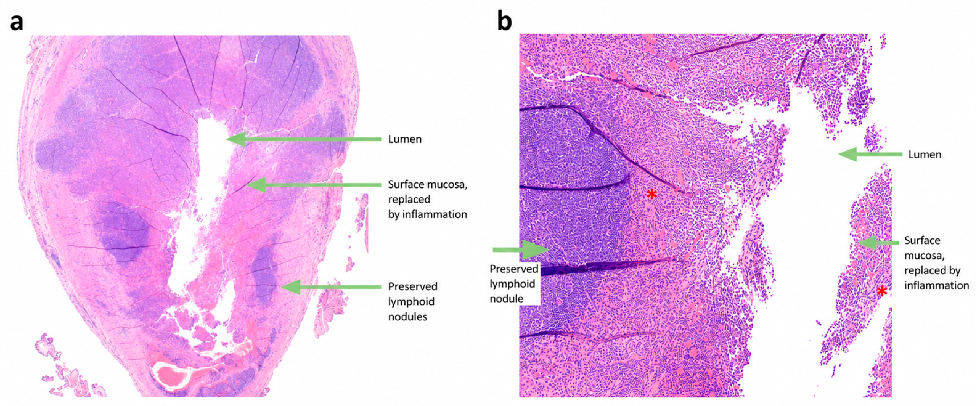

Table 1.
Summary of Emergency Department Visits and Inpatient management. .
| Date | 6/11/2024 | 6/12/2024 | 6/14/24 | 6/18/2024 | 8/23/2024 |
| Temperature (celcius) | 36.4 | 38.9 | 39.4 | 36.8 | - |
| Blood Pressure (mmHg) | - | 99/68 | - | 109/85 | - |
| Heart Rate (beats/ minute) | 134 | 159 | - | 150 | - |
| Respiratory Rate (Breaths/ minute) | 24 | 22 | 24 | 22 | - |
| Wong Baker/ FACES | 0 | 2 | - | - | - |
| White Blood Cell Count (WBC) | - | - | 13.3 x 10e6/L | - | - |
| Absolute Neutrophil Count (as percentage of WBC) | - | - | 48%3 | - | - |
| Band Count (as percentage of WBC) | - | - | 17%4 | - | - |
| Anion Gap | - | - | 17 mmol/L | - | - |
| Lactic Acid | - | - | 1.0 mmol/L | - | - |
| Imaging | CXR1 | AXR2 | CT scan5 | Abdominal US6 | - |
| Diagnosis | Gastroenteritis | Constipation | Perforated appendicitis with Abscess | Perforated Appendicitis with multiple abscesses | Perforated appendicitis for interval appendectomy |
| Treatment | ondansetron, ibuprofen and oral fluids | ondansetron, ibuprofen alternating with acetaminophen | Interval appendectomy pathway | percutaneous drain | Laparoscopic Appendectomy. |
| Pathology Report | - | - | - | Perforated vermiform appendix7 |
*1 No acute cardiopulmonary disease. 2 No free air, air fluid levels or appendicolith identified. 3 Absence of significant neutrophilia, possibly related to chronic steroid use. 4 Left shift with bandemia consistent with acute infection. Bandemia remains a useful marker of infection in patients on steroids. 5 Perforated appendix with walled off abscess (See Figure 1). 6 Multiple intraabdominal abscesses. 7 See Figure 2.
3. Discussion
This case illustrates how the combination of severe ASD, DMD, and chronic corticosteroid therapy obscured the clinical presentation of acute appendicitis and contributed to delayed diagnosis.
Our patient lacked the classic history associated with acute appendicitis, including periumbilical pain migrating to the right lower quadrant and exacerbation of pain with movement. This diagnostic challenge stemmed from the atypical presentation of pain in children with ASD as well as his inability to localize pain. In this population, visceral pain frequently manifests as acute behavioral changes, such as heightened irritability, self-injurious behaviors, or aggression along with poor cooperation with physical examination [6,7]. Caregiver interviews are therefore essential to establish the child’s behavioral baseline and identify meaningful deviations. Furthermore, clinicians should be aware that standard self-report pain assessment tools like the Wong-Baker FACES Pain Rating Scale can be difficult to interpret in neurodivergent children, who may have difficulty associating abstract pictorial expressions with their internal sensory experiences [10]. Despite the absence of classic localized pain findings, our patient demonstrated other signs of advanced peritonitis —such as anorexia, vomiting, and decreased bowel output—which may reflect secondary paralytic ileus and should prompt careful reassessment [11].
Classic peritoneal signs, including abdominal rigidity and guarding, were absent in our patient. Although causality cannot be established, coexisting DMD may have contributed to the absence of these findings. Clinicians should recognize that patients with advanced DMD may lack these classic signs because progressive weakness of the abdominal wall musculature may diminish protective muscle contraction [12,13]. Nevertheless, secondary signs of peritonitis, including abdominal distension and hypoactive bowel sounds, were evident.
Chronic corticosteroid therapy may attenuate fever and leukocytosis while increasing susceptibility to infection, making appendicitis more difficult to recognize [13]. Although corticosteroid-induced demargination can complicate interpretation of the leukocyte count, bandemia remains an important indicator of acute bacterial infection, as was observed in our patient. Emerging evidence suggests that procalcitonin may be less affected by corticosteroid therapy than C-reactive protein (CRP) and may therefore serve as a useful adjunctive biomarker for evaluating bacterial infection [14].
Plain abdominal radiography has limited diagnostic utility in suspected acute appendicitis and should not be used to exclude the diagnosis [15]. In our patient, the initial abdominal radiograph demonstrated only a nonspecific bowel gas pattern and mild stool burden, contributing little to the diagnostic evaluation. When clinical suspicion persists despite a reassuring physical examination or nondiagnostic radiographs, particularly in children with communication barriers or neuromuscular disorders, clinicians should maintain a low threshold for ultrasonography or CT.
Nonoperative management with antibiotics is an accepted treatment strategy for perforated appendicitis with localized abscess formation [16]. However, careful patient selection and close clinical monitoring remain essential. In our patient, a localized abscess progressed despite antibiotic therapy, ultimately requiring percutaneous drainage of multiple intra-abdominal abscesses. This case illustrates that medically complex or immunosuppressed children undergoing nonoperative management may require escalation of care despite appropriate initial therapy.
4. Conclusions
This case highlights the diagnostic and management challenges of acute appendicitis in a nonverbal child with coexisting ASD and DMD. Communication barriers, atypical pain behaviors, and attenuation of classic abdominal findings contributed to delayed recognition and subsequent perforation with abscess formation. Clinicians should maintain a high index of suspicion for serious intra-abdominal pathology in children with neurodevelopmental and neuromuscular disorders and consider early advanced imaging when symptoms persist or deviations from baseline behavior are present despite an initially reassuring evaluation. This case underscores the importance of close monitoring during nonoperative management of perforated appendicitis in medically complex children, as escalation of care may be required.
Author Contributions
Mona Makhlouf and Stanley Calderwood conceptualized and designed the study, drafted the initial manuscript, and critically reviewed and revised the manuscript. Stanley Calderwood designed the data collection instruments, collected data, carried out the initial analyses, and critically reviewed and revised the manuscript. All authors have read and agreed to the published version of the manuscript.
Funding
This research received no external funding.
Institutional Review Board Statement
Not applicable.
Informed Consent Statement
Written permission was obtained from the patient’s guardians to publish this case and the corresponding photos or imaging studies.
Acknowledgments
Thank you to Dr. Chris Le-Pong for providing pathology interpretation and to Dr. Bernard Maristany for providing pathology interpretation.
Conflicts of Interest
The authors declare no conflicts of interest.
Abbreviations
The following abbreviations are used in this manuscript:
| ASD | Autism Spectrum Disorder |
| DMD | Duchenne Muscular Dystrophy |
| CT | Computed Tomography |
References
- Becker C, Kharbanda A. Acute appendicitis in pediatric patients: an evidence-based review. Pediatr Emerg Med Pract. 2019 Sep;16(9):1-20. Epub 2019 Sep 2. PMID: 31461613.
- Moris D, Paulson EK, Pappas TN. Diagnosis and Management of Acute Appendicitis in Adults: A Review. JAMA. 2021 Dec 14;326(22):2299-2311. PMID: 34905026. [CrossRef]
- Glass CC, Rangel SJ. Overview and diagnosis of acute appendicitis in children. Semin Pediatr Surg. 2016 Aug;25(4):198-203. Epub 2016 May 10. PMID: 27521708. [CrossRef]
- Becker T, Kharbanda A, Bachur R. Atypical clinical features of pediatric appendicitis. Acad Emerg Med. 2007 Feb;14(2):124-9. Epub 2006 Dec 27. PMID: 17192449. [CrossRef]
- Michelson KA, Reeves SD, Grubenhoff JA, et al. Clinical Features and Preventability of Delayed Diagnosis of Pediatric Appendicitis. JAMA Netw Open. 2021;4(8):e2122248. [CrossRef]
- Johnson E, van Zijl K, Kuyler A. Pain communication in children with autism spectrum disorder: A scoping review. Paediatr Neonatal Pain. 2023 Oct 13;5(4):127-141. PMID: 38149220; PMCID: PMC10749405. [CrossRef]
- Allely CS. Pain sensitivity and observer perception of pain in individuals with autistic spectrum disorder. ScientificWorldJournal. 2013 Jun 13;2013:916178. PMID: 23843740; PMCID: PMC3697411. [CrossRef]
- Lee SY, Byrd EA, Kohler JE. Children with neurodevelopmental disabilities have worse appendicitis outcomes. Journal of Pediatric Surgery Open. 2025;11:100210. [CrossRef]
- Reeves PT, Susi A, Hisle-Gorman E, Gorman GH, Nylund C. Brief Report: Association of Complicated Appendicitis in Children with Autism Spectrum Disorders. J Autism Dev Disord. 2020 Dec;50(12):4535-4540. PMID: 32297124. [CrossRef]
- Ely E, Chen-Lim ML, Carpenter KM 2nd, Wallhauser E, Friedlaender E. Pain Assessment of Children with Autism Spectrum Disorders. J Dev Behav Pediatr. 2016 Jan;37(1):53-61. PMID: 26703326. [CrossRef]
- Almaramhy HH. Acute appendicitis in young children less than 5 years: Review article. Italian Journal of Pediatrics. 2017;43:15. [CrossRef]
- Birnkrant DJ, Bushby K, Bann CM, Apkon SD, Blackwell A, Brumbaugh D, Case LE, Clemens PR, Hadjiyannakis S, Pandya S, Street N, Tomezsko J, Wagner KR, Ward LM, Weber DR; DMD Care Considerations Working Group. Diagnosis and management of Duchenne muscular dystrophy, part 1: diagnosis, and neuromuscular, rehabilitation, endocrine, and gastrointestinal and nutritional management. Lancet Neurol. 2018 Mar;17(3):251-267. Epub 2018 Feb 3. Erratum in: Lancet Neurol. 2018 Jun;17(6):495. doi: 10.1016/S1474-4422(18)30125-X. PMID: 29395989; PMCID: PMC5869704. [CrossRef]
- Komaki H. Duchenne muscular dystrophy: Evolving therapeutic strategies and multidimensional evaluation approaches. Brain Dev. 2025 Oct;47(5):104397. Epub 2025 Jul 24. PMID: 40712356. [CrossRef]
- Raess N, Schuetz P, Cesana-Nigro N, Winzeler B, Urwyler SA, Schaedelin S, Rodondi N, Blum MR, Briel M, Mueller B, Christ-Crain M, Blum CA. Influence of Prednisone on Inflammatory Biomarkers in Community-Acquired Pneumonia: Secondary Analysis of a Randomized Trial. J Clin Pharmacol. 2021 Nov;61(11):1406-1414. Epub 2021 Jul 27. PMID: 34031890; PMCID: PMC8242868. [CrossRef]
- Koberlein GC, Trout AT, Rigsby CK, et al. ACR Appropriateness Criteria® Suspected Appendicitis–Child. J Am Coll Radiol. 2019;16(5S):S252-S263. [CrossRef]
- Fugazzola P, Ceresoli M, Agnoletti V, et al. Early appendectomy vs. conservative management in complicated acute appendicitis in children with abscess or phlegmon: A systematic review and meta-analysis. J Pediatr Surg. 2019.
Disclaimer/Publisher’s Note: The statements, opinions and data contained in all publications are solely those of the individual author(s) and contributor(s) and not of MDPI and/or the editor(s). MDPI and/or the editor(s) disclaim responsibility for any injury to people or property resulting from any ideas, methods, instructions or products referred to in the content. |
© 2026 by the authors. Licensee MDPI, Basel, Switzerland. This article is an open access article distributed under the terms and conditions of the Creative Commons Attribution (CC BY) license (http://creativecommons.org/licenses/by/4.0/).
Copyright: This open access article is published under a Creative Commons CC BY 4.0 license, which permit the free download, distribution, and reuse, provided that the author and preprint are cited in any reuse.


