

Submitted:
06 August 2026
Posted:
07 August 2026
You are already at the latest version
Abstract
Seizures occur more frequently in the neonatal period than at any other time in life. Seizures and epileptic burden in the neonatal period- is correlated to neurodevelopmental outcomes and overall prognosis. Accurate diagnosis is essential for appropriate management; however, there are many diagnostic dilemmas. Seizures in the neonatal period are difficult to diagnose accurately. Many seizures are electrographic only with no clinical correlates, different kinds of paroxysmal events in neonates may mimic electroclinical seizures, and diagnosis based on clinical phenomena alone may result in inaccuracies . There is no universally accepted definition of a neonatal seizure or neonatal status epilepticus (Conventional EEG (CEEG) is the gold standard for diagnosis of a neonatal seizure. Continuous conventional video EEG monitoring (cVEEG) for 24 to 48 hours may be needed for detecting seizures in those at risk, to monitor therapy and to assess epileptic burden; it is not available for clinical care in most parts of the world . There are many therapeutic challenges in the treatment of seizures in the neonate. Most neonatal seizures are provoked or due to symptomatic aetiology. Genetic, metabolic and structural epilepsies are less frequent causes for neonatal seizures. The treatment paradigm is often similar initially, till a specific aetiology is suspected or identified. When to treat, what to treat with, how much to treat, and how long to treat are still being debated, with no uniform strategy. Early distinction between provoked seizures and neonatal onset epilepsy is important to guide therapy. The emergence of targeted therapies (some quite expensive), while exciting, has added to the therapeutic challenges and inequities associated with management of neonates with seizures. This review addresses many of the diagnostic dilemmas and therapeutic challenges encountered by a clinician in the management of neonates with seizures.
Keywords:
neonatal seizures
; neonatal status epilepticus
; treatment of neonatal seizures
; diagnosis of neonatal seizures
1. Introduction
Seizures occur more frequently in the neonatal period than at any other time in life. Seizures and epileptic burden in the neonatal period- is correlated to neurodevelopmental outcomes and overall prognosis [1,2] Accurate diagnosis is essential for appropriate management; however, there are many diagnostic dilemmas. Seizures in the neonatal period are difficult to diagnose accurately. Many seizures are electrographic only with no clinical correlates, different kinds of paroxysmal events in neonates may mimic electroclinical seizures, and diagnosis based on clinical phenomena alone may result in inaccuracies [3,4,5]. There is no universally accepted definition of a neonatal seizure or neonatal status epilepticus [6,7,8,9] (Conventional EEG (CEEG) is the gold standard for diagnosis of a neonatal seizure. Continuous conventional video EEG monitoring (cVEEG) for 24 to 48 hours may be needed for detecting seizures in those at risk, to monitor therapy and to assess epileptic burden; it is not available for clinical care in most parts of the world [10,11,12,13,14,15].
There are many therapeutic challenges in the treatment of seizures in the neonate. Most neonatal seizures are provoked or due to symptomatic aetiology. Genetic, metabolic and structural epilepsies are less frequent causes for neonatal seizures. The treatment paradigm is often similar initially, till a specific aetiology is suspected or identified. When to treat, what to treat with, how much to treat, and how long to treat are still being debated, with no uniform strategy. Early distinction between provoked seizures and neonatal onset epilepsy is important to guide therapy. The emergence of targeted therapies (some quite expensive), while exciting, has added to the therapeutic challenges and inequities associated with management of neonates with seizures.
This review addresses many of the diagnostic dilemmas and therapeutic challenges encountered by a clinician in the management of neonates with seizures.
Figure 1.
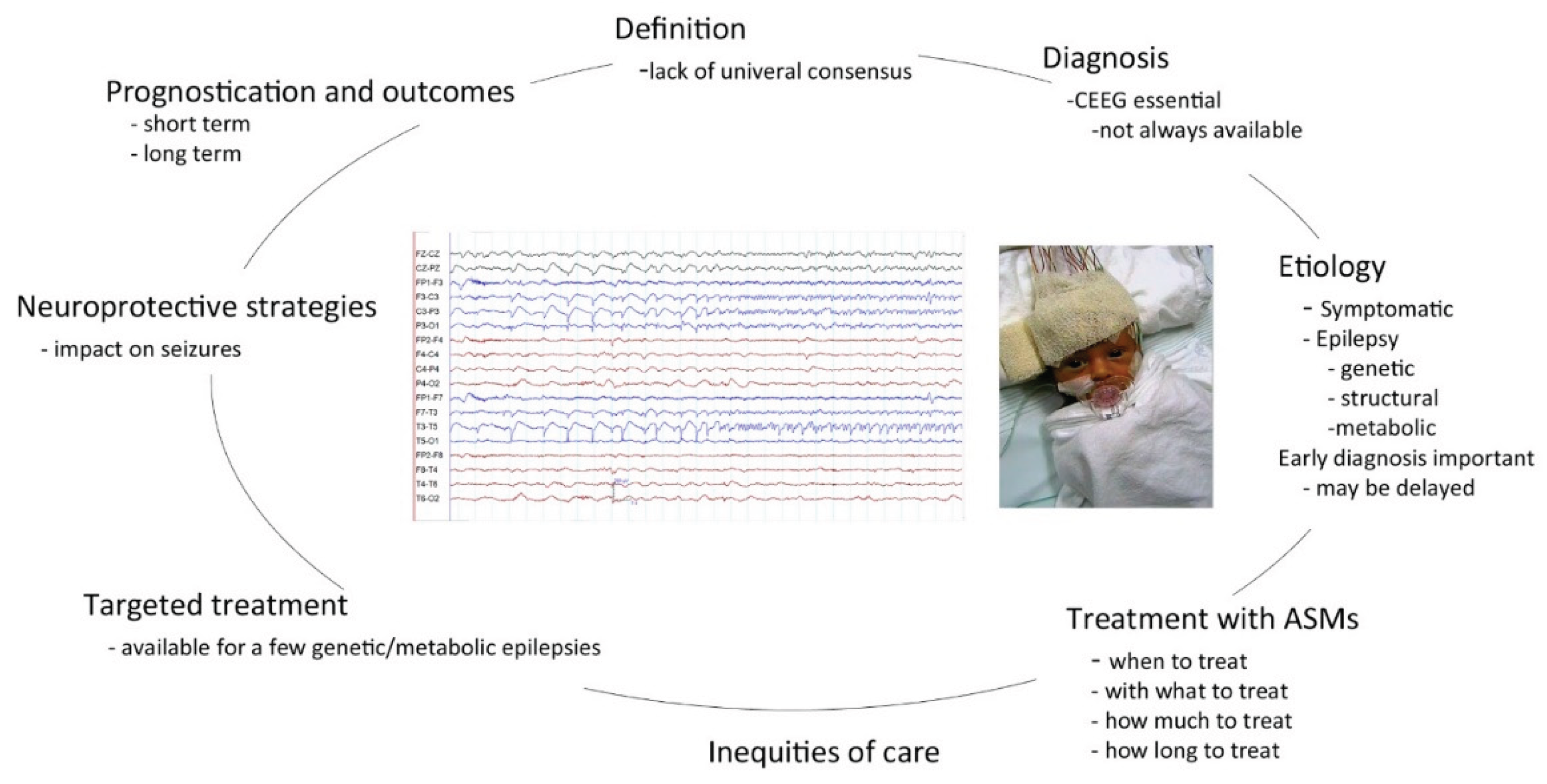
Challenges in diagnosis and management of neonatal seizures. EEG shows an electrographic seizure from the left hemisphere. EEG is double banana montage, calibration bars in figure. Picture shows baby having a CEEG. Abbreviations: ASM: Antiseizure medications, CEEG: conventional EEG.
Figure 1.
Challenges in diagnosis and management of neonatal seizures. EEG shows an electrographic seizure from the left hemisphere. EEG is double banana montage, calibration bars in figure. Picture shows baby having a CEEG. Abbreviations: ASM: Antiseizure medications, CEEG: conventional EEG.
1.1. Classification of Seizures in Neonates
Seizures in neonates have unique EEG signatures and are predominantly electrographic only [10,16,17,18]. Clinical features, when present can be subtle and may be missed in a busy neonatal intensive care unit. Non-epileptic paroxysmal movements may mimic electroclinical seizures [5] and even experienced health professionals with expertise in neonatal seizures may be incorrect. The ILAE classification of Seizures and the Epilepsies: Modification for seizures in the neonate [6] addresses many of the issues identified in application of the previous classification schemes to seizures in the neonate [19,20,21]. It is the only ILAE classification scheme that recognises electrographic only seizures as a type of seizure. The classification emphasizes the need for a clinical event to have an EEG correlate to call it an epileptic seizure. This may protect babies with seizure mimics from receiving unnecessary antiseizure medications which may have acute and long-term adverse effects.
1.2. Definition of Neonatal Seizure
Substantial variability occurs in the definition of a neonatal seizure in the literature. Conventionally a neonatal seizure was defined as one in which a rhythmic EEG discharge lasted 10 seconds or longer, had an identifiable start and finish, and could exhibit variable morphology, frequency, and field [22,23,24]. Electrographic only seizures were those without clinical correlates, and electroclinical seizures had associated clinical phenomena [16,25]. However, the position paper by the ILAE Task Force on Neonatal Seizures [6] does not nominate a specific duration for an electrographic event to be called a seizure. The relevance of BERDS (brief EEG rhythmic discharges lasting less than 10s), which is considered as mini seizures by some groups, is considered as part of the ictal/interictal continuum [26,27]. The lack of a defined duration in the latest EEG definition of a seizure has the advantage of being able to include seizure types like myoclonic seizures and infantile spasms that often have an ictal discharge of less than 10s duration. However, lack of uniformity in identification of a seizure poses difficulties for clinical research studies and for comparison between future and past studies [11]
1.3. Definition of Neonatal Status Epilepticus
The lack of a universally accepted definition of neonatal status epilepticus (NSE) makes diagnosis and management challenging [7]
The definition of the ILAE task force on Classification [28]) of Status epilepticus is conceptual, with two operational dimensions. The first is the length of the seizure and the time point (t1) beyond which the seizure should be regarded as “continuous seizure activity.” The second time point (t2) is the time of ongoing seizure activity after which there is a risk of long-term consequences. In the case of convulsive (tonic-clonic) status epilepticus. both time points (t1 at 5 minutes and t2 at 30 minutes) are based on animal experiments and clinical research. The relevance and applicability of these time points to neonatal status epilepticus, based on current knowledge, is unclear [7] (nagarajan and ghosh, 2025). The ILAE task force on classification of seizures, modified for neonates [6], does not address this issue. Based on a review of outcomes, the likelihood of spontaneous seizure termination, and probability of response to antiseizure medications, it has been proposed that any single neonatal seizure lasting more than 5 minutes be considered the equivalent of t1-early neonatal status epilepticus [8]).
A scoping review by the ILAE Neonatal Task force [9] of the available literature demonstrated substantial variations in definition of neonatal status epilepticus. The review concluded that the common definitions were based around a 30-minute seizure duration criterion, but evidence was insufficient to support a 30-minute cut-off to define prolonged seizures or indicate that seizures exceeding this burden were more likely to be pharmaco-resistant or associated with worse outcomes. Some studies suggest a seizure burden of 10-13 minutes may be a more realistic definition of t2. The neonatal task force intends to develop a standardised approach to assessing and describing neonatal seizure burden and defining NSE.
2. Diagnosis of Neonatal Seizures
Neonatal seizures are reliant on EEG for diagnosis. Conventional EEG (CEEG) is the gold standard for diagnosis and monitoring, with amplitude integrated EEG (aEEG) being a less accurate but more widely available and used tool.
CEEG vs aEEG
CEEG is the gold standard for diagnosis and monitoring of neonatal seizures – both electrographic only and electroclinical seizures [6,7]. (Most CEEG machines and studies are based on synchronised Video-EEG (VEEG). Continuous monitoring with VEEG (cVEEG) over 24 to 48 hours is used for detecting seizures (when a brief recording may miss them), and to monitor therapy and the overall epileptic burden. cVEEG may be also useful to identify infrequent non-epileptic events and to estimate the seizure burden. However, cVEEG is not available in most neonatal intensive care units across the world. Expertise in acquiring and interpreting neonatal EEG is important as neonatal seizures may have unique EEG signatures. Normal background patterns vary, are dependent on gestational and chronological age, and influenced by therapeutic interventions [10,22,29,30,31,32] .Amplitude integrated EEG (aEEG) is a simplified tool that provides a visual trend over time [10,33] and is used far more commonly in clinical practice. A recent Cochrane review [34] comparing aEEG with CEEG for detection of neonatal seizures concluded aEEG has only moderate sensitivity and specificity for detecting ‘neonates with seizures’, and its ability to detect ‘individual seizures’ varies widely. aEEG may not be sufficiently accurate for diagnosing neonatal seizures as it can under-diagnose or over-diagnose seizures and lead to inadequate or unnecessary treatment with antiseizure medications[35,36,37,38]. Use of CEEG monitoring will potentially optimise antiseizure medication use in neonates and help prognosticate outcomes [35,39] In clinical practice often combinations of standard recordings (<I hour) and more prolonged recordings are undertaken. More recently quantitative analysis of EEG (QEEG) and trend analysis have become available to clinicians; they condense hours of data into functional maps and graphs based on frequency, amplitude and rhythmicity changes [40]. Combined displays of aEEG[41] and CEEG, ECG, along with QEEG and trend analysis will promote greater interaction between neonatologists and child neurologists and is likely to contribute to better management and outcomes for neonates with seizures.
A critical review of continuous conventional EEG (cVEEG) for seizure detection in the intensive care units highlighted the gaps in evidence, practical problems, funding shortfalls and areas of future [11]research [13]. It concluded that while cVEEG detects electrographic seizures in a significant proportion of at-risk neonates, children, and adults in the ICU, conferring poorer neurological outcomes, and guiding treatment in many settings, the health economic benefits of treating such seizures remain to be proven. The American Clinical Neurophysiology Society review [14] identified that while there was evidence for the accuracy of cVEEG for seizure diagnosis and prognosis formulation in a variety of conditions, there were gaps in evidence directly demonstrating that cVEEG use improves long-term outcomes for neonates.
Optimising cVEEG resource allocation is important [11,42] as cost, personnel and equipment requirements are high. A review on EEG monitoring in critically ill children [43,44] (concluded that cVEEG monitoring has the potential to be cost effective if electrographic status epilepticus identification and management improves patient outcomes by as little as 3%. Their study supported monitoring of critically ill children for 24 hours because the cost to identify a child with electrographic seizures was modest. It is important for child neurologists, and all health professionals involved in neonatal care to advocate for availability of CEEG and cVEEG, and expertise to interpret it, across the world.
Neonatal automated background analysis and seizure detection models along with advances in machine and deep learning may help reduce cost and make cVEEG monitoring more feasible [15,18,45,46,47,48,49].. Artificial intelligence may play a significant role in future in integrating biological models, neurophysiology, neuroimaging, genetics, neurobiochemical markers, with clinical profile for better outcomes for babies with seizures. Implementation of digital health strategies incorporating real-time and continuous video-aEEG/EEG monitoring has the potential to improve diagnostic accuracy for electrographic seizures, optimize antiseizure medication stewardship, and inform early neuroprotective interventions [12,50,51,52].
Neonatal neurocritical care (NNCC) has emerged as a specialised discipline to address the neurological needs of babies that are critically ill with impairment of brain function. Remote NNCC, supported by telemedicine and digital health tools, offers a promising approach to extend specialized neurological care beyond tertiary centres, particularly to underserved or resource-limited settings [51,53,54].
2.1. Role of EEG in Identifying Aetiology and in Prognostication in Neonatal Seizures
2.2. Aetiology
The therapeutic interventions for neonates with seizures is initially often similar irrespective of aetiology [7,60,61,62] However, it is essential to distinguish symptomatic seizures from epilepsies starting in the neonate [63] to individualise treatment bundles. Hypoxic ischaemic encephalopathy, neonatal strokes, intracranial bleeds, infections and metabolic disturbances remain the most frequent causes of seizures in the neonate [23,60] Genetic, metabolic and structural aetiologies (or combinations) are important to recognise early [64,65]. Children with post-neonatal epilepsy had a higher burden of pathogenic variants in epilepsy-associated genes compared to those without post-neonatal epilepsy [66]. Although targeted interventions such as disease specific and disease modifying treatments remain limited to a few conditions at present (Ziobro 2021), recent advances with unique approaches such as small molecules, cell therapy and other forms of genetic therapy are exciting[67,68]. Newborn screening programmes for metabolic disorders has been developed for many years to identify neonates with severe but treatable conditions [69] The feasibility of genome sequencing using DNA from dried blood spots for early actionable conditions is being explored [70] The challenges in this area are the ethical and social issues, limited availability across the world of appropriate genetic testing and newborn screening, the time to an aetiological diagnosis, absence of precision therapy for most conditions, and impact of a genetic diagnosis on the family. This sometimes leads to frustration, families seeking alternate therapies and anger at health systems and professionals [71,72,73,74].
3. Therapeutic Challenges
3.1. Treatment with Anti-Seizure Medication
3.1.1. What and When Should You Treat
There is consensus that both electrographic only and electroclinical seizures need treatment [1,7,74,75,76].
When should one start an antiseizure medication (ASM): should it be at the first seizure, or only after recurrent or prolonged seizures? There is no known optimal time to start antiseizure medication. Treatment may be time critical, so most neonatal seizures are treated when diagnosed [17,33,62,76,77,78].
Neonatal seizures usually last 1-5 minutes in duration[16,79]. Higher seizure burden and ictal fractions (percentage of recording with EEG seizure activity) have been associated with increased mortality and adverse neurodevelopmental outcomes [80,81,82,83] . However, a precise seizure duration beyond which a neonatal seizure is unlikely to self-terminate has not been established. In estimating seizure burden various metrics are included in different studies –longest seizure, ictal fraction, ictal EEG correlates of the seizure, number of electrodes involved, characteristics of the ictal discharge, the amount of brief rhythmic discharges, total duration of seizures and epileptic burden [6,7,8,26].
Balancing the risk of a high seizure burden on neurodevelopmental outcome versus possible neurotoxic effects of ASM continues to remain a challenge [17,52,84,85,86,87] . Exposure to antiseizure medication may induce an increase in neuronal death, upset the balance between survival and death of neurons in the developing brain, and result in maladaptive plasticity. Chronic exposure to ASM may result in comorbidities such as depression, anxiety, And impairments in memory and learning. [88]
3.1.2. With What to Treat
Phenobarbital is considered the first line therapy for neonatal seizures [7,60,62,75,78] irrespective of aetiology, based on current evidence. In specific situations some studies suggest that an alternative ASM may have more efficacy: for example phenytoin for seizures with arterial ischaemic stroke, levetiracetam in very low birth weight infants [89,90] and phenytoin, carbamazepine or other sodium channel drug if a channelopathy is suspected.
At the onset of seizures, often along with or after first ASM, neonates need initiation of bundles of care: cardiorespiratory stabilisation, correction of metabolic perturbances such as glucose or electrolyte abnormalities, starting antibiotics if appropriate and undertaking tests to establish aetiology (EEG monitoring, structural, metabolic, genetic).
If seizures have not stopped with the first ASM, phenytoin, levetiracetam, midazolam or lignocaine may be used as second line drugs (levetiracetam may the preferred second line in neonates with cardiac dysfunction), in accordance with ILAE guidelines [60] A trial of pyridoxine may be warranted if Vitamin B6 dependant epilepsy is a possibility. If seizures continue, then a wide range of options such as vigabatrin, topiramate, lacosamide, perampanel, and ketogenic diet are often considered [7,76].
When second line ASMs are not effective aetiology specific interventions often become more relevant – removal of toxins, supplementation of missing cofactors, dietary interventions (ketogenic diets, elimination or addition of specific nutrients). Targeted therapy considerations such as Vigabatrin in some babies with Tuberous sclerosis (TSC) for prevention and treatment of neonatal seizures, quinidine in KCNT1 related DEE (Developmental and Epileptic Encephalopathy), ganaxolone for CDKL5 related DEE, and gene therapies may be available for some aetiologies in some parts of the world [1,64,67,75,76,91,92].
Seizure cessation is the primary aim of treatment. Efficacy of antiseizure medications are evaluated for seizure free outcomes or a predetermined (often 50%) reduction in seizures The treatment goals need to be better defined and individualized, based on short and long-term outcomes. Long term neurodevelopmental outcomes, though important, are difficult to predict in the acute situation and are also dependant on aetiology. Future trials [93] ( soul 26), with innovative designs, may explore other features such as postmenstrual age, aetiology, timing of seizures and other seizure metrics for analysing seizure burden and meaningful reduction with antiseizure medication.
3.1.3. How Long to Treat
When to stop ASM in symptomatic seizures is important and often a balancing act. ASMs maybe harmful to the neonatal brain while seizures are also harmful [94]).
It is safe for most neonates to stop ASM after resolution of acute provoked seizures, prior to discharge from the hospital, often in the first week [60,94,95]. A small number of neonates, for example with severe HIE, hypoglycaemia or infections may never stop seizing and progress to epilepsy in the neonatal period, needing continuation of ASM [23,24,94].
Neonates with genetic developmental and epileptic encephalopathies (DEE), cortical malformation disorders with early onset of seizures, metabolic disorders with seizures as a manifestation, also need continuation of ASM or other interventions beyond the neonatal period [15,30,60,61,75]. Duration of therapy should be individualised and prolonged ASM treatment should be avoided whenever possible.
3.1.4. Neuroprotective Strategies
Neonatal seizures may contribute to adverse long-term outcomes by independently adding further brain injury to the initial insult [23,85,96]. Many promising interventions (such as magnesium sulphate, erythropoietin, topiramate, citicoline, melatonin, bumetanide, correction of metabolic abnormalities, Dexmetomidine, possible immune modulation, stem cell therapy), have been and are being evaluated [1,67,75,97,98,99,100,101] for efficacy.
Therapeutic hypothermia (TH) remains the only intervention with robust evidence for neuroprotective efficacy. TH is well established as a standard treatment for neonates with moderate and severe HIE and has been shown to reduce mortality and severe neurodevelopmental disability [102,103]. Evidence based recommendation from the ILAE task force on treatment of neonatal seizures [60] states TH may reduce seizure burden in neonates with HIE. It’s role in mild HIE is being explored.
Research in neonatal encephalopathy and neonatal seizures is primarily based in high income countries (HIC), while neonates in low and middle-income countries are disproportionately affected [102,104,105,106]). The challenges raised by this situation in clinical care of ill neonates is illustrated by the HELIX trial[107] that concluded that TH should not be offered as treatment for neonatal encephalopathy in low-income and middle-income countries (LMICs). They reported that TH did not reduce the combined outcome of death or disability at 18 months after neonatal encephalopathy in LMICs, but significantly increased death alone. A systematic review[105] discusses the differences in some outcomes from LMICs and HICs and concludes further research is needed to explain the differences.
3.2. Prognosis and Neurodevelopmental Outcome
Seizures are an ominous sign in the neonatal period, often occurring in the first hours to days after birth. Seizures occur frequently, with wide variance in estimates of incidence: 1-15/1000 live births [3,108,109,110] The neonatal period is a unique time of brain development where there is rapid growth and maturation of cortical neurons and formation of exuberant synapses and connections. It is a period when genetic programs are influenced by early experiences. As a result, the brain reacts differently to injury and the consequences of injury may include the disruption of the proper sequence of development [111] Animal studies indicates that seizures adversely affect the young brain, and result in longstanding changes in signalling properties of neurons [85,86,87]
Neonatal seizures are the most common manifestation of neurological disorders in the newborn period and an important determinant of long-term outcome. The estimation of risk of adverse outcomes due to neonatal seizures is complex, as many factors, including aetiology, are involved. Several studies indicate that seizures themselves, irrespective of underlying aetiology contribute to adverse outcomes [82,96,108,110]. In the acute situation it is important to utilise history, clinical examination and profile, all investigations available – EEG, MRI, blood tests, metabolic and genetic tests, and response to treatment to prognosticate outcome.
Neurodevelopmental outcomes after neonatal seizures have improved with time, though disparities persist across the globe. Neonatal seizures are associated with increased risk of adverse outcomes (25-70%) that often co-occur[2,15,22,81,83,108,109,112,113,114,115]. Global developmental delay (upto 45%), epilepsy (20 -35%) and cerebral palsy (20-35%) are the most frequent adverse outcomes. Intellectual Disability is also common (10-15%), and autism, ADHD and impaired social behaviour (4 - 30%) also occur. Despite advances in NNNC, mortality (7-58%) still occurs in preterm and term neonates with seizures and is higher with NSE[116]. The large variance is due to different study cohorts, in different parts of the world, studied at different timeframes and different modalities of assessment. The high incidence of neurodevelopmental disorders, the variance in prevalence and the multidomain involvement seen, highlight the need for standardised follow up programs.
4. Conclusion
Many challenges remain in the clinical care of neonates with seizures. Diagnostic dilemmas, inequities in resources, patchy clinical expertise, availability of CEEG, ability to undertake appropriate investigations in a timely fashion, choice of ASMs, neuroprotective strategies, good long term follow-up studies, and the paucity of randomised clinical trials to establish best clinical practise across the gestational ages, aetiologies and socioeconomic scenarios need to be addressed. Advocacy for the neonate with seizures is required to address the inequities in resources and expertise across the world. Breakthroughs in mechanistic understanding, in neuromonitoring, innovative trial design, and novel therapeutic interventions promote enthusiasm and optimism for clinicians caring for neonates with seizures.:
References
- Gettings, J.V.; Soul, J.S. Updates in Neonatal Seizures. Clin. Perinatol. 2025, 52(2), 375–93. [Google Scholar] [CrossRef] [PubMed]
- Yozawitz, E. Neonatal Seizures. N Engl. J. Med. 2023, 388(18), 1692–700. [Google Scholar] [CrossRef] [PubMed]
- Nagarajan, L.; Ghosh, S. The role of the video EEG in neonates with seizures. Neonatal Seizures: Current management and future challenges; Mac Keith Press, 2016; pp. 12–29. [Google Scholar]
- Murray, D.M.; Boylan, G.B.; Ali, I.; Ryan, C.A.; Murphy, B.P.; Connolly, S. Defining the gap between electrographic seizure burden, clinical expression and staff recognition of neonatal seizures. Arch. Dis. Child Fetal Neonatal Ed. 2008, 93(3), F187–91. [Google Scholar] [PubMed]
- Malone, A.; Ryan, C.A.; Fitzgerald, A.; Burgoyne, L.; Connolly, S.; Boylan, G.B. Interobserver agreement in neonatal seizure identification. Epilepsia 2009, 50(9), 2097–101. [Google Scholar] [CrossRef] [PubMed]
- Pressler, R.M.; Cilio, M.R.; Mizrahi, E.M.; Moshé, S.L.; Nunes, M.L.; Plouin, P.; et al. The ILAE classification of seizures and the epilepsies: Modification for seizures in the neonate. Position paper by the ILAE Task Force on Neonatal Seizures. Epilepsia 2021, 62(3), 615–28. [Google Scholar] [CrossRef] [PubMed]
- Nagarajan, L.; Ghosh, S. Status epilepticus in the neonate. BMJ Paediatr. Open 2025, 9(1). [Google Scholar] [CrossRef] [PubMed]
- Spagnoli, C.; Ramantani, G.; Sharpe, C.; Pisani, F. Rethinking the definition of neonatal status epilepticus. Eur. J. Pediatr. 2025, 184(9), 598. [Google Scholar] [CrossRef] [PubMed]
- Nunes, M.L.; Yozawitz, E.G.; Wusthoff, C.J.; Shellhaas, R.A.; Olivas-Peña, E.; Wilmshurst, J.M.; et al. Defining neonatal status epilepticus: A scoping review from the ILAE neonatal task force. Epilepsia Open 2025, 10(1), 40–54. [Google Scholar] [PubMed]
- Ryan, M.A.J.; Malhotra, A. Electrographic monitoring for seizure detection in the neonatal unit: current status and future direction. Pediatr. Res. 2024, 96(4), 896–904. [Google Scholar] [CrossRef] [PubMed]
- Rajajee, V. Can Neurocritical Care Guidelines Developed in High-Income Countries be Relevant to Low- and Middle-Income Countries? Neurocrit Care 2025, 43(3), 713–8. [Google Scholar] [CrossRef] [PubMed]
- Variane, G.F.T.; Leandro, D.M.K.; Azevedo, S.S.; Rodrigues, R.F.; Sampaio, L.B.; Girotto, P.N.; et al. Digital neonatal neurocritical care in Brazil: a retrospective multicentre cohort study of over 11,000 remotely monitored infants. Lancet Reg. Health Am. 2025, 51, 101233. [Google Scholar] [CrossRef] [PubMed]
- Waak, M.; Laing, J.; Nagarajan, L.; Lawn, N.; Harvey, A.S. Continuous electroencephalography in the intensive care unit: A critical review and position statement from an Australian and New Zealand perspective. Crit. Care Resusc. 2023, 25(1), 9–19. [Google Scholar] [CrossRef] [PubMed]
- Wusthoff, C.J.; Numis, A.L.; Pressler, R.M.; Chu, C.J.; Massey, S.; Clancy, R.R.; et al. The American Clinical Neurophysiology Society Guideline on Indications for Continuous Electroencephalography Monitoring in Neonates. J. Clin. Neurophysiol. 2025, 42(1), 1–11. [Google Scholar] [CrossRef]
- Ziobro, J.; Pilon, B.; Wusthoff, C.J.; Benedetti, G.M.; Massey, S.L.; Yozawitz, E.; et al. Neonatal Seizures: New Evidence, Classification, and Guidelines. Epilepsy Curr. 2024, 15357597241253382. [Google Scholar] [PubMed]
- Nagarajan, L.; Ghosh, S.; Palumbo, L. Ictal electroencephalograms in neonatal seizures: characteristics and associations. Pediatr. Neurol. 2011, 45(1), 11–6. [Google Scholar] [CrossRef] [PubMed]
- Kharoshankaya, L.; Stevenson, N.J.; Livingstone, V.; Murray, D.M.; Murphy, B.P.; Ahearne, C.E.; et al. Seizure burden and neurodevelopmental outcome in neonates with hypoxic-ischemic encephalopathy. Dev. Med. Child Neurol. 2016, 58(12), 1242–8. [Google Scholar] [CrossRef] [PubMed]
- Okumura, A. Electroencephalography in neonatal epilepsies. Pediatr. Int. 2020, 62(9), 1019–28. [Google Scholar] [CrossRef] [PubMed]
- Berg, A.T.; Berkovic, S.F.; Brodie, M.J.; Buchhalter, J.; Cross, J.H.; van Emde Boas, W.; et al. Revised terminology and concepts for organization of seizures and epilepsies: report of the ILAE Commission on Classification and Terminology, 2005-2009. Epilepsia 2010, 51(4), 676–85. [Google Scholar] [CrossRef] [PubMed]
- Fisher, R.S.; Cross, J.H.; D’Souza, C.; French, J.A.; Haut, S.R.; Higurashi, N.; et al. Instruction manual for the ILAE 2017 operational classification of seizure types. Epilepsia 2017, 58(4), 531–42. [Google Scholar] [CrossRef] [PubMed]
- Scheffer, I.E.; Berkovic, S.; Capovilla, G.; Connolly, M.B.; French, J.; Guilhoto, L.; et al. ILAE classification of the epilepsies: Position paper of the ILAE Commission for Classification and Terminology. Epilepsia 2017, 58(4), 512–21. [Google Scholar] [CrossRef] [PubMed]
- Nagarajan, L.; Palumbo, L.; Ghosh, S. Neurodevelopmental outcomes in neonates with seizures: a numerical score of background encephalography to help prognosticate. J. Child Neurol. 2010, 25(8), 961–8. [Google Scholar] [CrossRef] [PubMed]
- Pisani, F.; Spagnoli, C.; Falsaperla, R.; Nagarajan, L.; Ramantani, G. Seizures in the neonate: A review of etiologies and outcomes. Seizure 2021, 85, 48–56. [Google Scholar] [CrossRef] [PubMed]
- Shellhaas, R.A. Neonatal seizures reach the mainstream: The ILAE classification of seizures in the neonate. Epilepsia 2021, 62(3), 629–31. [Google Scholar] [CrossRef] [PubMed]
- Nagarajan, L.; Palumbo, L.; Ghosh, S. Classification of clinical semiology in epileptic seizures in neonates. Eur. J. Paediatr. Neurol. 2012, 16(2), 118–25. [Google Scholar] [CrossRef] [PubMed]
- Nagarajan, L.; Palumbo, L.; Ghosh, S. Brief electroencephalography rhythmic discharges (BERDs) in the neonate with seizures: their significance and prognostic implications. J. Child Neurol. 2011, 26(12), 1529–33. [Google Scholar] [CrossRef] [PubMed]
- Oliveira, A.J.; Nunes, M.L.; Haertel, L.M.; Reis, F.M.; da Costa, J.C. Duration of rhythmic EEG patterns in neonates: new evidence for clinical and prognostic significance of brief rhythmic discharges. Clin. Neurophysiol. 2000, 111(9), 1646–53. [Google Scholar] [CrossRef] [PubMed]
- Trinka, E.; Cock, H.; Hesdorffer, D.; Rossetti, A.O.; Scheffer, I.E.; Shinnar, S.; et al. A definition and classification of status epilepticus--Report of the ILAE Task Force on Classification of Status Epilepticus. Epilepsia 2015, 56(10), 1515–23. [Google Scholar] [CrossRef] [PubMed]
- Nagarajan, L.; Pisani, F.; Ghosh, S. CARFS(7): A guide and proforma for reading a preterm neonate’s EEG. Neurophysiol. Clin. 2022, 52(4), 265–79. [Google Scholar] [CrossRef] [PubMed]
- Boscarino, G.; Cresta, E.; Leonardi, L.; Di Chiara, M.; Spalice, A.; Terrin, G. Neonatal Epilepsy: Beyond Seizures in a Developing Brain-A Narrative Review. Brain Sci. 2026, 16(6). [Google Scholar] [CrossRef] [PubMed]
- André, M.; Lamblin, M.D.; d’Allest, A.M.; Curzi-Dascalova, L.; Moussalli-Salefranque, F.; T, S.N.T.; et al. Electroencephalography in premature and full-term infants. Developmental features and glossary. Neurophysiol. Clin. 2010, 40(2), 59–124. [Google Scholar] [CrossRef] [PubMed]
- Wilmshurst, J.M.; Catsman-Berrevoets, C.; Gilbert, D.L.; Nagarajan, L.; Samia, P.; Serdaroglu, E.; et al. Access to Pediatric Neurology Training and Services Worldwide: A Survey by the International Child Neurology Association. Neurology 2023, 101(18), 798–808. [Google Scholar] [CrossRef] [PubMed]
- Pavel, A.M.; Rennie, J.M.; de Vries, L.S.; Blennow, M.; Foran, A.; Shah, D.K.; et al. Neonatal Seizure Management: Is the Timing of Treatment Critical? J. Pediatr. 2022, 243, 61–8.e2. [Google Scholar] [CrossRef] [PubMed]
- Rakshasbhuvankar, A.A.; Nagarajan, L.; Zhelev, Z.; Rao, S.C. Amplitude-integrated electroencephalography compared with conventional video-electroencephalography for detection of neonatal seizures. Cochrane Database Syst. Rev. 2025, 8(8), Cd013546. [Google Scholar] [PubMed]
- Rakshasbhuvankar, A.; Rao, S.; Ghosh, S.; Nathan, E.A.; Nagarajan, L. Why do neonates receive antiseizure medications? J. Matern Fetal Neonatal Med. 2022, 35(18), 3433–7. [Google Scholar] [PubMed]
- Rakshasbhuvankar, A.A.; Wagh, D.; Athikarisamy, S.E.; Davis, J.; Nathan, E.A.; Palumbo, L.; et al. Inter-rater reliability of amplitude-integrated EEG for the detection of neonatal seizures. Early Hum. Dev. 2020, 143, 105011. [Google Scholar] [CrossRef] [PubMed]
- Rakshasbhuvankar, A.; Rao, S.; Palumbo, L.; Ghosh, S.; Nagarajan, L. Amplitude Integrated Electroencephalography Compared With Conventional Video EEG for Neonatal Seizure Detection: A Diagnostic Accuracy Study. J. Child Neurol. 2017, 32(9), 815–22. [Google Scholar] [CrossRef] [PubMed]
- Buttle, S.G.; Lemyre, B.; Sell, E.; Redpath, S.; Bulusu, S.; Webster, R.J.; et al. Combined Conventional and Amplitude-Integrated EEG Monitoring in Neonates: A Prospective Study. J. Child Neurol. 2019, 34(6), 313–20. [Google Scholar] [CrossRef] [PubMed]
- Fitzgerald, M.P.; Massey, S.L.; Fung, F.W.; Puopolo, K.M.; Posencheg, M.; Allen-Napoli, L.; et al. Expanding Access to Continuous EEG Monitoring in Neonatal Intensive Care Units. J. Clin. Neurophysiol. 2021, 38(6), 525–9. [Google Scholar] [PubMed]
- Hwang, J.; Cho, S.M.; Ritzl, E.K. Recent applications of quantitative electroencephalography in adult intensive care units: a comprehensive review. J. Neurol. 2022, 269(12), 6290–309. [Google Scholar] [CrossRef] [PubMed]
- Hellström-Westas, L. Amplitude-integrated electroencephalography for seizure detection in newborn infants. Semin Fetal Neonatal Med. 2018, 23(3), 175–82. [Google Scholar] [CrossRef] [PubMed]
- Fung, F.W.; Parikh, D.S.; Donnelly, M.; Jacobwitz, M.; Topjian, A.A.; Xiao, R.; et al. EEG Monitoring in Critically Ill Children: Establishing High-Yield Subgroups. J. Clin. Neurophysiol. 2024, 41(4), 305–11. [Google Scholar] [PubMed]
- Abend, N.S.; Topjian, A.A.; Williams, S. Could EEG Monitoring in Critically Ill Children Be a Cost-effective Neuroprotective Strategy? J. Clin. Neurophysiol. 2015, 32(6), 486–94. [Google Scholar] [CrossRef] [PubMed]
- Abend, N.S.; Topjian, A.A.; Williams, S. How much does it cost to identify a critically ill child experiencing electrographic seizures? J. Clin. Neurophysiol. 2015, 32(3), 257–64. [Google Scholar] [CrossRef] [PubMed]
- O’Shea, A.; Lightbody, G.; Boylan, G.; Temko, A. Neonatal seizure detection from raw multi-channel EEG using a fully convolutional architecture. Neural Netw. 2020, 123, 12–25. [Google Scholar] [CrossRef] [PubMed]
- Montazeri, S.; Nevalainen, P.; Metsäranta, M.; Stevenson, N.J.; Vanhatalo, S. Clinical outcome prediction with an automated EEG trend, Brain State of the Newborn, after perinatal asphyxia. Clin. Neurophysiol. 2024, 162, 68–76. [Google Scholar] [CrossRef] [PubMed]
- Daly, A.; Lightbody, G.; Temko, A. Analysis of the impact of deep learning know-how and data in modelling neonatal EEG. Sci. Rep. 2024, 14(1), 28059. [Google Scholar] [CrossRef] [PubMed]
- Tegenaw, G.S.; Song, H.; Ward, T. Generalization or mirage? Data leakage and reported performance in neonatal EEG seizure detection models: a systematic review. BioData Min. 2026, 19(1). [Google Scholar] [CrossRef] [PubMed]
- Mader, J.; Hartmann, M.; Klebermass-Schrehof, K.; Werther, T.; Dressler, A.; Oberdorfer, L.; et al. Automated estimation of EEG maturity in preterm neonates and its association with long-term outcome. Clin. Neurophysiol. 2026, 181, 2111432. [Google Scholar] [CrossRef] [PubMed]
- de Azevedo, S.S.; Leandro, D.M.K.; Santos, T.S.; Mimica, M.J.; Rodrigues, P.G.; Teixeira, J.Q.; et al. Implementing a Digital Neurocritical Care Unit for Neonates in Brazil: A 4-Year Experience. Am. J. Perinatol. 2025. [Google Scholar] [CrossRef] [PubMed]
- Variane, G.F.T.; Leandro, D.M.K.; de Azevedo, S.S.; Teixeira, J.Q.; Mimica, M.J. Remote neonatal neurocritical care: from the bedside to the cloud. Semin Perinatol. 2025, 49(8), 152124. [Google Scholar] [CrossRef] [PubMed]
- Abend, N.; Abramsky, R.; Acun, C.; Afifi, J.; Almansa, A.S.; Alvarez, M.J.; et al. Proceedings of the 15(th) International Newborn Brain Conference: Neonatal Neurocritical Care, seizures, and continuous aEEG and /or EEG monitoring: Fota Island, Cork, Ireland, February 28(th) - March 2(nd) 2024. J. Neonatal Perinat. Med. 2024, 17(3), S345–s92. [Google Scholar] [CrossRef] [PubMed]
- Sharpe, C.; Davis, S.L.; Reiner, G.E.; Lee, L.I.; Gold, J.J.; Nespeca, M.; et al. Assessing the Feasibility of Providing a Real-Time Response to Seizures Detected With Continuous Long-Term Neonatal Electroencephalography Monitoring. J. Clin. Neurophysiol. 2019, 36(1), 9–13. [Google Scholar] [CrossRef] [PubMed]
- Mohammad, K.; Craig, A.K.; Chang, T.; Tam, E.W.Y.; Ayed, M.; de Vries, L.S.; et al. Training in neonatal neurocritical care: a proposal for a hybrid model of competence by design and time-based methods. Pediatr. Res. 2024, 96(4), 1013–20. [Google Scholar] [PubMed]
- Vilan, A.; Grangeia, A.; Ribeiro, J.M.; Cilio, M.R.; de Vries, L.S. Distinctive Amplitude-Integrated EEG Ictal Pattern and Targeted Therapy with Carbamazepine in KCNQ2 and KCNQ3 Neonatal Epilepsy: A Case Series. Neuropediatrics 2024, 55(1), 32–41. [Google Scholar] [PubMed]
- Holmes, G.L.; Lombroso, C.T. Prognostic value of background patterns in the neonatal EEG. J. Clin. Neurophysiol. 1993, 10(3), 323–52. [Google Scholar] [CrossRef] [PubMed]
- McLaren, J.; Holmes, G.L.; Berg, M.T. Functional Connectivity in Term Neonates With Hypoxic-Ischemic Encephalopathy Undergoing Therapeutic Hypothermia. Pediatr. Neurol. 2019, 94, 74–9. [Google Scholar] [CrossRef] [PubMed]
- Glass, H.C.; Bonifacio, S.L.; Shimotake, T.; Ferriero, D.M. Neurocritical care for neonates. Curr. Treat. Options Neurol. 2011, 13(6), 574–89. [Google Scholar] [CrossRef] [PubMed]
- Brunsch, C.L.; De Crescenzo, S.; Meiners, L.C.; Ter Horst, H.J.; Paoletti, V.; Pizzoli, C.; et al. Multimodal neuromonitoring in neonatal hypoxic-ischemic encephalopathy and the association with neurodevelopmental outcomes: A Multicenter Study. J. Pediatr. 2026, 115222. [Google Scholar] [CrossRef] [PubMed]
- Pressler, R.M.; Abend, N.S.; Auvin, S.; Boylan, G.; Brigo, F.; Cilio, M.R.; et al. Treatment of seizures in the neonate: Guidelines and consensus-based recommendations-Special report from the ILAE Task Force on Neonatal Seizures. Epilepsia 2023, 64(10), 2550–70. [Google Scholar] [CrossRef] [PubMed]
- Yozawitz, E.G.; Pressler, R.M.; Mizrahi, E.M. Neonatal seizures: Advances in diagnosis and management. In Epilepsia Open; 2025. [Google Scholar]
- Abiramalatha, T.; Thanigainathan, S.; Ramaswamy, V.V.; Pressler, R.; Brigo, F.; Hartmann, H. Anti-seizure medications for neonates with seizures. Cochrane Database Syst. Rev. 2023, 10(10), Cd014967. [Google Scholar] [CrossRef] [PubMed]
- Zuberi, S.M.; Wirrell, E.; Yozawitz, E.; Wilmshurst, J.M.; Specchio, N.; Riney, K.; et al. ILAE classification and definition of epilepsy syndromes with onset in neonates and infants: Position statement by the ILAE Task Force on Nosology and Definitions. Epilepsia 2022, 63(6), 1349–97. [Google Scholar] [CrossRef] [PubMed]
- Numis, A.L.; Cornet, M.C.; Glass, H.C. Genetic epilepsies as a cause of seizures in Neonates. Semin Perinatol. 2025, 49(8), 152146. [Google Scholar] [CrossRef] [PubMed]
- Cornet, M.C.; Cilio, M.R. Genetics of neonatal-onset epilepsies. Handb. Clin. Neurol. 2019, 162, 415–33. [Google Scholar] [CrossRef] [PubMed]
- Numis, A.L.; da Gente, G.; Sherr, E.H.; Glass, H.C. Whole-exome sequencing with targeted analysis and epilepsy after acute symptomatic neonatal seizures. Pediatr. Res. 2022, 91(4), 896–902. [Google Scholar] [CrossRef] [PubMed]
- Casillas-Espinosa, P.M.; Wong, J.C.; Grabon, W.; Gonzalez-Ramos, A.; Mantegazza, M.; Yilmaz, N.C.; et al. WONOEP appraisal: Targeted therapy development for early onset epilepsies. Epilepsia 2025, 66(2), 328–40. [Google Scholar] [CrossRef] [PubMed]
- Nabbout, R.; Kuchenbuch, M. Embracing the future: Neonatal screening for epileptic syndromes. Epilepsia 2025, 66(6), 1843–53. [Google Scholar] [CrossRef] [PubMed]
- Chien, Y.H.; Hwu, W.L. The modern face of newborn screening. Pediatr. Neonatol. 2023, 64 Suppl 1, S22–s9. [Google Scholar] [CrossRef] [PubMed]
- Ziegler, A.; Koval-Burt, C.; Kay, D.M.; Suchy, S.F.; Begtrup, A.; Langley, K.G.; et al. Expanded Newborn Screening Using Genome Sequencing for Early Actionable Conditions. Jama 2025, 333(3), 232–40. [Google Scholar] [CrossRef] [PubMed]
- Therrell, B.L.; Padilla, C.D.; Borrajo, G.J.C.; Khneisser, I.; Schielen, P.; Knight-Madden, J.; et al. Current Status of Newborn Bloodspot Screening Worldwide 2024: A Comprehensive Review of Recent Activities (2020-2023). Int. J. Neonatal Screen. 2024, 10(2). [Google Scholar] [CrossRef] [PubMed]
- Stenshorne, I.; Syvertsen, M.R.; Ramm-Pettersen, A.; Selmer, K.K.; Koht, J.; Henriksen, M.W. Impact of genetic testing in developmental and epileptic encephalopathy- parents’ perspective. Epilepsy Behav. 2025, 163, 110174. [Google Scholar] [CrossRef] [PubMed]
- Velarde-García, J.F.; Güeita-Rodríguez, J.; Jiménez-Antona, C.; García-Bravo, C.; Aledo-Serrano, Á.; Gómez-Sánchez, S.M.; et al. The impact of developmental and epileptic encephalopathies on families: a qualitative study. Eur. J. Pediatr. 2024, 183(9), 4103–10. [Google Scholar] [CrossRef] [PubMed]
- Salcedo-Perez-Juana, M.; Florencio, L.L.; San-Martín-Gómez, A.; Pérez-Corrales, J.; García-Bravo, C.; Aledo-Serrano, Á.; et al. Daily Care Concerns Among Mothers of Children With Developmental and Epileptic Encephalopathies: A Qualitative Study Using In-Depth Interviews. Child Care Health Dev. 2026, 52(1), e70215. [Google Scholar] [CrossRef] [PubMed]
- Falsaperla, R.; Saporito, M.A.N.; Scalia, B. Treatment of neonatal seizures: from guidelines to precision therapy. Mol. Cell Pediatr. 2025, 12(1), 8. [Google Scholar] [CrossRef] [PubMed]
- Nagarajan, L. Treatment of neonatal seizures. In Neonatal Seizures: Current Management and Future Challenges; Mac Keith Press, 2016; pp. 128–45. [Google Scholar]
- Thompson, K.W.; Suchomelova, L.; Wasterlain, C.G. Neonatal seizures and GABAergic drugs: Scylla and Charybdis? Epilepsia Open 2026. [Google Scholar] [CrossRef] [PubMed]
- Moawad, M.; Elettreby, A.M.; Alkhawaldeh, I.M.; Abdul-Hafez, H.A.; Ali, A.; Helal, O.M.; et al. Levetiracetam versus phenobarbital as first-line therapy for neonatal seizures: a comprehensive systematic review and meta-analysis with meta-regression of 26 studies involving 9,854 neonates. BMC Pediatr. 2026, 26(1). [Google Scholar] [CrossRef] [PubMed]
- Clancy, R.R.; Legido, A. The exact ictal and interictal duration of electroencephalographic neonatal seizures. Epilepsia 1987, 28(5), 537–41. [Google Scholar] [CrossRef] [PubMed]
- Glass, H.C.; Shellhaas, R.A.; Wusthoff, C.J.; Chang, T.; Abend, N.S.; Chu, C.J.; et al. Contemporary Profile of Seizures in Neonates: A Prospective Cohort Study. J. Pediatr. 2016, 174, 98–103.e1. [Google Scholar] [CrossRef] [PubMed]
- Glass, H.C.; Numis, A.L.; Soul, J.S.; Wusthoff, C.J.; Lemmon, M.E.; Benedetti, G.M.; et al. Profiles and Predictors of Neurodevelopmental Outcome at 5-6 Years in Children With a History of Acute Provoked Neonatal Seizures. Ann. Neurol. 2026, 99(3), 777–91. [Google Scholar] [CrossRef] [PubMed]
- Pisani, F.; Fusco, C.; Nagarajan, L.; Spagnoli, C. Acute symptomatic neonatal seizures, brain injury, and long-term outcome: The role of neuroprotective strategies. Expert Rev. Neurother. 2021, 21(2), 189–203. [Google Scholar] [CrossRef] [PubMed]
- Pisani, F.; Facini, C.; Bianchi, E.; Giussani, G.; Piccolo, B.; Beghi, E. Incidence of neonatal seizures, perinatal risk factors for epilepsy and mortality after neonatal seizures in the province of Parma, Italy. Epilepsia 2018, 59(9), 1764–73. [Google Scholar] [CrossRef] [PubMed]
- Kim, J.S.; Kondratyev, A.; Tomita, Y.; Gale, K. Neurodevelopmental impact of antiepileptic drugs and seizures in the immature brain. Epilepsia 2007, 48 Suppl 5, 19–26. [Google Scholar] [CrossRef] [PubMed]
- Mizrahi, E.M. Acute and chronic effects of seizures in the developing brain: lessons from clinical experience. Epilepsia. 1999;40 Suppl 1:S42–50; discussion S64–6. [PubMed]
- Holmes, G.L.; Ben-Ari, Y. The neurobiology and consequences of epilepsy in the developing brain. Pediatr. Res. 2001, 49(3), 320–5. [Google Scholar] [CrossRef] [PubMed]
- Holmes, G.L. The long-term effects of neonatal seizures. Clin Perinatol. 2009;36(4):901–14, vii–viii. [PubMed]
- Oliveira, M.S.; Fernandes, R.A.; Pinto, L.S.; Moreira, F.A.; Castro, O.W.; Santos, V.R. Balancing efficacy and safety: The dual impact of antiseizure medications on the developing brain. Epilepsy Behav. 2025, 167, 110400. [Google Scholar] [CrossRef] [PubMed]
- Pegoraro, V.; Viellevoye, R.; Malfilatre, G.; Dilena, R.; Proietti, J.; Mauro, I.; et al. Effectiveness of sodium channel blockers in treating neonatal seizures due to arterial ischemic stroke. Epilepsia 2025, 66(2), 394–406. [Google Scholar] [CrossRef] [PubMed]
- Armağan, C.; Nişancı Öğünçlü, B.; Halk, M.; Yiş, U.; Hız Kurul, A.S.; Erdoğan, F.; et al. Superior seizure control with first-line levetiracetam in very low birth weight neonates: a 10-year comparative retrospective study. Drug Metab. Pers. Ther. 2026. [Google Scholar] [CrossRef] [PubMed]
- Falsaperla, R.; Scalia, B.; Giugno, A.; Pavone, P.; Motta, M.; Caccamo, M.; et al. Treating the symptom or treating the disease in neonatal seizures: a systematic review of the literature. Ital. J. Pediatr. 2021, 47(1), 85. [Google Scholar] [CrossRef] [PubMed]
- Pellegrin, S.; Mastrangelo, M.; Accorsi, P.; Casellato, S.; Cesaroni, E.; De Cosmo, L.; et al. Electrographic-only seizures and status epilepticus in neonates with Tuberous Sclerosis Complex. Pediatr. Res. 2026. [Google Scholar] [CrossRef] [PubMed]
- Soul, J.S.; Wang, S.; Sharpe, C.; Pilon, B.; Pressler, R.M.; Allen, M.C.; et al. Updated recommendations for the design of therapeutic trials for neonatal seizures. Pediatr. Res. 2026. [Google Scholar] [CrossRef] [PubMed]
- Volpe, J.J. Commentary - Early discontinuation of antiseizure medication in neonatal seizures - Proceed with caution. J. Neonatal Perinat. Med. 2022, 15(2), 203–7. [Google Scholar] [CrossRef] [PubMed]
- Glass, H.C.; Numis, A.L.; Soul, J.S.; Wusthoff, C.J.; Lemmon, M.E.; Chu, C.J.; et al. Long-term safety of early discontinuation of antiseizure medication after resolution of acute provoked neonatal seizures. Epilepsia 2026, 67(6), 2836–44. [Google Scholar] [CrossRef] [PubMed]
- McBride, M.C.; Laroia, N.; Guillet, R. Electrographic seizures in neonates correlate with poor neurodevelopmental outcome. Neurology 2000, 55(4), 506–13. [Google Scholar] [CrossRef] [PubMed]
- Cocchi, E.; Shabani, J.; Aceti, A.; Ancora, G.; Corvaglia, L.; Marchetti, F. Dexmedetomidine as a Promising Neuroprotective Sedoanalgesic in Neonatal Therapeutic Hypothermia: A Systematic Review and Meta-Analysis. Neonatology 2025, 122(4), 495–504. [Google Scholar] [PubMed]
- Fang, C.; Yang, L.; Xiao, F.; Yan, K.; Zhou, W. Genotype and phenotype features and prognostic factors of neonatal-onset pyridoxine-dependent epilepsy: A systematic review. Epilepsy Res. 2024, 202, 107363. [Google Scholar] [CrossRef] [PubMed]
- Gowda, B.B.; Rath, C.; Muthusamy, S.; Nagarajan, L.; Rao, S. Outcomes of Neonates with Hypoxic-Ischemic Encephalopathy Treated with Magnesium Sulfate: A Systematic Review with Meta-analysis. J. Pediatr. 2023, 262, 113610. [Google Scholar] [CrossRef] [PubMed]
- Rao, S.; Farhat, A.; Rakshasbhuvankar, A.; Athikarisamy, S.; Ghosh, S.; Nagarajan, L. Effects of bumetanide on neonatal seizures: A systematic review of animal and human studies. Seizure 2023, 111, 206–14. [Google Scholar] [CrossRef] [PubMed]
- Ayeni, V.A.; Abdullahi, A.A.; Isah, A.; Ayamolowo, L.B.; Adeyemi, T.; Oladimeji, M.A.; et al. Pharmacologic neuroprotective agents for the treatment of perinatal asphyxia in low-income and lower-middle-income countries: A systematic review and meta-analysis of randomised controlled trials. PLoS ONE 2025, 20(12), e0337798. [Google Scholar] [CrossRef]
- Bedetti, L.; Lugli, L.; Guidotti, I.; Roversi, M.F.; Muttini, E.D.C.; Pugliese, M.; et al. Real-World Therapeutic Hypothermia for Neonatal HIE: Neurodevelopmental Outcomes and Predictors. Acta Paediatr. 2025, 114(11), 2874–84. [Google Scholar] [CrossRef] [PubMed]
- Jacobs, S.E.; Berg, M.; Hunt, R.; Tarnow-Mordi, W.O.; Inder, T.E.; Davis, P.G. Cooling for newborns with hypoxic ischaemic encephalopathy. Cochrane Database Syst. Rev. 2013, 2013(1), Cd003311. [Google Scholar] [CrossRef] [PubMed]
- Sadoo, S.; Wanduru, P.; Loucaides, E.; Nanyunja, C.; Hu, H.; Webb, E.L.; et al. Predictors of outcome following neonatal encephalopathy in low- and middle-income countries: a systematic review and meta-analysis. Front Pediatr. 2025, 13, 1668799. [Google Scholar] [CrossRef] [PubMed]
- Lee, H.C.; Costa-Nobre, D.T.; Katheria, A.C.; Mausling, R.; Nakwa, F.L.; Schmölzer, G.M.; et al. Therapeutic Hypothermia in Low- and Middle-Income Countries: A Systematic Review and Meta-Analysis. J. Pediatr. 2026, 288, 114793. [Google Scholar] [CrossRef] [PubMed]
- Onyeje, C.K.; Onyeje, B.T.; Udennaka, C.; Edokwe, E.S.; Echendu, S.T.; Ngonadi, S.C.; et al. Early adverse outcomes of neonatal hypoxic-ischemic encephalopathy in a resource-constrained setting. BMC Pediatr. 2026. [Google Scholar] [CrossRef] [PubMed]
- Thayyil, S.; Pant, S.; Montaldo, P.; Shukla, D.; Oliveira, V.; Ivain, P.; et al. Hypothermia for moderate or severe neonatal encephalopathy in low-income and middle-income countries (HELIX): a randomised controlled trial in India, Sri Lanka, and Bangladesh. Lancet Glob. Health 2021, 9(9), e1273–e85. [Google Scholar] [CrossRef] [PubMed]
- Uria-Avellanal, C.; Marlow, N.; Rennie, J.M. Outcome following neonatal seizures. Semin Fetal Neonatal Med. 2013, 18(4), 224–32. [Google Scholar] [CrossRef] [PubMed]
- Westergren, H.; Hesla, H.M.; Altman, M.; Wickström, R. Neurological Outcomes beyond Epilepsy following Electroencephalographically Verified Neonatal Seizures: A Swedish Nationwide Cohort Study. Neuroepidemiology 2026, 1–9. [Google Scholar] [CrossRef] [PubMed]
- Alharbi, H.M.; Pinchefsky, E.F.; Tran, M.A.; Salazar Cerda, C.I.; Parokaran Varghese, J.; Kamino, D.; et al. Seizure Burden and Neurologic Outcomes After Neonatal Encephalopathy. Neurology 2023, 100(19), e1976–e84. [Google Scholar] [CrossRef] [PubMed]
- Ghosh, S. Development of the neonatal cerebral cortex; Mc Keith Press, 2016. [Google Scholar]
- Tekgul, H.; Gauvreau, K.; Soul, J.; Murphy, L.; Robertson, R.; Stewart, J.; et al. The current etiologic profile and neurodevelopmental outcome of seizures in term newborn infants. Pediatrics 2006, 117(4), 1270–80. [Google Scholar] [CrossRef] [PubMed]
- Garfinkle, J.; Shevell, M.I. Cerebral palsy, developmental delay, and epilepsy after neonatal seizures. Pediatr. Neurol. 2011, 44(2), 88–96. [Google Scholar] [CrossRef] [PubMed]
- Nagarajan, L. Risk of epilepsy in neonates with seizures. Dev. Med. Child Neurol. 2025, 67(9), 1112–3. [Google Scholar] [CrossRef] [PubMed]
- Tinggaard, J.; Pedersen, S.V.; Larsen, M.L.; Jensen, A.K.; Greisen, G.; Hansen, B.M.; et al. The risk of epilepsy after neonatal seizures. Dev. Med. Child Neurol. 2025, 67(9), 1157–64. [Google Scholar] [CrossRef] [PubMed]
- Lugli, L.; Bariola, M.C.; Guidotti, I.; Pugliese, M.; Roversi, M.F.; Bedetti, L.; et al. Neurodevelopmental outcome of neonatal seizures: A longitudinal study. Eur. J. Paediatr. Neurol. 2024, 49, 17–26. [Google Scholar] [CrossRef] [PubMed]
Disclaimer/Publisher’s Note: The statements, opinions and data contained in all publications are solely those of the individual author(s) and contributor(s) and not of MDPI and/or the editor(s). MDPI and/or the editor(s) disclaim responsibility for any injury to people or property resulting from any ideas, methods, instructions or products referred to in the content. |
© 2026 by the authors. Licensee MDPI, Basel, Switzerland. This article is an open access article distributed under the terms and conditions of the Creative Commons Attribution (CC BY) license.
Copyright: This open access article is published under a Creative Commons CC BY 4.0 license, which permit the free download, distribution, and reuse, provided that the author and preprint are cited in any reuse.


