

Submitted:
05 August 2026
Posted:
06 August 2026
You are already at the latest version
Abstract
Kounis syndrome is an acute coronary syndrome occurring in the context of an allergic, hypersensitivity, or anaphylactic reaction. Its established classification includes coronary vasospasm, coronary atherosclerotic plaque thrombosis, and hypersensitivity-associated coronary stent thrombosis. Coronary artery bypass graft (CABG) involvement has recently emerged as a potential fourth (Type IV) variant but remains poorly characterized, with evidence limited to a few published case reports. We performed a focused literature review to identify all published cases of suspected or confirmed Kounis syndrome involving arterial or venous coronary bypass grafts. The available evidence encompasses anaphylaxis-associated myocardial infarction due to saphenous vein graft thrombosis, hypersensitivity-related thrombosis of a drug-eluting stent implanted within a saphenous vein graft, diffuse vasospasm involving arterial and venous bypass conduits, and probable transient graft vasomotor dysfunction. These observations support recognition of bypass graft spasm and thrombosis as distinct manifestations of allergic coronary disease. Saphenous vein grafts appear particularly vulnerable because accelerated atherosclerosis, endothelial dysfunction, disturbed shear stress, and a highly thrombogenic substrate may amplify the consequences of mast-cell activation. Early recognition requires simultaneous assessment of the allergic reaction, native coronary arteries, and all bypass conduits. Electrocardiography, coronary angiography, serum tryptase, and, when appropriate, intracoronary imaging and thrombus histology may improve diagnostic confidence. This is the first review specifically dedicated to Kounis syndrome involving coronary bypass grafts. Although current evidence remains limited, this review provides a comprehensive synthesis of the available literature and identifies the major diagnostic and therapeutic knowledge gaps that should guide future investigations.
Keywords:
Kounis syndrome
; coronary artery bypass graft
; saphenous vein graft
; anaphylaxis
; allergic myocardial infarction
; graft thrombosis
; coronary vasospasm
; graft vasospasm
1. Introduction
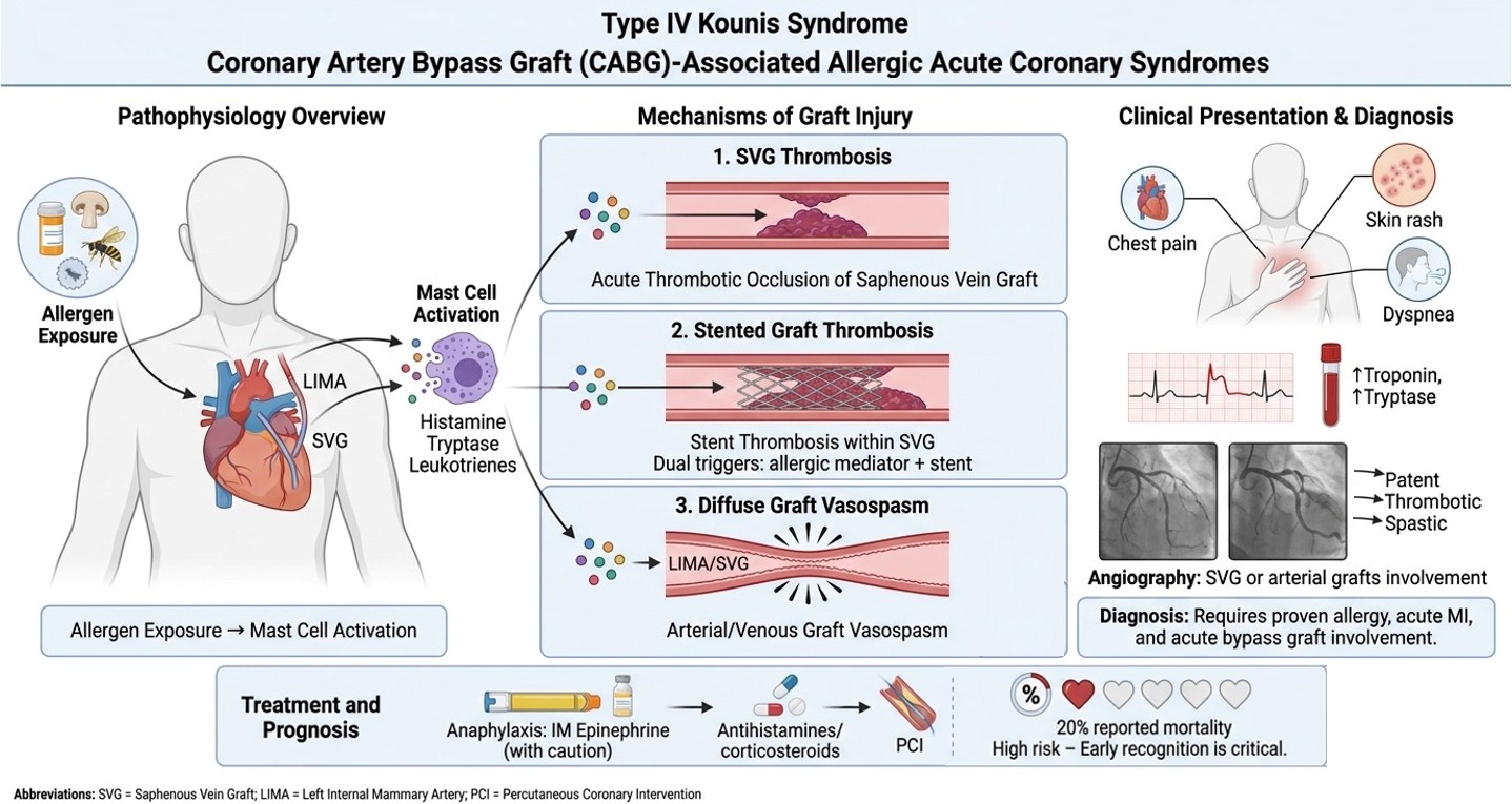
Kounis syndrome (KS) describes the concurrence of an acute coronary syndrome (ACS) with an allergic, hypersensitivity, anaphylactic, or anaphylactoid reaction. Mast-cell activation and the release of histamine, tryptase, platelet-activating factor, leukotrienes, prostaglandins, cytokines, chemokines, and proteases may cause coronary vasoconstriction, plaque erosion or rupture, endothelial dysfunction, platelet activation and thrombosis [1,2]. Since 2010, three variants have traditionally been recognized [2,3]. Type I KS occurs in patients with normal or near-normal coronary arteries and is predominantly mediated by epicardial vasospasm and/or microvascular disfunction. Type II develops in patients with pre-existing coronary vulnerable atherosclerotic plaque, in whom allergic mediators provoke destabilization and subsequent thrombosis [2,4]. Type III comprises acute thrombosis (IIIa) or, less frequently, restenosis (IIIb) of an intracoronary stent, ideally supported by the demonstration of eosinophils or mast cells within aspirated thrombotic material [5,6].
This classification does not adequately encompass patients with previous coronary artery bypass grafting (CABG). In non-allergic settings, acute myocardial ischaemia after CABG may result from graft vasospasm, acute graft thrombosis occurring with or without underlying atherosclerotic plaque destabilization, or, in previously stented grafts, stent thrombosis, involving either arterial or venous conduits [7,8,9,10].
In the context of allergic ACS, involvement of CABG remains poorly investigated and insufficiently characterized.
The first published report of anaphylaxis-associated myocardial infarction involving a coronary graft suggested that this presentation represented a novel variant of KS, which was designated as Type IV KS [11,12]. Subsequent reports have strengthened the biological plausibility of a graft-specific phenotype laying the foundation for the emerging concept of type IV KS.
This focused review summarizes the available clinical evidence, examines potential mechanisms of graft vulnerability, discusses diagnostic and therapeutic challenges.
2. Literature Search and Case Selection
A focused literature search was performed through July 27, 2026, using MEDLINE (via PubMed), Scopus, and Google Scholar. The search strategy combined the terms “Kounis syndrome,” “allergic myocardial infarction,” “anaphylaxis,” “coronary artery bypass graft,” “CABG,” “coronary graft,” “saphenous vein graft,” “internal mammary artery,” “graft spasm,” and “graft thrombosis,” using appropriate Boolean operators. To maximize case retrieval, backward citation searching (screening the reference lists of all eligible articles) and forward citation tracking (identifying publications citing the retrieved reports) were systematically performed. No restrictions regarding publication year were applied. Articles published in English were considered eligible. Conference abstracts were included when they provided original clinical data relevant to bypass graft involvement in KS. Reference lists of relevant case reports and reviews were also screened. Reports were considered directly relevant when an allergic or anaphylactic event was temporally associated with acute myocardial ischemia and angiographic, interventional, or clinical evidence implicated a coronary bypass graft.Cases occurring in patients with previous CABG but with patent grafts or exclusive involvement of native coronary arteries were retained as contextual evidence but were not classified as definite graft-related KS. Likewise, perioperative allergic coronary events during cardiac surgery were excluded unless a newly created or pre-existing bypass conduit was specifically implicated.
The search identified a very limited number of directly relevant reports, precluding quantitative synthesis. The evidence was therefore evaluated as a mechanistic and case-based focused review.
3. Clinical Evidence
Using this search strategy, five cases of suspected type IV KS were identified, including four definite cases of CABG involvement and one probable case in which transient graft vasomotor dysfunction was considered a plausible mechanism despite angiographically patent grafts.
3.1. The Original Description of Myocardial Infarction Within a Coronary Graft
Dazy et al. in 2013 [11] reported the first case of KS involving a coronary artery bypass graft, thereby introducing the concept of a potential type IV variant of the syndrome. The patient was a 69-year-old man with multivessel coronary artery disease (CAD) previously treated with percutaneous coronary intervention (PCI) followed by three-vessel CABG, who also had aspirin-exacerbated respiratory disease, severe asthma and allergic rhinitis. He underwent aspirin desensitization protocol to allow long-term antiplatelet therapy. During the first day he experienced mild respiratory symptoms that responded to bronchodilators and anti-allergic treatment. On the second day, after tolerating cumulative aspirin doses up to 81 mg, he developed a delayed systemic hypersensitivity reaction approximately one hour after discharge, characterized by diffuse urticaria, swelling of the hands and feet, tongue and lip edema, wheezing, stridor, dyspnea, and conjunctival injection. On arrival at the emergency department, serum tryptase was markedly elevated (41.2 ng/mL), confirming extensive mast cell activation. He was promptly treated with intramuscular epinephrine, nebulized albuterol, intravenous diphenhydramine, methylprednisolone, and famotidine, with initial improvement of the allergic manifestations. Less than 30 minutes later, however, he developed severe substernal chest pain associated with new ST-segment elevation in leads III and aVF and reciprocal ST-segment depression in leads aVL, V2, and V3, consistent with an acute inferior ST-segment elevation myocardial infarction. Emergency coronary angiography demonstrated acute thrombotic occlusion of the proximal saphenous vein graft (SVG) to the right posterior descending artery (PDA), whereas the remaining bypass grafts were reported to be patent, although their specific conduit type was not explicitly stated. Primary PCI with implantation of two overlapping bare-metal stents successfully restored coronary flow, and the patient was discharged after 72 hours receiving a corticosteroid taper, dual H1/H2 blockade, lifelong avoidance of aspirin and non-steroidal anti-inflammatory drugs, and prasugrel as antiplatelet therapy.
This report represents a milestone in the evolution of KS because it demonstrated for the first time that allergic coronary events are not confined to native coronary arteries or intracoronary stents but may also involve surgically implanted coronary conduits. The temporal association between documented anaphylaxis, marked serum tryptase elevation, and angiographically confirmed acute graft thrombosis provides compelling evidence supporting mast cell-mediated coronary injury as the underlying pathogenic mechanism. The release of vasoactive and prothrombotic mediators, including histamine, leukotrienes, thromboxane, platelet-activating factor, and tryptase, likely promoted endothelial dysfunction, platelet activation, tissue factor expression, and activation of the coagulation cascade, ultimately precipitating thrombotic occlusion of a vulnerable SVG. The authors also discussed the possible contribution of epinephrine, acknowledging that although it remains the cornerstone of anaphylaxis management and should never be withheld in life-threatening reactions, it may theoretically exacerbate myocardial ischemia through increased myocardial oxygen demand and coronary vasoconstriction. Nevertheless, the markedly elevated tryptase concentration, the unequivocal systemic allergic manifestations, and the selective thrombosis of the bypass graft strongly suggest that epinephrine was, at most, a facilitating cofactor rather than the primary cause of the coronary event. Most importantly, this case expanded fort the first time the last adopted classification of KS by proposing the existence of a fourth subtype involving CABG.
3.2. Hypersensitivity-Associated Stent Thrombosis in a Saphenous Vein Graft
Jariwala et al. [13] in 2022 described a 67-year-old man with type 2 diabetes and previous CABG performed in 2001 who presented with a two-hour history of chest pain. Electrocardiography showed significant ST-segment depression in the lateral leads, while transthoracic echocardiography demonstrated hypokinesia of the lateral wall and apical anterior segments with a left ventricular ejection fraction of 48%. A detailed clinical history revealed ingestion of mushrooms contained in a pizza approximately 24 hours before symptom onset, followed by the development of a diffuse cutaneous rash that resolved spontaneously without progression to anaphylactic shock. High-sensitivity cardiac troponin I was markedly elevated, and the patient was diagnosed with non-ST-segment elevation myocardial infarction (NSTEMI). The patient had previously experienced an NSTEMI in 2008, at which time a 3.5 × 28 mm first-generation sirolimus-eluting stent (Cypher Select) had been implanted in the mid-segment of the SVG to the first diagonal branch (D1). Coronary angiography during the current admission demonstrated chronic total occlusion of the native left anterior descending and right coronary arteries, non-obstructive disease of the left circumflex artery, and patent left internal mammary artery (LIMA) and SVG-to-right coronary artery grafts. The culprit lesion was identified as “very late” (>1 years) acute stent thrombosis involving the previously implanted drug-eluting stent (DES) within the SVG to D1. The patient received loading doses of aspirin, ticagrelor, and atorvastatin before undergoing PCI. Despite manual thrombus aspiration and repeated balloon dilatation, restoration of flow was initially unsuccessful because of a refractory no-reflow phenomenon. Intracoronary sodium nitroprusside and nicorandil were subsequently administered, resulting in recovery of TIMI 3 flow, after which two overlapping second-generation everolimus-eluting stents were implanted across the previously thrombosed stent. Owing to the substantial thrombotic burden, tirofiban infusion was continued for 24 hours. The patient was discharged without complications on prolonged dual antiplatelet therapy with aspirin and ticagrelor.
This report is particularly interesting because it lies at the intersection between type III and type IV KS. From a mechanistic perspective, the immediate culprit lesion was an extremely late (> 5 years) I generation DES thrombosis, fulfilling the classical definition of type III KS [6]. However, unlike previously reported cases, the thrombosed stent was not located in a native coronary artery but rather, for the first time, within a saphenous vein bypass graft, thereby extending allergic stent thrombosis to a surgically implanted coronary conduit. The temporal relationship between mushroom ingestion, transient allergic manifestations, and subsequent myocardial infarction strongly supports mast cell-mediated activation as the trigger for thrombosis. Importantly, the absence of anaphylactic shock and the fact that no epinephrine was administered eliminate potential confounding effects related to catecholamine-induced coronary vasoconstriction, further strengthening the causal role of the allergic reaction itself. Importantly, conventional predisposing factors for stent thrombosis appeared to be limited. The patient had uneventful post-PCI course and completed the recommended duration of dual antiplatelet therapy, remained on aspirin, and exhibited good lipid control, with an LDL cholesterol level of 67 mg/dL, meeting the guideline-recommended lipid goals of that era. These findings further support the hypothesis that the allergic reaction acted as the principal trigger for thrombosis rather than poorly controlled cardiovascular risk factors and highlights how allergic inflammation may overcome conventional mechanisms of thrombotic protection.
The allergic insult acted on two distinct thrombogenic substrates simultaneously: the implanted coronary device and the degenerated venous conduit. This dual substrate is likely to amplify the prothrombotic consequences of mast cell activation beyond those expected in isolated native-vessel stent thrombosis.
Unlike native coronary arteries, saphenous vein grafts undergo extensive structural and biological remodeling after implantation, characterized by endothelial injury and dysfunction, vascular inflammation, diffuse intimal hyperplasia, and accelerated atherosclerosis, ultimately resulting in a highly thrombogenic conduit frequently containing lipid-rich and friable lesions [14,15].
The implantation of a DES within such a conduit therefore creates a unique pathological environment in which hypersensitivity-related platelet activation may occurs on a background of pre-existing graft degeneration. In this setting, allergic mediator release may interact not only with the stent polymer and metallic platform but also with the chronically diseased graft wall, potentially explaining the extensive thrombotic burden and the refractory no-reflow phenomenon observed during PCI. Therefore, although formally representing a type III KS, this case is highly relevant to the evolving concept of type IV KS, as it demonstrates that allergic thrombosis may involve not only native coronary stents but also stents implanted within bypass grafts, suggesting a significant overlap between these two variants, in which the allergic reaction simultaneously targets both the intracoronary device and the bypass conduit. This distinction is clinically relevant because the pathophysiological mechanisms extend beyond stent hypersensitivity to include the intrinsic biological vulnerability of aging vein grafts.
An additional noteworthy observation is that, despite the systemic nature of the allergic reaction, thrombosis selectively involved the only bypass graft containing a previously implanted DES, whereas both the left internal mammary artery graft (LIMA) and the second SVG remained patent. This selective localization further supports the concept that the intracoronary device may have acted as the preferential site for allergic thrombus formation within an already vulnerable venous conduit, suggesting a synergistic interaction between stent-related hypersensitivity mechanisms and graft degeneration rather than a purely systemic thrombotic response.
3.3. Fatal Anaphylaxis-Associated Saphenous Vein Graft Occlusion
Hamideh et al. [16] in 2025 reported a 69-year-old man with a history of multivessel CAD treated with CABG, obstructive pulmonary disease, hypertension, hyperlipidaemia, and hymenoptera venom allergy who developed anaphylactic shock following a wasp sting. Intramuscular epinephrine (0.5 mg) improved the hypotension and was likely lifesaving during the initial anaphylactic phase. Shortly thereafter, however, the patient developed, respiratory failure, cardiogenic shock and cardiac arrest requiring cardiopulmonary resuscitation. 12-leads ECG showed infero-postero-lateral STEMI and emergency coronary angiography demonstrated complete occlusion of the SVG that could not be successfully recanalized. Echocardiography showed severe left ventricular systolic dysfunction (LVEF 15–20%), and the patient required extracorporeal membrane oxygenation together with inotropes, vasopressors and advanced intensive care support. His clinical course was further complicated by severe hepatic failure, disseminated intravascular coagulation and anoxic brain injury, ultimately leading to death. Although the authors classified this event as type II KS, the acute coronary event directly involved a SVG rather than a native coronary artery, making this case more appropriately interpretable as a type IV KS. Importantly, while epinephrine may have contributed to the subsequent coronary deterioration through its vasoconstrictive properties, it was also instrumental in reversing the life-threatening anaphylactic shock and should therefore not be regarded as the primary cause of the coronary event [17].
3.4. Expanding the Clinical Spectrum of Type IV KS: Diffuse Vasospasm
Ferdous et al. [18] in 2025 reported a 50-year-old man who developed profound haemodynamic collapse 20 minutes after CABG immediately following protamine administration. Severe hypotension rapidly progressed to pulseless electrical activity requiring cardiopulmonary resuscitation, extracorporeal membrane oxygenation, and subsequent defibrillation for ventricular fibrillation. Emergency coronary angiography demonstrated diffuse vasospasm involving all native coronary arteries as well as all bypass grafts (LIMA and SVGs), without evidence of fixed mechanical graft failure. Intracoronary nitroglycerin produced rapid angiographic and haemodynamic resolution of the vasospasm. Mechanical circulatory support was maintained for two days and subsequently transitioned to Impella. Left ventricular systolic function recovered completely and the patient was discharged with a favourable outcome. The diffuse simultaneous involvement of both native coronary arteries and bypass conduits shortly after protamine exposure strongly suggests a systemic vasospastic process rather than isolated graft dysfunction. It also expands the clinical spectrum of type IV KS by demonstrating that hypersensitivity-related bypass involvement may present as reversible graft spasm, in addition to the thrombotic phenotype previously reported.
3.5. Other Possible CABG-Related KS Reports
Kundumadam et al. [19] in 2017 described the case of possible type IV KS, a 70-year-old man with heart failure with reduced ejection fraction, previous triple CABG and recurrent episodes of angioedema/anaphylaxis of unknown origin who developed an NSTEMI ( chest pain, dyspnea, troponin elevation, negative anterior T waves) 48 hours after treatment with intramuscular epinephrine (with sodium metabisulfite preservative) for angioedema, which had also been administered during previous hypersensitivity episodes. Coronary angiography demonstrated severe native coronary artery disease but widely patent bypass grafts (LIMA-to-LAD and SVGs to obtuse marginal branch and PDA), excluding acute graft occlusion as the mechanism of ACS. Despite this, the patient experienced a marked deterioration in left ventricular systolic function, with and ejection fraction falling from 35-40% to 15%. No specific allergen was identified, complement studies were negative, serum tryptase was not measured, thus no evidence of mast-cell activation was documented. The authors hypothesized that delayed inflammatory mediators rather than the immediate hypersensitivity response accounted for the delayed myocardial injury. Interestingly, the initial electrocardiogram (before epinephrine injection and NSTEMI diagnosis) showed diffuse ST-segment depression with reciprocal ST elevation in aVR, an ischemic pattern that may reflect extensive subendocardial ischemia secondary to transient multivessel graft spasm or severe coronary microvascular dysfunction rather than focal graft thrombosis. At the time of NSTEMI diagnosis, approximately 48 hours after the initial allergic presentation, the initial electrocardiographic pattern had resolved, giving way to widespread asymmetric T-wave inversions, particularly evident in the anterior precordial leads (V1–V4). Although this case cannot be definitively classified as type IV KS, it represents a plausible CABG-related variant. The presence of patent arterial and venous grafts despite NSTEMI argues against fixed mechanical obstruction and raises the possibility of transient allergic vasomotor dysfunction involving the bypass conduits or the coronary microcirculation that had resolved before angiography. However, these mechanisms remain speculative because provocative vasospasm testing, biomarkers of mast-cell activation, and CMR were not performed. Furthermore, the contribution of epinephrine administration to the subsequent ischemic event cannot be completely excluded, particularly in a patient with advanced coronary artery disease. This report should be regarded as a possible, thus intriguing, rather than definitive, example of CABG-associated KS.
Table 1 Cases of type IV KS. Cases are categorized according to the predominant bypass-related mechanism (isolated graft thrombosis, diffuse graft vasospasm, or stent thrombosis within a bypass graft). Kundumadam et al. is included separately because angiography demonstrated patent bypass grafts and therefore the diagnosis of graft-related KS remains speculative. Abbreviations: CABG, coronary artery bypass grafting; DES, drug-eluting stent; D1, first diagonal branch; ECG, electrocardiogram; KS, Kounis syndrome; LIMA, left internal mammary artery; NSTEMI, non-ST-segment elevation myocardial infarction; PDA, posterior descending artery; PCI, percutaneous coronary intervention; PEA, pulseless electrical activity; STE, ST-segment elevation; STEMI, ST-segment elevation myocardial infarction; SVG, saphenous vein graft; VF, ventricular fibrillation.
4. Biological Bases of CABG Vulnerability to Allergic Mediators
4.1. Saphenous Vein Graft
Saphenous veins undergo profound structural and functional adaptation after implantation into the arterial circulation. Exposure to arterial pressure, pulsatile flow, elevated wall tension, and abnormal shear stress promotes endothelial injury, smooth-muscle-cell proliferation, intimal hyperplasia, lipid accumulation, inflammation, and accelerated atherosclerosis. Late saphenous vein graft lesions are often diffuse, friable, lipid-rich, and highly thrombogenic with substantial rates of graft degeneration and failure during long-term follow-up [14,15]. These characteristics provide an ideal substrate upon which mast-cell mediators, platelet-activating factor, tissue factor activation, and systemic prothrombotic responses may act. Histamine and leukotrienes can provoke vasoconstriction not only in native coronary arteries but also in vascular conduits containing functional smooth muscle [18]. Proteases released during mast-cell activation may degrade the extracellular matrix and weaken the fibrous cap of graft atherosclerotic plaque, simultaneously, platelet activation can generate acute graft thrombosis.
4.2. Arterial Grafts
Internal mammary and radial artery grafts retain a more organized arterial wall and generally demonstrate superior long-term patency compared with saphenous vein grafts. Nevertheless, arterial conduits remain capable of vasomotor responses and may develop intense spasm, perioperative and postoperative arterial graft spasm is a recognized, although uncommon, complication of CABG. Compared with the radial artery, the internal thoracic artery exhibits lower intrinsic vasoreactivity; however, clinically significant vasospasm may still occur. Current evidence indicates that arterial graft spasm is a multifactorial phenomenon mediated by potent vasoconstrictors including endothelin-1, norepinephrine, serotonin, thromboxane A2, angiotensin II, and vasopressin, as well as endothelial dysfunction, surgical manipulation, catecholamine surges, and platelet activation [20]. This phenomenon can also be triggered during systemic hypersensitivity due to the presence of some of these inflammatory mediators [18]. Finally, no documented case of allergic thrombosis involving an internal mammary artery graft has been reported to date, although native coronary arteries are a well-established substrate for allergic thrombosis in Type II KS [2]. This apparent discrepancy likely reflects the intrinsic biological differences between arterial conduits and native atherosclerotic coronary vessels, as well as the extreme rarity of this clinical presentation rather than the absence of a plausible pathophysiological mechanism.
4.3. Stented Bypass Grafts
In the pathogenesis of type III KS, potential antigenic and inflammatory stimuli include metallic components, durable or biodegradable polymers, antiproliferative drugs, that promote vascular injury leading to stent thrombosis during an hypersensitivity local or systemic reaction [2,6]. When thrombosis occurs in a stented graft during an allergic event, classification solely as type III KS may be incomplete. SVG stent failure often presents as acute MI and with SVG occlusion [9]. A stent implanted within a saphenous vein graft creates a particularly complex immuno-thrombotic environment. The graft substrate contributes additional endothelial, atherosclerotic, thrombotic vulnerability and residual platelet reactivity despite antithrombotic treatment, [9,14,15,21]. Such events may more accurately be regarded as a graft-specific subtype of type IV disease.
5. Diagnostic Approach
The diagnosis of type IV KS requires simultaneous confirmation of three components: a compatible allergic or hypersensitivity reaction, the objective evidence of acute myocardial ischemia and the acute involvement of a coronary bypass graft.
Initial evaluation should include detailed allergic history, clinical examination, serial electrocardiography, cardiac necrosis biomarkers including high sensitivity troponin, complete blood count with eosinophils, serum tryptase in the first hours after presentation, echocardiography and urgent coronary angiography [2].
Coronary angiography should include systematic assessment of the native coronary circulation, arterial and venous bypass grafts, proximal and distal anastomoses, previously implanted graft stents, and distal vessel runoff. When vasospasm is suspected, angiographic findings should be reassessed after intracoronary nitrate administration. Thrombus burden, distal embolization, and no-reflow should also be specifically evaluated, particularly in degenerated saphenous vein grafts [22,23,24]. A previous angiogram or coronary CT angiogram is particularly valuable for documenting prior graft patency and thereby supporting the distinction between acute graft occlusion and pre-existing chronic graft failure [25,26]. Intravascular imaging may be particularly useful in type IV Kounis syndrome to differentiate acute allergic thrombosis from chronic graft degeneration, characterize neoatherosclerosis and plaque disruption within saphenous vein grafts, and identify stent-related mechanisms when bypass graft stents are involved. Nevertheless, because degenerated SVGs are highly friable and prone to distal embolization, the potential diagnostic benefit of additional intracoronary instrumentation should always be balanced against procedural risk, particularly in haemodynamically unstable patients [22,27,28]. Whenever aspiration thrombectomy is performed, histopathological examination of the retrieved thrombus with specific staining for eosinophils and mast cells should be considered. By analogy with type III KS, identification of eosinophilic and mast-cell infiltration within the thrombus would provide one of the strongest pathological supports for an allergic thrombotic mechanism and could help distinguish type IV KS from conventional graft thrombosis. Serum tryptase should ideally be obtained during the acute phase and repeated after recovery. Specific IgE testing, skin testing, and specialist allergological assessment should be performed after stabilization.
6. Therapeutic Consideration
Management is challenging because treatment of anaphylaxis and treatment of acute coronary thrombosis may have competing haemodynamic effects.
Intramuscular epinephrine remains the first-line therapy for life-threatening anaphylaxis. Its use should not be withheld in patients with airway compromise or severe anaphylactic shock solely because KS is suspected. However, intravenous boluses and dosing errors should be avoided whenever possible because abrupt alpha-adrenergic vasoconstriction may aggravate coronary or graft spasm [2,17]. Antihistamines and corticosteroids may be used as adjunctive therapies but must not delay epinephrine in genuine anaphylaxis. Intravenous fluids should be administered carefully, particularly when left ventricular dysfunction or pulmonary congestion is present. Nitrates and calcium-channel blockers may relieve graft or native coronary vasospasm but can worsen hypotension. Their use should therefore be individualized according to blood pressure and haemodynamic status [2]. Confirmed graft thrombosis should be treated according to ACS principles, including antiplatelet and anticoagulant therapy, while considering the potential for aspirin or other drugs to have acted as allergic triggers. Percutaneous intervention of degenerated saphenous vein grafts carries a high risk of distal embolization and no-reflow. Distal protection, thrombus aspiration in selected cases, intracoronary vasodilators, and mechanical circulatory support may be required [22,28]. The choice between graft PCI and native-vessel PCI should be individualized. When technically feasible, intervention on the native coronary artery may avoid manipulation of a friable, thrombotic venous graft [28]. Beta-blockers deserve caution because they may worsen vasospasm through unopposed alpha-adrenergic activity and reduce the response to epinephrine. In patients receiving chronic beta-blockade with refractory anaphylactic shock, glucagon may be considered [2].
7. Prognosis, Prevention and Evidence Gaps
Although the available evidence is limited and subject to substantial publication bias, the reported clinical spectrum includes cardiogenic shock, cardiac arrest, refractory no-reflow, and death. Among the currently available cases, mortality was approximately 20% (1 of 5 patients), numerically exceeding that observed in our previous systematic review of type III KS (18%) and substantially higher than that reported for type I [2,6]. However, these observations should be interpreted with extreme caution given the very small number of published cases. Patients with previous CABG who develop anaphylaxis-associated troponin elevation should not be assumed to have only supply-demand mismatch. Direct assessment of graft patency may be warranted, particularly when electrocardiographic changes or regional wall-motion abnormalities are present.
Of note to date no documented case of allergic internal mammary artery graft thrombosis was identified in the present search.
Secondary prevention should rely on identification and strict avoidance of the culprit trigger, formal allergological assessment, optimization of graft and native coronary artery disease, and individualized reassessment of antithrombotic therapy. Although no recurrent cases of type IV KS have been reported to date, systematic allergological evaluation remains advisable because recurrency is a recognized phenomenon in other variants and prevention of re-exposure represents the cornerstone of long-term management [2,29]. Current evidence is restricted to isolated reports with incomplete and heterogeneous documentation. Key missing information includes: previous graft patency; exact interval from CABG to the allergic event; graft material and target vessel; distinction between chronic and acute occlusion; serum tryptase and eosinophil values; intracoronary imaging; thrombus histology; formal allergological testing; long-term graft and clinical outcomes. Future case reports should follow a standardized reporting framework. Establishing an international KS registry (for all subtypes) would allow reliable characterization of graft involvement and assessment of whether it carries a different prognosis from native-vessel or stent-related disease.
Figure 1 summarizes the proposed pathophysiological mechanisms, clinical characteristics of the reported cases, diagnostic approach, management principles, and future perspectives of type IV KS.
8. Conclusions
CABG involvement represents an exceptionally rare but biologically plausible manifestation of KS. The currently available evidence consists of only five reported cases, including four definite and one probable type IV presentations, encompassing anaphylaxis-associated thrombosis of saphenous vein grafts, thrombosis of a coronary stent implanted within a saphenous vein graft, and diffuse vasospasm involving both arterial and venous bypass conduits. Although limited, these observations support the concept that hypersensitivity reactions may target surgically implanted coronary conduits through mechanisms analogous to those described in native coronary arteries and intracoronary stents. SVG appear to be particularly vulnerable because accelerated atherosclerosis, endothelial dysfunction, altered haemodynamics, chronic inflammation, and a highly thrombogenic plaque phenotype may synergistically interact with mast-cell activation and allergic mediator release. Consequently, the clinical spectrum of type IV KS appears remarkably heterogeneous. Recognition of graft-related disease as a distinct type IV KS may improve diagnostic accuracy and promote a more systematic evaluation of patients with allergic acute coronary syndromes after CABG. Future reports should include comprehensive angiographic characterization of native vessels and grafts, intracoronary imaging whenever feasible, histopathological analysis of aspirated thrombotic material, acute and baseline serum tryptase measurements, and formal allergological assessment. Such standardized documentation will be essential to better define the true incidence and prognosis of type IV KS, and improve the diagnosis and management of this emerging clinical entity.
Author Contributions
Conceptualization, L.G.G.; resources, L.G.G., M.M., G.A., C.d.G.; writing—original draft preparation, L.G.G., M.M.; writing—review and editing, L.G.G.; visualization, L.G., G.A. C.d.G; supervision, C.d.G.; project administration, L.G.G. All authors have read and agreed to the published version of the manuscript.
Funding
This research received no external funding.
Institutional Review Board Statement
Not applicable.
Informed Consent Statement
Not applicable.
Data Availability Statement
No new data were created or analyzed in this study.
Acknowledgments
During the preparation of this manuscript, the authors used AI tools (ChatGPT and FigureLabs) exclusively for grammar/syntax refinement and for assisting in the generation of graphical elements. No AI system was involved in generating the scientific content or drafting the manuscript. The authors have reviewed and edited the output and take full responsibility for the content of this publication.
Conflicts of Interest
The authors declare no conflicts of interest.
Permission
No third-party copyrighted material has been used in the manuscript figures. All images/figures were entirely created by the authors specifically for this submission; therefore, no permissions are required and no copyright attribution is applicable.
References
- de Gregorio C, Granata L, Raspanti D, Giannino F, Cimino C, Koniari I, Andò G, Kounis NG. Cephalosporin triggered Kounis syndrome: Pathophysiological and clinical insights. Int J Cardiol. 2025 Jul 15;431:133249. [CrossRef]
- Granata, L. G.; Andò, G.; Marchetta, M.; Giubilato, S.; Kounis, N. G.; de Gregorio, C. Kounis Syndrome in the Modern Era: A Comprehensive Review of Allergic Acute Coronary Syndromes. Preprints 2026, 2026072027. [CrossRef]
- Biteker M. A new classification of Kounis syndrome. Int J Cardiol. 2010 Dec 3;145(3):553. [CrossRef]
- Kounis NG. Kounis syndrome: an update on epidemiology, pathogenesis, diagnosis and therapeutic management. Clin Chem Lab Med. 2016 Oct 1;54(10):1545–59. [CrossRef]
- Itoh T, Nakajima Y, Morino Y. Proposed classification for a variant of Kounis syndrome. Clinical Chemistry and Laboratory Medicine (CCLM). 2017 Jun 1;55(6):e107–e107. [CrossRef]
- Granata LG, Marchetta M, Alagna G, Giubilato S, Trio O, Ando’ G. Stent thrombosis: an updated clinical over-view from epidemiology to classification and management. Heart. 2026 May 8;heartjnl-2026-327899. [CrossRef]
- Gaudino M, Antoniades C, Benedetto U, Deb S, Di Franco A, Di Giammarco G, Fremes S, Glineur D, Grau J, He GW, Marinelli D, Ohmes LB, Patrono C, Puskas J, Tranbaugh R, Girardi LN, Taggart DP; ATLANTIC (Arterial Grafting International Consortium) Alliance. Mechanisms, Consequences, and Prevention of Coronary Graft Failure. Circulation. 2017 Oct 31;136(18):1749-1764. [CrossRef]
- He GW, Taggart DP. Spasm in Arterial Grafts in Coronary Artery Bypass Grafting Surgery. Ann Thorac Surg. 2016 Mar;101(3):1222-9. [CrossRef]
- Lichtenwalter C, de Lemos JA, Roesle M, Obel O, Holper EM, Haagen D, Saeed B, Iturbe JM, Shunk K, Bissett JK, Sachdeva R, Voudris VV, Karyofillis P, Kar B, Rossen J, Fasseas P, Berger P, Banerjee S, Brilakis ES. Clinical presentation and angiographic characteristics of saphenous vein graft failure after stenting: insights from the SOS (stenting of saphenous vein grafts) trial. JACC Cardiovasc Interv. 2009 Sep;2(9):855-60. [CrossRef]
- Sandner S, Antoniades C, Caliskan E, Czerny M, Dayan V, Fremes SE, Glineur D, Lawton JS, Thielmann M, Gaudino M. Intra-operative and post-operative management of conduits for coronary artery bypass grafting: a clinical consensus statement of the European Society of Cardiology Working Group on Cardiovascular Surgery and the European Association for Cardio-Thoracic Surgery Coronary Task Force. Eur Heart J. 2025 Jan 3;46(1):19-34. [CrossRef]
- Dazy K, Walters D, Holland C, Baldwin J. Anaphylaxis mediated myocardial infarction in a coronary graft: A new variant of Kounis syndrome (a case report). International Journal of Cardiology. 2013 Sep;168(2):e84–5. [CrossRef]
- Giovannini M, Koniari I, Mori F, Barni S, Novembre E, Kounis NG. Kounis syndrome: Towards a new classifi-cation. Int J Cardiol. 2021 Oct 15;341:13–4. [CrossRef]
- Jariwala P, Jadhav K, Khetan S. Allergic myocardial infarction (type III Kounis syndrome) secondary to mushroom ingestion: An unusual cause of very late stent thrombosis of saphenous venous graft and refractory no-reflow phenomenon. J Cardiol Cases. 2022 Mar 22;26(1):51-55. [CrossRef]
- Guida G, Ward AO, Bruno VD, George SJ, Caputo M, Angelini GD, Zakkar M. Saphenous vein graft disease, pathophysiology, prevention, and treatment. A review of the literature. J Card Surg. 2020 Jun;35(6):1314-1321. [CrossRef]
- Back L, Ladwiniec A. Saphenous Vein Graft Failure: Current Challenges and a Review of the Contemporary Percutaneous Options for Management. J Clin Med. 2023 Nov 15;12(22):7118. [CrossRef]
- Hamideh M, Rhodes A, Srinivasan A, Chakraborty S, Hawkins C, Kumar A, Golzarian H. Epinephrine and the paradox of Kounis syndrome: a case series and review of management. Eur Heart J Case Rep. 2025 Sep 3;9(9):ytaf414. [CrossRef]
- L. G. Granata, S. Giubilato, e M. Marchetta, «Epinephrine in Kounis syndrome: life-saving treatment or cardiac risk?», European Heart Journal - Case Reports, vol. 9, fasc. 12, p. ytaf636, nov. 2025. [CrossRef]
- Ferdous, T, Reiche, M, Waites, S. et al. A CASE OF DIFFUSE CORONARY ARTERY VASOSPASM AFTER CORONARY ARTERY BYPASS GRAFT SURGERY WITH POSSIBLE KOUNIS SYNDROME. JACC. 2025 Apr, 85 (12_Supplement) 3620. [CrossRef]
- Kundumadam S, Reddy V, Mallikethi Reddy S, Kathi P, Abidov A. An Unusual Case of Recurrent Hypersensitivity Reaction Associated with Kounis-Like Acute Coronary Syndrome. Case Rep Cardiol. 2017;2017:6421208. [CrossRef]
- Sandner S, Antoniades C, Caliskan E, Czerny M, Dayan V, Fremes SE, Glineur D, Lawton JS, Thielmann M, Gaudino M. Intra-operative and post-operative management of conduits for coronary artery bypass grafting: a clinical consensus statement of the European Society of Cardiology Working Group on Cardiovascular Surgery and the European Association for Cardio-Thoracic Surgery Coronary Task Force. Eur Heart J. 2025 Jan 3;46(1):19-34. [CrossRef]
- Redfors B, Généreux P, Witzenbichler B, McAndrew T, Diamond J, Huang X, Maehara A, Weisz G, Mehran R, Kirtane AJ, Stone GW. Percutaneous Coronary Intervention of Saphenous Vein Graft. Circ Cardiovasc Interv. 2017 May;10(5):e004953. [CrossRef]
- Lee MS, Park SJ, Kandzari DE, Kirtane AJ, Fearon WF, Brilakis ES, Vermeersch P, Kim YH, Waksman R, Mehilli J, Mauri L, Stone GW. Saphenous vein graft intervention. JACC Cardiovasc Interv. 2011 Aug;4(8):831-43. [CrossRef]
- Vishnevsky A, Julien HM, Fischman DL, Walinsky P, David Ogilby J, Ruggiero NJ 2nd, Jasti B, Savage MP. Unrecognized coronary vasospasm in patients referred for percutaneous coronary intervention: Intracoronary nitroglycerin, the forgotten stepchild of cardiovascular guidelines. Catheter Cardiovasc Interv. 2017 Dec 1;90(7):1086-1090. [CrossRef]
- Tamis-Holland JE, Abbott JD, Al-Azizi K, Barman N, Bortnick AE, Cohen MG, Dehghani P, Henry TD, Latif F, Madjid M, Yong CM, Sandoval Y. SCAI Expert Consensus Statement on the Management of Patients With STEMI Referred for Primary PCI. J Soc Cardiovasc Angiogr Interv. 2024 Oct 7;3(11):102294. [CrossRef]
- Yoo JY, Choi JH, Kim JA, Chun EJ. Role of Coronary CT Angiography in Coronary Revascularization. Cardiovasc Imaging Asia. 2018 Jan;2(1):1-11. [CrossRef]
- Hamon M, Lepage O, Malagutti P, Riddell JW, Morello R, Agostini D, Hamon M. Diagnostic performance of 16- and 64-section spiral CT for coronary artery bypass graft assessment: meta-analysis. Radiology. 2008 Jun;247(3):679-86. [CrossRef]
- McDaniel MC, Eshtehardi P, Sawaya FJ, Douglas JS Jr, Samady H. Contemporary clinical applications of coronary intravascular ultrasound. JACC Cardiovasc Interv. 2011 Nov;4(11):1155-67. [CrossRef]
- Xenogiannis I, Tajti P, Hall AB, Alaswad K, Rinfret S, Nicholson W, Karmpaliotis D, Mashayekhi K, Furkalo S, Cavalcante JL, et al. Update on Cardiac Catheterization in Patients With Prior Coronary Artery Bypass Graft Surgery. JACC Cardiovasc Interv. 2019 Sep 9;12(17):1635-1649. [CrossRef]
- Brancaccio R, Bonzano L, Cocconcelli A, Boyko R, Ienopoli G, Motolese A. Recurrent Kounis Syndrome: A Case Report and Literature Review. J Clin Med. 2024 Mar 13;13(6):1647. [CrossRef]
Figure 1.
Overview of type IV Kounis syndrome. Schematic representation of the proposed mechanisms underlying allergic involvement of coronary artery bypass grafts, highlighting the particular susceptibility of saphenous vein grafts to vasospasm, plaque destabilization, and thrombosis. The figure also summarizes the main clinical features reported in published cases, key diagnostic principles, acute management strategies, secondary prevention measures, and current knowledge gaps requiring further investigation. Abbreviations: ACS, acute coronary syndrome, IC, intracoronary; KS, Kounis syndrome; LIMA, left internal mammary artery, NSTEMI, Non ST-segment elevation myocardial infarction; PCI, percutaneous coronary intervention; STEMI, ST-segment elevation myocardial infarction; SVG, saphenous vein graft.
Figure 1.
Overview of type IV Kounis syndrome. Schematic representation of the proposed mechanisms underlying allergic involvement of coronary artery bypass grafts, highlighting the particular susceptibility of saphenous vein grafts to vasospasm, plaque destabilization, and thrombosis. The figure also summarizes the main clinical features reported in published cases, key diagnostic principles, acute management strategies, secondary prevention measures, and current knowledge gaps requiring further investigation. Abbreviations: ACS, acute coronary syndrome, IC, intracoronary; KS, Kounis syndrome; LIMA, left internal mammary artery, NSTEMI, Non ST-segment elevation myocardial infarction; PCI, percutaneous coronary intervention; STEMI, ST-segment elevation myocardial infarction; SVG, saphenous vein graft.
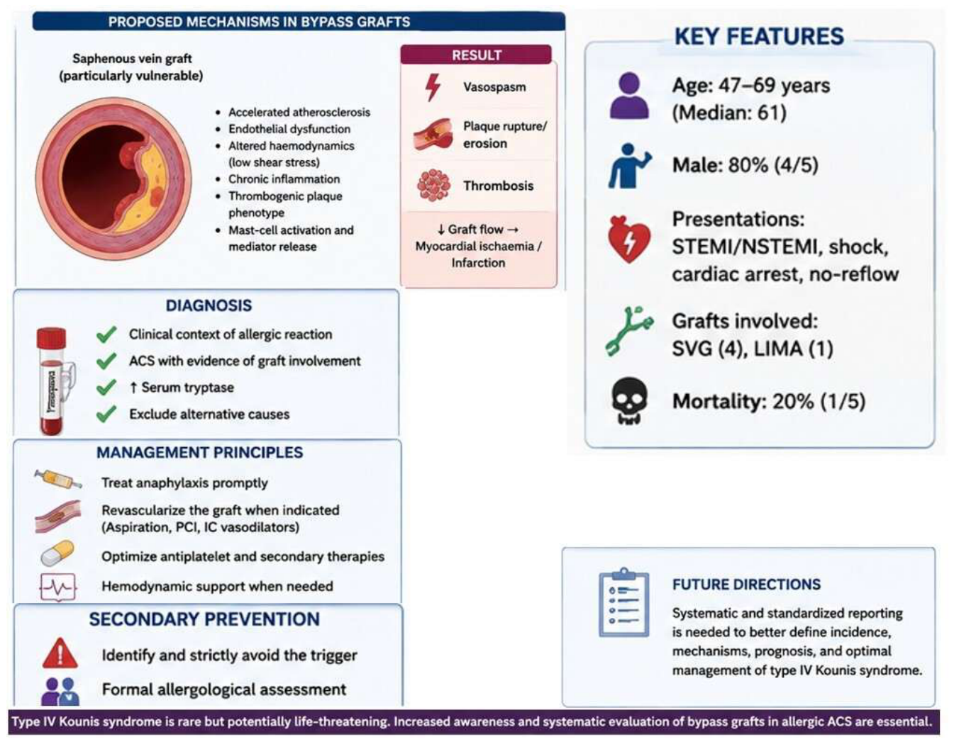

Table 1.
summarizes the principal clinical characteristics, angiographic findings and outcomes of all currently available reports.
Table 1.
summarizes the principal clinical characteristics, angiographic findings and outcomes of all currently available reports.
| First Author | Dazy [11] | Jariwala [13] | Hamideh [16] | Ferdous [18] | Kundumadam* [19] |
|---|---|---|---|---|---|
| Year | 2013 | 2022 | 2025 | 2025 | 2017 |
| Age/Sex | 69/M | 67/M | 69/M | 50/M | 70/M |
| Previous CABG | Not reported | Year 2001 | Not reported | 22 minutes before | Year 2007 |
| Trigger | Aspirin | Mushroom | Wasp sting | Protamine after CABG | Recurrent Angioedema due to unknown trigger |
| ECG | Inferior STEMI |
NSTEMI, lateral ST depression | Infero-posterolateral STEMI | Not specifically reported | NTEMI, Initial diffuse ST depression with STE in aVR followed by anterior T-wave inversion |
| Graft involvement | Acute thrombosis of SVG to PDA, remaining grafts patent | Very late thrombosis of first-generation DES in SVG to D1 | Complete SVG occlusion | Diffuse vasospasm involving LIMA, SVGs and native coronary arteries | No angiographic graft occlusion (LIMA and SVGs patent) |
| Severity | STEMI requiring PCI | Refractory no-reflow during PCI | Cardiogenic shock and cardiac arrest | Cardiogenic shock, PEA/VF VA-ECMO and Impella support |
Severe LV dysfunction (LVEF ↓ to 15%) |
| Outcome | Survived | Survived | Death | Survived | Survived |
*Possible type IV KS (hypothetical transient graft spasm or microvascular spasm). † Coronary angiography demonstrated patent bypass grafts; therefore, direct graft involvement was not documented and the diagnosis of type IV KS remains probable. Note: Acute serum tryptase, one of the principal biomarkers supporting mast-cell activation in KS, was documented and elevated only in the report by Dazy et al. [11], whereas it was unavailable or not reported in all subsequent cases.
Disclaimer/Publisher’s Note: The statements, opinions and data contained in all publications are solely those of the individual author(s) and contributor(s) and not of MDPI and/or the editor(s). MDPI and/or the editor(s) disclaim responsibility for any injury to people or property resulting from any ideas, methods, instructions or products referred to in the content. |
© 2026 by the authors. Licensee MDPI, Basel, Switzerland. This article is an open access article distributed under the terms and conditions of the Creative Commons Attribution (CC BY) license (http://creativecommons.org/licenses/by/4.0/).
Copyright: This open access article is published under a Creative Commons CC BY 4.0 license, which permit the free download, distribution, and reuse, provided that the author and preprint are cited in any reuse.