

Submitted:
04 August 2026
Posted:
05 August 2026
You are already at the latest version
Abstract
Type 1 Diabetes Mellitus (T1DM) is preceded by a prolonged asymptomatic autoimmune phase during which islet autoantibodies represent the earliest detectable biomarkers of disease. Although radiobinding assays (RBA) remain the reference method for autoantibody detection, their use is limited by the requirement of radioactive reagents, specialized facilities and high operational costs, highlighting the need for sensitive, non-radiometric assays suitable for large-scale screening. In this study, we developed a bridge Fluorophore-Linked Immunosorbent Assay (b-FLIA) for the simultaneous detection of glutamic acid decarboxylase autoantibodies (GADA) and zinc transporter 8 autoantibodies (ZnT8A) using a recombinant ZnT8/GAD65 chimeric antigen expressed in a baculovirus–insect cell system. Assay performance was evaluated using sera from healthy controls and diabetic patients previously characterized by RBA. The b-FLIA achieved an area under the receiver operating characteristic curve of 0.9867, with 90.5% sensitivity and 100% specificity for the detection of ZnT8A and/or GADA. Furthermore, the assay showed an almost perfect agreement with RBA (κ = 0.912). These findings demonstrate that the combination of a baculovirus-expressed ZnT8/GAD65 chimeric antigen with a bridge fluorescence immunoassay constitutes a practical and sensitive platform for multiplex detection of diabetes-associated autoantibodies, with potential application in routine laboratories and future population-based screening programs.
Keywords:
type 1 diabetes mellitus
; islet autoantibodies
; GAD65
; ZnT8
; chimeric antigen
; baculovirus expression system
; multiplex autoantibody detection
; population screening
1. Introduction
Type 1 Diabetes Mellitus (T1DM) is a chronic autoimmune disease characterized by the selective destruction of insulin-producing pancreatic β-cells, leading to absolute insulin deficiency and lifelong dependence on exogenous insulin therapy [1,2,3,4]. The worldwide incidence of T1DM has increased steadily during the last decades, with an estimated annual rise of approximately 3–5%, particularly among children and adolescents [5]. Once clinical symptoms appear, nearly 80% of the β-cell mass has already been lost, and many patients present with diabetic ketoacidosis, a life-threatening metabolic complication associated with increased morbidity and mortality. In addition, long-term complications including retinopathy, nephropathy, neuropathy and cardiovascular disease remain major causes of disability and reduced life expectancy in affected individuals.
T1DM is a highly predictable disease because it is preceded by a prolonged asymptomatic autoimmune phase that may last months or even years before the appearance of hyperglycemia [6]. During this presymptomatic period, circulating autoantibodies directed against pancreatic islet antigens constitute the earliest measurable biomarkers of the autoimmune process and provide a unique opportunity for disease prediction and early intervention. The major diabetes-associated autoantibodies include glutamic acid decarboxylase autoantibodies (GADA), zinc transporter 8 autoantibodies (ZnT8A), insulin/proinsulin autoantibodies (IAA/PAA) and insulinoma-associated protein-2 autoantibodies (IA-2A). Among these, GADA and ZnT8A represent two of the most prevalent markers in pediatric and adolescent patients, being detected in approximately 70–80% and 60–80% of newly diagnosed cases, respectively. These autoantibodies may appear during the first years of life, long before the onset of dysglycemia, and often persist for decades [6,7,8,9].
The number and combination of diabetes-associated autoantibodies strongly correlate with disease progression. Individuals carrying two or more islet autoantibodies have an almost inevitable risk of developing clinical T1DM, forming the basis of the currently accepted staging system for presymptomatic disease [6,7]. Consequently, early identification of autoantibody-positive individuals allows classification into preclinical stages of T1DM, facilitates metabolic monitoring, reduces the incidence of diabetic ketoacidosis at diagnosis, enables patient education and psychological preparation, and, importantly, permits enrollment in clinical trials evaluating disease-modifying therapies aimed at delaying or preventing clinical onset [6]. Early identification has become even more relevant following the approval of immune interventions capable of delaying disease progression in high-risk individuals [10,11].
Historically, screening strategies have focused mainly on first-degree relatives of patients with T1DM because their disease risk is approximately 15-fold higher than that of the general population [12]. However, nearly 85–90% of newly diagnosed patients have no affected first-degree relatives, indicating that family-based screening alone fails to identify the majority of future T1DM cases. Consequently, several large-scale population screening programs, including Fr1da in Germany, ASK in the United States, TrialNet and other international initiatives, have demonstrated the feasibility and public health value of screening islet autoantibodies in the general pediatric population [13]. These programs have highlighted the urgent need for diagnostic platforms capable of processing large numbers of samples rapidly, accurately and at low cost [14].
Currently, the reference methodology for measuring islet autoantibodies is the Radioligand Binding Assay (RBA), which exhibits excellent analytical performance and has served as the gold standard in most national and international clinical studies. Nevertheless, RBA requires radioactive tracers, specialized infrastructure, authorized laboratories and dedicated waste management, making the assay labor-intensive, environmentally undesirable and relatively expensive. These limitations make RBA unsuitable for the implementation of cost-effective, high-throughput screening programs targeting either individuals at increased genetic risk or the general population [15].
To overcome these limitations, several non-radioactive immunoassay platforms, including Enzyme Linked ImmunoSorbent Assays (ELISA), electrochemiluminescence (ECL) and multiplex immunoassays, have been developed. Although these technologies have substantially improved assay accessibility, conventional ELISA formats generally show lower sensitivity than RBA, whereas ECL-based systems require dedicated instrumentation that may not be readily available in many clinical laboratories [16,17,18,19].
In this context, the aim of the present work was to develop a highly sensitive and semi-quantitative bridge Fluorophore-Linked Immunosorbent Assay (b-FLIA) for the simultaneous detection of GADA and ZnT8A using a recombinant ZnT8/GAD65 chimeric antigen produced in a baculovirus expression system. By combining two major diabetes autoantigens into a single properly folded recombinant molecule and employing fluorescence-based detection, we sought to develop a non-radioactive, cost-effective and high-throughput assay suitable for large-scale screening of individuals at risk for T1DM and, ultimately, for implementation in population-based screening programs.
2. Materials and Methods
2.1. Recombinant Baculovirus Construction
As it was previously described [20], the chimeric construct comprised the coding sequence of the C-terminal domain of human ZnT8 arranged as a dimer containing amino acids 268–369 with residues R325 and W325, fused to the full-length human GAD65 sequence. An N-terminal hexahistidine tag (His₆) was included to facilitate detection and purification. The gene was synthesized by GenScript Corporation (Piscataway, NJ, USA) with codon optimization for expression in insect cells.
The chimeric cassette was cloned into the pFastBac™ Dual vector (Thermo Fisher Scientific, Waltham, MA, USA) under the control of the polyhedrin (polh) promoter using EcoRI and XbaI restriction sites. This vector had been previously modified to contain enhanced green fluorescent protein (EGFP) cDNA driven by the p10 promoter [21].
Recombinant baculovirus was generated using the Bac-to-Bac™ baculovirus expression system (Thermo Fisher Scientific, Waltham, MA, USA) following the manufacturer’s instructions.
2.2. Baculovirus-Mediated Expression and Purification of the His6-ZnT8/GAD65 Chimeric Protein
The chimeric His6-ZnT8/GAD65 molecule was expressed in Sf9 insect cells as we previously described [20]. Briefly, independent suspension cultures of Sf9 cells (serial passage 30) in logarithmic growth phase were used at a density of 4 × 10⁷ cells in 30 mL (equivalent to 2 × 10⁶ cells/mL). Cells were infected with Acpolh-ZnT8/GAD65 virus at a multiplicity of infection (MOI) of 1. After 4 days of incubation at 27 °C in the dark under continuous agitation, the cells were harvested by centrifugation. The recombinant chimera was recovered following previously described lysis and affinity purification protocols using a nickel resin [22]. The correct expression and purification of the chimeric molecule were evaluated by SDS-PAGE and Western blot using specific antibodies against ZnT8, GAD65 and His-Tag. Total protein concentration was determined by a Bradford microassay [23] using the Coomassie Plus™ Protein Assay reagent (Thermo Fisher Scientific, Waltham, MA, USA).
The recombinant His6-ZnT8/GAD65 chimera was stored in a solution containing 50% v/v glycerol, 0.05% v/v Tween 20 and 0.1% w/v aprotinin and kept at −20 °C until use.
2.3. Bridge Fluorophore-Linked Immunosorbent Assay for ZnT8A and GADA Screening
2.3.1. Sera Collection
Blood samples were collected from diabetic patients showing positivity for GADA and/or ZnT8A by radiometric reference method (n = 21). Sera were obtained after overnight fasting and were stored at −20 °C until assayed. Sera were selected among the samples collected in our laboratory during the routine detection of autoantibodies (Servicios Tecnológicos de Alto Nivel, STAN-CONICET, Buenos Aires, Argentina).
A total of 25 sera from healthy control individuals with no personal or family history of autoimmune diseases were used to establish the cut-off value for the immunoassay.
All experiments were done in accordance with the relevant guidelines and regulations. Written informed consent was obtained from all participants.
This work was performed with the approval of the Ethical Committee of José de San Martín Clinical Hospital, Buenos Aires, Argentina. Furthermore, the experimental protocol has the approval of the Comité de Ética en Investigación Clínica of Facultad de Farmacia y Bioquímica, UBA (EX-2024-02657401- -UBA-DME#SSA_FFYB).
2.3.2. Biotinylation of His6-ZnT8/GAD65 Protein
The purified His6-ZnT8/GAD65 chimera was subjected to buffer exchange to phosphate buffered saline (PBS: 1.5 mM KH2PO4, 8.1 mM Na2HPO4, 140 mM NaCl, 2.7 mM KCl, pH 7.4) using a PD-10 desalting column (GE Healthcare, Chicago, IL, USA) according to the manufacturer’s instructions. The desalted protein (250 ug) was then incubated for 2 h at 0 °C with a 800-fold molar excess of sulfo-NHS-biotin (Pierce Biotechnology, Rockford, IL, USA). Free biotin was removed on a new PD-10 desalting column.
Biotinylated His6-ZnT8/GAD65 chimeric protein was stored as previously described in 2.2.
2.3.3. Bridge Fluorophore-Linked Immunosorbent Assay Protocol (b-FLIA)
A fluorescence-based double-paratope protocol was developed for the detection of autoantibodies in patient sera (Figure 1). The bridge design of Fluorophore-Linked Immunosorbent Assay (b-FLIA) relies on the use of antigen immobilized on a multiwell plate together with a soluble antigen labeled with biotin. Polystyrene microplates were coated overnight at 4 °C with 0.1 µg of purified His6-ZnT8/GAD65 chimera per well. After washing with PBS, 200 µL of blocking solution (3% skim milk in PBS) were added per well and incubated for 1.5 h. Plates were then washed five times with PBS containing 0.05% Tween 20 (PBS-T) and serum samples (50 µL) were added in duplicate. Following 1 h of incubation at room temperature, plates were washed again and 56.0 ng of His6-ZnT8/GAD65-biotin were added to each well. After an additional hour, the plates were washed and the bound His6-ZnT8/GAD65-biotin was detected by adding streptavidin-phycoerythrin (Miltenyi Biotec, Bergisch Gladbach, Germany).
Finally, microplates were washed five times with PBS-T followed by a final wash with PBS, and fluorescence emission at 565 nm was measured using a Sapphire™ Biomolecular Imager (Azure Biosystems, CA, USA). Blank controls were prepared by replacing serum samples with 3% skim milk in PBS-T diluent. Normal human sera were processed simultaneously to determine the cut-off value. Results were expressed as standard deviation scores (SDs):
SDs = (OD – ODc) / SDc,
where ODc corresponds to the mean optical density (OD) of 25 normal human sera and SDc to their standard deviation. Samples were considered positive when SDs > 1.5.
2.4. Statistical Analysis
The selection of optimal cut-off values was based on curves constructed by plotting the calculated specificity and sensitivity vs. the corresponding cut-off values. The performance of the b-FLIA was analyzed by determining the area under the curve (AUC) of receiver operating characteristic (ROC) curves. Statistical significance was assessed by parametric tests: Student's t-test for unpaired samples with Welch correction; or non-parametric tests: Mann-Whitney U test for unpaired data, when applicable. All calculations were performed using GraphPad Prism version 8.0.2 for Windows (GraphPad Software, San Diego, CA, USA, www.graphpad.com). A p value < 0.05 was considered statistically significant.
3. Results
3.1. His6-ZnT8/GAD65 Expression and Purification
The recombinant His6-ZnT8/GAD65 chimera was produced in Sf9 insect cells infected at a MOI of 1, as we previously reported. Efficient expression was observed four days post-infection, yielding approximately 30 mg of recombinant protein per liter of culture with an estimated purity of 80% following affinity purification.
SDS-PAGE analysis of total cell lysates and purification fractions revealed a prominent band of ~80 kDa, consistent with the predicted molecular weight of the His6-ZnT8/GAD65 chimera. The identity of the recombinant protein was further confirmed by Western blot using antibodies directed against GAD65, ZnT8, and the His₆ tag, all of which specifically recognized the same ~80 kDa band [20].
3.2. Bridge Fluorophore-Linked Immunosorbent Assay (b-FLIA) for ZnT8A and GADA Screening
The assay described in this work is based on the dual interaction of specific antibodies with their corresponding antigens, as we previously reported [16,24,25,26]. In this design, His6-ZnT8/GAD65 is immobilized on the surface of the well (solid phase), allowing one paratope of the specific antibody to bind the antigen while leaving the second paratope available to interact with the biotin-labeled antigen in the fluid phase. This specific antigen–antibody interaction is subsequently detected through fluorescence emission generated by excitation of streptavidin–phycoerythrin bound to the biotinylated antigen (Figure 1).
After establishing the optimal assay conditions—namely the concentration of antigen immobilized on the plate, the dilution of the biotin-labeled antigen, and the dilution of the streptavidin–phycoerythrin detection reagent—, serum samples from diabetic patients ZnT8A and/or GADA positive by the reference method were analyzed.
A total of 25 sera from healthy control individuals (NHS) with no history of autoimmune diseases, and 21 sera from diabetic patients positive for one or both markers were evaluated. The test performance was optimized in terms of sensitivity and specificity by evaluating the effect of different cut-off values (in SDs) on receiver operating characteristic curves (ROC) (Figure 2A). As shown in Figure 2B, the area under the ROC curve (AUC) was 0.9867, indicating that the method had high accuracy to distinguish between samples from the two groups under study [27].
When a cut-off value of 1.5 SDs was established, 19 out of 21 diabetic patient samples positive for ZnT8A and/or GADA by RBA (90.5%) were detected as positive by the b-FLIA when the His6-ZnT8/GAD65 chimera was used as antigen, with a median SDs of 5.69 and a range from 1.03 to 6.62. The specificity, calculated as 100% minus the percentage of true negative samples (normal human sera, n = 25) detected as positive, was 100.0%; median: 0.16; SDs range: −1.61 to 1.50 (Figure 3 and Table 1).
3.3. Comparison of GADA and ZnT8A Detected by RBA and b-FLIA Using Recombinant His6-ZnT8/GAD65 Produced in Insect Cells
The Venn´s diagram in Figure 4 depicts the integrated results of the reference radiometric method (RBA) and the method proposed in this work (b-FLIA) for the determination of GADA and/or ZnT8A. Analyzing the integrated results, out of 21 samples of diabetic patients, 11 were positive for GADA and 19 for ZnT8A, by RBA. The b-FLIA was able to detect 19 positive samples for at least one of these markers.
Despite the difference in the physicochemical principles that govern the RBA and b-FLIA used to detect GADA and/or ZnT8A, the concordance between both methods estimated with the kappa index was 0.912 (95% confidence interval); number of observed agreements: 44 (95.65% of the observations), indicating an almost perfect agreement [28]. Thus, the correlation between methods for the T1DM markers was real and not by random sampling.
4. Discussion
T1DM is preceded by a prolonged asymptomatic autoimmune phase during which circulating islet autoantibodies constitute the earliest measurable biomarkers of β-cell autoimmunity. Consequently, the availability of highly sensitive, specific and cost-effective assays for autoantibody detection has become increasingly important not only for disease classification but also for prediction, patient stratification and implementation of population-based screening programs [6,7,13,14,15,29]. Although radiobinding assays remain the reference methodology, their dependence on radioactive tracers, specialized facilities and individual determination of each autoantibody considerably limits their widespread use in routine laboratories and large-scale screening initiatives [14,30].
In the present study, we developed a bridge Fluorophore-Linked Immunosorbent Assay (b-FLIA) based on a recombinant ZnT8/GAD65 chimeric antigen produced in a baculovirus expression system. The assay demonstrated excellent analytical performance, achieving an AUC of 0.9867, 90.5% sensitivity and 100% specificity for the simultaneous detection of GADA and ZnT8A. Moreover, comparison with the reference RBA showed an almost perfect agreement (κ = 0.912), indicating that the fluorescence-based bridge assay provides analytical performance comparable to that of the reference radiometric methodology despite relying on completely different physicochemical detection principles. According to the classification proposed by Landis and Koch, kappa coefficients above 0.81 indicates almost perfect agreement, further supporting the robustness of the proposed assay [28].
One of the major strengths of the present methodology is the simultaneous detection of two of the most prevalent diabetes-associated autoantibodies within a single analytical reaction. Current RBA protocols require independent determination of each autoantibody specificity, increasing reagent consumption, sample volume, processing time and laboratory costs. In contrast, the chimeric ZnT8/GAD65 antigen allows concurrent detection of GADA and ZnT8A using a single recombinant protein and one analytical procedure. This multiplex strategy simplifies assay workflow while preserving excellent analytical accuracy, making the assay particularly attractive for laboratories processing large numbers of samples.
Multiplex detection has become increasingly relevant as screening strategies progressively move beyond first-degree relatives toward general population screening. Approximately 85–90% of newly diagnosed patients with T1DM have no affected first-degree relatives, indicating that family-based screening alone fails to identify the majority of future cases. Large prospective studies such as Fr1da in Germany, ASK in the United States and TrialNet have demonstrated that population screening for islet autoantibodies is feasible and markedly reduces the frequency of diabetic ketoacidosis at diagnosis while enabling metabolic surveillance and enrollment into preventive clinical trials [13,14]. However, successful implementation of these programs depends on analytical platforms capable of processing thousands of samples rapidly and at low cost. Under these conditions, assays combining high sensitivity with multiplex capability, simplified laboratory workflow and reduced reagent consumption become particularly advantageous.
Another relevant aspect of the present study is the use of a recombinant chimeric antigen expressed in insect cells using the baculovirus expression system. Diabetes-associated autoantibodies predominantly recognize conformational epitopes, making preservation of native tertiary structure critical for optimal assay sensitivity. Unlike bacterial expression systems, baculovirus-infected insect cells provide a eukaryotic protein folding environment together with post-translational processing that better preserves native antigenic conformations. This characteristic is particularly important for a relatively large recombinant protein such as the ZnT8/GAD65 chimera, whose correct folding would likely be compromised in prokaryotic expression systems. The high concordance observed with RBA suggests that the recombinant antigen successfully preserved the relevant conformational epitopes recognized by patient autoantibodies.
The bridge assay format itself also contributes to the high analytical performance observed. Conventional indirect ELISAs frequently exhibit lower sensitivity because immobilization of recombinant antigens on solid surfaces may alter conformational epitopes or generate nonspecific background. In contrast, bridge immunoassays rely on the simultaneous interaction of the two antigen-binding sites of immunoglobulins with immobilized and labeled antigen molecules, thereby favoring the detection of high-affinity autoantibodies while improving analytical specificity. Similar principles have been successfully applied in electrochemiluminescence (ECL) bridge assays, which currently represent one of the most sensitive non-radiometric methodologies for diabetes autoantibody detection [18,31]. However, ECL assays require dedicated proprietary instrumentation that is not routinely available in many clinical laboratories. By comparison, fluorescence-based detection using commercially available laser scanning systems provides a simpler analytical platform while maintaining excellent diagnostic performance.
Several alternative technologies have recently been proposed for multiplex detection of diabetes-associated autoantibodies, including electrochemiluminescence assays, luciferase immunoprecipitation systems (LIPS), agglutination-PCR (ADAP) and bead-based multiplex immunoassays [32,33,34]. Although these platforms provide excellent analytical sensitivity, they generally require sophisticated instrumentation, complex analytical workflows or expensive reagents, limiting their widespread implementation in routine diagnostic laboratories. In contrast, the methodology described here combines the analytical advantages of bridge immunoassays with the simplicity of fluorescence detection and the use of a single recombinant chimeric antigen, resulting in a robust and practical platform suitable for routine laboratory implementation.
An additional advantage of the proposed strategy is its potential cost-effectiveness. The simultaneous determination of two major diabetes autoantibodies using a single recombinant antigen substantially reduces antigen production, purification and quality-control costs while decreasing sample volume and reagent consumption. These characteristics become particularly important for future population-wide screening initiatives, where millions of samples may require testing to identify relatively few individuals progressing toward clinical T1DM. Economic analyses have demonstrated that implementation of population screening programs critically depends on the availability of inexpensive, high-throughput analytical methods capable of maintaining excellent diagnostic performance [14,15].
Despite these encouraging findings, the present study has some limitations. The number of analyzed sera was relatively limited and all diabetic samples had been previously characterized by RBA. Therefore, larger multicenter validation studies including prospectively collected samples will be necessary to further establish clinical performance. In addition, the present assay was designed for the simultaneous detection of GADA and ZnT8A, whereas incorporation of IA-2A and insulin autoantibodies could further increase diagnostic sensitivity, particularly in pediatric populations and in early stages of disease development.
5. Conclusions
The present study describes the development of b-FLIA for the simultaneous detection of GADA and ZnT8A based on a recombinant ZnT8/GAD65 chimeric antigen produced in a baculovirus–insect cell expression system. The assay demonstrated excellent analytical performance with high sensitivity, complete specificity in the evaluated cohort, and an almost perfect agreement with the reference radiobinding assay, supporting its suitability as a reliable non-radiometric alternative for the detection of diabetes-associated autoantibodies.
Beyond its analytical performance, the proposed methodology combines several technological advantages, including multiplex detection in a single reaction, reduced sample volume and reagent consumption, simplified laboratory workflow and compatibility with routine fluorescence-based instrumentation. Importantly, the integration of a rationally designed chimeric antigen, a eukaryotic expression platform that preserves conformational epitopes, and a bridge fluorescence immunoassay format represents a novel analytical strategy for diabetes autoantibody detection. This integrated platform provides a practical and potentially cost-effective approach for large-scale screening of individuals at risk of developing T1DM and establishes a versatile framework that could be expanded to incorporate additional islet autoantigens in future multiplex diagnostic assays.
Although this study focused on the simultaneous detection of GADA and ZnT8A, the strategy described here is not restricted to these biomarkers. The combination of chimeric antigen engineering, baculovirus-mediated expression and bridge fluorescence detection constitutes a versatile platform that may be adapted to incorporate additional diabetes-associated autoantigens or extended to other autoimmune diseases requiring multiplex autoantibody detection.
Author Contributions
Conceptualization, S.N.V. and A.T.; methodology, A.T., A.M.T., G.F.R., F.F.R.F and S.S.B; validation, R.F.I. and J.I.M; formal analysis, A.T., S.S.B. and S.N.V; investigation, A.T., S.S.B, G.F.R. and F.F.R.F.; writing—original draft preparation, A.T.; writing—review and editing, S.N.V.; supervision, S.N.V, M.V.M and E.P; funding acquisition, S.N.V., A.T. and S.S.B. All authors have read and agreed to the published version of the manuscript.
Funding
This research was funded by Sociedad Argentina de Diabetes (SAD), National Research Council (CONICET) PIP (11220200102551CO), National Research Council (CONICET) PICT-I-B 2018 (PICT-2018-01560), FONCYT Program of the National Agency for Science and Technology Promotion (ANPCYT) PICT 2021-I-A-00345 and the University of Buenos Aires, Buenos Aires, Argentina (UBACyT 20020190100184BA).
Institutional Review Board Statement
The study was conducted in accordance with the Declaration of Helsinki and approved by the Ethics Committee of Facultad de Farmacia y Bioquímica, UBA (EX-2024-02657401- -UBA-DME#SSA_FFYB).
Informed Consent Statement
Informed consent was obtained from all subjects involved in the study.
Conflicts of Interest
The authors declare no conflicts of interest.
Acknowledgments
We thank División de Hemoterapia, Hospital de Clínicas José de San Martín (Buenos Aires, Argentina) for collecting and providing sera from control individuals.
Abbreviations
The following abbreviations are used in this manuscript:
| T1DM | Type 1 Diabetes Mellitus |
| GAD65 | Glutamic acid decarboxylase |
| ZnT8 | Zinc Transporter 8 |
| GADA | Autoantibodies to GAD65 |
| ZnT8A | Autoantibodies to ZnT8 |
| IAA/PAA | Insulin/proinsulin autoantibodies |
| IA-2A | Insulinoma-associated tyrosine phosphatase 2 autoantibodies |
| RBA | Radioligand Binding Assay |
| ELISA | Enzyme-Linked ImmunoSorbent Assay |
| ECL | Electrochemiluminescence |
| b-FLIA | bridge Fluorophore-Linked Immunosorbent Assay |
| EGFP | Enhanced green fluorescent protein |
| MOI | Multiplicity of Infection |
| PBS | Phosphate Buffered Saline |
| PBS-T | PBS containing 0.05% Tween 20 |
| SDs | Standard deviation scores |
| OD | Optical Density |
| NHS | Normal Human Sera |
| ROC | Receiver Operating Characteristic |
| AUC | Area Under the Curve |
| DM | Diabetes Mellitus |
| LIPS | Luciferase Immunoprecipitation Systems |
| ADAP | Agglutination-PCR |
References
- Atkinson, M.A.; Eisenbarth, G.S.; Michels, A.W. Type 1 Diabetes. Lancet 2014, 383, 69–82. [Google Scholar] [CrossRef] [PubMed]
- Knip, M.; Siljander, H. Autoimmune Mechanisms in Type 1 Diabetes. Autoimmun. Rev. 2008, 7, 550–557. [Google Scholar] [CrossRef] [PubMed]
- Bluestone, J.A.; Herold, K.; Eisenbarth, G. Genetics, Pathogenesis and Clinical Interventions in Type 1 Diabetes. Nature 2010, 464, 1293. [Google Scholar] [CrossRef] [PubMed]
- Barker, J.M. Clinical Review: Type 1 Diabetes-Associated Autoimmunity: Natural History, Genetic Associations, and Screening. J. Clin. Endocrinol. Metab. 2006, 91, 1210–1217. [Google Scholar] [CrossRef] [PubMed]
- Green, A.; Hede, S.M.; Patterson, C.C.; Wild, S.H.; Imperatore, G.; Roglic, G.; Beran, D. Type 1 Diabetes in 2017: Global Estimates of Incident and Prevalent Cases in Children and Adults. Diabetologia 2021, 64, 2741–2750. [Google Scholar] [CrossRef] [PubMed]
- Insel, R.A.; Dunne, J.L.; Atkinson, M.A.; Chiang, J.L.; Dabelea, D.; Gottlieb, P.A.; Greenbaum, C.J.; Herold, K.C.; Krischer, J.P.; Lernmark, A.; et al. Staging Presymptomatic Type 1 Diabetes: A Scientific Statement of JDRF, the Endocrine Society, and the American Diabetes Association. Diabetes Care 2015, 38, 1964–1974. [Google Scholar] [CrossRef] [PubMed]
- Ziegler, A.G.; Rewers, M.; Simell, O.; Simell, T.; Lempainen, J.; Steck, A.; Winkler, C.; Ilonen, J.; Veijola, R.; Knip, M.; et al. Seroconversion to Multiple Islet Autoantibodies and Risk of Progression to Diabetes in Children. JAMA 2013, 309, 2473. [Google Scholar] [CrossRef] [PubMed]
- Krischer, J.P.; Lynch, K.F.; Schatz, D.A.; Ilonen, J.; Lernmark, Å.; Hagopian, W.A.; Rewers, M.J.; She, J.X.; Simell, O.G.; Toppari, J.; et al. The 6 Year Incidence of Diabetes-Associated Autoantibodies in Genetically at-Risk Children: The TEDDY Study. Diabetologia 2015, 58, 980–987. [Google Scholar] [CrossRef] [PubMed]
- Steck, A.K.; Vehik, K.; Bonifacio, E.; Lernmark, A.; Ziegler, A.G.; Hagopian, W.A.; She, J.; Simell, O.; Akolkar, B.; Krischer, J.; et al. Predictors of Progression from the Appearance of Islet Autoantibodies to Early Childhood Diabetes: The Environmental Determinants of Diabetes in the Young (TEDDY). Diabetes Care 2015, 38, 808–813. [Google Scholar] [CrossRef]
- Herold, K.C.; Bundy, B.N.; Long, S.A.; Bluestone, J.A.; DiMeglio, L.A.; Dufort, M.J.; Gitelman, S.E.; Gottlieb, P.A.; Krischer, J.P.; Linsley, P.S.; et al. An Anti-CD3 Antibody, Teplizumab, in Relatives at Risk for Type 1 Diabetes. N. Engl. J. Med. 2019, 381, 603–613. [Google Scholar] [CrossRef] [PubMed]
- Sims, E.K.; Bundy, B.N.; Stier, K.; Serti, E.; Lim, N.; Long, S.A.; Geyer, S.M.; Moran, A.; Greenbaum, C.J.; Evans-Molina, C.; et al. Teplizumab Improves and Stabilizes Beta Cell Function in Antibody-Positive High-Risk Individuals. Sci. Transl. Med. 2021, 13. [Google Scholar] [CrossRef] [PubMed]
- Haller, M.J.; Bell, K.J.; Besser, R.E.J.; Casteels, K.; Couper, J.J.; Craig, M.E.; Larsson, H.E.; Jacobsen, L.; Lange, K.; Oron, T.; et al. ISPAD Clinical Practice Consensus Guidelines 2024: Screening, Staging, and Strategies to Preserve Beta-Cell Function in Children and Adolescents with Type 1 Diabetes. Horm. Res. Pædiatrics 2024, 97, 529. [Google Scholar] [CrossRef] [PubMed]
- Ziegler, A.G.; Kick, K.; Bonifacio, E.; Haupt, F.; Hippich, M.; Dunstheimer, D.; Lang, M.; Laub, O.; Warncke, K.; Lange, K.; et al. Yield of a Public Health Screening of Children for Islet Autoantibodies in Bavaria, Germany. JAMA 2020, 323, 339–351. [Google Scholar] [CrossRef] [PubMed]
- Jia, X.; Yu, L. Effective Assay Technologies Fit for Large-Scale Population Screening of Type 1 Diabetes. Front. Clin. Diabetes Healthc. 2022, 3, 1034698. [Google Scholar] [CrossRef]
- McQueen, R.B.; Rasmussen, C.G.; Waugh, K.; Frohnert, B.I.; Steck, A.K.; Yu, L.; Baxter, J.; Rewers, M. Cost and Cost-Effectiveness of Large-Scale Screening for Type 1 Diabetes in Colorado. Diabetes Care 2020, 43, 1496–1503. [Google Scholar] [CrossRef] [PubMed]
- Villalba, A.; Valdez, S.N.; Iacono, R.F.; Poskus, E. Development of 2 Alternative Enzyme-Linked Immunosorbent Assays for Routine Screening of Glutamic Acid Decarboxylase Autoantibodies. Clin. Chim. Acta 2007, 376, 82–87. [Google Scholar] [CrossRef] [PubMed]
- Miao, D.; Guyer, K.M.; Dong, F.; Jiang, L.; Steck, A.K.; Rewers, M.; Eisenbarth, G.S.; Yu, L. GAD65 Autoantibodies Detected by Electrochemiluminescence Assay Identify High Risk for Type 1 Diabetes. Diabetes 2013, 62, 4174. [Google Scholar] [CrossRef] [PubMed]
- Miao, D.; Steck, A.K.; Zhang, L.; Guyer, K.M.; Jiang, L.; Armstrong, T.; Muller, S.M.; Krischer, J.; Rewers, M.; Yu, L. Electrochemiluminescence Assays for Insulin and Glutamic Acid Decarboxylase Autoantibodies Improve Prediction of Type 1 Diabetes Risk. Diabetes Technol. Ther. 2015, 17, 119. [Google Scholar] [CrossRef] [PubMed]
- Steck, A.K.; Fouts, A.; Miao, D.; Zhao, Z.; Dong, F.; Sosenko, J.; Gottlieb, P.; Rewers, M.J.; Yu, L. ECL-IAA and ECL-GADA Can Identify High-Risk Single Autoantibody-Positive Relatives in the TrialNet Pathway to Prevention Study. Diabetes Technol. Ther. 2016, 18, 410. [Google Scholar] [CrossRef] [PubMed]
- Trabucchi, A.; Bombicino, S.S.; Sabljic, A.V.; Marfía, J.I.; Targovnik, A.M.; Iacono, R.F.; Miranda, M.V.; Valdez, S.N. Development of an Immunoassay for the Simultaneous Detection of GADA and ZnT8A in Autoimmune Diabetes Using a ZnT8/GAD65 Chimeric Molecule. Front. Immunol. 2023, 14. [Google Scholar] [CrossRef] [PubMed]
- Targovnik, A.M.; Ferrari, A.; Mc Callum, G.J.; Arregui, M.B.; Smith, I.; Bracco, L.F.; Alfonso, V.; López, M.G.; Martínez-Solís, M.; Herrero, S.; et al. Highly Efficient Production of Rabies Virus Glycoprotein G Ectodomain in Sf9 Insect Cells. 3 Biotech. 2019, 9. [Google Scholar] [CrossRef] [PubMed]
- Trabucchi, A.; Bombicino, S.S.; Targovnik, A.M.; Marfía, J.I.; Sabljic, A. V.; Faccinetti, N.I.; Guerra, L.L.; Iacono, R.F.; Miranda, M. V.; Valdez, S.N. Expression of Recombinant Glutamic Acid Decarboxylase in Insect Larvae and Its Application in an Immunoassay for the Diagnosis of Autoimmune Diabetes Mellitus. Sci. Rep. 2019, 9, 824. [Google Scholar] [CrossRef] [PubMed]
- MM, B. A Rapid and Sensitive Method for the Quantitation of Microgram Quantities of Protein Utilizing the Principle of Protein-Dye Binding. Anal. Biochem. 1976, 72, 248–254. [Google Scholar] [CrossRef]
- Guerra, L.L.; Trabucchi, A.; Faccinetti, N.I.; Iacono, R.F.; Ureta, D.B.; Poskus, E.; Valdez, S.N. Flow Cytometric Microsphere-Based Immunoassay as a Novel Non-Radiometric Method for the Detection of Glutamic Acid Decarboxylase Autoantibodies in Type 1 Diabetes Mellitus. Analyst 2014, 139, 3017–3025. [Google Scholar] [CrossRef] [PubMed]
- Faccinetti, N.I.; Guerra, L.L.; Sabljic, A. V.; Bombicino, S.S.; Rovitto, B.D.; Iacono, R.F.; Poskus, E.; Trabucchi, A.; Valdez, S.N. Prokaryotic Expression and Characterization of the Heterodimeric Construction of ZnT8 and Its Application for Autoantibodies Detection in Diabetes Mellitus. Microb. Cell Fact. 2017, 16, 196. [Google Scholar] [CrossRef] [PubMed]
- Sabljic, A.V.; Bombicino, S.S.; Marfía, J.I.; Guerra, L.L.; Steinhardt, A.P.; Faccinetti, N.I.; Iacono, R.F.; Poskus, E.; Trabucchi, A.; Valdez, S.N. Novel Flow Cytometric Immunoassay for Detection of Proinsulin Autoantibodies in Diabetes Mellitus Employing a Recombinant Autoantigen Expressed in E. Coli. Front. Immunol. 2021, 12. [Google Scholar] [CrossRef] [PubMed]
- Carter, J. V.; Pan, J.; Rai, S.N.; Galandiuk, S. ROC-Ing along: Evaluation and Interpretation of Receiver Operating Characteristic Curves. Surgery 2016, 159, 1638–1645. [Google Scholar] [CrossRef] [PubMed]
- Landis, J.R.; Koch, G.G. The Measurement of Observer Agreement for Categorical Data. Biometrics 1977, 33, 159–174. [Google Scholar] [CrossRef]
- Ghalwash, M.; Dunne, J.L.; Lundgren, M.; Rewers, M.; Ziegler, A.G.; Anand, V.; Toppari, J.; Veijola, R.; Hagopian, W. Two-Age Islet-Autoantibody Screening for Childhood Type 1 Diabetes: A Prospective Cohort Study. Lancet. Diabetes Endocrinol. 2022, 10, 589–596. [Google Scholar] [CrossRef] [PubMed]
- Bonifacio, E.; Yu, L.; Williams, A.K.; Eisenbarth, G.S.; Bingley, P.J.; Marcovina, S.M.; Adler, K.; Ziegler, A.G.; Mueller, P.W.; Schatz, D.A.; et al. Harmonization of Glutamic Acid Decarboxylase and Islet Antigen-2 Autoantibody Assays for National Institute of Diabetes and Digestive and Kidney Diseases Consortia. J. Clin. Endocrinol. Metab. 2010, 95, 3360–3367. [Google Scholar] [CrossRef] [PubMed]
- Gu, Y.; Zhao, Z.; High, H.; Yang, T.; Yu, L. Islet Autoantibody Detection by Electrochemiluminescence (ECL) Assay. J. Clin. Cell. Immunol. 2017, 8. [Google Scholar] [CrossRef] [PubMed]
- de Jesus Cortez, F.; Gebhart, D.; Robinson, P. V.; Seftel, D.; Pourmandi, N.; Owyoung, J.; Bertozzi, C.R.; Wilson, D.M.; Maahs, D.M.; Buckingham, B.A.; et al. Sensitive Detection of Multiple Islet Autoantibodies in Type 1 Diabetes Using Small Sample Volumes by Agglutination-PCR. PLoS ONE 2020, 15, e0242049. [Google Scholar] [CrossRef] [PubMed]
- de Jesus Cortez, F.; Gebhart, D.; Tandel, D.; Robinson, P. V.; Seftel, D.; Wilson, D.M.; Maahs, D.M.; Buckingham, B.A.; Miller, K.W.P.; Tsai, C.T. Automation of a Multiplex Agglutination-PCR (ADAP) Type 1 Diabetes (T1D) Assay for the Rapid Analysis of Islet Autoantibodies. SLAS Technol. 2022, 27, 26–31. [Google Scholar] [CrossRef] [PubMed]
- Bombicino, S.S.; Trabucchi, A.; Sabljic, A. V.; Marfía, J.I.; Iacono, R.F.; Valdez, S.N. Development of a Novel Flow Cytometry Multiplex Immunoassay for the Diagnosis of Celiac Disease Simultaneously with Autoimmune Diabetes. Eur. J. Med. Res. 2025, 31, 6. [Google Scholar] [CrossRef] [PubMed]
Figure 1.
Schematic representation of bridge Fluorophore-Linked Immunosorbent Assay (b-FLIA) for detection of GADA and/or ZnT8A.
Figure 1.
Schematic representation of bridge Fluorophore-Linked Immunosorbent Assay (b-FLIA) for detection of GADA and/or ZnT8A.
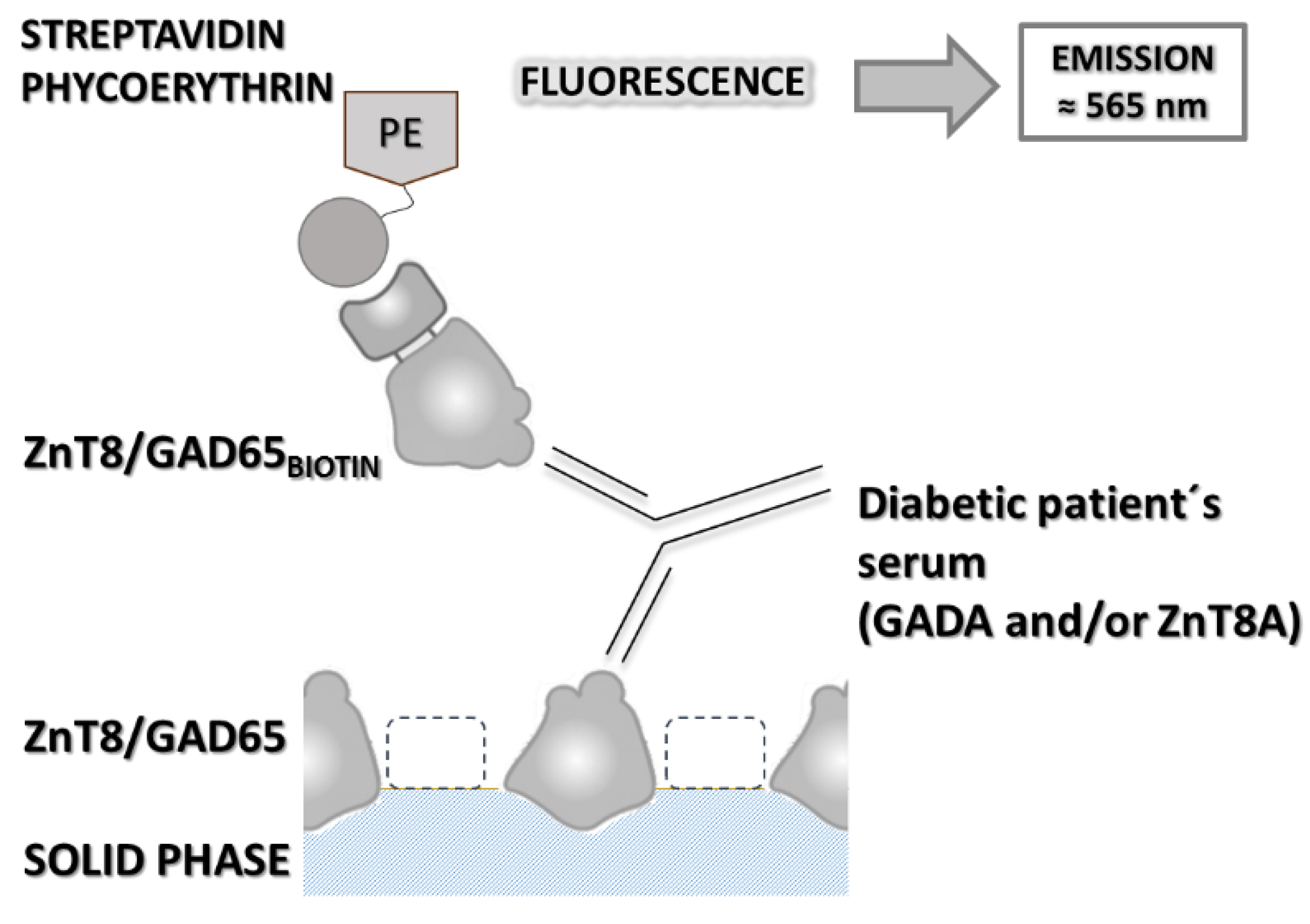
Figure 2.
Analysis of the performance of b-FLIA resulting from the study of 25 sera from normal control individuals and 21 sera from DM patients who tested positive for ZnT8A and/or GADA by RBA. (A) Sensitivity (o) and specificity (x) curve as function of the possible cut-off values. The vertical dashed line indicates the cut-off value with the optimized sensitivity and specificity parameters (cut-off = 1.5). (B) ROC curve analysis of b-FLIA, AUC is included.
Figure 2.
Analysis of the performance of b-FLIA resulting from the study of 25 sera from normal control individuals and 21 sera from DM patients who tested positive for ZnT8A and/or GADA by RBA. (A) Sensitivity (o) and specificity (x) curve as function of the possible cut-off values. The vertical dashed line indicates the cut-off value with the optimized sensitivity and specificity parameters (cut-off = 1.5). (B) ROC curve analysis of b-FLIA, AUC is included.
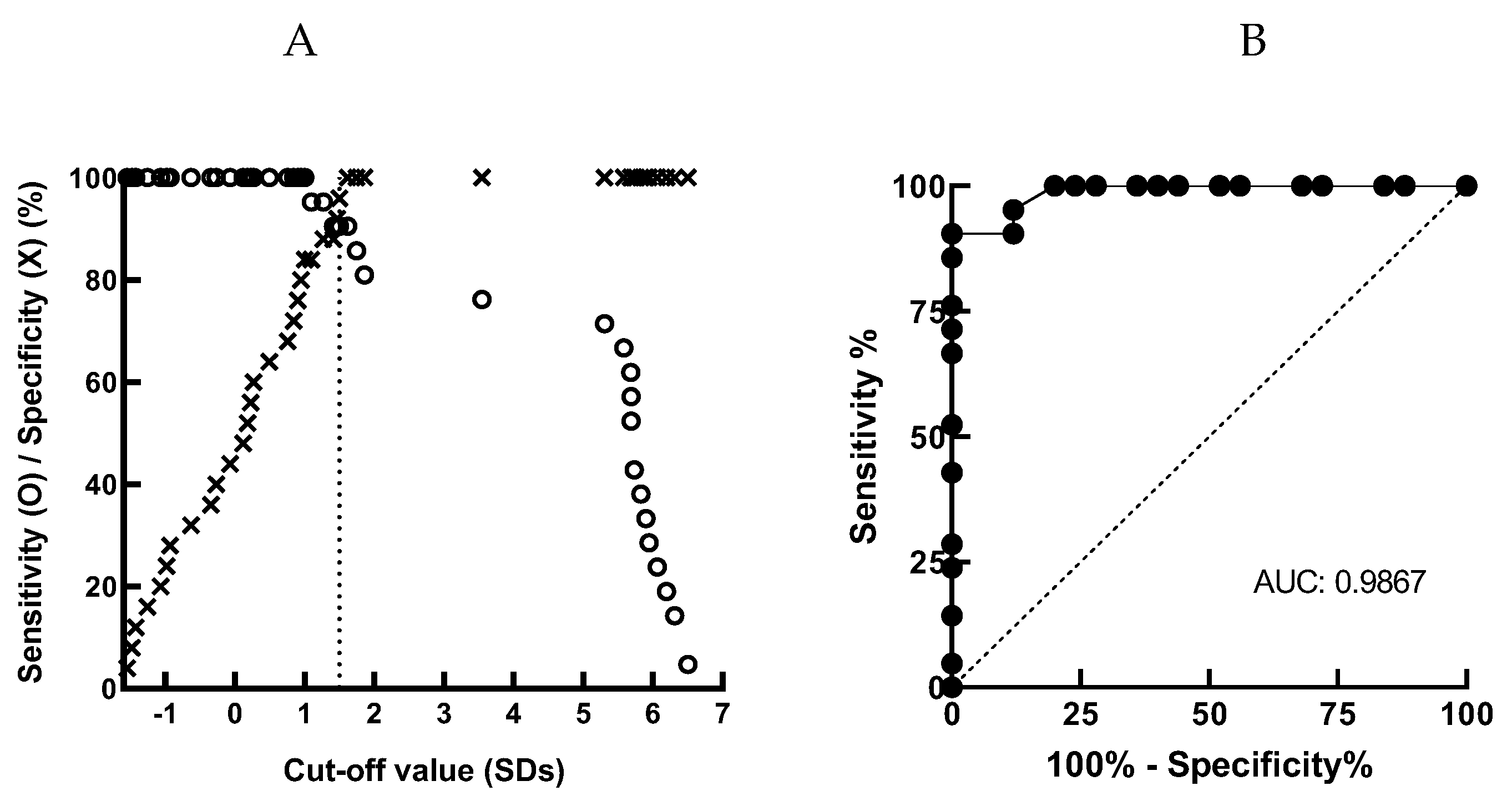
Figure 3.
Autoantibodies results obtained by b-FLIA from 25 normal human sera (NHS) and 21 diabetic samples positive for ZnT8A and/or GADA by RBA. Results are expressed as Standard Deviation score (SDs). The cut-off value (SDs = 1.5) is indicated by a dotted line and median SDs for each group is indicated by a solid line **** p < 0.0001.
Figure 3.
Autoantibodies results obtained by b-FLIA from 25 normal human sera (NHS) and 21 diabetic samples positive for ZnT8A and/or GADA by RBA. Results are expressed as Standard Deviation score (SDs). The cut-off value (SDs = 1.5) is indicated by a dotted line and median SDs for each group is indicated by a solid line **** p < 0.0001.
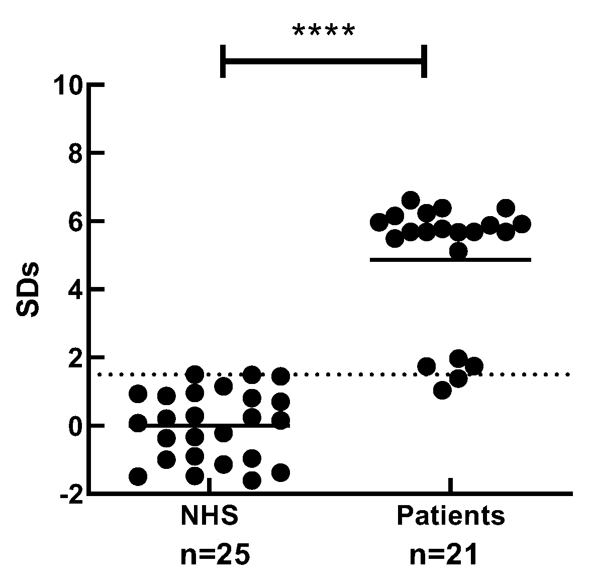
Figure 4.
Venn’s diagram of the integrated results for the determination of GADA and ZnT8A using the reference radiometric assay (RBA), and GADA and or ZnT8A by b-FLIA.
Figure 4.
Venn’s diagram of the integrated results for the determination of GADA and ZnT8A using the reference radiometric assay (RBA), and GADA and or ZnT8A by b-FLIA.
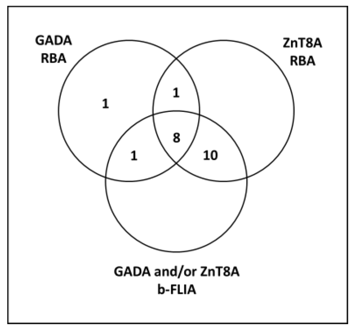

Table 1.
Analytical parameters of b-FLIA from normal human controls (NHS) and positive patients for ZnT8A and/or GADA by RBA.
Table 1.
Analytical parameters of b-FLIA from normal human controls (NHS) and positive patients for ZnT8A and/or GADA by RBA.
| NHS | ZnT8A+ and /or GADA+ | |
|---|---|---|
| n | 25 | 21 |
| Mean (SDs) | 0.00 | 4.87 |
| Median (SDs) | 0.16 | 5.69 |
| Range (SDs) | (-1.61) – 1.50 | 1.03 – 6.62 |
| Sensitivity1 (%) | 90.5 | |
| Specificity2 (%) | 100 | |
1 Percentage of patients RBA positive that were also positive by b-FLIA. 2 100 minus the percentage of false positives.
Disclaimer/Publisher’s Note: The statements, opinions and data contained in all publications are solely those of the individual author(s) and contributor(s) and not of MDPI and/or the editor(s). MDPI and/or the editor(s) disclaim responsibility for any injury to people or property resulting from any ideas, methods, instructions or products referred to in the content. |
© 2026 by the authors. Licensee MDPI, Basel, Switzerland. This article is an open access article distributed under the terms and conditions of the Creative Commons Attribution (CC BY) license (http://creativecommons.org/licenses/by/4.0/).
Copyright: This open access article is published under a Creative Commons CC BY 4.0 license, which permit the free download, distribution, and reuse, provided that the author and preprint are cited in any reuse.