
Submitted:
01 August 2026
Posted:
03 August 2026
You are already at the latest version
Abstract
Micro- and nanoplastics (MNPs) are reported in blood, urine, and tissues, including the kidney, raising concern that the urinary system may be both a target and a route of elimination. This narrative review integrates analytical, toxicokinetic, experimental, epidemiological, and dialysis-related evidence on MNPs and renal health. Human studies demonstrate renal access and urinary detection, but estimates are strongly method-dependent and do not yet define tissue dose, clearance, or a disease threshold. Cell, kidney-organoid, and animal studies consistently identify oxidative and mitochondrial stress, endoplasmic-reticulum dysfunction, inflammation, altered autophagy, regulated cell death, senescence, and fibrotic remodelling. Effects vary with polymer, size, shape, surface ageing, route, dose, and chemical co-exposures, and many experiments remain difficult to map to typical human exposure. Direct human outcome evidence is limited to cross-sectional or exploratory studies; plasticizer epidemiology is informative for chemical co-exposure but cannot establish particle-specific toxicity. Patients with chronic kidney disease may be more susceptible, while kidney dysfunction may also alter measured blood or urinary concentrations. Dialysis introduces an additional, incompletely quantified exposure system. MNP-related renal injury is biologically plausible, but causal contribution to human kidney disease and clinical actionability remain unproven. Standardized methods, paired-matrix kinetics, prospective cohorts, and complete dialysis mass-balance studies are priorities.
Keywords:
microplastics
; nanoplastics
; kidney
; nephrotoxicity
; chronic kidney disease
; biomonitoring
; dialysis
; xenobiotics
1. Introduction
Plastic production and use have created a persistent continuum of debris ranging from visible fragments to particles below the resolution of routine microscopy. Microplastics are commonly defined as plastic particles smaller than 5 mm, whereas the boundary assigned to nanoplastics varies among disciplines and is often operational rather than biological [1,2]. This terminology matters because a millimetre-scale fibre, a 5-µm fragment, and a 50-nm particle differ markedly in surface-area-to-mass ratio, aggregation, protein-corona formation, cellular uptake, and ability to cross biological barriers. The term micro- and nanoplastics (MNPs) is therefore convenient for describing a pollution continuum, but it should not imply that all particles share a common toxicokinetic profile or renal hazard.
Human exposure occurs through ingestion, inhalation, and contact with consumer or medical products. Plastics also contain additives and residual monomers and can adsorb other environmental contaminants; thus, the biologically encountered material may comprise the particle, its surface corona, leachable chemicals, and co-pollutants. The public-health implications of this mixture extend from the full plastics life cycle to individual exposure [3]. Analytical studies have reported plastic-associated signals in human blood [4], placenta [5], lung [6], kidney and urine [7], atherosclerotic plaque [8], and post-mortem organs including brain, liver, and kidney [9]. These observations support internal exposure. They do not, by themselves, establish that the detected material caused tissue injury or disease, and several high-profile measurements remain challenging to compare because the reported measurand may be particle number, polymer mass, percentage of positive samples, or a spectroscopic match.
The kidney is a plausible target for circulating MNPs and their chemical cargo. Renal blood flow creates extensive contact with the glomerular microvasculature; filtration and tubular transport concentrate many xenobiotics; proximal tubular cells have high energetic requirements; and resident and recruited immune cells can amplify injury. At the same time, the glomerular filtration barrier is highly selective, and the renal fate of a particle cannot be inferred from nominal diameter alone. Effective size and surface properties change through aggregation, ageing, and protein adsorption. A particle detected after digestion of kidney tissue may have been intravascular, trapped in a glomerulus, internalized by tubular or immune cells, retained in the interstitium, or introduced during collection and processing. Similarly, a urinary signal may reflect filtration, secretion, epithelial shedding, downstream urinary-tract sources, or contamination.
Interest in renal effects has expanded rapidly. Recent reviews summarize kidney and cardiovascular mechanisms [10] and earlier evidence for MNP deposition and nephrotoxicity [11]. Since those syntheses, the field has added multimodal human tissue studies, urinary biomonitoring, kidney organoids, more realistic particle geometries, models of pre-existing renal disease, and the first patient-level investigations of dialysis-associated exposure. The central question has consequently shifted from whether a plastic-associated signal can be found to whether the available evidence supports a clinically relevant contribution to kidney disease.
This narrative review evaluates that question across five linked domains: human exposure and renal access; analytical validity; renal handling; experimental injury and mechanism; and human clinical relevance, including chronic kidney disease (CKD), development, transplantation, nephrolithiasis, renal cancer, and dialysis. PubMed was searched through 26 July 2026 using combinations of microplastic, nanoplastic, kidney, renal, urine, glomerular, tubular, organoid, and dialysis. Reference lists of relevant reviews and the source articles supplied for this review were also examined. Priority was given to primary studies with polymer-confirming analysis, renal-specific endpoints, appropriate controls, or mechanistic intervention. Because study designs, particles, matrices, size windows, and outcome measures were highly heterogeneous, the evidence was synthesized narratively rather than pooled quantitatively.
2. Human Exposure, Systemic Distribution, and Renal Access
2.1. From Environmental Exposure to the Circulation
Demonstrating systemic entry is a prerequisite for haematogenous delivery to the kidney. In a pioneering study of 22 healthy volunteers, double-shot pyrolysis gas chromatography–mass spectrometry (Py-GC/MS) quantified plastic polymers of at least approximately 700 nm in blood, with a mean total concentration of 1.6 µg/mL [4]. Other studies identified micrometre-scale particles in placenta and lung using Raman or Fourier-transform infrared (FTIR) microspectroscopy [5,6]. These reports are consistent with uptake after ingestion or inhalation, but their outputs are not interchangeable: thermal analysis quantifies polymer mass after destruction of the sample, whereas microspectroscopy counts individual particles within an instrument-specific size window.
Evidence from tissues supports distribution beyond the portal and pulmonary interfaces. In carotid endarterectomy specimens, polyethylene was detected by Py-GC/MS in 58.4% of plaques and polyvinyl chloride in 12.1%; the presence of plastic-associated material was associated with a higher composite risk of myocardial infarction, stroke, or death during follow-up [8]. This study is important because it linked a tissue measurement with longitudinal outcomes, but it concerned a selected cardiovascular population and does not define renal risk. Post-mortem analyses using complementary thermal, spectroscopic, and electron-microscopic methods have also reported MNPs in kidney, liver, and brain, with pronounced variation among tissues and sampling periods [9,12]. Autopsy studies broaden the biodistribution map but remain vulnerable to post-mortem sampling, digestion, recovery, and contamination effects and generally cannot determine temporality.
Animal and modelling studies indicate that systemic delivery is strongly size- and route-dependent. After a single exposure, fluorescently labelled particles produced time-dependent blood and urinary signals in rodents, although fluorescence and electron microscopy did not always provide concordant evidence for intact particles [13]. A physiologically based toxicokinetic model predicted substantial size dependence around the submicrometre range and emphasized gastrointestinal absorption and faecal excretion as determinants of internal dose [14]. More recent mammalian tracing experiments likewise found that translocation across biological barriers varied with size [15]. These findings make renal access plausible but do not support a universal filtration threshold or conversion from an administered dose to a human renal dose.
2.2. Direct Evidence in Human Kidney and Urinary-System Specimens
Massardo and colleagues provided the first polymer-confirmed evidence in human kidney tissue and urine using micro-Raman spectroscopy [7]. Healthy portions of ten nephrectomy kidneys and ten urine samples from healthy donors were analysed. The study identified 26 particles across kidney and urine specimens, with sizes of 1–29 µm in kidney and 3–13 µm in urine; polyethylene and polystyrene were the most frequent polymers. Extensive procedural, reagent, water, and filter controls were a major strength. However, complete digestion removed anatomical localization, the kidney and urine samples were not paired for mass-balance analysis, and no histological or functional phenotype was linked to the particles. The result therefore establishes renal presence, not renal accumulation, filtration, toxicity, or disease causation.
Subsequent studies have extended detection to clinically obtained renal-system tissues. Multimodal analysis of tissue from 28 kidney donors reported polymer signals in kidney, renal vessels, ureter, and adrenal gland [16]. Exploratory correlations with recipients’ blood pressure 15 days after transplantation were mostly non-significant after correction for multiple testing, and the measured particles—many reported between 20 and 500 µm—raise unresolved questions about tissue localization and the relation between particle counts and polymer mass. In paired clear-cell renal cell carcinoma (ccRCC) and adjacent tissue, laser direct infrared spectroscopy, scanning electron microscopy, and Py-GC/MS showed differences in polymer composition and higher total signals in tumour specimens [17]. Detection in tumour tissue is not evidence that MNPs initiated cancer: altered vascularity, tissue architecture, inflammation, and sampling may all affect retention or measurement.
Kidney stones provide another long-lived matrix in which material may be incorporated over time. A 2026 multimodal study reported six polymer types in all examined stones, with a median polymer mass of 12.89 µg/g and predominantly irregular particles in the 20–100 µm range; accompanying multi-omics experiments linked chronic low-dose exposure to mitochondrial and inflammatory responses in renal cells [18]. The finding is hypothesis-generating for nephrolithiasis, but a stone-associated particle may be a nidus, a passenger incorporated during growth, or an external contaminant. Demonstrating a lithogenic role requires prospective exposure data and comparison with rigorously processed stone-free controls.
2.3. Urinary Biomonitoring: An Attractive But Unresolved Matrix
Urine is non-invasive and clinically familiar, which makes it an appealing exposure matrix. Early Raman-based work reported microplastics in the urine of a small number of adults [19]. Later studies found regional differences in urinary MNP signals [20], documented the importance of collection and sampling conditions [21], and showed that the apparent particle profile changes when smaller size fractions are interrogated [22]. Double-shot Py-GC/MS with internal-standard calibration has extended mass-based detection toward particles reported at or above 300 nm [23]. Cascaded microfiltration followed by Py-GC/MS achieved matrix-matched quantification of six polymers and showed that a 0.7–1.2 µm operational fraction accounted for much of the measured mass in multiple biological matrices [24].
Two 2026 studies further illustrate both the potential and the uncertainty of urinary biomonitoring. Paired blood and urine measurements in 98 environmentally exposed volunteers detected at least one of ten polymers in every participant, with higher overall detection in urine than blood and polymer-specific partitioning patterns [25]. In 29 schoolchildren, MNPs below 10 µm were reported in every first-morning urine sample using scanning electron microscopy/energy-dispersive X-ray analysis with Py-GC/MS polymer characterization [26]. These studies suggest that urine contains structured exposure information, but neither validates a clearance model. A concentration in a spot sample can vary with urine flow, recent exposure, collection materials, lower size limit, blank subtraction, and normalization. Larger particles also challenge simple assumptions of glomerular filtration.
Urinary MNPs should therefore not yet be described as a biomarker of “kidney burden.” A rigorous renal-handling study would require repeated paired blood and timed urine sampling, measurement of urine flow and filtration, size-resolved polymer confirmation in both matrices, full procedural blanks, recovery experiments, and ideally tissue or imaging information. Until such data exist, a urinary signal may indicate exposure or excretion but does not distinguish filtration, tubular release, urinary-tract shedding, or contamination.
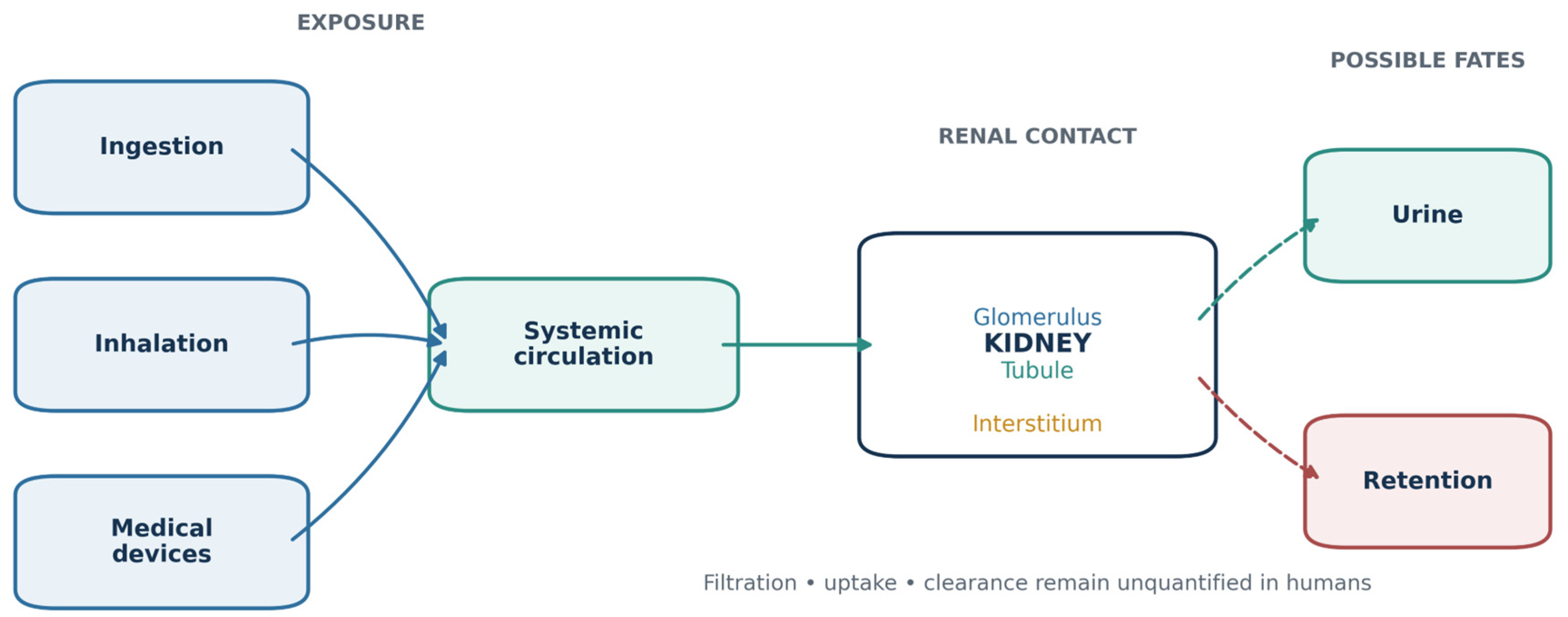
Figure 1.
Exposure pathways and renal disposition of micro- and nanoplastics. Ingestion and inhalation are established environmental routes, whereas medical devices can add healthcare-associated exposure. Systemically available material may remain in blood, contact renal vascular and epithelial compartments, be retained, or appear in urine. The relative contributions of glomerular passage, tubular handling, urinary-tract shedding, and contamination remain unresolved; arrows indicate plausible pathways rather than quantified human fluxes.
Figure 1.
Exposure pathways and renal disposition of micro- and nanoplastics. Ingestion and inhalation are established environmental routes, whereas medical devices can add healthcare-associated exposure. Systemically available material may remain in blood, contact renal vascular and epithelial compartments, be retained, or appear in urine. The relative contributions of glomerular passage, tubular handling, urinary-tract shedding, and contamination remain unresolved; arrows indicate plausible pathways rather than quantified human fluxes.
3. Analytical Challenges in Renal Matrices
MNP analysis is not a single assay but a chain of collection, digestion, separation, identification, and quantification steps. Reproducible reporting requires particle characterization, quality assurance and quality control, field and laboratory blanks, recovery data, matrix effects, and explicit treatment of values near detection limits [27,28]. These requirements are particularly demanding in human studies, where the expected signal is low, tissue is chemically complex, and plastics are ubiquitous in the clinical and laboratory environment.
Spectroscopic methods and thermal methods answer different questions. Raman and FTIR microspectroscopy preserve particle morphology and can assign polymer identity, but their efficiency falls as particles approach the nanoscale, and pigments, fluorescence, weathering, or biological residues may obscure spectra. Py-GC/MS can quantify low polymer masses and detect polymers that microscopy misses, but it destroys size and shape information and may capture dissolved or non-particulate polymer-associated material unless separation is carefully defined [29]. Reviews of human biomonitoring have emphasized that differences in sampling, digestion, size cut-offs, spectral libraries, and blank correction frequently prevent direct comparison [30].
Cross-platform evidence confirms these method-dependent contrasts. In paired endometrial and urine samples, Raman microscopy characterized individual particles of approximately 1.2–7 µm, whereas Py-GC/MS identified additional polymer co-occurrences in samples initially classified as single-polymer by Raman [31]. A multistep digestion protocol for human tissue improved organic-matrix removal while preserving particles for micro-Raman analysis, illustrating that digestion chemistry must be validated for the target polymers and matrix [32]. No method currently captures particle number, polymer mass, shape, nanoscale fraction, surface chemistry, and anatomical localization simultaneously.
Contamination control is not optional. Renal surgery, tissue storage, urinary collection, dialysis tubing, laboratory clothing, filters, and airborne fibres are all potential sources. Procedural blanks should accompany every batch; collection containers and instruments should be polymer-audited; recovery should be assessed across relevant particle sizes and polymers; and both raw and blank-corrected results should be reported. Fluorescent tracing requires additional controls because dyes can leach from labelled particles and accumulate independently, while biological autofluorescence can create false-positive localization [33].
The newest systematic assessment of methods in human biological materials still finds marked heterogeneity and limited validation across matrices [34]. Consequently, agreement within one platform is not equivalent to external validity. Orthogonal confirmation is especially important when an unexpected tissue concentration, very small particle, or unusually large urinary particle drives the biological conclusion.

Table 1.
Principal analytical approaches and their interpretation in renal research.
| Approach | Primary output | Main strength | Main limitation | Minimum renal-study safeguards |
| Raman or micro-Raman spectroscopy | Particle count, size, shape, polymer spectrum | Particle-specific chemical identification; morphology retained | Fluorescence, spectral ambiguity, low throughput, limited nanoscale detection | Procedural blanks; library and match threshold; recovery; proportion of suspected particles confirmed |
| µFTIR or laser direct infrared spectroscopy | Particle count, size, shape, polymer spectrum | Rapid mapping of micrometre-scale particles | Size-dependent sensitivity; substrate and matrix effects | Blank correction; size window; substrate controls; manual validation of automated classification |
| Py-GC/MS | Polymer mass | Sensitive multi-polymer quantification | Destructive; no morphology or anatomical localization; particulate state may be ambiguous | Matrix-matched calibration; separation definition; polymer-specific limits; blank subtraction |
| Electron microscopy with elemental analysis | Ultrastructure and elemental composition | Nanoscale morphology and localization | Polymer identity often indirect; small field of view | Correlative spectroscopy; representative sampling; contamination controls |
| Fluorescence-labelled particles | Experimental kinetics and cellular uptake | High temporal and spatial sensitivity | Dye leaching, photobleaching, autofluorescence, label–particle dissociation | Free-dye and dialysate controls; orthogonal confirmation of intact particles |
| Multimodal analysis | Complementary particle and mass information | Reduces platform-specific blind spots | Complex, expensive, and still dependent on harmonized sampling | Prespecified concordance rules; shared blanks; transparent handling of discordant results |
4. Renal Handling and Toxicokinetics
The renal fate of MNPs depends on their state in biological fluids rather than their nominal diameter in a stock suspension. Protein adsorption can alter charge, aggregation, macrophage recognition, and effective hydrodynamic size. Weathering and degradation change surface oxidation and the release of additives or oligomers. Shape also influences flow and tissue contact. A rigid spherical bead is therefore an imperfect surrogate for an irregular, aged environmental fragment.
The glomerular filtration barrier comprises fenestrated endothelium, glomerular basement membrane, and podocyte slit diaphragms. It is not a passive pore-size filter. Charge, deformability, protein binding, haemodynamics, and barrier integrity all influence passage. Larger circulating particles may remain intravascular or be taken up by endothelial and phagocytic cells, whereas sufficiently small or deformable particles may enter the filtrate. Peritubular delivery and basolateral uptake provide potential routes that do not require glomerular filtration. Once in tubular cells, particles may undergo lysosomal sequestration, exocytosis, epithelial shedding, or prolonged retention.
Experimental kinetics support rapid faecal elimination of most ingested material and much smaller systemic fractions [13,14,15]. A 2026 study comparing realistic irregular fragments with spherical particles reported that more than 99% of the administered material was excreted in faeces, yet irregular fragments persisted longer in circulation, appeared in glomeruli and tubules, attracted phagocytic cells, and produced albumin hyperfiltration and local lesions; comparable effects were not observed with spheres [35]. Although this work improves shape realism, administered dose, labelling, and species differences still limit direct extrapolation.
Renal excretion should be distinguished from renal clearance. Detection in urine establishes that material or a polymer-associated signal reached the sample; clearance requires a validated relation between plasma concentration, urine concentration, flow, and time. Tissue concentration likewise requires a defined denominator and knowledge of whether the signal was intravascular or parenchymal. Human studies have not yet provided a complete blood–kidney–urine mass balance. This gap is central to causal interpretation because CKD could increase circulating concentrations by reducing elimination, decrease urinary concentrations by lowering filtration, increase urinary particles through barrier injury or epithelial shedding, or alter the biological response to an unchanged exposure.
5. Experimental Evidence of Renal Injury
5.1. Renal Cells and Kidney Organoids
Proximal tubular cells are the most frequently studied human renal model. In HK-2 cells, polystyrene microplastics increased mitochondrial reactive oxygen species (ROS), endoplasmic-reticulum (ER) stress, inflammatory signals, and autophagy; the mitochondrial antioxidant MitoTEMPO attenuated several of these responses [36]. Polystyrene nanoplastics induced size- and dose-dependent apoptosis through oxidative stress and p38/ERK mitogen-activated protein kinase signalling [37]. A cross-polymer comparison subsequently showed that uptake and short-term toxicity were not determined by size alone: polyethylene and poly(methyl methacrylate) nanoparticles produced greater morphological disruption than several polystyrene preparations, whereas low concentrations had minimal effects and marked viability loss occurred mainly at 100–200 µg/mL [38]. These studies demonstrate hazard and property dependence, but direct immersion of an immortalized monolayer does not reproduce the delivered dose, corona, flow, or clearance of the human proximal tubule.
Real-world exposure is also a mixture problem. In human proximal tubular epithelial cells, combined polyethylene microplastic and bisphenol A exposure reduced viability and increased oxidative and inflammatory responses more than either exposure alone [39]. Similar enhancement has been reported with metals and other adsorbed chemicals in experimental systems. Such findings show that particle–chemical interactions can modify toxicity, but they do not justify attributing the combined effect solely to the physical particle.
Human pluripotent-stem-cell-derived kidney organoids add multicellular architecture and developmental patterning. Exposure to 1-µm polystyrene particles during the nephron-progenitor stage reduced organoid size, increased ROS and apoptosis, and disturbed proximal and distal tubular patterning through altered Notch signalling [40]. Follow-up work linked growth retardation and abnormal tubules to mitochondrial ROS, DNA damage, reduced glycolysis, and compensatory tricarboxylic-acid-cycle activity [41]. Other studies identified DDIT4-mediated mTOR inhibition, autophagy, and apoptosis [42] and activation of the BCL-2/Bax/caspase pathway [43]. Silencing DDIT4 attenuated autophagy and apoptosis, strengthening the causal role of that pathway within the model [42].
Organoids are relevant to developmental susceptibility, but they remain immature and lack mature filtration, perfusion, immune-cell trafficking, and urinary excretion. Moreover, several studies selected nominal concentrations by reference to polymer mass reported in whole blood, even though a blood mass concentration does not establish the concentration of intact particles reaching nephron progenitors. An inhalation study that combined mice, HK-2 cells, and kidney organoids found greater sensitivity in organoids than in the immortalized cell line [44], again suggesting that model choice materially affects the apparent hazard.
5.2. Whole-Kidney Injury in Animal Models
Animal models integrate absorption, circulation, immune responses, and renal physiology. Oral exposure to polystyrene particles of 50 nm to 4 µm produced size-dependent aggregation in the gastrointestinal tract, renal signals, oxidative stress, inflammation, altered biochemical markers, and histological damage in mice [45]. In combined in-vitro and in-vivo work, polystyrene exposure produced tubular stress and renal histopathology consistent with the cell responses described above [36]. These studies established early proof of renal hazard but used pristine particles and exposure regimens designed for effect detection.
More recent experiments have moved toward organ crosstalk and pathway perturbation. Polystyrene microplastics impaired the intestinal barrier and activated the renal complement C5a/C5aR pathway; an antibiotic intervention and the C5aR inhibitor PMX53 attenuated injury, supporting a gut–kidney component [46]. Co-exposure with di(2-ethylhexyl) phthalate intensified oxidative stress, NF-κB/NLRP3 activation, pyroptosis, and inflammation; N-acetylcysteine and the NLRP3 inhibitor MCC950 reduced several effects [47]. These interventions increase mechanistic confidence, although antibiotics, antioxidants, and signalling inhibitors can have broad actions and do not prove that a single pathway is exclusive.
Several models describe progression from tubular stress to remodelling. Six months of polystyrene exposure induced inflammation, ferroptosis, and renal fibrosis; ferrostatin-1 attenuated epithelial ferroptosis and fibroblast activation [48]. A separate model linked fibrosis to Klotho/Wnt/β-catenin-dependent tubular senescence and showed improvement after restoration of Klotho function [49]. Nanoplastic-induced lysosomal dysfunction impaired autophagic flux, whereas lysosomal exocytosis contributed to particle clearance and recovery after exposure [50]. Ultraviolet ageing enhanced polystyrene nanoplastic toxicity by altering transferrin interactions, intracellular iron, lipid peroxidation, and ferroptosis [51]. Together, these studies indicate that persistence, surface transformation, and failure of cellular clearance can convert early stress into maladaptive repair.
Renal dysfunction has also been assessed at the organ level. After 12 weeks of polyethylene exposure, radionuclide imaging with 99mTc-DMSA and 99mTc-DTPA, serum and urinary markers, and histology indicated impaired renal function in mice [52]. In Wistar rats, oral polyethylene microplastics increased serum creatinine, urea, cystatin C, albumin-to-creatinine ratio, and kidney injury molecule-1 in a dose-dependent manner while suppressing mitochondrial respiratory complexes and producing glomerular and tubular lesions [53]. The inclusion of polyethylene helps counter the field’s strong polystyrene bias, but daily doses of 15 and 60 mg/kg remain far above any established human renal dose.
Newer studies refine the identity of the affected compartment and the importance of particle properties. Chronic nanoplastic exposure produced tubular-specific injury, mitochondrial oxidative-phosphorylation failure, STING-dependent inflammation, senescence, and fibrosis; activation of PGC1α improved mitochondrial function and attenuated these outcomes [54]. Irregular fragments, but not matched spheres, disrupted glomerular capillary flow and barrier integrity [35]. Polystyrene microplastics activated non-canonical TGF-β signalling, epithelial–mesenchymal transition, metabolic reprogramming, and early fibrosis [55]. Partially degraded polylactic-acid particles were taken up by renal macrophages through MSR1 and produced greater inflammatory injury than higher-molecular-weight polymer particles; inhibition of MSR1 or PI3K/AKT narrowed the toxicity difference [56]. These results argue against treating “microplastic exposure” as a uniform dose.
The urinary system may also be vulnerable to disease-specific interactions. In a calcium-oxalate nephrolithiasis model, polystyrene nanoplastics changed crystal morphology, increased crystal adhesion to tubular cells, promoted macrophage recruitment and ferroptosis, and aggravated stone formation; polymers were also reported in human stones [57]. The human observation and experimental mechanism are mutually informative, but neither establishes that ordinary exposure initiates stones in the population.
Table 2.
Representative experimental evidence for MNP-associated renal injury.
| Model and exposure | Principal renal finding | Mechanistic support | Main translational limitation |
| HK-2 cells; polystyrene MPs/NPs [36,37] | Mitochondrial ROS, ER stress, autophagy, MAPK activation, apoptosis | MitoTEMPO attenuation; pathway coherence | Direct nominal exposure; immortalized monolayer; high concentrations |
| HK-2 cells; PS, PMMA, and PE NPs [38] | Polymer-, size-, and concentration-dependent uptake and cytotoxicity | Cross-material comparison and dose response | Short exposure; no biological ageing or clearance |
| Proximal tubular cells; PE-MPs plus BPA [39] | Greater oxidative, inflammatory, and viability effects with co-exposure | Comparison with single exposures | Does not separate particle from chemical contribution in vivo |
| Human kidney organoids [40,41,42,43,44] | Reduced growth, abnormal nephron patterning, metabolic rewiring, autophagy, apoptosis | DDIT4 silencing; convergent structural and omics findings | Developmental immaturity; no perfusion, filtration, or excretion |
| Repeated-dose mice; PS particles [36,45,46,47,48,49,50,51,54,55] | Tubular/glomerular injury, inflammation, senescence, ferroptosis, fibrosis | Complement, NLRP3, Klotho, ferroptosis, lysosome, PGC1α, and TGF-β interventions | Predominantly pristine PS; administered dose not anchored to human renal dose |
| Polyethylene rodent models [52,53] | Imaging, biochemical, mitochondrial, glomerular, and tubular dysfunction | Dose response and organ-function measures | High oral dose; uncertain environmental comparability |
| Irregular versus spherical particles [35] | Shape-dependent circulation, glomerular lesions, and albumin hyperfiltration | Matched-shape comparison; intravital and isolated-kidney methods | Short experimental exposure; label and species effects |
| Degraded versus polymeric PLA [56] | Greater macrophage uptake and renal inflammation with oligomeric material | MSR1 and PI3K/AKT inhibition | Model degradation state; human exposure unknown |
| CaOx crystal and nephrolithiasis models [57] | Increased crystal adhesion, ferroptosis, inflammation, and stone formation | LRP6 manipulation and in-vivo confirmation | Co-exposure regimen; human temporal relation unknown |
6. Mechanisms of MNP-Associated Renal Toxicity
6.1. Oxidative Stress and Mitochondrial Dysfunction
Oxidative stress is the most reproducible signal across renal models, but it should be interpreted as a common response rather than a particle-specific mechanism. MNP contact, internalization, lysosomal stress, surface redox activity, or leached chemicals can increase ROS. Proximal tubular cells are especially vulnerable because they depend on mitochondrial oxidative phosphorylation. Consequences reported across studies include loss of membrane potential, reduced ATP production, mitochondrial structural damage, lipid peroxidation, DNA damage, and activation of stress kinases [36,37,38,39,41,44,53,54].
Mechanistic depth is strongest where mitochondrial injury precedes a renal phenotype and targeted intervention provides rescue. MitoTEMPO attenuated mitochondrial ROS and downstream ER-stress and autophagy signals [36]. PGC1α activation restored mitochondrial function and reduced inflammation, senescence, and fibrotic remodelling in cells and mice [54]. In organoids, altered glycolytic and tricarboxylic-acid-cycle flux accompanied impaired nephron growth [41]. These studies support mitochondrial dysfunction as a central event within several models, while leaving open whether equivalent intracellular doses occur in humans.
6.2. ER Stress, Autophagy, and Lysosomal Handling
The ER–autophagy–lysosome axis is repeatedly disturbed after MNP exposure. ER-stress proteins, LC3, and Beclin 1 increase in tubular models [36]. DDIT4-mediated mTOR inhibition linked exposure to autophagy and apoptosis in kidney organoids [42]. Nanoplastics can promote autophagosome formation while impairing lysosomal degradation, leading to blocked autophagic flux and fibrosis; recovery may depend on lysosomal exocytosis and particle clearance [50]. This dual role is biologically plausible because lysosomes are both the destination for internalized particles and a determinant of whether damaged proteins, organelles, and particles can be removed.
Autophagy markers alone do not reveal whether flux is increased or blocked. Future experiments should combine time-resolved flux assays, lysosomal function, intracellular particle localization, and recovery after exposure. Without those elements, increased LC3 may reflect protective recycling, impaired degradation, or both.
6.3. Inflammation, Innate Immunity, and the Gut–Kidney Axis
MNPs can activate inflammatory signalling after direct renal contact or through extra-renal pathways. Reported mechanisms include NF-κB, NLRP3 inflammasome activation, complement C5a/C5aR, STING signalling after mitochondrial injury, chemokine-mediated macrophage recruitment, and extracellular-vesicle communication. The gut–kidney study provides intervention-supported evidence that intestinal barrier disruption and complement activation contribute to renal injury [46]. Co-exposure with DEHP activated NF-κB/NLRP3 and pyroptosis [47]. Degraded polylactic-acid particles were decoded by macrophage scavenger receptor 1, generating a CCL2-dependent inflammatory loop [56].
These pathways converge with established mechanisms of AKI and CKD and may therefore amplify rather than uniquely initiate disease. This is particularly relevant in susceptible kidneys, where basal inflammation, oxidative stress, endothelial dysfunction, and impaired repair are already present. It also means that generic cytokine changes cannot identify an MNP-specific signature.
6.4. Regulated Cell Death, Senescence, and Fibrosis
Apoptosis, pyroptosis, and ferroptosis appear in different experimental contexts. Oxidative and ER stress can activate BCL-2-family and caspase pathways [37,43]. NLRP3/caspase-1 and gasdermin-dependent pyroptosis has been described in co-exposure and kidney-injury models [47,58]. Iron dysregulation and lipid peroxidation support ferroptosis in chronic, aged-particle, and stone models [48,51,57]. These processes can release inflammatory signals, promote fibroblast activation, and impair tubular repair.
Senescence supplies a plausible bridge from repeated sublethal stress to chronic remodelling. Klotho/Wnt/β-catenin-dependent tubular senescence increased TGF-β1 signalling and fibroblast activation [49], while persistent mitochondrial dysfunction induced senescence and fibrosis through a partially separable pathway from STING-mediated inflammation [54]. Non-canonical TGF-β signalling and epithelial phenotypic change have also been observed [55]. Although the term epithelial–mesenchymal transition should be used cautiously in vivo, the combined evidence supports maladaptive tubular–interstitial crosstalk as an experimental outcome.
6.5. Particle Properties and Chemical Co-Exposure
Toxicity varies with polymer chemistry, size, shape, surface charge, ageing, aggregation, and degradation state [35,38,45,51,56]. Smaller particles may have greater cellular access, but aggregation can make an intermediate nominal size more biologically active [45]. Irregular fragments behave differently from spheres [35]. Ultraviolet ageing can increase oxidative surface chemistry and change protein binding [51]. Biodegradable plastic is not necessarily biologically inert: oligomeric polylactic-acid material produced a different renal immune response from the parent polymer [56].
Particles also transport or co-occur with plasticizers, metals, persistent organic pollutants, and other xenobiotics. Experiments with BPA or DEHP demonstrate enhanced renal injury under combined exposure [39,47]. Accordingly, three distinct questions should be kept separate: the toxicity of the physical particle, the toxicity of particle-associated chemicals, and interaction between them. Real-world risk may reflect all three, but a study measuring only a urinary chemical metabolite does not measure intact MNP exposure.
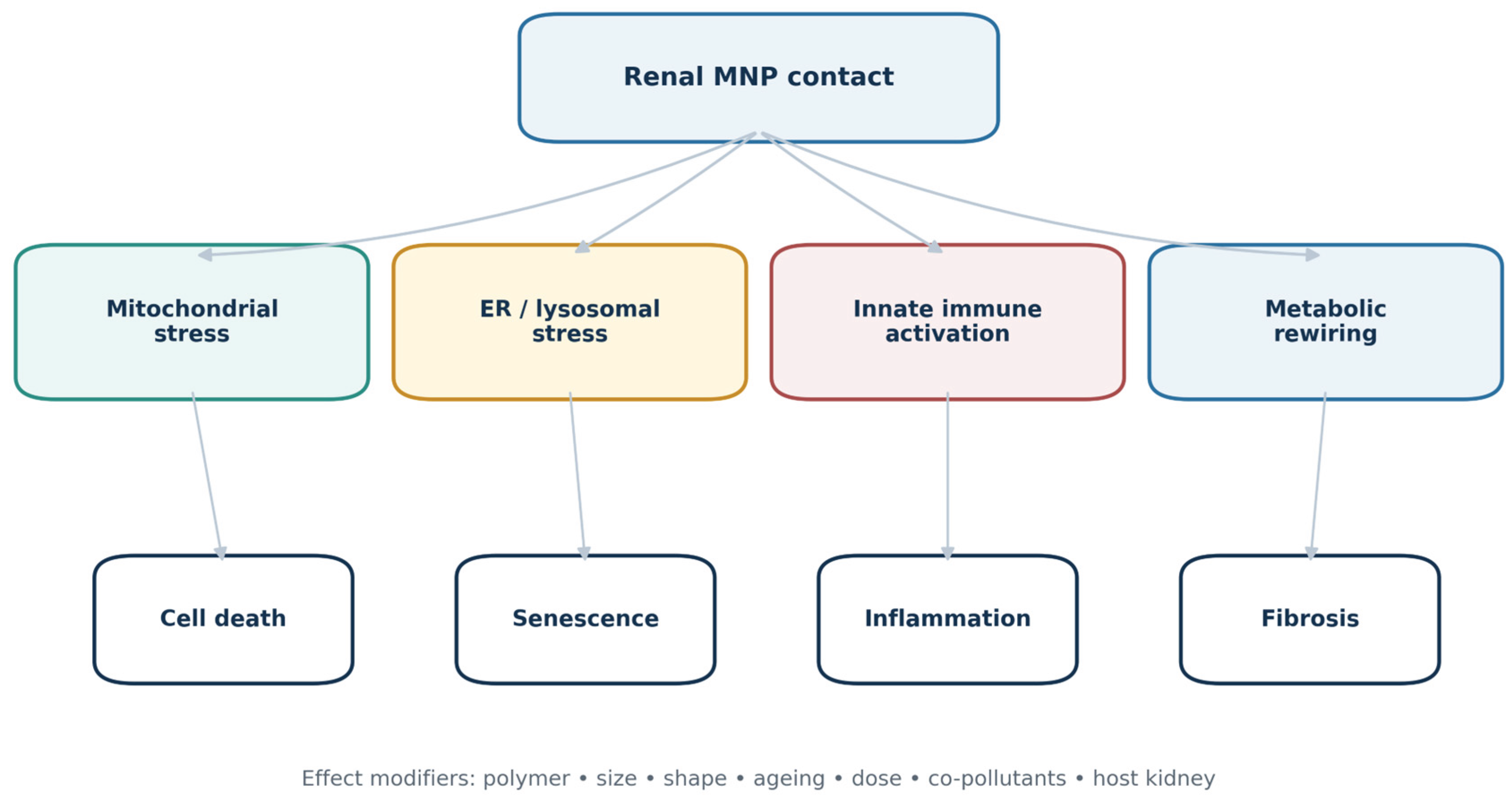
Figure 2.
Mechanistic pathways proposed for MNP-associated renal injury. Particle properties and chemical cargo influence renal delivery and cellular contact. Mitochondrial and ER/lysosomal stress, innate immune activation, and altered energy metabolism converge on regulated cell death, senescence, and maladaptive repair. These pathways are supported primarily by experimental models; their operation at human renal doses has not been established.
Figure 2.
Mechanistic pathways proposed for MNP-associated renal injury. Particle properties and chemical cargo influence renal delivery and cellular contact. Mitochondrial and ER/lysosomal stress, innate immune activation, and altered energy metabolism converge on regulated cell death, senescence, and maladaptive repair. These pathways are supported primarily by experimental models; their operation at human renal doses has not been established.
7. Human Evidence for Renal Outcomes
7.1. Direct Particle Measurements and Renal Phenotypes
Human outcome evidence remains much smaller than the detection and experimental literatures. In 50 healthy adults from Barcelona, six polymers between approximately 0.7 and 20 µm were measured in stool and spot urine by high-performance liquid chromatography coupled to high-resolution mass spectrometry [59]. At least one urinary polymer was detected in 48% of participants. Presence of any urinary polymer was associated with a higher urinary albumin-to-creatinine ratio (ratio of means 2.18; 95% confidence interval 1.17–4.01). However, all albumin-to-creatinine ratios remained below 30 mg/g, the design was cross-sectional, individual polymer detections were sparse, and the associations did not remain significant after false-discovery-rate correction. The study provides a preliminary renal signal, not evidence of albuminuric CKD.
A Chinese birth cohort of 1350 pregnancies reported microplastics in placental samples and positive mixture associations—driven mainly by polyvinyl chloride—with cord-blood creatinine and cystatin C [60]. The sample size and mixture modelling are notable, but placental counts are a proxy for fetal exposure, cord-blood biomarkers have gestational and perinatal determinants, and the study cannot establish postnatal renal dysfunction. Replication with standardized placental analysis, neonatal measured or longitudinal estimated GFR, and childhood follow-up is needed.
The transplant-donor study found widespread polymer signals across donor kidney-associated tissues but no robust relation with early recipient blood pressure after multiple-testing correction [16]. ccRCC studies observed more polymer-associated material in tumour than adjacent tissue and linked experimental polystyrene exposure to NF-κB and TGF-β signalling and tumour growth [17,61]. These findings raise a research question but cannot distinguish cause from tumour-related retention, inflammation, or altered tissue composition. Similarly, detection in kidney stones and supportive experimental lithogenic mechanisms [18,57] require prospective validation before MNPs can be considered a clinical stone risk factor.
No direct-particle study has yet established that a measured exposure precedes incident AKI, sustained eGFR decline, persistent albuminuria, a defined tubulopathy, CKD progression, or kidney failure. No exposure threshold or reference interval has been validated, and no assay has demonstrated clinical utility.
7.2. Plastic-Associated Chemicals Are Not Particle Biomarkers
Large epidemiological studies of phthalates, bisphenols, and other plastic-associated chemicals are often discussed alongside MNPs. They are relevant to the plastics exposome and to particle–chemical mixtures, but urinary metabolites measure soluble chemical exposure and metabolism, not the number or mass of intact particles. Renal function can also affect the concentration and creatinine normalization of urinary metabolites, creating reverse-causality and collider-bias concerns.
In 9008 NHANES participants, several phthalate metabolites and bisphenol A were associated with lower eGFR or higher albumin-to-creatinine ratio, with results influenced by the method used to adjust for urine dilution [62]. Among 9989 adults, MBzP, MCPP, and MEOHP were positively associated with low eGFR, whereas other metabolites showed inverse associations and no overall association with albuminuria was found [63]. In 1610 adolescents, a higher phthalate-mixture concentration was associated with higher eGFR, interpreted as possible hyperfiltration, but the authors explicitly acknowledged reverse causality [64]. Newer analyses of PFAS–phthalate mixtures [65] and urinary or circulating plasticizer metabolites [66] likewise report heterogeneous associations.
This epidemiology supports concern about plastic-associated chemicals and renal function. It cannot be used as confirmatory human evidence for particle toxicity. Combining direct MNP measurements and chemical-metabolite studies into one effect estimate would conflate different exposures, kinetics, and causal pathways.
7.3. What Can Currently Be Concluded from Human Data
The human evidence supports four graded conclusions. First, systemic exposure and renal access are plausible and directly observed. Second, urine can contain polymer-confirmed particles or polymer mass, but its relation to systemic dose and renal clearance is unresolved. Third, preliminary associations with low-range albumin excretion, fetal renal biomarkers, renal tumours, or stones warrant replication but do not establish causal renal disease. Fourth, no direct study currently supports patient-level diagnosis, prognosis, or treatment based on an MNP measurement.
Table 3.
Direct human and dialysis evidence relevant to renal health.
| Population or matrix | Exposure measurement | Renal observation | Principal limitation | Interpretation |
| Nephrectomy kidney and urine [7] | Micro-Raman-confirmed particles | Particles detected in kidney and urine | Ten kidneys; unpaired matrices; no localization or phenotype | Supports renal access and urinary presence |
| Kidney-donor tissues [16] | Py-GC/MS, LD-IR, and electron microscopy | Exploratory early post-transplant blood-pressure correlations | Multiple tissues and tests; no robust corrected association | Detection is not graft toxicity |
| ccRCC and adjacent tissue [17,61] | Multimodal tissue analysis; experimental tumour models | Higher tumour-associated signals and pathway activation | Reverse causality and differential tissue retention | Oncological hypothesis, not human causation |
| Human kidney stones [18,57] | Multimodal polymer analysis plus mechanistic models | Polymers in stones; experimental promotion of CaOx injury | Temporal sequence absent; contamination and incorporation unresolved | Potential lithogenic cofactor requiring prospective study |
| Healthy adults, n = 50 [59] | Urinary MNP presence by HPLC-HRMS | Higher low-range uACR | Cross-sectional; multiple testing; all uACR <30 mg/g | Preliminary association only |
| Birth cohort, n = 1350 [60] | Placental particle counts | Cord creatinine and cystatin-C mixture associations | Placenta is a proxy; perinatal determinants; no follow-up | Developmental signal requiring replication |
| HD/PD solutions [67] | FTIR-confirmed suspected particles | Particles in treatment fluids | Small number; large particles; no patient transfer | Identifies a possible treatment-system source |
| HD patients, n = 9 [68] | LDIR counts pre/post session | Higher post-HD blood counts | Very small study; no full mass balance or outcome | Suggests session-associated entry, not clinical harm |
8. Susceptible Populations and Bidirectional Risk
8.1. Chronic Kidney Disease
CKD may increase both exposure-related vulnerability and uncertainty in biomonitoring. Reduced nephron number and renal reserve, endothelial dysfunction, oxidative stress, chronic inflammation, impaired mitochondrial function, and maladaptive repair could magnify a particle-associated insult. Conversely, reduced filtration, altered protein binding, tubular dysfunction, or changes in urine concentration could modify blood or urinary MNP measurements independently of exposure. A higher circulating level might therefore reflect greater intake, reduced clearance, redistribution, or analytical differences; a lower urinary level might indicate lower exposure, lower filtration, or greater retention.
Experimental evidence supports susceptibility but not its magnitude in patients. In adenine-induced CKD, polystyrene exposure further reduced creatinine clearance and increased plasma urea and creatinine, albuminuria, tubular injury, inflammation, oxidative stress, apoptosis, and interstitial fibrosis; effects were associated with NF-κB and ERK/p38 activation and reduced sirtuin-1 [69]. In db/db mice, polystyrene exposure worsened biochemical injury, inflammation, inflammasome activation, and fibrosis [70]. Prior exposure also aggravated renal ischaemia–reperfusion injury and NLRP3–gasdermin-mediated pyroptosis [58]. These models show effect modification by a diseased kidney, but the exposure route and dose, the animal disease model, and the predominance of polystyrene limit quantitative translation.
Prospective human studies should treat kidney function as a time-varying determinant of both measured exposure and outcome. Baseline and repeated eGFR, measured GFR in nested studies, albuminuria, tubular injury markers, urine flow, medication use, diet, socioeconomic factors, and co-pollutants should be incorporated. Analyses that simply adjust a spot urinary particle concentration for creatinine risk introducing rather than removing bias.
8.2. Development, Pregnancy, and Early Life
Nephron formation is completed before birth, so developmental disruption may have lifelong consequences even without an immediate change in serum creatinine. Kidney organoids consistently show altered nephron-progenitor survival, tubular patterning, and energy metabolism after polystyrene exposure [40,41,42,43,44]. Maternal exposure in mice produced sex-specific reductions in glomerular number, immune and apoptotic responses, and fibrosis in offspring [71]. Human placental measurements associated with cord-blood filtration markers [60] provide an epidemiological signal, but they do not yet connect exposure with nephron endowment, childhood blood pressure, albuminuria, or longitudinal GFR.
Research in pregnancy and children requires especially stringent contamination control because collection devices, diapers, specimen bags, and clinical tubing may introduce plastics. It also requires age-appropriate outcomes and long follow-up. The detection of urinary MNPs in children [26] should motivate standardized exposure studies, not screening or attribution of developmental symptoms to MNPs.
8.3. Nephrolithiasis, Renal Malignancy, and Transplantation
Kidney stones, renal tumours, and transplanted kidneys are emerging but preliminary contexts. Stone matrices could archive long-term contact and experimental data support interactions with calcium-oxalate crystallization [18,57]. Tumour tissues may retain a different polymer profile and experimental exposure can modify oncogenic signalling [17,61]. Donor kidneys and associated tissues can contain polymer signals before implantation [16]. In each setting, temporal direction is unresolved and tissue architecture may determine particle retention. These studies should be viewed as starting points for disease-specific cohorts rather than evidence for clinical testing.
9. Dialysis and Healthcare-Associated Exposure
Dialysis creates a distinctive exposure environment because treatment fluids and blood encounter large surface areas of polymeric tubing, bags, filters, connectors, and membranes. Haemodialysis (HD) additionally uses hundreds of litres of treated water per week, while peritoneal dialysis (PD) involves repeated storage, warming, and transfer of sterile fluid through plastic systems. These features make dialysis a priority setting for MNP research [72]. They do not establish that particles cross the dialyser or peritoneum, accumulate in the patient, or cause outcomes.
The first direct comparison of dialysis fluids analysed 30 HD and PD solution samples and reported 36 suspected particles, predominantly fibres, with polyethylene, polyvinyl chloride, and ethylene-vinyl acetate among the identified polymers [67]. Concentrations were similar in HD and PD fluids (approximately 0.29 and 0.34 particles/L), but estimated weekly particle contact was higher for PD because of the larger sampled volume assumption. The particles were relatively large—mean size approximately 1.31 mm in HD and 0.64 mm in PD—making systemic passage uncertain. The study established possible contamination of treatment fluids, not the delivered patient dose.
A subsequent dual-simulation clinical study measured water before and after passage through HD equipment and blood before and after a session in nine patients [68]. No particles were detected in pre-dialysis water samples, whereas four post-equipment samples contained particles, mostly 20–50 µm. Median blood counts increased from 46.11 before HD to 96.11 after HD (p = 0.027), and several polymers appeared only after treatment. The paired design is informative, but the very small sample, absence of a full flow-normalized mass balance, possible haemoconcentration, vascular-access and sampling contamination, and lack of clinical endpoints prevent inference about retained dose or harm.
PD introduces different questions. Particles may originate from bags, warming, connectors, or transfer sets; conversely, effluent may contain material removed from the peritoneal cavity, shed by mesothelial or inflammatory processes, or introduced during collection. A narrative analysis of dialysis effluent and possible remediation approaches underscores the need for source apportionment [73]. Metal–organic frameworks or other experimental removal systems are not ready for clinical application because adsorption efficiency, biocompatibility, leaching, sterility, and effects on dialysis performance must first be demonstrated.
Current dialysis-fluid standards specify chemical and microbiological quality but do not provide particle-specific limits for MNPs [74]. Adding a regulatory threshold would be premature without a validated sampling protocol, harmonized size range and unit, blank strategy, relation to patient dose, and evidence of health effects. The appropriate next step is a complete mass-balance design rather than isolated concentration measurements.
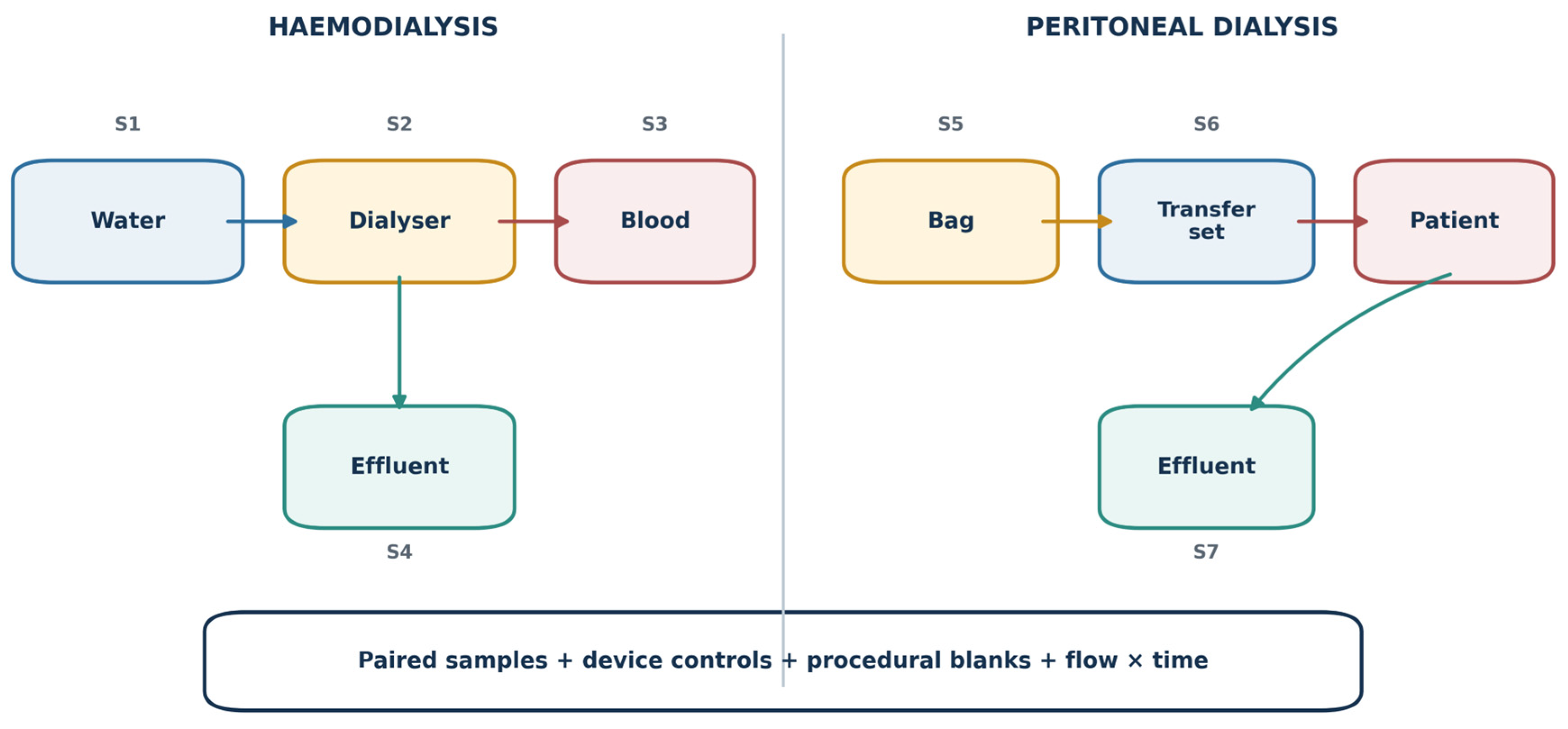
For HD, samples should be obtained from source water, post-treatment water, concentrate, freshly prepared dialysate, dialyser inlet and outlet, spent dialysate, and patient blood immediately before and after treatment. Blood volume, ultrafiltration, haematocrit, treatment time, membrane, tubing lot, and access type should be recorded. For PD, unopened bag fluid, post-warming fluid, transfer-set effluent, and timed drained dialysate should be compared. In both modalities, device-only circulation controls and full procedural blanks are essential. Only a flow- and time-integrated balance can distinguish introduction, adsorption to equipment, redistribution, and removal.
Figure 3.
Dialysis as a particle mass-balance problem. In haemodialysis, source water, concentrate, dialysate, tubing, membrane, patient blood, and spent dialysate are potential sources or sinks. In peritoneal dialysis, the bag, warming, transfer set, peritoneal cavity, and drained effluent must be considered. Paired inflow/outflow and pre/post-patient measurements, normalized to volume and time and accompanied by device controls and procedural blanks, are required to estimate net patient exposure.
Figure 3.
Dialysis as a particle mass-balance problem. In haemodialysis, source water, concentrate, dialysate, tubing, membrane, patient blood, and spent dialysate are potential sources or sinks. In peritoneal dialysis, the bag, warming, transfer set, peritoneal cavity, and drained effluent must be considered. Paired inflow/outflow and pre/post-patient measurements, normalized to volume and time and accompanied by device controls and procedural blanks, are required to estimate net patient exposure.
10. Clinical Implications and Research Priorities
10.1. What Clinicians Should and Should Not Do
The current evidence does not support routine measurement of MNPs in blood, urine, kidney tissue, dialysis fluid, or dialysate for individual clinical care. There is no validated reference interval, disease threshold, prognostic model, or treatment decision linked to a result. A positive research assay cannot identify the source, duration, retained renal dose, or attributable fraction of a patient’s kidney disease. Commercial “microplastic testing” or “detoxification” should not be recommended on the basis of the present renal literature.
No pharmacological intervention has clinical evidence for MNP-associated kidney injury. Antioxidants, ferroptosis inhibitors, complement blockade, sirtuin or PGC1α modulation, curcumin, and other agents have attenuated selected experimental pathways, but these findings identify mechanisms rather than therapies. Extrapolating doses or agents from animals and organoids would expose patients to uncertain benefit and potential harm.
Likewise, current data do not justify changing dialysis modality, membrane, prescription, or access. The available dialysis studies are designed for detection and source discovery, not comparative effectiveness or safety. Dialysis units can nevertheless support research-ready good practice by documenting tubing, membranes, bags, filters, and lots; minimizing unnecessary plastic contact where validated alternatives exist; and preserving samples during product investigations. Any material substitution must maintain sterility, biocompatibility, solute clearance, and mechanical safety.
General measures that reduce avoidable plastic use or unnecessary heating of plastic food-contact materials may be environmentally prudent, but no renal outcome trial has established a kidney-specific benefit. Clinical communication should acknowledge uncertainty: exposure is widespread, experimental kidney injury is credible, and human renal causation remains unproven.
10.2. Priorities for Translational Research
The next generation of studies should connect exposure, internal dose, renal compartment, phenotype, and time. Analytical studies should use harmonized collection and blank procedures, orthogonal confirmation, standardized reporting of number and mass, and explicit size windows. Kinetic studies should obtain paired blood and timed urine, quantify flow and renal function, and distinguish intact particles from polymer-associated chemical signals. Where kidney tissue is clinically available, spatially resolved methods should preserve glomerular, tubular, vascular, and interstitial localization.
Experimental designs should compare polymers beyond polystyrene, irregular and aged particles, environmentally plausible mixtures, chronic low-dose exposure, recovery, and pre-existing kidney disease. Reporting nominal concentration is insufficient; studies should quantify the administered, systemic, and renal delivered dose wherever possible. Organoids and microphysiological kidney systems would benefit from perfusion, immune and endothelial components, and measurement of transepithelial transport.
Prospective human cohorts should prespecify incident AKI, sustained eGFR decline, persistent albuminuria, tubular dysfunction, nephrolithiasis, CKD progression, and kidney failure rather than combine nonspecific biomarkers. Repeated direct MNP measures should precede outcomes, and models must account for kidney function as both a confounder and possible mediator. Biobanks linked to renal phenotyping could enable nested multimodal studies without exposing patients to research biopsies.
Dialysis research should progress from detection to source apportionment, mass balance, reproducibility across equipment and lots, and patient outcomes. A multicentre study should pair device-only circuits with patient sessions, correct blood concentrations for plasma-volume change, integrate flow and treatment duration, and sample both blood and effluent. Only then can mitigation strategies be evaluated.
Table 4.
Research agenda needed to establish clinical relevance.
| Question | Minimum design | Critical measurements | Decision enabled |
| What reaches the human kidney? | Paired blood–urine kinetics with clinically available tissue | Polymer, size, shape, mass, count, blanks, recovery, renal localization | Valid internal and renal dose metric |
| Are urinary MNPs a renal biomarker? | Repeated timed urine across kidney-function strata | Urine flow, paired blood, eGFR/measured GFR, tubular markers, within-person variability | Interpretation of urinary concentrations |
| Do MNPs cause kidney disease? | Prospective cohort with repeated direct exposure measurement | Incident AKI/CKD, sustained eGFR slope, persistent albuminuria, co-exposures, causal modelling | Human risk estimate and threshold |
| Which particle properties matter? | Factorial chronic models with realistic particles | Polymer, size, shape, ageing, corona, degradation, delivered renal dose | Hazard ranking and material design |
| Who is susceptible? | CKD, diabetes, pregnancy, childhood, and older-adult cohorts | Baseline disease, medication, inflammation, renal reserve, longitudinal outcomes | Risk stratification |
| Does dialysis add or remove MNPs? | Multicentre full mass-balance studies | Device-only controls, inflow/outflow, pre/post blood, effluent, flow, time, haemoconcentration | Source control and comparative equipment evaluation |
| Can exposure be reduced safely? | Controlled engineering or clinical intervention | Particle reduction, dialysis/clinical performance, adverse effects, renal outcomes | Evidence-based mitigation |
11. Conclusions
MNPs have been detected in human blood, urine, and kidney-related tissues, and the experimental literature provides a coherent account of how selected particles can injure renal systems. Mitochondrial and ER stress, disrupted autophagy and lysosomal handling, innate immune activation, regulated cell death, tubular senescence, and fibrosis recur across cell, organoid, and animal models. The strength of this evidence is biological plausibility, not quantitative human risk.
The principal uncertainty lies between experimental hazard and human disease. Direct human studies are few, largely cross-sectional or exploratory, analytically heterogeneous, and unable to establish temporality or a renal dose–response relation. Urinary particles are not yet validated biomarkers of renal burden or injury. Epidemiology based on phthalate or other plastic-associated chemical metabolites addresses a related but distinct exposure. CKD may increase susceptibility while simultaneously altering measured concentrations, and dialysis may introduce, redistribute, or remove particles depending on the treatment system.
Accordingly, MNP-associated renal injury should be considered a credible emerging toxicological concern, but not a current clinical diagnosis. Progress will depend on standardized multimodal analysis, paired-matrix toxicokinetics, realistic and dose-anchored models, prospective renal cohorts, and complete dialysis mass-balance studies. Until these links are established, clinicians should neither dismiss the signal nor overinterpret it into testing, treatment, or dialysis changes unsupported by evidence.
Author Contributions
Conceptualization, A.T. and P.C.D.; methodology, A.T. and P.C.D.; investigation, A.T. and P.C.D.; writing - original draft preparation, A.T. and P.C.D.; writing - review and editing, A.T. and P.C.D.; visualization, A.T. and P.C.D. Both authors contributed equally to this work. Both authors have read and agreed to the published version of the manuscript.
Funding
This research received no external funding.
Institutional Review Board Statement
Not applicable.
Informed Consent Statement
Not applicable.
Data Availability Statement
No new data were created or analysed in this study. The literature sources supporting this narrative review are cited in the article.
Conflicts of Interest
The author declares no conflicts of interest.
Acknowledgments
None.
Abbreviations
| Abbreviation | Definition |
| AKI | Acute kidney injury |
| BPA | Bisphenol A |
| CaOx | Calcium oxalate |
| ccRCC | Clear-cell renal cell carcinoma |
| CKD | Chronic kidney disease |
| DEHP | Di(2-ethylhexyl) phthalate |
| eGFR | Estimated glomerular filtration rate |
| ER | Endoplasmic reticulum |
| FTIR | Fourier-transform infrared spectroscopy |
| HD | Haemodialysis |
| HPLC-HRMS | High-performance liquid chromatography coupled to high-resolution mass spectrometry |
| LD-IR | Laser direct infrared spectroscopy |
| MNPs | Micro- and nanoplastics |
| NPs | Nanoplastics |
| PD | Peritoneal dialysis |
| PE | Polyethylene |
| PMMA | Poly(methyl methacrylate) |
| PS | Polystyrene |
| Py-GC/MS | Pyrolysis gas chromatography–mass spectrometry |
| ROS | Reactive oxygen species |
| uACR | Urinary albumin-to-creatinine ratio |
References
- Hartmann, N.B.; Hüffer, T.; Thompson, R.C.; Hassellöv, M.; Verschoor, A.; Daugaard, A.E.; Rist, S.; Karlsson, T.; Brennholt, N.; Cole, M.; et al. Are we speaking the same language? Recommendations for a definition and categorization framework for plastic debris. Environ. Sci. Technol. 2019, 53, 1039–1047. [Google Scholar] [CrossRef] [PubMed]
- Gigault, J.; Ter Halle, A.; Baudrimont, M.; Pascal, P.-Y.; Gauffre, F.; Phi, T.-L.; El Hadri, H.; Grassl, B.; Reynaud, S. Current opinion: What is a nanoplastic? Environ. Pollut. 2018, 235, 1030–1034. [Google Scholar] [CrossRef] [PubMed]
- Landrigan, P.J.; Raps, H.; Cropper, M.; Bald, C.; Brunner, M.; Canonizado, E.M.; Charles, D.; Chiles, T.C.; Donohue, M.J.; Enck, J.; et al. The Minderoo-Monaco Commission on Plastics and Human Health. Ann. Glob. Health 2023, 89, 23. [Google Scholar] [CrossRef] [PubMed]
- Leslie, H.A.; van Velzen, M.J.M.; Brandsma, S.H.; Vethaak, A.D.; Garcia-Vallejo, J.J.; Lamoree, M.H. Discovery and quantification of plastic particle pollution in human blood. Env. Int. 2022, 163, 107199. [Google Scholar] [CrossRef] [PubMed]
- Ragusa, A.; Svelato, A.; Santacroce, C.; Catalano, P.; Notarstefano, V.; Carnevali, O.; Papa, F.; Rongioletti, M.C.A.; Baiocco, F.; Draghi, S.; et al. Plasticenta: First evidence of microplastics in human placenta. Env. Int. 2021, 146, 106274. [Google Scholar] [CrossRef] [PubMed]
- Jenner, L.C.; Rotchell, J.M.; Bennett, R.T.; Cowen, M.; Tentzeris, V.; Sadofsky, L.R. Detection of microplastics in human lung tissue using μFTIR spectroscopy. Sci. Total Env. 2022, 831, 154907. [Google Scholar] [CrossRef] [PubMed]
- Massardo, S.; Verzola, D.; Alberti, S.; Caboni, C.; Santostefano, M.; Eugenio Verrina, E.; Angeletti, A.; Lugani, F.; Ghiggeri, G.M.; Bruschi, M.; et al. MicroRaman spectroscopy detects the presence of microplastics in human urine and kidney tissue. Env. Int. 2024, 184, 108444. [Google Scholar] [CrossRef] [PubMed]
- Marfella, R.; Prattichizzo, F.; Sardu, C.; Fulgenzi, G.; Graciotti, L.; Spadoni, T.; D’Onofrio, N.; Scisciola, L.; La Grotta, R.; Frigé, C.; et al. Microplastics and Nanoplastics in Atheromas and Cardiovascular Events. N Engl. J. Med. 2024, 390, 900–910. [Google Scholar] [CrossRef] [PubMed]
- Nihart, A.J.; Garcia, M.A.; El Hayek, E.; Liu, R.; Olewine, M.; Kingston, J.D.; Castillo, E.F.; Gullapalli, R.R.; Howard, T.; Bleske, B.; et al. Bioaccumulation of microplastics in decedent human brains. Nat. Med. 2025, 31, 1114–1119. [Google Scholar] [CrossRef] [PubMed]
- Lee, Y.H.; Zheng, C.M.; Wang, Y.J.; Wang, Y.L.; Chiu, H.W. Effects of microplastics and nanoplastics on the kidney and cardiovascular system. Nat. Rev. Nephrol. 2025, 21, 585–596. [Google Scholar] [CrossRef] [PubMed]
- La Porta, E.; Exacoustos, O.; Lugani, F.; Angeletti, A.; Chiarenza, D.S.; Bigatti, C.; Spinelli, S.; Kajana, X.; Garbarino, A.; Bruschi, M.; et al. Microplastics and Kidneys: An Update on the Evidence for Deposition of Plastic Microparticles in Human Organs, Tissues and Fluids and Renal Toxicity Concern. Int. J. Mol. Sci. 2023, 24. [Google Scholar] [CrossRef] [PubMed]
- Dzierżyński, E.; Blicharz-Grabias, E.; Komaniecka, I.; Panek, R.; Forma, A.; Gawlik, P.J.; Puźniak, D.; Flieger, W.; Choma, A.; Suśniak, K.; et al. Post-mortem evidence of microplastic bioaccumulation in human organs: insights from advanced imaging and spectroscopic analysis. Arch. Toxicol. 2025, 99, 4051–4066. [Google Scholar] [CrossRef] [PubMed]
- Sun, W.; Jin, C.; Bai, Y.; Ma, R.; Deng, Y.; Gao, Y.; Pan, G.; Yang, Z.; Yan, L. Blood uptake and urine excretion of nano- and micro-plastics after a single exposure. Sci. Total Env. 2022, 848, 157639. [Google Scholar] [CrossRef] [PubMed]
- Chen, C.Y.; Kamineni, V.N.; Lin, Z. A physiologically based toxicokinetic model for microplastics and nanoplastics in mice after oral exposure and its implications for human dietary exposure assessment. J. Hazard Mater. 2024, 480, 135922. [Google Scholar] [CrossRef] [PubMed]
- Zhang, H.J.; Li, S.; Wang, X.L.; Zhang, K.D.; Fang, H.T.; Wu, X.; Huang, Z.; Jiang, W.; Yang, L.; Tan, Q.G.; et al. Size-dependent translocation of polystyrene nanoplastics across biological barriers in mammals. Nat. Commun. 2025, 17, 1116. [Google Scholar] [CrossRef] [PubMed]
- Wang, T.; Li, N.; Zhao, Z.; Zhang, Y.; Lin, L.; Ding, Y.; Feng, R.; Yang, Z.; Du, W.; Zeng, Y. Microplastic contamination in kidney transplant donor tissues and its association with early post-transplant blood pressure changes in recipients: A multi-modal analytical study. J. Hazard Mater. 2025, 497, 139728. [Google Scholar] [CrossRef] [PubMed]
- Zhang, K.; Yu, S.; Chen, S.; Zhou, Z.; Zhan, Y.; Zhang, X. Multimodal detection and analysis of microplastics in human clear cell renal cell carcinoma. Env. Pollut. 2025, 383, 126729. [Google Scholar] [CrossRef] [PubMed]
- Zeng, Q.; Tang, J.; Zhan, L.; Liang, R.; Yang, C.; Cai, R.; Luo, J.; Xu, J.; Liu, T.; Wei, H.; et al. Multimodal detection of microplastics in human kidney stones and multi-omics exploration of renal cell metaflammation. J. Hazard Mater. 2026, 512, 142443. [Google Scholar] [CrossRef] [PubMed]
- Pironti, C.; Notarstefano, V.; Ricciardi, M.; Motta, O.; Giorgini, E.; Montano, L. First Evidence of Microplastics in Human Urine, a Preliminary Study of Intake in the Human Body. Toxics 2022, 11. [Google Scholar] [CrossRef] [PubMed]
- Song, X.; Chen, T.; Chen, Z.; Du, L.; Qiu, X.; Zhang, Y.; Li, Y.; Zhu, Y.; Tan, Z.; Mo, Y.; et al. Micro(nano)plastics in human urine: A surprising contrast between Chongqing’s urban and rural regions. Sci. Total Env. 2024, 917, 170455. [Google Scholar] [CrossRef] [PubMed]
- Rotchell, J.M.; Austin, C.; Chapman, E.; Atherall, C.A.; Liddle, C.R.; Dunstan, T.S.; Blackburn, B.; Mead, A.; Filart, K.; Beeby, E.; et al. Microplastics in human urine: Characterisation using μFTIR and sampling challenges using healthy donors and endometriosis participants. Ecotoxicol. Env. Saf. 2024, 274, 116208. [Google Scholar] [CrossRef] [PubMed]
- Zhu, L.; Wu, Z.; Dong, J.; Zhao, S.; Zhu, J.; Wang, W.; Ma, F.; An, L. Unveiling Small-Sized Plastic Particles Hidden behind Large-Sized Ones in Human Excretion and Their Potential Sources. Env. Sci. Technol. 2024, 58, 11901–11911. [Google Scholar] [CrossRef] [PubMed]
- Ji, S.; Wang, W.; Wang, Y.; Bai, H.; Li, Z.; Huo, Z.; Luo, K. Quantitative Detection of Micro- and Nanoplastics (≥300 nm) in Human Urine Using Double-Shot Py-GC/MS with Internal Standard Calibration. Toxics 2025, 13. [Google Scholar] [CrossRef] [PubMed]
- Kim, U.J.; Ghalamkari, M.; Alvarez, P.; Lee, A.M.; Kim Nguyen, J. Quantifying micro- and nanoplastics in blood, urine, and soft tissues using optimized cascaded microfiltration and pyrolysis-GC/MS for exposomic investigations. Env. Pollut. 2026, 405, 128553. [Google Scholar] [CrossRef] [PubMed]
- Rodríguez-Gutiérrez, J.; Torres-Agullo, A.; Lacorte, S.; Jasso-Arreola, Y.; Santolalla-Vargas, C.E.; Ayllon-Vergara, J.C.; Arreola-Mendoza, L. First study on the toxicokinetic approach of MNPs in an environmentally exposed human population, through their evaluation in blood and urine samples by Pyr-GC-MS and multivariate analysis. Env. Pollut. 2026, 406, 128688. [Google Scholar] [CrossRef] [PubMed]
- Castrogiovanni, M.; Rapisarda, P.; Oliveri Conti, G.; Carroccio, S.C.; Riccobene, P.M.; Ferrante, M.; Makris, K.C. Plastic signatures in childhood: first evidence of urinary micro- and nanoplastics in primary school children from Cyprus. Part Fibre Toxicol. 2026. [Google Scholar] [CrossRef] [PubMed]
- Cowger, W.; Booth, A.M.; Hamilton, B.M.; Thaysen, C.; Primpke, S.; Munno, K.; Lusher, A.L.; Dehaut, A.; Vaz, V.P.; Liboiron, M.; et al. Reporting Guidelines to Increase the Reproducibility and Comparability of Research on Microplastics. Appl. Spectrosc. 2020, 74, 1066–1077. [Google Scholar] [CrossRef] [PubMed]
- Hermsen, E.; Mintenig, S.M.; Besseling, E.; Koelmans, A.A. Quality criteria for the analysis of microplastic in biota samples: A critical review. Environ. Sci. Technol. 2018, 52, 10230–10240. [Google Scholar] [CrossRef] [PubMed]
- Ivleva, N.P. Chemical analysis of microplastics and nanoplastics: Challenges, advanced methods, and perspectives. Chem. Rev. 2021, 121, 11886–11936. [Google Scholar] [CrossRef] [PubMed]
- Kutralam-Muniasamy, G.; Shruti, V.C.; Pérez-Guevara, F.; Roy, P.D. Microplastic diagnostics in humans: “The 3Ps” Progress, problems, and prospects. Sci. Total Env. 2023, 856, 159164. [Google Scholar] [CrossRef] [PubMed]
- Zhang, L.; Tian, J.; Zhu, X.; Wang, L.; Yun, X.; Liang, L.; Duan, S. Cross-platform detection of microplastics in human biological tissues: Comparing spectroscopic and chromatographic approaches. J. Hazard Mater. 2025, 492, 138133. [Google Scholar] [CrossRef] [PubMed]
- Pascali, J.P.; Litti, L.; Fornasari, A.; Giorgetti, A.; Pozzebon, M.; Barone, R.; Ragusa, A.; Fais, P. A Robust Multistep Digestion Method for Microplastics Detection in Human Tissue by MicroRaman Analysis. Drug Test. Anal. 2026, 18, 370–375. [Google Scholar] [CrossRef] [PubMed]
- Catarino, A.I.; Frutos, A.; Henry, T.B. Use of fluorescent-labelled nanoplastics (NPs) to demonstrate NP absorption is inconclusive without adequate controls. Sci. Total Env. 2019, 670, 915–920. [Google Scholar] [CrossRef] [PubMed]
- Dmitrowicz, A.; Sacharczuk, M.; Skiba, D. Micro- and nanoplastics in human biological materials: a systematic review of detection methods and methodological challenges. Arch. Toxicol. 2026. [Google Scholar] [CrossRef] [PubMed]
- Triebel, H.; De Luca, F.; Baumann, S.J.; Wieberneit, A.J.; Oppermann, M.; Baeumner, A.J.; Schweda, F.; Schiessl, I.M.; Castrop, H. Irregularly Shaped Microplastic Particles Compromise the Integrity of the Glomerular Filtration Barrier of the Kidney. Kidney360 2026. [Google Scholar] [CrossRef] [PubMed]
- Wang, Y.L.; Lee, Y.H.; Hsu, Y.H.; Chiu, I.J.; Huang, C.C.; Huang, C.C.; Chia, Z.C.; Lee, C.P.; Lin, Y.F.; Chiu, H.W. The Kidney-Related Effects of Polystyrene Microplastics on Human Kidney Proximal Tubular Epithelial Cells HK-2 and Male C57BL/6 Mice. Env. Health Perspect. 2021, 129, 57003. [Google Scholar] [CrossRef] [PubMed]
- Zhu, Z.; Liao, R.; Shi, Y.; Li, J.; Cao, J.; Liao, B.; Wu, J.; Li, G. Polystyrene nanoplastics induce apoptosis of human kidney proximal tubular epithelial cells via oxidative stress and MAPK signaling pathways. Env. Sci. Pollut. Res. Int. 2023, 30, 110579–110589. [Google Scholar] [CrossRef] [PubMed]
- Gillings, H.L.; Rojas-Canales, D.M.; Wong, S.W.; Bhuskute, K.R.; Kaur, A.; Delcheva, I.; Gleadle, J.M.; MacGregor, M. Nanoplastic toxicity and uptake in kidney cells: differential effects of concentration, particle size, and polymer type. Cell Biol. Toxicol. 2026, 42, 21. [Google Scholar] [CrossRef] [PubMed]
- Verzola, D.; Rumeo, N.; Alberti, S.; Loiacono, F.; La Maestra, S.; Passalacqua, M.; Artini, C.; Russo, E.; Verrina, E.; Angeletti, A.; et al. Coexposure to microplastic and Bisphenol A exhacerbates damage to human kidney proximal tubular cells. Heliyon 2024, 10, e39426. [Google Scholar] [CrossRef] [PubMed]
- Zhou, B.; Wei, Y.; Chen, L.; Zhang, A.; Liang, T.; Low, J.H.; Liu, Z.; He, S.; Guo, Z.; Xie, J. Microplastics exposure disrupts nephrogenesis and induces renal toxicity in human iPSC-derived kidney organoids. Env. Pollut. 2024, 360, 124645. [Google Scholar] [CrossRef] [PubMed]
- Zhou, B.; Zhang, A.; Wang, Y.; Feng, S.; Xue, Q.; Liu, Z.; Zhao, H.; Jing, Z.; Xie, J. Microplastics induce human kidney development retardation through ATP-mediated glucose metabolism rewiring. J. Hazard Mater. 2025, 486, 137002. [Google Scholar] [CrossRef] [PubMed]
- Wang, Y.; Zhang, A.; Liang, T.; Chen, L.; Feng, S.; Zhao, Z.; Jing, Z.; Lv, J.; Xie, J.; Zhou, B. Polystyrene microplastics induce nephrotoxicity through DDIT4-mediated autophagy and apoptosis. Ecotoxicol. Env. Saf. 2025, 294, 118066. [Google Scholar] [CrossRef] [PubMed]
- Zhang, A.; Wang, Y.; Xue, Q.; Yao, J.; Chen, L.; Feng, S.; Shao, J.; Guo, Z.; Zhou, B.; Xie, J. Polystyrene microplastics disrupt kidney organoid development via oxidative stress and Bcl-2/Bax/caspase pathway. Chem. Biol. Interact. 2025, 419, 111642. [Google Scholar] [CrossRef] [PubMed]
- Chen, L.; Han, B.; Yang, S.; Guo, L.; Zhao, L.; Liu, P.; Hong, X.; Zhao, Y.; Peng, Y.; Qi, S.; et al. Toxicological effects and mechanisms of renal injury induced by inhalation exposure to airborne nanoplastics. J. Hazard Mater. 2025, 488, 137393. [Google Scholar] [CrossRef] [PubMed]
- Meng, X.; Zhang, J.; Wang, W.; Gonzalez-Gil, G.; Vrouwenvelder, J.S.; Li, Z. Effects of nano- and microplastics on kidney: Physicochemical properties, bioaccumulation, oxidative stress and immunoreaction. Chemosphere 2022, 288, 132631. [Google Scholar] [CrossRef] [PubMed]
- Liang, Y.; Liu, D.; Zhan, J.; Liu, X.; Li, P.; Ma, X.; Hou, H.; Wang, P. Polystyrene microplastics induce kidney injury via gut barrier dysfunction and C5a/C5aR pathway activation. Env. Pollut. 2024, 342, 122909. [Google Scholar] [CrossRef] [PubMed]
- Li, S.; Gu, X.; Zhang, M.; Jiang, Q.; Xu, T. Di (2-ethylhexyl) phthalate and polystyrene microplastics co-exposure caused oxidative stress to activate NF-κB/NLRP3 pathway aggravated pyroptosis and inflammation in mouse kidney. Sci. Total Env. 2024, 926, 171817. [Google Scholar] [CrossRef] [PubMed]
- Hong, R.; Shi, Y.; Fan, Z.; Gao, Y.; Chen, H.; Pan, C. Chronic exposure to polystyrene microplastics induces renal fibrosis via ferroptosis. Toxicology 2024, 509, 153996. [Google Scholar] [CrossRef] [PubMed]
- Pan, C.; Wang, X.; Fan, Z.; Mao, W.; Shi, Y.; Wu, Y.; Liu, T.; Xu, Z.; Wang, H.; Chen, H. Polystyrene microplastics facilitate renal fibrosis through accelerating tubular epithelial cell senescence. Food Chem. Toxicol. 2024, 191, 114888. [Google Scholar] [CrossRef] [PubMed]
- Lu, Y.Y.; Zhu, W.; Hua, W.; Ren, H.Y.; Tian, M.; Luo, L.; Zhang, Y.; Huang, Q. Reversibility of Renal Fibrosis Induced by Exposure to Polystyrene Nanoplastics: The Dual Role of Lysosomes. Env. Sci. Technol. 2025, 59, 14932–14943. [Google Scholar] [CrossRef] [PubMed]
- He, S.; Cai, J.; Jia, T.; Mao, Z.; Zhou, L.; Zhang, X.; Jiang, S.; Huang, P. New Sight of Renal Toxicity Caused by UV-Aged Polystyrene Nanoplastics: Induced Ferroptosis via Adsorption of Transferrin. Small 2024, 20, e2309369. [Google Scholar] [CrossRef] [PubMed]
- Zaheer, J.; Shanmugiah, J.; Kim, S.; Kim, H.; Ko, I.O.; Byun, B.H.; Cheong, M.A.; Lee, S.S.; Kim, J.S. 99mTc-DMSA and 99mTc-DTPA identified renal dysfunction due to microplastic polyethylene in murine model. Chemosphere 2024, 364, 143108. [Google Scholar] [CrossRef] [PubMed]
- Kehinde, S.A.; Olajide, A.T.; Ogunsanya, S.T.; Fatokun, T.P.; Olulana, D.I.; Olabiyi, F.A.; Faokunla, O.; Tham, C.L.; Chusri, S. Polyethylene microplastics disrupt renal function, mitochondrial bioenergetics, redox homeostasis, and histoarchitecture in Wistar rats. Sci. Rep. 2025, 15, 41120. [Google Scholar] [CrossRef] [PubMed]
- Kim, B.M.; Yang, S.B.; Ha, S.; Kim, M.J.; Kim, J.; Park, D.E.; Chung, H.Y.; Jung, Y.S.; Lee, J.; Lee, H.; et al. Nanoplastic exposure induces mitochondrial dysfunction-driven inflammation and senescence in renal tubular epithelial cells. Free Radic. Biol. Med. 2026, 253, 18–35. [Google Scholar] [CrossRef] [PubMed]
- Dubey, R.; Baby, S.; Singh, A.; Singh, S.; Singh, N.; Goswami, P.; Kushwaha, S.; Khantwal, D.; Sharma, V.; Patnaik, S.; et al. Polystyrene Microplastics Activate Noncanonical TGF-β Signaling and Metabolomic Reprogramming to Promote Epithelial-Mesenchymal Transition and Fibrosis in the Kidney. ACS Omega 2026, 11, 33395–33415. [Google Scholar] [CrossRef] [PubMed]
- Chen, X.; Deng, Y.; Huang, X.; Yang, X.; Li, Z.; Huang, Y.; Zhong, Y.; Li, H.; Ma, L.; Tang, S.; et al. Degradation reshapes the toxic identity of polylactic acid microplastics through MSR1-dependent immune decoding in mouse kidney. Part Fibre Toxicol. 2026. [Google Scholar] [CrossRef] [PubMed]
- Su, X.; Yang, Y.; Xiang, H.; Dong, C.; Shi, M.; Wang, Y.; Zhou, Q.; Song, Q.; He, Z.; Jiang, Q.; et al. Supplemental exposure to polystyrene nanoplastics synergistically amplifies calcium oxalate crystal-induced injury to renal tubular epithelium, accelerating the formation of calcium oxalate kidney stones. Part Fibre Toxicol. 2026, 23. [Google Scholar] [CrossRef] [PubMed]
- Kuang, Q.; Gao, L.; Feng, L.; Xiong, X.; Yang, J.; Zhang, W.; Huang, L.; Li, L.; Luo, P. Toxicological effects of microplastics in renal ischemia-reperfusion injury. Env. Toxicol. 2024, 39, 2350–2362. [Google Scholar] [CrossRef] [PubMed]
- Miguela-Benavides, M.; Calikanzaros, E.; Donat-Vargas, C.; Aguilar, R.; Raimondi, F.; Iraola, S.; Dobaño, C.; de Cid, R.; Llorca, M.; Farré, M.; et al. Association of faecal and urinary micro- and nanoplastics with markers of gut integrity and renal function. Env. Res. 2026, 298, 124197. [Google Scholar] [CrossRef] [PubMed]
- Yin, L.; Liu, M.; Jiang, H.; Shao, H.; Wang, X.; Zhang, B. Prenatal exposure to microplastics and biomarkers of renal dysfunction in umbilical cord blood: Evidence from a birth cohort in China. Ecotoxicol. Env. Saf. 2025, 307, 119318. [Google Scholar] [CrossRef] [PubMed]
- Ye, S.; Xiang, J.; Zhou, S.; Ge, Q.; Anwaier, A.; Chang, K.; Wei, G.; Lu, J.; Tian, X.; Zhu, S.; et al. Polystyrene Microplastics Exposure Aggravates Clear Cell Renal Cell Carcinoma Progression via the NF-κB and TGF-β Signaling Pathways. Adv. Sci. (Weinh) 2026, 13, e18500. [Google Scholar] [CrossRef] [PubMed]
- Kang, H.; Lee, J.P.; Choi, K. Exposure to phthalates and environmental phenols in association with chronic kidney disease (CKD) among the general US population participating in multi-cycle NHANES (2005-2016). Sci. Total Env. 2021, 791, 148343. [Google Scholar] [CrossRef] [PubMed]
- Wang, Z.; Sun, Y.; Gu, L.; Zhang, T.; Liu, S.; Wang, S.; Wang, Z. Association of urinary phthalate metabolites with renal function among 9989 US adults. Ecotoxicol. Env. Saf. 2022, 242, 113930. [Google Scholar] [CrossRef] [PubMed]
- Xiang, S.T.; Cao, Y.; Dong, J.; Li, C.; Duan, Y.; Li, X.; Qiu, J. Association of urine phthalate metabolites levels with kidney function in 1610 US adolescents. Env. Sci. Pollut. Res. Int. 2023, 30, 70519–70527. [Google Scholar] [CrossRef] [PubMed]
- Rahman, H.H.; Stokey, W.R.; Munson-McGee, S.H. Exposure to per- and polyfluoroalkyl substances (PFAS) and phthalate metabolites and their association with low estimated glomerular filtration rates in the US population. Food Chem. Toxicol. 2026, 207, 115853. [Google Scholar] [CrossRef] [PubMed]
- Liu, Y.; Zhang, L.; Sun, C.; Wang, H.; Guo, Y.; Xue, R.; Xing, Y. Urinary and circulatory levels of plasticizer metabolites and their associations with renal function: a cross-sectional analysis of the NHANES cohort. Ann. Med. 2025, 57, 2559125. [Google Scholar] [CrossRef] [PubMed]
- Kara, E.; Konur, K.; Koca, Y.Ş.; Aytan, Ü. Unveiling hidden contaminants: a systematic quantification and characterization of microplastics in hemodialysis and peritoneal dialysis fluids. BMC Nephrol. 2025, 26, 359. [Google Scholar] [CrossRef] [PubMed]
- Deng, Y.; Sun, T.; Long, Z.; Luo, X.; Geng, X.; Lin, Z.; Chen, C.; Li, X. Microplastic entry into bloodstream via hemodialysis: A dual-simulation clinical study. Env. Pollut. 2026, 398, 128085. [Google Scholar] [CrossRef] [PubMed]
- Beegam, S.; Al-Salam, S.; Zaaba, N.E.; Elzaki, O.; Greish, Y.E.; Ali, B.H.; Nemmar, A. Polystyrene microplastics exacerbate experimental chronic kidney disease via inflammatory and oxidative pathways involving NF-κB, ERK/p38 MAPK, and sirtuin-1. Life Sci. 2026, 385, 124142. [Google Scholar] [CrossRef] [PubMed]
- Feng, L.; Chen, C.; Xiong, X.; Wang, X.; Li, X.; Kuang, Q.; Wei, X.; Gao, L.; Niu, X.; Li, Q.; et al. PS-MPs promotes the progression of inflammation and fibrosis in diabetic nephropathy through NLRP3/Caspase-1 and TGF-β1/Smad2/3 signaling pathways. Ecotoxicol. Env. Saf. 2024, 273, 116102. [Google Scholar] [CrossRef] [PubMed]
- Chen, X.; Wang, L.; Liu, K.; Wang, Q.; Li, R.; Niu, L.; Wu, H. Maternal exposure to polystyrene nanoplastics induces sex-specific kidney injury in offspring. Ecotoxicol. Env. Saf. 2025, 293, 118006. [Google Scholar] [CrossRef] [PubMed]
- Passos, R.S.; Davenport, A.; Busquets, R.; Selden, C.; Silva, L.B.; Baptista, J.S.; Barceló, D.; Campos, L.C. Microplastics and nanoplastics in haemodialysis waters: Emerging threats to be in our radar. Env. Toxicol. Pharmacol. 2023, 102, 104253. [Google Scholar] [CrossRef] [PubMed]
- Kanwal, A.; Han, J.; Xu, J.; Xue, X.; Akhmedova, N.; Gao, Z.; Fu, R. Microplastics in dialysis effluent: Environmental chemistry insights and metal-organic frameworks-based remediation prospects. Env. Toxicol. Pharmacol. 2026, 124, 105033. [Google Scholar] [CrossRef] [PubMed]
- International Organization for Standardization. ISO 23500-5:2024ISO 23500-5:2024: Preparation and Quality Management of Fluids for Haemodialysis and Related Therapies—Part 5: Quality of Dialysis Fluid for Haemodialysis and Related Therapies; ISO: Geneva, Switzerland, 2024; Available online: https://www.iso.org/standard/84372.html (accessed on 26 July 2026).
Disclaimer/Publisher’s Note: The statements, opinions and data contained in all publications are solely those of the individual author(s) and contributor(s) and not of MDPI and/or the editor(s). MDPI and/or the editor(s) disclaim responsibility for any injury to people or property resulting from any ideas, methods, instructions or products referred to in the content. |
© 2026 by the authors. Licensee MDPI, Basel, Switzerland. This article is an open access article distributed under the terms and conditions of the Creative Commons Attribution (CC BY) license (http://creativecommons.org/licenses/by/4.0/).
Copyright: This open access article is published under a Creative Commons CC BY 4.0 license, which permit the free download, distribution, and reuse, provided that the author and preprint are cited in any reuse.