

Submitted:
02 August 2026
Posted:
03 August 2026
You are already at the latest version
Abstract
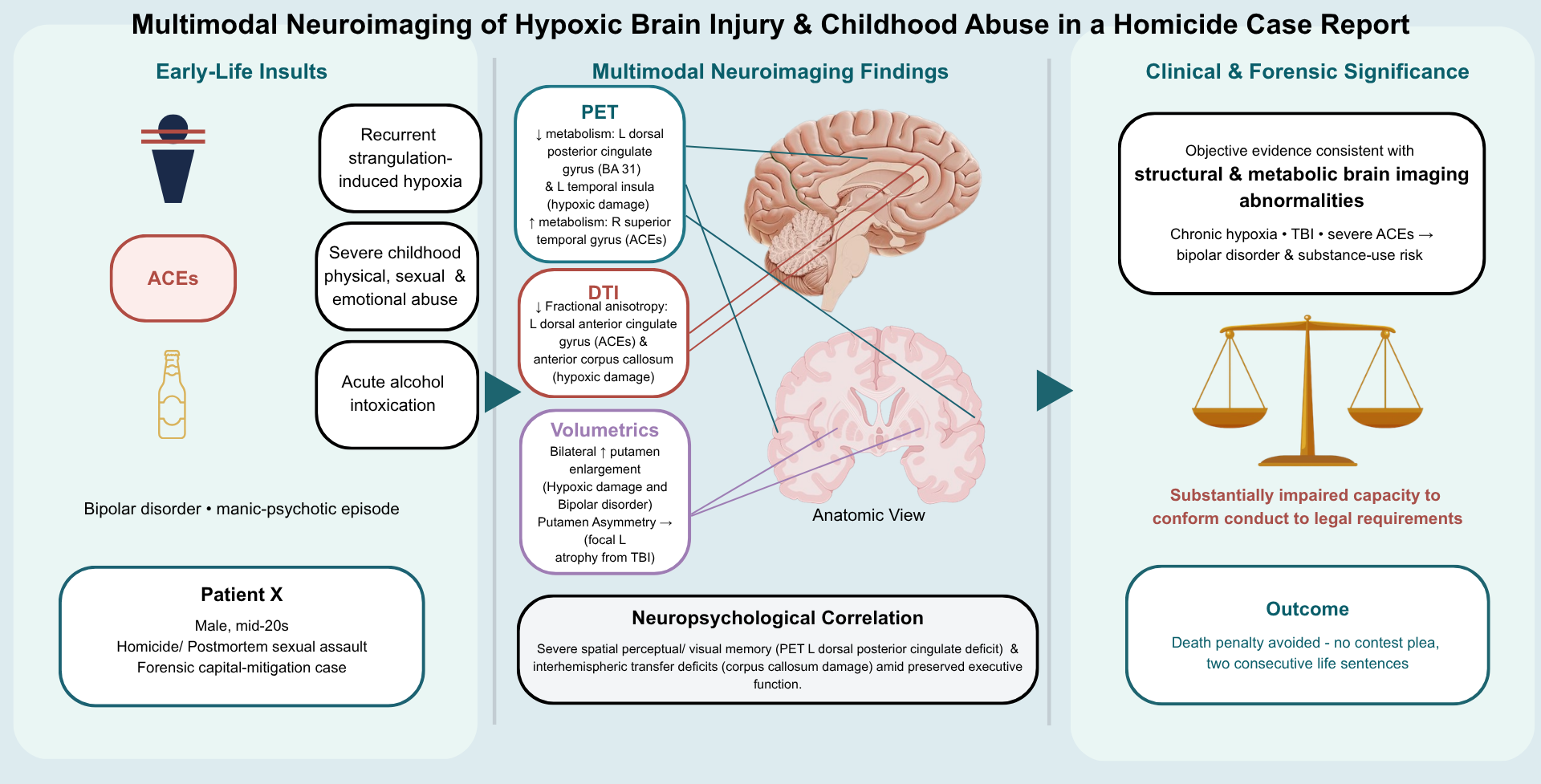
Background/Objectives: Hypoxic brain damage and adverse childhood experiences (ACEs) are strongly associated with neurodevelopmental disorders, adverse behavioral outcomes, and increased risk of bipolar disorder. This case study evaluates the neurological sequelae of severe childhood trauma and recurrent hypoxia in a man in his mid-20s with a history of extreme violent criminal and sexual behavior, with the aim of clarifying the neurological factors associated with his actions. Methods: Multimodal neuroimaging was performed in a subject with recurrent childhood strangulation-induced hypoxia and sexual, physical, and emotional abuse. Assessments included positron emission tomography (PET) with Z-mapping for metabolic analysis, diffusion tensor imaging (DTI) for white matter integrity, and MRI quantitative volumetrics (QV) for structural evaluation. Results: PET revealed hypometabolism in the left temporal insular cortex and left dorsal posterior cingulate gyrus, a decreased neocortical-to-cerebellar metabolic ratio, and hypermetabolism in the right temporal cortex. DTI showed profound decreases in fractional anisotropy (FA) in the anterior corpus callosum consistent with hypoxic injury. DTI also showed decreased FA in left dorsal anterior cingulate consistent with adverse childhood events. MRI QV demonstrated bilateral putamen enlargement, consistent with multiple hypoxic episodes and bipolar disorder, as well as significant asymmetry, with the left putamen smaller than the right, suggesting relative left-right putamen asymmetry secondary to traumatic brain injury. Conclusions: Multimodal neuroimaging identified marked metabolic, white matter, and volumetric abnormalities consistent with severe childhood hypoxia, childhood abuse, bipolar disorder, and traumatic brain injury. Objective abnormalities in networks involved in impulse control and aggression provide neurobiological context relevant to mitigation considerations in aberrant homicidal behavior.
Keywords:
positron emission tomography (PET) brain scan
; diffusion tensor imaging (DTI)
; homicide
; hypoxic brain injury
; quantitative brain volumetrics (QV)
1. Introduction
Adverse childhood experiences (ACEs) and hypoxic brain injuries are established risk factors for profound neurodevelopmental, cognitive, and psychiatric deficits [1,2]. The compounding effects of early developmental trauma, including severe abuse and recurrent hypoxia, can fundamentally alter brain architecture, often presenting clinically as severe bipolar disorder, impaired impulse control, and an increased propensity for violent behavior. In forensic psychiatry, the application of multimodal neuroimaging to assess these neurological deficits has become a critical, albeit debated, tool. While some researchers caution against directly linking neuroimaging anomalies to specific criminal acts, there is broad consensus that structural and metabolic brain abnormalities significantly impair behavioral regulation and judgment, rendering neuroimaging highly relevant in capital mitigation contexts.
This case study examines the neurological profile of Patient X, a mid-20s Hispanic male with a documented history of severe childhood emotional and physical abuse, alcoholism, severe mood disorder, and multiple childhood hypoxic brain injuries and traumatic brain injuries. In late 2016, while heavily intoxicated, the patient committed a homicide followed by postmortem sexual assault in the midst of a manic psychotic episode. Given the extreme severity of the crime and the patient’s extensive history of neurotrauma, multimodal neuroimaging—including Positron Emission Tomography (PET), Diffusion Tensor Imaging (DTI), and MRI Quantitative Volumetrics—was utilized to objectively evaluate metabolic activity, axonal integrity, and structural volume, serving as mitigating clinical evidence against the death penalty.
Current literature strongly supports the use of these modalities in detecting abnormalities consistent with past hypoxic events [3]. PET, which measures glucose cerebral metabolic activity [4], frequently reveals hypometabolism in the temporal cortices and dorsal posterior cingulate gyrus following acute hypoxia [5,6,7,8,9,10]. Furthermore, DTI, which tracks water diffusion to assess white matter integrity [11,12,13], has consistently demonstrated decreased fractional anisotropy (FA) in the corpus callosum with hypoxic encephalopathy [14,15], indicating severe axonal disruption. Building upon these functional and microstructural deficits, MRI Quantitative Volumetrics (QV) provides high-precision, observer-independent data on the macrostructural consequences of these injuries. QV is critical for identifying asymmetric structural atrophy and abnormal inflammatory responses resulting in abnormally enlarged brain regions commonly associated with hypoxic and traumatic brain injury and severe emotional abuse [16,17]. Most notably, QV enables the precise measurement of subcortical anomalies such as profound bilateral enlargement of the putamen, which serves as a structural biomarker consistent with the compounded, catastrophic effects of severe bipolar disorder and chronic intermittent hypoxia [18].
The principal aim of this study is to provide a clinical correlation between the patient’s documented history of extreme early-life physical, emotional, and sexual trauma and his subsequent criminal behavior through advanced neuroimaging. Ultimately, this report concludes that the patient’s multimodal scans revealed profound, objective abnormalities in metabolic function, white matter integrity, and regional brain volumes. These findings are highly consistent with chronic hypoxic injury, severe emotional trauma, and bipolar disorder, demonstrating a substantially impaired neurological capacity for behavioral regulation and conforming conduct to the requirements of the law.
2. Materials and Methods
2.1. Ethical Approval and Data Availability
The multimodal neuroimaging evaluation and associated forensic analyses were performed pursuant to court order as part of a capital mitigation assessment, and the present article represents a retrospective analysis of previously generated data involving no additional interaction, intervention, or data collection; therefore, no separate institutional ethics approval was obtained. The information reported in this article was presented during judicial proceedings and is available in the public court record pursuant to Florida’s public-records framework, commonly referred to as the Florida Sunshine Law, including Article I, Section 24(a) of the Florida Constitution and Florida Rule of General Practice and Judicial Administration 2.420. Direct identifiers have been omitted, and the underlying DICOM files will not be publicly available because they contain sensitive medical information and potentially identifying metadata.
2.2. Neuropsychological Assessment
A comprehensive face-to-face neuropsychological evaluation was administered in early 2018, over a 5.5-hour period utilizing a standardized multi-domain testing battery. Functional performance was calculated across cognitive, memory, executive, and sensorimotor domains using instruments including the WAIS-IV, RAVLT, RCFT, and Dichotic Listening Test, with diagnostic validity rigorously confirmed via standalone performance measures (MSVT and NV-MSVT). The subject’s raw data were converted to demographically corrected T-scores, and aberrant functional domains were subsequently cross-referenced with objective neuroimaging biomarkers to establish clinical correlations with the observed hypoxic and traumatic brain damage.
2.3. Positron Emission Tomography (PET) Acquisition and Processing
In early 2019, F-18 fluorodeoxyglucose [^18F] FDG positron emission tomography was performed using a GE Discovery LS system. The patient received 10.9 mCi of [^18F] FDG after fasting for at least four hours. The uptake interval was 33 min 38 s. During uptake, the patient rested with eyes open in a dimly lit room. The patient had been weaned off all psychoactive medications at least two weeks before the PET scan. The acquisition consisted of a single 10-min static emission frame. Data were reconstructed using three-dimensional Fourier rebinning with iterative reconstruction (3D FORE-IR) into 35 transaxial slices on a 128 × 128 matrix, with an in-plane pixel size of 2.34375 × 2.34375 mm, a slice thickness of 4.25 mm, and a reconstruction diameter of 300 mm. Measured attenuation correction using a linear attenuation coefficient of 0.096 cm−1¹ and model-based scatter correction were applied.
The resulting PET brain scans, in standard DICOM format, were transferred to University Neurocognitive Imaging (UNI) for advanced processing. Image processing was conducted utilizing Statistical Parametric Mapping (SPM) software operating within a MATLAB environment. The preprocessing pipeline included spatial normalization to a standard template, spatial smoothing, and noise reduction. To evaluate statistical significance, the patient’s PET scans were compared with a normative database of 16 normal controls (7 female, 9 male; mean age 34.9 years and SD 13.1 years) to generate a Z-Map using a p-value < 0.01 and a 30-voxel extent threshold. The incorporation of the 30-voxel extent threshold significantly reduces the likelihood of false-positive clusters, since all voxels in a 30-voxel cluster must have p < 0.01. These observer-independent Z-Maps, which are widely accepted by the Society of Nuclear Medicine, provide significantly greater sensitivity for detecting metabolic abnormalities than traditional visual analysis [19,20]. Region of interest statistical z-score differences were calculated for key structures highlighted by the statistical thresholding method.
2.4. Diffusion Tensor Imaging (DTI) Processing and Analysis
Diffusion-weighted MRI was acquired in mid-2019 using a 3.0-T GE Discovery MR750 system with an HNS Head receive coil. A two-dimensional spin-echo echo-planar imaging sequence was acquired with 25 diffusion-encoding directions, repetition time/echo time = 10,000/90 ms, flip angle = 90°, one signal average, 56 contiguous 3-mm axial slices, a 210-mm reconstruction field of view, and a 128 × 128 acquisition matrix. Images were reconstructed onto a 256 × 256 matrix with a pixel spacing of 0.8203 × 0.8203 mm, producing reconstructed voxel dimensions of 0.8203 × 0.8203 × 3.0 mm; the nominal acquired in-plane resolution was approximately 1.64 × 1.64 mm. The acquisition contained 25 diffusion-weighted volumes and one non-diffusion-weighted b0 volume. The nominal acquisition duration was approximately 4 min 20 s.
Raw DICOM files were converted into Neuroimaging Informatics Technology Initiative (NIfTI) format, and fractional anisotropy (FA) images were generated using the Functional MRI of the Brain Software Library (FSL) [21,22]. The FA images were subsequently aligned, normalized, and converted into standard space via Tract-Based Spatial Statistics (TBSS) using nonlinear registration.
Voxel-wise analysis of the patient’s white matter tracts was conducted utilizing the standardized FA images [23,24]. Using SPM, a two-sample t-test was performed to compare the patient’s FA data with that of a cohort of 42 normal controls (14 female, 28 male; mean age 34.7 years and SD of 11.0 years). To ensure statistical accuracy, linear regression covariates were applied to control for the patient’s age and sex. Z-Maps in the transaxial, coronal, and sagittal planes were constructed to identify regions demonstrating significantly abnormal structural integrity (defined by a p-value < 0.01 and a 30-voxel extent threshold) [25] which has been shown to have high sensitivity and specificity [26]. Vinet et al. 2024 validates the SPM thresholding used in this article.
2.5. Region of Interest (ROI) Analysis
Following the generation of PET and DTI Z-Maps, multiple regions of interest (ROIs) were quantified from the transaxial slice using VINCI software. The mean metabolic rates and mean FA values within these specific ROIs were extracted and compared with corresponding neuroanatomical regions in the normal control group.
2.6. Quantitative Volumetrics
High-resolution T1-weighted structural MRI was acquired in mid-2019 on a 3.0-T GE Discovery MR750 system. A sagittal three-dimensional inversion-prepared fast spoiled gradient-echo sequence was acquired with repetition time/echo time/inversion time = 6.628/3.004/600 ms, flip angle = 8°, one signal average, a 240-mm field of view, a 256 × 256 acquisition matrix, 0.9375 × 0.9375-mm in-plane resolution, and 158 contiguous 1.2-mm partitions, yielding reconstructed voxel dimensions of 0.9375 × 0.9375 × 1.2 mm.
Volumetric analysis was performed using NeuroQuant software suite. Volumetric differences were calculated for multiple cortical and subcortical structures [25]. The patient’s neuroimaging volumetric data were statistically compared against 42 healthy controls (14 female, 28 male; mean age 34.7 years and SD of 11.0 years), and aberrant regions of interest were subsequently cross-referenced with established areas of clinical significance known to be impacted in patients with traumatic and hypoxic brain injuries.
2.7. AI Usage
During preparation of this manuscript, the authors used Google Gemini 3.6 Flash and OpenAI ChatGPT (GPT-5.6 Thinking; accessed June-July 2026) to assist with identifying potentially relevant literature, providing editorial suggestions, and performing superficial text editing and formatting. Every source and related claim was independently verified by the authors against the original publication. The authors reviewed and edited all AI-assisted output and take full responsibility for the accuracy, interpretation, and content of the manuscript and its citations.
3. Results
3.1. Neuropsychological Assessment Findings
The neuropsychological evaluation yielded a valid profile, with the subject successfully passing standalone performance validity metrics (MSVT and NV-MSVT), indicating adequate effort and reliable results. Premorbid intellectual functioning was estimated in the Average range (T=48), while current global functioning was in the Low Average range (Overall Battery Mean T=44). Despite an intact baseline Full Scale IQ (T=45), the assessment revealed highly specific, lateralized deficits contrasted against several preserved cognitive domains. The subject’s higher-order cognition remains largely intact, falling within the Average to Superior ranges across executive functioning (domain T=49; Category Test T=64), attention and working memory (domain T=48), and verbal reasoning and language (domain T=44; Sentence Repetition T=66). This pattern establishes that the subject’s deficits are highly localized rather than the result of a generalized cognitive decline.
In stark contrast to his preserved executive functioning, the subject demonstrated profound impairments correlating with organic right-hemisphere, limbic, and callosal dysfunction. Most notably, he exhibited severe deficits in nonverbal and visual memory encoding and retrieval. Performance on the Rey Complex Figure Test (RCFT) revealed Severe impairment in Immediate Recall (T=16) alongside Moderate impairment in both Delayed Recall (T=20) and Recognition (T=26). This was accompanied by Moderately impaired visuospatial construction (RCFT Copy T=21), indicating heavily compromised spatial-perceptual organization and right parietal network dysfunction. Furthermore, overall verbal learning and memory demonstrated Mild impairment (RAVLT Total Recall T=33), reflecting underlying hippocampal vulnerability and disrupted verbal encoding.
Additionally, the subject demonstrated marked functional deficits in auditory processing and interhemispheric transfer. Performance on the Dichotic Listening Test was Borderline in the left ear (T=35) and Moderately impaired in both the right ear (T=21) and simultaneous bilateral processing (T=24), functionally corroborating significant disruption of the corpus callosum. Finally, lateralized sensorimotor testing revealed atypical fine motor coordination deficits isolated to the subject’s dominant hand. Despite being strictly left-handed, his Grooved Pegboard performance was Mildly impaired for his dominant left hand (T=30), while his non-dominant right hand performed entirely within the Average range (T=50).
3.2. Positron Emission Tomography (PET) Metabolic Analysis
PET neuroimaging data were acquired from the patient as part of a comprehensive clinical forensic evaluation. The PET scan acquisitions were of high technical quality, and the raw data were reconstructed and visualized across transaxial, coronal, and sagittal planes (Figure 1).
3.2.1. Neocortical-Cerebellar Metabolic Ratios
Analysis revealed a significant decrease in neocortical metabolism relative to cerebellar metabolism.
3.2.2. Regional Z-Map Statistical Analysis
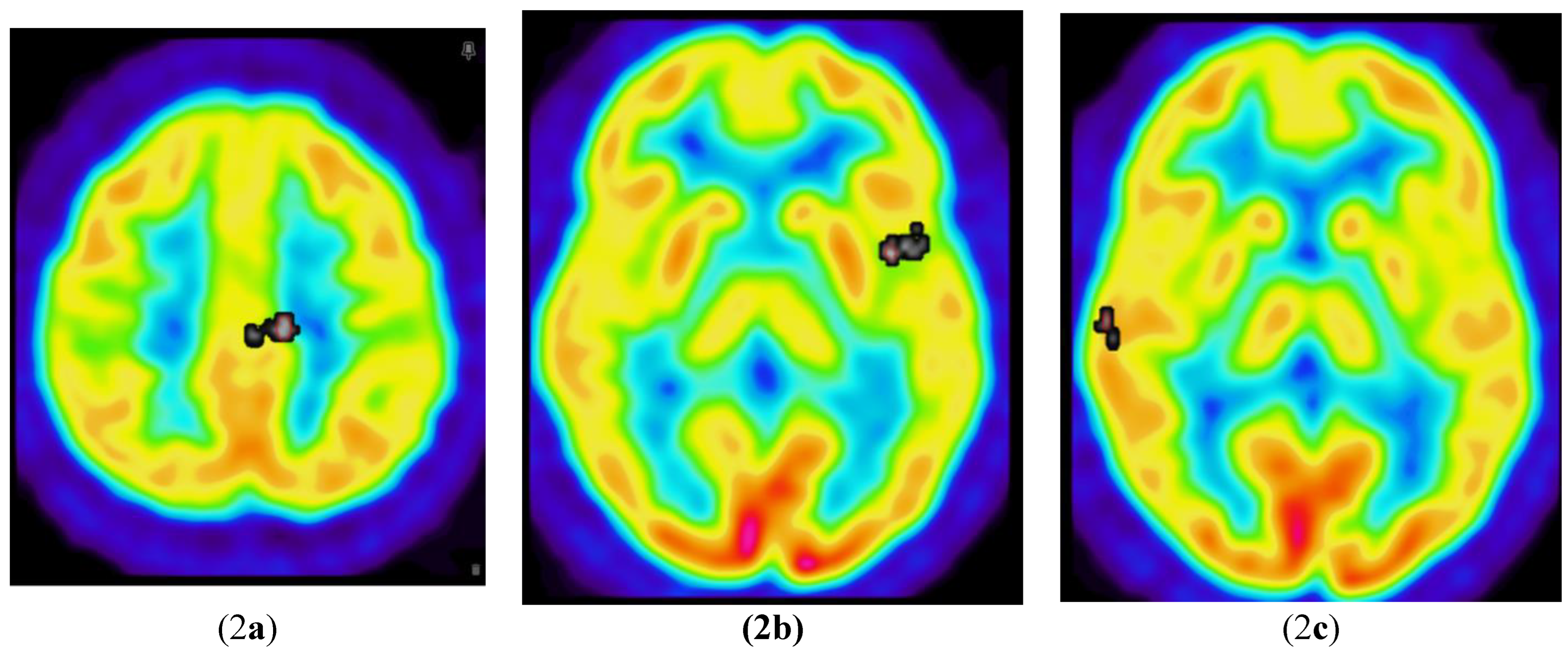
Voxel-wise Z-map analysis identified multiple localized regions exhibiting statistically significant metabolic deviations from the normative control group (Table 2). Specifically, the analysis revealed abnormal hypermetabolism (significant increases) in the right temporal cortex. Conversely, focal hypometabolism (significant decreases) was identified in both the left temporal insular cortex and the left dorsal posterior cingulate gyrus.

Table 1.
PET values demonstrating a statistically significant decrease in the neocortex-to-cerebellum metabolic ratio.
Table 1.
PET values demonstrating a statistically significant decrease in the neocortex-to-cerebellum metabolic ratio.
| ROI | Patient value | Control Mean | Std. Dev. | Z-Score | P-Value |
| Neocortex | 1.02 | 0.98 | 0.02 | 2.00 | 4.6 × 10⁻² |
| Cerebellum | 1.18 | 0.97 | 0.08 | 2.63 | 8.5 × 10⁻³ |
| Neocortex/ Cerebellum |
0.86 | 1.02 | 0.08 | -2.00 | 4.6 × 10⁻² |
Table 2.
PET values demonstrating statistically significant increased metabolism in the right temporal cortex and decreased metabolism in the left temporal insula and left dorsal posterior cingulate gyrus.
Table 2.
PET values demonstrating statistically significant increased metabolism in the right temporal cortex and decreased metabolism in the left temporal insula and left dorsal posterior cingulate gyrus.
| ROI | Patient value | Control Mean | Std. Dev. | Z-Score | P-Value |
| L. dorsal posterior cingulate gyrus (BA31) | 0.77 | 1.39 | 0.10 | -6.20 | 5.7 × 10⁻¹⁰ |
| L. temporal insula | 1.05 | 1.27 | 0.10 | -2.20 | 2.8 × 10⁻² |
| R. Superior Temporal Gyrus (BA42) | 1.56 | 1.24 | 0.18 | 1.77 | 3.9 × 10⁻² (one-tailed) |
3.3. Diffusion Tensor Imaging Analysis
3.3.1. The patient’s MRI-DTI scan was compared with scans from 42 healthy controls. The scans revealed profound decreases in fractional anisotropy (FA) in the anterior corpus callosum (p-value 6.1 × 10⁻⁵) (Figure 3a and Table 3) and in the left dorsal anterior cingulate gyrus (p-value 8.6 × 10⁻⁴) (Figure 3b and Table 3). There were decreases in fiber tract length in the left anterior and mid corpus callosum on tractography relative to the right side (Figure 4).
3.4. Quantitative MRI Volumetric Analysis
3.4.1. Patient X had a T1-weighted MRI sequence, which was processed using quantitative volumetric analysis. Statistical analysis determined clinical significance using relative volumes as a function of the proportion of total intracranial volume occupied by each region. The volumes of various brain regions were compared against 42 controls. The right putamen was significantly increased (two-tailed). There was a significant difference between the left and right putamen, with the left side being smaller than the right (two-tailed). Post hoc analysis of the left putamen showed a one-tailed increase (Table 4). There are bilateral increases in volume in the left and right putamina, with a more pronounced increase on the right side.
4. Discussion
The multimodal neuroimaging findings in the case of Patient X provide compelling, objective evidence consistent with severe, chronic hypoxic brain injury and profound neurodevelopmental disruption secondary to extreme adverse childhood experiences (ACEs). The observed structural and metabolic anomalies align closely with established literature regarding the neurological consequences of early developmental trauma and recurrent asphyxiation, offering a critical biological context for the subject’s severe neuropsychiatric deficits and subsequent violent behavior. These imaging findings are also consistent with neuropsychological deficits that were found, which provide additional cross-modal validation.
4.1. Pathophysiology and Neuroimaging of Chronic Hypoxia
Hypoxic brain damage occurs in response to a critical deficiency of energy in the human brain, which relies on a constant supply of glucose and oxygen to produce adenosine triphosphate (ATP) for neuronal maintenance and function [27,28]. During his development, the subject suffered recurrent, severe episodes of hypoxia resulting from frequent strangulation by his stepfather, often to the point of unconsciousness. Such global hypoxic-ischemic injuries trigger neuronal cell death and significantly alter normal brain development, notably interfering with synaptic pruning. This disruption of dendritic and axonal arborization leads to inefficient neural networks and abnormal regional brain volumes.
These physiological alterations are explicitly reflected in the subject’s PET and MRI-DTI analyses. PET imaging revealed a marked decrease in the ratio of neocortical metabolism relative to the cerebellar cortex (ratio: 0.86; control: 1.02 ± 0.08), placing the subject 2.0 standard deviations (SD) below the mean—a finding noted in individuals with sustained hypoxic damage [29]. Furthermore, the subject exhibited significant hypometabolism in the parasagittal area (dorsal posterior cingulate, Brodmann area 31) [30,31], presenting a relative metabolic rate of 0.77 (control: 1.39 ± 0.10). This represents a deficit of 6.2 SD below the norm, with the probability of this occurring by chance being 5.7 in 10 billion. Damage to Brodmann area 31 is strongly implicated in spatial memory deficits [32] and directly correlates with the subject’s profound functional impairments on neuropsychological evaluation. Specifically, the subject exhibited Severe impairment in nonverbal memory encoding (RCFT Immediate Recall T=16) alongside moderate impairment in spatial-perceptual organization (RCFT Copy T=21). Furthermore, decreased left temporal insular metabolism (1.05 vs. control 1.27 ± 0.10; z = -2.20; p = 0.03) further corroborates the hypoxic etiology [33] and directly aligns with his mild functional impairments in verbal memory recall (RAVLT Total Recall T=33).
Structurally, the subject’s DTI scans demonstrated a profound ~21% decrease in fractional anisotropy (FA) within the corpus callosum, greatly exceeding the ~4% decrease typically observed in severe alcohol dependence [34,35], and aligning instead with the profound axonal disruption of ~20% seen in chronic hypoxic brain damage [36]. Specifically, his anterior corpus callosum FA score was 0.60 (control: 0.76 ± 0.04), placing him 4.01 SD below the mean (p = 6.1 × 10⁻⁵). This profound microstructural degradation clinically manifests as a severe deficit in interhemispheric transfer. Neuropsychological testing revealed marked impairment on the Dichotic Listening Test (Right Ear T=21; Both Ears T=24). Because simultaneous auditory processing requires signals to efficiently cross the corpus callosum, this functional processing deficit provides direct behavioral corroboration of the objective white matter destruction seen on the DTI.
Finally, MRI quantitative volumetrics revealed highly abnormal bilateral putamen enlargement (left: +20.6%; right: +38.7%). To contextualize this severity, individuals with chronic intermittent hypoxia typically exhibit only a 6.1% and 6.6% higher global putamen volume on the left and right sides, respectively [18], while individuals with bipolar disorder show enlargements of 4.7% on the left and 3.2% on the right [37]. In stark contrast to these expected baselines, the subject’s right putamen volume measured 0.43% ICV (control: 0.31% ICV ± 0.03), an expansion over 3.38 SD from the norm (p = 0.00073). Such pronounced, bilateral putamen enlargement heavily underscores the compounded, catastrophic effects of his chronic intermittent hypoxia, which would be consistent with bipolar disorder [18,37,38].
The specific neurobiological link between these types of hypoxic injuries and extreme violent or sexual offending is well-established in forensic literature. Research involving adolescent sexual homicide offenders has demonstrated a significant prevalence of prior hypoxic brain damage, often accompanied by secondary temporal lobe-induced seizures [39]. Similarly, comprehensive studies of adult sexual homicide offenders reveal high rates of past head trauma leading to unconsciousness, alongside temporal lobe irregularities and ventricular dilation [40]. Furthermore, severe head injuries and subsequent hypoxia frequently compromise the hippocampus, a condition strongly associated with the severe dysregulation of volitional impulses and an increased propensity for homicidal violence [41]. In an interview with detectives, Patient X struggled to remember parts of the homicide sexual assault, consistent with some disruption of hippocampal function. Recent neuroanatomical research corroborates that hypoxia-induced lesions in these specific cortical and subcortical networks are strongly correlated with increased violent criminality, mirroring the severe behaviors exhibited in this case [42]. Consequently, the profound structural and metabolic anomalies observed in the patient, particularly within the temporo-limbic regions, closely align with the established neurological profiles of sexual homicide offenders, illustrating how recurrent hypoxic trauma fundamentally damages the neurobiological mechanisms necessary for behavioral control.
4.2. Neurological Consequences of Adverse Childhood Experiences (ACEs)
Beyond hypoxic trauma, the subject endured extensive ACEs, including long-term sexual abuse by family members and severe, targeted physical and emotional abuse. The ACE scoring framework demonstrates a graded, dose-response relationship between early trauma and negative health, behavioral, and developmental outcomes, substantially increasing the risk for affective dysregulation, substance abuse, and aggression [2,43]. Chronic ACEs precipitate chronic inflammatory responses and permanently alter endocrine and metabolic systems, significantly impacting structures such as the amygdala and limbic networks [44,45].
The subject’s neuroimaging data provides a direct clinical correlation with this history of severe maltreatment. DTI analysis revealed a significant decrease in FA in the left dorsal anterior cingulate (0.38 vs. control 0.66 ± 0.08; z = -3.33; p = 8.6 × 10⁻⁴), a structural anomaly widely reported in individuals subjected to severe early childhood abuse and neglect [46]. Additionally, PET findings demonstrated abnormal limbic irritability, which is strongly correlated with severe developmental neglect and abuse [47]. Specifically, this was evidenced by hypermetabolism in the right superior temporal gyrus [48].
4.3. Risk factors for developing Bipolar Disorder and Comorbid Alcohol Use Disorder
A history of multiple hypoxic brain injuries significantly increases the clinical likelihood of developing severe affective conditions, such as bipolar disorder [49]. This neurobiological risk is profoundly compounded by his history of adverse childhood experiences. Exposure to childhood trauma during early neurodevelopmental stages is a major contributing factor to the development of bipolar disorder, with affected individuals being 2.63 times more likely to report a history of childhood trauma compared to healthy controls [49]. Critically, current literature establishes a direct link between severe maltreatment, specifically physical neglect and sexual abuse, and an enhanced risk for the first onset of mania [50]. This psychiatric trajectory is clinically corroborated by Patient X’s documented history of multiple suicide attempts.
His compounded developmental trauma further placed him at an elevated risk for developing alcohol use disorder [51]. Broekhof et al. found that males who were emotionally abused as children were 5.8 times more likely to develop alcohol use disorder later in life. On the date of the offense (late 2016), the patient was under the acute influence of alcohol, a substance well-documented to severely aggravate bipolar volatility [52]. Indeed, current forensic research concludes that the vast majority of violence committed by individuals with bipolar disorder is directly catalyzed by comorbid substance abuse [53,54]. The extreme hyperaggressiveness and hypersexuality exhibited during the homicide and subsequent postmortem sexual interference are highly consistent with such an alcohol-induced manic psychotic break. The neuroimaging evidence consistent with hypoxic damage and traumatic brain injury in conjunction with his clinical history of depression and manic behavior provided the basis for forensic expert opinion that Patient X was operating under the influence of an extreme mental and emotional disturbance at the time of the felony. His capacity to conform his conduct to the requirements of the law was substantially impaired by the likely catastrophic convergence of his neuroimaging-confirmed hypoxic and traumatic brain injuries, severe early childhood abuse, uncontrolled bipolar disorder, and acute alcohol intoxication.
4.4. Traumatic Brain Injury Implications
While the patient’s bilateral putamen enlargement is consistent with his history of chronic intermittent hypoxia and severe mood disorders, the profound asymmetry between the two hemispheres warrants distinct clinical attention. Typically, volumetric increases associated with intermittent hypoxia present with relative symmetry, demonstrating an absolute difference of only about 0.5% between the left (+6.1%) and right (+6.6%) putamen [18]. Similarly, volumetric enlargements associated with bipolar disorder reflect a minor absolute difference of approximately 1.5% between the left (+4.7%) and right (+3.2%) sides [37].
In stark contrast, Patient X exhibits a highly irregular 18.1% absolute difference between the enlargement of his left (+20.6%) and right (+38.7%) putamen. This enormous degree of asymmetry strongly suggests a compounding neurological insult beyond hypoxia or affective dysregulation. From a forensic neurobiological perspective, the medically most probable etiology for this extreme discrepancy is superimposed focal atrophy of the left putamen resulting from traumatic brain injury (TBI). While the baseline hypoxic and affective conditions drove global bilateral enlargement, structural damage from the patient’s history of severe physical trauma likely caused simultaneous, localized atrophic volume loss on the left side, blunting its expansion relative to the right. This interpretation is heavily supported by established MRI quantitative volumetric literature, which consistently demonstrates localized brain atrophy in damaged regions among patients suffering from chronic neuropsychiatric symptoms secondary to TBI [17,55].
Importantly, comprehensive neuropsychological testing supported the validity of the subject’s test performance, as he passed the standalone performance-validity measures MSVT and NV-MSVT and showed no evidence of inadequate effort. The assessment revealed a stark diagnostic contrast: his baseline intelligence and higher-order executive functioning remain largely intact (Executive Function Domain T=49; Category Test T=64), yet he suffers from catastrophic, highly specific memory, spatial, and interhemispheric processing deficits. This clinical contrast supports the interpretation that his impairment is not the result of a generalized or fabricated cognitive decline, but rather highly specific cognitive impairments.
4.5. Clinical Correlates and Broader Implications
The convergence of chronic hypoxic brain injury, traumatic brain injury, and profound developmental trauma severely impaired the subject’s neurological capacity. While most individuals with bipolar disorder, childhood maltreatment, TBI, and hypoxic injury are not seriously violent, there is a greater likelihood of violence with multiple risk factors. The resulting temporo-limbic, subcortical, and white matter structural damage manifested clinically as severe mood instability, chronic suicidal ideation (with documented attempts in 2014, 2016, and 2019), and self-medicating substance abuse. These neurodevelopmental disruptions severely compromised brain regions that regulate impulse control, emotional regulation, memory, and interhemispheric processing. Consequently, the catastrophic neurological damage documented in these scans provides a critical neurobiological explanation for his substantially impaired capacity to conform his conduct to the requirements of the law, ultimately culminating in the violent homicide and postmortem sexual interference committed in late 2016.
Functional neuroimaging modalities, particularly PET, have been consistently validated as reliable tools for corroborating the extent of brain injury months or even years post-insult [22,56]. This case underscores the necessity of utilizing high-resolution multimodal neuroimaging in forensic psychiatric evaluations, particularly in capital mitigation contexts where complex developmental trauma and occult brain injuries are suspected. Future research should continue to define the specific neuroimaging biomarkers that differentiate traumatic anoxic injuries from other neurodevelopmental disorders, expanding the utility of observer-independent neuroimaging in complex legal and clinical applications.
5. Conclusions
Chronic hypoxic brain injury, severe adverse childhood experiences (ACEs), and traumatic brain injury induce profound, lasting disruptions to neurodevelopment and structural brain architecture, which increase the likelihood of developing alcohol use disorder and bipolar disorder. In the case of Patient X, multimodal neuroimaging (PET, MRI-DTI, and Quantitative Volumetrics) revealed extensive objective neurological deficits that clinically correlate with his well-documented history of severe early-life physical, emotional, and sexual abuse, recurrent asphyxiation, traumatic brain injury, and subsequent neuropsychiatric sequelae, most notably his bipolar disorder and severe substance abuse. These findings are also consistent with the neurological deficits discussed previously.
Specific neuroimaging biomarkers, namely, profoundly decreased fractional anisotropy (FA) within the corpus callosum, significant hypometabolism in the parasagittal cortex, and bilaterally enlarged putamen volumes, substantiate the clinical history of recurrent, strangulation-induced cerebral hypoxia. Furthermore, indicators of severe developmental trauma and resultant limbic kindling were evidenced by abnormal hypermetabolism in the right temporal cortex and significantly decreased FA in the left dorsal anterior cingulate gyrus. Additionally, the asymmetry between the putamen volumes is likely due to traumatic brain injury with resultant atrophy of the damaged side.
The compounded structural and metabolic damage from these intersecting neurotraumas significantly degraded the patient’s temporo-limbic and subcortical networks, increasing his susceptibility to bipolar disorder and maladaptive substance abuse. The catastrophic convergence of these chronic neuropsychiatric impairments and acute alcohol intoxication provided the foundation for the forensic expert opinion that Patient X had an uncontrolled manic episode with psychotic features on the night of the offense, characterized by profound hyperaggressiveness and hypersexuality. Collectively, these neurodevelopmental and psychiatric impairments fundamentally compromised the patient’s impulse control and behavioral regulation. These documented deficits serve as a critical biological context for his substantially impaired capacity to conform his conduct to the law, culminating in his criminal behavior in December 2016. Ultimately, this case demonstrates the vital utility of advanced, observer-independent neuroimaging in forensic psychiatry, providing objective, quantifiable corroboration of occult brain damage and offering a neurobiological framework for severe, otherwise incomprehensible violent behavior.
Limitations include that this was a retrospective, uncontrolled case report. The likely timing of the imaging abnormalities was inferred based through expert analysis of case history, imaging findings, and relevant literature; however, these conclusions are not definitive.
The district attorney initially sought the death penalty for Patient X. After the district attorney agreed to his plea of no contest, the judge accepted his no-contest plea to charges of murder in the first degree, armed burglary of a dwelling with assault or battery, abuse of a dead human body, petit theft, and tampering with evidence. Patient X received two consecutive life sentences. The neuroimaging evidence may have helped persuade the district attorney and the judge to accept a no-contest plea in exchange for two consecutive life sentences.
Author Contributions
“Conceptualization, J.W.; methodology, J.W.; software, J.W.; resources, J.W.; data curation, J.W.; writing—original draft preparation, S.S. and N.W.; writing—review and editing, N.W.; supervision, J.W. All authors have read and agreed to the published version of the manuscript.”.
Funding
This research received no external funding. The underlying forensic neuroimaging evaluation was commissioned and funded by the defense.
Institutional Review Board Statement
No separate institutional review board approval was obtained because this individual case report is a retrospective analysis of data previously generated pursuant to court order as part of a forensic capital mitigation assessment and involved no additional interaction, intervention, or prospective data collection. The manuscript is limited to information presented in open court and available in the public judicial record under Florida’s public-records framework, including Article I, Section 24(a) of the Florida Constitution and Florida Rule of General Practice and Judicial Administration 2.420. The patient’s name and other direct identifiers have not been disclosed.
Informed Consent Statement
Informed consent for participation is not required as per local legislation and applicable federal regulations. The neuroimaging examination and associated analyses were not conducted as a research intervention but were performed pursuant to a court order as part of a forensic capital mitigation evaluation. The present case report constitutes a secondary analysis of information generated for that nonresearch judicial proceeding. Pursuant to 45 C.F.R. § 46.104(d)(4)(i), secondary research involving identifiable private information for which consent is not required may qualify as exempt when the information is publicly available. The clinical, neuroimaging, neuropsychological, and historical information reported in this article was presented in the judicial proceeding and incorporated into the public court record, which is accessible under Article I, Section 24(a) of the Florida Constitution and Rule 2.420 of the Florida Rules of General Practice and Judicial Administration, subject to applicable statutory exemptions and confidentiality protections. No additional interaction, intervention, examination, or prospective collection of data from the individual was undertaken for the preparation of this article. Direct personal identifiers have been omitted from the manuscript. The underlying DICOM files will not be made publicly available because they contain sensitive metadata that could increase the risk of identification.
Data Availability Statement
Original imaging data is not available because the metadata contains information that is confidential under HIPAA. The metadata was not presented in open court.
Conflicts of Interest
Manuscript preparation received no external funding. The funder had no role in analysis or interpretation, manuscript preparation or the decision to submit. J.W. independently interpreted the forensic neuroimaging analysis in his capacity as a retained expert witness. J.W. received compensation for forensic neuroimaging analysis through University Neurocognitive Imaging. N.W. and S.S. declare no conflicts of interest.
Acknowledgments
During the preparation of this manuscript/study, the author(s) used Google Gemini and ChatGPT for the purpose of obtaining relevant citations for the literature. The authors have reviewed and edited the output and take full responsibility for the content of this publication.
Abbreviations
The following abbreviations are used in this manuscript:
| PET | Positron Emission Tomography |
| MRI | Magnetic Resonance Imaging |
| ACEs | Adverse Childhood Experiences |
| DTI | Diffusion Tensor Imaging |
| QV | Quantitative Volumetrics |
| FA | Fractional anisotropy |
| HIPAA | Health Insurance Portability and Accountability Act |
| DICOM | Digital Imaging and Communications in Medicine |
| WAIS-IV | Wechsler Adult Intelligence Scale – Fourth Edition |
| RAVLT | Rey Auditory Verbal Learning Test |
| RCFT | Rey Complex Figure Test |
| MSVT | Medical Symptom Validity Test |
| NV-MSVT | Nonverbal Medical Symptom Validity Test |
| ^18F | Fluorine-18 |
| FDG | Fluorodeoxyglucose |
| UNI | University Neurocognitive Imaging |
| SPM | Statistical Parametric Mapping |
| MATLAB | Matrix Laboratory |
| NIfTI | Neuroimaging Informatics Technology Initiative |
| FSL | Functional MRI of the Brain Software Library |
| TBSS | Tract-Based Spatial Statistics |
| ROI | Region of interest |
| AI | Artificial Intelligence |
| IQ | Intelligence quotient |
| BA | Brodmann area |
| ICV | Intracranial volume |
| SD | Standard deviation |
| ATP | Adenosine triphosphate |
| TBI | Traumatic brain injury |
| IRB | Institutional Review Board |
References
- Stein, M.B.; Koverola, C.; Hanna, C.; Torchia, M.G.; McClarty, B. Hippocampal volume in women victimized by childhood sexual abuse. Psychol. Med. 1997, 27, 951-959. [CrossRef]
- Gilgoff, R.; Singh, L.; Koita, K.; Gentile, B.; Marques, S.S. Adverse childhood experiences, outcomes, and interventions. Pediatr. Clin. N. Am. 2020, 67, 259-273. [CrossRef]
- Manasova, D.; Belloli, L.M.L.; Rosenfelder, M.J.; Willacker, L.; Rama, E.F.; Valota, C.; et al. Multimodal multicentre investigation of diagnostic and prognostic markers in disorders of consciousness. Brain 2026, awaf412. [CrossRef]
- Zimmer, L. Positron emission tomography neuroimaging for a better understanding of the biology of ADHD. Neuropharmacology 2009, 57, 601-607. [CrossRef]
- He, Z.; Lu, R.; Guan, Y.; Wu, Y.; Ge, J.; Liu, G.; et al. Brain Metabolic Connectivity Patterns in Patients with Prolonged Disorder of Consciousness after Hypoxic-Ischemic Injury: A Preliminary Study. Brain Sci. 2022, 12, 892. [CrossRef]
- De Reuck, J.; Santens, P.; Goethals, P.; Strijckmans, K.; Lemahieu, I.; Boon, P.; et al. Positron emission tomographic study of post-ischaemic-hypoxic amnesia. Eur. Neurol. 2003, 49, 131-136. [CrossRef]
- Gale, S.D.; Hopkins, R.O.; Weaver, L.K.; Bigler, E.D.; Booth, E.J.; Blatter, D.D. MRI, quantitative MRI, SPECT, and neuropsychological findings following carbon monoxide poisoning. Brain Inj. 1999, 13, 229-243. [CrossRef]
- Markowitsch, H.J.; Weber-Luxemburger, G.; Ewald, K.; Kessler, J.; Heiss, W.D. Patients with heart attacks are not valid models for medial temporal lobe amnesia. A neuropsychological and FDG-PET study with consequences for memory research. Eur. J. Neurol. 1997, 4, 178-184. [CrossRef]
- Pinkston, J.B.; Wu, J.C.; Gouvier, W.D.; Varney, N.R. Quantitative PET scan findings in carbon monoxide poisoning: deficits seen in a matched pair. Arch. Clin. Neuropsychol. 2000, 15, 545-553. [CrossRef]
- Tengvar, C.; Johansson, B. Frontal lobe and cingulate cortical metabolic dysfunction in acquired akinetic mutism: A PET study of the interval form of carbon monoxide poisoning. Brain Inj. 2004, 18, 615-625. [CrossRef]
- Le Bihan, D.; Mangin, J.F.; Poupon, C.; Clark, C.A.; Pappata, S.; Molko, N.; Chabriat, H. Diffusion tensor imaging: concepts and applications. J. Magn. Reson. Imaging 2001, 13, 534-546. [CrossRef]
- Armstrong, R.C.; Mierzwa, A.J.; Sullivan, G.M.; Sanchez, M.A. Myelin and oligodendrocyte lineage cells in white matter pathology and plasticity after traumatic brain injury. Neuropharmacology 2016, 110, 654-659. [CrossRef]
- Mori, S.; Zhang, J. Principles of diffusion tensor imaging and its applications to basic neuroscience approach. Neuron 2006, 51, 527-539. [CrossRef]
- Gerdes, J.S.; Walther, E.U.; Jaganjac, S.; Makrigeorgi-Butera, M.; Meuth, S.G.; Deppe, M. Early detection of widespread progressive brain injury after a cardiac arrest: A single case DTI and post-mortem histology study. PLoS ONE 2014, 9, e92103. [CrossRef]
- Luyt, C.-E.; Galanaud, D.; Perlbarg, V.; Vanhaudenhuyse, A.; Stevens, R.D.; Gupta, R.; Besancenot, H.; Krainik, A.; Audibert, G.; Combes, A.; Chastre, J.; Benali, H.; Laureys, S.; Puybasset, L. Diffusion tensor imaging to predict long-term outcome after cardiac arrest. Anesthesiology 2012, 117, 1311–1321. [CrossRef]
- Ross, D.E.; Ochs, A.L.; DeSmit, M.E.; Seabaugh, J.M.; Abildskov, T.J. Patients with chronic mild or moderate traumatic brain injury have abnormal longitudinal brain volume enlargement more than atrophy. J. Concussion 2021, 5, 20597002211018049. [CrossRef]
- Ross, D.E.; Ochs, A.L.; DeSmit, M.E.; Seabaugh, J.M.; Abildskov, T.J. Updated review of the evidence supporting the medical and legal use of NeuroQuant® and NeuroGage® in patients with traumatic brain injury. Front. Hum. Neurosci. 2022, 16, 715807. [CrossRef]
- Kumar, R.; Farahvar, S.; Ogren, J.A.; Macey, P.M.; Thompson, P.M.; Woo, M.A.; Yan-Go, F.L.; Harper, R.M. Brain putamen volume changes in newly-diagnosed patients with obstructive sleep apnea. Neuroimage Clin. 2014, 4, 383-391. [CrossRef]
- Perani, D.; Della Rosa, P.A.; Cerami, C.; Gallivanone, F.; Fallanca, F.; Antonova, E.; et al. Validation of an optimized SPM procedure for FDG-PET in dementia diagnosis in a clinical setting. Neuroimage Clin. 2014, 6, 445-454. [CrossRef]
- Waxman, A.D.; Herholz, K.; Lewis, D.H.; Herscovitch, P.; Minoshima, S.; Ichise, M.; Drzezga, A.E.; Devous, M.D., Sr.; Mountz, J.M. Procedure Guideline for FDG-PET Brain Imaging, Version 1.0; Society of Nuclear Medicine: Reston, VA, USA, 2009.
- Smith, S.M.; Jenkinson, M.; Woolrich, M.W.; Beckmann, C.F.; Behrens, T.E.J.; Johansen-Berg, H.; Bannister, P.R.; De Luca, M.; Drobnjak, I.; Flitney, D.E.; Niazy, R.K.; Saunders, J.; Vickers, J.; Zhang, Y.; De Stefano, N.; Brady, J.M.; Matthews, P.M. Advances in functional and structural MR image analysis and implementation as FSL. NeuroImage 2004, 23 (Suppl 1), S208-S219. [CrossRef]
- Provenzano, F.A.; Jordan, B.; Tikofsky, R.S.; Saxena, C.; Van Heertum, R.L.; Ichise, M. F-18 FDG PET imaging of chronic traumatic brain injury in boxers. Nucl. Med. Commun. 2010, 31, 952–957. [CrossRef]
- Abe, O.; Takao, H.; Gonoi, W.; Sasaki, H.; Murakami, M.; Kabasawa, H.; Kawaguchi, H.; Goto, M.; Yamada, H.; Yamasue, H.; Kasai, K.; Aoki, S.; Ohtomo, K. Voxel-based analysis of the diffusion tensor. Neuroradiology 2010, 52, 699-710. [CrossRef]
- Aoki, Y.; Inokuchi, R.; Gunshin, M.; Yahagi, N.; Suwa, H. Diffusion tensor imaging studies of mild traumatic brain injury: a meta-analysis. J. Neurol. Neurosurg. Psychiatry 2012, 83, 870-876. [CrossRef]
- Loizidou, P.; Wieczorek-Flynn, R.E.; Wu, J.C. The State of Florida v. Kelvin Lee Coleman Jr.: the implications of neuroscience in the courtroom through a case study. Psychol. Crime Law 2023, 29, 339-360. [CrossRef]
- Vinet, M.D.; Cifuentes, S.R.; D’Elia, F.J.; Del Rey, J.C.H.; de Freitas Rizenti, N.; et al. Validation of diffusion tensor imaging for diagnosis of traumatic brain injury. Neurosci. Inform. 2024, 4, 100161. [CrossRef]
- Lacerte, M.; Hays Shapshak, A.; Mesfin, F.B. Hypoxic brain injury. In StatPearls; StatPearls Publishing: Treasure Island, FL, USA, 2020.
- Heinz, U.E.; Rollnik, J.D. Outcome and prognosis of hypoxic brain damage patients undergoing neurological early rehabilitation. BMC Res. Notes 2015, 8, 243. [CrossRef]
- Thorngren-Jerneck, K.; Ohlsson, T.; Sandell, A.; Ryding, E.; Svenningsen, N.W.; Ilves, P.; et al. Cerebral glucose metabolism measured by positron emission tomography in term newborn infants with hypoxic ischemic encephalopathy. Pediatr. Res. 2001, 49, 495-501. [CrossRef]
- Lawley, J.S.; Macdonald, J.H.; Oliver, S.J.; Mullins, P.G. Unexpected reductions in regional cerebral perfusion during prolonged hypoxia. J. Physiol. 2017, 595, 935-947. [CrossRef]
- Laureys, S.; Owen, A.M.; Schiff, N.D. Brain function in coma, vegetative state, and related disorders. Lancet Neurol. 2004, 3, 537-546. [CrossRef]
- Baumann, O.; Mattingley, J.B. Medial Parietal Cortex Encodes Perceived Heading Direction in Humans. J. Neurosci. 2010, 30, 12897-12901. [CrossRef]
- Baril, A.A.; Gagnon, K.; Arbour, C.; Soucy, J.P.; Montplaisir, J.; Gagnon, J.F.; Gosselin, N. Regional cerebral blood flow during wakeful rest in older subjects with mild to severe obstructive sleep apnea. Sleep 2015, 38, 1439-1449. [CrossRef]
- Wang, Y.; Li, X.; Zhang, C.; Zhu, Y.; Li, Y.; Li, H.; et al. Selective micro-structural integrity impairment of the isthmus subregion of the corpus callosum in alcohol-dependent males. BMC Psychiatry 2019, 19, 96. [CrossRef]
- Smith, K.W.; Gierski, F.; Andre, J.; Cousin, E.; Chappard, C.; Vucovich, O.; et al. Altered white matter integrity in whole brain and segments of corpus callosum, in young social drinkers with binge drinking pattern. Addict. Biol. 2017, 22, 490-501. [CrossRef]
- Nagy, Z.; Lindstrom, K.; Westerberg, H.; Skare, S.; Andersson, J.; Lilja, A.; et al. Diffusion Tensor Imaging on Teenagers, Born at Term With Moderate Hypoxic-ischemic Encephalopathy. Pediatr. Res. 2005, 58, 936-940. [CrossRef]
- DelBello, M.P.; Zimmerman, M.E.; Mills, N.P.; Getz, G.E.; Strakowski, S.M. Magnetic resonance imaging analysis of amygdala and other subcortical brain regions in adolescents with bipolar disorder. Bipolar Disord. 2004, 6, 43-52. [CrossRef]
- Thomas-Odenthal, F.; Stein, F.; Vogelbacher, C.; Alexander, N.; Bechdolf, A.; Bermpohl, F.; Bröckel, K.; Brosch, K.; Correll, C.U.; Evermann, U.; et al. Larger putamen in individuals at risk and with manifest bipolar disorder. Psychol. Med. 2024, 54, 3071–3081. [CrossRef]
- Myers, W.C. Juvenile Sexual Homicide; Academic Press: San Diego, CA, USA, 2002.
- Briken, P.; Habermann, N.; Berner, W.; Hill, A. The influence of brain abnormalities on psychosocial development, criminal history and paraphilias in sexual murderers. J. Forensic Sci. 2005, 50, 1-5. [CrossRef]
- Cope, L.M.; Ermer, E.; Gaudet, L.M.; Steele, V.R.; Eckhardt, A.L.; Arbabshirani, M.R.; Caldwell, M.F.; Calhoun, V.D.; Kiehl, K.A. Abnormal brain structure in youth who commit homicide. NeuroImage Clin. 2014, 4, 800–807. [CrossRef]
- Kletenik, I.; Filley, C.M.; Cohen, A.L.; Drew, W.; Churchland, P.S.; Darby, R.R.; Fox, M.D. White matter disconnection in acquired criminality. Mol. Psychiatry 2025, 30, 4815-4823. [CrossRef]
- Anda, R.F.; Felitti, V.J.; Bremner, J.D.; Walker, J.D.; Whitfield, C.; Perry, B.D.; et al. The enduring effects of abuse and related adverse experiences in childhood. A convergence of evidence from neurobiology and epidemiology. Eur. Arch. Psychiatry Clin. Neurosci. 2006, 256, 174-186. [CrossRef]
- Deighton, S.; Neville, A.; Pusch, D.; Dobson, K. Biomarkers of adverse childhood experiences: A scoping review. Psychiatry Res. 2018, 269, 719-732. [CrossRef]
- Armio, R.L.; Laurikainen, H.; Ilonen, T.; Walta, M.; Hirvonen, J.; Tuominen, L.; et al. Amygdala subnucleus volumes in psychosis high-risk state and first-episode psychosis. Schizophr. Res. 2020, 215, 284-292. [CrossRef]
- Rodriguez, A.; Petropoulos, H.; Sanjuan, P.M.; Wang, Y.P.; Wilson, T.W.; Calhoun, V.D.; Stephen, J.M. Childhood adversity and white matter microstructure: White matter differences associated with trauma exposure. Stresses 2025, 5, 19. [CrossRef]
- Teicher, M.H.; Andersen, S.L.; Polcari, A.; Anderson, C.M.; Navalta, C.P.; Kim, D.M. The neurobiological consequences of early stress and childhood maltreatment. Neurosci. Biobehav. Rev. 2003, 27, 33-44. [CrossRef]
- van Schie, C.C.; van Harmelen, A.-L.; Hauber, K.; Boon, A.; Crone, E.A.; Elzinga, B.M. The neural correlates of childhood maltreatment and the ability to understand mental states of others. Eur. J. Psychotraumatol. 2017, 8, 1272788. [CrossRef]
- Giannopoulou, I.; Pagida, M.A.; Briana, D.D.; Panayotacopoulou, M.T. Perinatal hypoxia as a risk factor for psychopathology later in life: The role of dopamine and neurotrophins. Hormones 2018, 17, 25-32. [CrossRef]
- Quidé, Y.; Tozzi, L.; Corcoran, C.; Cannon, T.D.; Dauvermann, M.R. The impact of childhood trauma on developing bipolar disorder. Neuropsychiatr. Dis. Treat. 2020, 16, 3095-3115. [CrossRef]
- Broekhof, R.; Nordahl, H.M.; Tanum, L.; Selvik, S.G. Adverse childhood experiences and their association with substance use disorders in adulthood: A general population study (Young-HUNT). Addict. Behav. Rep. 2023, 17, 100488. [CrossRef]
- Di Nicola, M.; Moccia, L.; Ferri, V.R.; Panaccione, I.; Janiri, L. Alcoholism in Bipolar Disorders: An Overview of Epidemiology, Common Pathogenetic Pathways, Course of Disease, and Implications for Treatment. In Neuroscience of Alcohol; Preedy, V.R., Ed.; Academic Press: San Diego, CA, USA, 2019; pp. 363-371. [CrossRef]
- Pickard, H.; Fazel, S. Substance abuse as a risk factor for violence in mental illness: Some implications for forensic psychiatric practice and clinical ethics. Curr. Opin. Psychiatry 2013, 26, 349-354. [CrossRef]
- Fazel, S.; Lichtenstein, P.; Grann, M.; Goodwin, G.M.; Långström, N. Bipolar disorder and violent crime: New evidence from population-based longitudinal studies and systematic review. Arch. Gen. Psychiatry 2010, 67, 931-938. [CrossRef]
- Ross, D.E.; Ochs, A.L.; Zannoni, M.D.; Seabaugh, J.M. Back to the future: estimating pre-injury brain volume in patients with traumatic brain injury. Neuroimage 2014, 102, 565-578. [CrossRef]
- Gross, H.; Kling, A.; Henry, G.; Herndon, C.; Lavretsky, H. Local cerebral glucose metabolism in patients with long-term behavioral and cognitive deficits following mild traumatic brain injury. J. Neuropsychiatry Clin. Neurosci. 1996, 8, 324-334. [CrossRef]
Figure 1.
PET Z-map of the patient’s PET image in transaxial view with an overlay of results that were significantly lower (a) and significantly higher (b) than controls. Images are in radiographic orientation. (Image left = patient right).
Figure 1.
PET Z-map of the patient’s PET image in transaxial view with an overlay of results that were significantly lower (a) and significantly higher (b) than controls. Images are in radiographic orientation. (Image left = patient right).
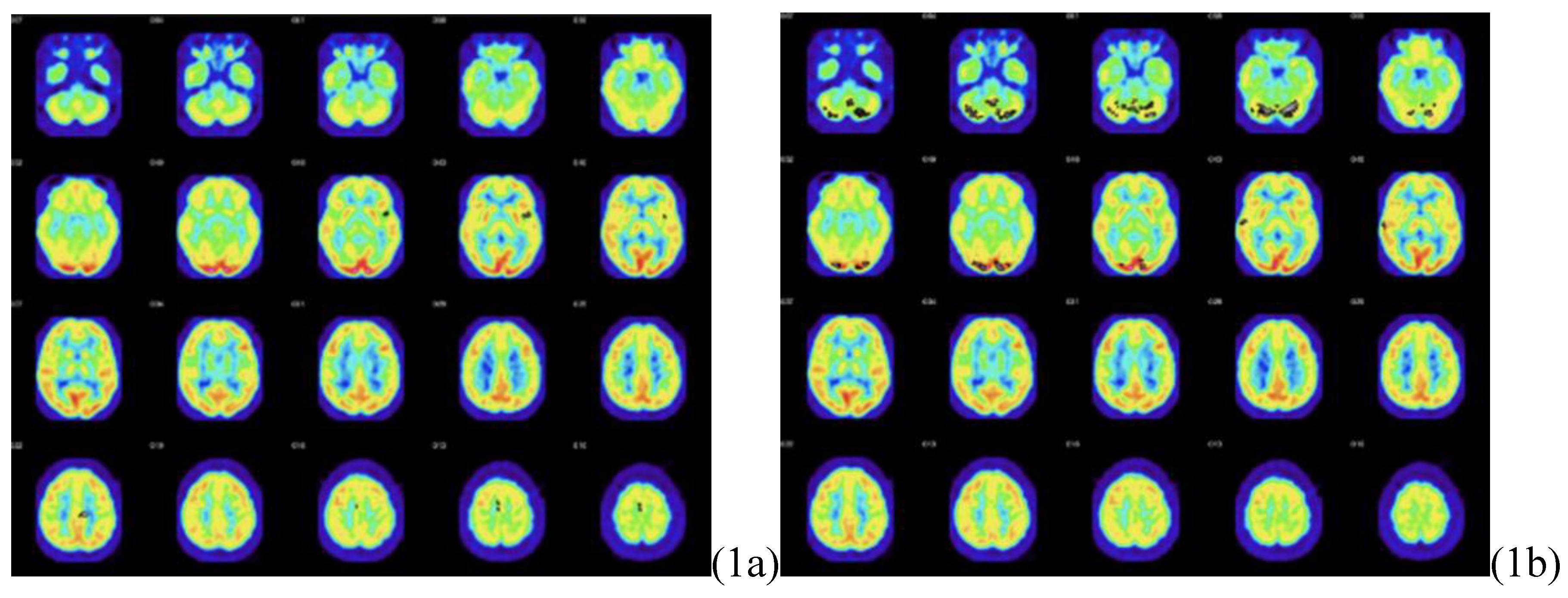
Figure 3.
Figure 3a is a transaxial DTI image showing significantly decreased fractional anisotropy in the patient’s anterior corpus callosum. Figure 3b is a transaxial DTI image showing significantly decreased fractional anisotropy in the patient’s left dorsal anterior cingulate region (Brodmann area 32). Images are displayed in anatomic orientation (image left = patient left).
Figure 3.
Figure 3a is a transaxial DTI image showing significantly decreased fractional anisotropy in the patient’s anterior corpus callosum. Figure 3b is a transaxial DTI image showing significantly decreased fractional anisotropy in the patient’s left dorsal anterior cingulate region (Brodmann area 32). Images are displayed in anatomic orientation (image left = patient left).
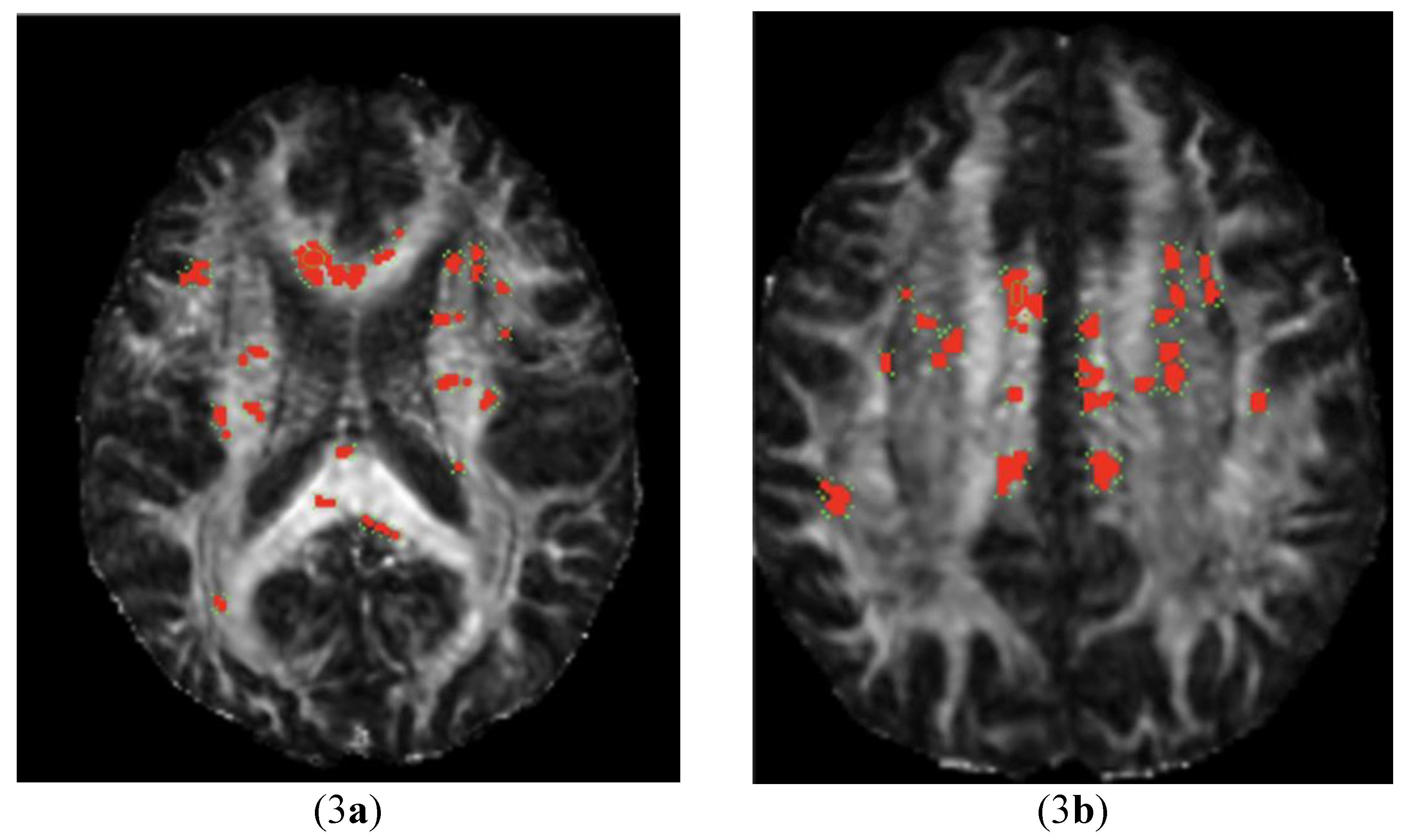
Figure 4.
Observable qualitative tractography difference, showing decreased fiber tract length in the left (4a) and right (4b) anterior and mid corpus callosum. Age and gender matched normal control corpus callosum tractography (4c, 4d) is presented for comparison.
Figure 4.
Observable qualitative tractography difference, showing decreased fiber tract length in the left (4a) and right (4b) anterior and mid corpus callosum. Age and gender matched normal control corpus callosum tractography (4c, 4d) is presented for comparison.
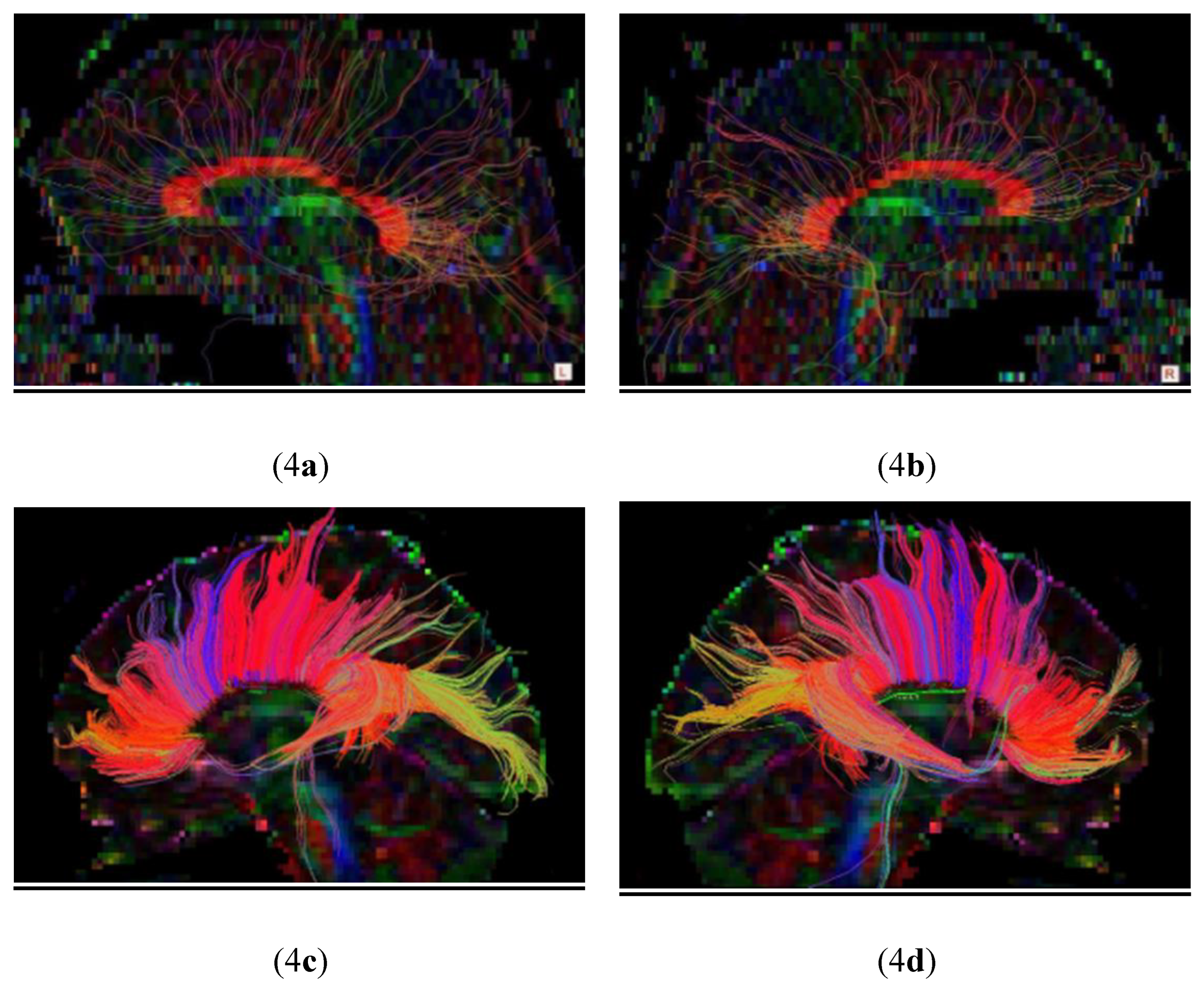
Table 3.
Fractional anisotropy values in the patient and healthy control group for the anterior corpus callosum and left cingulate region.
Table 3.
Fractional anisotropy values in the patient and healthy control group for the anterior corpus callosum and left cingulate region.
| Anterior Corpus Callosum (Figure 3a) | Lt Dorsal Ant Cingulate (Figure 3b) |
|
| Patient | 0.60 | 0.38 |
| Control Mean | 0.76 | 0.66 |
| Std. Deviation | 0.04 | 0.08 |
| Z-Score | -4.01 | -3.33 |
| P-Value | 6.1 × 10⁻⁵ | 8.6× 10⁻⁴ |
Table 4.
MRI quantitative volumetric (QV) sequence of relative volume as a percentage of Intracranial Volume (ICV) showing statistically significant differences in the patient’s right putamen and left minus right putamen.
Table 4.
MRI quantitative volumetric (QV) sequence of relative volume as a percentage of Intracranial Volume (ICV) showing statistically significant differences in the patient’s right putamen and left minus right putamen.
| Relative Volume (% ICV) | Patient | Mean | SD | Z-Score | P-Value |
| Left Putamen | 0.41 | 0.34 | 0.04 | 1.73 | 4.2 × 10⁻² |
| Right Putamen | 0.43 | 0.31 | 0.03 | 3.38 | 7.3 × 10⁻⁴ |
| Left-Right Putamen | -0.02 | 0.02 | 0.02 | -2.00 | 4.6 × 10⁻² |
Disclaimer/Publisher’s Note: The statements, opinions and data contained in all publications are solely those of the individual author(s) and contributor(s) and not of MDPI and/or the editor(s). MDPI and/or the editor(s) disclaim responsibility for any injury to people or property resulting from any ideas, methods, instructions or products referred to in the content. |
© 2026 by the authors. Licensee MDPI, Basel, Switzerland. This article is an open access article distributed under the terms and conditions of the Creative Commons Attribution (CC BY) license (http://creativecommons.org/licenses/by/4.0/).
Copyright: This open access article is published under a Creative Commons CC BY 4.0 license, which permit the free download, distribution, and reuse, provided that the author and preprint are cited in any reuse.