

Submitted:
28 July 2026
Posted:
29 July 2026
You are already at the latest version
Abstract
Background: Immunotherapy and targeted therapy have gained ground over conven-tional chemotherapy in treating various cancers. While pulmonary toxicity associated with these agents is rare, it represents a significant factor in both mortality and morbidity and may influence the overall success of cancer treatment. This case series report adds to the emerging evidence of cancer therapy-induced pneumonitis features and cortico-therapy outcomes. Patients and methods: This case series report consists of a retrospective analysis of 12 cases of patients undergoing immunotherapy (4 Nivolumab, 4 Pembrolizumab) or targeted therapy (3 Obinutuzumab, 1 Abemaciclib) for cancer (7 lung, 3 non-Hodgkin lymphoma, 1 breast, 1 renal) that developed pneumonitis during their follow-up. Results: The onset of pneumonitis ranged from 6 to 48 months (median = 18.5), and in 4 cases it occurred after discontinuation of oncologic treatment. The diagnosis was estab-lished with a high probability based only on clinical picture, radiologic features, and concomitant oncologic therapy. The cases with onset after discontinuation of oncologic treatment required a more thorough investigation (including bronchoscopy with BAL analysis). The main symptom was dyspnea (10/12 cases), and 3 of 12 patients had res-piratory failure (SpO2≤88%). The CTCAE severity grades were: 1 mild, 7 moderate, 3 severe, and 1 life-threatening. The CT scan showed different patterns (7 OP, 4 NSIP, and 1 HP). 11 patients received oral methylprednisolone (0.40 to 0.82 mg/kg) for 5 to 16 weeks. 2 patients continued oncologic treatment, and 6 discontinued. All patients showed fa-vorable outcomes. Conclusions: Immunotherapy and targeted therapy-induced pneumonitis can express various features and severities, and prompt recognition and diagnosis based on clinical, radiologic and contextual elements is mandatory. Corticotherapy regimens used, based on severity and treatment response, had a favorable outcome. The follow-up can consist only of clinical assessment and chest X-ray, and extensive tests are reserved for non-responsive cases.
Keywords:
immune-induced pneumonitis
; target agents-induced pneumonitis
; immune checkpoint inhibitor
; anti-CD20-targeted agents
; CDK 4/6 inhibitor
1. Introduction
In recent years, the treatment of certain types of cancer has been revolutionized by the approval of targeted therapies and immunotherapy. Although pulmonary toxicity is a rare adverse effect, the increasing use of these therapeutic agents has facilitated the recognition of such reactions.
For active immunotherapy involving immune checkpoint inhibitors, the incidence of therapy-induced pneumonitis ranges between 2.7% and 10%[1,2,3,4,5,6], with conflicting evidence that a higher incidence is observed in cases involving the combination of immunotherapeutic agents[7]. Concomitant chemotherapy seems to reduce the risk of immune-related pneumonitis[7]. In the case of molecular-targeted therapies, incidence rates vary depending on the specific agent. For instance, anti-CD20 agents such as Rituximab have been associated with a risk of inducing non-infectious pneumonitis ranging from 3.9% to 8.4%[8,9]. Conversely, the literature describes a single reported case of pneumonitis caused by the anti-CD20 agent Obinutuzumab[10]. Additionally, CDK 4/6 inhibitors rarely cause pneumonitis; however, the FDA has issued warnings regarding the potential severity of these adverse events[11].
The prompt recognition of pulmonary toxicity is crucial to preventing potentially irreversible or fatal consequences. Pneumonitis is one of the leading causes of immunotherapy discontinuation[3]. Suspicion usually starts with common symptoms (progressive exertional and even at-rest dyspnea, cough, fever) in a patient with cancer undergoing targeted oncologic therapy for several months. The median time to onset is 3 months, but there is a high variability (9 days to 19 months)[3], and pneumonitis can occur even several months after cessation of therapy[12]. Patients can present with mild hypoxemia (PaO2 more than 60 mmHg and SpO2 more than 88-90% but less than normal ~95%) or various degrees of respiratory failure (PaO2 less than 60 mmHg and SpO2 less than 88-90%). Clinical exam of the thorax can be normal, and lung crackles may be audible in rare cases. Postero-anterior chest X-rays can reveal a patchy interstitial pattern with mild opacity consistent mostly with ground-glass attenuation on CT scan, or more intense opacity consistent with lung consolidation. Most papers that present radiologic features of oncologic targeted therapy-induced pneumonitis discuss less simple chest X-ray images and focus mainly on CT scan patterns. Inflammatory markers can have increased values but are not currently used for diagnosis. These patterns can be localized or diffuse. These clinical (symptoms, signs), contextual (cancer following targeted therapy), and paraclinical elements (chest X-ray, inflammatory markers) can suffice for a high-probability diagnosis, but most clinicians follow a more prudent approach and perform other more invasive and costly medical investigations to ”confirm” the diagnosis.
HRCT scan is frequently used to characterize and localize more precisely the lung lesions as it has a higher sensitivity and specificity for interstitial lung diseases[13]; the most typical and frequent patterns are patchy ground-glass attenuation and consolidation zones distributed in both lungs which are quite similar with the aspects encountered in SARS-Cov2 pneumonia (diffuse alveolar damage/acute interstitial pneumonia/acute respiratory distress syndrome – DAD/AIP/ARDS) or organizing pneumonia (OP) [14,15]. Other patterns were also described: nonspecific interstitial pneumonia (NSIP), hypersensitivity pneumonia (HP), bronchiolitis (BL), radiation recall pneumonitis (RRP)[15], diffuse alveolar hemorrhage (DAH) or sarcoid-like reactions (SLR)[17]. Also, localized lung lesions can be more rarely encountered[10].
Bronchoscopy and lung biopsy can be performed to further characterize the existing lung injury by cell distribution patterns but often cannot distinguish other diagnoses such as various types of non-bacterial pneumonia, alveolar hemorrhage, lung congestion, non-cardiogenic pulmonary edema, sarcoidosis, lymphangitic carcinomatosis etc. The bronchoalveolar lavage is important to exclude an infectious cause (viral, bacterial or fungal). The bronchoalveolar lavage reveals lymphocyte-predominant cellularity most of the time [14]. Still, the existence of other patterns cannot exclude the oncologic targeted therapy lung injury and the presence of malignant cells can be a feature of the subsidiary oncologic disease. For these considerations, these furthermore complex medical tests cannot confirm the etiologic diagnosis. Lung biopsies can also be performed, but the high variability of histopathologic lung injury patterns generated by different drugs and even within a specific class of therapeutic agents[3,14] correlated with the risks, makes this approach less desirable in terms of risk/benefit ratio.
The management of these patients is guided by symptomatology, levels of severity or presence of respiratory failure and the extent of pulmonary involvement[15,16] and is guided by severity grades by CTCAE criteria. For asymptomatic patients, imaging surveillance alone is sufficient, without interrupting immunotherapy or targeted therapy. Depending on the severity of symptoms, discontinuation of immunotherapy or targeted therapy and the initiation of corticosteroid treatment should be considered in moderately to severe patients. If considering corticotherapy, currently there is no standard recommendation of treatment for this type of pneumonitis or of discontinuation of the incriminated therapy. Most recommendations are based on clinical experience, consensus opinion or observational studies or case series[16] and include corticotherapy regimens of various doses (0.5-1 mg/kg) for various lengths (minimum of 6 weeks and no maximum duration recommendation) and with no standard dose tapering rhythm (at least 4 weeks, based on the initial severity, clinical response and tendency to relapse with dose decrease)[18,19].
2. Materials and Methods
2.1. Selection and Description of Participants
This case series includes twelve cases of pneumonitis induced by immunotherapy or targeted therapy diagnosed and managed by the authors in the Marius Nasta Institute of Pulmonology in Bucharest, Romania, between January 2022 and June 2025. The medical data of these cases were collected in July 2025 based on the personal data collection consent form signed by the patients on admission to the hospital, which also covers research purposes. Besides this, verbal consent for the purpose of publishing anonymized medical data was obtained from the subjects who were alive at the moment of the decision to start this study. A formal request was sent to the Ethics Committee of Marius Nasta Institute of Pulmonology on 11.07.2025. The study was conceptualized by the main research team members by the end of July 2025. This research was approved by the local ethics committee of Marius Nasta Institute of Pulmonology (reference number 16362/19.08.2025) and was conducted in accordance with the Helsinki Declaration. The manuscript was reviewed by all team members.
The medical data used in this study are available and can be provided by the corresponding author upon request.
The study aims to add more information to the emerging evidence of diagnostic features and management outcomes of these cases.
2.2. Data Collection and Measurements
A retrospective search was performed to identify the clinical cases diagnosed and managed by the authors in the hospital’s electronic database using diagnostic criteria of pneumonitis induced by oncologic immune/targeted therapy. No specific exclusion criteria were used. Clinical, imagistic, biological, treatment, and outcome data were collected and anonymized. A formal approval for data collection was obtained from the Marius Nasta Institute in accordance with the internal and Ethics Committee regulations.
The main characteristics of the patients included in the case series are detailed in Table 1.
Each clinical case was studied in terms of history, clinical picture, basic tests (chest X-rays, blood tests), further tests (CT scans, bronchoscopy with BAL), treatment and outcome.
Imaging studies (chest X-rays and CT scans) were downloaded from the hospital’s database, anonymized, and further studied with the open-source cross-platform imaging software “Weasis – uptodate version.” A descriptive assessment of the initial CT scan was performed using the suggested features of CT patterns previously identified in the literature [15]. Based on the symptomatology, SaO2, and extent of lung involvement, the CTCAE (Common Terminology Criteria for Adverse Events) criteria were used to grade pneumonitis severity at the initial diagnosis (Table 2). A subjective, semi-quantitative comparison of lung involvement was performed for the imaging exams obtained for each patient at the time of diagnosis/start of treatment and after a time interval (during or at the end of treatment) to assess the outcome in imaging terms. Comparative assessments were made preferably between same-type images (chest X-rays or CT scans) based on availability. These features were added to the follow-up clinical assessment (improvement or worsening of symptoms) to establish the outcome (improved, stationary, worsened, death).
The essential collected data was centralized in tables of results and general observations and considerations were issued by the research team. No statistical analysis could be performed on this type of data.
3. Results
3.1. Onset, Symptoms and Severity
The average and median onset duration was 19.4 and 18.5 months, respectively, ranging from 6 to 48 months. One case had an exceptionally long time to onset of 48 months.
The most frequent symptom observed in 10 out of 12 patients was progressive dyspnea, while only two patients reported dry cough as the sole complaint. In addition to dyspnea, 4 patients also presented with cough. Fever was present in a single patient (case 8), and 2 patients reported loss of appetite (case 3) or chest pain (case 4). 3 patients presented with respiratory failure (SpO2≤88%) (Table 3).
Oxygen saturation at diagnosis was available for all 12 patients (median 97%, range 70–99%), and 3 patients (cases 8, 11 and 12) met the criterion for respiratory failure (SpO2 ≤ 88%). Lung function testing was feasible only in part of the series. DLCO was obtained in 8 of 12 patients (median 44.6%, range 17.4–68% of predicted) and was below 80% of predicted in all of them, with a mild reduction (60–79%) in 2 cases, a moderate reduction (40–59%) in 2 cases and a severe reduction (<40%) in 4 cases. FVC was obtained in 8 of 12 patients (median 66.3%, range 46.2–84.7% of predicted) and was below 80% of predicted in 7 of them, consistent with a predominantly restrictive ventilatory pattern.
The degree of functional impairment did not parallel the CTCAE severity grade. The lowest DLCO of the series (case 2, 17.4% of predicted) was recorded in a patient graded 2 whose oxygen saturation was normal (96%), and 3 of the 4 patients with a severe reduction in DLCO (<40%) were graded 2. Lung function tests could not be obtained in 3 patients (cases 6, 11 and 12), including the most severe case (case 11, grade 4) and one of the grade 3 patients (case 12); the reported functional values therefore reflect predominantly the lower severity grades (Table 3).
3.2. Radiologic and Bronchoscopy Features
In 4 of 12 cases, CT scan showed patchy opacities with mild intensity (ground-glass – GGO) predominantly at the periphery of both lung fields, and these were more consistent with the nonspecific interstitial pneumonia (NSIP) pattern. In 7 of 12 cases, the opacities were more intense, expressing features of lung consolidation, and in 2 of them the consolidations were peribronchovascular. These cases were more consistent with an organizing pneumonia (OP) radiologic pattern. Only one case showed diffuse bilateral centrilobular nodules more consistent with a hypersensitivity pneumonitis (HP) radiologic pattern. The CT patterns are centralized in Table 4.
In 3 cases CT scan was not performed at the control visit given the clinician's consideration that the information provided by the clinical picture of the patient and the chest X-ray is sufficient for assessing the improvement.
Only one case (case 11) was not evaluated with a chest CT scan at the moment of pneumonitis diagnosis, as the patient refused to be admitted to the hospital and perform a CT scan, although the severity was of grade 4 and the diagnosis relied only on the clinical data, the nivolumab treatment, and the chest X-ray that showed a patchy interstitial pattern with mild intensity opacities. The patient received corticotherapy and oxygen therapy at home and improved clinically and radiologically at 1 month and the CT scan performed at 1 month showed an improved NSIP pattern. A comparison was made between chest X-rays performed at the diagnosis and at 3-month intervals and showed complete remission of pneumonitis.
The three types of radiologic patterns (OP, NSIP and HP) are illustrated in Figure 1.
Bronchoalveolar lavage (BAL) was performed in 8 of the 12 included patients, revealing lymphocyte-predominant cellularity in all cases, with no detection of neoplastic cells. In all cases with late onset (cases 2,3,4 and 9), after months of oncologic treatment discontinuation, BAL was performed to exclude alternative diagnoses, but only in 4 of 8 cases of pneumonitis that occurred during oncologic treatment, BAL was performed. In the other 3 cases, BAL was not considered necessary based on the high probability of oncologic treatment-associated pneumonitis, and in case 11 the patient declined the procedure, which would in any case have carried an unacceptable risk of further respiratory deterioration given the severe respiratory failure.
3.3. Treatment and Outcome
Pneumonitis treatment consisted of oral methylprednisolone (Table 5) starting at moderate doses (ranging from 0.4 to 0.82 mg/kg with an average of 0.55 mg/kg) in 11 out of 12 patients, with gradual dose tapering throughout 5 to 16 weeks, depending on each patient's clinical and imaging evolution. The treatment regimens were highly variable and are illustrated in Figure 2. The decision to start with a dose close to the inferior or superior limit of the general recommended interval (equivalent to a prednisone dosage of 0.5 to 1 mg/kg) was made by each clinician based on severity and comorbidities and the following regimen was also adjusted by each clinician based on treatment response and clinical experience with different types of pneumonitis treatment with corticotherapy.
Immunotherapy or targeted oncological therapy was discontinued in 6 patients, while in 4 cases, discontinuation was not necessary as pneumonitis developed after completion of treatment. In 2 patients, the decision was made to continue immunotherapy (in case 1 without and in case 5 in parallel with corticotherapy).
It is noteworthy that the mild symptomatic patient in whom targeted therapy was not discontinued was monitored solely through imaging, without corticosteroid administration, and exhibited spontaneous resolution of the interstitial lung lesions (Case 1).
Symptomatic improvement was achieved in all 12 patients, with resolution of parenchymal lesions in 11 out of 12 cases. The imaging evolution of each individual patient is illustrated in the Appendix. In Case 7, the oncologic treatment was discontinued, and the patient had experienced improvement after 2 months of corticotherapy. Immunotherapy was reintroduced, in parallel with methylprednisolone administration, and this led to a worsening of pulmonary lesions, despite an improvement in clinical symptoms. Ultimately, the patient died of cancer complications.
4. Discussion
Reviewing these 12 cases, a series of discussions are raised.
The first aspect is related to diagnosis, which in our opinion should rely mostly on clinical, onco-therapeutic, and radiologic features and only in limited cases should necessitate more invasive techniques such as BAL or biopsy. Common Terminology Criteria for Adverse Events (CTCAE) are suited for initial severity assessment. For high-grade severity cases, the diagnosis criteria should be more permissive, as bronchoscopy with bronchoalveolar lavage could further increase the patient’s risk. Thus, clinical, radiological, and circumstantial data (history of oncologic immune or targeted therapy) could be sufficient for diagnosis.
The second aspect is related to the so-called CT radiologic patterns. Although they can be helpful in imagistic descriptions, some of them are purely descriptive without pathophysiological correspondence. This is the case, for example, of NSIP, which can lead to confusion with idiopathic or connective tissue diseases NSIP which has a different mechanism. Therefore, this pattern should be labeled differently, for example as acute interstitial pneumonia (AIP) as most of the cases have features similar with viral pneumonia and have closer mechanisms that center around T-cell activity.
The third discussion is related to follow-up. In this case series, 3 cases were managed successfully without the need for a CT scan at follow-up. Clinical evaluation and chest X-ray were sufficient to assess the outcome. Although a native CT scan is non-invasive, using only chest X-ray and not a mandatory CT scan could reduce the costs and discomfort of the patients in case of improvement. Cases with non-improvement or worsening necessitate an extensive follow-up including CT scan. Follow-up assessment time interval should be based on severity and based on this assessment (improved, stationary, worsened) the treatment regimen can be adjusted. Criteria for these follow-up temporary outcomes are yet to be established but it is also a matter of triviality. Mandatory Chest X-ray and SpO2 should be performed at follow-up alongside clinical assessment and in case of improvement no further tests are necessary.
The fourth discussion is related to treatment. Currently, there is no standard recommendation on dosages and duration of treatment. In this case series, using highly variable regimens of treatment, the patients had good outcomes, pointing out that following the current recommendations that are flexible based on severity and response to treatment is the proper conduct so far. Data suggest a severity-dose approach with a starting dose close to 1 mg/kg prednisone for high-grade severity cases and close to 0.5 mg/kg for low-moderate grade severity cases. Studies could be performed in the future using lower-dose and shorter duration regimens or more standardized starting doses and tapering schemes to optimize corticotherapy for the lowest necessary cumulative dose to decrease exposure to known side effects (Cushing syndrome, diabetes, osteoporosis).
The fifth discussion concerns whether to maintain or discontinue oncologic treatment. In this subject, there is some controversy, and the decision is a matter of consensus between the pneumologist and oncologist and in our opinion the decision should be based mostly on severity. In mild-to-moderate severity cases, it could be maintained, and for grades 3 and 4, stopping the treatment should be strongly considered.
5. Conclusions
Immunotherapy and targeted therapy have revolutionized cancer treatment, yielding more favorable outcomes for many patients compared to classic chemotherapy. Although pulmonary toxicity is not a frequent adverse effect, it represents a significant cause of morbidity and mortality in this patient population. Current literature reports a low incidence of immunotherapy- or targeted therapy-induced pneumonitis; however, the trend appears to be increasing due to the growing use of these agents—often in combination—as well as improved recognition of clinical symptoms and interstitial lung abnormalities.
Further complex investigations (bronchoscopy, CT scan) may not be needed for the purpose of obtaining a clearer diagnosis and may only increase the financial costs and medical stress for these patients. Also repeated CT scans for follow-up may not be needed and at least for clinically improving patient’s chest X-ray can suffice for confirmatory reason.
Timely identification of symptoms, radiological changes, assessment of severity and prompt start of treatment play a crucial role in the management and prognosis of affected patients. Current guidelines, largely based on expert consensus, recommend discontinuing oncologic treatment in all cases of therapy-induced pneumonitis. However, considering the risk-benefit ratio, our center’s experience suggests that oral corticosteroid therapy in low to moderate doses administered concurrently with immunotherapy or targeted therapy can yield favorable outcomes. Close and continuous monitoring of patients—through regular clinical, functional, and imaging assessments at intervals of one to three months—is essential.
Further studies are needed to develop clear therapeutic protocols, particularly those addressing the continuation, discontinuation, or reintroduction of immunotherapy following the resolution of the acute episode, as well as to identify specific risk factors that may predispose patients to pulmonary toxicity.
Author Contributions
Conceptualization, C.L.T., S.F.O. and D.C.Z.; methodology, C.L.T., S.F.O. and D.C.Z.; software, S.F.O. and D.C.Z.; validation, C.L.T., S.F.O., S.D.R., I.N.B., D.J.D., C.C., A.M.C., C.C.D. and D.C.Z; formal analysis, C.L.T., S.F.O. and D.C.Z.; investigation, C.L.T., S.F.O. and D.C.Z.; resources, C.L.T., S.F.O., S.D.R., I.N.B., D.J.D., C.C., A.M.C., C.C.D. and D.C.Z.; data curation, C.L.T., S.F.O. and D.C.Z; writing—original draft preparation, , C.L.T., S.F.O. and D.C.Z.; writing—review and editing, C.L.T., S.F.O., S.D.R., I.N.B., D.J.D., C.C., A.M.C., C.C.D. and D.C.Z ; visualization, C.L.T., S.F.O. and D.C.Z.; supervision, C.L.T., S.F.O. and D.C.Z.; project administration, C.L.T. S.F.O. and D.C.Z. All authors have read and agreed to the published version of the manuscript.
Funding
This research received no external funding.
Institutional Review Board Statement
The study was conducted in accordance with the Declaration of Helsinki and approved by the local ethics committee of Marius Nasta Institute of Pulmonology (reference number 16362/19.08.2025).
Informed Consent Statement
Informed consent was obtained from all subjects involved in the study through the institution’s general informed consent on admission to collect data for scientific and educational purposes.
Data Availability Statement
The original contributions presented in this study are included in the article. The individual patient data supporting the reported results are not publicly available due to privacy and ethical restrictions regarding patient confidentiality, but anonymized data can be made available on reasonable request from the corresponding authors.
Conflicts of Interest
The authors declare no conflicts of interest.
Acknowledgments
The authors would like to thank the medical and nursing staff of the Pneumology IV Department, as well as the radiology and bronchoscopy teams of the Marius Nasta Institute of Pulmonology, Bucharest, for their support in patient management and data collection.
Abbreviations
The following abbreviations are used in this manuscript:
| AIP | Acute interstitial pneumonia |
| ARDS | Acute respiratory distress syndrome |
| BAL | Bronchoalveolar lavage |
| BL | Bronchiolitis |
| CD20 | Cluster of differentiation 20 |
| CDK 4/6 | Cyclin-dependent kinase 4 and 6 |
| CT | Computed tomography |
| CTCAE | Common Terminology Criteria for Adverse Events |
| DAD | Diffuse alveolar damage |
| DAH | Diffuse alveolar hemorrhage |
| FDA | Food and Drug Administration |
| HP | Hypersensitivity pneumonitis |
| HRCT | High-resolution computed tomography |
| NSIP | Nonspecific interstitial pneumonia |
| OP | Organizing pneumonia |
| PaO2 | Arterial partial pressure of oxygen |
| RRP | Radiation recall pneumonitis |
| SARS-COV2 | Severe acute respiratory syndrome coronavirus 2 |
| SLR | Sarcoid-like reactions |
| SpO2 | Peripheral oxygen saturation of hemoglobin |
Appendix A
Appendix A.1
Figure A1.
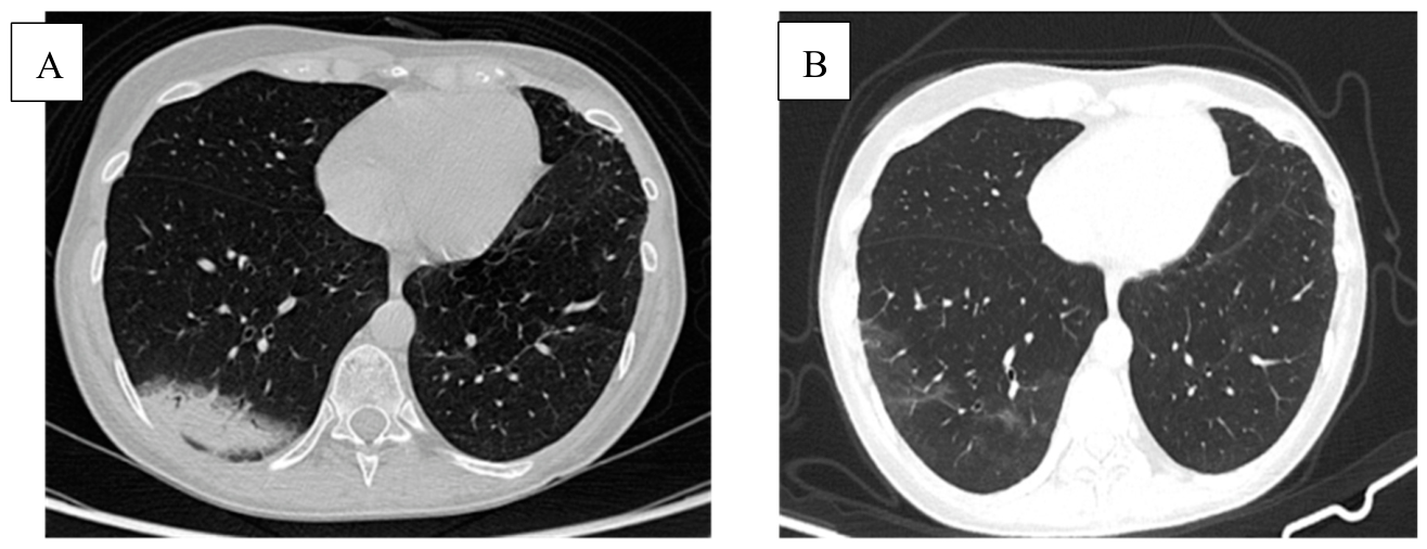
Case 1: patient with breast cancer treated with Abemaciclib presented with an organizing pneumonia (OP) pattern on chest CT (A). The lesions resolved after 3 months without corticosteroid therapy or interruption of the oncologic treatment (B).
Figure A1.
Case 1: patient with breast cancer treated with Abemaciclib presented with an organizing pneumonia (OP) pattern on chest CT (A). The lesions resolved after 3 months without corticosteroid therapy or interruption of the oncologic treatment (B).
Appendix A.2.
Figure A2.
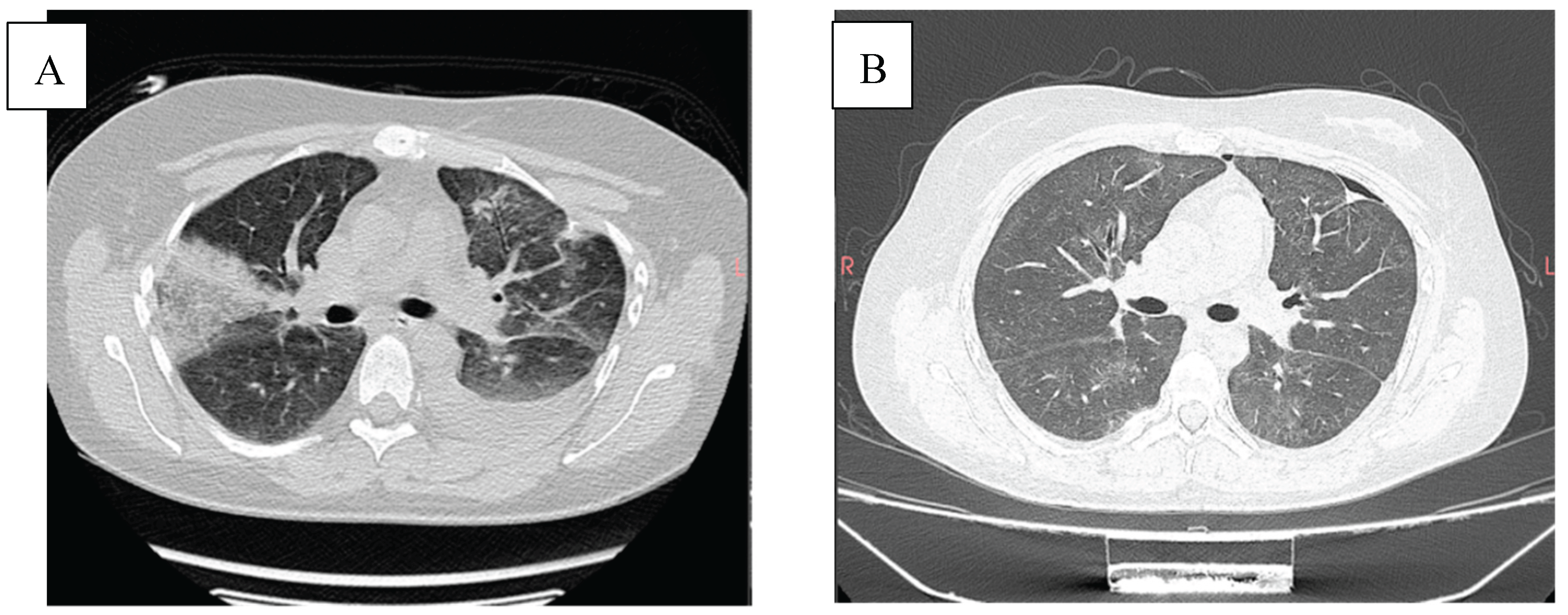
Case 2: A patient with non-Hodgkin lymphoma treated with Obinutuzumab, presenting with an organizing pneumonia (OP) pattern on CT and a left pleural effusion (A), with improvement of the pulmonary consolidations and resolution of the pleural effusion after 2 weeks of oral corticosteroid therapy (B).
Figure A2.
Case 2: A patient with non-Hodgkin lymphoma treated with Obinutuzumab, presenting with an organizing pneumonia (OP) pattern on CT and a left pleural effusion (A), with improvement of the pulmonary consolidations and resolution of the pleural effusion after 2 weeks of oral corticosteroid therapy (B).
Appendix A.3.
Figure A3.
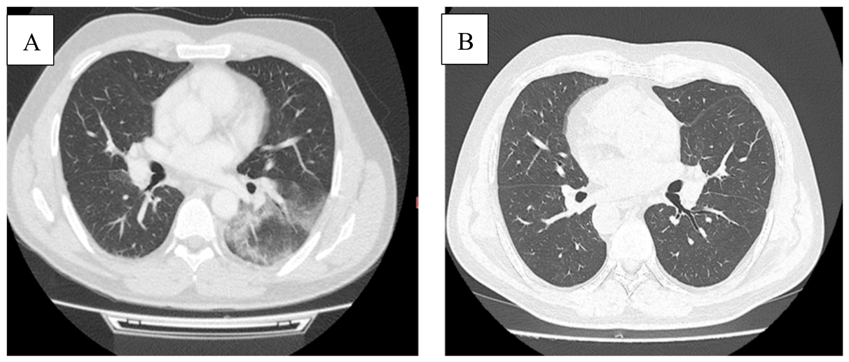
Case 3: patient with non-Hodgkin lymphoma treated with Obinutuzumab presented with an organizing pneumonia (OP) pattern on chest CT (A). The lesions resolved after 2 months with corticosteroid therapy (B).
Figure A3.
Case 3: patient with non-Hodgkin lymphoma treated with Obinutuzumab presented with an organizing pneumonia (OP) pattern on chest CT (A). The lesions resolved after 2 months with corticosteroid therapy (B).
Appendix A.4.
Figure A4.
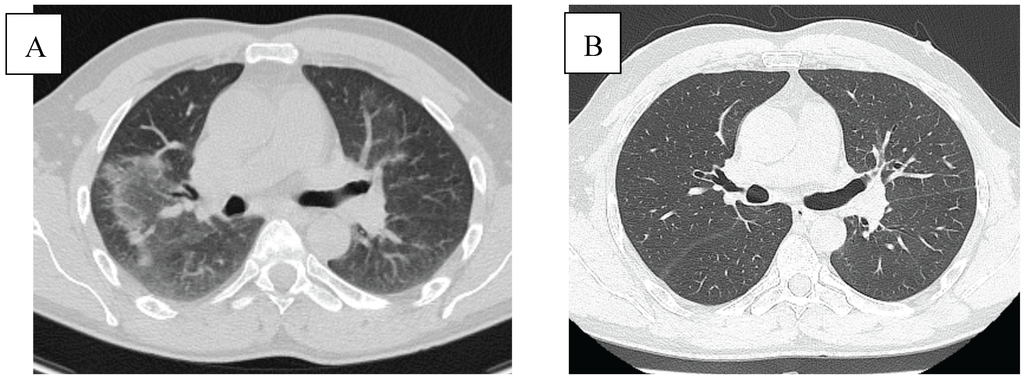
Case 4: patient with non-Hodgkin lymphoma treated with Obinutuzumab presented with NSIP pattern on chest CT (A). Complete resolution of ground glass opacities after 3 months of oral corticosteroid therapy (B).
Figure A4.
Case 4: patient with non-Hodgkin lymphoma treated with Obinutuzumab presented with NSIP pattern on chest CT (A). Complete resolution of ground glass opacities after 3 months of oral corticosteroid therapy (B).
Appendix A.5.
Figure A5.
Case 5: patient with lung cancer treated with Pembrolizumab presented with an organizing pneumonia (OP) pattern on chest CT (A). Improvement of the pulmonary perihilar lesions after 1 month with corticosteroid therapy and no interruption of the oncologic treatment (B).
Figure A5.
Case 5: patient with lung cancer treated with Pembrolizumab presented with an organizing pneumonia (OP) pattern on chest CT (A). Improvement of the pulmonary perihilar lesions after 1 month with corticosteroid therapy and no interruption of the oncologic treatment (B).
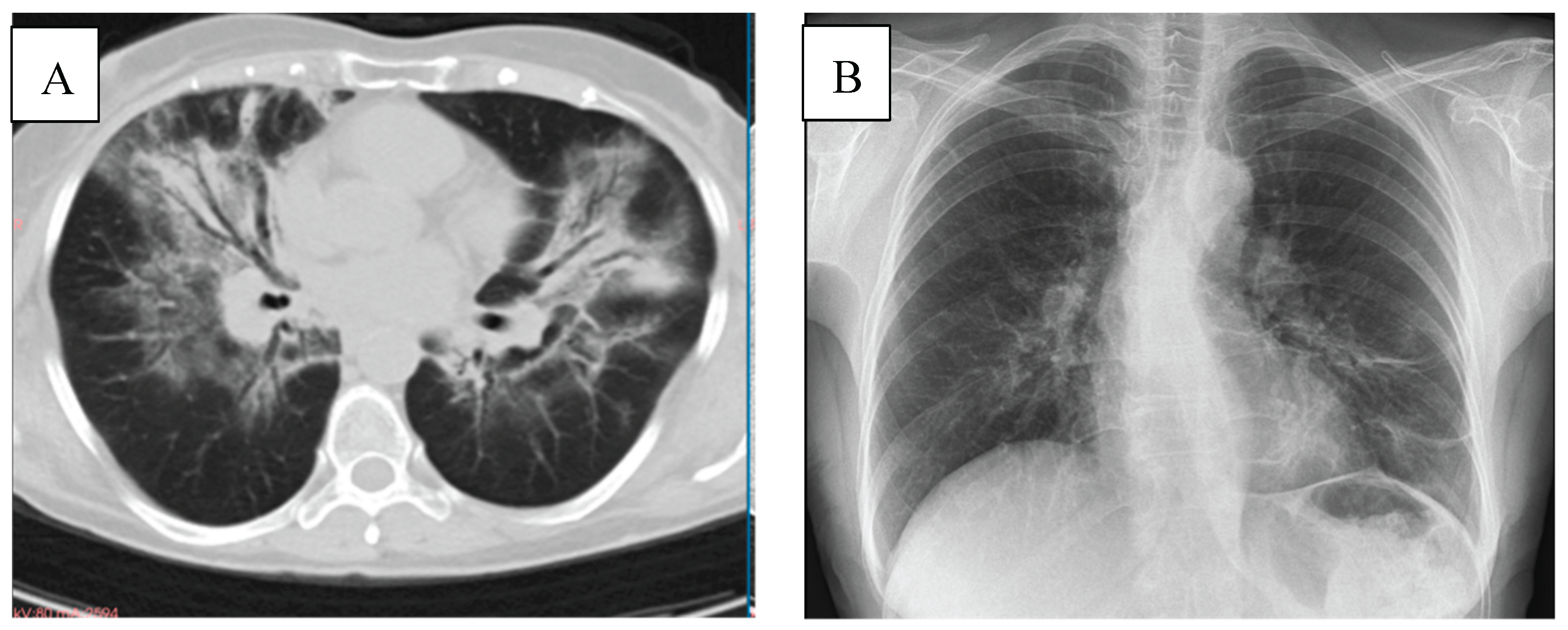
Appendix A.6.
Figure A6.
Case 6: patient with lung cancer treated with Pembrolizumab presented with an organizing pneumonia (OP) pattern on chest CT (A), with improvement of the lesions on the X-ray after one month of corticosteroid therapy and interruption of the oncologic treatment (B).
Figure A6.
Case 6: patient with lung cancer treated with Pembrolizumab presented with an organizing pneumonia (OP) pattern on chest CT (A), with improvement of the lesions on the X-ray after one month of corticosteroid therapy and interruption of the oncologic treatment (B).
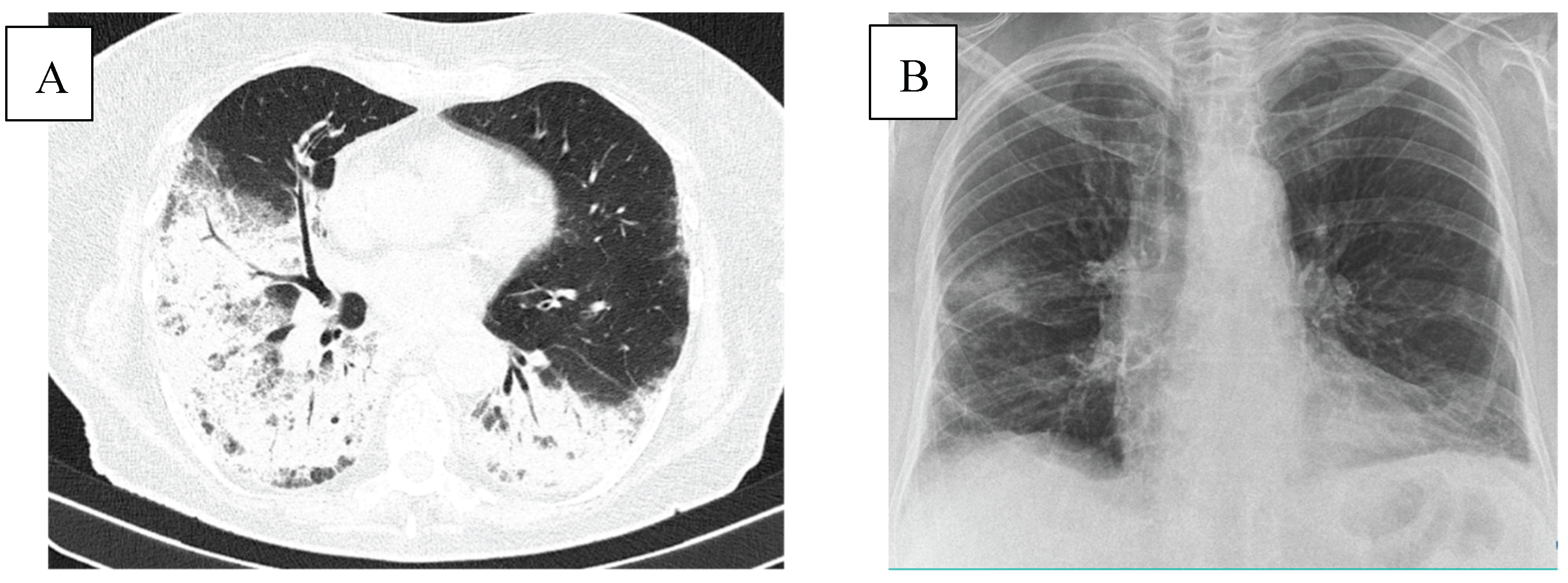
Appendix A.7.
Figure A7.
Case 7: patient with lung cancer treated with Pembrolizumab presented with an organizing pneumonia (OP) pattern on chest CT (A) with progression of the lesions after reintroduction of the immunotherapy in parallel with corticotherapy (B).
Figure A7.
Case 7: patient with lung cancer treated with Pembrolizumab presented with an organizing pneumonia (OP) pattern on chest CT (A) with progression of the lesions after reintroduction of the immunotherapy in parallel with corticotherapy (B).
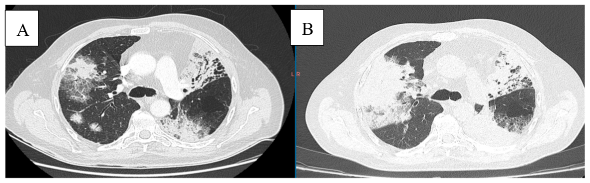
Appendix A.8.
Figure A8.
Case 8: patient with lung cancer treated with Pembrolizumab presented with bilateral reticular and alveolar opacities on chest X-Ray (A). Improvement of the lesions on the X-ray after one month of corticosteroid therapy and interruption of the oncologic treatment (B).
Figure A8.
Case 8: patient with lung cancer treated with Pembrolizumab presented with bilateral reticular and alveolar opacities on chest X-Ray (A). Improvement of the lesions on the X-ray after one month of corticosteroid therapy and interruption of the oncologic treatment (B).
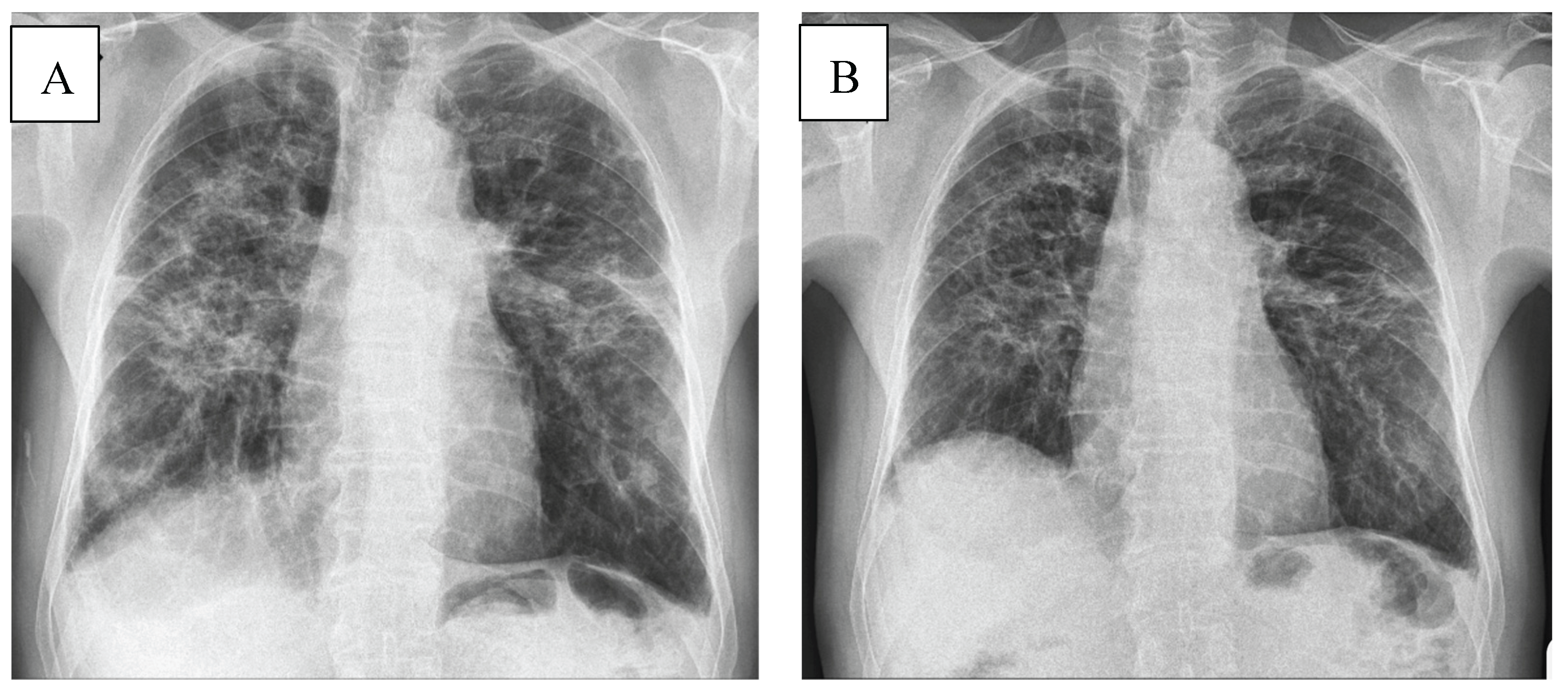
Appendix A.9.
Figure A9.
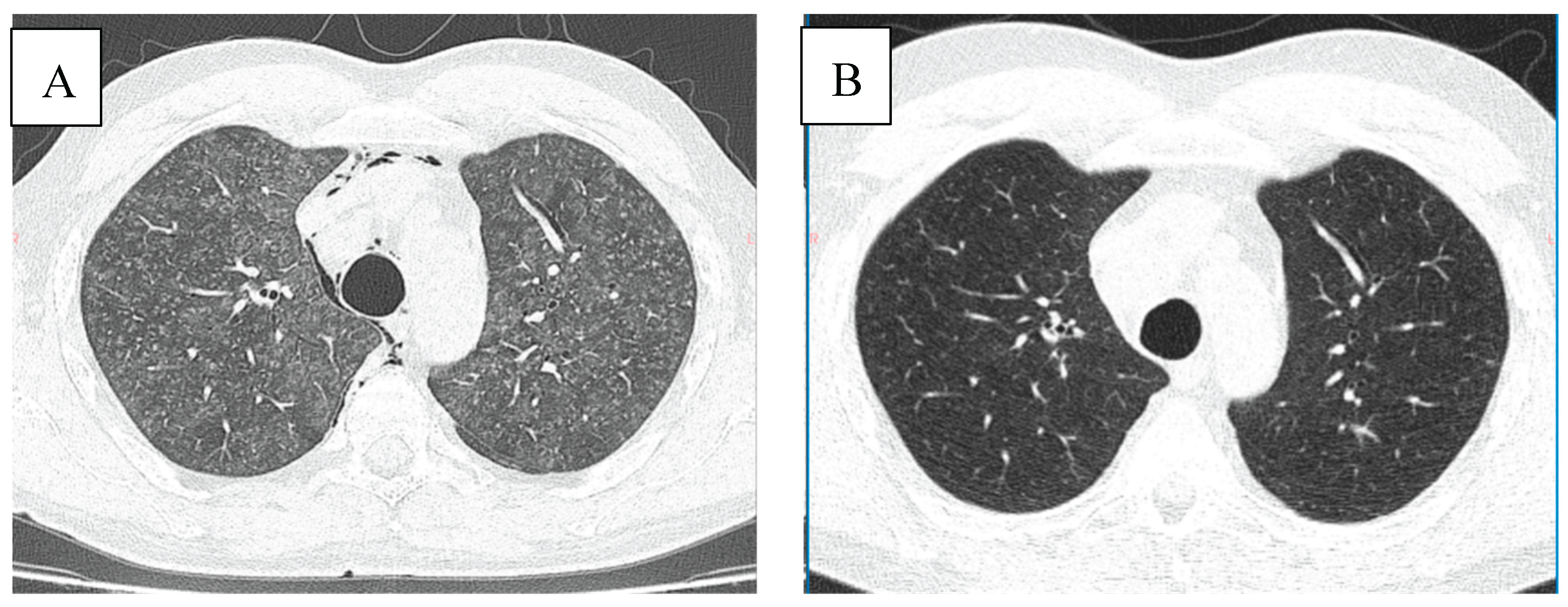
Case 9: patient with renal cancer treated with Ipilimumab/Nivolumab presented with an HP pattern on chest CT (A). The lesions resolved after one month of corticosteroid therapy (B).
Figure A9.
Case 9: patient with renal cancer treated with Ipilimumab/Nivolumab presented with an HP pattern on chest CT (A). The lesions resolved after one month of corticosteroid therapy (B).
Appendix A.10.
Figure A10.
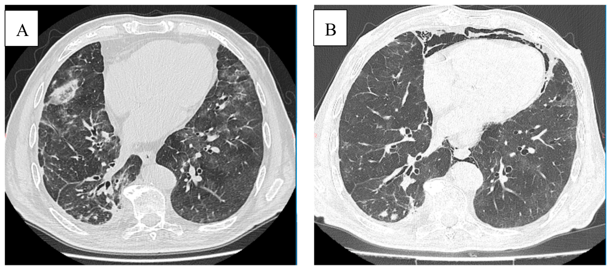
Case 10: patient with lung cancer treated with Nivolumab presented with an NSIP pattern on chest CT (A). Improvement of the lesions on CT after one month of corticosteroid therapy and interruption of the oncologic treatment (B).
Figure A10.
Case 10: patient with lung cancer treated with Nivolumab presented with an NSIP pattern on chest CT (A). Improvement of the lesions on CT after one month of corticosteroid therapy and interruption of the oncologic treatment (B).
Appendix A.11.
Figure A11.
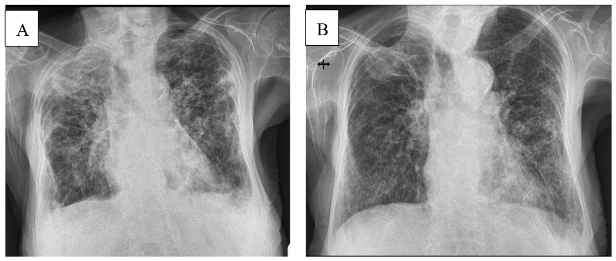
Case 11: patient with lung cancer treated with Nivolumab presented with bilateral reticular opacities on chest X-Ray (A). Improvement of the lesions on the X-ray after one month of corticosteroid therapy and interruption of the oncologic treatment (B).
Figure A11.
Case 11: patient with lung cancer treated with Nivolumab presented with bilateral reticular opacities on chest X-Ray (A). Improvement of the lesions on the X-ray after one month of corticosteroid therapy and interruption of the oncologic treatment (B).
Appendix A.12.
Figure A12.
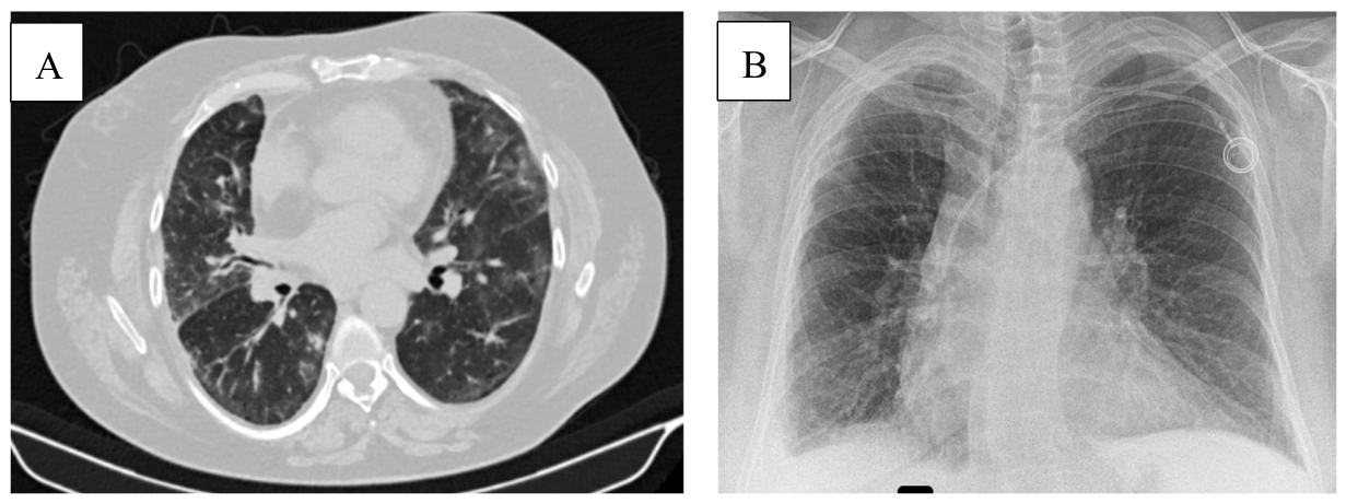
Case 12: patient with lung cancer treated with Nivolumab presented with NSIP pattern on chest CT (A). The X-Ray showed no new lesions, only small pleural effusion on the right side after one month of corticosteroid therapy and interruption of the oncological treatment (B).
Figure A12.
Case 12: patient with lung cancer treated with Nivolumab presented with NSIP pattern on chest CT (A). The X-Ray showed no new lesions, only small pleural effusion on the right side after one month of corticosteroid therapy and interruption of the oncological treatment (B).
References
- Zhang S, Liang F, Zhu J, Chen Q. Risk of pneumonitis associated with programmed cell death 1 inhibitors in cancer patients: A meta-analysis. Mol Cancer Ther. 2017 Aug 1;16(8):1588–95, PubMed PMID: 28446641. [CrossRef]
- Nishino M, Giobbie-Hurder A, Hatabu H, Ramaiya NH, Hodi FS. Incidence of programmed cell death 1 inhibitor-related pneumonitis in patients with advanced cancer a systematic review and meta-analysis. JAMA Oncol. 2016 Dec 1;2(12):1607–16, PubMed PMID: 27540850. [CrossRef]
- Naidoo J, Wang X, Woo KM, Iyriboz T, Halpenny D, Cunningham J, et al. Pneumonitis in patients treated with anti-programmed death-1/programmed death ligand 1 therapy. Journal of Clinical Oncology. 2017 Mar 1;35(7):709–17, PubMed PMID: 27646942. [CrossRef]
- Delaunay M, Cadranel J, Lusque A, Meyer N, Gounaut V, Moro-Sibilot D, et al. Immune-checkpoint inhibitors associated with interstitial lung disease in cancer patients. European Respiratory Journal. 2017 Aug 1;50(2), PubMed PMID: 28798088. [CrossRef]
- Tiu BC, Zubiri L, Iheke J, Pahalyants V, Theodosakis N, Ugwu-Dike P, et al. Real-world incidence and impact of pneumonitis in patients with lung cancer treated with immune checkpoint inhibitors: A multi-institutional cohort study. J Immunother Cancer. 2022 Jun 15;10(6), PubMed PMID: 35705313. [CrossRef]
- Khunger M, Rakshit S, Pasupuleti V, Hernandez A V., Mazzone P, Stevenson J, et al. Incidence of Pneumonitis With Use of Programmed Death 1 and Programmed Death-Ligand 1 Inhibitors in Non-Small Cell Lung Cancer: A Systematic Review and Meta-Analysis of Trials. Chest. 2017 Aug 1;152(2):271–81. PubMed PMID: 28499515. [CrossRef]
- Chen X, Zhang Z, Hou X, Zhang Y, Zhou T, Liu J, et al. Immune-related pneumonitis associated with immune checkpoint inhibitors in lung cancer: A network meta-analysis. J Immunother Cancer. 2020 Aug 30;8(2), PubMed PMID: 32863271. [CrossRef]
- Salmasi G, Li M, Sivabalasundaram V, Panzarella T, Tsang R, Kukreti V, et al. Incidence of pneumonitis in patients with non-Hodgkin lymphoma receiving chemoimmunotherapy with rituximab. Leuk Lymphoma. 2015 Jun 1;56(6):1659–64, PubMed PMID: 25356925. [CrossRef]
- Liu X, Hong XN, Gu YJ, Wang BY, Luo ZG, Cao J. Interstitial pneumonitis during rituximab-containing chemotherapy for non-Hodgkin lymphoma. Leuk Lymphoma. 2008;49(9):1778–83, PubMed PMID: 18798110. [CrossRef]
- Saunders S, Fryman C. An Original Occurrence of Organizing Pneumonia From Obinutuzumab. American Thoracic Society International Conference Meetings Abstracts American Thoracic Society International Conference Meetings Abstracts. 2024 May;A3854–A3854. [CrossRef]
- FDA warns about rare but severe lung inflammation with Ibrance, Kisqali, and Verzenio for breast cancer | FDA [Internet]. [cited 2025 Jun 30]. Available from: https://www.fda.gov/drugs/drug-safety-and-availability/fda-warns-about-rare-severe-lung-inflammation-ibrance-kisqali-and-verzenio-breast-cancer.
- Kimura H, Sone T, Araya T, Murata A, Yamamura K, Ohkura N, et al. Late-onset programmed cell death protein-1 inhibitor-induced pneumonitis after cessation of nivolumab or pembrolizumab in patients with advanced non-small cell lung cancer: A case series. Transl Lung Cancer Res. 2021 Mar 1;10(3):1576–81. [CrossRef]
- Grenier P, Valeyre D, Cluzel P, Brauner MW, Lenoir S, Chastang C. Chronic diffuse interstitial lung disease: Diagnostic value of chest radiography and high-resolution CT. Radiology. 1991;179(1):123–32, PubMed PMID: 2006262. [CrossRef]
- Shannon VR. Cancer treatment-related lung injury. Oncologic Critical Care. 2019 Oct 12;531–56. [CrossRef]
- Kalisz KR, Ramaiya NH, Laukamp KR, Gupta A. Immune checkpoint inhibitor therapy–related pneumonitis: Patterns and management. Radiographics. 2019 Nov 1;39(7):1923–37, PubMed PMID: 31584861. [CrossRef]
- Choi J, Anderson R, Blidner A, Cooksley T, Dougan M, Glezerman I, et al. Multinational Association of Supportive Care in Cancer (MASCC) 2020 clinical practice recommendations for the management of severe dermatological toxicities from checkpoint inhibitors. Supportive Care in Cancer. 2020 Dec 1;28(12):6119–28, PubMed PMID: 32856211. [CrossRef]
- Berthod G, Lazor R, Letovanec I, Romano E, Noirez L, Stalder JM, et al. Pulmonary sarcoid-like granulomatosis induced by ipilimumab. Journal of Clinical Oncology. 2012 Jun 10;30(17), PubMed PMID: 22547608. [CrossRef]
- Puzanov I, Diab A, Abdallah K, Bingham CO, Brogdon C, Dadu R, et al. Managing toxicities associated with immune checkpoint inhibitors: Consensus recommendations from the Society for Immunotherapy of Cancer (SITC) Toxicity Management Working Group. J Immunother Cancer. 2017 Nov 21;5(1), PubMed PMID: 29162153. [CrossRef]
- Rapoport BL, Cooksley T, Johnson DB, Anderson R. Supportive care for new cancer therapies. Curr Opin Oncol. 2021 Jul 1;33(4):287–94. PubMed PMID: 33756517. [CrossRef]
Figure 1.
Radiologic patterns of oncologic treatment-induced pneumonitis; A1: case 10 treated with Nivolumab– nonspecific interstitial pneumonia (NSIP) pattern; A2: case 10 after 1 months of corticotherapy, with pneumomediastinum; B1: Case 6 treated with Pembrolizumab – organizing pneumonia (OP) pattern; B2: case 6 after 1 month of corticotherapy; C1: case 9 treated with Ipilimumab/Nivolumab – hypersensitivity pneumonitis (HP) pattern; C2: case 9 after 1 month of corticotherapy. (see Table 5 and Figure 2 for corticotherapy regimens).
Figure 1.
Radiologic patterns of oncologic treatment-induced pneumonitis; A1: case 10 treated with Nivolumab– nonspecific interstitial pneumonia (NSIP) pattern; A2: case 10 after 1 months of corticotherapy, with pneumomediastinum; B1: Case 6 treated with Pembrolizumab – organizing pneumonia (OP) pattern; B2: case 6 after 1 month of corticotherapy; C1: case 9 treated with Ipilimumab/Nivolumab – hypersensitivity pneumonitis (HP) pattern; C2: case 9 after 1 month of corticotherapy. (see Table 5 and Figure 2 for corticotherapy regimens).
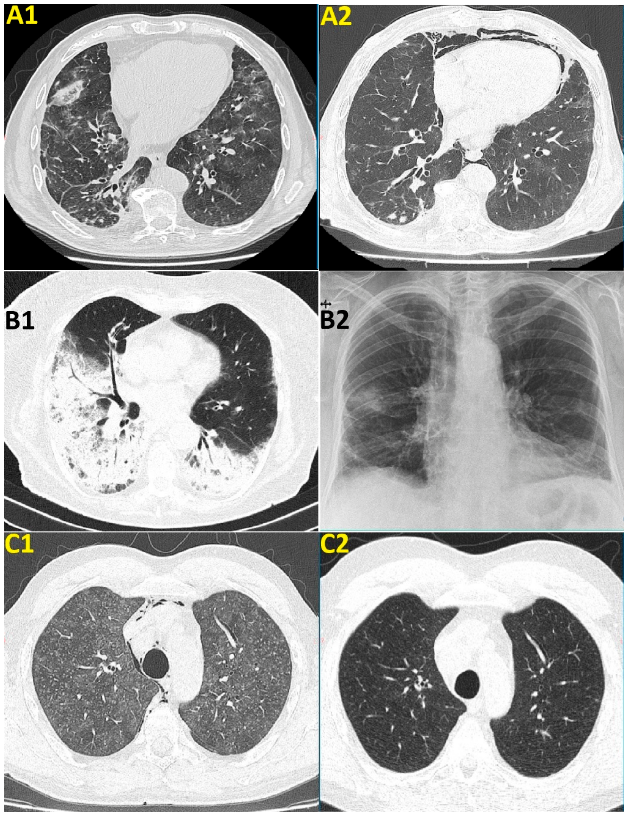
Figure 2.
3D Graphic illustration of methylprednisolone regimens; each strip represents one case labeled on the Y axis; The numbers on the strips represent the daily dose of methylprednisolone (labeled on the Z axis) received during each week from the regimen, and the number of weeks is labeled on the X axis;.
Figure 2.
3D Graphic illustration of methylprednisolone regimens; each strip represents one case labeled on the Y axis; The numbers on the strips represent the daily dose of methylprednisolone (labeled on the Z axis) received during each week from the regimen, and the number of weeks is labeled on the X axis;.
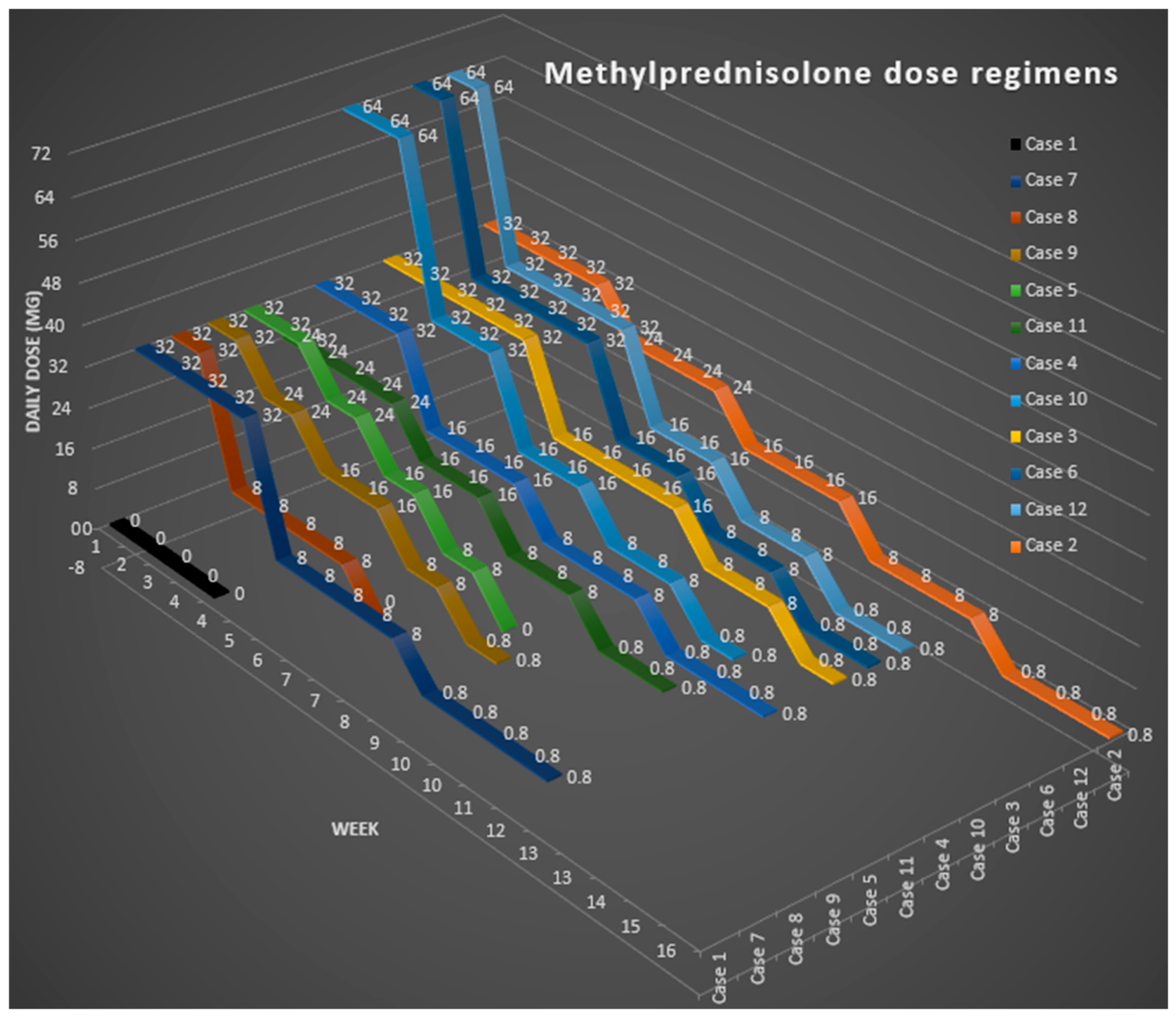

Table 1.
Patient characteristics.
| No | Age | S | Cancer | Therapy |
|---|---|---|---|---|
| 1 | 50 | F | Breast | Abemaciclib |
| 2 | 34 | F | NHL | Obinutuzumab |
| 3 | 47 | M | NHL | Obinutuzumab |
| 4 | 50 | M | NHL | Obinutuzumab |
| 5 | 74 | F | Lung | Pembrolizumab |
| 6 | 69 | F | Lung | Pembrolizumab |
| 7 | 67 | M | Lung | Pembrolizumab |
| 8 | 67 | M | Lung | Pembrolizumab |
| 9 | 52 | M | Renal | Ipilimumab, Nivolumab |
| 10 | 82 | M | Lung | Nivolumab |
| 11 | 79 | F | Lung | Nivolumab |
| 12 | 62 | F | Lung | Nivolumab |
Age = age in years; S = sex (M=male; F=female); NHL = non-Hodgkin lymphoma.
Table 2.
CTCAE criteria for grading the severity of the pneumonitis.
| Grade | Symptoms | Management | |
|---|---|---|---|
| 1 | Mild | Asymptomatic or mild symptoms | Clinical or diagnostic observations only; intervention not indicated. |
| 2 | Moderate | Limiting age-appropriate instrumental ADL. | Minimal, local, or noninvasive intervention indicated. |
| 3 | Severe | Severe or medically significant, but not immediately life-threatening; disabling; limiting self-care ADL. |
Hospitalization or prolongation of hospitalization indicated. |
| 4 | Life-threatening | Life-threatening consequences | Urgent intervention indicated. |
| 5 | Death | Death related to the adverse even. |
|
ADL = Activities of Daily Living.
Table 3.
Onset time in months from start of oncologic treatment, symptoms, severity and lung function tests data.
Table 3.
Onset time in months from start of oncologic treatment, symptoms, severity and lung function tests data.
| No | Onset (Months) | Symptoms | Severity Grade | SpO2 (%) | FVC (%) |
DLCO(%) | |||
|---|---|---|---|---|---|---|---|---|---|
| Dyspnea | Cough | Fever | Others | ||||||
| 1 | 19 | No | Yes | No | No | 1 | 99 | 77 | 54 |
| 2 | 24 | Yes | No | No | No | 2 | 96 | 59,4 | 17,4 |
| 3 | 24 | Yes | Yes | No | LA | 2 | 99 | NA* | 68 |
| 4 | 48 | No | Yes | No | CP | 2 | 98 | 61 | 65 |
| 5 | 12 | Yes | No | No | No | 2 | 95 | 75,8 | 50,5 |
| 6 | 18 | Yes | Yes | No | No | 2 | 98 | NP | NP |
| 7 | 16 | Yes | No | No | No | 2 | 99 | 64,3 | 38,6 |
| 8 | 10 | Yes | No | Yes | No | 3 | 88 | 84,7 | NP |
| 9 | 24 | Yes | No | No | No | 3 | 92 | 68,2 | 21,9 |
| 10 | 24 | Yes | No | No | No | 2 | 98 | 46,2 | 29,6 |
| 11 | 8 | Yes | Yes | No | No | 4 | 70 | NP | NP |
| 12 | 6 | Yes | Yes | No | No | 3 | 88 | NP | NP |
LA = loss of appetite; Cp = chest pain; SpO2 = oxygen saturation of hemoglobin; DLCO = Lung diffusion of carbon monoxide in percentage of predicted value; FVC = forced vital capacity in percentage of predicted value; NA = not available; NP = not performed. * normal values.
Table 4.
Radiographic findings.
| Case | Radiographic pattern | CT findings | BAL |
|---|---|---|---|
| 1 | OP | Bilateral subpleural consolidations | Yes |
| 2 | OP | Bilateral peripheral consolidations, left pleural effusion | Yes |
| 3 | OP | Bilateral multifocal and migratory GGOs | Yes |
| 4 | NSIP | Bilateral GGOs | Yes |
| 5 | OP | Bilateral peribronchovascular consolidations | Yes |
| 6 | OP | Bilateral multifocal consolidations and GGOs | No |
| 7 | OP | Bilateral large lung consolidations | Yes |
| 8 | OP | Bilateral peribronchovascular consolidations | Yes |
| 9 | HP | Diffuse bilateral centrilobular ground-glass nodules, mosaic attenuation caused by the air trapping; pneumomediastinum | Yes |
| 10 | NSIP | Bilateral predominantly peripheral reticular opacities and GGOs; pneumomediastinum | No |
| 11 | NSIP | Bilateral GGOs | No |
| 12 | NSIP | Bilateral predominantly peripheral reticular opacities and GGOs | No |
OP = Organizing pneumonia; HP = Hypersensitivity pneumonitis; NSIP = Nonspecific interstitial pneumonitis; GGOs = Ground glass opacities; BAL = bronchoalveolar lavage made/not made.
Table 1.
Treatment, decision to stop oncologic treatment and outcome.
| No | Weight | Dose | Oral CS |
Weeks of methylprednisolone dose (mg/day) | Hold | Outcome | ||||||
|---|---|---|---|---|---|---|---|---|---|---|---|---|
| kg | mg/kg | (weeks) | 64 | 32 | 24 | 16 | 8 | 8/0 | ||||
| 1 | 64 | N/A | 0 | 0 | 0 | 0 | 0 | 0 | 0 | No++ | Improved | |
| 2 | 70 | 0.46 | 16 | 0 | 4 | 3 | 3 | 3 | 3 | No need | Improved | |
| 3 | 78 | 0.41 | 12 | 0 | 5 | 0 | 4 | 2 | 1 | No need | Improved | |
| 4 | 70 | 0.46 | 12 | 0 | 3 | 0 | 3 | 3 | 3 | No need | Improved | |
| 5 | 65 | 0.49 | 5 | 0 | 2 | 1 | 1 | 1 | 0 | No++ | Improved | |
| 6 | 78 | 0.82 | 11 | 1 | 4 | 0 | 2 | 2 | 2 | Yes | Improved | |
| 7 | 79 | 0.41 | 12 | 0 | 4 | 0 | 0 | 4 | 4 | Yes+ | Death* | |
| 8 | 80 | 0.40 | 5 | 0 | 1 | 0 | 0 | 4 | 0 | Yes | Improved | |
| 9 | 75 | 0.43 | 6 | 0 | 1 | 1 | 2 | 1 | 1 | No need | Improved | |
| 10 | 82 | 0.78 | 9 | 2 | 2 | 0 | 2 | 2 | 1 | Yes | Improved | |
| 11 | 42 | 0.57 | 12 | 0 | 0 | 4 | 2 | 2 | 2 | Yes | Improved | |
| 12 | 78 | 0.82 | 11 | 1 | 4 | 0 | 2 | 2 | 2 | Yes | Improved | |
CS = corticosteroid (duration in weeks); Hold = hold oncologic targeted treatment. +Initially stopped and after improvement, received immunotherapy and oral corticosteroid simultaneously. ++ Ongoing treatment. *Initial improvement; death by cancer complications.
Disclaimer/Publisher’s Note: The statements, opinions and data contained in all publications are solely those of the individual author(s) and contributor(s) and not of MDPI and/or the editor(s). MDPI and/or the editor(s) disclaim responsibility for any injury to people or property resulting from any ideas, methods, instructions or products referred to in the content. |
© 2026 by the authors. Licensee MDPI, Basel, Switzerland. This article is an open access article distributed under the terms and conditions of the Creative Commons Attribution (CC BY) license (http://creativecommons.org/licenses/by/4.0/).
Copyright: This open access article is published under a Creative Commons CC BY 4.0 license, which permit the free download, distribution, and reuse, provided that the author and preprint are cited in any reuse.


