

Submitted:
28 July 2026
Posted:
28 July 2026
You are already at the latest version
Abstract
Surface electromyography (sEMG) signals acquired with low-cost sensors tend to exhibit variable degradation that can compromise the reliability of myoelectric control systems outside controlled conditions. This work presents an adaptive processing pipeline for sEMG signals composed of three chained stages: a motor-intent classifier based on dilated temporal convolutional networks, whose function is to determine whether a signal window contains muscle activity associated with a voluntary gesture; a dual-output quality assessor that estimates a continuous score and a binary acceptability label, aimed at deciding whether the signal can be used directly or requires intervention; and a convolutional autoencoder that recovers the morphology of degraded windows before they are used in prosthetic control. The decision policy for reconstruction operates on two independent thresholds and classifies each window into one of three states: signal discard, direct acceptance, or active restoration. The models within the proposed architecture are trained on the public NinaPro DB1, DB3, and DB10 datasets and validated without recalibration on signals recorded from two participants with transtibial amputation over four weekly sessions. The results show that the pipeline correctly handles signal profiles with opposing characteristics. Inference latency remained below 5 ms at the 50th percentile across all scenarios, consistent with real-time operation. An integrated prototype using an Arduino UNO R4 WiFi was used to evaluate the results, and validate the feasibility of the system on physical hardware.
Keywords:
surface electromyography (sEMG)
; myoelectric prosthesis control
; intent detection
; signal quality assessment
; convolutional autoencoder
; temporal convolutional network
; adaptive signal restoration
; NinaPro dataset
1. Introduction
Surface electromyography (sEMG) signals constitute one of the information sources used for non-invasive myoelectric control in upper-limb prostheses, reflecting the muscle activation associated with the user’s motor intent. However, their practical application still faces challenges related to inter-subject variability, electrode shift, the presence of noise, muscle fatigue, and performance degradation outside controlled laboratory conditions. These factors explain why, despite advances in pattern recognition, transfer to clinical and everyday-use settings remains limited [1,2].
sEMG signals captured with low-cost sensors such as the MyoWare, a commercial single-channel device widely used in gestural control and prosthetic applications, frequently exhibit three types of degradation. Motion artifacts are electrical signals generated by the displacement of the electrode over the skin that mix with, and distort, the actual muscle activity. Power-line electromagnetic noise is a 60 Hz interference introduced from the user’s electrical environment. Baseline drift is a slow shift of the signal that causes its reference level to rise or fall gradually during recording, without corresponding to any real muscle contraction, together with saturation effects [3].
In prosthetic rehabilitation contexts, sEMG signal degradation can produce erroneous recordings and false interpretations of motor intent, phenomena that interrupt the tactile-visual feedback loop and generate high levels of user frustration, accelerating abandonment of the assistive device [4]. Faced with this problem, most existing solutions in the specialized literature address noise removal or pattern classification in a strictly independent and sequential manner [5]. This operational isolation ignores the fact that fixed-frequency filters can alter the morphology and discriminant spectral features of the useful signal, indirectly compromising the performance of the downstream classifier. The present work proposes unifying both tasks into a pipeline – a sequence of ordered data-processing elements in which the output of one element is the input of the next – with adaptive decision logic: Restoration is activated only when movement intent is present and a quality threshold is not met, thereby avoiding unnecessary processing of resting windows.
To compare myoelectric control algorithms, evaluations are commonly carried out on the same data, such as NinaPro, a public database built by Atzori and colleagues that gathers sEMG recordings from healthy individuals and individuals with transradial amputation, acquired using different protocols and devices [6,7]. This makes NinaPro a reference not only for the sample it offers, but also for its coverage of a diversity of users, movements, and acquisition conditions. The use of this dataset allows the results of this work to be directly comparable with those of other teams that have worked on the same data.
Building on this limitation, the present work proposes a cascaded processing pipeline composed of three stages: intent detection, quality assessment, and signal restoration. The process begins with the segmentation of the sEMG signal into temporal windows, which are analyzed to determine whether they contain muscle activity associated with motor intent. Windows that meet this criterion are subsequently evaluated in terms of quality, understood as the signal’s capacity to preserve relevant muscle information with a noise level that does not compromise the interpretation of muscle intent. When quality is considered sufficient, the signal can be used directly in downstream recognition or control tasks. Otherwise, the window is sent to a restoration stage based on an autoencoder, whose objective is to recover useful features before final processing. In this way, the proposed approach incorporates validation and recovery mechanisms aimed at bringing the system closer to real-time operating scenarios for prosthetic devices.
2. Related Work
The development of myoelectric control systems for upper-limb prostheses has evolved from classical linear analysis toward pattern recognition based on deep learning [8,9]. Despite the high accuracy rates reported under controlled conditions, transfer to everyday clinical use remains limited, owing to the susceptibility of surface electromyography (sEMG) signals to multiple sources of degradation [6,8].
2.1. EMG Signals
Surface electromyography (sEMG) is a non-invasive technique that records the electrical activity generated by skeletal muscle fibers during voluntary contraction. From a physiological standpoint, when the central nervous system issues a motor command, the recruited motor units produce action potentials that propagate along the muscle fiber; the temporal and spatial superposition of these potentials constitutes the signal that surface electrodes capture on the skin. The resulting signal is a complex superposition of multiple electrical sources whose amplitude and frequency composition depend on several factors that interact simultaneously: how deep and how large the activated motor units are, how the subcutaneous tissue between the muscle and the electrode is organized, and the firing rate of those units [8]. Small variations in any of these elements affect what the sensor ultimately captures and records.
In the residual limb of a person with an amputation, sEMG is practically the only non-invasive channel available to infer which movement the user intends to perform. Beyond reflecting contraction intensity, what makes sEMG valuable is that it preserves temporal and spectral patterns distinctive enough for trained classifiers to differentiate between gestures from windows only tens of milliseconds long [10]. Difficulties arise when the signal arrives degraded: as described above, low-cost sensors used outside the laboratory introduce different types of degradation that reduce the signal-to-noise ratio in a variable and unpredictable way [11], directly compromising the classifier’s ability to correctly interpret the user’s intent.
From a spectral standpoint, the muscle energy considered useful is distributed between 20 and 500 Hz. In superficial forearm muscles, the median frequency typically lies between 50 and 100 Hz under normal contraction conditions [8].
Muscle fatigue plays an important role, as it progressively shifts this median frequency toward lower values – a phenomenon that occurs during prolonged rehabilitation sessions, when the user trains residual-limb activation. Likewise, inter-subject variability in anatomy, adipose tissue thickness, and electrode placement introduces differences in amplitude and morphology that limit the transferability of models trained on one subject to another, a problem known as cross-subject generalization degradation [12]. These factors justify the need for adaptive processing systems that assess the quality of each signal segment before using it for control decisions.
2.2. Robustness in Bioelectric Control and Inter-Subject Variability
Intuitive prosthetic control depends on the system’s ability to generalize across different users and chronological sessions [9,13]. Scheme and Englehart [12] identified the main sources of this problem as anatomical differences between individuals, variations in adipose tissue thickness, and slight electrode shifts between sessions. Any one of these factors is enough to cause a classifier trained with fixed thresholds to lose performance drastically when the user does not recalibrate the system – something that happens frequently in clinical practice.
This phenomenon is evident when working with the NinaPro databases (DB1, DB3, and DB10), which include signals from both non-amputee individuals and individuals with transradial amputation, exposing the morphological asymmetry of action potentials in residual muscles and the performance degradation that occurs outside the laboratory [6,13]. Krasoulis et al. [13] used the DB10 database to demonstrate the impact of long-term user adaptation: while a neuromuscular learning process can stabilize the signal over several weeks, static models struggle against the stochastic, time-varying nature that characterizes sEMG.
2.3. Temporal Window Size Selection and Its Impact on Latency
For fluid human-machine interaction, the delay introduced by digital signal processing must remain imperceptible to the user [14,15]. Since the foundational work of Hudgins et al. [10], it has been established that the temporal segmentation window should range between 100 and 200 ms to extract representative features of muscle contraction without breaking the real-time control loop. This is where convolutional networks make their contribution: Kiranyaz et al. [16] documented the advantages of one-dimensional convolutional neural networks for operating directly on raw signals.
Smith et al. [15] approached this problem with a formal mathematical framework, evaluating how window length, classification error, and controller delay interact with one another. Their results show that 200 ms represents a reasonable trade-off point, sufficient to capture the relevant spectral content of sEMG (which in superficial muscles is distributed between 20 and 500 Hz) without compromising responsiveness on platforms with limited computational resources. Regarding overlap between contiguous windows, values close to 50% have been shown to reduce the loss of transient events at segment boundaries, which is particularly useful for signals with brief or unpredictable activations [10].
2.4. Signal Modeling and Restoration Using Convolutional Networks
Recurrent architectures such as LSTMs or RNNs carry inherent workflow complexities: their sequential nature hinders parallelization and accumulates latency, making them impractical for real-time scenarios [16]. Convolutional networks currently dominate this space. Kiranyaz et al. [16] documented the advantages of one-dimensional convolutional networks for operating directly on raw signals, noting that they extract local features automatically without requiring manual preprocessing. Zanghieri et al. [17] showed that temporal convolutional networks with dilated convolutions substantially widen the receptive field, enabling the tracking of complex temporal dynamics in sEMG even on low-power IoT processors while keeping inference latency low.
The restoration of degraded signals has followed a different path: convolutional autoencoders. Although methodologies such as that of Chiang et al. [18] were originally developed for denoising electrocardiography (ECG) signals using fully convolutional autoencoders, their approach – based on composite loss functions combining L1 and MSE – proves highly transferable to other types of biosignals. Combining these two metrics consolidates the robustness of the L1 loss against isolated transient artifacts, such as spontaneous spikes, while the MSE term preserves the fidelity and overall morphological structure of the reconstructed signal.
2.5. Signal Degradation and Low-Cost Control
De Luca et al. [8] systematically describe the sources of contamination in surface electromyography, categorizing four critical disturbances: noise inherent to electronic components, baseline drift, amplifier saturation from poor contact, and transient artifacts from electrode movement. This problem intensifies considerably in the transition from multichannel medical-grade acquisition equipment to low-cost single-channel sensors such as the MyoWare module [11].
Benalcázar et al. [11] examined the feasibility of these commercial devices for muscle gesture recognition, concluding that reducing the input space to a single channel (or emulating it through operators such as the per-channel median) requires strict adaptive schemes. For pattern-based control to become functional, Englehart and Hudgins [14] indicate that the minimum clinically acceptable signal-to-noise ratio (SNR) is around 12 dB. Drops below this threshold degrade classifier confidence, justifying the need for cascaded pipelines that continuously assess window quality before executing any prosthetic command.
3. Proposal
The methodology proposed in this work is oriented toward motor-intent detection and signal-quality improvement in myoelectric control applications. Unlike traditional approaches focused exclusively on pattern classification or gesture recognition, the proposed model introduces a scheme based on a progressive, integral evaluation of the acquired signal, taking into account both its informational content and its quality before issuing a final decision on the analyzed signal.
This work starts from a specific diagnosis: current systems treat noise removal and classification as separate problems, when in practice both occur on the same signal and at the same time. The response proposed here is a three-step sequential processing pipeline – motor-intent detection, quality assessment, and signal restoration – in which each stage that a window passes feeds into the next.
The process begins with the signal segmented into fixed-duration 200 ms windows, equivalent to 400 samples at a 2000 Hz sampling rate. Each window is evaluated by the intent detector, which determines whether it contains muscle activity linked to a voluntary movement or corresponds to rest. Windows that do not pass this threshold are discarded immediately, without further processing.
Windows classified as valid are then subjected to a quality assessment which determines whether they present characteristics suitable for use in control and assistance applications. A signal is considered to have sufficient quality if it preserves relevant muscle information and exhibits a noise level that does not compromise the interpretation of motor intent. Windows that fail to meet this criterion are sent to a restoration stage based on an autoencoder, aimed at recovering useful signal features before subsequent use.
4. Architecture
Figure 1 presents the architecture used for adaptive sEMG processing, starting from the raw signal.
The internal workflow of each model is presented next, together with its layers and loss functions; the synthetic corruption scheme used during training is also included. Figure 2 shows how each model is integrated within the overall pipeline.
5. Materials and Methods
5.1. General System Design
The proposed system is built around a model based on temporal convolutional networks (TCNs) for identifying intentional muscle activity, followed by a quality-assessment model that determines the reliability of the signal for direct use. In cases where intent is detected but signal quality is insufficient, a reconstruction module based on autoencoders is activated, whose function is to recover the relevant structure of the signal without losing the semantic information associated with the muscle movement.
The complete pipeline classifies each window into one of three states: discard, direct acceptance, or restoration. Unlike traditional fixed-threshold classifiers, which make a decision from a single binary criterion, each state is triggered by the probabilities delivered independently, first by the intent detector and, if that criterion is met, by the quality assessor. Combining these two probabilistic estimates gives the system greater specificity in responding to variable signal conditions, without requiring manual recalibration between sessions [12].
The methodology was designed with real-time feasibility in mind, including its integration with embedded systems, which reinforces its applicability to human-machine interaction systems and myoelectric prosthetic control.
5.2. Data Acquisition and Preparation
This work relies on public databases from the NinaPro family, supported by the Swiss National Science Foundation – specifically the DB1, DB3, and DB10 sets – selected for their relevance to prosthetic-control research and for including scenarios useful for evaluating generalization across users, movements, and acquisition conditions. The creators of NinaPro describe has a reference resource for developing and evaluating natural hand-prosthesis control algorithms [6,13].
The DB1, DB3, and DB10 databases are used to build separate training, validation, and test subsets, and to evaluate the model’s ability to operate on signals with a different origin and structure from those originally obtained by the NinaPro team.
The data used in this work come from the public NinaPro sets. Before training any model, the multichannel signal was reduced to a single channel by computing the median across channels for each time sample. This decision responds to a hardware constraint of the acquisition device: the MyoWare sensor, described earlier, which operates with a single differential surface channel. Training under this same constraint from the outset allows the models to learn representations that are directly compatible with that device, without requiring later adaptation. The reduced signal is then normalized using a method based on the median and the median absolute deviation (MAD).
The exported files from the DB1, DB3, and DB10 databases are in .npz format; each file contains a multichannel EMG matrix and a label vector (restimulus or stimulus). The preprocessing reduces the channels to one via the per-sample median, emulating the single channel of the MyoWare sensor. This is followed by the robust_norm_1d normalization, based on the median absolute deviation (MAD), which ensures robustness to unrepresentative observations by producing standardized signals in the range .
The normalized signal is segmented into 400-sample windows (200 ms at 2000 Hz) with a 200-sample stride, corresponding to 50% overlap. Each window is labeled according to a majority criterion: it is marked as positive for intent if more than half of its samples corresponds to an active gesture; otherwise, it is defined as rest or no intent.
Once labeled, the data are split into three subsets. The first gathers clean windows used to train the intent detector. The second incorporates artificially degraded versions of those same windows, each paired with its quality score, to train the assessor. The third subset is the most restrictive, as it groups pairs of corrupted signals and their clean counterparts, drawn from windows with active intent, and is used exclusively to train the restoration autoencoder.
5.3. Signal Segmentation
Each sEMG signal is divided into blocks of 400 samples at 2000 Hz; this size corresponds to a 200 ms window, a range recognized in the state of the art as adequate for capturing the dynamics of a muscle contraction without the accumulated latency becoming perceptible to the user. Shorter windows sacrifice spectral content, while longer windows delay the response. The chosen size balances both extremes.
5.4. Normalization
Before any analysis, each window undergoes normalization based on the median and the median absolute deviation (MAD). These statistics were chosen for being less sensitive to extreme values than the classical mean and standard deviation – extreme values that occur more frequently in signals captured with low-cost sensors. The result is a standardized signal that preserves the relevant morphological structure without a single artifact distorting the overall scale.
5.5. Synthetic Corruption Generation
To train the quality assessor and the autoencoder on degraded signals, each clean window was artificially corrupted before use. Corruption does not follow a fixed pattern: for each signal, a random combination of four degradation types is activated, each with its own probability, calibrated to reflect the relative frequency with which each type of artifact appears in sEMG recordings from low-cost sensors. The first is additive Gaussian noise with a standard deviation between 0.05 and 0.35, present in 65% of cases. The second is a sinusoidal baseline drift (amplitude between 0.2 and 1.2, frequency between 0.2 and 1.5 Hz) with a probability of 0.45, corresponding to low-frequency motion interference. The third is amplitude-clipping saturation, applied to 30% of windows, associated with abrupt contractions that exceed the amplifier range. Finally, random-amplitude transient spikes, with a probability of 0.20, model sporadic electrical interference.
For each corrupted window, a quality score is computed from the signal-to-noise ratio between the clean and degraded versions. This ratio, originally expressed in decibels over the range to 25 dB, is compressed to the interval via a monotonic function. Windows with a score of 0.60 or higher are considered of sufficient quality for direct use; those below 0.60 are routed to the restoration module.
5.6. Model Architecture
The architecture processes each window through four chained stages: intent detection, quality assessment, restoration when necessary, and the final decision on the signal.
The first stage is the binary gestural-intent classification model. Here, the input is a normalized 400-sample window. The first convolutional layer captures low-frequency components; stacked on top of it are three temporal convolutional blocks with dilations of 1, 2, and 4, which progressively widen the receptive field without adding parameters. Each block has its own residual connection, which keeps the gradient stable during training. At the end of this chain, global pooling collapses the temporal dimension, and two dense layers with dropout produce a single scalar value. A sigmoid function converts it into a probability between 0 and 1, indicating how likely that window is to contain a voluntary gesture.
This stage is trained using binary cross-entropy with positive-class weighting, which is necessary because rest windows vastly outnumber gesture windows in any real recording. The chosen optimizer is Adam with a learning rate of and L2 regularization of . Early stopping is triggered after five epochs without improvement in a combined metric: F1 weighted at 0.6 and area under the ROC curve weighted at 0.4.
The next stage is the dual-output signal quality assessor, which receives the same 400-sample window. It consists of three convolutional blocks with batch normalization, nonlinear activation, and downsampling that reduce the temporal length to one eighth of the original. A global pooling operation followed by two regularized dense layers builds a shared internal representation. From this point, the model branches. The first branch produces a continuous quality score in via a sigmoid function; the second branch directly produces the probability that the signal is acceptable. The loss function assigns more weight to the regression branch (70% versus 30%), whose output determines how each window is handled in the next stage – passed through directly, discarded, or sent to the restorer. The second branch acts as an auxiliary task to help optimize training, so its output does not directly participate in the final decision.
The third stage is the convolutional autoencoder for signal restoration, whose architecture follows a symmetric compression-expansion scheme. The encoder takes the 400-sample window and reduces it to a 50-element representation using three convolutional layers with a receptive field of five samples. Before expansion begins, an intermediate layer works on this compressed representation to refine it without further reduction; its role is to adjust the learned patterns without losing additional information. The decoder reverses this path: three transposed-convolution stages with an expansion factor of 2 reconstruct the original 400 samples. The loss function combines mean absolute error (70%) with mean squared error (30%). The former is more tolerant of isolated spikes; the latter penalizes sustained deviations more severely. Together they cover the two most frequent degradation patterns in signals acquired with low-cost sensors outside the laboratory: high-amplitude point corruption and continuous low-frequency distortion.
The fourth and final stage is the decision policy. Each window arrives at this point with two values: the probability that it contains a voluntary gesture and the signal’s quality score. If the intent probability falls below the established threshold, the window is discarded, indicating that it contains no relevant muscle activity and therefore no useful signal to process. If the probability exceeds the threshold and the quality score is sufficient, the signal is passed on to the next stage. If intent is present but quality falls below the threshold, the window is sent to the autoencoder for reconstruction before continuing.
The two thresholds (gesture intent and signal quality) were set using a grid search over three values – 0.40, 0.50, and 0.60 – for each criterion, resulting in nine possible combinations. These were evaluated on the test set in terms of SNR and mean absolute error. The (0.50, 0.50) configuration offered the best balance between coverage of active windows and restoration accuracy.
5.7. Experimental Evaluation
Once a signal is restored, its metrics before and after the process are compared, analyzing mean absolute error, mean squared error, signal-to-noise ratio (dB), and the Pearson correlation coefficient. This work focuses on determining how many windows show a simultaneous improvement in mean absolute error and SNR, the most rigorous indicator for the restoration of a real signal.
Processing time is a critical factor; the analysis therefore focuses on per-window latency, measured with a high-resolution timer and summarized using the 50th, 90th, and 95th percentiles. The 50th percentile describes the model’s typical behavior, while the 90th and 95th percentiles reveal what happens in the slowest cases – the ones that ultimately determine whether the system is viable for real-time operation.
5.8. Real-Time Implementation
Inference was executed on a host computer using Python. The three models described in the Architecture section (the intent classifier, the quality assessor, and the restoration autoencoder) operate sequentially on each window, following the decision policy defined in the corresponding section. The system supports two operating modes. In file mode, the signal is read from a CSV file whose rows can represent either complete 400-sample windows or a continuous column that is segmented into blocks; in simulation mode, windows are generated with different activation profiles to verify the pipeline’s behavior without acquisition hardware. Both modes can be run without a microcontroller, logging the decisions obtained during execution to the console and to a log file.
When a microcontroller is connected – an Arduino UNO R4 WiFi in this work – a string containing the decision and the numeric values of intent probability, quality score, and latency, terminated with a newline, is transmitted over the serial port at 115200 baud. The Arduino UNO R4 WiFi firmware reads the string, parses it, and activates one of three visual patterns on the board’s integrated 8×12 LED matrix.
When a signal is accepted, a check mark is displayed; for a discarded window, an X mark is shown; and a pulsing circle is displayed while a window is being restored.
The temporal evaluation of the complete pipeline was carried out on 74 windows from a test signal. Mean latency was 4.49 ms, with a 50th-percentile value of 3.71 ms, a 90th-percentile value of 5.29 ms, and a 95th-percentile value of 10.79 ms – all below the 200 ms window period. The higher values at the upper end of the distribution correspond to windows routed to the autoencoder, which executes over the full 400 samples and therefore carries a greater computational cost than the discard or direct-pass branches.
6. Results
To evaluate the generalization capacity of the proposed pipeline, we processed 18 sEMG recordings from 10 non-amputee individuals together with the longitudinal recordings of two participants with transradial amputation; Table 1 shows the characteristics of the amputee participants. In all cases, the decision thresholds used were the same ones validated on NinaPro, with no per-subject or per-session adjustment.
Amputee participants were recruited on a voluntary-availability basis. The only inclusion criterion was having a unilateral transradial amputation and the ability to voluntarily activate residual forearm musculature. No additional exclusion criteria were applied.
P1 has a mature residual limb with complete tissue remodeling and consolidated residual muscle-activation potential. Given the time elapsed since amputation, P2’s residual limb is likewise consolidated, but residual muscle activation can still show noticeable variation between sessions – expected when the user is still adjusting neuromuscular control. Both participants completed four weekly sessions, identified as WEEK 1, WEEK 2, WEEK 3, and WEEK 4. The three-electrode sensor was placed on the residual limb at each visit without a standardized repositioning protocol, meaning the exact electrode position could vary slightly from session to session – replicating real-world conditions of ambulatory rehabilitation.
For the evaluation of non-amputee individuals, 10 people with different profiles participated, detailed in Table 2, all performing the same exercise with both the left and right limbs in a single 60-second session.
Amputee participants show a different pattern. P1 had 39.2% of windows reconstructed, with only 2.4% accepted directly, reflecting a residual-limb signal with detectable muscle intent but low quality. P2 had 25% direct acceptance and 9.1% reconstruction, a distribution closer to that of healthy participants with better signal quality.
Figure 3 illustrates the three types of decisions on representative windows from P1’s first session. The accepted window corresponds to a segment of progressive muscle activation with a quality score sufficient for direct use without intervention. The reconstructed window shows how the autoencoder recovers the morphology of the degraded signal, with a mean squared error of 0.048 and a correlation of 0.994, preserving both the activation envelope and the temporal transitions. The discarded window shows low-amplitude activity with no recognizable muscle pattern, which the classifier correctly identifies as a resting period.
What Figure 4 reveals is not merely a numerical distribution, but a difference in nature between the two groups. In non-amputee individuals, the pipeline discards most windows because the open/close-hand task produces long resting periods between contractions, and the intent classifier does exactly what it should: it does not process windows that carry no useful gestural information. In amputee participants, the residual-limb signal generates more continuous activation, which explains the higher proportion of reconstructed windows in P1 and of directly accepted windows in P2. The fact that the two groups show different distributions under the same threshold confirm that the pipeline adapts to the signal profile without manual intervention.
Figure 5 presents four quality and reconstruction metrics. The average quality score for intact-limb participants ranged from 0.28 to 0.67, with a mean of 0.44, below the 0.50 threshold for most recordings. P2, at 0.52, was the only participant to exceed that threshold; P1, at 0.25, recorded the lowest value in the entire study. That healthy participants mostly falling below the threshold does not indicate a problem – it indicates that the intact-forearm signal at rest has spectral characteristics different from those seen during training on the residual limb, and the classifier responds by consistently discarding those segments.
The correlation and mean-squared-error values in Figure 5 are not directly comparable between groups, though both show high correlation overall. In non-amputee participants, high correlation combined with low error indicates that when the autoencoder intervenes, it does so on signals with moderate, recoverable degradation. The two exceptions – S4 left and S7 right, with errors of 6.27 and 19.9, respectively – correspond to signals with pronounced baseline drift, the type of degradation most challenging for the autoencoder because it distorts the overall morphology rather than affecting only isolated points. In amputee participants, the lower errors (0.26 and 0.85) reflect that the residual-limb signal, despite its lower quality score, has a more predictable temporal structure that the model learned to reconstruct with greater fidelity. The complete numeric values for each recording are presented in Table 3.
Table 3 allows a direct comparison between the 18 non-amputee recordings and the two longitudinal profiles of the amputee participants. Three patterns stand out. First, no recording, amputee or non-amputee, exceeded 2 ms of latency at the 50th percentile, confirming the pipeline’s temporal feasibility across all evaluated profiles. Second, reconstruction correlation remained above 0.92 in every case where the autoencoder was activated, regardless of the signal’s origin. Third, P1’s average quality score (0.254) was the lowest in the entire table, even lower than non-amputee recordings with poorer electrode-skin contact, which explains the high proportion of reconstructed windows for that participant across all sessions.
Figure 6 shows an example of a reconstructed window for the amputee participants in their first available session. In P1, the signal shows an abrupt transition from activation to rest, which the autoencoder reproduces with high fidelity (, MSE). In P2, the signal exhibits a regular, higher-frequency oscillatory pattern – a morphology more predictable for the model – which is reflected in a near-exact reconstruction (, MSE). The closeness between the normalized input curve and the reconstructed signal in both cases confirms that the autoencoder captures the relevant morphological structure regardless of the participant’s profile.
Taken together, the results obtained between the two evaluated groups show that the pipeline operates coherently and consistently across heterogeneous signal profiles, from an intact forearm performing a controlled task to a residual limb under ambulatory conditions, without modifying a single parameter between recordings. The distribution of decisions, the reconstruction metrics, and the inference latency are consistent with a system that detects, assesses, and corrects the signal according to the actual conditions of each window, rather than fixed assumptions about the user or the environment.
7. Discussion
Evaluation on non-amputee recordings is not part of the study’s clinical objective; nonetheless, it provides evidence of the architecture’s generalization capacity. When processing 18 recordings from 10 non-amputee participants, the models operated without errors or reconfiguration, correctly discarding resting periods and identifying and reconstructing degraded windows whenever appropriate. Reconstruction correlation remaining between 0.88 and 0.99 across all non-amputee recordings confirms that the autoencoder is not overfitted to the particular characteristics of the NinaPro databases but instead captures morphological patterns that generalize to signals of different anatomical origin.
The results presented constitute a preliminary pilot-scale validation. With two participants and four weeks of follow-up, the scope of the study does not support statistically generalizable conclusions. The decline in P1’s signal quality from the second week onward does not have a single certain cause: possible sources include sensor degradation, changes in residual-limb volume, variations in electrode position between sessions, or a combination of the three. Future work should expand the sample, incorporate real-time impedance logging, and establish a standardized sensor-repositioning protocol between sessions, making it possible to attribute this variability to its actual cause.
This scheme gives rise to a decision policy that classifies each signal window into one of three possible operational states: rejection, direct acceptance, or restoration, allowing for more robust information handling compared to traditional approaches [12]. The policy operates on two concrete probabilistic outputs: the gestural-intent probability delivered by the classifier and the quality score produced by the assessor. Using these two numeric values instead of fixed binary decisions allows the model’s behavior to adjust to different signal conditions without requiring retraining.
The (0.50, 0.50) configuration was selected through an exhaustive search over a grid of nine combinations of the values 0.40, 0.50, and 0.60 for each threshold, evaluated on the test set in terms of SNR and mean absolute error. The selected configuration offered the best balance between coverage of active windows and restoration accuracy; the detailed comparison results are not included here for reasons of length.
The system was trained and validated using the public NinaPro DB1, DB3, and DB10 databases, sets that cover a wide diversity of individuals, movements, and acquisition conditions and constitute the de facto reference for evaluating myoelectric control algorithms.
In terms of speed, the 50th-percentile latency remained below 5 ms across all scenarios. Relative to a 200 ms window, this margin is considered comfortable. The Arduino UNO R4 WiFi prototype confirmed that the hardware side does not introduce bottlenecks. It is worth noting that activating the autoencoder adds roughly 7 ms of latency relative to the direct-pass branch, which is reflected in the gap between the 50th-percentile (3.71 ms) and 95th-percentile (10.79 ms) values of the latency distribution.
To place the contribution of this work in context relative to existing approaches, Table 4 compares the capabilities of the most relevant state-of-the-art methods across four dimensions: inference latency, signal restoration, intent detection, and quality assessment. Prior work addresses some of these capabilities in isolation – intent classification without quality assessment, or signal restoration without intent detection – whereas the proposed approach integrates them into a single pipeline with an adaptive decision policy. It is the only method in the comparison that operates simultaneously across all four dimensions with latencies compatible with real-time control.
Integrating these four capabilities into a single pipeline with sub-5 ms latency at the 50th percentile represents this work’s differential contribution relative to existing methods, none of which combine intent detection, quality assessment, and adaptive real-time restoration on prosthetic-control signals.
8. Conclusions
Testing the model on two amputee individuals, under ambulatory conditions and without adjusting thresholds, is a more demanding test than any benchmark on a controlled database. P1, five years post-amputation, showed good signal quality in the first session, which progressively deteriorated in later sessions. P2, eight years post-amputation, followed the opposite trajectory, starting with a degraded signal and ending at its best point, with a correlation of 0.957 in Week 4. Both participants, with opposite trajectories, were evaluated under the same pipeline, with no adjustments.
The additional evaluation on 18 recordings from non-amputee individuals, with intact-forearm signals whose characteristics differ from those of the residual limb, showed that the autoencoder maintains reconstruction correlations between 0.88 and 0.99 without reconfiguration, confirming that the model is not overfitted to the training databases.
Taken together, the results show that the proposed pipeline responds differentially to contrasting signal profiles, maintains inference latencies compatible with real-time control across all evaluated scenarios, and generalizes to conditions and users not seen during training in controlled clinical settings.
This work presents a three-component chained architecture – intent detection, quality assessment, and adaptive restoration – that, unlike existing approaches, treats noise and classification as an integrated problem rather than as independent stages. The main contribution is not any individual model, but the decision policy that connects them: by operating on continuous probabilistic outputs rather than fixed binary thresholds, the system adjusts its response to the actual conditions of each window without requiring recalibration between sessions or users.
9. Future Work
Three open lines of future work are proposed:
- Expanding the sample: two participants are not sufficient to establish statistical significance, and diversity of amputation profiles is important.
- Measuring electrode-skin impedance in real time during sessions: without this data, it is difficult to determine whether signal deterioration originates from the sensor, the residual limb, or electrode placement. Practical methods exist, such as injecting a small, safe alternating current between the electrode and a reference point, or incorporating bioimpedance techniques based on low-amplitude alternating signals.
- Porting inference to hardware: migrating the models to a microcontroller with a floating-point unit to remove the dependency on a host computer, enabling a fully autonomous device.
Each of these lines has a concrete foundation in this work from which to build.
Author Contributions
Conceptualization, J.O.C.; methodology, J.O.C.; software, J.O.C.; validation, J.O.C.; formal analysis, J.O.C.; investigation, J.O.C.; resources, J.O.C.; data curation, J.O.C.; writing—original draft preparation, J.O.C.; writing—review and editing, I.M.A.B.; visualization, I.M.A.B.; supervision, I.M.A.B. and E.R.L.; project administration, I.M.A.B. All authors have read and agreed to the published version of the manuscript.
Funding
This research received no external funding.
Institutional Review Board Statement
This study was conducted in accordance with the ethical principles of the Declaration of Helsinki. Participation was voluntary and informed verbal consent was obtained from all participants prior to data collection. Formal ethics committee approval was not required as no invasive procedures were performed and no identifiable personal data were collected.
Informed Consent Statement
Informed consent was obtained from all subjects involved in the study.
Data Availability Statement
The NinaPro datasets (DB1, DB3, DB10) analyzed in this study are publicly available at http://ninapro.hevs.ch/. The data collected from the amputee and non-amputee participants in this study are available from the corresponding author on reasonable request.
Conflicts of Interest
The authors declare no conflict of interest.
References
- Farina, D.; Jiang, N.; Rehbaum, H.; Holobar, A.; Graimann, B.; Dietl, H.; Aszmann, O.C. The Extraction of Neural Information from the Surface EMG for the Control of Upper-Limb Prostheses: Emerging Avenues and Challenges. IEEE Trans. Neural Syst. Rehabil. Eng. 2014, 22, 797–809. [Google Scholar] [CrossRef] [PubMed]
- Peerdeman, B.; Boere, D.; Witteveen, H.; in ’t Veld, R.H.; Hermens, H.; Stramigioli, S.; Rietman, H.; Veltink, P.; Misra, S. Myoelectric Forearm Prostheses: State of the Art from a User-Centered Perspective. J. Rehabil. Res. Dev. 2011, 48, 719. [Google Scholar] [CrossRef] [PubMed]
- Durandau, G.; Farina, D.; Sartori, M. Robust Real-Time Musculoskeletal Modeling Driven by Electromyograms. IEEE Trans. Biomed. Eng. 2018, 65, 556–564. [Google Scholar] [CrossRef] [PubMed]
- Castellini, C.; Artemiadis, P. Proceedings of the First Workshop on Peripheral Machine Interfaces: Going Beyond Traditional Surface Electromyography. Front. Neurorobotics 2014, 8, 22. [Google Scholar] [CrossRef] [PubMed]
- Kaufmann, P.; Englehart, K. Fluctuating EMG Signals: Investigating Long-Term Effects of Pattern Matching Algorithms. In Proceedings of the Proceedings of the Annual International Conference of the IEEE Engineering in Medicine and Biology Society (EMBC), 2010; pp. 6357–6360. [Google Scholar] [CrossRef] [PubMed]
- Atzori, M.; Gijsberts, A.; Kuzborskij, I.; Elsig, S.; Hager, A.G.M.; Deriaz, O. Characterization of a Benchmark Database for Myoelectric Movement Classification. IEEE Trans. Neural Syst. Rehabil. Eng. 2015, 23, 73–83. [Google Scholar] [CrossRef] [PubMed]
- Atzori, M.; Müller, H. Electromyography Data for Non-Invasive Naturally-Controlled Robotic Hand Prostheses. Sci. Data 2014, 1, 140053. [Google Scholar] [CrossRef] [PubMed]
- De Luca, C.J.; Gilmore, L.D.; Kuznetsov, M.; Roy, S.H. Filtering the Surface EMG Signal: Movement Artifact and Baseline Noise Contamination. J. Biomech. 2010, 43, 1573–1579. [Google Scholar] [CrossRef] [PubMed]
- Kuiken, T.; Englehart, K. Electromyogram Pattern Recognition for Control of Powered Upper-Limb Prostheses: State of the Art and Challenges for Clinical Use. J. Rehabil. Res. Dev. 2011, 643–659. [Google Scholar] [CrossRef]
- Hudgins, B.; Parker, P.; Scott, R.N. A New Strategy for Multifunction Myoelectric Control. IEEE Trans. Biomed. Eng. 1993, 40, 82–94. [Google Scholar] [CrossRef] [PubMed]
- Benalcázar, M.E.; Motoche, C.; Zea, J.A.; Jaramillo, A.G.; Anchundia, C.E.; Zambrano, P. Real-Time Hand Gesture Recognition Using the Myo Armband and Muscle Activity Detection. In Proceedings of the IEEE Second Ecuador Technical Chapters Meeting (ETCM), 2017; pp. 1–6. [Google Scholar] [CrossRef]
- Scheme, E.J.; Englehart, K.B. Selective Classification for Improved Robustness of Myoelectric Control Under Nonideal Conditions. IEEE Trans. Biomed. Eng. 2011, 58, 1698–1705. [Google Scholar] [CrossRef] [PubMed]
- Krasoulis, A.; Vijayakumar, S.; Nazarpour, K. Effect of User Practice on Prosthetic Finger Control with an Intuitive Myoelectric Decoder. Front. Neurosci. 2019, 13, 891. [Google Scholar] [CrossRef] [PubMed]
- Englehart, K.; Hudgins, B. A Robust, Real-Time Control Scheme for Multifunction Myoelectric Control. IEEE Trans. Biomed. Eng. 2003, 50, 848–854. [Google Scholar] [CrossRef] [PubMed]
- Smith, L.H.; Hargrove, L.J.; Lock, B.A.; Kuiken, T.A. Determining the Optimal Window Length for Pattern Recognition-Based Myoelectric Control: Balancing the Competing Effects of Classification Error and Controller Delay. IEEE Trans. Neural Syst. Rehabil. Eng. 2011, 19, 186–192. [Google Scholar] [CrossRef] [PubMed]
- Kiranyaz, S.; Avci, O.; Abdeljaber, O.; Ince, T.; Gabbouj, M.; Inman, D.J. 1D Convolutional Neural Networks and Applications: A Survey. Mech. Syst. Signal Process. 2021, 151, 107398. [Google Scholar] [CrossRef]
- Zanghieri, M.; Benatti, S.; Burrello, A.; Kartsch, V.; Conti, F.; Benini, L. Robust Real-Time Embedded EMG Recognition Framework Using Temporal Convolutional Networks on a Multicore IoT Processor. IEEE Trans. Biomed. Circuits Syst. 2020, 14, 244–256. [Google Scholar] [CrossRef] [PubMed]
- Chiang, H.T.; Hsieh, Y.Y. Noise Reduction in ECG Signals Using Fully Convolutional Denoising Autoencoders. IEEE Access 2019, 7, 60806–60813. [Google Scholar] [CrossRef]
Figure 1.
Adaptive sEMG signal processing pipeline.
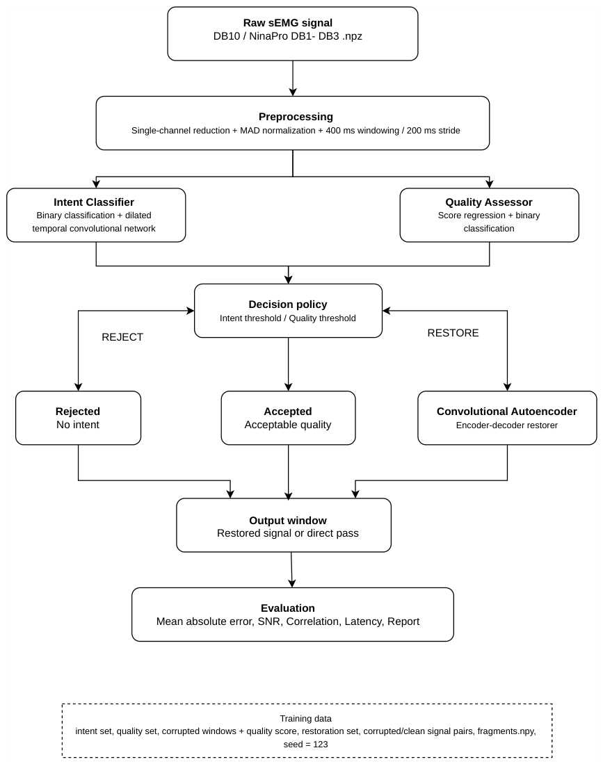
Figure 2.
Internal structure of the three models and the synthetic corruption scheme used during training.
Figure 2.
Internal structure of the three models and the synthetic corruption scheme used during training.
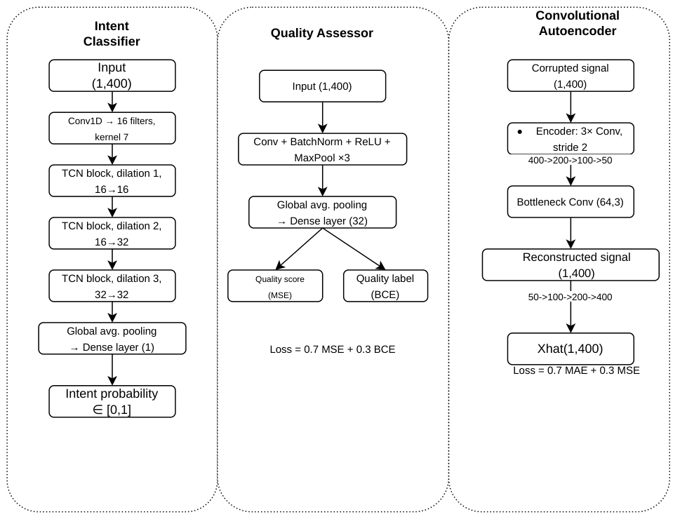
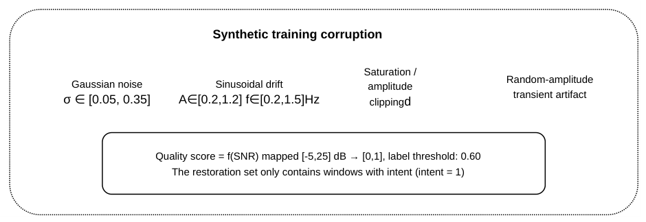
Figure 3.
Representative examples of the three pipeline decisions for P1 (female), Week 1.
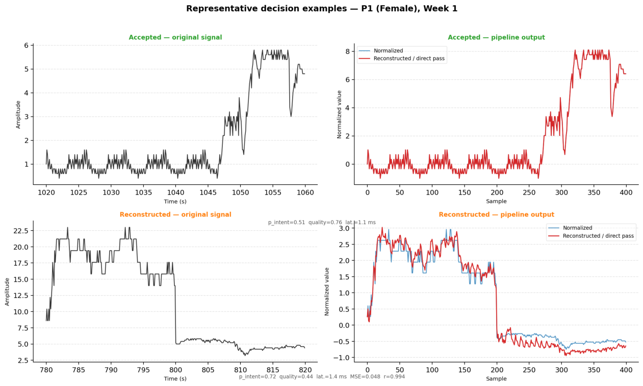
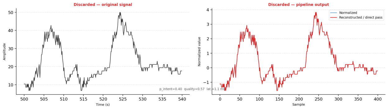
Figure 4.
Distribution of pipeline decisions by study participant.
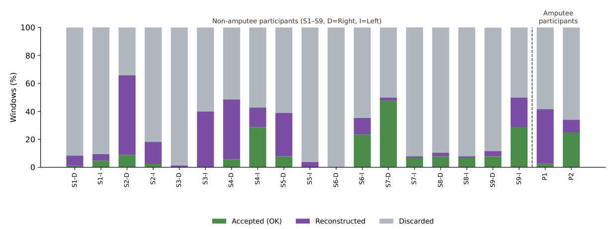
Figure 5.
Comparative metrics by participant.
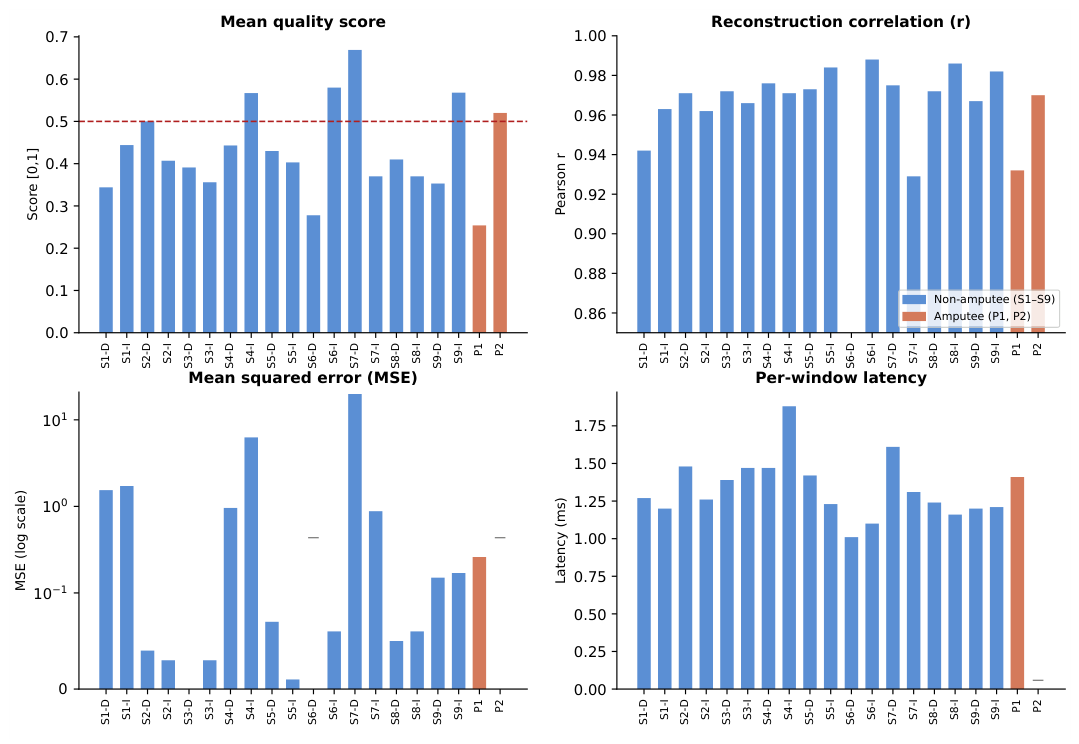
Figure 6.
Original degraded signal and signal reconstructed by the convolutional autoencoder – P1 (female) and P2 (male), first available session.
Figure 6.
Original degraded signal and signal reconstructed by the convolutional autoencoder – P1 (female) and P2 (male), first available session.
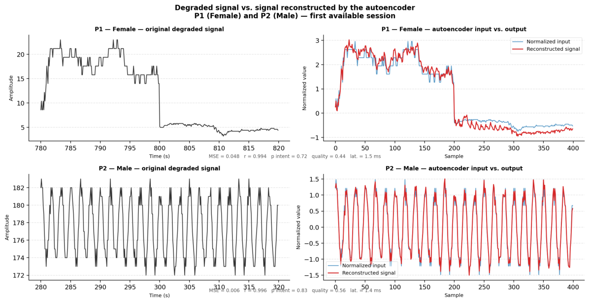

Table 1.
Participants with transradial amputation.
| Participant | Gender | Age | Post-amp. | Amputation type | Weeks | Task |
|---|---|---|---|---|---|---|
| P1 | Female | 31 | 5 years | Unilateral transradial | W1–W4 | Residual muscle activation |
| P2 | Male | 28 | 8 years | Unilateral transradial | W1–W4 | Residual muscle activation |
Table 2.
Healthy study participants.
| Participant | Age | Gender | Muscle | Task | Limbs |
|---|---|---|---|---|---|
| S1 | 54 | Male | Flexor carpi radialis | Open/close hand | Left/Right |
| S2 | 28 | Male | Flexor carpi radialis | Open/close hand | Left/Right |
| S3 | 29 | Male | Flexor carpi radialis | Open/close hand | Left/Right |
| S4 | 27 | Male | Flexor carpi radialis | Open/close hand | Left/Right |
| S5 | 23 | Female | Flexor carpi radialis | Open/close hand | Left/Right |
| S6 | 32 | Female | Flexor carpi radialis | Open/close hand | Left/Right |
| S7 | 26 | Female | Flexor carpi radialis | Open/close hand | Left/Right |
| S8 | 24 | Female | Flexor carpi radialis | Open/close hand | Left/Right |
| S9 | 20 | Female | Flexor carpi radialis | Open/close hand | Left/Right |
| S10 | 20 | Female | Flexor carpi radialis | Open/close hand | Left/Right |
Table 3.
Summary of metrics per recording.
| Group | ID | Participant | Limb | OK (%) | Reconstructed (%) | Discarded (%) | Mean quality | Correlation | MSE |
|---|---|---|---|---|---|---|---|---|---|
| Non-amputee | P14 | S1 | R | 1.0 | 7.3 | 91.7 | 0.344 | 0.942 | 1.54 |
| Non-amputee | P15 | S1 | L | 4.7 | 4.7 | 90.7 | 0.444 | 0.963 | 1.72 |
| Non-amputee | P16 | S2 | R | 8.9 | 57.0 | 34.1 | 0.500 | 0.971 | 0.04 |
| Non-amputee | P17 | S2 | L | 2.3 | 15.9 | 81.8 | 0.407 | 0.962 | 0.03 |
| Non-amputee | P18 | S3 | R | 0.0 | 1.4 | 98.6 | 0.391 | 0.972 | 0.00 |
| Non-amputee | P19 | S3 | L | 0.0 | 40.0 | 60.0 | 0.356 | 0.966 | 0.03 |
| Non-amputee | P20 | S4 | R | 5.7 | 42.9 | 51.4 | 0.443 | 0.976 | 0.96 |
| Non-amputee | P21 | S4 | L | 28.7 | 14.0 | 57.3 | 0.567 | 0.971 | 6.27 |
| Non-amputee | P22 | S5 | R | 7.8 | 31.2 | 61.0 | 0.430 | 0.973 | 0.07 |
| Non-amputee | P23 | S5 | L | 0.0 | 3.9 | 96.1 | 0.403 | 0.984 | 0.01 |
| Non-amputee | P24 | S6 | R | 0.0 | 0.0 | 100.0 | 0.278 | — | — |
| Non-amputee | P25 | S6 | L | 23.5 | 11.8 | 64.7 | 0.580 | 0.988 | 0.06 |
| Non-amputee | P26 | S7 | R | 47.7 | 2.3 | 50.0 | 0.669 | 0.975 | 19.9 |
| Non-amputee | P27 | S7 | L | 6.9 | 1.0 | 92.2 | 0.370 | 0.929 | 0.88 |
| Non-amputee | P29 | S8 | R | 7.8 | 2.6 | 89.6 | 0.410 | 0.972 | 0.05 |
| Non-amputee | P30 | S8 | L | 6.9 | 1.0 | 92.2 | 0.370 | 0.986 | 0.06 |
| Non-amputee | P31 | S9 | R | 7.8 | 3.9 | 88.2 | 0.353 | 0.967 | 0.15 |
| Non-amputee | P32 | S9 | L | 28.9 | 21.1 | 50.0 | 0.568 | 0.982 | 0.17 |
| Amputee | — | P1 (Female) | Residual limb | 2.4 | 39.2 | 58.4 | 0.254 | 0.932 | 0.26 |
| Amputee | — | P2 (Male) | Residual limb | 25.0 | 9.1 | 65.9 | 0.520 | 0.970 | — |
Note: “—” indicates that no reconstructed windows existed in that recording. R = Right, L = Left.
Disclaimer/Publisher’s Note: The statements, opinions and data contained in all publications are solely those of the individual author(s) and contributor(s) and not of MDPI and/or the editor(s). MDPI and/or the editor(s) disclaim responsibility for any injury to people or property resulting from any ideas, methods, instructions or products referred to in the content. |
© 2026 by the authors. Licensee MDPI, Basel, Switzerland. This article is an open access article distributed under the terms and conditions of the Creative Commons Attribution (CC BY) license (http://creativecommons.org/licenses/by/4.0/).
Copyright: This open access article is published under a Creative Commons CC BY 4.0 license, which permit the free download, distribution, and reuse, provided that the author and preprint are cited in any reuse.