
Submitted:
27 July 2026
Posted:
28 July 2026
You are already at the latest version
Abstract
Nanocarrier medicine has entered a paradox. The chemistry to make targeted, multi-functional carriers is now routine, and the clinical need - most sharply in cancers such as colorectal carcinoma - is unambiguous, yet fewer than one in ten preclinical candidates reaches patients, and the fraction of an injected dose that reaches a solid tumour remains small. The bottleneck is not synthetic capacity but interpretation: nanocarrier physicochemistry, in vitro biological behaviour and patient-level clinical variability are measured in disconnected assays, and no single framework couples them into an actionable prediction for a specific patient. We propose that this coupling is best cast as a three-layer virtual twin. Layer 1 is the nanocarrier physicochemical feature space (size, shape, coating, corona, payload). Layer 3 is the patient biological profile drawn from routine clinical-laboratory data (molecular status, tumour markers, systemic-inflammation indices). Between them sits Layer 2, an intermediate integrating layer of multi-modal AI/ML biosensing. We formalise the concept, show how each layer is populated with concrete quantitative examples (drawn from a validated microfluidic biosensing platform and from published colorectal-cancer laboratory data), and argue that a personalised, safety-gated decision index emerges naturally from the fusion. We conclude with a maturation trajectory that turns this concept into a practical instrument for personalised nanomedicine.
Keywords:
virtual twin
; nanocarrier
; multi-modal biosensing
; label-free microscopy
; magnetosensing
; fluorescence
; AI/ML
; colorectal cancer
; patient stratification
; personalised nanomedicine
1. Nanomedicine – A Field That Performs Well in Lab but Predicts Poorly in Clinic
Nanomedicine has made extraordinary technical progress. Ionisable-lipid nanoparticles delivered mRNA vaccines to hundreds of millions of people; superparamagnetic iron-oxide contrast agents are clinically routine; polymeric, lipid and inorganic carriers now permit exquisite control over size, shape, surface chemistry, biological corona and payload release. Yet the translational record for anticancer nanocarriers remains disappointing: more than nine in ten candidates fail before reaching patients (van der Meel et al., 2019), and a widely cited meta-analysis found that only about 0.7% of an intravenously administered nanoparticle dose accumulates in a solid tumour (Wilhelm et al., 2016). Colorectal cancer (CRC), the second leading cause of cancer death in Europe, exemplifies the paradox: nanocarrier formulations of chemotherapy and RNA payloads are entering trials at speed, while the tools to predict which formulation will work for which patient lag far behind.
The gap is not synthetic. It is a gap of integration. Nanocarrier physicochemistry is characterised in one laboratory with one set of assays; the in vitro biological behaviour of the carriers - barrier crossing, uptake, intracellular fate - is measured elsewhere in still other systems; and the patient’s clinical-laboratory profile that will ultimately determine the response lives in an entirely separate data ecosystem, the hospital electronic health record. Nothing routinely couples the three. Consequently, when a candidate carrier fails, the failure mode - was it physicochemistry, transport, patient biology, or an interaction of all three? - cannot be attributed, and the design cycle for the next iteration is uninformed.
We take this Concept Article to argue that the missing piece is a virtual twin: an interpretable, continuously learning model that jointly represents nanocarrier chemistry, in vitro biological behaviour and patient biology in a single decision-oriented framework. The idea sits within a wider movement. In precision oncology, digital-twin frameworks have moved rapidly from thought experiments to explicit clinical proposals - Hernandez-Boussard and colleagues describe them as "a paradigm shift for precision cancer care" (Hernandez-Boussard et al., 2021), Björnsson and colleagues formalise them as patient-specific computational models integrating clinical, imaging, genomic and therapy-response data (Björnsson et al., 2020), and recent commentary highlights data fusion and the marriage of mechanistic modelling with machine learning as the operational challenges (Laubenbacher et al., 2024). What has been missing from this conversation, and what we contribute here, is a twin specifically for the interface between a nanocarrier and a patient. Two commitments make the concept practical rather than aspirational. First, we distinguish three data layers explicitly, treating the second - the intermediate biosensing layer - as an integrating tissue between design and patient rather than as a scattered set of characterisation methods. Second, we treat all biosensing modalities (label-free AI-microscopy, magnetosensing, fluorescence, and further techniques) as equally-valid, complementary channels of one biosensing layer rather than as competing methods; their fusion is the point. The rest of the article develops this three-layer concept, shows how each layer is populated with concrete example data, and sets out a maturation path to a personalised, safety-gated decision instrument for colorectal-cancer nanomedicine.
2. The Three-Layer Concept
A virtual twin, in the sense we use the term here, is a population-level, data-driven predictive model that jointly represents three feature spaces: what the carrier is (Layer 1), how it behaves in a controlled biological system (Layer 2), and what the patient looks like (Layer 3). Each layer is a matrix of quantitative features; the three matrices are coupled by shared observations, so that a change in one layer is reflected in the others. Figure 1 sets out the whole concept in one picture; the remainder of Section 2 defines the layers and their coupling, and Section 3, Section 4 and Section 5 develop each in turn.
2.1. Layer 1 - Nanocarrier Physicochemistry
The first layer captures the intrinsic design variables of the carrier that a chemist can specify. For polymeric and lipid nanoparticles this is on the order of fifteen features: core size and shape, surface chemistry (biodegradable polymer vs stealth PEG vs ionisable lipid), PEG grafting density, zeta potential, protein-corona composition (Bertrand et al., 2017), and, for nucleic-acid carriers, the ionisable-lipid pKₐ that governs endosomal escape. For inorganic and hybrid carriers the layer additionally includes magnetisation, plasmonic properties and coating architecture. Layer 1 is where a chemist thinks - it is the input a formulation team can choose.
2.2. Layer 3 - Patient Clinical-Laboratory Profile
The third layer captures the biological environment the carrier must navigate - the patient. Unlike Layer 1, it is not chosen but observed. For colorectal cancer, the most tractable of contexts, Layer 3 is populated almost entirely from data routinely measured in clinical practice: tumour molecular status (KRAS, BRAF, NRAS, mismatch-repair/MSI), serum tumour markers (CEA, CA19-9), systemic-inflammation indices (NLR, LDH, the CRP/albumin-based modified Glasgow Prognostic Score), demographics (age, biological sex) and prior therapy. Where research-grade covariates are available (gut microbiome, endothelial phenotype), they slot in; but the working power of the layer is that its baseline is already in every patient’s chart.
2.3. Layer 2 - The Intermediate Integrating Biosensing Layer
Between the designer’s variables and the patient’s biology sits the layer that has, historically, been most fragmented and is the crux of this Concept Article: the in vitro biological behaviour of the carrier. Traditional characterisation splinters this into a permeability assay here, an uptake assay there, a viability endpoint elsewhere. The concept we advance is that this middle layer is best treated as one integrating biosensing layer fed by several complementary channels - label-free AI-microscopy (organelle darkening upon intracellular accumulation), magnetosensing of ferromagnetic-core carriers (giant- or tunnel-magnetoresistive sensors, magnetorelaxometry, AC susceptibility), fluorescence with AI-based segmentation (labelled carriers or reporter-based readouts), and further modalities such as impedance-based barrier monitoring (TEER), Raman/surface-enhanced Raman scattering, surface-plasmon resonance and holographic microscopy. These are not competing methods to be chosen among; they are equally-valid channels each providing partial, complementary information, whose fusion under a common data schema yields a richer, more physically-grounded feature vector than any single modality alone. Section 4 develops this in detail; the key conceptual move is that the biosensing layer is one entity, not a menu.
The three layers are coupled: Layer 2 measures Layer 1 in a biological system whose properties Layer 3 partially specifies (donor sex, endothelial phenotype, prior therapy). This is why we describe the twin as three-layer rather than as a stack of independent predictors: each layer conditions the others, and the model must be trained to represent the couplings, not just the layers.
3. Layer 1 in Practice - the Nanocarrier Feature Space
What features should populate Layer 1? The choice reflects a balance between mechanistic informativeness and measurability. Size and shape govern the wrapping energy at which the cell membrane uptake the particle, with a well-documented curvature optimum near 10–25 nm hydrodynamic diameter for spherical carriers (Champion and Mitragotri, 2006; Decuzzi and Ferrari, 2007). Surface chemistry - charge, coating identity, PEG density - governs opsonisation and corona formation (Nel et al., 2009; Bertrand et al., 2017), which in turn set the biological identity the carrier presents to cells (Behzadi et al., 2017). For nucleic-acid carriers, ionisable-lipid pKₐ and pH-triggered release control endosomal escape and thus payload survival.
Layer 1 therefore comprises, for a typical polymeric or lipid nanocarrier, roughly fifteen features: core size (mean and PDI), shape/aspect ratio, coating identity and grafting density, zeta potential, hydrodynamic corona in serum, ionisable-lipid pKₐ (where applicable), payload identity and loading, and physicochemical release kinetics. Each is measurable by standard characterisation. The important design choice is that these are not endpoints in themselves - they are the input to Layer 2, and only their coupling with Layer 2 (transport, uptake, kinetics) makes them predictive of biological behaviour.
Example - a factorial exploration of Layer 1
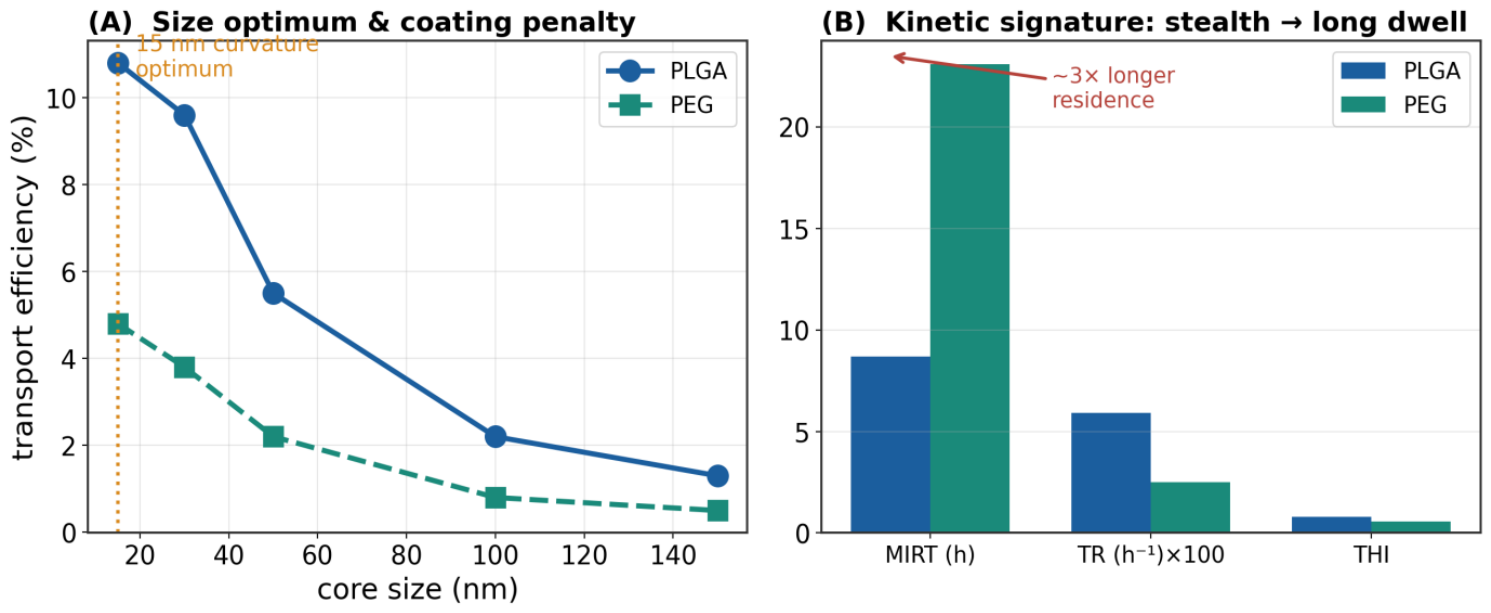
As an illustrative example (drawn from Goranov and Haranava, 2026), Layer 1 was populated across a controlled factorial matrix: two coatings (biodegradable polymer vs stealth PEG), five core sizes (15–150 nm), four concentrations (10–500 µg mL⁻¹) and two barriers. Passed through the intermediate biosensing layer (Section 4), the coupling recovers the classical, mechanism-consistent structure of the field: a sharp 15-nm curvature optimum (transport efficiency 10.8% for the biodegradable carrier at 100 µg mL⁻¹ in systemic endothelium, falling to 5.5% at 50 nm and 1.3% at 150 nm), a large stealth-coating penalty (PEG yields only 30–45% of biodegradable-coating transport, peak 4.8% vs 10.8%; Owens and Peppas, 2006; Sahay et al., 2010), and a two-to-three-fold longer intracellular residence time for stealth carriers (MIRT 23 h vs 9 h). These numbers are given as one example of what Layer 1 looks like once coupled to Layer 2; they are not the point of the concept.
Figure 2.
Example. Layer 1 as revealed by Layer 2 biosensing. (A) A sharp 15-nm curvature optimum and a large stealth-coating penalty (systemic endothelium, 100 µg mL⁻¹). (B) Kinetic signature: stealth PEG coating triples intracellular residence time, the key risk factor for acid-labile cargoes. Data adapted from Goranov and Haranava (2026); shown as an example, not as the concept’s evidence base.
Figure 2.
Example. Layer 1 as revealed by Layer 2 biosensing. (A) A sharp 15-nm curvature optimum and a large stealth-coating penalty (systemic endothelium, 100 µg mL⁻¹). (B) Kinetic signature: stealth PEG coating triples intracellular residence time, the key risk factor for acid-labile cargoes. Data adapted from Goranov and Haranava (2026); shown as an example, not as the concept’s evidence base.
4. Layer 2 - One Biosensing Layer, Several Complementary Channels
The intermediate layer is where the concept lives or dies. In the field’s current practice, in vitro nanocarrier evaluation is fragmented among assays that speak incompatible languages: Transwell permeability and inductively-coupled plasma spectroscopy for mass balance, flow cytometry for population uptake, fluorescence microscopy for localisation, giant-magnetoresistive sensing for magnetic-core carriers, and so on. Each captures a partial truth; none is by itself sufficient; and they are rarely acquired co-temporally on the same biological system. The concept we advance is to invert this stance: the intermediate layer is one entity, and its several modalities are complementary channels of that single layer, curated under a shared data schema, gated on shared barrier-integrity criteria, and fused into a common feature tensor.
Three properties define a biosensing channel that belongs to Layer 2. First, it must be quantitative, returning a numerical readout rather than a qualitative image. Second, it must be cell-compatible, delivering its readout without perturbing the biology being measured (this is where label-free approaches shine, but AI-augmented fluorescence with low-phototoxicity dyes also qualifies). Third, it must be time-resolved, producing a kinetic rather than a static endpoint, so that transport rates and intracellular residence times - not only endpoint fractions - are recoverable. We treat modalities that meet these three criteria as equivalent channels; their differences lie in what specifically they constrain. Section 4.1, Section 4.2, Section 4.3 and Section 4.4 develop each channel, and Section 4.5 sets out the shared quality-control gate that makes their fusion coherent.
4.1. Label-Free AI-Microscopy
Label-free brightfield or phase-contrast microscopy, interpreted by a residual-attention U-Net (ResAt-UNet; Ronneberger et al., 2015; Diakogiannis et al., 2020) or comparable segmentation network (Moen et al., 2019), reads the optical consequence of intracellular nanocarrier accumulation - characteristic organelle darkening produced by electron-dense cores concentrating in endolysosomes. As an example implementation, Goranov and Haranava (2026) report a five-stage AI/ML pipeline (preprocessing, ResAt-UNet segmentation, k-means uptake classification, transport-fraction quantification, gradient-boosted feature attribution) reaching intersection-over-union 0.85, precision 93.2% and recall 86.9% on a held-out test set, comfortably above the 70–85% consistency typical of manual annotation and free of inter-operator variance. The channel’s virtues are that it needs no label (avoiding the physicochemical perturbations imposed by fluorescent tags; Wu et al., 2019), it is time-resolved at minute-level cadence, and the same imaging data yield both transport-fraction and kinetic descriptors. Its principal limitation is that it reports a morphometric surrogate rather than a molecular species, so it cannot on its own resolve dissolved from particulate material for iron-oxide cores, nor identify the payload post-delivery.
4.2. Magnetosensing
For carriers bearing a ferromagnetic core - superparamagnetic iron-oxide nanoparticles (SPIONs) and their hybrids - magnetosensing (giant- or tunnel-magnetoresistive sensors, magnetorelaxometry, AC-susceptibility measurements; Laurent et al., 2008) provides a channel that is orthogonal in an important way: it reports absolute magnetic mass directly and non-destructively, without optical access, and it can be used to measure transport through opaque samples (organoids, three-dimensional matrices) that microscopy cannot penetrate. Where a magnetic core is native to the carrier design this is a first-class channel; where it is not, a passively-sequestered magnetic tracer (≤ 3–4 wt%) may be included solely to enable the readout, without perturbing the surface chemistry or corona formation that dominate biological behaviour. The channel’s limitation is scope - it applies only to magnetically-labelled carriers - and its calibration must be checked against a reference standard for each session.
4.3. Fluorescence with AI Segmentation
Fluorescence-based channels remain indispensable where molecular specificity is required. A payload conjugated to a low-phototoxicity fluorophore, or a genetically encoded reporter in the target cells (for example, a KRAS-silencing readout in an organoid), delivers a molecule-specific signal that neither label-free microscopy nor magnetosensing can provide. AI segmentation of the fluorescent channel (Moen et al., 2019) allows the same automated, reproducible quantification as in Section 4.1 and Section 4.2. The channel’s classical limitation - the physicochemical perturbation of the carrier by the fluorescent label - is a legitimate concern (Wu et al., 2019) and constrains fluorescence’s role in Layer 2 to (i) validation of the label-free channel against an orthogonal readout, and (ii) reporter-based downstream measurement (payload delivery, target-gene silencing) rather than primary transport quantification of the unlabelled clinical formulation.
4.4. Further Channels of the Biosensing Layer
The list is not exhaustive. Impedance-based transendothelial-electrical-resistance imaging (TEER) provides continuous barrier-integrity monitoring - not a transport measurement in itself, but an indispensable validity gate on every other channel, because apparent transport across a torn monolayer is paracellular leak rather than transcytosis. Raman and surface-enhanced Raman scattering deliver chemical fingerprinting; surface-plasmon resonance provides binding-kinetic readouts on functionalised carriers; holographic microscopy adds a third dimension without labels. Any of these that satisfies the three criteria of Section 4 (quantitative, cell-compatible, time-resolved) joins Layer 2 as another channel. The channels are unified not by shared instrumentation but by the shared data schema they emit into: a per-condition, time-resolved, barrier-integrity-gated vector of transport, uptake and kinetic descriptors.
4.5. Fusion Under a Shared Quality-Control Gate
Across all channels, the fusion is governed by a single dynamic barrier-integrity gate (Weksler et al., 2013; Helms et al., 2016). This shared gate is what makes the channels commensurable: without it, one channel’s leaky-barrier artefact would be admitted while another’s would be filtered, and the fused readout would be dominated by whichever channel had the loosest quality-control. In one concrete example, Goranov and Haranava (2026) show that applying such a gate raises the cross-validated R² of a downstream transport-prediction model from 0.69 (uncleaned) to 0.89 (cleaned) with a bootstrap-significant gain, and that the excluded points are predicted wrongly for the right reason - the model reads their large-core, high-dose, tight-barrier design as low-transport rather than tracking the leak-inflated observations - confirming that the exclusion is principled rather than cosmetic. The intermediate integrating layer is the discipline as much as it is the instrumentation.
5. Layer 3 - The Patient Biological Profile
5.1. The Concept - Why Patient Biology Is a First-Class Layer
Layer 3 answers a question that the nanomedicine field has, until recently, largely ignored: for whom is the carrier intended? Physicochemical design (Layer 1) and in vitro behaviour (Layer 2) determine what a carrier can do; the patient’s biological state determines what it will do once administered. That the third is crucial has been documented repeatedly: reviewers of the field’s translational record now attribute much of the clinical-trial attrition of nanoparticulate nanomedicines to insufficient attention to "the relationship between disease pathophysiology and the heterogeneity of the disease in humans" - patient variables that reshape biodistribution, clearance and target engagement (Hare et al., 2017; Lammers and Ferrari, 2020; Abo Qoura et al., 2026). Even the protein corona that forms around a nanoparticle upon exposure to biological fluids - the very interface that determines its biological identity - varies with patient sex, age, ancestry, environment and disease state (Ahsan et al., 2024), so a Layer 1 specification is only half the story.
A twin that treats the patient as a first-class layer must therefore encode Layer 3 with the same explicit, quantitative feature-space discipline it applies to Layer 1. Four families of variables recur across cancer contexts. Molecular status captures oncogenic drivers, tumour-suppressor loss, mismatch-repair state and, increasingly, whole-transcriptomic subtype; these are mechanistically consequential for uptake, retention and response (Björnsson et al., 2020; Hernandez-Boussard et al., 2021). Circulating markers- organ-specific tumour markers, cell-free DNA, and, where feasible, corona-modifying serum proteins - index disease burden and biological identity. Systemic-inflammation indices (neutrophil-to-lymphocyte ratio, C-reactive protein / albumin, LDH) reshape the endothelium a nanocarrier must cross and predict overall prognosis independently of tumour stage. And demographic and treatment-history variables (age, biological sex, prior therapy, comorbidities, microbiome status) modulate baseline physiology in ways that measurably affect nanocarrier fate. The Layer 3 principle is that these should populate a defined feature vector, be measured routinely as part of the twin’s admission workflow, and be treated by the predictive model as primary predictors rather than nuisance covariates (Katsoulakis et al., 2024; Laubenbacher et al., 2024).
Two implications follow. First, Layer 3 is cancer-specific in its features but generalisable in its logic: the four families above transfer across disease areas, while the specific variables and their thresholds vary. Second, Layer 3 is routine in its baseline: with rare exceptions, the variables that most matter for a first-generation twin are already recorded in every patient’s chart, so the layer is not a research-only construct but an operational one, ready to be assembled from the electronic health record. The next subsection makes this concrete with colorectal cancer as a worked example.
5.2. Colorectal Cancer as a Specific Example
Colorectal cancer (CRC) is a natural first example because the four families of Section 5.1 each map cleanly onto variables that oncology already records. Two are dominant: the tumour’s molecular status, and the patient’s serum and haematological profile.
Tumour molecular status. KRAS is mutated in roughly 40–45% of CRC (predominantly at codons 12/13), BRAF V600E in ~7–9%, NRAS in ~4–5%, and mismatch-repair deficiency / high microsatellite instability (MSI-H) in ~12–15% of cases (with stage-dependent prevalence, from ~20% in stage II to ~4% in stage IV; Li et al., 2022). These are mechanistically consequential for nanocarrier uptake. Oncogenic KRAS upregulates macropinocytosis (Commisso et al., 2013), so nanocarrier uptake in KRAS-mutant cells depends more on fluid-phase, receptor-independent internalisation than on receptor-targeted binding - meaning genotype and formulation interact rather than add. MSI-H tumours have a distinct immune context that shapes downstream response to nanocarrier-delivered payloads.
Serum and haematological indices. The tumour markers CEA and CA19-9 index disease burden and prognosis. In a large single-centre cohort of 1487 CRC patients, five-year overall survival fell from ~69% at CEA < 5 ng mL⁻¹ to ~44% at 5–200 and ~7% at ≥ 200 ng mL⁻¹, and elevated CA19-9 (≥ 37 U mL⁻¹) independently predicted poorer survival (Lakemeyer et al., 2021). Systemic inflammation - which reshapes the vascular endothelium a nanocarrier must cross - is captured by the neutrophil-to-lymphocyte ratio (NLR; values > 2.5 are a recognised adverse marker; Li et al., 2014), lactate dehydrogenase, and the CRP/albumin-based modified Glasgow Prognostic Score. Biological sex modifies the endothelial phenotype at the level of the cell the biosensing layer actually cultures (female-donor endothelial cells express higher eNOS and produce more nitric oxide; Cattaneo et al., 2017). The gut microbiome modulates barrier tightness through short-chain fatty acids (Tabat et al., 2020). Prior oxaliplatin/FOLFOX exposure raises baseline endothelial stress through endothelial-to-mesenchymal transition (Kovacic et al., 2019). Table 1 lists the variables, their reference values, and their hypothesised role in the twin.
6. Integration - From Three Layers to a Personalised Decision
With the three layers defined, integration becomes a modelling question with a specific shape. The joint feature tensor - nanocarrier physicochemistry (Layer 1) crossed with the biosensing readout in a defined biological context (Layer 2) crossed with the patient profile (Layer 3) - is passed to an interpretable predictive model whose outputs are the three components of a personalised decision: a Biosafety Index (BSI, from morphometric and barrier-integrity readouts in the biosensing layer), a Nanocarrier Transport Efficiency (NTE, the endpoint and kinetic transport readout across the modelled barrier), and a Tumour Response Index (TRI, from patient-derived organoid or organ-chip readouts downstream of the barrier). Each is a scalar output of the fused model.
A working example of how these combine is the Personalised Therapeutic Index (PTI). An earlier proposal wrote PTI = (TRI × NTE) / BSI. This is mathematically unsound and instructive to correct: dividing by safety makes the index diverge as BSI → 0, so a barrier-toxic formulation is rewarded rather than suppressed - the opposite of the intended semantics. A bounded, safety-gated form is PTI = TRI · NTE · σₖ(BSI − θ), where σₖ(x) = 1/(1 + e^(−kx)) is a logistic gate centred at an acceptability threshold θ (Figure 3B). All inputs are normalised to [0, 1], so PTI is bounded, monotonically increasing in safety, and collapses toward zero when safety falls below threshold. Because the three predictions are themselves uncertain, PTI is reported not as a point value but as a distribution obtained by Monte-Carlo propagation of the component credible intervals. We stress that this formula is one example of an integration function; the deeper goal of the twin is to learn the combination from outcome data rather than to impose an algebraic form.
6.1. Interpretability and the Physical Anchor
For a twin that must support clinical decisions, the fused model must be interpretable and physically consistent. Interpretability is delivered by exact TreeSHAP (Lundberg et al., 2020) or comparable game-theoretic attribution, which decomposes each prediction into per-feature contributions in the same units as the prediction target. Physical consistency is delivered by cross-channel agreement within Layer 2: no single channel is trusted in isolation; agreement between two or more independent channels (label-free ↔ magnetosensing, label-free ↔ fluorescence) is the working criterion for admitting a datum to the training set. This is why we describe Layer 2 as integrating: it not only fuses inputs, it also enforces internal consistency before those inputs reach the predictive model.
6.2. An Illustrative Case in Colorectal Cancer
As one concrete illustration, consider two siRNA lipid-nanoparticle formulations for KRAS-mutant colorectal cancer that a conventional in vitro potency assay cannot distinguish (both give IC₅₀ near 20 nM), evaluated for one patient context: a 65-year-old, KRAS-G12D, FOLFOX-pre-treated female donor with elevated CEA (~40 ng mL⁻¹) and NLR ~4. Layer 2 biosensing (label-free + magnetosensing + fluorescent KRAS-silencing reporter) resolves them completely. Formulation A (highly cationic) induces early endothelial stress (BSI 0.60), transports poorly with long lysosomal dwell (NTE 12%, MIRT 4.2 h), and gives a modest response in the patient-derived organoid downstream (TRI 0.30). Formulation B (ionisable, near-neutral) preserves the barrier (BSI 0.95), transports efficiently with rapid endosomal escape (NTE 28%, MIRT 1.8 h), and drives deep organoid penetration with confirmed KRAS silencing (TRI 0.78). Under the corrected gated index (θ = 0.6, k = 10), the toxic Formulation A is gated to about half credit while the safe Formulation B passes near-fully: PTIA ≈ 0.018 versus PTIB ≈ 0.21, a roughly twelve-fold, correctly-signed separation. The numbers are illustrative; the point is that the three-layer integration turns a case that conventional assays leave undecided into a mechanistically attributed decision.
7. Layer Couplings - Why the Three Layers Must Be Trained Together
A defining move of the three-layer concept is that the layers are not independent inputs to be concatenated. They interact, and much of the twin’s predictive value lies in modelling the interactions explicitly. Three couplings are of particular importance.
Layer 1 × Layer 3. Formulation and patient interact rather than add. The KRAS example above is the archetype: an ionisable-lipid nanoparticle that performs modestly in a KRAS-wild-type endothelium may perform excellently in a KRAS-mutant tumour, precisely because the fluid-phase, macropinocytotic route it exploits is upregulated by the mutation (Commisso et al., 2013). Encoding genotype only as a nuisance covariate would flatten a signal that is genuinely genotype-dependent. Interaction terms between Layer 1 features and Layer 3 features must be explicit in the model.
Layer 2 × Layer 3. The biosensing layer measures the carrier in a biological system whose properties Layer 3 partially specifies. Female-donor endothelial cells produce more nitric oxide (Cattaneo et al., 2017); patient microbiome status modulates baseline barrier tightness (Tabat et al., 2020); prior chemotherapy raises baseline endothelial stress (Kovacic et al., 2019). If the biological substrate of Layer 2 varies with Layer 3 covariates, then Layer 2 readouts are conditional on Layer 3, and the twin must be trained on paired (formulation, patient) observations rather than on formulations alone.
Layer 1 × Layer 2. Cross-channel consistency within Layer 2 (Section 6.1) also implicitly conditions the twin’s admissible Layer 1 features. A carrier whose optical readout disagrees with its magnetometric readout by more than a defined threshold is flagged, not silently averaged; a persistent disagreement is often a signal that the corona composition of that Layer 1 candidate is not what its nominal chemistry would predict.
8. Relation to Existing Frameworks
The three-layer virtual twin is not a break with the field but a reorganisation of pieces that have been maturing in parallel. It is useful to say what it borrows from and what it does differently, because the relationships determine which existing tools slot in and which do not.
Organ-on-chip and microphysiological systems provide the biological substrate on which the intermediate biosensing layer operates. The chip is not the twin; it is the platform through which Layer 2 sees Layer 1 in a context conditioned by Layer 3. What the concept adds is that the chip is treated as one channel of the biosensing layer, not as a stand-alone predictor - its readouts must be gated on shared barrier-integrity criteria and reconciled with orthogonal channels before entering the model.
Physiologically-based pharmacokinetic (PBPK) modelling provides the whole-body context that the tissue-level twin lacks. Where PBPK excels - systemic clearance, mononuclear-phagocyte handling, tissue-to-tissue distribution - the tissue-level twin is silent, and vice versa. The concept explicitly frames the two as complements to be coupled at Stage 3 of the maturation trajectory (Section 9), not competitors: the twin predicts tissue delivery given a plasma exposure, PBPK predicts plasma exposure given a dose, and their coupling yields dose-to-response prediction.
Quantitative structure–activity relationships (QSAR) for nanoparticles are the ancestor of Layer 1 alone. The concept generalises them by insisting that Layer 1 is not predictive by itself - it must be coupled to a rich Layer 2 readout in a defined biological system - and that Layer 3 is the missing dimension that made classical QSAR under-predictive of clinical outcome.
Patient-derived organoids and patient-derived xenografts provide validation anchors for Layer 3, not substitutes for it. Their documented ability to predict clinical chemotherapy response in colorectal cancer (Vlachogiannis et al., 2018; Su et al., 2023; Smabers et al., 2025) makes them the natural downstream reference for the tumour-response arm of the twin, precisely because they already encode patient biology at a level the twin can be trained against.
9. Maturation of the Three-Layer Twin
A concept is only useful if it admits a realistic maturation path. The trajectory we envisage (Figure 4) proceeds in three overlapping stages. Stage 1 - already realised on the biosensing layer alone - trains a tree-ensemble predictor on Layer 1 physicochemistry using a well-populated intermediate biosensing layer, in one well-characterised biological context. As an example, on a factorial dataset generated by a validated label-free biosensing platform, a gradient-boosted model (Chen and Guestrin, 2016) reaches an honest, condition-grouped cross-validated R² = 0.89 on transport prediction, with feature attributions that agree perfectly across three independent statistical routes (Kendall’s W = 1.00); a bootstrap-significant fraction of that performance is delivered by the shared barrier-integrity gate of Section 4.5, quantifying the value of the discipline. Stage 2 adds Layer 3, extending the model to a patient-conditioned multi-task neural network with shared encoder and task-specific heads for BSI, NTE and TRI, plus calibrated uncertainty; this stage requires the first prospective patient-derived-cell cohort in a specific context (colorectal cancer). Stage 3 couples the tissue-level twin to systemic pharmacokinetic modelling and validates against downstream outcome data, turning the concept into a deployable service.
Two commitments make the maturation credible. First, interpretability must remain first-class throughout - attributions in native prediction units, cross-channel consistency as an admission criterion, human-in-the-loop review of mechanistically implausible feature dependences. Second, physical validation via cross-channel agreement within Layer 2 must remain mandatory: no single channel is trusted in isolation, and no fused prediction is trusted without cross-channel corroboration. These commitments are inexpensive to state and expensive to enforce, and they distinguish the concept from a purely data-driven surrogate that would fail the first out-of-distribution query.
10. Discussion
The strongest reading of the concept advanced here is that in vitro nanocarrier evaluation has been organised around the wrong unit of analysis. The unit has been the assay - permeability, uptake, viability, mass balance - each producing a partial number that must then be reconciled with the others by expert judgement. The concept we advance is that the unit should be the layer, and specifically the intermediate biosensing layer, in which several complementary channels produce a single fused, quality-controlled, kinetic readout of the carrier’s biological behaviour in a defined system. Layer 1 (physicochemistry) and Layer 3 (patient) are then coupled to that layer through interpretable predictive models. The reorganisation is conceptual before it is technical; the technical pieces - label-free AI-microscopy, magnetosensing, AI-augmented fluorescence, TEER-gated data curation, tree-ensemble and multi-task-DNN predictors - already exist. What is new is the discipline of treating them as one layer.
Adopted at scale, this reorganisation would deliver several practical gains. It would let a formulation team ask, for a specific patient profile, which of a set of candidate carriers has the highest personalised index and receive a decision with a calibrated uncertainty and a mechanistic attribution rather than a bare ranking. It would let a clinician stratify patients by their expected response to a nanocarrier therapy on the basis of routine laboratory variables they already collect. It would let the field’s aggregate data grow into a resource that satisfies the FAIR guiding principles for scientific data management (Wilkinson et al., 2016) - findable, accessible, interoperable, reusable - and thus support transfer learning across institutions rather than fragmenting into per-laboratory silos. And it would let regulatory conversations about New Approach Methodologies proceed on the basis of a defined, auditable framework rather than an ad-hoc collection of assays, so that dossier-quality prediction of tissue-level delivery and response becomes an achievable rather than aspirational deliverable.
The concept also reframes what counts as validation. In the assay paradigm, a method is validated against a chosen reference - typically an animal model - by a single-number correlation. In the three-layer twin, validation is plural: cross-channel agreement within Layer 2 (label-free ↔ magnetosensing ↔ fluorescence) is one axis; agreement between the twin’s PTI prediction and downstream patient-derived-organoid response, patient-derived-xenograft outcome, and, where available, clinical response is another. No single reference is treated as ground truth; the twin’s credibility grows with the number of independent axes on which it agrees.
10.1. Practical Limits Worth Stating
The concept, however visionary, must respect two practical limits. First, the tissue-level twin models transport and local cellular response; it does not, by itself, model systemic clearance, circulation pharmacokinetics or adaptive immunity. A high PTI is necessary but not sufficient for clinical efficacy, and the twin must ultimately be coupled to a physiologically-based pharmacokinetic model to close that gap. Second, the layer-coupling story is only as strong as the biological substrate of Layer 2. Patient-derived cells introduce donor-to-donor variability; organoids drift with passage; barrier models have known phenotypic limits. A credible twin encodes these as explicit competence boundaries and abstains from prediction outside them, rather than extrapolating silently. Neither limit undermines the concept; both bound what may honestly be claimed.
11. Conclusions
Nanocarrier medicine’s translational bottleneck is a bottleneck of integration between three data layers that today live apart: nanocarrier physicochemistry, in vitro biological behaviour and patient clinical-laboratory data. We have argued that a virtual twin naturally organises these into three layers, with a middle integrating biosensing layer fed by several complementary AI/ML channels - label-free microscopy, magnetosensing, fluorescence, and further modalities - treated as equally-valid, complementary streams of one biosensing layer rather than as competing methods. The three layers are coupled: their interactions are the point, and the twin must be trained to model them jointly. For colorectal cancer specifically, the three layers can be populated concretely today - Layer 1 by standard nanocarrier characterisation, Layer 2 by a validated multi-modal biosensing platform, Layer 3 by routine clinical laboratory data - and combined into an interpretable, safety-gated personalised decision index. What remains is not proof of concept but the disciplined build-out: a shared FAIR data schema, cross-channel consistency as an admission criterion, calibrated uncertainty as a first-class output, and outcome-validated coupling to systemic pharmacokinetic modelling. The concept is realistic, near-term, and, we believe, the natural organising framework for the next decade of personalised nanomedicine.
Author Contributions
V.A.G.: conceptualisation, three-layer framework, virtual-twin architecture, figures, manuscript drafting; biosensing methodology, feature engineering, critical revision.
Funding
This work was supported by Biodevice Systems s.r.o.
Data and Code Availability
Illustrative example data drawn from Goranov and Haranava (2026) are available in the cited preprint. Colorectal-cancer laboratory reference values are from the published literature cited in Table 1 and the reference list. No patient cohort was generated for this Concept Article.
Conflicts of Interest
The author do not declare any conflicts of interest.
References
- Abo Qoura, L.; Kostyushev, D.; Parodi, A.; et al. Nanoparticle–host interactions: the impact of physiological and pathological factors on biodistribution, immune processes, and translational challenges. Cellular and Molecular Bioengineering 2026, 19, 1–27. [Google Scholar] [CrossRef] [PubMed]
- Ahsan, A.; Wang, J.; Liu, W.; et al. The role of patient-specific variables in protein corona formation and therapeutic efficacy in nanomedicine. Journal of Nanobiotechnology 2024, 22, 732. [Google Scholar] [CrossRef]
- Behzadi, S.; Serpooshan, V.; Tao, W.; et al. Cellular uptake of nanoparticles: journey inside the cell. Chemical Society Reviews 2017, 46(14), 4218–4244. [Google Scholar] [CrossRef] [PubMed]
- Bertrand, N.; Grenier, P.; Mahmoudi, M.; et al. Mechanistic understanding of in vivo protein-corona formation on polymeric nanoparticles and impact on pharmacokinetics. Nature Communications 2017, 8(1), 777. [Google Scholar] [CrossRef] [PubMed]
- Björnsson, B.; Borrebaeck, C.; Elander, N.; (Swedish Digital Twin Consortium); et al. Digital twins to personalize medicine. Genome Medicine 2020, 12(1), 4. [Google Scholar]
- Cattaneo, M.G.; Vanetti, C.; Decimo, I.; et al. Sex-specific eNOS activity and function in human endothelial cells. Scientific Reports 2017, 7, 9612. [Google Scholar] [CrossRef] [PubMed]
- Champion, J.A.; Mitragotri, S. Role of target geometry in phagocytosis. Proceedings of the National Academy of Sciences USA 2006, 103(13), 4930–4934. [Google Scholar] [CrossRef]
- Chen, T.; Guestrin, C. XGBoost: a scalable tree-boosting system. In Proceedings of the 22nd ACM SIGKDD Conference; 2016; pp. 785–794. [Google Scholar]
- Commisso, C.; Davidson, S.M.; Soydaner-Azeloglu, R.G.; et al. Macropinocytosis of protein is an amino-acid supply route in Ras-transformed cells. Nature 2013, 497(7451), 633–637. [Google Scholar] [CrossRef] [PubMed]
- Decuzzi, P.; Ferrari, M. The role of specific and non-specific interactions in receptor-mediated endocytosis of nanoparticles. Biomaterials 2007, 28(18), 2915–2922. [Google Scholar] [CrossRef] [PubMed]
- Diakogiannis, F.I.; Waldner, F.; Caccetta, P.; Wu, C. ResUNet-a: a deep-learning framework for semantic segmentation of remotely sensed data. ISPRS Journal of Photogrammetry and Remote Sensing 2020, 162, 94–114. [Google Scholar] [CrossRef]
- Goranov, V.A.; Haranava, Y. Integrated platform for quantification of nanoparticle transport across biological barriers using AI/ML analysis. Preprints 2026, 2026, 202606.2090. [Google Scholar] [CrossRef]
- Hare, J.I.; Lammers, T.; Ashford, M.B.; et al. Challenges and strategies in anti-cancer nanomedicine development: an industry perspective. Advanced Drug Delivery Reviews 2017, 108, 25–38. [Google Scholar] [CrossRef] [PubMed]
- Helms, H.C.; Abbott, N.J.; Burek, M.; et al. In vitro models of the blood–brain barrier: an overview and guidelines for their use. Journal of Cerebral Blood Flow & Metabolism 2016, 36(5), 862–890. [Google Scholar] [CrossRef]
- Hernandez-Boussard, T.; Macklin, P.; Greenspan, E.J.; et al. Digital twins for predictive oncology will be a paradigm shift for precision cancer care. Nature Medicine 2021, 27(12), 2065–2066. [Google Scholar] [CrossRef] [PubMed]
- Katsoulakis, E.; Wang, Q.; Wu, H.; et al. Digital twins for health: a scoping review. npj Digital Medicine 2024, 7, 77. [Google Scholar] [CrossRef] [PubMed]
- Kovacic, J.C.; Dimmeler, S.; Harvey, R.P.; et al. Endothelial to mesenchymal transition in cardiovascular disease: JACC state-of-the-art review. Journal of the American College of Cardiology 2019, 73(2), 190–209. [Google Scholar] [CrossRef] [PubMed]
- Lakemeyer, L.; Sander, S.; Wittau, M.; Henne-Bruns, D.; Kornmann, M.; Lemke, J. Diagnostic and prognostic value of CEA and CA19-9 in colorectal cancer. Diseases 2021, 9(1), 21. [Google Scholar] [CrossRef] [PubMed]
- Lammers, T.; Ferrari, M. The success of nanomedicine. Nano Today 2020, 31, 100853. [Google Scholar] [CrossRef] [PubMed]
- Laubenbacher, R.; Mehrad, B.; Shmulevich, I.; Trayanova, N. Digital twins in medicine. Nature Computational Science 2024, 4, 184–191. [Google Scholar] [CrossRef] [PubMed]
- Laurent, S.; Forge, D.; Port, M.; et al. Magnetic iron-oxide nanoparticles: synthesis, stabilization, vectorization, physicochemical characterizations and biological applications. Chemical Reviews 2008, 108(6), 2064–2110. [Google Scholar] [CrossRef] [PubMed]
- Li, M.X.; Liu, X.M.; Zhang, X.F.; et al. Prognostic role of neutrophil-to-lymphocyte ratio in colorectal cancer: a systematic review and meta-analysis. International Journal of Cancer 2014, 134(10), 2403–2413. [Google Scholar] [CrossRef] [PubMed]
- Li, Y.; Xiao, J.; Zhang, T.; Zheng, Y.; Jin, H. Analysis of KRAS, NRAS and BRAF mutations, microsatellite instability and prognosis in early colorectal cancer. Frontiers in Oncology 2022, 12, 897548. [Google Scholar] [CrossRef] [PubMed]
- Lundberg, S.M.; Erion, G.; Chen, H.; et al. From local explanations to global understanding with explainable AI for trees. Nature Machine Intelligence 2020, 2(1), 56–67. [Google Scholar] [CrossRef] [PubMed]
- Moen, E.; Bannon, D.; Kudo, T.; et al. Deep learning for cellular image analysis. Nature Methods 2019, 16(12), 1233–1246. [Google Scholar] [CrossRef] [PubMed]
- Nel, A.E.; Mädler, L.; Velegol, D.; et al. Understanding biophysicochemical interactions at the nano–bio interface. Nature Materials 2009, 8(7), 543–557. [Google Scholar] [CrossRef] [PubMed]
- Owens, D.E.; Peppas, N.A. Opsonization, biodistribution and pharmacokinetics of polymeric nanoparticles. International Journal of Pharmaceutics 2006, 307(1), 93–102. [Google Scholar] [CrossRef] [PubMed]
- Ronneberger, O.; Fischer, P.; Brox, T. U-Net: convolutional networks for biomedical image segmentation. MICCAI 2015, LNCS 9351, 234–241. [Google Scholar] [CrossRef]
- Sahay, G.; Alakhova, D.Y.; Kabanov, A.V. Endocytosis of nanomedicines. Journal of Controlled Release 2010, 145(3), 182–195. [Google Scholar] [CrossRef] [PubMed]
- Smabers, L.P.; Wensink, G.E.; Verissimo, C.S.; et al. Patient-derived organoids predict treatment response in metastatic colorectal cancer. Clinical Cancer Research 2025. [Google Scholar] [CrossRef] [PubMed]
- Su, C.; Olsen, K.A.; Bond, C.E.; Whitehall, V.L.J. The efficacy of using patient-derived organoids to predict treatment response in colorectal cancer. Cancers 2023, 15(3), 805. [Google Scholar] [CrossRef] [PubMed]
- Tabat, M.W.; Marques, T.M.; Markgren, M.; et al. Acute effects of butyrate on induced hyperpermeability and tight-junction protein expression in human colonic tissues. Biomolecules 2020, 10(5), 766. [Google Scholar] [CrossRef] [PubMed]
- van der Meel, R.; Sulheim, E.; Shi, Y.; et al. Smart cancer nanomedicine. Nature Nanotechnology 2019, 14(11), 1007–1017. [Google Scholar] [CrossRef] [PubMed]
- Vlachogiannis, G.; Hedayat, S.; Vatsiou, A.; et al. Patient-derived organoids predict chemoradiotherapy response of metastatic gastrointestinal cancers. Science 2018, 359(6378), 920–926. [Google Scholar] [CrossRef] [PubMed]
- Weksler, B.; Romero, I.A.; Couraud, P.-O. The hCMEC/D3 cell line as a model of the human blood–brain barrier. Fluids and Barriers of the CNS 2013, 10(1), 16. [Google Scholar] [CrossRef] [PubMed]
- Wilhelm, S.; Tavares, A.J.; Dai, Q.; et al. Analysis of nanoparticle delivery to tumours. Nature Reviews Materials 2016, 1, 16014. [Google Scholar] [CrossRef]
- Wilkinson, M.D.; Dumontier, M.; Aalbersberg, I.J.; et al. The FAIR guiding principles for scientific data management and stewardship. Scientific Data 2016, 3, 160018. [Google Scholar] [CrossRef] [PubMed]
- Wu, Y.; Ihme, S.; Feuring-Buske, M.; et al. Fluorescent labelling alters nanoparticle physicochemistry and cellular uptake. Journal of Controlled Release 2019, 305, 267–276. [Google Scholar]
Figure 1.
The three-layer virtual twin. Layer 1 encodes the nanocarrier physicochemical feature space; Layer 3 encodes the patient clinical-laboratory profile; between them sits Layer 2, an intermediate integrating layer of multi-modal AI/ML biosensing - label-free microscopy, magnetosensing, fluorescence and further modalities treated as complementary channels of a single biosensing layer. The three feature spaces are fused into an interpretable predictive model that emits a personalised, safety-gated decision index and closes a continuous-learning loop back to nanocarrier reformulation.
Figure 1.
The three-layer virtual twin. Layer 1 encodes the nanocarrier physicochemical feature space; Layer 3 encodes the patient clinical-laboratory profile; between them sits Layer 2, an intermediate integrating layer of multi-modal AI/ML biosensing - label-free microscopy, magnetosensing, fluorescence and further modalities treated as complementary channels of a single biosensing layer. The three feature spaces are fused into an interpretable predictive model that emits a personalised, safety-gated decision index and closes a continuous-learning loop back to nanocarrier reformulation.
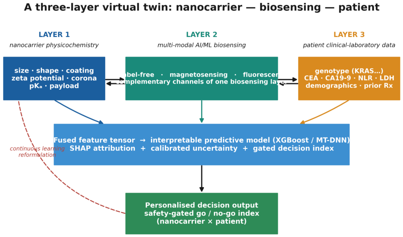
Figure 3.
Example. (A) Approximate prevalence in colorectal cancer of measurable patient-layer variables. (B) The corrected, safety-gated Personalised Therapeutic Index. Dividing by a safety index makes the score diverge as safety fails, perversely rewarding barrier-toxic candidates; a bounded logistic gate σ(k(BSI − θ)) suppresses unsafe candidates and preserves the intended safety semantics. Shown as one example of how the three layers combine into a decision output.
Figure 3.
Example. (A) Approximate prevalence in colorectal cancer of measurable patient-layer variables. (B) The corrected, safety-gated Personalised Therapeutic Index. Dividing by a safety index makes the score diverge as safety fails, perversely rewarding barrier-toxic candidates; a bounded logistic gate σ(k(BSI − θ)) suppresses unsafe candidates and preserves the intended safety semantics. Shown as one example of how the three layers combine into a decision output.
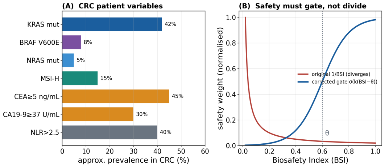
Figure 4.
Maturation of the three-layer virtual twin. The biosensing backbone (Stage 1) is already realised on Layer 1 with a well-populated intermediate layer; Stage 2 introduces the patient layer for a defined clinical context (colorectal cancer); Stage 3 couples to systemic pharmacokinetic modelling and outcome-validated benchmarking. A FAIR-curated dataset feeds periodic retraining, driving cumulative capability growth.
Figure 4.
Maturation of the three-layer virtual twin. The biosensing backbone (Stage 1) is already realised on Layer 1 with a well-populated intermediate layer; Stage 2 introduces the patient layer for a defined clinical context (colorectal cancer); Stage 3 couples to systemic pharmacokinetic modelling and outcome-validated benchmarking. A FAIR-curated dataset feeds periodic retraining, driving cumulative capability growth.
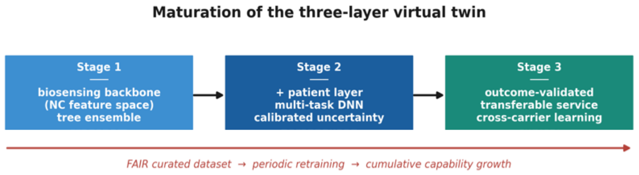

Table 1.
Example. Layer 3 for colorectal cancer. Every variable is measured in routine clinical practice; molecular prevalences and prognostic weights are from the published literature (Li et al., 2014; Lakemeyer et al., 2021; Li et al., 2022; Cattaneo et al., 2017; Tabat et al., 2020; Kovacic et al., 2019). These populate the patient feature space until a prospective cohort refines them. The same logic transfers to other cancer contexts with disease-specific variables in each of the four families of Section 5.1.
Table 1.
Example. Layer 3 for colorectal cancer. Every variable is measured in routine clinical practice; molecular prevalences and prognostic weights are from the published literature (Li et al., 2014; Lakemeyer et al., 2021; Li et al., 2022; Cattaneo et al., 2017; Tabat et al., 2020; Kovacic et al., 2019). These populate the patient feature space until a prospective cohort refines them. The same logic transfers to other cancer contexts with disease-specific variables in each of the four families of Section 5.1.
| Patient variable | Reference / threshold | Prevalence or weight | Role in the twin |
|---|---|---|---|
| KRAS mutation | codons 12/13 | ~40–45% | macropinocytotic uptake |
| BRAF V600E | - | ~7–9% | resistance; response index |
| MSI-H / dMMR | - | ~12–15% (stage-dep.) | immune context; organoid response |
| CEA | <5 / 5–200 / ≥200 ng mL⁻¹ | 5-yr OS ~69/44/7% | tumour-burden covariate |
| CA19-9 | ≥37 U mL⁻¹ | adverse prognosis | burden / prognosis |
| NLR | ≈2.5 threshold | adverse if high | systemic inflammation → endothelium |
| LDH | above upper normal | adverse | metabolic / hypoxic load |
| Biological sex | - | eNOS/NO higher in females | baseline barrier phenotype |
| Microbiome (SCFA) | butyrate status | context-dependent | barrier-tightness modifier |
| Prior FOLFOX | lines of therapy | EndoMT | elevated baseline barrier stress |
Disclaimer/Publisher’s Note: The statements, opinions and data contained in all publications are solely those of the individual author(s) and contributor(s) and not of MDPI and/or the editor(s). MDPI and/or the editor(s) disclaim responsibility for any injury to people or property resulting from any ideas, methods, instructions or products referred to in the content. |
© 2026 by the author. Licensee MDPI, Basel, Switzerland. This article is an open access article distributed under the terms and conditions of the Creative Commons Attribution (CC BY) license.
Copyright: This open access article is published under a Creative Commons CC BY 4.0 license, which permit the free download, distribution, and reuse, provided that the author and preprint are cited in any reuse.