

Submitted:
24 July 2026
Posted:
28 July 2026
You are already at the latest version
Abstract
Alzheimer’s disease (AD) in humans shares several features with canine cognitive dysfunction (CCD), a disease that affects older dogs. Therefore, CCD serves as a natural model for the study of AD. Several studies have documented early pathological changes in both AD and CCD, including widespread damage to the cerebral cortex and hippocampus, with this pathology attributed to the deposition of neurotoxic proteins such as beta-amyloid. Magnetic resonance imaging (MRI) is currently the only advanced imaging modality routinely used to support the diagnosis and prognosis of CCD. However, it is not commonly applied for the diagnosis or longitudinal monitoring of aging dogs with cognitive dysfunction. MRI can detect brain atrophy, hippocampal atrophy, ventricular enlargement, widening of the cerebral sulci, lesions in the temporal lobes of the cerebral cortex, and progressive thinning of the interthalamic adhesion in dogs undergoing natural or pathological aging. Accordingly, this imaging modality may serve as a valuable diagnostic marker for CCD in both early and advanced stages.
Keywords:
canine cognitive dysfunction
; magnetic resonance imaging
; Alzheimer´s disease
; dog
; dementia
1. Introduction
As in humans, aging in dogs induces progressive and irreversible changes in the body. These changes are often associated with cognitive deficits that manifest as alterations in interactive behaviors, such as elimination and spatial navigation [1]. Cognitive dysfunction has been widely studied in dogs, and research has shown that it is the canine analogue of Alzheimer’s disease (AD) in humans [2]. Canine cognitive dysfunction (CCD) is characterized by slowly progressive signs of mental impairment, dementia, and cognitive decline that are not attributable to normal aging or other medical conditions [3]. Research has further shown that CCD is the most common progressive neurodegenerative disorder in elderly dogs [4]. It is estimated that 14% to 35% of geriatric dogs may develop CCD [5].
The disorder is characterized by cortical atrophy, ventricular enlargement, demyelination, axonal degeneration, neuronal loss, meningeal calcification, lipofuscin accumulation, apoptotic bodies, and β-amyloid plaque deposition in various regions of the cerebral parenchyma [6]. Not all dogs develop signs of dementia as they age; some remain cognitively healthy. Dogs older than 8 years, an age associated with cerebral deposition of protein compounds and increased oxidative stress, may show multiple geriatric behavioral changes that can be broadly divided into 2 groups: changes consistent with normal aging and changes associated with CCD [7]. Pet owners and veterinarians who are unfamiliar with CCD often mistake its early clinical signs for normal aging. Furthermore, misconceptions persist that CCD is uncommon in dogs and that no therapeutic options are available to slow neurodegenerative changes in affected patients. These misconceptions contribute to misdiagnosis, delays in diagnosis, and inadequate treatment implementation [8]. In veterinary medicine, brief screening tests currently represent the only rapid diagnostic tools used to identify early and late stages of canine cognitive decline. However, screening tests alone cannot monitor the progression of the quantitative and qualitative changes in CCD over time, which is essential for recognizing the early stages of the disease. The use of advanced imaging modalities such as structural magnetic resonance imaging (MRI), in conjunction with brief screening tests, offers an opportunity for earlier diagnosis in dogs with CCD, thereby greatly increasing the likelihood of successful treatment [6]. In light of the lack of recent reviews on this topic, this manuscript examines the current state of knowledge regarding conventional MRI in the diagnosis of CCD. It emphasizes the use of MRI as a diagnostic marker for CCD by highlighting the distinct imaging findings associated with this condition. Combined with brief screening tools, these imaging findings may help prioritize early diagnosis and support more appropriate clinical and therapeutic decision-making for elderly dogs with this disease.
2. Pathophysiology
The most important risk factor for the development of dementia in both humans and animals is age [9,10]. It has been demonstrated that the cognitive capacities of both humans and dogs are affected by aging [11]. In a 2-year prospective longitudinal study, the prevalence of CCD was reported to be 28% in dogs aged 11 to 12 years and 68% in dogs aged 15 to 16 years, confirming that CCD increases with age in elderly dogs, similarly to AD in humans. Among dogs older than 8 years, 33% of those with normal cognition progressed to mild dementia, and 22% of those with mild dementia progressed to advanced CCD [12,13]. CCD can affect any breed; its prevalence does not differ significantly among breeds, and no marked breed-related differences have been reported in the presentation of clinical signs or in disease pathology. However, because large-breed dogs have a shorter life expectancy than small breeds, clinical signs of CCD are observed more frequently in small-breed dogs [3].
Beta-amyloid (Aβ) levels in brain tissue and cerebrospinal fluid (CSF) were measured in a companion dog study, in which CSF Aβ levels were found to correlate with body weight (BW). [13] One might expect large-breed dogs to develop CCD earlier, as occurs with other age-related pathologies, despite their shorter lifespan [14]. Nevertheless, one study showed that large-breed dogs do not appear to be affected by CCD at younger ages [15]. Various investigations have reported that dementia in both humans and dogs is multifactorial in origin, which complicates the study and understanding of its pathophysiology [16]. Most cases of AD are late-onset and of unknown etiology [17]. Companion dogs age more rapidly than humans despite sharing their owners’ environment and being exposed to several risk factors for AD and related disorders. This accelerated aging may help improve understanding of the environmental and genetic factors associated with age-related neurodegenerative diseases [13,18]. To date, no gene alterations have been reported in dogs with CCD [3]. Notably, CCD arises in diverse genetic backgrounds, and dogs are homozygous for the AD gene APOE4 [19].
The pathophysiological changes that occur in CCD show many similarities to those observed in humans, particularly with respect to deposits of Aβ and phosphorylated tau (TAU), which have also been reported in the brains of geriatric dogs [20]. Aβ is a neurotoxic protein that, in addition to forming plaques that accumulate in the cerebral parenchyma, is also deposited in neurons and the vascular endothelium, a feature observed in both AD and CCD [21]. Canine amyloid precursor protein (APP) is 98% similar to human APP, and the canine Aβ42 peptide is similar to that of humans [13]. In AD, the total amyloid burden is linked to the accumulation of Aβ42, which appears in various brain regions before Aβ40. The Aβ42/Aβ40 ratio in CSF reflects cerebral amyloid deposition because Aβ42 declines with age, whereas Aβ40 remains constant [10,22]. According to some canine studies, young dogs have higher plasma concentrations of Aβ than older dogs or dogs with CCD, suggesting a peripheral decrease in Aβ as cerebral deposition increases [16]. Aβ plaques are present in different areas of the cerebral parenchyma in elderly dogs with symptoms of CCD and may be detectable before clinical signs appear [3,10]. In dogs, Aβ deposition occurs in the prefrontal cortex, temporal cortex, hippocampus, and occipital cortex [23]. One study found higher Aβ levels in the prefrontal cortex of females than males, suggesting that females may be more susceptible to CCD [13]. Lesions in different brain areas are associated with behavioral deficits; thus, the proposed pattern of Aβ deposition suggests that the emergence of cognitive deficits reflects progression of the neurological disease [24].
In the aged canine brain, in addition to Aβ, microtubule-associated tau protein, the precursor of neurofibrillary tangles (NFTs), has also been described. [5] Similar to other species, not all dogs develop fully formed NFTs, which represent the second major neuropathological hallmark of AD [25]. Therefore, only humans develop the full pathology of AD, including Aβ plaques, NFTs, and cognitive impairment [26]. As a result, CCD may be regarded as a partial analogue of AD, reflecting age-related and Aβ-related processes, but not the full pathology observed in humans [27]. Recent studies have used methods such as transcriptomics to identify new mechanisms involved in AD. Transposable elements and microRNAs (miRNAs), which are carried in extracellular vesicles (EVs), are among the transcriptomic mechanisms implicated in AD and human brain aging. The prefrontal cortex transcriptome of older dogs with CCD exhibits patterns resembling those observed in aging and AD, according to a study conducted in companion dogs [28]. Neurodegenerative diseases render neurons vulnerable to multiple factors that compromise their integrity during disease progression, a process compounded by limited regenerative capacity with advancing age [29].
In addition to amyloid plaques, activation of astrocytes and glial cells has been observed in older dogs with CCD, corresponding to astrogliosis with astrocyte hypertrophy [30]. Dogs with CCD exhibit degeneration of noradrenergic neurons associated with Aβ deposits in the prefrontal cortex [31]. In both human and canine patients, cholinergic neurons are vulnerable to Aβ deposition; synaptic loss is related to disease progression, and destruction of cholinergic neurons contributes to dementia [32]. Mitochondrial dysfunction contributes to the development of both AD and CCD, as mitochondria undergo morphological and functional changes, including altered mitochondrial gene expression due to Aβ deposition.
Mitochondrial capacity has therefore been shown to decline in AD and CCD [23]. Evidence indicates that reduced endogenous antioxidant defenses in the brain, neuronal DNA damage, impaired mitochondrial function, increased free radical-mediated cellular damage, vascular compromise, and inflammation are interrelated processes that drive progressive cognitive decline in CCD [33].
Damage to the blood-brain barrier (BBB) in humans has been linked to neurodegenerative diseases and cognitive decline. Changes in BBB permeability can be detected and used as a biomarker for AD and other brain diseases by combining subtraction enhancement analysis (SEA) with immunomagnetic reduction (IMR) technology. BBB alterations in dogs with CCD were identified and measured in one study. However, although IMR detected changes in BBB permeability, SEA was unable to associate these changes with the clinical manifestations of CCD [10,34]. Neurobiological changes may explain some, but not all, of the clinical signs of cognitive decline in aging dogs [35]. Behavioral changes typically observed in CCD include disorientation, altered social interactions and house-training behavior, changes in the sleep-wake cycle and overall activity [6], and deterioration in memory and learning [36]. Traditionally, the clinical signs of CCD in dogs are assessed using the DISHA test, which evaluates disorientation, interaction changes, sleep-wake alterations, house-soiling, and activity changes [16]. Changes in behavior and daily routines are considered clinical markers of CCD. Dogs with CCD commonly display signs such as staring into space, wandering, avoidance of petting, and difficulty locating food that has been offered [37].
Behavioral manifestations of CCD show similarities to those observed in humans. Frequent findings include reduced coping ability, increased anxiety, and altered behavior [38]. In AD, noncognitive neuropsychiatric symptoms (NPS) are considered common. In a study using data from the Dog Aging Project, which included more than 10,000 dogs older than 8 years, behaviors resembling NPS in humans were identified in elderly dogs and in dogs with CCD. Dogs with CCD were found to be less active, to show reduced motivation and learned behavior, to sleep more during the day, to show greater reactivity to novelty and separation anxiety, to have reduced appetite, and to display more aggression [39]. Age-related social dysfunction in dogs may also be associated with sudden, spontaneous aggression or unprovoked behavior toward owners, unfamiliar people, or other dogs, which may indicate an underlying neurological or medical disorder [40]. Behavioral problems in elderly dogs may therefore represent either a continuation of issues acquired over the lifespan or characteristic features of age-related dementia [6].
Recent AD research has shown that patients not only exhibit prominent symptoms affecting cognition, mood, thought processes, and behavior but also demonstrate motor disturbances [41]. In human medicine, motor signs (MOSI) have been documented in AD [42]. In veterinary medicine, one study evaluated neurological examination findings and behavior in elderly dogs to identify potential associations. The study showed that neurological disorders occurred twice as often in elderly dogs with signs of CCD as in cognitively healthy dogs [11]. In veterinary medicine, rapid repetitive postural myoclonus of the pelvic limbs has occasionally been reported in elderly dogs; it is considered benign, rarely affects all 4 limbs, and may progress with age. In a recent review of involuntary contractions in dogs, tremors were classified as a form of myoclonic activity. Further research in elderly dogs is needed to determine whether an association exists between the presence of MOSI and CCD [43].
3. Diagnosis
The appropriate selection of available diagnostic tests is essential to support the clinical suspicion of CCD and to promote early supportive interventions. Consequently, the study of the clinical phenotype of CCD, its progression, and prognosis has become increasingly important in small animal practice [44]. Currently, the diagnosis of CCD relies on clinical history and signs compatible with the disorder [5]. In humans, Alzheimer’s disease (AD) is diagnosed in a similar stepwise manner, beginning with the exclusion of other possible causes of the symptoms, followed by analysis of biomarker levels in CSF and plasma, as well as assessment of memory, attention, and language. These evaluations may be supplemented by conventional and functional MRI and positron emission tomography (PET) [45,46]. As in dogs, the definitive etiology of dementia in humans can only be established via post-mortem studies [3]. In human medicine, dementia is commonly assessed using brief screening tools such as the Mini-Mental State Examination [47]. These tests are easy to administer and are used to evaluate dementia severity and document subsequent cognitive changes [48]. At present, no standardized guidelines have been established for the diagnosis of CCD. In veterinary medicine, rating scales are valuable tools for diagnosing and staging CCD because they allow evaluation and monitoring of clinical signs and assessment of therapeutic response [6,46]. Such scales are based on owner questionnaires and clinical ratings completed by veterinarians [38,49]. Results of CCD evaluations must always be interpreted alongside a clinical assessment to rule out or confirm systemic or primary disorders that may explain the patient’s signs [50].
The tests developed for CCD include a broad range of items assessing water and food consumption, aimless activity, social interactions, appetite, vocalization, diurnal and nocturnal rhythms, adaptive and perceptual capacity, disorientation, memory, and behavioral changes [49,51]. However, one major limitation is that the assessment of mental status in elderly dogs often depends on untrained owners evaluating behavioral traits, which introduces substantial variability [52,53]. To address these limitations, several rapid screening tools have been developed, among which the Canine Dementia Scale (CADES) is particularly notable. This brief questionnaire, which can be administered in clinical practice in approximately 30 min, evaluates cognitive domains such as orientation, social interaction, circadian rhythm, and learned household habits. It also enables identification of early memory changes and monitoring of decline over time.
The CADES scale has shown high utility and represents a significant advance over previously used CCD assessment tools. With CADES, three stages of cognitive impairment in elderly dogs have been identified, namely mild, moderate, and severe, and the degree of impairment at each stage has been quantified over time, including progression from normal cognition to mild impairment and from mild to moderate cognitive decline at 6 months and 1 year. This has allowed elderly dogs to be staged according to behavioral patterns [6,10]. Nevertheless, although current findings suggest that CCD is a multidomain behavioral disorder with rapid progression over time, the neuroanatomical changes associated with each stage of impairment defined by CADES cannot yet be identified or correlated with the observed cognitive decline in elderly dogs without complementary diagnostic markers such as MRI. MRI is a widely available, sensitive, specific, and noninvasive imaging modality. As a biomarker, it can provide additional information to support or refute a diagnosis of CCD and to monitor disease progression [49]. The combination of MRI and cognitive testing may therefore improve the diagnostic accuracy of CCD.
4. Biomarkers
AD presents a particular challenge in human medicine because it is difficult to identify and diagnose in its early stages. The identification of blood biomarkers that could enable early diagnosis or even prediction of the disease remains under investigation [3]. Compounds have been detected in both blood and CSF, but none has yet been adopted for routine clinical use. The biomarkers most frequently used in humans include CSF levels of amyloid-beta protein and total tau protein, together with PET molecular imaging, particularly for the diagnosis of early-stage AD [54]. Studies have shown that plasma changes in amyloid beta 1 (Aβ1) and amyloid beta 42 (Aβ42) associated with cerebral amyloid deposition are not readily detectable in dogs with CCD.
In recent years, increasing attention has been given to blood concentrations of neurofilament light chain (NfL) in AD, using new-generation immunoassay methods that allow quantification of structural brain markers in peripheral samples across a range of clinical conditions, including neurodegenerative diseases. Blood concentrations of NfL in dogs are low and are not detectable using conventional assays. Therefore, NfL has been regarded as a nonspecific marker that may help evaluate the extent of brain damage and quantify ongoing neuropathology when measured using immunoassay methods [55,56]. Advances in ultrasensitive detection methods have now made it possible to measure NfL in canine plasma [57]. According to one study, plasma NfL concentrations in dogs with CCD can be detected using immunomagnetic reduction (IMR), and dogs with CCD were shown to have higher plasma NfL levels than dogs without CCD [55]. Another study found that plasma NfL concentration may also be useful for evaluating cognitive impairment in elderly dogs. It was shown that plasma NfL levels could be used to assess cognitive damage when combined with validated CCD rating scales and IMR [58].
However, no biological markers are currently available for routine use in the diagnosis of CCD in dogs. In general, assessment of memory using currently available cognitive tests, together with exclusion of other diseases that produce similar signs, remains the primary approach for confirming the diagnosis once the disease has progressed. Therefore, implementation of novel diagnostic markers, such as structural MRI, for detecting CCD in both early and advanced stages would be of considerable value in the clinical setting for patients suspected of having this condition [3].
5. MRI as a Diagnostic Marker in CCD
Study of the pathophysiology of degenerative diseases of the central nervous system (CNS) is limited by the inaccessibility of brain tissue in living patients. Consequently, noninvasive imaging technologies such as MRI have long been of great value in both human and veterinary medicine. Within this context, the ability to monitor the progression of brain changes in vivo is considered a major objective in clinical studies of neurodegenerative diseases [9]. Initial structural MRI studies in humans with dementia were used to detect brain atrophy. Early diagnoses of AD were based on neuropathological examinations and computed tomography (CT), which revealed extensive medial temporal lobe (MTL) atrophy [59].
However, the low resolution and contrast of CT, structural MRI became the technique of choice for identifying CNS lesions. Early MRI studies in AD confirmed expected structural changes by demonstrating MTL degeneration, which was initially assessed through visual rating scales and later through measurements of total hippocampal volume [60]. As research progressed from small patient groups to larger cohorts, MRI became a central tool in longitudinal studies [9].
All MRI analytic methods have shown consistent findings, which have also been supported by visual assessment of hippocampal and posterior cingulate atrophy, precuneus involvement, and bilateral cortical thinning [61]. In veterinary medicine, the use of MRI as a diagnostic tool for CNS disorders has been comparatively limited; however, its application has expanded as an adjunct to cognitive evaluation in elderly dogs. As a result, MRI has gained increasing importance in the study of canine neurodegenerative disease. Currently, MRI is the only imaging modality considered practical for the diagnosis of CCD. In clinical veterinary practice, structural MRI is used to confirm lesions and identify their location in a variety of CNS disorders, including neoplastic, inflammatory, vascular, and congenital conditions. Nevertheless, MRI is not routinely used for the diagnosis or monitoring of geriatric patients, largely because many owners are reluctant to subject their dogs to MRI owing to the associated cost and anesthetic risk. In addition, limited awareness among veterinarians regarding the diagnostic value of MRI in CCD, the pathophysiology of the disease, and available therapeutic options further contributes to its underuse. Consequently, information on MRI features of CCD remains limited compared with the extensive body of evidence available for AD in human medicine [24].
MRI provides a noninvasive means of investigating brain changes associated with CCD in elderly dogs [62]. MRI examinations in dogs with CCD may reveal cerebral atrophy, hippocampal atrophy, enlargement of the ventricular system (ventriculomegaly), T2-weighted hyperintensities in the periventricular white matter, enlargement of the subarachnoid space with widened and well-defined cerebral sulci, lesions in the MTL cortex, progressive thinning of the interthalamic adhesion, and CSF accumulation in the cerebellomedullary cistern [11]. These findings, which are observed in both natural and pathological aging, support the use of MRI as a diagnostic marker of CCD in both early and advanced stages (Figure 1) [21]. Although histological features of CNS disease cannot be directly identified by MRI, clinical diagnosis can be supported by MRI findings that reflect the macroscopic morphological features of lesions. Differential diagnoses are established by integrating imaging findings with patient data and, when available, histopathological results. Confidence that an MRI abnormality is clinically relevant depends on the relationship between the lesion location and the neuroanatomical localization of the patient’s signs.
Several reports have described MRI findings in dogs with age-related neurodegeneration. These studies provide information on MRI abnormalities associated with such disorders and assist in the interpretation of images obtained from other patients. Retrospective evaluations of large groups of geriatric dogs with suspected CCD may also provide valuable information regarding the incidence of these abnormalities in dogs examined by MRI. Knowledge of the frequency of macroscopic and histological lesions in specific pathologies is particularly useful when interpreting MRI findings in dogs suspected of having CCD [63]. MRI features may also prove useful for monitoring treatment response and predicting prognosis in affected dogs.
6. Longitudinal MRI Designs for the Diagnosis of CCD
Cross-sectional MRI designs have certain limitations when used as the sole method for comparing brain changes in dogs with suspected CCD. One such limitation is interindividual variability in brain conformation, which reduces sensitivity for detecting structural differences among dogs of different ages and sexes. In addition, estimates of progressive atrophy based on a single scan assume that aging occurs linearly in all patients, whereas important abnormalities may be masked in single-scan evaluations that do not fully capture structural detail because of biological variability. Nevertheless, normalization of scans to total intracranial volume has been shown to reduce this variability [23]. In longitudinal MRI studies, each patient serves as its own control, thereby allowing quantification of progressive atrophy at the individual level. Longitudinal MRI studies over periods of 1 to 6 years have demonstrated different rates of cortical atrophy in human patients with AD compared with healthy controls. However, such studies in humans are complex to perform because of the long periods required to detect relevant morphometric differences [62].
In veterinary medicine, postmortem studies have demonstrated a correlation between canine brain degeneration and aging, including studies in beagles evaluated with various MRI techniques [64]. MRI studies of dementia in beagles are not affected by the same degree of difficulty as longitudinal studies in humans, because dogs share many of the cognitive, behavioral, and pathological features of aging seen in humans [65,66]. It has also been shown that canine life expectancy varies by breed, with large-breed dogs having shorter lifespans than smaller dogs. Therefore, biological age may differ among breeds even when chronological age is the same [67]. Moreover, dogs have much shorter lifespans than humans and age more rapidly. Beagles have an average life expectancy of approximately 13 years, with no major difference between sexes. A 5-year-old beagle is considered roughly comparable to a 40-year-old human, whereas a 9-year-old beagle corresponds approximately to a 60-year-old human. Beyond 2 years of age, 4 canine years roughly correspond to 16 years of human aging in this breed. This makes the beagle an appropriate model for studying brain aging in vivo by MRI over relatively short time intervals [23,68].
7. Visual Rating Scales in MRI for the Diagnosis of CCD
In humans, brain atrophy can be assessed using several MRI-based approaches, including visual rating scales. Visual rating methods have been shown to be faster, simpler, and more suitable for evaluating large numbers of scans [7,69]. An MRI study in dogs used an adapted version of a simple visual assessment originally designed for humans to determine whether progressive cerebral atrophy could serve as a marker of aging in dogs. This study included 27 dogs of different breeds, ages, and sexes and aimed to determine whether involutional changes in brain tissue were associated with aging. Because canine skulls vary considerably in shape and size, the images were also analyzed after classifying the dogs as dolichocephalic, mesaticephalic, or brachycephalic in order to determine whether cerebral degeneration was dependent on head conformation.
Focal brain atrophy was assessed in the frontal, parietal, temporal, and occipital regions. Global cerebral atrophy and hippocampal atrophy were also evaluated in each dog by MRI. The results showed that age-related cerebral degeneration was significantly greater in brachycephalic and mesaticephalic dogs, possibly because of genetic predisposition or ventricular dilation. The findings confirmed the presence of progressive age-related cerebral atrophy in dogs, including hippocampal atrophy, which parallels changes observed in aging humans, in whom progressive cerebral atrophy has also been identified by MRI. Furthermore, hippocampal atrophy was shown to play an important role in canine cerebral degeneration and may be considered an early marker of brain aging detectable by MRI. The visual scale showed good reliability when compared with linear and volumetric MRI measurements in humans [2,70,71].
Unlike normal aging, AD is typically characterized by atrophy of the MTL, entorhinal cortex, and hippocampus. These regional changes facilitate early detection of AD by MRI [72]. Qualitative assessments of focal brain atrophy in humans, when performed by experienced observers, have also been shown to be more useful than quantitative measures for evaluating hippocampal atrophy, mesial temporal sclerosis, and chronological aging [73]. Although quantitative assessments of cerebral atrophy using planimetric and volumetric MRI measurements show good discriminative ability, they are labor-intensive, require training, and may be costly in veterinary practice. By contrast, semiquantitative MRI evaluation of the canine brain using a visual rating scale provides highly repeatable measures of cerebral atrophy that correlate with aging. This visual scale therefore represents an accessible, reliable, rapid, and broadly applicable method for assessing cerebral atrophy in dogs using conventional MRI, with the added advantage that it does not require specialized training and can be used by veterinarians in routine clinical practice [7,74].
8. Cerebral Atrophy on MRI in the Diagnosis of CCD
MRI provides information on abnormalities affecting specific cerebral regions associated with aging [5]. Postmortem examinations in aged dogs have shown a decrease in the volume of the cerebral cortex and subcortical white matter as a consequence of enlargement of the ventricular system, resulting in marked ventriculomegaly [75]. Likewise, cortical degeneration identified on MRI as prominent sulci, thinning of brain tissue, and ventricular dilation in elderly Beagles and German Shepherds has been shown to progress with age and is considered a characteristic feature of aging in many dogs [11,76]. Periventricular signal changes are commonly observed on MRI in elderly dogs and are often evaluated when investigating potential causes of seizures, vestibular disease, or behavioral alterations (Figure 2 and Figure 3). Symmetrical bilateral T2-weighted hyperintensities in the white matter are thought to be associated with demyelination and gliosis [23].
9. Interthalamic Adhesion on MRI in the Diagnosis of Cerebral Atrophy in Dogs
Cross-sectional MRI studies in human patients with AD generally show differences in cortical atrophy between normal and pathological aging. Several studies have reported that cerebral atrophy is accompanied by enlargement of the third ventricle as a result of thalamic and possibly cortical atrophy (Figure 4) [62]. In veterinary medicine, a retrospective study of 78 dogs was conducted to define criteria for cerebral atrophy in dogs. The thickness of the interthalamic adhesion was evaluated on transverse MRI sections in healthy geriatric dogs and dogs with dementia. The interthalamic adhesion, located near the center of the sagittal section of the brain parenchyma, is a circular structure formed by fusion of the right and left thalami across the third ventricle. Its thickness was measured and compared to investigate normal variation, determine correlations with breed, age, and BW, and assess whether it could serve as a predictor of CCD. Measurements were obtained from T1-weighted imaging (T1WI) and T2-weighted imaging (T2WI) transverse images that included both the interthalamic adhesion and the third ventricle.
The results showed a negative correlation between interthalamic adhesion thickness and age and a positive correlation with BW. Breed type also influenced the results, with brachycephalic dogs showing significantly thinner adhesions. It was suggested that a thickness of 5.0 mm or less may represent a critical threshold for defining cerebral atrophy. Although global brain atrophy is unlikely to be diagnosed solely on the basis of interthalamic adhesion thickness, this parameter is quantifiable and may serve as an effective complementary marker of cerebral atrophy in dogs with suspected CCD [77]. Canine brains and skulls vary substantially in size and shape [78]. Body weight, breed, age, imaging sequence, and measurement plane all influence the measured height of the interthalamic adhesion [49].
In one study of elderly dogs, 2 metrics were used to assess brain atrophy: the ratio of lateral ventricle size to brain height and the ratio of interthalamic adhesion thickness to brain height (ITAr). Interthalamic adhesion thickness values were lower in dogs with CCD than in younger and older control dogs. The study also showed that using interthalamic adhesion thickness alone does not account for external sources of variation, such as age, brain size, or BW, across different breeds. By contrast, ITAr was found to be a reliable parameter for evaluating brain atrophy because it accounts for differences in brain size, BW, and skull type while remaining simple to measure and showing low variability [78]. In another study, T1-weighted (T1W) and T2-weighted (T2W) MRI sequences were used to measure interthalamic adhesion thickness (ITAt) and the ratio of ITAt to brain height across different breeds in order to adjust for age, sex, breed, and BW. Reference ranges for ITAt and ITAr were established while accounting for BW, MRI sequence type, and imaging plane. BW had no effect on ITAr, supporting its value as a stable parameter.
Diagnostic accuracy across breeds was further improved by using ITAt reference values within body weight-classified groups. In addition, ITAt values obtained from T1W sequences were higher than those from T2W sequences, likely because of differences in cerebrospinal fluid signal characteristics. The partial volume effect was also found to contribute to variations in ITAt measurements between sagittal and transverse planes. For this reason, the use of high-resolution 3-dimensional isotropic T1-weighted sequences with a slice thickness of 1 mm or less has been recommended [79]. To improve the accuracy of ITAt measurement, the use of BRAVO (brain volume imaging) or MPRAGE (magnetization-prepared rapid gradient echo) has also been suggested [49]. Nevertheless, MRI sequence type and imaging plane have been shown to affect both ITAt and hippocampal measurements. Accordingly, independent, nonstandardized reference values should be used for each sequence and measurement plane when assessing ITAt. This supports the need for prior experimental studies establishing reference values for each sequence, imaging plane, and anatomical structure of interest before these newer sequences are used routinely [79].
In the clinical setting, baseline sequences obtained with a standard brain MRI protocol across different imaging systems can be used to measure hippocampal height and interthalamic adhesion height [49,80]. Therefore, when T1-weighted baseline sequences are used together with validated reference values and standardized measurement planes, linear interthalamic adhesion parameters serve as accurate indicators of canine brain atrophy on conventional MRI (Figure 4). These measurements show strong intraobserver and interobserver agreement and are easy to apply in clinical practice, even by inexperienced personnel [78,80].
10. Hippocampal Atrophy on MRI in the Diagnosis of CCD
In human medicine, hippocampal atrophy can be evaluated by MRI, and this imaging modality is regarded as an important diagnostic marker of AD [21]. MRI-based volumetric measurements of the hippocampus have also been investigated as a means of assessing responses to future treatments targeting hippocampal function in AD. Hippocampal neuronal loss and synaptic alterations are considered key early features of AD in humans and are directly related to hippocampal atrophy, which in turn is associated with cognitive decline [81]. In veterinary medicine, an MRI study was conducted to quantify hippocampal volume in dogs with CCD. The aim of the study was to compare total hippocampal volume between dogs affected by CCD and cognitively healthy elderly dogs. The results showed that dogs with CCD had reduced hippocampal volumes compared with healthy dogs of similar age.
Although hippocampal alterations have been described in the brain parenchyma of dogs with cognitive dysfunction, hippocampal degeneration has not yet been definitively established as a pathological hallmark of the disorder [21]. Beyond its role as a central pathophysiological feature of AD in humans, the hippocampus is also a source of neural progenitor cells, and ongoing studies are investigating its role in the pathogenesis of AD as well as potential ways to positively influence hippocampal function in treatment [82]. Because CCD is a natural canine analogue of AD, research focused on the hippocampus in dogs with CCD may have translational value for both veterinary patients and humans with AD (Figure 5) [37].
11. Brain Volume and Ventricular Volume on MRI in the Diagnosis of Cortical Atrophy in CCD
Volumetric MRI has made it possible to identify morphometric changes in elderly dogs that are comparable to those observed in aging humans, including reductions or enlargements involving the frontal, parietal, temporal, and occipital lobes, as well as changes in total brain volume, ventricular volume, and hippocampal volume [7,83]. From a clinical perspective, the cerebral ventricles are diagnostically important cavities within the brain, and their variation in size may provide valuable information [84]. A longitudinal study involving 47 healthy beagle dogs that underwent annual MRI examinations over 3 years evaluated brain volume changes using volumetric region-of-interest techniques. The study assessed total brain volume, lateral ventricular volume, and longitudinal changes in cortical atrophy, drawing parallels with MRI studies of aging in human patients and beagles.
The results revealed several age-related morphometric changes in the canine brain. An increase in ventricular volume was observed and quantified beginning at 11 years of age. However, total brain volume did not change during this period, suggesting that cortical degeneration may manifest as deepening of the gyri and widening of the sulci rather than as overall brain contraction (Figure 3) [62,85]. The study also showed that ventricular size changed significantly with age but not with sex, suggesting that age-related changes are driven by the morphological and functional demands of aging on the canine brain, which indirectly affect the ventricles as well as other CNS structures [64].
Serial MRI scans also quantified regional changes and revealed the development of age-related lesions in the caudate nucleus and frontal cerebral cortex. Dogs began developing aging-related lesions after 11 years of age, and most had such lesions by 14 years of age. The MRI appearance of these lesions suggested that they may represent lacunar infarcts associated with cerebrovascular changes in dogs or, alternatively, a form of neurocysticercosis (Figure 6) [86]. Age-related increases in structural lesion formation in the frontal cerebral cortex were also shown to correlate with other markers of brain aging in dogs. Amyloid-beta plaque deposition occurs initially in the frontal cortex of elderly dogs before appearing in other cortical and subcortical regions. The frontal lobe is also particularly affected by aging, with its volume beginning to decrease between 8 and 10 years of age, in parallel with functional decline. Similarly, lesions were frequently observed in the caudate nucleus, a region with extensive reciprocal neuronal networks linking the frontal cortex and thalamocaudate regions. The lesions identified in the frontal and caudate regions may therefore reflect age-related changes within this frontothalamocaudate network. These findings provide strong support for the frontal lobe aging hypothesis in the beagle dog [66,87].
12. Intraparenchymal Cerebral Hemorrhage on MRI in CCD
Although microhemorrhages are commonly identified on MRI in AD, their prevalence in dogs with CCD has not been thoroughly investigated. Intraparenchymal cerebral hemorrhage has been reported in both human patients with AD and dogs with CCD [88]. T1-weighted and T2-weighted MRI sequences have proven useful for visualizing and differentiating hemorrhagic brain lesions in both human and canine patients. In AD, evidence of microhemorrhages and, in some cases, macrohemorrhages is frequently observed on T2-weighted images and has been associated with the disease. In one investigation of cerebral microhemorrhages in dogs with suspected CCD, T2-weighted MRI sequences showed that these lesions occurred more frequently in elderly small-breed dogs. This study also found that such dogs were more likely to present with vestibular disorders [5].
Microhemorrhages are readily identifiable on T2-weighted images in the brain parenchyma of geriatric dogs evaluated for suspected seizure activity, acute vestibular dysfunction, or behavioral abnormalities. In addition, cases of macrohemorrhage have been documented in elderly dogs without an identifiable underlying cause of spontaneous cerebral bleeding [89].
13. Neurodegenerative and Metabolic Changes on MRI in the Diagnosis of CCD
Neurodegenerative diseases and disorders caused by metabolic abnormalities are uncommon conditions that arise as a result of disturbances in endogenous homeostasis or exogenous toxic effects on neurotransmitters, physiological pH, blood flow, metabolic substrates, and electrolytes [16]. The nature of the changes observed in affected patients depends on the extent of metabolic product accumulation, the specificity of the affected cell population, the anatomical localization of neuronal damage, and the stage of maturation of the brain parenchyma [5]. It has been shown that lysosomal storage diseases (LSDs) are relevant to the study of brain aging in dogs. Investigations in dogs with hereditary metabolic disorders have provided valuable insight into the mechanisms of brain aging, including those involved in lysosomal storage diseases, leukodystrophies, and mitochondrial disorders. These disorders produce neuropathological changes similar to those observed in geriatric dogs. Knowledge derived from these conditions has supported both cross-sectional and longitudinal MRI studies aimed at evaluating disease progression [23].
Several hereditary lysosomal diseases have been reported, all of which are characterized by memory loss, cerebral atrophy, neuronal loss in specific brain regions, and demyelination, all of which resemble changes associated with aging. Some LSDs primarily affect the cerebral white matter. Among them, neuronal ceroid lipofuscinosis is characterized by particularly severe cerebral atrophy. Neurodegenerative and metabolic diseases share similar clinical and MRI features, which makes diagnosis challenging and often necessitates multiple tests to achieve a definitive diagnosis. In veterinary medicine, numerous case reports have examined the MRI characteristics of different metabolic encephalopathies in small animals [90]. MRI findings are typically characterized by symmetrical bilateral lesions in the brain parenchyma. However, these abnormalities may be nonspecific, thereby broadening the list of differential diagnoses and delaying definitive diagnosis (Figure 7) [91].
One study in dogs aimed to classify patients with different LSDs and neurodegenerative disorders on the basis of MRI characteristics, which allowed the development of a pattern-recognition system to guide diagnosis of these conditions. All patients in that study showed MRI features of severe cerebral atrophy affecting the prosencephalon and cerebellum, resulting from accumulation of lipofuscin-like lipopigments, in agreement with previous studies [92]. In companion animals with lipofuscinosis, diffuse pachymeningeal enhancement has also been described, although this finding has not been reported in humans. MRI additionally revealed diffuse T2-weighted hyperintensity of the cerebral white matter, particularly in the corona radiata and internal capsule (Figure 8). These findings are consistent with previous veterinary reports [93]. However, because of the limited number of reported cases, it remains difficult to establish MRI features that are specific to lysosomal storage diseases. Further studies are needed to evaluate these diseases more comprehensively. At present, clinicians must rely on MRI findings in combination with histopathological examination and molecular genetic testing for each lysosomal disease to reach a diagnosis.
14. Treatment
The treatments currently available for AD and CCD are symptomatic and must be tailored to each patient because, unfortunately, no proven curative therapies yet exist for neurodegenerative disorders affecting humans or animals. Successful management of AD and CCD is further complicated by the challenge of preserving the patient’s social role and quality of life [3]. The nature of both conditions means that available treatment options vary in their degree of success. Although some drugs have shown evidence of exerting physiological effects on the neuroanatomy of affected patients, their clinical impact is often limited. Despite approval by the US Food and Drug Administration (FDA) of Aducanumab for AD, no widely effective and accessible treatment for dementia has been available since 2004, highlighting the urgent need for innovative therapeutic strategies [94].
Accordingly, a range of diagnostic and therapeutic approaches has been developed for CCD, which naturally shares features with AD in humans [95]. Recent studies have shown that cell-based therapies for neurodegenerative diseases in human patients, using canine models, aim to restore neurons and synapses through donor cells and EV such as cell-derived exosomes [96]. One study described a novel method for cultivating unipotent skin-derived neural precursors (SKNs) from canine tissue. Unlike rodent models used in previous studies, these cells do not exhibit gene alterations or express pluripotency markers. They can be generated reliably and uniformly from a small donor skin sample, are capable of proliferation and differentiation in vitro into viable neurons, and do not express glial proteins. This study reported the first veterinary clinical application of SKN cell therapy for CCD and suggested a possible role in restoring lost brain tissue and addressing the biological basis of neurodegeneration and cognitive dysfunction [97].
Epidemiological studies have suggested that nutraceutical products may provide some behavioral and lifestyle benefits in humans and dogs with neurodegenerative diseases, although scientific evidence supporting their use remains limited. Therefore, pharmacological and nutraceutical interventions in AD and CCD are primarily intended to reduce the physiological changes associated with pathological brain aging while continuing to support the patient’s ability to function in a social environment [98]. It has been reported that treatment outcomes in humans and animals are similar with different drugs used for AD and CCD, which suggests that dogs may serve as a valuable model not only for studying pathogenesis but also for developing future treatments for AD. Dogs with CCD generally respond well to available treatment options, particularly when the disease is diagnosed in its early stages. Routine use of novel diagnostic tools such as MRI for neurodegenerative disease, in addition to existing behavioral questionnaires, may allow earlier initiation of therapeutic strategies. Early intervention generally provides therapeutic benefit with few or no adverse effects [3].
Several studies suggest that geriatric dogs with CCD may benefit from treatments including anxiolytics, anticonvulsants, cerebral perfusion enhancers, antioxidant preparations, and behavioral training programs (Figure 9) [38,98]. Research in AD has demonstrated the effectiveness of transcranial photobiomodulation (tPBMT) as a treatment for cognitive impairment. Transcranial laser therapy has been shown to increase cerebral blood flow, promote mitochondrial adenosine triphosphate synthesis, and reduce beta-amyloid production and accumulation around blood vessels and neurons. In addition, it promotes the synthesis of neurotrophic factors while reducing oxidative damage, inflammation, and neuronal and dendritic loss. A study in dogs reported improvement in CCD cognitive scores after 60 days of tPBMT use [99].
In dogs with CCD, treatment with cholinesterase inhibitors, secretase inhibitors, and anti-amyloid-beta immunotherapy has been investigated, with some results reported to exceed those observed in AD clinical trials in humans [5]. One possible target for alleviating symptoms of cognitive dysfunction is butyrylcholinesterase (BChE). In one study, a BChE inhibitor known as (R)-(-)-3 was developed and may help improve cholinergic dysfunction and cognitive ability in AD and CCD [100]. Another study evaluated the safety and efficacy of donepezil in dogs. This selective acetylcholinesterase inhibitor was shown to be well tolerated and to exert neuroprotective effects, improving cholinergic transmission in AD and CCD and thereby enhancing cognition, social interaction, and coexistence [53].
By regulating neurotransmitter release, synaptic activity, potentiation, depression, and memory, calcineurin (CN), a key brain signaling molecule, plays an essential role in cognition. Increased CN activity and activation of nuclear factor of activated T-cells (NFAT) have been associated with synaptic loss, neuroinflammation, amyloid-beta production, neurodegeneration, and cognitive decline in AD. In one preventive study, 37 middle-aged beagles received Tacrolimus, an FDA-approved drug that suppresses CN activity and inhibits T-cell activation and NFAT signaling. Targeting the CN/NFAT pathway showed protective and inhibitory effects, as evidenced by improvements in learning, memory, and attention [101]. Cognitive therapy combined with an antioxidant-rich diet has also demonstrated neuroprotective benefits that help slow progression of neurodegeneration in both humans and elderly dogs [5,23]. Thus, the cornerstone of CCD treatment lies in early detection and timely diagnosis, which allow implementation of strategies that prolong good quality of life for the patient, supported by the active involvement of an engaged and interactive family.
15. A Future Perspective on the Diagnosis of CCD through Functional MRI
Previous studies have provided evidence of white matter abnormalities in AD, including axonal loss and demyelination, and some research suggests that myelin damage may precede formation of neurofibrillary tangles, raising the possibility that AD may also be considered, at least in part, a demyelinating disorder. For this reason, development of imaging techniques that assess white matter integrity has become increasingly important. White matter loss may also reveal new targets for early therapeutic intervention [102]. Early amyloid deposition in the motor cortex, together with demyelination, has been suggested to contribute to cognitive changes and may also underlie the motor disorders observed in AD in the absence of neuropathological confirmation [11].
Diffusion techniques routinely used in the diagnosis of AD in humans, including diffusion-weighted imaging (DWI) and diffusion tensor imaging (DTI), are valuable methods for assessing white matter demyelination in vivo in dogs with CCD. At present, these techniques in veterinary medicine have been applied only in controlled experimental studies involving specific breeds in laboratory settings rather than in routine clinical practice. However, their promising results suggest that in the future they may serve as additional diagnostic markers that complement the structural MRI techniques already used in the diagnosis of CCD in dogs. Functional MRI techniques are expected to gain importance in veterinary medicine as MRI becomes better understood, more widely available, and more accessible to both veterinarians and pet owners [103].
It has been demonstrated that the aging canine brain exhibits age-dependent myelin loss similar to the white matter abnormalities associated with dementia in humans [83]. Additional quantitative techniques for assessing cellular inflammation, neuronal loss, demyelination, and gliosis may also be useful for identifying specific lesions. These include magnetization transfer ratio (MTR), which has been used to improve characterization of white matter neurodegeneration through magnetization transfer imaging [104], as well as DWI for detection of cellular inflammation and other techniques for evaluating neuronal loss and gliosis [105].
16. DWI
DWI is a functional MRI technique used in the diagnosis of neurodegenerative diseases in humans. DWI detects the random motion of water within the cerebral white matter, which is altered by various cerebral lesions, including neurodegenerative processes, cerebral infarction, and tumors, all of which have been shown to modify water distribution within fibers of the CNS [63]. The apparent diffusion coefficient (ADC) provides a quantitative estimate of water movement in brain tissue and is calculated from DWI using images acquired with different b values. Water within the ventricles typically shows decreased signal intensity on DWI and increased ADC values. In human patients with acute cerebral infarction, the resulting edema appears hyperintense on DWI and hypointense on ADC maps [106].
Decreased ADC values are associated with reduced extracellular volume in cerebral ischemia as a result of cellular swelling. Once this cellular swelling resolves and necrosis develops, ADC values tend to increase [107]. Similar associations between reduced ADC values and cellular swelling have been demonstrated in conditions such as hypoglycemia, toxin exposure, and status epilepticus [108]. ADC measurements can also be used to distinguish vasogenic edema, which increases extracellular volume, from cytotoxic edema, which reduces it [109]. Beyond reflecting cellular swelling and extracellular space volume, extracellular water mobility also appears to be influenced by its intrinsic properties and the presence of different cell types. Accordingly, ADC has also been used in human patients to assess brain injury [110], differentiate cysts from abscesses and tumors, and determine tumor cellularity and grade [111].
In veterinary medicine, the routine use of DWI and ADC in small animal practice and in the diagnosis of CCD remains limited. However, several experimental studies in dogs have reported the use of DWI and ADC for diagnosing and characterizing acute and chronic ischemic stroke conditions associated with CCD [112], as well as for identifying edema following status epilepticus and cerebral infarction [113]. In domestic cats, ADC has been used to identify brain tissue inflammation associated with experimental ischemia and with naturally occurring alpha-mannosidosis, a lysosomal storage disease. In cats affected by alpha-mannosidosis, brain tissue inflammation, gliosis, and astrogliosis occur simultaneously and all contribute to reduced ADC values [114].
17. DTI
In neuroscience research, there is a need for methods that record neurological function and quantify the effects of brain disease on both structure and function [115]. DTI is a functional MRI technique that quantifies restriction of Brownian motion by measuring the directionality and magnitude of water diffusion within cerebral white matter fibers. In human medicine, it is used to investigate the intrinsic properties of white matter in the CNS [116]. DTI has proved highly valuable in the brains of human patients for detecting and quantifying age-related neurodegeneration, and it has shown strong potential for predicting progression to age-related dementia [117]. DTI-based images have also shown promise for assessing the integrity of cerebral white matter in both normal aging and Alzheimer’s disease (AD) in humans [118]. Fractional anisotropy (FA) is the primary diffusion metric derived from DTI and quantifies the anisotropy of diffusion in brain tissue [119].
In cerebral white matter, FA largely reflects axonal structure that restricts diffusion in directions not aligned with tract orientation. Although DTI measurements in white matter are influenced by multiple cellular characteristics, including fiber orientation, myelination, and axonal density, FA is generally interpreted as a marker of axonal integrity [120]. Other tensor-derived metrics, including radial diffusivity (RD), mean diffusivity (MD), and axial diffusivity (AxD), provide additional characterization of water diffusion within a voxel. RD and AxD have been associated with axonal loss and demyelination in preclinical studies and may therefore aid interpretation of functional MRI findings [121]. Studies in healthy older humans have shown widespread decreases in FA and increases in MD in cerebral white matter, following a gradient of neurodegeneration over time [122]. This aging pattern is characterized by degeneration that begins anteriorly in the frontal lobe and progresses in an anterior-to-posterior gradient along the z-axis of structural MRI scans. MRI studies have also identified an inverse relationship between FA, myelination, and lifespan, particularly in the corpus callosum and frontal lobe [123].
These findings in humans correspond well with ex vivo studies showing age-dependent myelin loss in the cerebral white matter of elderly dogs [124]. Because dogs live closely with humans as companion animals, they are exposed to environmental factors similar to those affecting older people, among whom progression of neurodegenerative disease is frequently documented. Functional MRI studies have demonstrated associations between human and canine reasoning, particularly in the temporo-occipital regions during presentation of different objects, as well as activation of canine olfactory pathways associated with species recognition. DTI has also been used to generate diffusion tensor models that reconstruct orientation trajectories of cerebral white matter. Consequently, atlases have been developed for several research species, including dogs, nonhuman primates, rodents, cats, sheep, and horses, thereby enabling comparative studies of white matter tracts across species [115,125]. DTI has additionally been used to delineate specific cerebral white matter tracts in the canine brain by tractography, which assists in identifying distinct white matter pathways [126] and in detecting cerebral diffusivity alterations associated with aging [83], epilepsy [127], and glaucoma [128]. These studies represent an important contribution to the emerging field of functional MRI in small animal species and to comparative investigations of canine brain evolution and function [129].
One study used DTI to monitor cerebral white matter changes associated with aging in a group of dogs by quantifying in vivo changes in cerebral white matter fibers. Decreases in FA and AxD were observed in elderly dogs compared with younger dogs. Other age-related tensor changes included reduced FA in the fornix, corpus callosum, and temporal region, as well as decreased AxD in the frontal lobe and midbrain. These findings suggest axonal degeneration or loss and may help distinguish normal aging from pathological demyelination in elderly dogs. The generalized age-related decreases in FA identified in this study were consistent with findings from most human aging studies [130]. The results further highlight the importance of dogs as a translational model for neurological aging, since regional decreases in diffusion metrics were identified in canine brain regions analogous to those affected by aging in humans [131]. Consistent findings in humans and dogs, together with proposed shared mechanisms of aging, the detection of beta-amyloid in elderly dogs with CCD, and the similar lifestyles of these companion animals, provide a novel perspective for studying aging in elderly dogs using DTI.
18. Magnetization Transfer Imaging (MTI) and MTR
MTI is a complementary technique to structural MRI, DWI, and DTI in the diagnosis of neurodegenerative diseases such as AD in humans. This technique increases lesion detection sensitivity by enhancing contrast in MR angiography, gadolinium-enhanced imaging, and T2-weighted imaging for identification of various demyelinating diseases affecting the cerebral parenchyma in humans and animals [132]. MTI of brain tissue generates contrast through the signal intensity produced by magnetization exchange between protons in brain parenchyma and protons in water. By selectively saturating the signal of macromolecules such as myelin in brain tissue, MTI substantially increases tissue contrast and improves sensitivity for detecting CNS disease in neurodegenerative processes such as AD and CCD [133].
The MTR is the quantitative expression of MTI and enables identification of cerebral white matter abnormalities. Its value is well illustrated in imaging studies of multiple sclerosis in humans, because T2-weighted images alone are insufficient to characterize the heterogeneity of lesions with respect to demyelination, remyelination, inflammation, axonal injury, and gliosis [104]. MTR analysis can reveal histological differences between affected and unaffected brain tissue. It has also been used to distinguish demyelination from other conditions such as edema and to quantify myelin maturation, an important feature in disease characterization through detection of periventricular white matter hyperintensities in older patients [134]. In addition, MTR has been used to quantify cerebral white matter lesions in traumatic brain injury and neurodegenerative processes [135]. Although MTI and MTR are not yet available as routine diagnostic tools for neurodegenerative diseases in dogs, their wide range of applications in humans suggests that they may represent promising future options in the diagnosis of CCD in elderly dogs.
19. Conclusions
Given the many similarities between AD and CCD, CCD is highly valuable as a model for studying neurodegenerative disease in humans. Simultaneously, CCD is recognized as an important and growing health problem in elderly dogs and is therefore of considerable interest for the development of new veterinary diagnostic markers. Among these, MRI, when used in combination with existing diagnostic tools, offers strong potential for the early detection of CCD. Early diagnosis would allow the implementation of available treatment options during the initial stages of the disorder, thereby improving the quality of life of affected dogs as well as that of their owners.
Author Contributions
Conceptualization M.S.A.G.; methodology M.S.A.G..; software, J.S.H.A.; validation, E.C.S.M. and R.E.M.A..; formal analysis, M.S.A.G.; investigation, M.S.A.G. and J.S.H.A..; resources, M.S.A.G..; data curation M.S.A.G. and J.S.H.A..; writing—original draft preparation, M.S.A.G..; writing—review and editing, M.S.A.G. and J.S.H.A..; visualization, E.C.S.M and R.E.M.A.; supervision, J.S.H.A., E.C.S.M. and R.E.M.A..; project administration, M.S.A.G..;. All authors have read and agreed to the published version of the manuscript.
Funding
This research received no external funding.
Institutional Review Board Statement
Not applicable.
Informed Consent Statement
Not applicable.
Data Availability Statement
The original contributions presented in this study are included in the article. Further inquiries can be directed to the corresponding author.
Acknowledgments
We would like to thank Editage (www.editage.com) for English language editing.
Conflicts of Interest
The authors declare no conflicts of interest.
Abbreviations
The following abbreviations are used in this manuscript:
Aβ beta-amyloid
Aβ1 amyloid beta 1
Aβ40 amyloid beta 40
Aβ42 amyloid beta 42
ADC apparent diffusion coefficient
AD Alzheimer’s disease
APOE4 apolipoprotein E4
APP amyloid precursor protein
AxD axial diffusivity
BBB blood-brain barrier
BChE butyrylcholinesterase
BW body weight
CADES Canine Dementia Scale
CCD canine cognitive dysfunction
CN calcineurin
CNS central nervous system
CSF cerebrospinal fluid
CT computed tomography
DISHA disorientation, interaction changes, sleep-wake alterations, house-soiling, and activity changes
DTI diffusion tensor imaging
DWI diffusion-weighted imaging
EV extracellular vesicles
FA fractional anisotropy
FDA Food and Drug Administration
IMR immunomagnetic reduction
ITAr ratio of interthalamic adhesion thickness to brain height
ITAt interthalamic adhesion thickness
LSD lysosomal storage disease
MD mean diffusivity
miRNAs microRNAs
MOsI motor signs
MPRAGE magnetization-prepared rapid gradient echo
MRI magnetic resonance imaging
MTL medial temporal lobe
MTI magnetization transfer imaging
MTR magnetization transfer ratio
NFAT nuclear factor of activated T-cells
NfL neurofilament light chain
NFTs neurofibrillary tangles
NPS neuropsychiatric symptoms
PET positron emission tomography
RD radial diffusivity
SEA subtraction enhancement analysis
TAU phosphorylated tau
T1W T1-weighted
T1WI T1-weighted imaging
T2W T2-weighted
T2WI T2-weighted imaging
tPBMT transcranial photobiomodulation
References
- Shepherd, A.J. Results of the 2007 AVMA survey of US pet-owning households regarding use of veterinary services and expenditures. J. Am. Vet. Med. Assoc. 2008, 233, 727–728. [Google Scholar] [CrossRef] [PubMed]
- Bosch, M.N.; Pugliese, M.; Gimeno-Bayón, J.; Rodríguez, M.J.; Mahy, N. Dogs with cognitive dysfunction syndrome: a natural model of Alzheimer’s disease. Curr. Alzheimer Res. 2012, 9, 298–314. [Google Scholar] [CrossRef] [PubMed]
- Prpar Mihevc, S.; Majdič, G. Canine cognitive dysfunction and Alzheimer’s disease—two facets of the same disease? Front Neurosci. 2019, 13, 604. [Google Scholar] [CrossRef] [PubMed]
- Landsberg, G.; Araujo, J.A. Behavior problems in geriatric pets. Vet. Clin. North Am. Small Anim. Pract. 2005, 35, 675–698. [Google Scholar] [CrossRef] [PubMed]
- Dewey, C.W.; Davies, E.S.; Xie, H.; Wakshlag, J.J. Canine cognitive dysfunction: pathophysiology, diagnosis, and treatment. Vet. Clin. North Am. Small Anim. Pract. 2019, 49, 477–499. [Google Scholar] [CrossRef] [PubMed]
- Madari, A.; Farbakova, J.; Katina, S.; Smolek, T.; Novak, P.; Weissova, T.; Novak, M.; Zilka, N. Assessment of severity and progression of canine cognitive dysfunction syndrome using the Canine DEmentia Scale (CADES). Appl. Anim. Behav. Sci. 2015, 171, 138–145. [Google Scholar] [CrossRef]
- Pugliese, M.; Carrasco, J.L.; Gomez-Anson, B.; Andrade, C.; Zamora, A.; Rodríguez, M.J.; Mascort, J.; Mahy, N. Magnetic resonance imaging of cerebral involutional changes in dogs as markers of aging: an innovative tool adapted from a human visual rating scale. Vet. J. 2010, 186, 166–171. [Google Scholar] [CrossRef] [PubMed]
- Fahnestock, M.; Marchese, M.; Head, E.; Pop, V.; Michalski, B.; Milgram, W.N.; Cotman, C.W. BDNF increases with behavioral enrichment and an antioxidant diet in the aged dog. Neurobiol. Aging 2012, 33, 546–554. [Google Scholar] [CrossRef] [PubMed]
- Blamire, A.M. MR approaches in neurodegenerative disorders. Prog. Nucl. Magn. Reson Spectrosc. 2018, 108, 1–16. [Google Scholar] [CrossRef] [PubMed]
- Yoon, J.-W.; Nam, C.-S.; Lee, K.-S.; Dan, T.-J.; Jeon, H.-J.; Kang, M.-A.; Park, H.-M. Evaluation of blood-based diagnostic biomarkers for canine cognitive dysfunction syndrome. Animals 2025, 15, 1974. [Google Scholar] [CrossRef] [PubMed]
- Golini, L.; Colangeli, R.; Tranquillo, V.; Mariscoli, M. Association between neurologic and cognitive dysfunction signs in a sample of aging dogs. J. Vet. Behav. 2009, 4, 25–30. [Google Scholar] [CrossRef]
- Salvin, H.E.; McGreevy, P.D.; Sachdev, P.S.; Valenzuela, M.J. Under diagnosis of canine cognitive dysfunction: a cross-sectional survey of older companion dogs. Vet. J. 2010, 184, 277–281. [Google Scholar] [CrossRef] [PubMed]
- Urfer, S.R.; Darvas, M.; Czeibert, K.; Sándor, S.; Promislow, D.E.L.; Creevy, K.E.; Kubinyi, E.; Kaeberlein, M. Canine cognitive dysfunction (CCD) scores correlate with amyloid beta 42 levels in dog brain tissue. GeroScience 2021, 43, 2379–2386. [Google Scholar] [CrossRef] [PubMed]
- Urfer, S.R.; Kaeberlein, M.; Promislow, D.E.L.; Creevy, K.E. Lifespan of companion dogs seen in three independent primary care veterinary clinics in the United States. Canine Med. Genet 2020, 7, 7. [Google Scholar] [CrossRef] [PubMed]
- Watowich, M.M.; MacLean, E.L.; Hare, B.; Call, J.; Kaminski, J.; Miklósi, Á.; Snyder-Mackler, N. Age influences domestic dog cognitive performance independent of average breed lifespan. Anim. Cogn. 2020, 23, 795–805. [Google Scholar] [CrossRef] [PubMed]
- Landsberg, G.M.; Nichol, J.; Araujo, J.A. Cognitive dysfunction syndrome: a disease of canine and feline brain aging. Vet. Clin. North Am. Small Anim. Pract. 2012, 42, 749–768. [Google Scholar] [CrossRef] [PubMed]
- Cacace, R.; Sleegers, K.; Van Broeckhoven, C. Molecular genetics of early-onset Alzheimer’s disease revisited. Alzheimers Dement 2016, 12, 733–748. [Google Scholar] [CrossRef] [PubMed]
- Schütt, T.; Helboe, L.; Pedersen, L.Ø.; Waldemar, G.; Berendt, M.; Pedersen, J.T. Dogs with cognitive dysfunction as a spontaneous model for early Alzheimer’s disease: a translational study of neuropathological and inflammatory markers. J. Alzheimers Dis. 2016, 52, 433–449. [Google Scholar] [CrossRef] [PubMed]
- Valenzuela, M.; Duncan, T.; Abey, A.; Johnson, A.; Boulamatsis, C.; Dalton, M.A.; Jacobson, E.; Brunel, L.; Child, G.; Simpson, D.; et al. Autologous skin-derived neural precursor cell therapy reverses canine Alzheimer dementia-like syndrome in a proof of concept veterinary trial. Stem Cell Res. Ther. 2022, 13, 261. [Google Scholar] [CrossRef] [PubMed]
- Head, E.; Moffat, K.; Das, P.; Sarsoza, F.; Poon, W.W.; Landsberg, G.; Cotman, C.W.; Murphy, M.P. β-amyloid deposition and tau phosphorylation in clinically characterized aged cats. Neurobiol. Aging 2005, 26, 749–763. [Google Scholar] [CrossRef] [PubMed]
- Dewey, C.W.; Rishniw, M.; Johnson, P.J.; Platt, S.; Robinson, K.; Sackman, J.; O’Donnell, M. Canine cognitive dysfunction patients have reduced total hippocampal volume compared with aging control dogs: a comparative magnetic resonance imaging study. Open Vet. J. 2020, 10, 438–442. [Google Scholar] [CrossRef] [PubMed]
- Nakamura, A.; Kaneko, N.; Villemagne, V.L.; Kato, T.; Doecke, J.; Doré, V.; Fowler, C.; Li, Q.X.; Martins, R.; Rowe, C.; et al. High performance plasma amyloid-β biomarkers for Alzheimer’s disease. Nature 2018, 554, 249–254. [Google Scholar] [CrossRef] [PubMed]
- Vite, C.H.; Head, E. Aging in the canine and feline brain. Vet. Clin. North Am. Small Anim. Pract. 2014, 44, 1113–1129. [Google Scholar] [CrossRef] [PubMed]
- Head, E.; Callahan, H.; Muggenburg, B.A.; Cotman, C.W.; Milgram, N.W. Visual-discrimination learning ability and β-amyloid accumulation in the dog. Neurobiol. Aging 1998, 19, 415–425. [Google Scholar] [CrossRef] [PubMed]
- Morys, J.; Narkiewicz, O.; Maciejewska, B.; Wegiel, J.; Wisniewski, H.M. Amyloid deposits and loss of neurones in the claustrum of the aged dog. NeuroReport 1994, 5, 1825–1828. [Google Scholar] [CrossRef] [PubMed]
- Kuroki, K.; Uchida, K.; Kiatipattanasakul, W.; Nakamura, S.; Yamaguchi, R.; Nakayama, H.; Doi, K.; Tateyama, S. Immunohistochemical detection of Tau protein in various non-human animal brains. Neuropathology 1997, 17, 174–180. [Google Scholar] [CrossRef]
- Abey, A.; Davies, D.; Goldsbury, C.; Buckland, M.; Valenzuela, M.; Duncan, T. Distribution of tau hyperphosphorylation in canine dementia resembles early Alzheimer’s disease and other tauopathies. Brain Pathol. 2021, 31, 144–162. [Google Scholar] [CrossRef] [PubMed]
- Osburn, S.C.; Easterday, D.S.; McEntee, C.M.; Latham, A.S.; Lark, D.S.; McGrath, S.; Moreno, J.A.; LaRocca, T.J. Evidence of emerging transcriptome mediators of Alzheimer’s disease in canine cognitive dysfunction. Brain Pathol. 2025, 36, e70062. [Google Scholar] [CrossRef] [PubMed]
- Saikia, B.; Dhanushkodi, A. Engineered exosome therapeutics for neurodegenerative diseases. Life Sci. 2024, 356, 123019. [Google Scholar] [CrossRef] [PubMed]
- Smolek, T.; Madari, A.; Farbakova, J.; Kandrac, O.; Jadhav, S.; Cente, M.; Brezovakova, V.; Novak, M.; Zilka, N. Tau hyperphosphorylation in synaptosomes and neuroinflammation are associated with canine cognitive impairment. J. Comp. Neurol. 2016, 524, 874–895. [Google Scholar] [CrossRef] [PubMed]
- Insua, D.; Suárez, M.-L.; Santamarina, G.; Sarasa, M.; Pesini, P. Dogs with canine counterpart of Alzheimer’s disease lose noradrenergic neurons. Neurobiol. Aging 2010, 31, 625–635. [Google Scholar] [CrossRef] [PubMed]
- Bell, K.F.S.; Ducatenzeiler, A.; Ribeiro-da-Silva, A.; Duff, K.; Bennett, D.A.; Cuello, A.C. The amyloid pathology progresses in a neurotransmitter-specific manner. Neurobiol. Aging 2006, 27, 1644–1657. [Google Scholar] [CrossRef] [PubMed]
- Chapagain, D.; Range, F.; Huber, L.; Virányi, Z. Cognitive aging in dogs. Gerontology 2018, 64, 165–171. [Google Scholar] [CrossRef] [PubMed]
- Merbl, Y.; Mondrus, E.; Hanael, E.; Shamir, M.H. Blood-brain barrier permeability changes in dogs with suspected canine cognitive dysfunction using magnetic resonance imaging subtraction enhancement analysis. Front Vet. Sci. 2025, 12, 1572286. [Google Scholar] [CrossRef] [PubMed]
- Landsberg, G.; Hunthausen, W.; Ackerman, L. Behavior Problems of the Dog and Cat; Elsevier Health Sciences, 2012. [Google Scholar]
- Azkona, G.; García-Belenguer, S.; Chacón, G.; Rosado, B.; León, M.; Palacio, J. Prevalence and risk factors of behavioural changes associated with age-related cognitive impairment in geriatric dogs. J. Small Anim. Pract. 2009, 50, 87–91. [Google Scholar] [CrossRef] [PubMed]
- Schütt, T.; Toft, N.; Berendt, M. Cognitive function, progression of age-related behavioral changes, biomarkers, and survival in dogs more than 8 years old. J. Vet. Intern Med. 2015, 29, 1569–1577. [Google Scholar] [CrossRef] [PubMed]
- Milgram, N.W.; Head, E.; Zicker, S.C.; Ikeda-Douglas, C.J.; Murphey, H.; Muggenburg, B.; Siwak, C.; Tapp, D.; Cotman, C.W. Learning ability in aged beagle dogs is preserved by behavioral enrichment and dietary fortification: a two-year longitudinal study. Neurobiol. Aging 2005, 26, 77–90. [Google Scholar] [CrossRef] [PubMed]
- Fisher, D.W.; Fisher, J.R.; Urfer, S.R.; Tran, N.A.; Kerr, K.F.; Dog Aging Project Consortium; Darvas, M. Higher burden of neuropsychiatric symptom-like behaviors associated with canine cognitive dysfunction compared to normal aging in the Dog Aging Project. GeroScience 2026, 1–24. [Google Scholar] [CrossRef] [PubMed]
- Heiblum, M.; Labastida, R.; Gris, G.C.; Tejeda, A. “Didy,” a clinical case of cognitive dysfunction syndrome. J. Vet. Behav. 2007, 2, 68–72. [Google Scholar] [CrossRef]
- Lebowitz, B.D.; Evans, J.D. Is Alzheimer disease a mental disorder? Am. J. Geriatr. Psychiatry 2004, 12, 443–445. [Google Scholar] [CrossRef] [PubMed]
- Scarmeas, N.; Hadjigeorgiou, G.M.; Papadimitriou, A.; Dubois, B.; Sarazin, M.; Brandt, J.; Albert, M.; Marder, K.; Bell, K.; Honig, L.S.; et al. Motor signs during the course of Alzheimer disease. Neurology 2004, 63, 975–982. [Google Scholar] [CrossRef] [PubMed]
- Podell, M. Tremor, Fasciculations, and movement disorders. Vet. Clin. North Am. Small Anim. Pract. 2004, 34, 1435–1452. [Google Scholar] [CrossRef] [PubMed]
- Fast, R.; Schütt, T.; Toft, N.; Møller, A.; Berendt, M. An observational study with long-term follow-up of canine cognitive dysfunction: clinical characteristics, survival, and risk factors. J. Vet. Intern Med. 2013, 27, 822–829. [Google Scholar] [CrossRef] [PubMed]
- Hane, F.T.; Robinson, M.; Lee, B.Y.; Bai, O.; Leonenko, Z.; Albert, M.S. Recent progress in Alzheimer’s disease research, part 3: Diagnosis and treatment. J. Alzheimers Dis. 2017, 57, 645–665. [Google Scholar] [CrossRef] [PubMed]
- Simon, K.E.; Gruen, M.E.; Olby, N.J. Current practices for diagnosis and management of canine cognitive dysfunction syndrome in the United States. Front Vet. Sci. 2025, 12, 1685430. [Google Scholar] [CrossRef] [PubMed]
- Velayudhan, L.; Ryu, S.-H.; Raczek, M.; Philpot, M.; Lindesay, J.; Critchfield, M.; Livingston, G. Review of brief cognitive tests for patients with suspected dementia. Int. Psychogeriatr. 2014, 26, 1247–1262. [Google Scholar] [CrossRef] [PubMed]
- Tombaugh, T.N.; McIntyre, N.J. The Mini-Mental State Examination: a comprehensive review. J. Am. Geriatr. Soc. 1992, 40, 922–935. [Google Scholar] [CrossRef] [PubMed]
- Olby, N.J.; Araujo, J.A.; Gruen, M.E.; Johnson, P.; Kubinyi, E.; Landsberg, G.; Latimer, C.S.; McGrath, S.; McKenzie, B.; Moreno, J.A.; et al. The Canine Cognitive Dysfunction Syndrome Working Group guidelines for diagnosis and monitoring of canine cognitive dysfunction syndrome. J. Am. Vet. Med. Assoc. 2026, 264, 498–505. [Google Scholar] [CrossRef] [PubMed]
- Rofina, J.E.; Van Ederen, A.M.; Toussaint, M.J.M.; Secrève, M.; van der Spek, A.; van der Meer, I.; Van Eerdenburg, F.J.; Gruys, E. Cognitive disturbances in old dogs suffering from the canine counterpart of Alzheimer’s disease. Brain Res. 2006, 1069, 216–226. [Google Scholar] [CrossRef] [PubMed]
- Pugliese, M.; Carrasco, J.L.; Andrade, C.; Mas, E.; Mascort, J.; Mahy, N. Severe cognitive impairment correlates with higher cerebrospinal fluid levels of lactate and pyruvate in a canine model of senile dementia. Prog. Neuropsychopharmacol. Biol. Psychiatry 2005, 29, 603–610. [Google Scholar] [CrossRef] [PubMed]
- Neilson, J.C.; Hart, B.L.; Cliff, K.D.; Ruehl, W.W. Prevalence of behavioral changes associated with age-related cognitive impairment in dogs. J. Am. Vet. Med. Assoc. 2001, 218, 1787–1791. [Google Scholar] [CrossRef] [PubMed]
- Kang, M.-H.; Kang, M.-A.; Jeon, H.-J.; Shin, H.-C.; Moon, H.; Lee, D.-G.; Park, H.-M. Evaluation of the safety and efficacy of a donepezil depot injection in dogs with canine cognitive dysfunction. Front Vet. Sci. 2025, 12, 1724060. [Google Scholar] [CrossRef] [PubMed]
- McKhann, G.M.; Knopman, D.S.; Chertkow, H.; Hyman, B.T.; Jack, C.R.; Kawas, C.H.; Klunk, W.E.; Koroshetz, W.J.; Manly, J.J.; Mayeux, R.; et al. The diagnosis of dementia due to Alzheimer’s disease: recommendations from the National Institute on Aging-Alzheimer’s Association workgroups on diagnostic guidelines for Alzheimer’s disease. Alzheimers Dement 2011, 7, 263–269. [Google Scholar] [CrossRef] [PubMed]
- Wu, C.-H.; Pan, X.-S.; Su, L.-Y.; Yang, S.-Y. Plasma neurofilament light chains as blood-based biomarkers for early diagnosis of canine cognitive dysfunction syndrome. Int. J. Mol. Sci. 2023, 24, 13771. [Google Scholar] [CrossRef] [PubMed]
- Bavato, F.; Barro, C.; Schnider, L.K.; Simrén, J.; Zetterberg, H.; Seifritz, E.; Quednow, B.B. Introducing neurofilament light chain measure in psychiatry: current evidence, opportunities, and pitfalls. Mol. Psychiatry 2024, 29, 2543–2559. [Google Scholar] [CrossRef] [PubMed]
- Mattsson, N.; Zetterberg, H.; Janelidze, S.; Insel, P.S.; Andreasson, U.; Stomrud, E.; Palmqvist, S.; Baker, D.; Tan Hehir, C.A.; Jeromin, A.; et al. Plasma tau in Alzheimer disease. Neurology 2016, 87, 1827–1835. [Google Scholar] [CrossRef] [PubMed]
- Fefer, G.; Panek, W.K.; Khan, M.Z.; Singer, M.; Westermeyer, H.D.; Mowat, F.M.; Murdoch, D.M.; Case, B.; Olby, N.J.; Gruen, M.E. Use of cognitive testing, questionnaires, and plasma biomarkers to quantify cognitive impairment in an aging pet dog population. J. Alzheimers Dis. 2022, 87, 1367–1378. [Google Scholar] [CrossRef] [PubMed]
- De Leon, M.J.; George, A.E.; Stylopoulos, L.A.; Smith, G.; Miller, D.C. Early marker for Alzheimer’s disease: the atrophic hippocampus. Lancet 1989, 2, 672–673. [Google Scholar] [CrossRef] [PubMed]
- Seab, J.P.; Jagust, W.J.; Wong, S.T.S.; Roos, M.S.; Reed, B.R.; Budinger, T.F. Quantitative NMR measurements of hippocampal atrophy in Alzheimer’s disease. Magn. Reson Med. 1988, 8, 200–208. [Google Scholar] [CrossRef] [PubMed]
- Watson, R.; Colloby, S.J.; Blamire, A.M.; O’Brien, J.T. Assessment of regional gray matter loss in dementia with Lewy bodies: a surface-based MRI analysis. Am. J. Geriatr. Psychiatry 2015, 23, 38–46. [Google Scholar] [CrossRef] [PubMed]
- Su, M.-Y.; Tapp, P.D.; Vu, L.; Chen, Y.-F.; Chu, Y.; Muggenburg, B.; Chiou, J.-Y.; Chen, C.; Wang, J.; Bracco, C.; et al. A longitudinal study of brain morphometrics using serial magnetic resonance imaging analysis in a canine model of aging. Prog. Neuropsychopharmacol. Biol. Psychiatry 2005, 29, 389–397. [Google Scholar] [CrossRef] [PubMed]
- Vite, C.H.; Cross, J.R. Correlating magnetic resonance findings with neuropathology and clinical signs in dogs and cats. Vet. Radiol. Ultrasound 2011, 52(1), S23–S31. [Google Scholar] [CrossRef] [PubMed]
- Borràs, D.; Ferrer, I.; Pumarola, M. Age-related changes in the brain of the dog. Vet. Pathol. 1999, 36, 202–211. [Google Scholar] [CrossRef] [PubMed]
- Adams, B.; Chan, A.; Callahan, H.; Milgram, N.W. The canine as a model of human cognitive aging: recent developments. Prog. Neuropsychopharmacol. Biol. Psychiatry 2000, 24, 675–692. [Google Scholar] [CrossRef] [PubMed]
- Tapp, P.D.; Siwak, C.T.; Gao, F.Q.; Chiou, J.Y.; Black, S.E.; Head, E.; Muggenburg, B.A.; Cotman, C.W.; Milgram, N.W.; Su, M.Y. Frontal lobe volume, function, and β-amyloid pathology in a canine model of aging. J. Neurosci. 2004, 24, 8205–8213. [Google Scholar] [CrossRef] [PubMed]
- Greer, K.A.; Canterberry, S.C.; Murphy, K.E. Statistical analysis regarding the effects of height and weight on life span of the domestic dog. Res. Vet. Sci. 2007, 82, 208–214. [Google Scholar] [CrossRef] [PubMed]
- Patronek, G.J.; Waters, D.J.; Glickman, L.T. Comparative longevity of pet dogs and humans: implications for gerontology research. J. Gerontol. A Biol. Sci. Med. Sci. 1997, 52, B171–B178. [Google Scholar] [CrossRef] [PubMed]
- Bresciani, L.; Rossi, R.; Testa, C.; Geroldi, C.; Galluzzi, S.; Laakso, M.P.; Beltramello, A.; Soininen, H.; Frisoni, G.B. Visual assessment of medial temporal atrophy on MR films in Alzheimer’s disease: comparison with volumetry. Aging Clin. Exp. Res. 2005, 17, 8–13. [Google Scholar] [CrossRef] [PubMed]
- Brehm, H.; Loeffler, K.; Komeyli, H. Schädelformen beim Hund [Skull forms in dogs]. Anat. Histol. Embryol. 1985, 14, 324–331. [Google Scholar] [CrossRef] [PubMed]
- Scheltens, P.; Launer, L.J.; Barkhof, F.; Weinstein, H.C.; Van Gool, W.A. Visual assessment of medial temporal lobe atrophy on magnetic resonance imaging: interobserver reliability. J. Neurol. 1995, 242, 557–560. [Google Scholar] [CrossRef] [PubMed]
- Du, A.T.; Schuff, N.; Zhu, X.P.; Jagust, W.J.; Miller, B.L.; Reed, B.R.; Kramer, J.H.; Mungas, D.; Yaffe, K.; Chui, H.C.; et al. Atrophy rates of entorhinal cortex in AD and normal aging. Neurology 2003, 60, 481–486. [Google Scholar] [CrossRef] [PubMed]
- Cheon, J.E.; Chang, K.H.; Kim, H.D.; Han, M.H.; Hong, S.H.; Seong, S.O.; Kim, I.O.; Lee, S.G.; Hwang, Y.S.; Kim, H.J. MR of hippocampal sclerosis: comparison of qualitative and quantitative assessments. AJNR Am. J. Neuroradiol. 1998, 19, 465–468. [Google Scholar] [PubMed]
- Tapp, P.D.; Head, K.; Head, E.; Milgram, N.W.; Muggenburg, B.A.; Su, M.-Y. Application of an automated voxel-based morphometry technique to assess regional gray and white matter brain atrophy in a canine model of aging. Neuroimage 2006, 29, 234–244. [Google Scholar] [CrossRef] [PubMed]
- Vandevelde, M.; Higgins, R.; Oevermann, A. Veterinary Neuropathology: Essentials of Theory and Practice; John Wiley & Sons: Chichester, United Kingdom, 2012. [Google Scholar]
- Kimotsuki, T.; Nagaoka, T.; Yasuda, M.; Tamahara, S.; Matsuki, N.; Ono, K. Changes of magnetic resonance imaging on the brain in beagle dogs with aging. J. Vet. Med. Sci. 2005, 67, 961–967. [Google Scholar] [CrossRef] [PubMed]
- Hasegawa, D.; Yayoshi, N.; Fujita, Y.; Fujita, M.; Orima, H. Measurement of interthalamic adhesion thickness as a criteria for brain atrophy in dogs with and without cognitive dysfunction (dementia). Vet. Radiol. Ultrasound 2005, 46, 452–457. [Google Scholar] [CrossRef] [PubMed]
- Noh, D.; Choi, S.; Choi, H.; Lee, Y.; Lee, K. Evaluation of interthalamic adhesion size as an indicator of brain atrophy in dogs with and without cognitive dysfunction. Vet. Radiol. Ultrasound 2017, 58, 581–587. [Google Scholar] [CrossRef] [PubMed]
- Ji, M.; Kwon, D.; Kim, S.; Park, H.; Lee, K.; Yoon, H. Interthalamic adhesion thickness and its ratio to cerebral height in canine brain MRI: influence of body weight, MRI sequence. Vet Radiol. Ultrasound 2025, T1w, Imaging Plane 66(5), e70069. [CrossRef] [PubMed]
- Gardini, A.; Taeymans, O.; Cherubini, G.B.; de Stefani, A.; Targett, M.; Vettorato, E. Linear magnetic resonance imaging measurements of the hippocampal formation differ in young versus old dogs. Vet. Rec. 2019, 185, 306–306. [Google Scholar] [CrossRef] [PubMed]
- Bayram, E.; Caldwell, J.Z.K.; Banks, S.J. Current understanding of magnetic resonance imaging biomarkers and memory in Alzheimer’s disease. Alzheimers Dement (N Y) 2018, 4, 395–413. [Google Scholar] [CrossRef] [PubMed]
- Kirschen, G.W.; Ge, S. Young at heart: insights into hippocampal neurogenesis in the aged brain. Behav. Brain Res. 2019, 369, 111934. [Google Scholar] [CrossRef] [PubMed]
- Barry, E.F.; Loftus, J.P.; Luh, W.-M.; de Leon, M.J.; Niogi, S.N.; Johnson, P.J. Diffusion tensor-based analysis of white matter in the healthy aging canine brain. Neurobiol. Aging 2021, 105, 129–136. [Google Scholar] [CrossRef] [PubMed]
- Head, E.; McCleary, R.; Hahn, F.F.; Milgram, N.W.; Cotman, C.W. Region-specific age at onset of β-amyloid in dogs☆. Neurobiol. Aging 2000, 21, 89–96. [Google Scholar] [CrossRef] [PubMed]
- Su, M.Y.; Head, E.; Brooks, W.M.; Wang, Z.; Muggenburg, B.A.; Adam, G.E.; Sutherland, R.; Cotman, C.W.; Nalcioglu, O. Magnetic resonance imaging of anatomic and vascular characteristics in a canine model of human aging. Neurobiol. Aging 1998, 19, 479–485. [Google Scholar] [CrossRef] [PubMed]
- Buback, J.L.; Schulz, K.S.; Walker, M.A.; Snowden, K.F. Magnetic resonance imaging of the brain for diagnosis of neurocysticercosis in a dog. J. Am. Vet. Med. Assoc. 1996, 208, 1846–1848. [Google Scholar] [CrossRef]
- Fuster, J.M. Synopsis of function and dysfunction of the frontal lobe. Acta Psychiatr. Scand. Suppl. 1999, 395, 51–57. [Google Scholar] [CrossRef] [PubMed]
- Colle, M.A.; Hauw, J.J.; Crespeau, F.; Uchihara, T.; Akiyama, H.; Checler, F.; Pageat, P.; Duykaerts, C. Vascular and parenchymal Aβ deposition in the aging dog: correlation with behavior. Neurobiol. Aging 2000, 21, 695–704. [Google Scholar] [CrossRef] [PubMed]
- Kerwin, S.C.; Levine, J.M.; Budke, C.M.; Griffin, IV, J.F.; Boudreau, C.E. Putative cerebral microbleeds in dogs undergoing magnetic resonance imaging of the head: a retrospective study of demographics, clinical associations, and relationship to case outcome. J. Vet. Intern Med. 2017, 31, 1140–1148. [CrossRef] [PubMed]
- Flegel, T.; Kornberg, M.; Mühlhause, F.; Neumann, S.; Fischer, A.; Wielaender, F.; König, F.; Pakozdy, A.; Quitt, P.R.; Trapp, A.M.; et al. A retrospective case series of clinical signs in 28 Beagles with Lafora disease. J. Vet. Intern Med. 2021, 35, 2359–2365. [Google Scholar] [CrossRef] [PubMed]
- Steenweg, M.E.; Vanderver, A.; Blaser, S.; Bizzi, A.; de Koning, T.J.; Mancini, G.M.S.; van Wieringen, W.N.; Barkhof, F.; Wolf, N.I.; van der Knaap, M.S. Magnetic resonance imaging pattern recognition in hypomyelinating disorders. Brain 2010, 133, 2971–2982. [Google Scholar] [CrossRef] [PubMed]
- Miguel-Garcés, M.; Gonçalves, R.; Quintana, R.; Álvarez, P.; Beckmann, K.M.; Alcoverro, E.; Moioli, M.; Ives, E.J.; Madden, M.; Gomes, S.A.; et al. Magnetic resonance imaging pattern recognition of metabolic and neurodegenerative encephalopathies in dogs and cats. Front Vet. Sci. 2024, 11, 1390971. [Google Scholar] [CrossRef] [PubMed]
- Hasegawa, D.; Tamura, S.; Nakamoto, Y.; Matsuki, N.; Takahashi, K.; Fujita, M.; Uchida, K.; Yamato, O. Magnetic resonance findings of the corpus callosum in canine and feline lysosomal storage diseases. PLoS ONE 2013, 8, e83455. [Google Scholar] [CrossRef] [PubMed]
- Panza, F.; Lozupone, M.; Logroscino, G.; Imbimbo, B.P. A critical appraisal of amyloid-β-targeting therapies for Alzheimer disease. Nat. Rev. Neurol. 2019, 15, 73–88. [Google Scholar] [CrossRef] [PubMed]
- Salvin, H.E.; McGreevy, P.D.; Sachdev, P.S.; Valenzuela, M.J. The canine cognitive dysfunction rating scale (CCDR): a data-driven and ecologically relevant assessment tool. Vet. J. 2011, 188, 331–336. [Google Scholar] [CrossRef] [PubMed]
- Valenzuela, M.J.; Dean, S.K.; Sachdev, P.; Tuch, B.E.; Sidhu, K.S. Neural precursors from canine skin: a new direction for testing autologous cell replacement in the brain. Stem Cells Dev. 2008, 17, 1087–1094. [Google Scholar] [CrossRef] [PubMed]
- Guo, M.; Yin, Z.; Chen, F.; Lei, P. Mesenchymal stem cell-derived exosome: a promising alternative in the therapy of Alzheimer’s disease. Alzheimers Res. Ther. 2020, 12, 109. [Google Scholar] [CrossRef] [PubMed]
- Cory, J. Identification and management of cognitive decline in companion animals and the comparisons with Alzheimer disease: a review. J. Vet. Behav. 2013, 8, 291–301. [Google Scholar] [CrossRef]
- Dewey, C.W.; Rishniw, M.; Brunke, M.W.; Gerardi, J.; Sakovitch, K. Transcranial photobiomodulation therapy improves cognitive test scores in dogs with presumptive canine cognitive dysfunction: a case series of five dogs. Open Vet. J. 2024, 14, 1167–1171. [Google Scholar] [CrossRef] [PubMed]
- Košak, U.; Strašek Benedik, N.; Knez, D.; Žakelj, S.; Trontelj, J.; Pišlar, A.; Horvat, S.; Bolje, A.; Žnidaršič, N.; Grgurevič, N.; et al. Lead optimization of a butyrylcholinesterase inhibitor for the treatment of Alzheimer’s disease. J. Med. Chem. 2025, 68, 11693–11723. [Google Scholar] [CrossRef] [PubMed]
- Sordo, L.; Ubele, M.F.; Boaz, K.A.; Mefford, J.L.; Jones, E.D.; McCarty, K.L.; van Rooyen, H.Y.; Smiley, J.; Bembenek Bailey, S.A.; Perpich, J.A.; et al. Calcineurin/NFAT inhibitors maintain cognition in a preclinical prevention study in an aging canine model of Alzheimer disease. Neurobiol. Aging 2025, 146, 1–14. [Google Scholar] [CrossRef] [PubMed]
- Nasrabady, S.E.; Rizvi, B.; Goldman, J.E.; Brickman, A.M. White matter changes in Alzheimer’s disease: a focus on myelin and oligodendrocytes. Acta Neuropathol. Commun. 2018, 6, 22. [Google Scholar] [CrossRef] [PubMed]
- Medina, D.; deToledo-Morrell, L.; Urresta, F.; Gabrieli, J.D.E.; Moseley, M.; Fleischman, D.; Bennett, D.A.; Leurgans, S.; Turner, D.A.; Stebbins, G.T. White matter changes in mild cognitive impairment and AD: a diffusion tensor imaging study. Neurobiol. Aging 2006, 27, 663–672. [Google Scholar] [CrossRef] [PubMed]
- Dousset, V.; Grossman, R.I.; Ramer, K.N.; Schnall, M.D.; Young, L.H.; Gonzalez-Scarano, F.; Lavi, E.; Cohen, J.A. Experimental allergic encephalomyelitis and multiple sclerosis: lesion characterization with magnetization transfer imaging. Radiology 1992, 182, 483–491. [Google Scholar] [CrossRef] [PubMed]
- Le Bihan, D.; Turner, R.; Douek, P.; Patronas, N. Diffusion MR imaging: clinical applications. AJR Am. J. Roentgenol. 1992, 159, 591–599. [Google Scholar] [CrossRef] [PubMed]
- Moseley, M.E.; Cohen, Y.; Mintorovitch, J.; Chileuitt, L.; Shimizu, H.; Kucharczyk, J.; Wendland, M.F.; Weinstein, P.R. Early detection of regional cerebral ischemia in cats: comparison of diffusion- and T2-weighted MRI and spectroscopy. Magn. Reson Med. 1990, 14, 330–346. [Google Scholar] [CrossRef] [PubMed]
- Davis, D.; Ulatowski, J.; Eleff, S.; Izuta, M.; Mori, S.; Shungu, D.; van Zijl, P.C. Rapid monitoring of changes in water diffusion coefficients during reversible ischemia in cat and rat brain. Magn. Reson Med. 1994, 31, 454–460. [Google Scholar] [CrossRef] [PubMed]
- Buckley, D.L.; Bui, J.D.; Phillips, M.I.; Zelles, T.; Inglis, B.A.; Plant, H.D.; Blackband, S.J. The effect of ouabain on water diffusion in the rat hippocampal slice measured by high resolution NMR imaging. Magn. Reson Med. 1999, 41, 137–142. [Google Scholar] [CrossRef]
- Schaefer, P.W.; Buonanno, F.S.; Gonzalez, R.G.; Schwamm, L.H. Diffusion-weighted imaging discriminates between cytotoxic and vasogenic edema in a patient with eclampsia. Stroke 1997, 28, 1082–1085. [Google Scholar] [CrossRef] [PubMed]
- Tofts, P. (Ed.) Quantitative MRI of the Brain: Measuring Changes Caused by Disease; John Wiley & Sons: Chichester, United Kingdom, 2005. [Google Scholar]
- Mechtler, L. Neuroimaging in neuro-oncology. Neurol. Clin. 2009, 27, 171–201. [Google Scholar] [CrossRef] [PubMed]
- Garosi, L.; McConnell, J.F.; Platt, S.R.; Barone, G.; Baron, J.C.; de Lahunta, A.; Schatzberg, S.J. Clinical and topographic magnetic resonance characteristics of suspected brain infarction in 40 dogs. J. Vet. Intern Med. 2006, 20, 311–321. [Google Scholar] [CrossRef]
- Hasegawa, D.; Orima, H.; Fujita, M.; Nakamura, S.; Takahashi, K.; Ohkubo, S.; Igarashi, H.; Hashizume, K. Diffusion-weighted imaging in kainic acid-induced complex partial status epilepticus in dogs. Brain Res. 2003, 983, 115–127. [Google Scholar] [CrossRef] [PubMed]
- Vite, C.H.; Magnitsky, S.; Aleman, D.; O’Donnell, P.; Cullen, K.; Ding, W.; Pickup, S.; Wolfe, J.H.; Poptani, H. Apparent diffusion coefficient reveals gray and white matter disease, and T2 mapping detects white matter disease in the brain in feline alpha-mannosidosis. AJNR Am. J. Neuroradiol. 2008, 29, 308–313. [Google Scholar] [CrossRef] [PubMed]
- Inglis, F.M.; Taylor, P.A.; Andrews, E.F.; Pascalau, R.; Voss, H.U.; Glen, D.R.; Johnson, P.J. A diffusion tensor imaging white matter atlas of the domestic canine brain. Imaging Neurosci. 2024, 2, 1–21. [Google Scholar] [CrossRef] [PubMed]
- Alexander, A.L.; Lee, J.E.; Lazar, M.; Field, A.S. Diffusion tensor imaging of the brain. Neurotherapeutics 2007, 4, 316–329. [Google Scholar] [CrossRef] [PubMed]
- Kruggel, F.; Masaki, F.; Solodkin, A. Alzheimer’s Disease Neuroimaging Initiative. Analysis of longitudinal diffusion-weighted images in healthy and pathological aging: an ADNI study. J. Neurosci. Methods 2017, 278, 101–115. [Google Scholar] [CrossRef] [PubMed]
- Madden, D.J.; Bennett, I.J.; Burzynska, A.; Potter, G.G.; Chen, N.-K.; Song, A.W. Diffusion tensor imaging of cerebral white matter integrity in cognitive aging. Biochim Biophys. Acta 2012, 1822, 386–400. [Google Scholar] [CrossRef] [PubMed]
- Basser, P.J.; Mattiello, J.; LeBihan, D. MR diffusion tensor spectroscopy and imaging. Biophys. J. 1994, 66, 259–267. [Google Scholar] [CrossRef] [PubMed]
- Madden, D.J.; Bennett, I.J.; Song, A.W. Cerebral white matter integrity and cognitive aging: contributions from diffusion tensor imaging. Neuropsychol. Rev. 2009, 19, 415–435. [Google Scholar] [CrossRef] [PubMed]
- Bennett, I.J.; Madden, D.J.; Vaidya, C.J.; Howard, J.H., Jr.; Howard, D.V. White matter integrity correlates of implicit sequence learning in healthy aging. Neurobiol. Aging 2011, 32, 2317.e1–2317.12. [Google Scholar] [CrossRef] [PubMed]
- Coutu, J.-P.; Chen, J.J.; Rosas, H.D.; Salat, D.H. Non-Gaussian water diffusion in aging white matter. Neurobiol. Aging 2014, 35, 1412–1421. [Google Scholar] [CrossRef] [PubMed]
- Bartzokis, G.; Lu, P.H.; Heydari, P.; Couvrette, A.; Lee, G.J.; Kalashyan, G.; Freeman, F.; Grinstead, J.W.; Villablanca, P.; Finn, J.P.; et al. Multimodal magnetic resonance imaging assessment of white matter aging trajectories over the lifespan of healthy individuals. Biol. Psychiatry 2012, 72, 1026–1034. [Google Scholar] [CrossRef] [PubMed]
- Chambers, J.K.; Uchida, K.; Nakayama, H. White matter myelin loss in the brains of aged dogs. Exp. Gerontol. 2012, 47, 263–269. [Google Scholar] [CrossRef] [PubMed]
- Henry, J.; Ukai, M.; Antonakakis, M.; Zhou, T.; Moreno, J.A.; Burzynska, A.Z.; McGrath, S. Diffusion tensor imaging analysis of aged dogs with and without canine cognitive dysfunction. BMC Vet. Res. 2025, 21, 550. [Google Scholar] [CrossRef] [PubMed]
- Anaya García, M.S.; Hernández Anaya, J.S.; Marrufo Meléndez, O.; Velázquez Ramírez, J.L.; Palacios Aguiar, R. In vivo study of cerebral white matter in the dog using diffusion tensor tractography. Vet. Radiol. Ultrasound 2015, 56, 188–195. [Google Scholar] [CrossRef] [PubMed]
- Hamamoto, Y.; Hasegawa, D.; Mizoguchi, S.; Yu, Y.; Wada, M.; Kuwabara, T.; Fujiwara-Igarashi, A.; Fujita, M. Changes in the interictal and early postictal diffusion and perfusion magnetic resonance parameters in familial spontaneous epileptic cats. Epilepsy Res. 2017, 133, 76–82. [Google Scholar] [CrossRef] [PubMed]
- Graham, K.L.; Johnson, P.J.; Barry, E.F.; Pérez Orrico, M.; Soligo, D.J.; Lawlor, M.; White, A. Diffusion tensor imaging of the visual pathway in dogs with primary angle-closure glaucoma. Vet. Ophthalmol. 2021, 24, 63–74. [Google Scholar] [CrossRef] [PubMed]
- Andics, A.; Miklósi, Á. Neural processes of vocal social perception: dog-human comparative fMRI studies. Neurosci. Biobehav Rev. 2018, 85, 54–64. [Google Scholar] [CrossRef] [PubMed]
- Abe, O.; Yamasue, H.; Aoki, S.; Suga, M.; Yamada, H.; Kasai, K.; Masutani, Y.; Kato, N.; Kato, N.; Ohtomo, K. Aging in the CNS: comparison of gray/white matter volume and diffusion tensor data. Neurobiol. Aging 2008, 29, 102–116. [Google Scholar] [CrossRef] [PubMed]
- Davis, S.W.; Dennis, N.A.; Daselaar, S.M.; Fleck, M.S.; Cabeza, R.; Que, P.A.S.A. Que PASA? The posterior-anterior shift in aging. Cereb. Cortex 2008, 18, 1201–1209. [Google Scholar] [CrossRef] [PubMed]
- Bagley, L.J.; Grossman, R.I.; McGowan, J.C. Magnetization transfer contrast: its utility as a technique and its application to central nervous system pathology. Neurology 1999, 53(5), S49–S51. [Google Scholar] [PubMed]
- Hajnal, J.V.; Baudouin, C.J.; Oatridge, A.; Young, I.R.; Bydder, G.M. Design and implementation of magnetization transfer pulse sequences for clinical use. J. Comput Assist Tomogr. 1992, 16, 7–18. [Google Scholar] [CrossRef] [PubMed]
- Rademacher, J.; Engelbrecht, V.; Bürgel, U.; Freund, H.J.; Zilles, K. Measuring in vivo myelination of human white matter fiber tracts with magnetization transfer MR. Neuroimage 1999, 9, 393–406. [Google Scholar] [CrossRef] [PubMed]
- McGowan, J.C.; McCormack, T.M.; Grossman, R.I.; Mendonça, R.; Chen, X.H.; Berlin, J.A.; Meaney, D.F.; Xu, B.N.; Cecil, K.M.; McIntosh, T.K.; et al. Diffuse axonal pathology detected with magnetization transfer imaging following brain injury in the pig. Magn. Reson Med. 1999, 41, 727–733. [Google Scholar] [CrossRef]
Figure 1.
MR images of a 14-year-old mixed-breed dog with generalized brain atrophy. A, D, and G, Transverse T2W images; B, E, and H, transverse T1W images; and C, F, and I, transverse T2 FLAIR images show prominence (deepening and widening) of the cerebral sulci, reduced ITAt, measured as 1.79 mm on the transverse T1W image (orange line), ITAr (purple line), LVr (blue line), and moderate generalized ventriculomegaly. J, The sagittal T2W image shows a small, abnormally triangular interthalamic adhesion. FLAIR, fluid-attenuated inversion recovery; ITAr, brain height; ITAt, interthalamic adhesion thickness; LVr, lateral ventricular height; MR, magnetic resonance; T1W, T1-weighted; T2W, T2-weighted.
Figure 1.
MR images of a 14-year-old mixed-breed dog with generalized brain atrophy. A, D, and G, Transverse T2W images; B, E, and H, transverse T1W images; and C, F, and I, transverse T2 FLAIR images show prominence (deepening and widening) of the cerebral sulci, reduced ITAt, measured as 1.79 mm on the transverse T1W image (orange line), ITAr (purple line), LVr (blue line), and moderate generalized ventriculomegaly. J, The sagittal T2W image shows a small, abnormally triangular interthalamic adhesion. FLAIR, fluid-attenuated inversion recovery; ITAr, brain height; ITAt, interthalamic adhesion thickness; LVr, lateral ventricular height; MR, magnetic resonance; T1W, T1-weighted; T2W, T2-weighted.
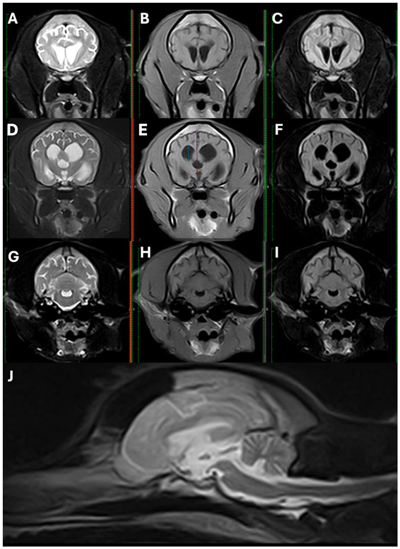
Figure 2.
MRI images of a 12-year-old Jack Russell Terrier with generalized brain atrophy. A and C, Transverse T2 FLAIR and T2W images show bilateral hyperintense periventricular white matter lesions consistent with leukoaraiosis. B, Generalized brain atrophy is also evident. FLAIR, fluid-attenuated inversion recovery; MRI, magnetic resonance imaging; T2W, T2-weighted.
Figure 2.
MRI images of a 12-year-old Jack Russell Terrier with generalized brain atrophy. A and C, Transverse T2 FLAIR and T2W images show bilateral hyperintense periventricular white matter lesions consistent with leukoaraiosis. B, Generalized brain atrophy is also evident. FLAIR, fluid-attenuated inversion recovery; MRI, magnetic resonance imaging; T2W, T2-weighted.
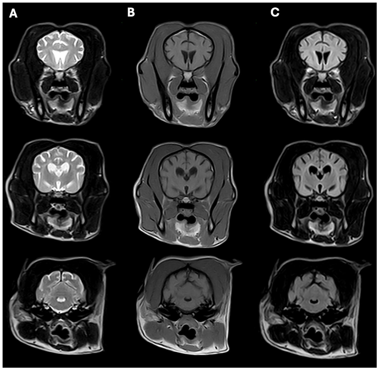
Figure 3.
MRI images of a 15-year-old West Highland White Terrier with generalized brain atrophy. Symmetrical ventricular enlargement is observed. C, Transverse T2 FLAIR image shows bilateral hyperintense periventricular white matter lesions. D, E, and F, T2W, T1W, and transverse T2 FLAIR images show hippocampal atrophy. FLAIR, fluid-attenuated inversion recovery; MRI, magnetic resonance imaging; T1W, T1-weighted; T2W, T2-weighted.
Figure 3.
MRI images of a 15-year-old West Highland White Terrier with generalized brain atrophy. Symmetrical ventricular enlargement is observed. C, Transverse T2 FLAIR image shows bilateral hyperintense periventricular white matter lesions. D, E, and F, T2W, T1W, and transverse T2 FLAIR images show hippocampal atrophy. FLAIR, fluid-attenuated inversion recovery; MRI, magnetic resonance imaging; T1W, T1-weighted; T2W, T2-weighted.
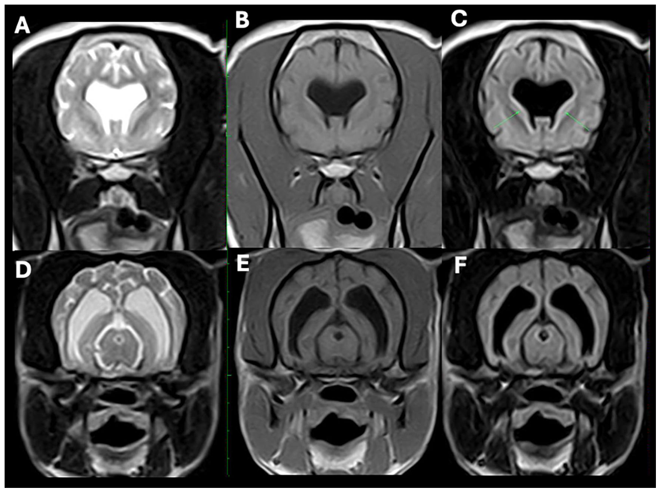
Figure 4.
MRI images of a 15-year-old Schnauzer with generalized brain atrophy. A, B, and C, Transverse T2W, T1W, and T2 FLAIR images show decreased thickness of the interthalamic adhesion, symmetrical ventricular enlargement, and enlargement of the subarachnoid space causing widened, well-defined cerebral sulci. D, The sagittal T2W image shows a small, abnormally triangular interthalamic adhesion. On the transverse T1W image (B), ITAt is indicated by the purple line, ITAr by the green line, and LVr is also measured. FLAIR, fluid-attenuated inversion recovery; ITAr, brain height; ITAt, interthalamic adhesion thickness; LVr, lateral ventricular height; MRI, magnetic resonance imaging; T1W, T1-weighted; T2W, T2-weighted.
Figure 4.
MRI images of a 15-year-old Schnauzer with generalized brain atrophy. A, B, and C, Transverse T2W, T1W, and T2 FLAIR images show decreased thickness of the interthalamic adhesion, symmetrical ventricular enlargement, and enlargement of the subarachnoid space causing widened, well-defined cerebral sulci. D, The sagittal T2W image shows a small, abnormally triangular interthalamic adhesion. On the transverse T1W image (B), ITAt is indicated by the purple line, ITAr by the green line, and LVr is also measured. FLAIR, fluid-attenuated inversion recovery; ITAr, brain height; ITAt, interthalamic adhesion thickness; LVr, lateral ventricular height; MRI, magnetic resonance imaging; T1W, T1-weighted; T2W, T2-weighted.
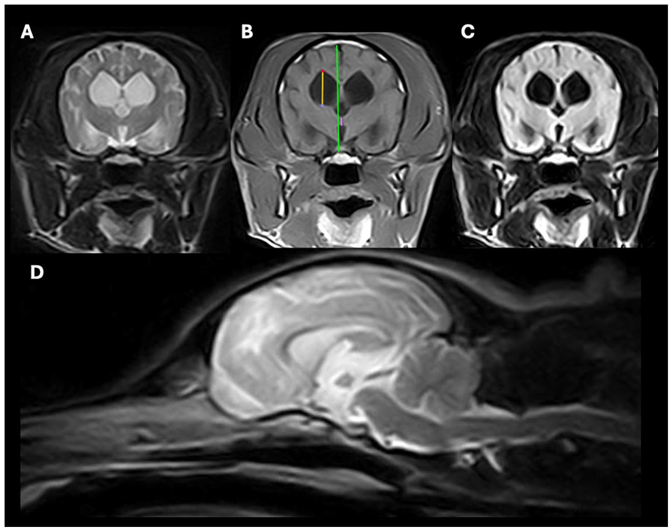
Figure 5.
MRI images of a 14-year-old Poodle with generalized brain atrophy. (A) Transverse T2W, (B) transverse T1W, and (C) transverse T2 FLAIR images show hippocampal atrophy, asymmetrical ventricular enlargement, diffuse and dispersed T2 hyperintensity in the periventricular white matter, and enlargement of the subarachnoid space causing widened, well-defined cerebral sulci. FLAIR, fluid-attenuated inversion recovery; MRI, magnetic resonance imaging; T1W, T1-weighted; T2W, T2-weighted.
Figure 5.
MRI images of a 14-year-old Poodle with generalized brain atrophy. (A) Transverse T2W, (B) transverse T1W, and (C) transverse T2 FLAIR images show hippocampal atrophy, asymmetrical ventricular enlargement, diffuse and dispersed T2 hyperintensity in the periventricular white matter, and enlargement of the subarachnoid space causing widened, well-defined cerebral sulci. FLAIR, fluid-attenuated inversion recovery; MRI, magnetic resonance imaging; T1W, T1-weighted; T2W, T2-weighted.
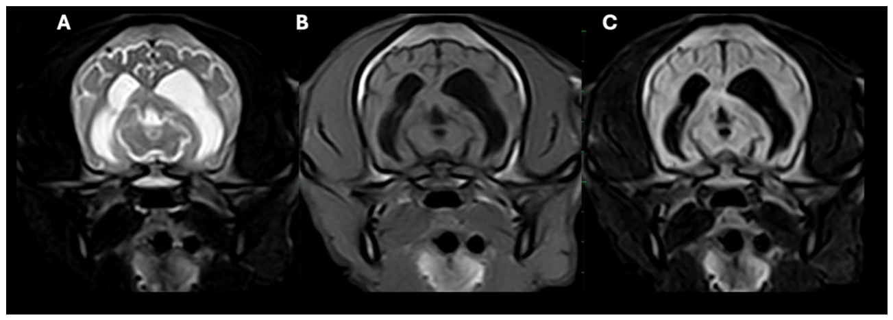
Figure 6.
MRI brain images of a 13-year-old mixed-breed dog with generalized brain atrophy. A , B, and C, Transverse T2W, Transverse T1W, and T2 FLAIR images show a lacunar infarction in the rostral portion of the right midbrain (green arrows), diffuse and dispersed T2 hyperintensity in the periventricular white matter, ventriculomegaly, and enlargement of the subarachnoid space causing widened, well-defined cerebral sulci. FLAIR, fluid-attenuated inversion recovery; MRI, magnetic resonance imaging; T2W, T2-weighted.
Figure 6.
MRI brain images of a 13-year-old mixed-breed dog with generalized brain atrophy. A , B, and C, Transverse T2W, Transverse T1W, and T2 FLAIR images show a lacunar infarction in the rostral portion of the right midbrain (green arrows), diffuse and dispersed T2 hyperintensity in the periventricular white matter, ventriculomegaly, and enlargement of the subarachnoid space causing widened, well-defined cerebral sulci. FLAIR, fluid-attenuated inversion recovery; MRI, magnetic resonance imaging; T2W, T2-weighted.
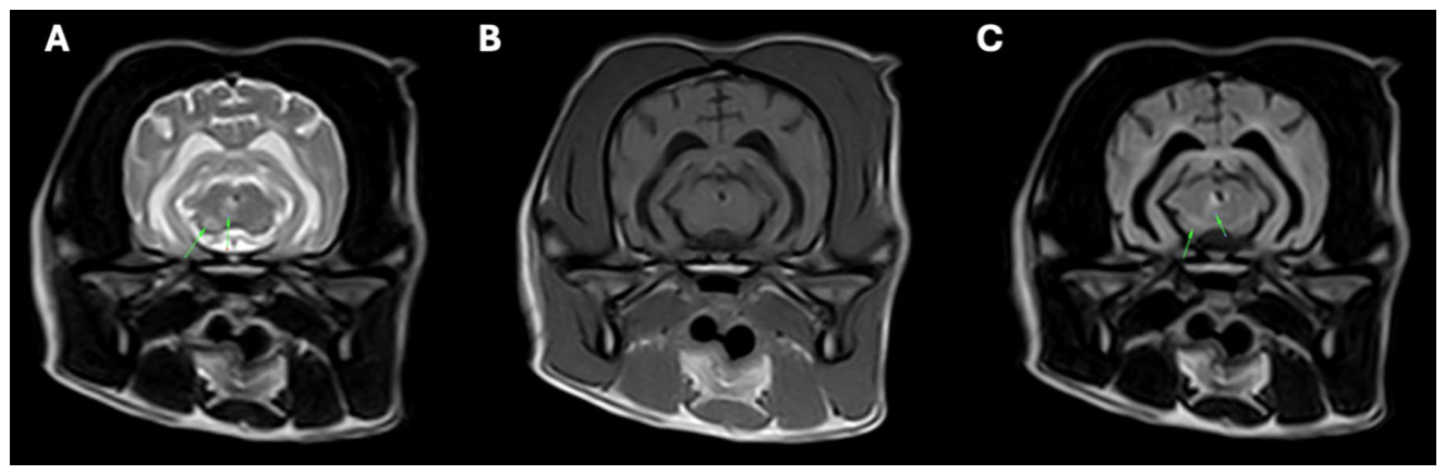
Figure 7.
MRI brain images of a 7-year-old Scottish Terrier with generalized brain atrophy associated with hepatic encephalopathy. A, B, and C, Transverse T2W, Transverse T1W, and T2 FLAIR images show generalized cerebral atrophy with widened sulci and ventriculomegaly, as well as bilaterally symmetric hyperintense lesions involving the thalamus (green arrows). FLAIR, fluid-attenuated inversion recovery; MRI, magnetic resonance imaging; T2W, T2-weighted.
Figure 7.
MRI brain images of a 7-year-old Scottish Terrier with generalized brain atrophy associated with hepatic encephalopathy. A, B, and C, Transverse T2W, Transverse T1W, and T2 FLAIR images show generalized cerebral atrophy with widened sulci and ventriculomegaly, as well as bilaterally symmetric hyperintense lesions involving the thalamus (green arrows). FLAIR, fluid-attenuated inversion recovery; MRI, magnetic resonance imaging; T2W, T2-weighted.
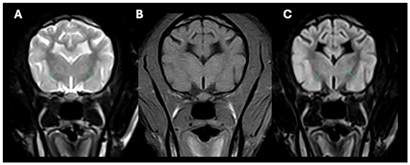
Figure 8.
MRI brain images of a 6-month-old male Weimaraner with generalized brain atrophy associated with neuronal ceroid lipofuscinosis. A, B, and C, Transverse T2W, T1W, and T2 FLAIR images show ventriculomegaly and enlargement of the subarachnoid space causing widened, well-defined cerebral sulci. FLAIR, fluid-attenuated inversion recovery; MRI, magnetic resonance imaging; T1W, T1-weighted; T2W, T2-weighted.
Figure 8.
MRI brain images of a 6-month-old male Weimaraner with generalized brain atrophy associated with neuronal ceroid lipofuscinosis. A, B, and C, Transverse T2W, T1W, and T2 FLAIR images show ventriculomegaly and enlargement of the subarachnoid space causing widened, well-defined cerebral sulci. FLAIR, fluid-attenuated inversion recovery; MRI, magnetic resonance imaging; T1W, T1-weighted; T2W, T2-weighted.
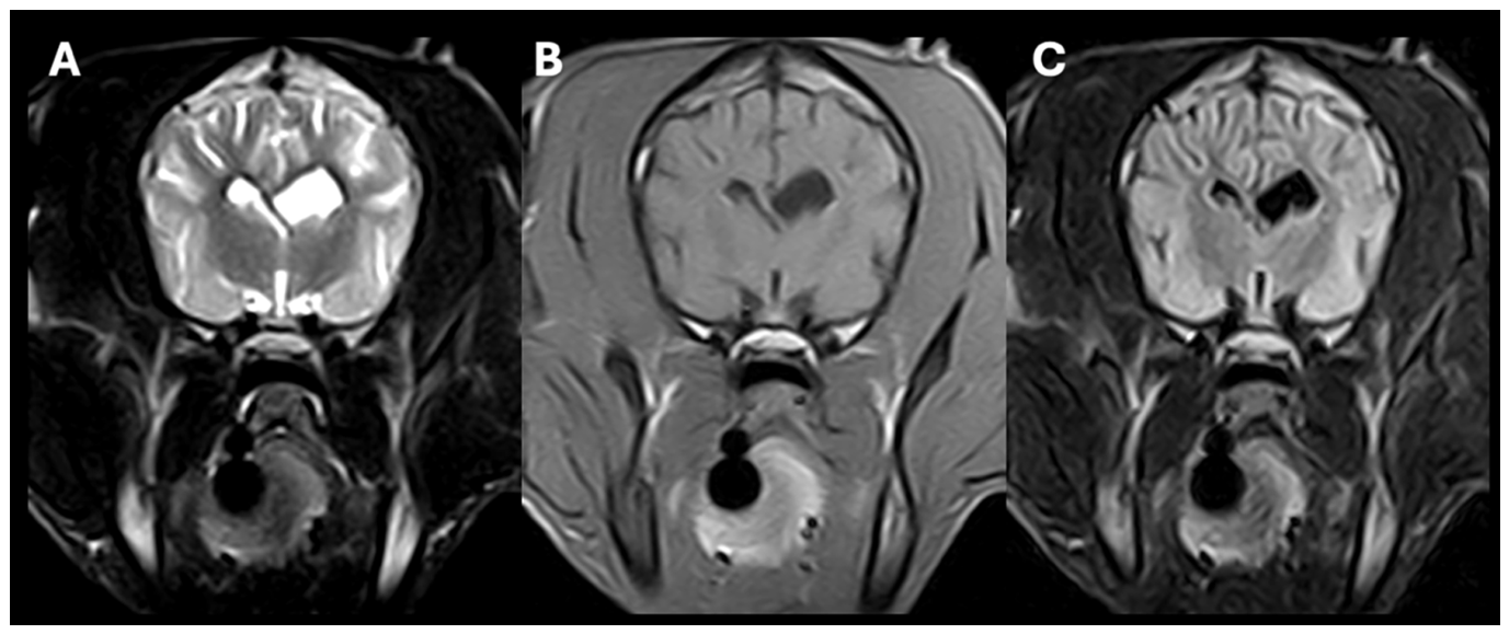
Figure 9.
Rehabilitation of a 15-year-old male Pug diagnosed with canine cognitive dysfunction.
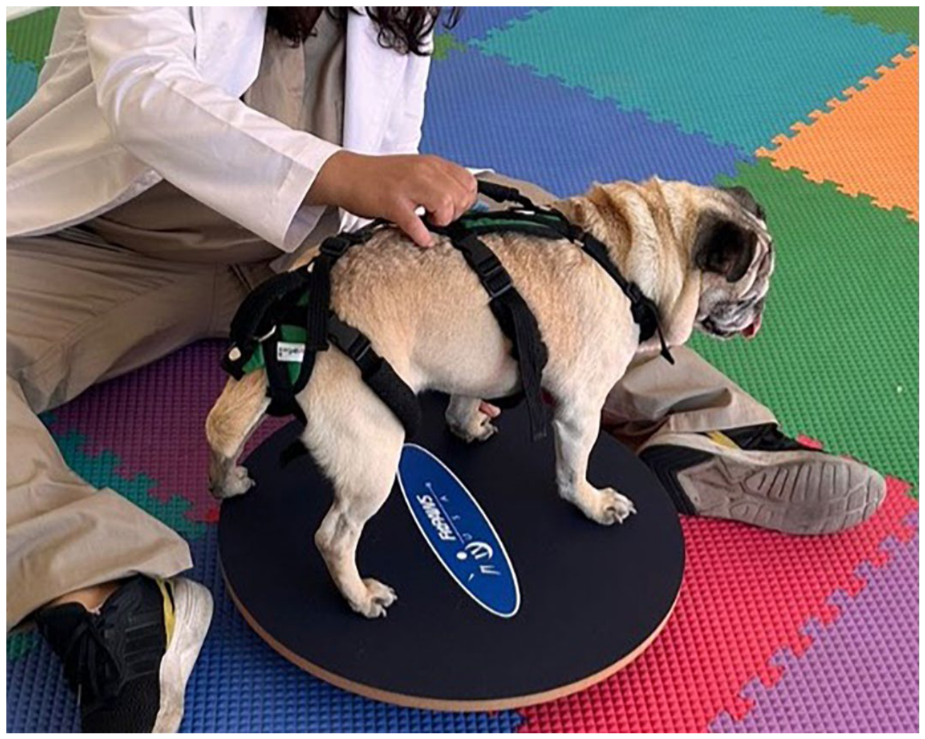
Disclaimer/Publisher’s Note: The statements, opinions and data contained in all publications are solely those of the individual author(s) and contributor(s) and not of MDPI and/or the editor(s). MDPI and/or the editor(s) disclaim responsibility for any injury to people or property resulting from any ideas, methods, instructions or products referred to in the content. |
© 2026 by the authors. Licensee MDPI, Basel, Switzerland. This article is an open access article distributed under the terms and conditions of the Creative Commons Attribution (CC BY) license (http://creativecommons.org/licenses/by/4.0/).
Copyright: This open access article is published under a Creative Commons CC BY 4.0 license, which permit the free download, distribution, and reuse, provided that the author and preprint are cited in any reuse.