

Submitted:
22 July 2026
Posted:
22 July 2026
You are already at the latest version
Abstract
Polyetheretherketone (PEEK) is increasingly attractive for patient-specific maxillofacial reconstruction because its elastic modulus approximates cortical bone, it is fully radiolucent, and it is compatible with additive manufacturing; its principal limitation is bioinertness, as the hydrophobic surface does not support protein adsorption or direct bone apposition. This study aimed to render the surface of fused-deposition-modelling (FDM)-printed, medical-grade PEEK bioactive while preserving these bulk advantages. To this aim, 3D printed specimens of implant-grade PEEK were activated by CO₂ plasma and coated with gelatin methacryloyl (GelMA) via EDC/NHS coupling followed by UV photocrosslinking. Surfaces were characterized by attenuated total reflectance Fourier-transform infrared (ATR-FTIR) spectroscopy, sessile-drop water contact angle measurement, gravimetric analysis, and scanning electron microscopy (SEM). Plasma treatment introduced oxygen-containing functional groups and reduced the water contact angle from 78.3° to 6.6°. Covalent GelMA grafting was confirmed by characteristic amide bands, was most pronounced for the 50 mg/mL formulation, and corresponded to a deposited mass of 11.81 ± 0.47 µg/mm²; SEM revealed a relatively uniform protein film along the printed filaments. These results establish a reproducible route toward bioactive, patient-specific PEEK maxillofacial implants.
Keywords:
polyetheretherketone
; PEEK
; additive manufacturing
; fused deposition modelling
; surface functionalization
; cold plasma
; gelatin methacryloyl
; GelMA
; osseointegration
; mandibular reconstruction
1. Introduction
Reconstruction of the mandible after segmental resection is one of the most demanding challenges in oral and maxillofacial surgery, because the mandible is a mobile, load-bearing structure essential to mastication, speech, deglutition, airway maintenance, and facial aesthetics. Loss of mandibular continuity—following oncologic resection, trauma, osteoradionecrosis, or aggressive benign pathology such as ameloblastoma—produces marked functional impairment, segment deviation, malocclusion, and considerable psychosocial burden [1,2]. Although the free vascularized fibula flap remains the reconstructive reference standard, its use is constrained by donor-site morbidity, the requirement for microvascular expertise, prolonged operative time, and several patient-related contraindications [3]. These limitations continue to motivate the development of patient-specific alloplastic solutions designed directly from the patient’s imaging data [2,4].
Titanium and its alloys have long dominated alloplastic mandibular reconstruction owing to their strength and biocompatibility. However, the very high elastic modulus of titanium (approximately 104–110 GPa, more than an order of magnitude greater than cortical bone) promotes stress shielding [5], and the metal generates severe streak and blooming artifacts on computed tomography and magnetic resonance imaging that hinder post-operative tumor surveillance and complicate radiotherapy planning [6]. Polyetheretherketone (PEEK), a high-performance, semi-crystalline thermoplastic, has therefore emerged as an attractive alternative: its elastic modulus of roughly 3.6–4 GPa closely approximates cortical bone, it offers excellent chemical and fatigue resistance, it is fully radiolucent, and it is compatible with additive manufacturing for genuine patient-specific design [7,8,9].
The biomechanical viability of PEEK for mandibular reconstruction has been established, including in recent finite-element studies demonstrating that customized PEEK plates and minimal-surface lattice structures can restore mandibular continuity, reduce stress shielding, and distribute masticatory loads within safe limits, provided minimum thickness requirements are respected [10,11]. These analyses, however, converge on the same fundamental limitation: PEEK is bioinert. Its hydrophobic, low-energy surface, essentially devoid of reactive functional groups, does not support protein adsorption, cell adhesion, or direct bone apposition, so even a mechanically optimal PEEK implant cannot by itself achieve the osseointegration required for durable, biologically anchored reconstruction [9,12,13]. In vivo, this manifests as fibrous encapsulation, no direct bone contact, compromising mechanical stability and increasing the risk of loosening under functional load [7,14].
A broad range of strategies has been investigated to convert the bioinert PEEK surface into a bioactive one. Bulk modification incorporates bioactive fillers such as hydroxyapatite into the polymer matrix, but high filler loading can reduce tensile strength and complicate processing, and the particles exert biological effects only where exposed at the surface [12]. Surface modification approaches preserve the bulk properties and include physical and energetic treatments (plasma, ultraviolet, laser nanotexturing, ion implantation), chemical treatments (sulfonation, amination, phosphonation), and coating or biological functionalization (hydroxyapatite and ceramic coatings, polydopamine intermediate layers, peptide and growth -factor immobilization) [13,15,16,17,18,19,20,21,22]. Combined sequential treatments frequently produce synergistic improvements in wettability, coating adhesion, and biological response [23].
Among these options, low-pressure plasma treatment is particularly well suited to patient-specific implants: it introduces functional groups and modifies surface energy, treats geometrically complex surfaces uniformly, induces negligible dimensional change, and can be applied as a final step after printing [16,24,25,26,27,28]. Plasma activation also creates clean, reactive sites that serve as anchoring points for the covalent immobilization of biomolecules [28]. Building on this, the present study couples plasma activation with methacryloyl gelatin (GelMA), a photocrosslinkable, cell-instructive macromolecule derived from denatured collagen that retains arginine–glycine–aspartate (RGD) adhesion motifs and matrix-metalloproteinase-degradable sequences, properties that make it a widely used substrate for promoting cell adhesion and tissue integration [23,29].
The objective of this study is therefore to transform the bioinert surface of additively manufactured, medical-grade PEEK into a bioactive one through a sequential strategy including CO₂ plasma activation and covalent GelMA grafting, without sacrificing the bulk mechanical and imaging advantages that make PEEK desirable.
2. Materials and Methods
2.1. Additive Manufacturing of PEEK Substrates
PEEK substrates were fabricated by industrial fused deposition modelling (FDM) using a 3DGence INDUSTRY F350 system (3DGence GmbH, Pfungstadt, Germany) equipped with a high-temperature M500 print-head module, which permits extrusion temperatures of up to 500 °C. Square test plates used for the surface-functionalization experiments were printed from implantable-grade VESTAKEEP® PEEK (Evonik Industries AG, Essen, Germany), supplied as 1.75 mm filament. The material is compliant with ASTM F2026, and its base resin has been evaluated according to ISO 10993-1 and USP Class VI. Plates measuring 15 × 15 × 0.7 mm were produced with a raster orientation of 0° and used for all functionalization experiments.
2.2. CO₂ Plasma Treatment
Prior to functionalization, the 15 × 15 × 0.7 mm PEEK plates were cleaned sequentially with detergent, double-distilled water (dH₂O), and ethanol, then dried in an oven at 37 °C. Plasma treatment was performed in a low-pressure plasma reactor (LFG1000, Diener Electronic GmbH & Co. KG, Ebhausen, Germany). Each face of the samples was exposed to CO₂ plasma at a power of 200 W and a gas flow rate of 20 standard cubic centimeters per minute (sccm), for either 60 s or 120 s per face, generating reactive oxygen-containing functional groups on the polymer surface.
2.3. GelMA Synthesis and Covalent Coating
Methacryloyl-modified porcine gelatin (GelMA) was synthesized from porcine gelatin (gel strength ~300 g Bloom, type A, from Sigma,), methacrylic anhydride (Aldrich, 94%), and phosphate-buffered saline (PBS, pH 7.4, from Sigma). The degree of methacrylation (approximately 30%) was determined by proton nuclear magnetic resonance (1H NMR) spectroscopy. For covalent coupling, 1-ethyl-3-(3-dimethylaminopropyl)carbodiimide (EDC, from Fluka), N-hydroxysuccinimide (NHS, Aldrich), 2-(N-morpholino)ethanesulfonic acid (MES, Sigma) hydrate, and sodium chloride were used as received. 2-Hydroxy-4′-(2-hydroxyethoxy)-2-methylpropiophenone (Irgacure 2959, Aldrich) was prepared as a 10% (w/v) solution in ethanol and used as photoinitiator.
Following plasma activation, GelMA was grafted via EDC/NHS crosslinking according to a previously described protocol [30]. Briefly, samples were immersed in an activation buffer of 0.1 M MES and 0.5 M NaCl (pH 6.0) containing the appropriate amounts of EDC and NHS. In parallel, GelMA solutions at three concentrations (1, 5, and 50 mg/mL) were prepared in PBS (pH 7.4), with the photoinitiator incorporated in a GelMA:Irgacure ratio of 100:1 (w/w). The GelMA solution was combined with the activation buffer, and samples were incubated in the dark at 37 °C for 24 h. Photocrosslinking was then carried out under ultraviolet (UV) irradiation at 312 nm for 15 min on each side. The complete workflow is summarized in Figure 1.
2.4. Plasma Treatment Assessment Through Attenuated Total Reflectance Fourier Transform Infrared
Plasma treatment was performed on the samples for one and two minutes, respectively. The presence of functional groups was investigated through attenuated total reflectance Fourier transform infrared (ATR-FTIR) using a Jasco 4200 spectrometer (JASCO Deutschland GmbH, Pfungstadt, Germany) equipped with a diamond Specac Golden Gate ATR device (Specac Ltd, Orpington, UK) at a 4 cm-1 resolution. The analyses were performed in the wavenumber interval 4000-600 cm-1. The registered spectra for the pre-established time points were compared with the spectra of the untreated PEEK sample (control).
2.5. Surface Wettability Modification Assessment Through Contact Angle
Plasma- treated PEEK wettability was assessed using the sessile drop technique with Drop Shape Analyzer 100 (DSA 100, KRÜSS GmbH, Hamburg, Germany). At least 3 different regions of each sample were selected for testing. Drops of 2 μl distilled water were placed on the samples and contact angle measurements were recorded for 60 s.
2.6. GelMA Coating Assessment
2.6.1. ATR-FTIR Spectroscopy
ATR-FTIR spectroscopy, performed using the same instrument described in Section 2.4, was employed to confirm the presence of the GelMA coating on plasma-treated PEEK. Spectra were acquired from both sides of the samples and revealed characteristic protein-associated vibrational bands consistent with the presence of GelMA.
2.6.2. Evaluation of Deposited GelMA Mass Per Surface Area
The amount of deposited GelMA was determined gravimetrically by measuring the mass difference before and after coating, photocrosslinking, and drying. The amount of deposited GelMA was normalized to the sample surface area according to Equation 1 and expressed as deposited mass per unit area (µg/mm²). The analysis was conducted in triplicate for the coating prepared with the selected 50 mg/mL GelMA formulation only
where:
= deposited GelMA mass per unit area (µg/mm²)
= initial sample mass
= final sample mass after coating
= sample surface area
2.6.3. Scanning Electron Microscopy
Scanning electron microscopy (SEM) was employed to evaluate the surface morphology and homogeneity of the protein coating. The analysis was performed using a FEI Quanta Inspect F50 microscope (FEI Company, Hillsboro, OR, USA) operated in secondary electron (SE) mode, at an accelerating voltage of 10 kV, a spot size of 3.5, and a working distance of 9.4‒9.6mm Prior to analysis, the samples were coated with a thin gold layer by direct-current magnetron sputtering for 60 s.
3. Results
The overall GelMA coating procedure applied to the plasma-activated PEEK substrates is summarized schematically in Figure 2, comprising surface activation, covalent EDC/NHS coupling, and UV photocrosslinking.
3.1. Plasma Activation: Surface Chemistry and Wettability
The ATR-FTIR spectrum of pristine PEEK displayed the characteristic absorption bands of the polymer backbone, including aromatic ring vibrations near 1500 cm⁻¹ and 1650 cm⁻¹ and multiple sharp bands in the 1220–1150 cm⁻¹ region assigned to C–O stretching [31].
After CO₂ plasma treatment (60 s or 120 s per side), new absorption bands appeared near 1730 cm⁻¹ and 1710 cm⁻¹, attributable to C=O stretching vibrations associated with carboxyl-group formation (Figure 3) [32]. The relatively low intensity of these bands is consistent with the surface-confined nature of plasma modification. For the 120 s sample, an additional band near 3400 cm⁻¹ (O–H stretching of carboxylic groups) and a band near 2600 cm⁻¹ (C–H of carboxylic acid groups) further confirmed the generation of oxygen-containing surface functionalities.
Water contact angle measurements quantified the corresponding change in wettability (Table 1). The contact angle of pristine PEEK was 78.32 ± 2.94°, confirming a hydrophobic surface. CO₂ plasma treatment reduced it to 18.35 ± 2.01° after 60 s and to 6.61 ± 0.57° after 120 s per side, indicating a pronounced, exposure-dependent increase in hydrophilicity (Figure 4). Considering the results obtained following ATR-FTIR and contact angle measurements, plasma treatment of 120 s per side was selected as the optimal activation condition for subsequent GelMA coupling.
3.2. Covalent GelMA Grafting
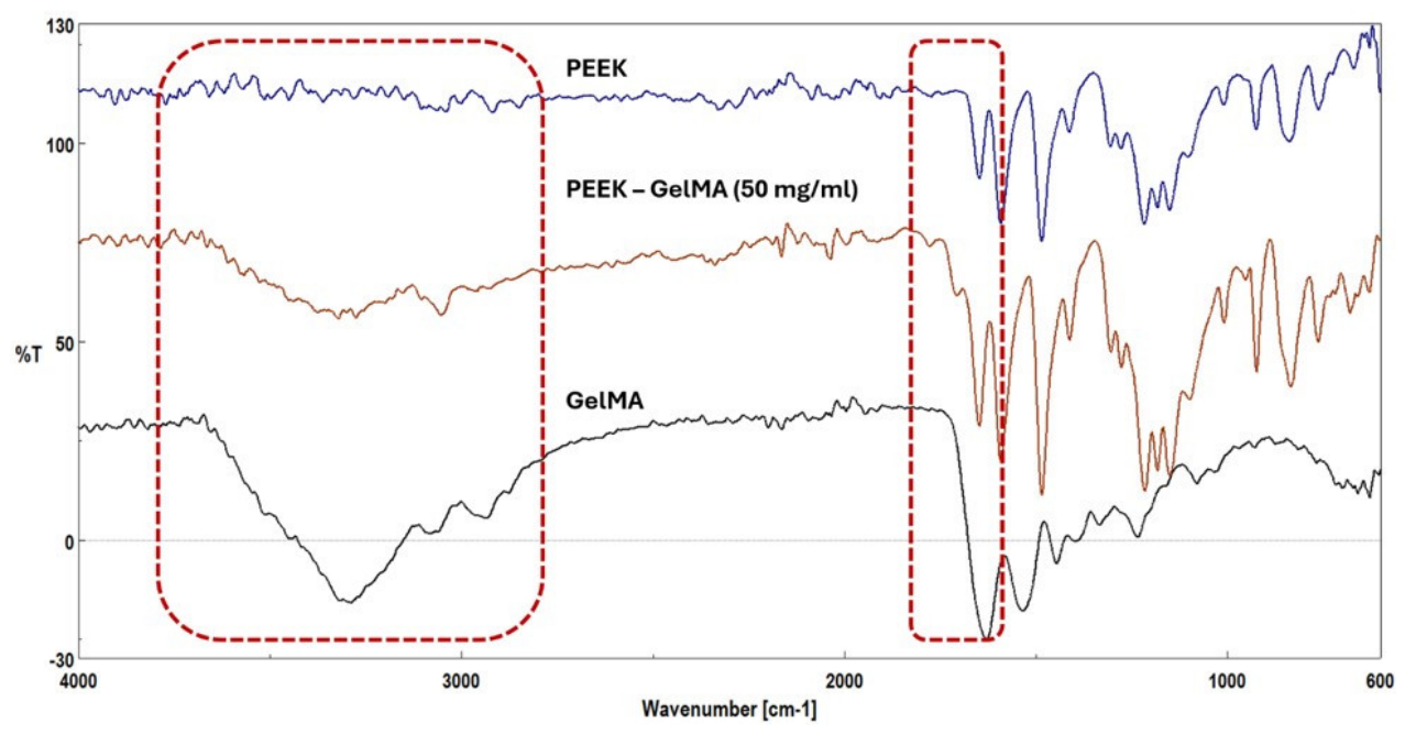
GelMA coatings were prepared using various concentrations of protein (1, 5, and 50 mg/mL) following the EDC/NHS coupling protocol. The reference GelMA spectrum exhibited a broad band at 3293 cm⁻¹ (overlapping O–H and N–H stretching), a band at 3086 cm⁻¹ (N–H stretching), a band at 2937 cm⁻¹ (aliphatic C–H stretching), and amide I and amide II bands at 1629 cm⁻¹ and 1533 cm⁻¹, respectively [32].
The spectrum of pristine PEEK shows the characteristic absorption bands of the polymer backbone, as discussed previously. Following coating, all GelMA-modified PEEK samples seem to display the emergence of these characteristic GelMA bands, confirming successful surface functionalization. Notably, the intensity of GelMA-related absorption bands increases with increasing polymer concentration, being most pronounced for the 50 mg/mL condition (supplementary Figure 1). This trend suggests a higher amount of deposited and crosslinked GelMA on the PEEK support.
The successful grafting of GelMA onto the plasma-treated PEEK surface was confirmed by ATR-FTIR spectroscopy, primarily through the appearance of a new broad absorption band centered at 3326 cm⁻¹, attributed to the overlapping contributions of amide A and O–H stretching vibrations characteristic of GelMA. Additional evidence of GelMA deposition is provided by the band observed at 1709 cm⁻¹ in the GelMA (50 mg/ml)-coated PEEK samples, which can be associated with the combined contribution of C=O vibrations from carboxylic groups generated during plasma treatment and the amide I band of GelMA (Supplementary Figure S1).
Figure 5.
ATR-FTIR spectra of PEEK, GelMA (50 mg/mL)-coated PEEK, and GelMA. The appearance of protein-associated amide bands and the broad band centred near 3326 cm⁻¹ in the coated sample confirm covalent immobilization of GelMA on the plasma-activated surface.
Figure 5.
ATR-FTIR spectra of PEEK, GelMA (50 mg/mL)-coated PEEK, and GelMA. The appearance of protein-associated amide bands and the broad band centred near 3326 cm⁻¹ in the coated sample confirm covalent immobilization of GelMA on the plasma-activated surface.
3.3. Evaluation of Deposited GelMA Mass Per Surface Area
Gravimetric analysis of samples coated with the 50 mg/mL GelMA formulation indicated a deposited mass of 11.81 ± 0.47 µg/mm² after coating, UV photocrosslinking, and drying. This result confirms the formation of a stable GelMA layer on the plasma-activated PEEK surface and is consistent with the spectroscopic observations, which showed the most pronounced protein-related bands for this formulation.
3.4. Surface Morphology by SEM
SEM micrographs of pristine PEEK revealed the characteristic layered topography of the FDM process—parallel deposition tracks with a relatively smooth inter-filament morphology—and, at higher magnification, a compact, homogeneous surface with only minor irregularities. The filament average diameter was approximately 100 microns, with a narrow size distribution, highlighting a high degree of dimensional uniformity, as well as the good control achieved during the extrusion process. Following GelMA coating, the overall filament architecture was preserved, but a thin surface film was distributed along the printed strands; the boundaries between adjacent filaments became significantly blurred, demonstrating the existence of an overlayer and confirming the effectiveness of the coating approach. At higher magnification, coated surfaces showed slightly increased roughness and discontinuous granular and film-like structures, particularly along inter-filament regions, which may indicate a locally disturbed surface modification process (Figure 6). The coating appeared relatively homogeneous across the analyzed area, indicating that the combined plasma activation and EDC/NHS coupling strategy promoted effective immobilization of the protein layer on the polymer substrate.
4. Discussion
The results demonstrate that a sequential process consisting of CO₂ plasma activation followed by covalent GelMA grafting strategy can convert the bioinert surface of additively manufactured, medical-grade PEEK into a hydrophilic, protein-bearing surface, This addresses the central limitation identified across biomechanical studies of PEEK mandibular implants: that mechanical adequacy alone does not guarantee biological integration [10,11,12,13].
Plasma activation was confirmed by two complementary lines of evidence. ATR-FTIR revealed the emergence of carbonyl and carboxyl features following CO₂ plasma exposure, consistent with the established mechanism by which reactive plasma species cleave surface C–H and C–C bonds and generate oxygen-containing groups upon reaction with plasma and atmospheric oxygen [25,27]. The accompanying drop in water contact angle from 78.3° to 6.6° is in line with the wettability improvements reported for oxygen-bearing plasma treatment of PEEK, where contact angles fall from the low-80° range to near-zero values under optimized conditions [24,26]. The exposure-dependence observed here—intermediate hydrophilicity at 60 s and maximal hydrophilicity at 120 s—supports the selection of a defined optimal treatment time not the maximal exposure, which is relevant because excessive treatment can over-etch the surface and because plasma-induced hydrophilicity is subject to partial hydrophobic recovery over time [16,26].
The role of plasma activation in this strategy is twofold: it raises surface energy to favor protein adsorption, and it introduces carboxyl groups that serve as covalent anchoring points. EDC/NHS chemistry exploits these groups to form amide bonds with the amine groups of GelMA, producing a zero-length covalent linkage rather than mere physical adsorption [30]. The spectroscopic appearance of amide I/II bands and the broad amide A/O–H feature in coated samples, increasing in intensity with GelMA concentration, is consistent with covalent immobilization, and the gravimetric value of 11.81 ± 0.47 µg/mm² confirms a stable, quantifiable coating. GelMA is a particularly suitable coating for this application because it retains the RGD adhesion motifs and matrix-metalloproteinase-degradable sequences of its parent collagen, providing cell-instructive cues that bioinert PEEK inherently lacks [23,29].
Relative to alternative bioactivation routes, the present approach offers specific advantages for patient-specific maxillofacial implants. Bulk incorporation of hydroxyapatite improves bioactivity but can reduce tensile strength and alter processing behavior, and it relies on surface-exposed particles [12]. Sulfonation generates a porous bioactive layer but uses aggressive reagents and can cause dimensional change, which is undesirable for components requiring precise anatomical fit [18]. Ceramic and nano-hydroxyapatite coatings improve osseointegration but often require line-of-sight deposition that is difficult to apply uniformly to undercut or internal lattice surfaces [18,20]. Low-pressure plasma, by contrast, treats complex geometries uniformly with negligible dimensional change and can be applied as a finishing step, and the subsequent GelMA layer is deposited from solution, allowing it to reach surfaces that line-of-sight methods cannot [16,28]. Combined treatments of this kind are consistent with the broader literature showing synergistic gains when physical activation is paired with biological functionalization [19,23].
Several limitations should be acknowledged. First, the present study characterizes the physicochemical and morphological outcome of functionalization but does not yet include biological validation; osteoblast adhesion, proliferation, and differentiation assays, followed by in vivo osseointegration studies, are required to confirm that the measured surface changes translate into improved biological performance. Second, the functionalization substrates were flat plates; transfer of the protocol to the full lattice geometry of a patient-specific implant, including verification of uniform coating within internal pores, remains to be demonstrated. Third, while the substrates were printed from an ASTM F2026-compliant implant grade, additively manufactured PEEK requires controlled post-processing and finishing before clinical use, and the durability of the GelMA layer under physiological and loading conditions was not assessed here. Finally, the corresponding biomechanical analysis has been reported separately [10] and was deliberately not repeated; integrating the mechanical and biological findings into a single validated, patient-specific construct is the natural next step.
5. Conclusions
This study establishes the feasibility of CO₂ plasma activation and covalent GelMA grafting strategy for improving the surface bioactivity of additively manufactured, medical-grade PEEK intended for maxillofacial reconstruction. CO₂ plasma treatment introduced oxygen-containing functional groups and increased surface hydrophilicity, reducing the water contact angle from 78.3° to 6.6°, with 120 s per face identified as the optimal activation condition. Covalent GelMA grafting via EDC/NHS coupling and UV photocrosslinking was confirmed by ATR-FTIR, gravimetric analysis, and SEM; the 50 mg/mL formulation provided the highest coating density and surface homogeneity, corresponding to a deposited mass of 11.81 ± 0.47 µg/mm². Together with the previously reported biomechanical validation of the patient-specific reconstruction geometry, these results support an integrated translational pathway—computational design, additive manufacturing in high-performance PEEK, and chemical surface biofunctionalization—toward a next generation of personalized mandibular implants that combine favorable biomechanics, imaging compatibility, and genuine biological integration. Future work will focus on biological validation, transfer of the coating protocol to the full implant geometry, and assessment of coating durability under physiological loading.
Supplementary Materials
The following supporting information can be downloaded at https://www.mdpi.com/article/doi/s1, Figure S1.
Author Contributions
Conceptualization, C.M.C., I.C.S. and A.S.; methodology, A.S., C.M.C., M.A.C., C.B., E.O., I.G.G. and I.C.S.; software, I.G.G. and E.L.; validation, A.S., I.C.S, M.A.C., C.B., E.O., I.G.G., E.L. and C.M.C.; formal analysis, A.S., I.C.S, M.A.C., C.B., and E.O ; investigation, A.S., I.C.S, M.A.C., C.B., E.O. and C.M.C.; resources, I.C.S. and C.M.C.; data curation, A.S., I.C.S, M.A.C., C.B., E.O., I.G.G., E.L. and C.M.C.; writing—original draft preparation, A.S., M.A.C., C.B., E.O., I.G.G., E.L. and C.M.C; writing—review and editing, I.C.S and C.M.C.; visualization, A.S., M.A.C., C.B., E.O., I.G.G., E.L. and C.M.C.; supervision, A.S., I.C.S. and C.M.C.; project administration, A.S., I.C.S. and C.M.C.; funding acquisition, I.C.S. and C.M.C. All authors have read and agreed to the published version of the manuscript.
Funding
I.C.S, A.S., E.O. and C.B. acknowledge funding through the project PED44/2025-PersOC4Reg, Development of a Personalized System for Osteo-Chondral Regeneration through converging 3D Printing and Bioactivation.
Institutional Review Board Statement
Not applicable.
Informed Consent Statement
Not applicable.
Data Availability Statement
The data supporting the findings of this study are available from the first author or the corresponding author upon reasonable request.
Conflicts of Interest
The authors declare no conflicts of interest.
Abbreviations
The following abbreviations are used in this manuscript:
| PEEK | Polyetheretherketone |
| GelMA | Methacryloyl Gelatin |
| ATR-FTIR | Attenuated total reflectance Fourier transform infrared |
| dH₂O | double-distilled water |
| FDM | Fused deposition modelling |
| PBS | Phosphate-buffered saline |
| NMR | Nuclear magnetic resonance |
| RGD | Arginine–glycine–aspartate |
| NHS | N-hydroxysuccinimide |
| EDC | 1-ethyl-3-(3-dimethylaminopropyl)carbodiimide |
| MES | 2-(N-morpholino)ethanesulfonic acid |
| Irgacure 2959 | 2-Hydroxy-4′-(2-hydroxyethoxy)-2-methylpropiophenone |
| SEM | Scanning electron microscopy |
References
- Khayat, S.; Sada Urmeneta, Á.; González Moure, B.; Fernández Acosta, D.; Benito Anguita, M.; López López, A.; Verdaguer Martín, J.J.; Navarro Cuéllar, I.; Falahat, F.; Navarro Cuéllar, C. Reconstruction of Segmental Mandibular Defects with Double-Barrel Fibula Flap and Osseo-Integrated Implants: A Systematic Review. J. Clin. Med. 2024, 13. [Google Scholar] [CrossRef] [PubMed]
- Tarsitano, A.; Battaglia, S.; Ricotta, F.; Bortolani, B.; Cercenelli, L.; Marcelli, E.; Cipriani, R.; Marchetti, C. Accuracy of CAD/CAM mandibular reconstruction: A three-dimensional, fully virtual outcome evaluation method. J. Craniomaxillofac. Surg. 2018, 46, 1121–1125. [Google Scholar] [CrossRef] [PubMed]
- Gu, Y.; Ma, H.; Shujaat, S.; Orhan, K.; Coucke, W.; Amoli, M.S.; Bila, M.; Politis, C.; Jacobs, R. Donor- and recipient-site morbidity of vascularized fibular and iliac flaps for mandibular reconstruction: A systematic review and meta-analysis. J. Plast. Reconstr. Aesthet. Surg. 2021, 74, 1470–1479. [Google Scholar] [CrossRef] [PubMed]
- Goodson, A.M.; Kittur, M.A.; Evans, P.L.; Williams, E.M. Patient-specific, printed titanium implants for reconstruction of mandibular continuity defects: A systematic review of the evidence. J. Craniomaxillofac. Surg. 2019, 47, 968–976. [Google Scholar] [CrossRef] [PubMed]
- Niinomi, M.; Nakai, M. Titanium-Based Biomaterials for Preventing Stress Shielding between Implant Devices and Bone. Int. J. Biomater. 2011. [Google Scholar] [CrossRef] [PubMed]
- Lommen, J.; Schorn, L.; Sproll, C.; Haussmann, J.; Kübler, N.R.; Budach, W.; Rana, M.; Tamaskovics, B. Reduction of CT Artifacts Using Polyetheretherketone (PEEK), Polyetherketoneketone (PEKK), Polyphenylsulfone (PPSU), and Polyethylene (PE) Reconstruction Plates in Oral Oncology. J. Oral Maxillofac. Surg. 2022, 80, 1272–1283. [Google Scholar] [CrossRef] [PubMed]
- Kurtz, S.M.; Devine, J.N. PEEK biomaterials in trauma, orthopedic, and spinal implants. Biomaterials 2007, 28, 4845–4869. [Google Scholar] [CrossRef] [PubMed]
- Qin, L.; Yao, S.; Zhao, J.; Zhou, C.; Oates, T.W.; Weir, M.D.; Wu, J.; Xu, H.H.K. Review on Development and Dental Applications of Polyetheretherketone-Based Biomaterials and Restorations. Mater. 2021, 14, 408. [Google Scholar] [CrossRef] [PubMed]
- Dallal, S.; Eslami, B.; Tiari, S. Recent Advances in PEEK for Biomedical Applications: A Comprehensive Review of Material Properties, Processing, and Additive Manufacturing. Polym. 2025, 17, 1968. [Google Scholar] [CrossRef] [PubMed]
- Ghionea, I.G.; Tarba, C.I.; Cristache, M.A.; Cristache, C.M. Comparative Evaluation of Symmetrical Titanium and Polyetheretherketone (PEEK) Hollow Structures for Mandibular Reconstruction: Strength, Geometry, and Biomechanical Performance. Symmetry 2025, 17, 499. [Google Scholar] [CrossRef]
- Ghionea, I.G.; Tarba, C.I.; Cristache, C.M.; Filipov, I.; Beuran, I.A. A Comparative Finite Element Analysis of Titanium, Autogenous Bone, and Polyetheretherketone (PEEK)-Based Solutions for Mandibular Reconstruction. Mater. 2025, 18, 314. [Google Scholar] [CrossRef] [PubMed]
- Manzoor, F.; Golbang, A.; Jindal, S.; Dixon, D.; McIlhagger, A.; Harkin-Jones, E.; Crawford, D.; Mancuso, E. 3D printed PEEK/HA composites for bone tissue engineering applications: Effect of material formulation on mechanical performance and bioactive potential. J. Mech. Behav. Biomed. Mater. 2021, 121, 104601. [Google Scholar] [CrossRef] [PubMed]
- Ma, R.; Tang, T. Current Strategies to Improve the Bioactivity of PEEK. Int. J. Mol. Sci. 2014, 15, 5426–5445. [Google Scholar] [CrossRef] [PubMed]
- Gupta, G.; Bansal, S.; Upinder, S.; Kapila, S. Osseointegration: Biological Basis, Materials Science, and Clinical Perspectives in Dental Implantology. IJFMR-Int. J. Multidiscip. Res. 2025, 7. [Google Scholar] [CrossRef]
- Najeeb, S.; Zafar, M.S.; Khurshid, Z.; Siddiqui, F. Applications of polyetheretherketone (PEEK) in oral implantology and prosthodontics. J. Prosthodont. Res. 2016. [Google Scholar] [CrossRef] [PubMed]
- Sundriyal, P.; Sahu, M.; Prakash, O.; Bhattacharya, S. Long-term surface modification of PEEK polymer using plasma and PEG silane treatment. Surf. Interfaces 2021, 25, 101253. [Google Scholar] [CrossRef]
- Wang, W.; Luo, C.J.; Huang, J.; Edirisinghe, M. PEEK surface modification by fast ambient-temperature sulfonation for bone implant applications. J. R. Soc. Interface 2019, 16. [Google Scholar]
- Barkarmo, S.; Wennerberg, A.; Hoffman, M.; Kjellin, P.; Breding, K.; Handa, P.; Stenport, V. Nano-hydroxyapatite-coated PEEK implants: A pilot study in rabbit bone. J. Biomed. Mater. Res. Part A 2013, 101A, 465–471. [Google Scholar] [CrossRef] [PubMed]
- Mostafa, D.; Kassem, Y.M.; Omar, S.S.; Shalaby, Y. Nano-topographical surface engineering for enhancing bioactivity of PEEK implants (in vitro—histomorphometric study). Clin. Oral Investig. 2023, 27, 6789–6799. [Google Scholar]
- Mahjoubi, H.; Buck, E.; Manimunda, P.; Farivar, R.; Chromik, R.; Murshed, M.; Cerruti, M. Surface phosphonation enhances hydroxyapatite coating adhesion on polyetheretherketone and its osseointegration potential. Acta Biomater. 2017, 47, 149–158. [Google Scholar] [CrossRef] [PubMed]
- Park, S.; Jung, T.G. Surface Modification of Polyetheretherketone (PEEK) Intervertebral Fusion Implant Using Polydopamine Coating for Improved Bioactivity. Bioeng. 2024, 11, 343. [Google Scholar] [CrossRef] [PubMed]
- Chen, Y.; Chen, Z.; Lei, K.; Ding, J.; Yu, L. Surface modification of polyetheretherketone for boosted osseointegration: a review. Biomater. Transl. 2025, 6, 181–201. [Google Scholar] [CrossRef] [PubMed]
- Cassari, L.; Balducci, C.; Messina, G.M.L.; Iucci, G.; Battocchio, C.; Bertelà, F.; Lucchetta, G.; Coward, T.; Di Silvio, L.; Marletta, G.; et al. Polyetheretherketone Double Functionalization with Bioactive Peptides Improves Human Osteoblast Response. Biomimetics 2024, 9, 767. [Google Scholar] [CrossRef] [PubMed]
- Štefaníková, R.; Kretková, T.; Kuzminova, A.; Hanuš, J.; Vaidulych, M.; Kylián, O.; Biederman, H. Influence of atmospheric pressure dielectric barrier discharge on wettability and drying of poly(ether-ether-ketone) foils. Polym. Degrad. Stab. 2018, 150, 114–121. [Google Scholar] [CrossRef]
- Liston, E.M.; Martinu, L.; Wertheimer, M.R. Plasma surface modification of polymers for improved adhesion: a critical review. J. Adhes. Sci. Technol. 1993, 7, 1091–1127. [Google Scholar] [CrossRef]
- Primc, G. Strategies for Improved Wettability of Polyetheretherketone (PEEK) Polymers by Non-Equilibrium Plasma Treatment. Polymers . 2022, 14, 5319. [Google Scholar] [CrossRef] [PubMed]
- Booth, J.P.; Mozetič, M.; Nikiforov, A.; Oehr, C. Foundations of plasma surface functionalization of polymers for industrial and biological applications. Plasma Sources Sci. Technol. 2022, 31, 103001. [Google Scholar] [CrossRef]
- Qiu, P.; Bennani, V.; Cooper, P.; Dias, G.; Ratnayake, J. Surface chemistry on PEEK surfaces: From enhanced biofunctionality to improved surface modifiability. Appl. Mater. Today 2024, 41, 102523. [Google Scholar] [CrossRef]
- Lou, J.; Meyer, C.; Chen, A.; Weitz, D.A.; Mooney, D.J. Immobilization of BMP-2 in porous hydrogels to spatially regulate osteogenesis. J. Control. Release 2025, 379, 944–950. [Google Scholar] [CrossRef] [PubMed]
- Procedure for Two-step Coupling of Proteins Using EDC and NHS or Sulfo-NHS. 2017. [CrossRef] [PubMed]
- Ahmad, A.; Iqbal, T.; Yasin, S.; Hanif, R.; Riaz, S.; Luckham, P.F. Stability of amorphous PEEK in organic solvents. J. Chem. Soc. Pak. 2018, 40, 810–818. [Google Scholar]
- Serafim, A.; Tucureanu, C.; Petre, D.G.; Dragusin, D.M.; Salageanu, A.; Van Vlierberghe, S.; Dubruel, P.; Stancu, I.C. One-pot synthesis of superabsorbent hybrid hydrogels based on methacrylamide gelatin and polyacrylamide. Effortless control of hydrogel properties through composition design. New J. Chem. 2014, 38, 3112–3126. [Google Scholar] [CrossRef]
Figure 1.
Sequential surface biofunctionalization workflow applied to FDM-printed PEEK substrates: additive manufacturing, low-pressure CO₂ plasma activation to introduce oxygen-containing groups, EDC/NHS covalent coupling of GelMA, and UV photocrosslinking to form a stable hydrogel film.
Figure 1.
Sequential surface biofunctionalization workflow applied to FDM-printed PEEK substrates: additive manufacturing, low-pressure CO₂ plasma activation to introduce oxygen-containing groups, EDC/NHS covalent coupling of GelMA, and UV photocrosslinking to form a stable hydrogel film.
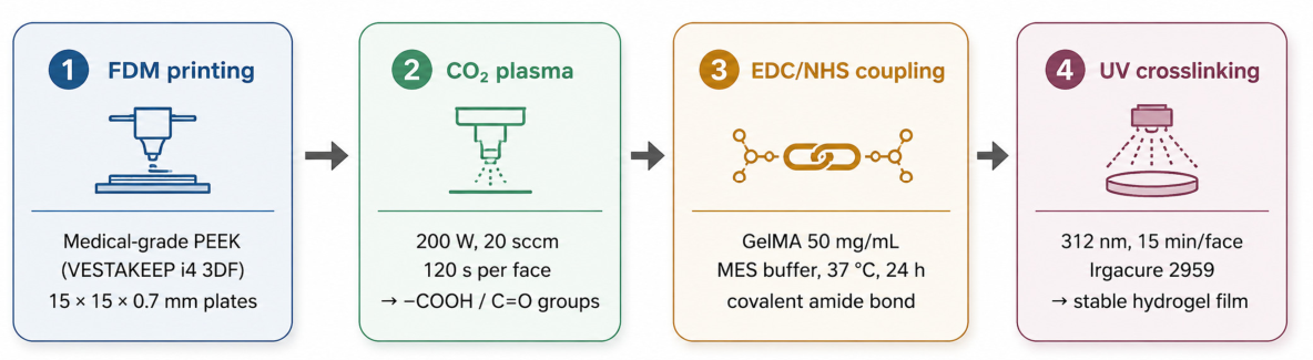
Figure 2.
Schematic illustration of the GelMA coating procedure applied to PEEK substrates.
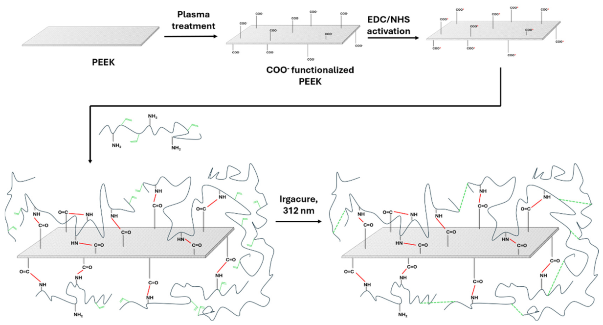
Figure 3.
ATR-FTIR spectra of pristine PEEK (control) and CO₂ plasma-treated (PT) samples (60 s and 120 s per side). New carbonyl/carboxyl bands near 1730 and 1710 cm⁻¹, and additional O–H and C–H features for the 120 s sample, confirm the introduction of oxygen-containing functional groups.
Figure 3.
ATR-FTIR spectra of pristine PEEK (control) and CO₂ plasma-treated (PT) samples (60 s and 120 s per side). New carbonyl/carboxyl bands near 1730 and 1710 cm⁻¹, and additional O–H and C–H features for the 120 s sample, confirm the introduction of oxygen-containing functional groups.
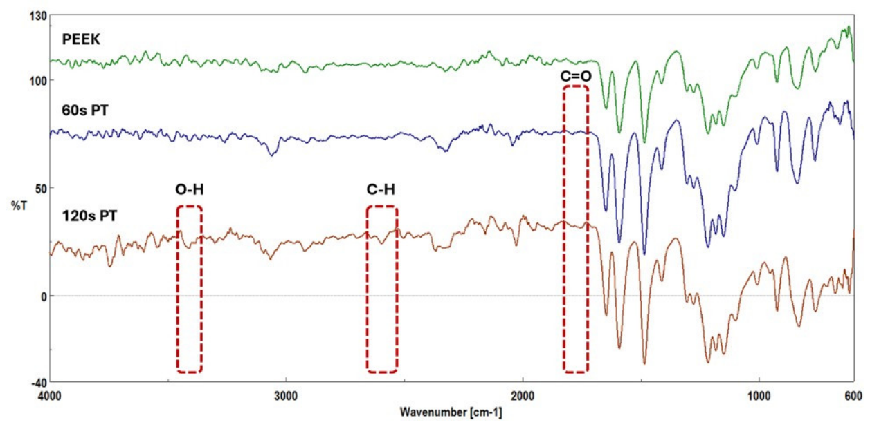
Figure 4.
Graphical representation of the contact angle values registered for PEEK. and the plasma treated samples. The progressive reduction in contact angle reflects increasing surface functionalization and hydrophilicity with plasma exposure time.
Figure 4.
Graphical representation of the contact angle values registered for PEEK. and the plasma treated samples. The progressive reduction in contact angle reflects increasing surface functionalization and hydrophilicity with plasma exposure time.
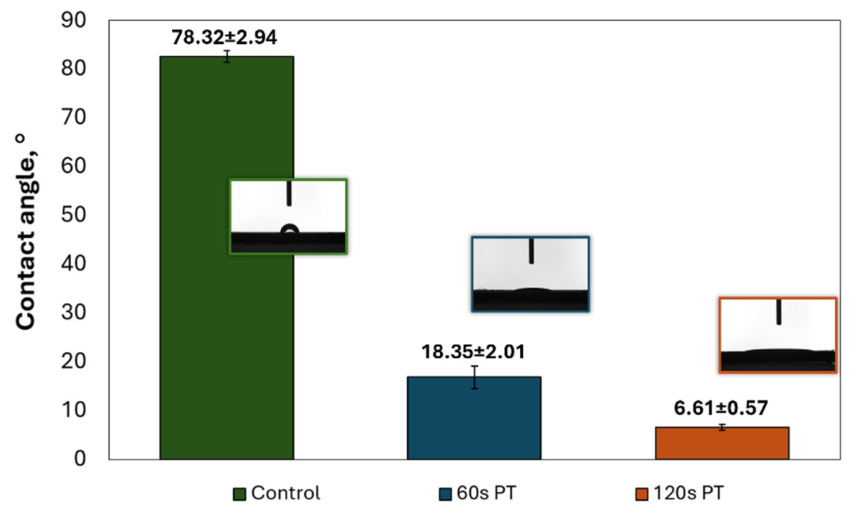
Figure 6.
SEM micrographs of pristine PEEK and GelMA-coated PEEK surfaces at different magnifications. Coated surfaces retain the printed filament architecture while exhibiting increased roughness and a relatively uniform protein film along the inter-filament regions, consistent with successful GelMA deposition and photocrosslinking. (A) Pristine PEEK at 100× magnification (scale bar 1 mm), showing the regular, parallel deposition tracks characteristic of the FDM process and a relatively smooth inter-filament topography. (B) Pristine PEEK at 500× magnification (scale bar 300 µm), revealing the compact, homogeneous surface of the individual printed filaments, with only minor surface irregularities and isolated debris. (C) GelMA-coated PEEK (PEEK–GelMA) at 100× magnification (scale bar 1 mm), in which the underlying filament architecture is preserved while a thin coating is distributed along the printed strands. (D) GelMA-coated PEEK at 500× magnification (scale bar 300 µm), showing increased surface roughness and discontinuous granular and film-like deposits localized predominantly along the inter-filament grooves, indicative of the immobilized GelMA layer. All micrographs were acquired in secondary-electron mode (ETD detector) at an accelerating voltage of 10.00 kV, spot size 3.5, and a working distance of 9.4–9.6 mm.
Figure 6.
SEM micrographs of pristine PEEK and GelMA-coated PEEK surfaces at different magnifications. Coated surfaces retain the printed filament architecture while exhibiting increased roughness and a relatively uniform protein film along the inter-filament regions, consistent with successful GelMA deposition and photocrosslinking. (A) Pristine PEEK at 100× magnification (scale bar 1 mm), showing the regular, parallel deposition tracks characteristic of the FDM process and a relatively smooth inter-filament topography. (B) Pristine PEEK at 500× magnification (scale bar 300 µm), revealing the compact, homogeneous surface of the individual printed filaments, with only minor surface irregularities and isolated debris. (C) GelMA-coated PEEK (PEEK–GelMA) at 100× magnification (scale bar 1 mm), in which the underlying filament architecture is preserved while a thin coating is distributed along the printed strands. (D) GelMA-coated PEEK at 500× magnification (scale bar 300 µm), showing increased surface roughness and discontinuous granular and film-like deposits localized predominantly along the inter-filament grooves, indicative of the immobilized GelMA layer. All micrographs were acquired in secondary-electron mode (ETD detector) at an accelerating voltage of 10.00 kV, spot size 3.5, and a working distance of 9.4–9.6 mm.
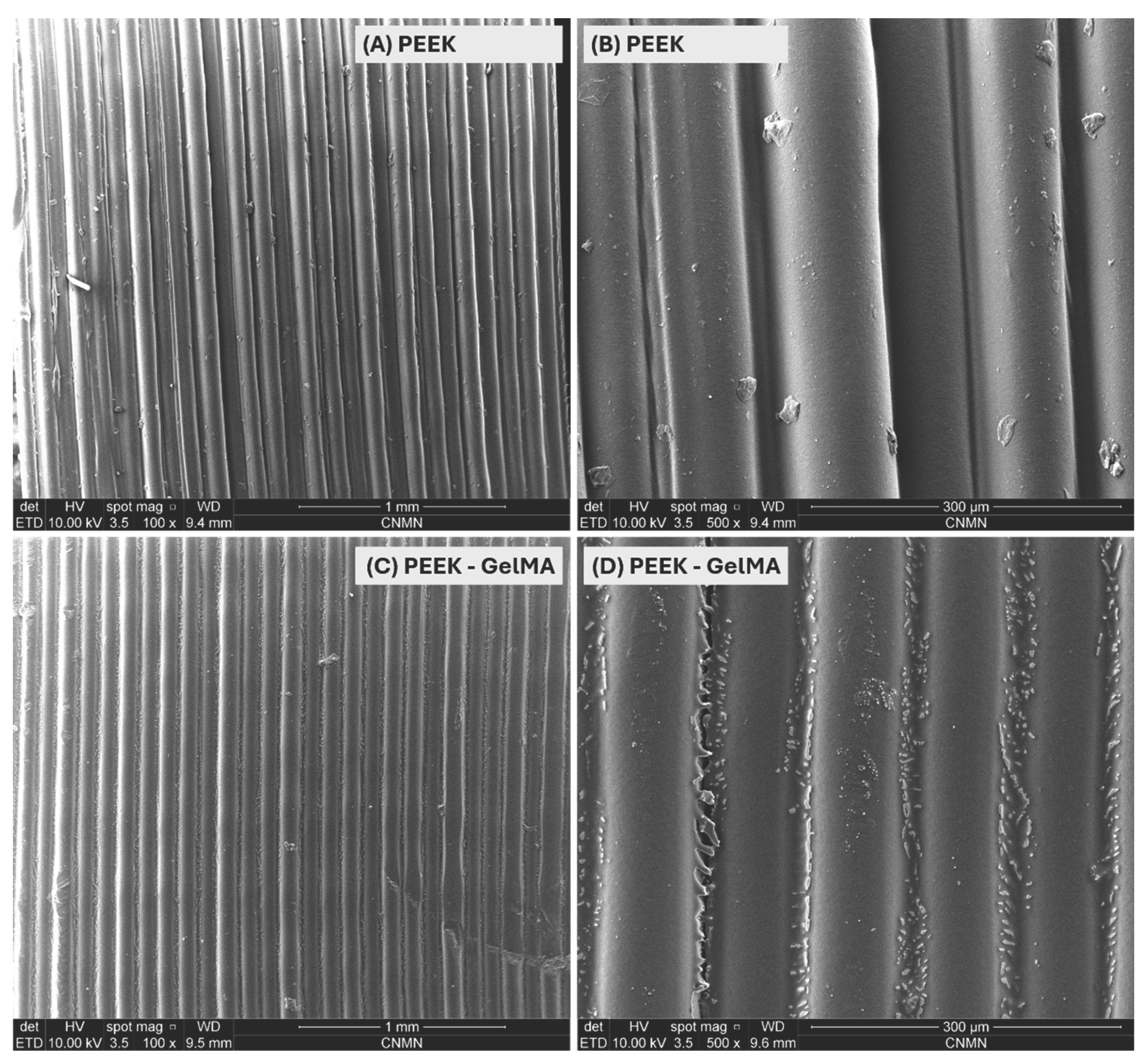

Table 1.
Water contact angle values for untreated and CO₂ plasma-treated PEEK samples (mean ± standard deviation).
Table 1.
Water contact angle values for untreated and CO₂ plasma-treated PEEK samples (mean ± standard deviation).
| Treatment condition | Contact angle (°) | Interpretation |
|---|---|---|
| Pristine PEEK (control) | 78.32 ± 2.94 | Hydrophobic surface |
| CO₂ plasma, 60 s per side | 18.35 ± 2.01 | Marked increase in hydrophilicity |
| CO₂ plasma, 120 s per side | 6.61 ± 0.57 | Maximum hydrophilicity (optimal) |
Disclaimer/Publisher’s Note: The statements, opinions and data contained in all publications are solely those of the individual author(s) and contributor(s) and not of MDPI and/or the editor(s). MDPI and/or the editor(s) disclaim responsibility for any injury to people or property resulting from any ideas, methods, instructions or products referred to in the content. |
© 2026 by the authors. Licensee MDPI, Basel, Switzerland. This article is an open access article distributed under the terms and conditions of the Creative Commons Attribution (CC BY) license (http://creativecommons.org/licenses/by/4.0/).
Copyright: This open access article is published under a Creative Commons CC BY 4.0 license, which permit the free download, distribution, and reuse, provided that the author and preprint are cited in any reuse.


