
Submitted:
14 July 2026
Posted:
21 July 2026
You are already at the latest version
Abstract
Paclitaxel chemotherapy frequently induces debilitating paclitaxel-induced peripheral neuropathic pain, which lacks effective treatments. This study demonstrates that dorsomorphin alleviates paclitaxel-induced peripheral neuropathic pain in mice by targeting neuroinflammation and pain pathways. Administering dorsomorphin (8 mg/kg, i.p.) alongside paclitaxel signifi-cantly reduced mechanical allodynia (von Frey test), thermal hyperalgesia (hot-plate test), and cold hyperalgesia (acetone test) compared to paclitaxel alone. Mechanistically, dorsomorphin suppressed spinal neuroinflammation by inhibiting microglial activation, reducing NF-κB pathway activity, and lowering pro-inflammatory cytokines TNF-α, IL-1β, and IL-6 in serum. Dorsomorphin also modulated key pain mediators, downregulating sodium channel Scn1a and lysophosphatidic acid (LPA) expression in the spinal cord. Crucially, dorsomorphin synergized with paclitaxel in A549 lung cancer cells in vitro, en-hancing the suppression of proliferation (CCK-8 assay), migration, and invasion (Transwell assays) at combined doses (e.g., 5 nM paclitaxel + 5 μM dorsomorphin) without compromising paclitaxel’s antitumor efficacy. These findings reveal dor-somorphin’s dual role: it mitigates paclitaxel-induced peripheral neuropathic pain by inhibiting neuroinflammation (via NF-κB/microglia/cytokines) and pain pathways (LPA/Scn1a), while concurrently enhancing paclitaxel’s anticancer activity. Dorsomorphin thus represents a promising adjunctive therapy for managing chemotherapy-induced neuropathy and im-proving treatment outcomes.
Keywords:
dorsomorphin
; paclitaxel-induced peripheral neuropathic pain
; inflammatory cytokines
1. Introduction
Paclitaxel remains a cornerstone chemotherapeutic agent for malignancies such as lung, ovarian, and breast cancer (Stage et al., 2020; Wang et al., 2022). It exerts antitumor effects by disrupting mitosis and inhibiting tumor growth (Weaver, 2014). However, its clinical utility is frequently complicated by paclitaxel-induced peripheral neuropathic pain, which affects 30–70% of patients (Staff et al., 2020; Park et al., 2008). Paclitaxel-induced peripheral neuropathic pain manifests as paresthesia, motor dysfunction, persistent pain, and thermal hypersensitivity (Flatters et al., 2017; Seretny et al., 2014), severely impairing the quality of life. Current analgesics provide only limited relief and are often associated with significant side effects (Chapman et al., 2020; Brewer et al., 2016).
The pathogenesis of paclitaxel-induced peripheral neuropathic pain involves multiple pathways distinct from those of inflammatory pain, including paclitaxel-induced oxidative nerve damage (Duggett et al., 2016), neuroinflammation via spinal glial activation and cytokine dysregulation (Sharma et al., 2016), altered neuronal excitability through dysregulation of sodium channels (e.g., Nav1.1 encoded by Scn1a) (Li et al., 2018; Shen et al., 2016; Yilmaz et al., 2017), and demyelination mediated by elevated lysophosphatidic acid (LPA) (Uchida et al., 2014).
Dorsomorphin (also known as compound C), initially identified as an inhibitor of AMPK phosphorylation, exhibits both AMPK-dependent and independent bioactivities (Dasgupta and Seibel, 2018). It has demonstrated synergistic antitumor effects with chemotherapeutic agents in models of breast cancer, leukemia, and colorectal cancer (Avsec et al., 2021; Li et al., 2024; Li et al., 2023), acting through the inhibition of proliferation, induction of apoptosis, and immune modulation (Avsec et al., 2021; Jaschke et al., 2020; Zhu et al., 2021). While these studies highlight its potential, the complete spectrum of dorsomorphin's antitumor mechanisms, particularly in the context of combination therapy, remains an active area of investigation. Furthermore, dorsomorphin possesses documented anti-inflammatory and analgesic properties. For instance, it suppresses spinal inflammatory mediators in murine inflammatory pain models via inhibition of the p38 MAPK/c-fos pathway (Yin et al., 2022). However, despite its efficacy in inflammatory pain models, the effect of dorsomorphin on chemotherapy-induced peripheral neuropathic pain has not been explored.
Therefore, this study was designed to investigate the potential of dorsomorphin to alleviate paclitaxel-induced peripheral neuropathic pain in mice, elucidating its effects on neuroinflammation and pain-related pathways specific to neuropathy. Additionally, we explored its antitumor activity with paclitaxel in A549 lung cancer cells. We aimed to elucidate the dual role of dorsomorphin in mitigating chemotherapy-induced neuropathy while potentially enhancing paclitaxel's anticancer efficacy, thereby proposing a novel strategy for improving chemotherapy outcomes.
2. Materials and Methods
2.1. Animals
Male Kunming mice (18–20 g) obtained from the Animal Center of North Sichuan Medical College (SCXK(川)2023 - 0018) were housed under a 12 h light/dark cycle with ad libitum access to food/water. All procedures complied with IASP guidelines for conscious animal pain research and were approved by the Institutional Animal Care and Use Committee of North Sichuan Medical College (IACUC No. 2024-029-132).
2.2. Treatment and Experimental Design
Mice were randomized into three groups (n = 6-8/group):
- Vehicle: Saline (i.p.)
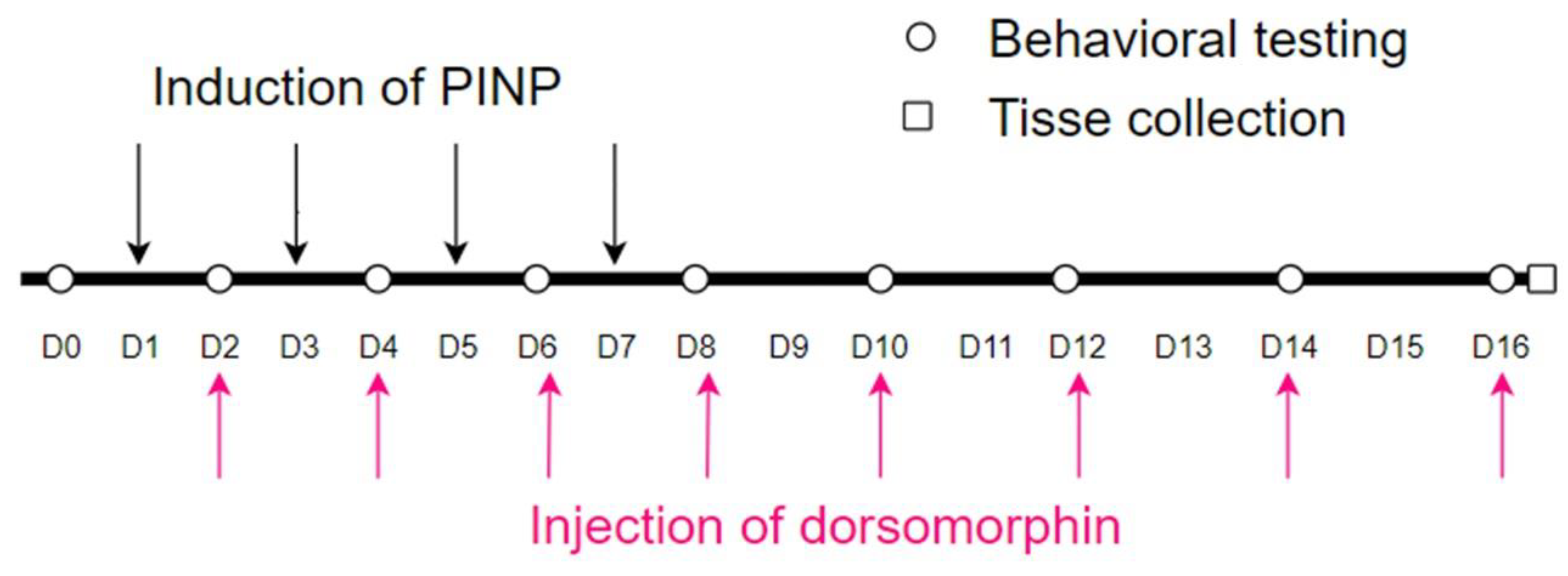
- Paclitaxel: Paclitaxel (8 mg/kg in corn oil, i.p.) on days 1, 3, 5, 7
- Paclitaxel + Dorsomorphin: paclitaxel (as above) + dorsomorphin (2, 4, or 8 mg/kg in saline, i.p.) on alternating days (days 2, 4, 6, 8, 10, 12, 14, 16). Dorsomorphin was administered intraperitoneally 1 hour prior to behavioral testing.
Behavioral tests were performed pre-treatment (day 0) and post-injection (days 2, 4, 6, 8, 10, 12, 14, 16). Tissues were collected ≤ 24 h after final behavioral assessment (Fig. 1). The dosing regimen and dosage in this experiment were based on previous studies (Caillaud, M et al., 2021; Chen, R et al., 2024; Kim HK et al., 2021; Yin et al., 2022).
Figure 1.
Experimental design showing the time point of dorsomorphin and paclitaxel administration, behavioral test, and tissue collection. (PINP: Paclitaxel-induced peripheral neuropathic pain).
Figure 1.
Experimental design showing the time point of dorsomorphin and paclitaxel administration, behavioral test, and tissue collection. (PINP: Paclitaxel-induced peripheral neuropathic pain).
2.3. Behavioral Assessments
2.3.1. Mechanical Withdrawal Threshold (Von Frey Test)
Mice acclimated 15 min in plexiglass chambers with wire mesh floors. The plantar hindpaw was stimulated using calibrated von frey filaments (Aesthesio, USA). Withdrawal threshold was defined as the minimal force (g) eliciting rapid paw retraction (mean of 3 trials/paw; 5-min intervals).
2.3.2. Thermal Withdrawal Latency (Hot-Plate Test)
Mice were placed on a 55 ± 1°C plate (Zhongshi Technology, China). Latency to jump or lick hindpaws was recorded (30-s cutoff; mean of 3 trials; 10-min intervals).
2.3.3. Cold Stimulation (Acetone Evaporation Test)
20 µL acetone was applied to the hindpaw plantar surface. Responses (flicking/licking/biting) were scored over 30 s: 0 (no response), 1 (brief withdrawal), 2 (prolonged withdrawal), 3 (repeated flicking/licking). (Dallazen et al., 2022).
2.4. Tissue Collection
Mice were anesthetized with isoflurane. Blood was collected via orbital enucleation, and serum was isolated (30 min RT, centrifugation, −80°C storage). Spinal cords (L4–L6 segments) were dissected, snap-frozen in liquid N₂, and stored at −80°C.
2.5. Biochemical Analyses
2.5.1. Enzyme-Linked Immunosorbent Assay (ELISA)
Serum TNF-α, IL-1β, and IL-6 levels were quantified using commercial kits (Yuduo, China) per manufacturer protocols.
2.5.2. Western Blot
Spinal cord tissues were homogenized (Whole Cell Lysis Assay, Wanleibio). Proteins (30 µg/lane) were separated by SDS-PAGE (8–12.5%), transferred to PVDF membranes, and probed overnight (4°C) with primary antibodies: anti-p65 (1:500; Wanleibio), anti-Scn1a (1:500; Upingbio), anti-Iba1 (1:500; Servicebio), anti–β-actin (1:2000; Beyotime). Membranes were incubated with HRP-conjugated secondary antibody (1:4000; Proteintech; 1 h, RT) and visualized via chemiluminescence. Band intensities were quantified (ImageJ) and normalized to β-actin.
2.5.3. Immunohistochemistry
Spinal cord sections (4 µm) were deparaffinized, antigen-retrieved (citrate buffer), blocked (3% H₂O₂, 3% BSA), and incubated with anti-LPA (1:300; Servicebio). DAB was used for detection, followed by hematoxylin counterstaining. LPA-positive cells were counted in 3 random fields/section.
2.6. In Vitro Assays (A549 Cells)
2.6.1. CCK-8 Proliferation
Cells (1,000/well) were treated with paclitaxel (1–20 nM), dorsomorphin (2.5–10 µM), or combinations (paclitaxel + dorsomorphin at 2.5 + 2.5, 5 + 5, 10 + 10 nM/µM). Viability was assessed at 0/24/48/72 h using CCK-8 reagent (Beyotime; OD₄₅₀ nm).
2.6.2. Migration/Invasion (Transwell)
Migration: Cells (1×10⁴) in serum-free medium were seeded into upper chambers; 10% FBS medium served as chemoattractant. After 48 h, migrated cells were fixed (4% PFA), stained (0.1% crystal violet), and counted (100×; 3 fields/chamber).
Invasion: Matrigel-coated chambers (1:6 dilution) were seeded with serum-starved cells (2.5×10⁵/mL). Invaded cells were quantified after 48 h as above.
2.7. Statistical Analysis
All statistical analyses were performed using SPSS 25.0 and GraphPad Prism 8.0. Data are expressed as mean ± SEM. For behavioral tests (Fig. 2), data were analyzed by two-way ANOVA with 'Treatment Group' and 'Time' as the two factors, followed by Bonferroni's post hoc tests. For all other comparisons among three or more groups (e.g., ELISA, Western blot, immunohistochemistry, CCK-8, Transwell), one-way ANOVA followed by Bonferroni's post hoc test was used. The detailed statistical values (F, degrees of freedom, and p-values) for main and interaction effects are provided in the figure legends or results section where appropriate. A p-value of < 0.05 was considered statistically significant.
3. Results
3.1. Dorsomorphin Alleviates Mechanical Allodynia, Thermal Hyperalgesia, and Cold Hyperalgesia in the Mouse Model of Paclitaxel-Induced Peripheral Neuropathic Pain
To evaluate the effects of dorsomorphin on paclitaxel-induced peripheral neuropathic pain, we treated mice and conducted behavioral assessments according to the experimental protocol. Prior to treatment, no significant differences existed in mechanical withdrawal threshold, thermal withdrawal latency, or total scores among the mouse groups. Compared with the vehicle group, mice in the paclitaxel group exhibited significantly decreased mechanical withdrawal threshold and thermal withdrawal latency, along with increased total scores from day 8 to day 16 (p < 0.05; Figure 2A-C), indicating the development of mechanical allodynia and thermal hyperalgesia. Significant differences in mechanical withdrawal threshold, thermal withdrawal latency, and total scores were observed between the paclitaxel group and the vehicle group.
Administration of dorsomorphin (8 mg/kg) significantly increased mechanical withdrawal threshold and thermal withdrawal latency from day 4 to day 16 (p < 0.05; Figure 2A, B), and significantly decreased total scores from day 4 to day 16 (p < 0.05; Figure 2C), compared with the paclitaxel group.
Similarly, mice treated with paclitaxel + dorsomorphin (4 mg/kg) showed increased mechanical withdrawal threshold from day 6 to day 16 (p < 0.05; Figure 2A) and increased thermal withdrawal latency from day 8 to day 16 (p < 0.05; Figure 2B), accompanied by decreased total scores from day 4 to day 16 (p < 0.05; Figure 2C).
Mice treated with paclitaxel + dorsomorphin (2 mg/kg) displayed a significant decrease in total scores only on day 8 (p < 0.05; Figure 2C) and an increase in thermal withdrawal latency on day 10 (p < 0.05; Figure 2B). However, no significant changes in mechanical withdrawal threshold were observed between the paclitaxel + dorsomorphin (2 mg/kg) group and the paclitaxel group at any time point during the experiment (Figure 2A).
3.2. Effect of Dorsomorphin on Inflammatory Cytokine Expression Levels
Serum levels of the inflammatory cytokines IL-6, IL-1β, and TNF-α were measured by ELISA in each experimental group. Compared with the vehicle group, mice in the paclitaxel group exhibited significantly elevated serum levels of IL-6, IL-1β, and TNF-α (p < 0.05; Figure 3A-C). Conversely, animals in the paclitaxel + dorsomorphin (8 mg/kg) group showed significantly reduced serum levels of IL-6, IL-1β, and TNF-α compared to the paclitaxel group (p < 0.01; Figure 3A-C).
To explore the mechanism underlying dorsomorphin's analgesic effects, we measured the expression of NF-κB p65, Iba1 (a microglial marker), and Scn1a (Nav1.1) proteins in mouse spinal cord tissues using western blotting. Compared to the vehicle group, paclitaxel treatment significantly increased the expression levels of Iba1 (p < 0.05; Figure 4A, B), NF-κB p65 (p < 0.05; Figure 4C, D), and Scn1a (p < 0.05; Figure 4C, D). Conversely, co-administration of dorsomorphin (8 mg/kg) with paclitaxel significantly attenuated these paclitaxel-induced increases: Iba1 expression was decreased (p < 0.05; Figure 4A, B), NF-κB p65 expression was reduced relative to the pacl itaxel group (p < 0.05; Figure 4C, D), and Scn1a expression was lowered (p < 0.05; Figure 4C, D).
To investigate the regulatory effect of dorsomorphin on LPA expression, we performed immunohistochemical analysis on spinal cord tissues from each experimental group. Compared to the vehicle-treated group, paclitaxel administration significantly increased the density of LPA-positive cells (p < 0.05; Figure 5A-D). Co-treatment with dorsomorphin (8 mg/kg) significantly attenuated this paclitaxel-induced upregulation, reducing LPA-positive cell density to levels comparable to the vehicle group (p < 0.05; Figure 5A-D). These results indicate that dorsomorphin effectively inhibits paclitaxel-induced LPA overexpression in the spinal cord, suggesting its potential to modulate LPA-mediated pathways in neuropathic pain.
3.5. Effects of Dorsomorphin and Paclitaxel on the Proliferation of A549 Cells
To assess the impact of dorsomorphin, paclitaxel, and their combination on the proliferation of A549 cancer cells, CCK-8 assays were conducted (Figure 6). Paclitaxel monotherapy (Figure 6A): Compared to the vehicle group, 1 nM, 2.5 nM, and 5 nM paclitaxel significantly reduced OD values at 72 h (p < 0.05), while 10 nM and 20 nM paclitaxel exhibited significant reductions at both 48 h and 72 h (p < 0.05).
Dorsomorphin monotherapy (Figure 6B): 2.5 μM, 5 μM, and 10 μM dorsomorphin significantly decreased OD values versus vehicle at 48 h and 72 h (p < 0.05).
These results indicate that both paclitaxel and dorsomorphin individually suppress the proliferation of A549 cells.
Effects of combination therapy: Compared to 2.5 nM paclitaxel alone, the combination of 2.5 nM paclitaxel + 2.5 µM dorsomorphin significantly reduced OD values only at 48 h (Figure 6C; p < 0.05). Compared to 5 nM paclitaxel alone, the combination of 5 nM paclitaxel + 5 µM dorsomorphin significantly decreased OD values at both 48 h and 72 h (Figure 6D; p < 0.001). However, at the highest dose (10 µM) where dorsomorphin alone achieved 100% suppression (Figure 6E), the specific contribution of the combination partners could not be evaluated. The observed effect at this dose is attributable to dorsomorphin.
3.6. Effects of Dorsomorphin and Paclitaxel on the Migration and Invasion of A549 Cells
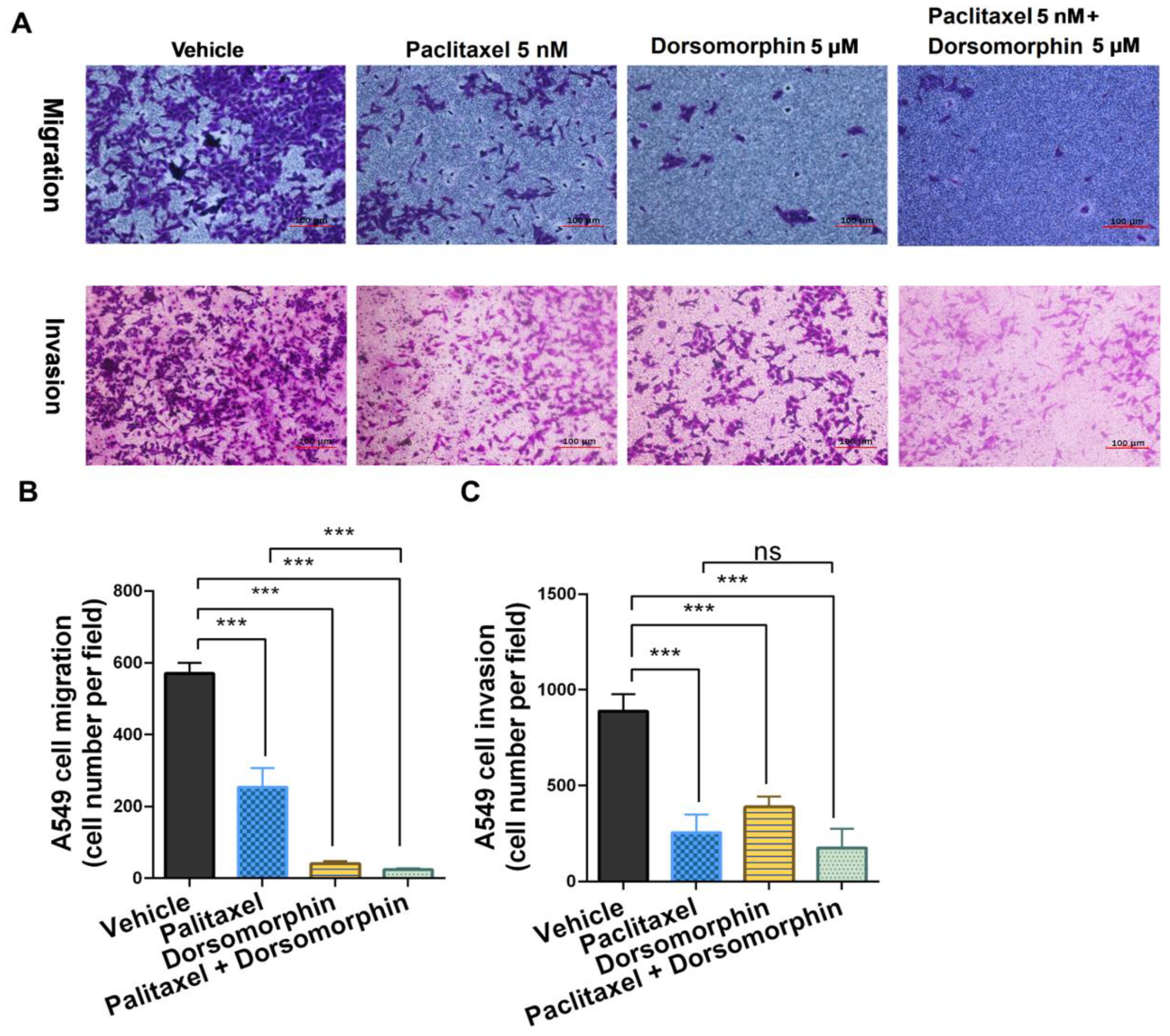
Transwell migration assays revealed that A549 cell migration was significantly suppressed in both the dorsomorphin-alone (5 μM) and 5 nM paclitaxel + 5 μM dorsomorphin groups compared with the vehicle group (Figure 7A-B). The anti-migratory effect of the 5 nM paclitaxel + 5 μM dorsomorphin combination was not significantly different from that of dorsomorphin-alone (5 μM) (Figure 7A-B), indicating that the suppression of migration was primarily attributable to DM.
Invasion assays showed similar inhibitory trends (Figure 7A-C): paclitaxel (5 nM), dorsomorphin (5 μM), and 5 nM paclitaxel + 5 μM dorsomorphin treatments all significantly decreased invaded cell numbers versus vehicle (p < 0.001). The combination treatment showed comparable efficacy to paclitaxel monotherapy (NS, not significant).
These results demonstrate that dorsomorphin and paclitaxel independently suppress A549 cell migration and invasion, while their combination yields anti-migratory and anti-invasion effects without compromising paclitaxel's activity.
4. Discussion
In the present study, we employed behavioral experiments to investigate the effects of dorsomorphin on paclitaxel-induced peripheral neuropathic pain. Paclitaxel, a cornerstone chemotherapeutic agent for various malignancies, frequently induces paclitaxel-induced peripheral neuropathic pain, significantly impairing patients' quality of life (Seretny et al., 2014). Unfortunately, current clinical analgesics demonstrate limited efficacy against paclitaxel-induced peripheral neuropathic pain (Derry et al., 2017).
Recent studies suggest that dorsomorphin exerts anti-inflammatory, analgesic, and antitumor effects through multi-target mechanisms (Li et al., 2024; Zhu et al., 2021; Peng et al., 2016; Yin et al., 2022). However, its specific effects on paclitaxel-induced peripheral neuropathic pain and the underlying mechanisms remained incompletely elucidated. Our findings demonstrate that dorsomorphin significantly alleviated mechanical allodynia, thermal hyperalgesia, and cold hyperalgesia in a mouse model of paclitaxel-induced peripheral neuropathic pain. Specifically, dorsomorphin (4, 8 mg/kg) treatment increased the mechanical withdrawal threshold and thermal withdrawal latency while reducing the total pain scores in the acetone evaporation test.
To elucidate the mechanisms underpinning dorsomorphin's analgesic effect in paclitaxel-induced peripheral neuropathic pain, we utilized ELISA, western blotting, and immunohistochemistry. Existing literature indicates that paclitaxel activates spinal microglia and the NF-κB pathway, resulting in the overproduction of pro-inflammatory cytokines (Wede et al., 2023; Zhao et al., 2020; Suo et al., 2020). The observed suppression of NF-κB signaling by dorsomorphin in our study highlights its role in attenuating neuroinflammation, consistent with its established anti-inflammatory profile (Yin et al., 2022). Furthermore, enhanced ion channel activity in peripheral sensory neurons, including Nav1.1 channels (Scn1a gene product), which are critical drivers of neuronal hyperexcitability in neuropathic pain (Inoue et al., 2008; Weuring et al., 2020; Li et al., 2018; Yilmaz et al., 2017), represents another proposed mechanism for paclitaxel-induced peripheral neuropathic pain. Our data revealed that dorsomorphin reduced serum levels of the inflammatory cytokines IL-6, IL-1β, and TNF-α. Moreover, western blot analysis demonstrated that dorsomorphin decreased the expression of NF-κB p65, Iba1 (a microglial marker), and Scn1a proteins in spinal cord tissue.
LPA, a bioactive lipid mediator, has been implicated in dorsal root ganglion (DRG) demyelination and neuronal hyperexcitability associated with neuropathic pain (Poon et al., 2024; Ueda, 2020; Shen et al., 2016). Given that paclitaxel-induced pain is linked to increased LPA production (Uchida et al., 2014), and considering that AMPK activation can influence NMDA receptor function (Shen et al., 2016) while dorsomorphin inhibits AMPK activation (Dasgupta and Seibel, 2018; Wang et al., 2020), dorsomorphin's analgesic effect may involve modulation of the NMDA/LPA pathway. Supporting this hypothesis, our immunohistochemical analysis showed that dorsomorphin effectively inhibited paclitaxel-induced LPA upregulation in the spinal cord.
Beyond its analgesic properties, dorsomorphin has demonstrated antitumor effects via multiple targets (Li et al., 2024; Zhu et al., 2021; Peng et al., 2016). Having established the efficacy of dorsomorphin in alleviating paclitaxel-induced peripheral neuropathic pain, we further explored its impact on tumor cells. CCK-8 assays revealed that dorsomorphin, paclitaxel, and their combination all inhibited A549 cell proliferation, with the combination therapy exhibiting a more potent effect. Transwell assays indicated that both dorsomorphin and paclitaxel suppressed the migration and invasion capabilities of A549 cells. Crucially, the combination did not compromise paclitaxel's antitumor efficacy, instead, it exhibited even more significant inhibitory effects on cellular proliferation, migration, and invasive potential.
However, the dorsomorphin 's effects on paclitaxel mechanical withdrawal threshold and thermal withdrawal latency are significantly higher than vehicle conditions without paclitaxel. Moreover, the effect of dorsomorphin (8mg/kg) on thermal withdrawal latency on paclitaxel-treated animals appears even before of paclitaxel-induced significant effects. Thus, the effect of dorsomorphin at 8 mg/kg by itself in both mechanical withdrawal threshold and specially in thermal withdrawal latency needs further explanation and experimental verification. It would be necessary to assess if dorsomorphin has a direct effect by itself on mechanical withdrawal threshold and thermal withdrawal latency to define if the real effect on paclitaxel is overestimated in the context of paclitaxel. If dorsomorphin has an effect by itself on the allodynia or the thermal hyperalgesia, other pharmacological effects should be considered in the CNS/PNS area (anasthesia vs analgesia, or motor impairment). All the issues above need to be studied in deeper in the future.
In conclusion, our findings demonstrate that dorsomorphin alleviates paclitaxel-induced peripheral neuropathic pain by suppressing neuroinflammation (via inhibition of the NF-κB pathway and microglial activation, leading to reduced pro-inflammatory cytokines) and modulating key pain-related molecules (LPA and Scn1a). Furthermore, dorsomorphin inhibits tumor cell proliferation, migration, and invasion without compromising paclitaxel’s antitumor efficacy. These dual therapeutic benefits position dorsomorphin as a promising candidate for improving chemotherapy outcomes by simultaneously managing neuropathic pain and enhancing antitumor efficacy, thereby potentially improving patient quality of life.
Data availability
Data will be made available on request.
Acknowledgments
This work was supported by Health Nanchong Research Center Program (NC26JK02), North Sichuan Medical College 2026 Graduate Education and Teaching Quality Project (PGJC2026034), 2026 North Sichuan Medical College International Student Teaching Engineering Project (ISJG2026-13). Nanchong City-North Sichuan Medical College Cooperative Scientific Research Project (22SXZRKX0014).
Declaration of competing interest
The authors declare that they have no known competing financial interests or personal relationships that could have appeared to influence the work reported in this paper.
References
- Avsec, D.; Jakoš Djordjevič, A. T.; Kandušer, M.; Podgornik, H.; Škerget, M.; Mlinarič-Raščan, I. Targeting autophagy triggers apoptosis and complements the action of venetoclax in chronic lymphocytic leukemia cells. Cancers 2021, 13, 4557. [Google Scholar] [CrossRef] [PubMed]
- Brewer, J. R.; Morrison, G.; Dolan, M. E.; Fleming, G. F. Chemotherapy-induced peripheral neuropathy: Current status and progress. Gynecol. Oncol. 2016, 140, 176–183. [Google Scholar] [CrossRef] [PubMed]
- Caillaud, M.; Patel, N. H.; White, A.; Wood, M.; Contreras, K.M.; Toma, W.; Alkhlaif, Y.; Roberts, J.L.; Tran, T.H.; Jackson, A.B.; Poklis, J.; Gewirtz, D.A.; Damaj, M.I. Targeting Peroxisome Proliferator-Activated Receptor-α (PPAR-α) to reduce paclitaxel-induced peripheral neuropathy. Brain Behav. Immun. 2021, 93, 172–185. [Google Scholar] [CrossRef] [PubMed]
- Chapman, E. J.; Edwards, Z.; Boland, J. W.; Maddocks, M.; Fettes, L.; Malia, C.; Mulvey, M.R.; Bennett, M.I. Practice review: Evidence-based and effective management of pain in patients with advanced cancer. Palliative Med. 2020, 34, 444–453. [Google Scholar] [CrossRef] [PubMed]
- Chen, R.; Hu, J.; Zhang, Y.; Liu, Y.; Zhu, J.; Pan, Z.; Yang, H.; Wang, Q.; Chen, Y.; Tang, S.; Min, B. Total glucosides of paeony ameliorates chemotherapy-induced neuropathic pain by suppressing microglia pyroptosis through the inhibition of KAT2A-mediated p38 pathway activation and succinylation. Sci. Rep. 2024, 14, 31875. [Google Scholar] [CrossRef] [PubMed]
- Dallazen, J. L.; da Luz, B. B.; Maria-Ferreira, D.; Nascimento, A.M.; Cipriani, T.R.; de Souza, L.M.; Geppetti, P.; de Paula Werner, M.F. Local effects of natural alkylamides from Acmella oleracea and synthetic isobutylalkyl amide on neuropathic and postoperative pain models in mice. Fitoterapia 2022, 160, 105224. [Google Scholar] [CrossRef] [PubMed]
- Dasgupta, B.; Seibel, W. Compound C/Dorsomorphin: its use and misuse as an ampk inhibitor. Methods Mol. Biol. (Clifton, N.J.) 2018, 1732, 195–202. [Google Scholar] [CrossRef] [PubMed]
- Derry, S.; Wiffen, P. J.; Kalso, E. A.; Bell, R. F.; Aldington, D.; Phillips, T.; Gaskell, H.; Moore, R.A. Topical analgesics for acute and chronic pain in adults - an overview of Cochrane Reviews. Cochrane Database Syst. Rev. 2017, 5, CD008609. [Google Scholar] [CrossRef] [PubMed]
- Duggett, N. A.; Griffiths, L. A.; McKenna, O. E.; de Santis, V.; Yongsanguanchai, N.; Mokori, E.B.; Flatters, S.J. Oxidative stress in the development, maintenance and resolution of paclitaxel-induced painful neuropathy. Neuroscience 2016, 333, 13–26. [Google Scholar] [CrossRef] [PubMed]
- Flatters, S. J. L.; Dougherty, P. M.; Colvin, L. A. Clinical and preclinical perspectives on Chemotherapy-Induced Peripheral Neuropathy (CIPN): a narrative review. Brit. J. Anaesth. 2017, 119, 737–749. [Google Scholar] [CrossRef] [PubMed]
- Inoue, M.; Ma, L.; Aoki, J.; Ueda, H. Simultaneous stimulation of spinal NK1 and NMDA receptors produces LPC which undergoes ATX-mediated conversion to LPA, an initiator of neuropathic pain. J. Neurochem. 2008, 107, 1556–1565. [Google Scholar] [CrossRef] [PubMed]
- Jaschke, N.; Kleymann, A.; Hofbauer, L. C.; Göbel, A.; Rachner, T.D. Dorsomorphin: a novel inhibitor of Dickkopf-1 in breast cancer. Biochem. Bioph. Res. Co. 2020, 524, 360–365. [Google Scholar] [CrossRef] [PubMed]
- Kim, H.K.; Lee, S.Y.; Koike, N.; Kim, E.; Wirianto, M.; Burish, M.J.; Yagita, K.; Lee, H.K.; Chen, Z.; Chung, J.M.; Abdi, S.; Yoo, S.H. Circadian regulation of chemotherapy-induced peripheral neuropathic pain and the underlying transcriptomic landscape. Sci. Rep. 2020, 10, 13844. [Google Scholar] [CrossRef] [PubMed]
- Li, K.; Sun, X.; Minami, K.; Tamari, K.; Ogawa, K.; Li, H.; Ma, H.; Zhou, M.; Na, S.; Li, B.Y.; Yokota, H. Proteomes from AMPK-inhibited peripheral blood mononuclear cells suppress the progression of breast cancer and bone metastasis. Theranostics 2023, 13, 1247–1263. [Google Scholar] [CrossRef] [PubMed]
- Li, X. P.; Cao, L. Q.; Yu, Z. Z.; He, K.; Ding, P.B.; Li, J.S.; Shan, Y.Y.; Su, Y.B.; Yuan, Z.M.; Shi, Z. Dorsomorphin attenuates ABCG2-mediated multidrug resistance in colorectal cancer. Fron. Pharmacol. 2024, 15, 1393693. [Google Scholar] [CrossRef] [PubMed]
- Li, Y.; North, R. Y.; Rhines, L. D.; Tatsui, C.E.; Rao, G.; Edwards, D.D.; Cassidy, R.M.; Harrison, D.S.; Johansson, C.A.; Zhang, H.; Dougherty, P.M. DRG voltage-gated sodium channel 1.7 is upregulated in paclitaxel-induced neuropathy in rats and in humans with neuropathic pain. J. Neurosci. 2018, 38, 1124–1136. [Google Scholar] [CrossRef] [PubMed]
- Park, S. B.; Krishnan, A. V.; Lin, C. S.; Goldstein, D.; Friedlander, M.; Kiernan, M.C. Mechanisms underlying chemotherapy-induced neurotoxicity and the potential for neuroprotective strategies. Curr. Med. Chem. 2008, 15, 3081–3094. [Google Scholar] [CrossRef] [PubMed]
- Peng, J.; Yoshioka, Y.; Mandai, M.; Matsumura, N.; Baba, T.; Yamaguchi, K.; Hamanishi, J.; Kharma, B.; Murakami, R.; Abiko, K.; Murphy, S.K.; Konishi, I. The BMP signaling pathway leads to enhanced proliferation in serous ovarian cancer-A potential therapeutic target. Mol. Carcinogen. 2016, 55, 335–345. [Google Scholar] [CrossRef] [PubMed]
- Poon, M. M.; Lorrain, K. I.; Stebbins, K. J.; Edu, G.C.; Broadhead, A.R.; Lorenzana, A.O.; Paulson, B.E.; Baccei, C.S.; Roppe, J.R.; Schrader, T.O.; Valdez, L.J.; Xiong, Y.; Chen, A.C.; Lorrain, D.S. Discovery of a brain penetrant small molecule antagonist targeting LPA1 receptors to reduce neuroinflammation and promote remyelination in multiple sclerosis. Sci. Rep. 2024, 14, 10573. [Google Scholar] [CrossRef] [PubMed]
- Roza, C.; Campos-Sandoval, J. A.; Gómez-García, M. C.; Peñalver, A.; Márquez, J. Lysophosphatidic acid and glutamatergic transmission. Front. Mol. Neurosci. 2019, 12, 138. [Google Scholar] [CrossRef] [PubMed]
- Saito, S.; Furuno, A.; Sakurai, J.; Park, H.R.; Shin-ya, K.; Tomida, A. Compound C prevents the unfolded protein response during glucose deprivation through a mechanism independent of AMPK and BMP signaling. PloS One 2012, 7, e45845. [Google Scholar] [CrossRef] [PubMed]
- Seretny, M.; Currie, G. L.; Sena, E. S.; Ramnarine, S.; Grant, R.; MacLeod, M.R.; Colvin, L.A.; Fallon, M. Incidence, prevalence, and predictors of chemotherapy-induced peripheral neuropathy: A systematic review and meta-analysis. Pain 2014, 155, 2461–2470. [Google Scholar] [CrossRef] [PubMed]
- Sharma, A.; Behl, T.; Sharma, L.; Shah, O.P.; Yadav, S.; Sachdeva, M.; Rashid, S.; Bungau, S.G.; Bustea, C. Exploring the molecular pathways and therapeutic implications of angiogenesis in neuropathic pain. Biomed. Pharmacother. 2023, 162, 114693. [Google Scholar] [CrossRef] [PubMed]
- Shen, K. Z.; Wu, Y. N.; Munhall, A. C.; Johnson, S.W. AMP kinase regulates ligand-gated K-ATP channels in substantia nigra dopamine neurons. Neuroscience 2016, 330, 219–228. [Google Scholar] [CrossRef] [PubMed]
- Staff, N. P.; Fehrenbacher, J. C.; Caillaud, M.; Damaj, M.I.; Segal, R.A.; Rieger, S. Pathogenesis of paclitaxel-induced peripheral neuropathy: A current review of in vitro and in vivo findings using rodent and human model systems. Exp. Neurol. 2020, 324, 113121. [Google Scholar] [CrossRef] [PubMed]
- Stage, T. B.; Mortensen, C.; Khalaf, S.; Steffensen, V.; Hammer, H.S.; Xiong, C.; Nielsen, F.; Poetz, O.; Svenningsen, Å.F.; Rodriguez-Antona, C.; Kroetz, D.L. P-Glycoprotein inhibition exacerbates paclitaxel neurotoxicity in neurons and patients with cancer. Clin. Pharmacol. Ther. 2020, 108, 671–680. [Google Scholar] [CrossRef] [PubMed]
- Suo, J.; Wang, M.; Zhang, P.; Lu, Y.; Xu, R.; Zhang, L.; Qiu, S.; Zhang, Q.; Qian, Y.; Meng, J.; Zhu, J. Siwei Jianbu decoction improves painful paclitaxel-induced peripheral neuropathy in mouse model by modulating the NF-κB and MAPK signaling pathways. Regen. Med. Res. 2020, 8, 2. [Google Scholar] [CrossRef] [PubMed]
- Uchida, H.; Nagai, J.; Ueda, H. Lysophosphatidic acid and its receptors LPA1 and LPA3 mediate paclitaxel-induced peripheral neuropathic pain in mice. Mol. Pain 2014, 10, 71. [Google Scholar] [CrossRef] [PubMed]
- Ueda, H. LPA receptor signaling as a therapeutic target for radical treatment of neuropathic pain and fibromyalgia. Pain Manag. 2020, 10, 43–53. [Google Scholar] [CrossRef] [PubMed]
- Wang, W.; Wang, J.; Liu, S.; Ren, Y.; Wang, J.; Liu, S.; Cui, W.; Jia, L.; Tang, X.; Yang, J.; Wu, C.; Wang, L. An EHMT2/NFYA-ALDH2 signaling axis modulates the RAF pathway to regulate paclitaxel resistance in lung cancer. Mol. Cancer 2022, 21, 106. [Google Scholar] [CrossRef] [PubMed]
- Wang, Y.; Tian, M.; Tan, J.; Pei, X.; Lu, C.; Xin, Y.; Deng, S.; Zhao, F.; Gao, Y.; Gong, Y. Irisin ameliorates neuroinflammation and neuronal apoptosis through integrin αVβ5/AMPK signaling pathway after intracerebral hemorrhage in mice. J. Neuroinflamm. 2022, 19, 82. [Google Scholar] [CrossRef] [PubMed]
- Weaver, B. A. How Taxol/paclitaxel kills cancer cells. Mol. Biol. Cell 2014, 25, 2677–2681. [Google Scholar] [CrossRef] [PubMed]
- Wedel, S.; Hahnefeld, L.; Schreiber, Y.; Namendorf, C.; Heymann, T.; Uhr, M.; Schmidt, M.V.; de Bruin, N.; Hausch, F.; Thomas, D.; Geisslinger, G.; Sisignano, M. SAFit2 ameliorates paclitaxel-induced peripheral neuropathic pain by reducing spinal gliosis and elevating pro-resolving lipid mediators. J. Neuroinflamm. 2023, 20, 149. [Google Scholar] [CrossRef] [PubMed]
- Weuring, W. J.; Singh, S.; Volkers, L.; Rook, M.B.; van't Slot, R.H.; Bosma, M.; Inserra, M.; Vetter, I.; Verhoeven-Duif, N.M.; Braun, K.P.J.; Rivara, M.; Koeleman, B.P.C. NaV1.1 and NaV1.6 selective compounds reduce the behavior phenotype and epileptiform activity in a novel zebrafish model for Dravet Syndrome. PloS One 2020, 15, e0219106. [Google Scholar] [CrossRef] [PubMed]
- Yilmaz, E.; Watkins, S. C.; Gold, M. S. Paclitaxel-induced increase in mitochondrial volume mediates dysregulation of intracellular Ca2+ in putative nociceptive glabrous skin neurons from the rat. Cell Calcium 2017, 62, 16–28. [Google Scholar] [CrossRef] [PubMed]
- Yin, X.Q.; Jing, Y.Y.; You, Z.P.; Ke, J.; Tan, X.; Hu, Y.M.; Zhu, C.X.; Duan, S.Y.; Yi, M.P.; Zhu, Y.L.; Chen, S.H.; Yan, H. Systemic administration of dorsomorphin relieves inflammatory nociception in the mouse formalin test. Int. Immunopharmacol. 2022, 113, 109337. [Google Scholar] [CrossRef] [PubMed]
- Zhao, Y.X.; Yao, M.J.; Liu, Q.; Xin, J.J.; Gao, J.H.; Yu, X.C. Electroacupuncture treatment attenuates paclitaxel-induced peripheral neuropathic pain in rats via inhibiting spinal glia and the TLR4/NF-κB pathway. J. Pain Res. 2020, 13, 239–250. [Google Scholar] [CrossRef] [PubMed]
- Zhu, G.; Song, J.; Chen, W.; Yuan, D.; Wang, W.; Chen, X.; Liu, H.; Su, H.; Zhu, J. Expression and role of Dickkopf-1 (Dkk1) in tumors: from the cells to the patients. Cancer Manag. Res. 2021, 13, 659–675. [Google Scholar] [CrossRef] [PubMed]
Figure 2.
Dorsomorphin attenuate paclitaxel-induced peripheral neuropathic pain in mice. (A) Mechanical allodynia assessed by von frey filament test. (B) Thermal hyperalgesia measured by hot plate test. (C) Cold hyperalgesia evaluated by acetone evaporation test. Mice received intraperitoneal (i.p.) injections of dorsomorphin (2, 4, or 8 mg/kg) during paclitaxel-induced peripheral neuropathic pain. Behavioral responses were recorded at baseline (day 0) and on days 2, 4, 6, 8, 10, 12, 14, and 16 post-paclitaxel initiation. Data are expressed as mean ± SEM (n = 6-8 per group). Statistical significance: paclitaxel vs. vehicle: # p < 0.05, ## p < 0.01, ### p < 0.001; paclitaxel + dorsomorphin (2, 4, or 8 mg/kg) vs. paclitaxel: * p < 0.05, ** p < 0.01, *** p < 0.001.
Figure 2.
Dorsomorphin attenuate paclitaxel-induced peripheral neuropathic pain in mice. (A) Mechanical allodynia assessed by von frey filament test. (B) Thermal hyperalgesia measured by hot plate test. (C) Cold hyperalgesia evaluated by acetone evaporation test. Mice received intraperitoneal (i.p.) injections of dorsomorphin (2, 4, or 8 mg/kg) during paclitaxel-induced peripheral neuropathic pain. Behavioral responses were recorded at baseline (day 0) and on days 2, 4, 6, 8, 10, 12, 14, and 16 post-paclitaxel initiation. Data are expressed as mean ± SEM (n = 6-8 per group). Statistical significance: paclitaxel vs. vehicle: # p < 0.05, ## p < 0.01, ### p < 0.001; paclitaxel + dorsomorphin (2, 4, or 8 mg/kg) vs. paclitaxel: * p < 0.05, ** p < 0.01, *** p < 0.001.
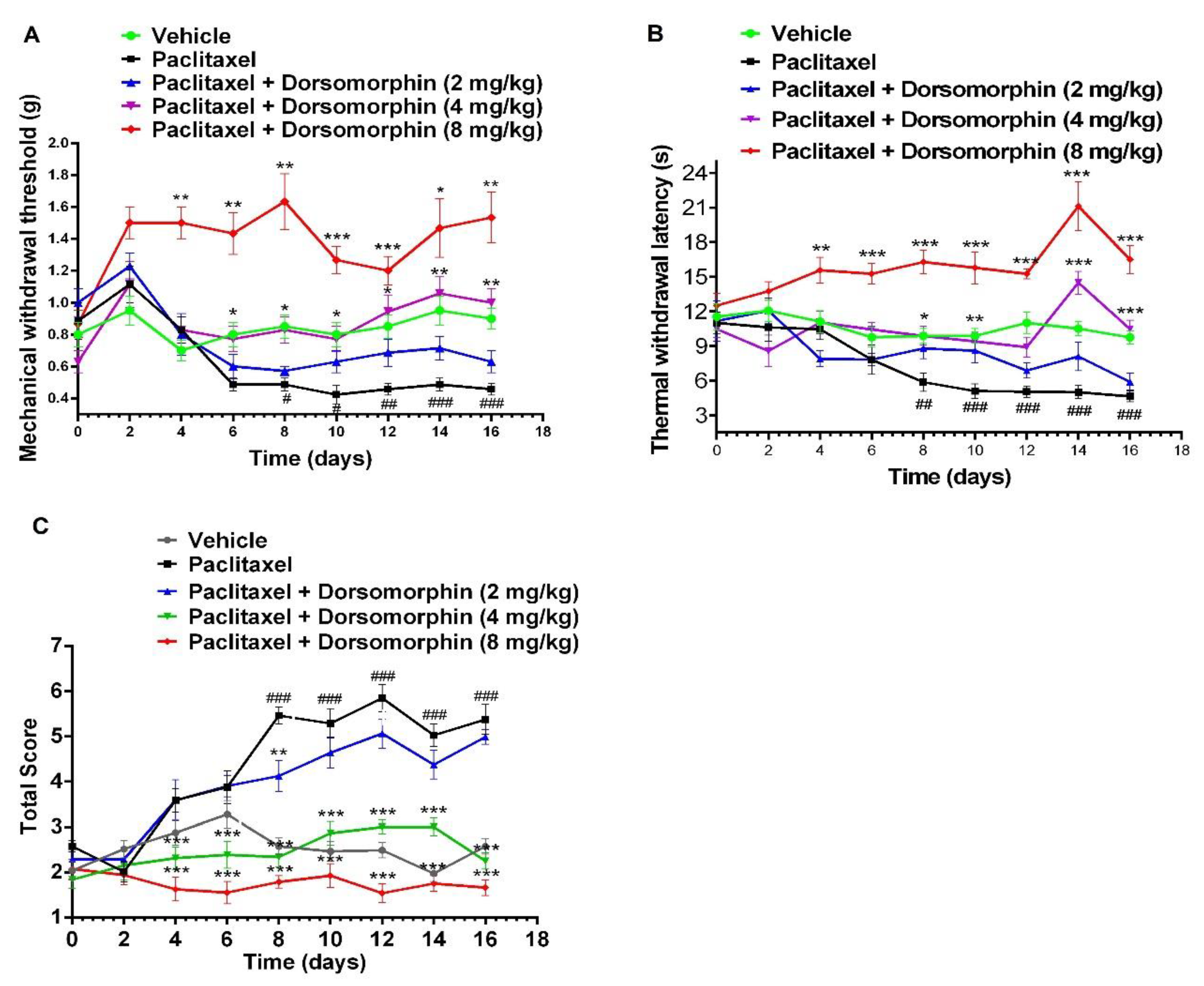
Figure 3.
Dorsomorphin (8 mg/kg) attenuates paclitaxel-induced increases in serum cytokine levels. Serum levels of (A) IL-6, (B) IL-1β, and (C) TNF-α were measured by ELISA upon completion of behavioral testing. Data are expressed as mean ± SEM (n = 4 per group). Statistical significance: * p < 0.05, ** p < 0.01, *** p < 0.001. 3.3. Effects of dorsomorphin on the expression of p65, Iba1, and Scn1a proteins.
Figure 3.
Dorsomorphin (8 mg/kg) attenuates paclitaxel-induced increases in serum cytokine levels. Serum levels of (A) IL-6, (B) IL-1β, and (C) TNF-α were measured by ELISA upon completion of behavioral testing. Data are expressed as mean ± SEM (n = 4 per group). Statistical significance: * p < 0.05, ** p < 0.01, *** p < 0.001. 3.3. Effects of dorsomorphin on the expression of p65, Iba1, and Scn1a proteins.
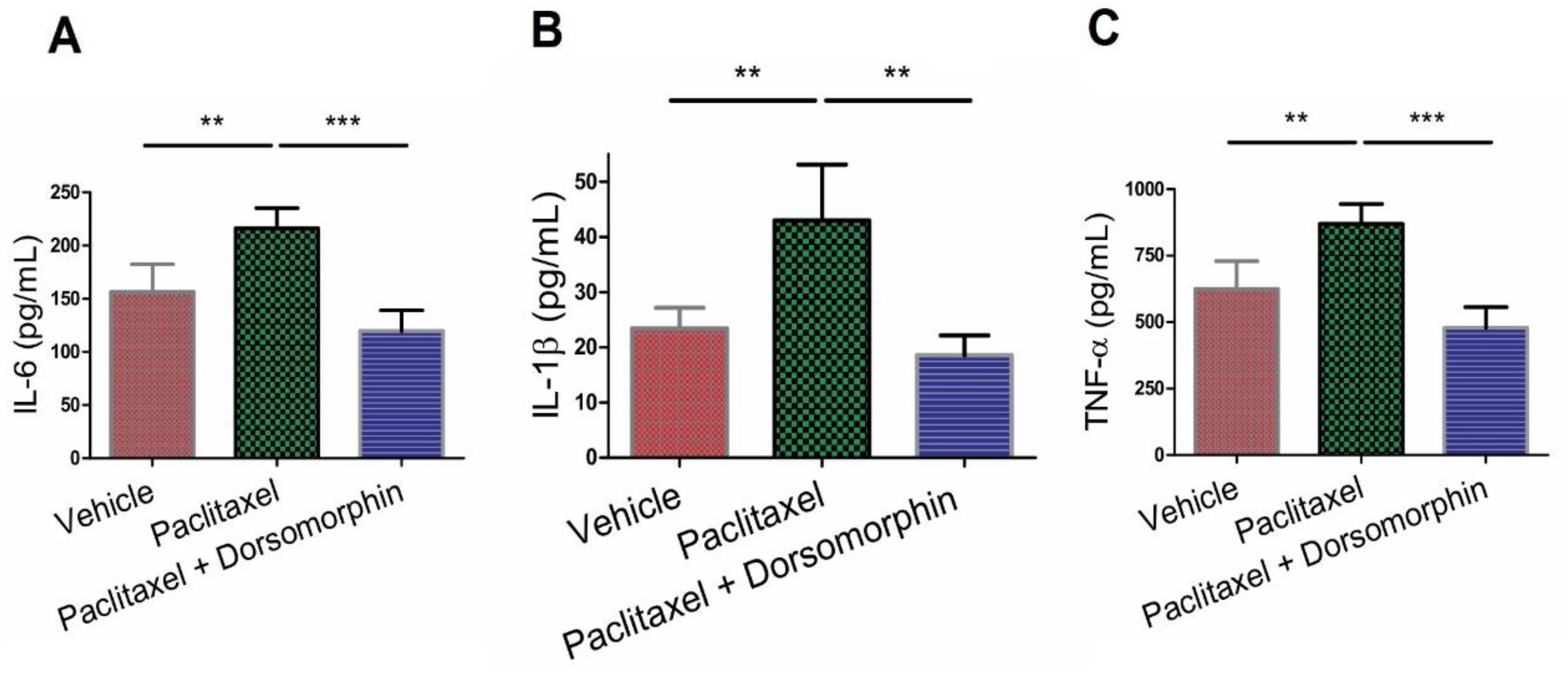
Figure 4.
Dorsomorphin (8 mg/kg) attenuates paclitaxel-induced upregulation of Iba1, p65, and Scn1a protein expression in mouse spinal cord. (A) Representative western blot images of Iba1 (microglial marker) and β-actin (loading control). (B) Quantitative analysis of Iba1 protein expression normalized to β-actin. (C) Representative western blot images of NF-κB p65, Scn1a (Nav1.1), and β-actin (loading control). (D) Quantitative analysis of p65 and Scn1a protein expression normalized to β-actin. Spinal cord tissues were collected upon completion of behavioral testing. Protein levels were quantified by densitometry and normalized to β-actin. Data are expressed as mean ± SEM (n = 3 per group). * p < 0.05, ** p < 0.01, *** p < 0.001. 3.4. Effect of dorsomorphin on LPA expression in the mouse spinal cord.
Figure 4.
Dorsomorphin (8 mg/kg) attenuates paclitaxel-induced upregulation of Iba1, p65, and Scn1a protein expression in mouse spinal cord. (A) Representative western blot images of Iba1 (microglial marker) and β-actin (loading control). (B) Quantitative analysis of Iba1 protein expression normalized to β-actin. (C) Representative western blot images of NF-κB p65, Scn1a (Nav1.1), and β-actin (loading control). (D) Quantitative analysis of p65 and Scn1a protein expression normalized to β-actin. Spinal cord tissues were collected upon completion of behavioral testing. Protein levels were quantified by densitometry and normalized to β-actin. Data are expressed as mean ± SEM (n = 3 per group). * p < 0.05, ** p < 0.01, *** p < 0.001. 3.4. Effect of dorsomorphin on LPA expression in the mouse spinal cord.
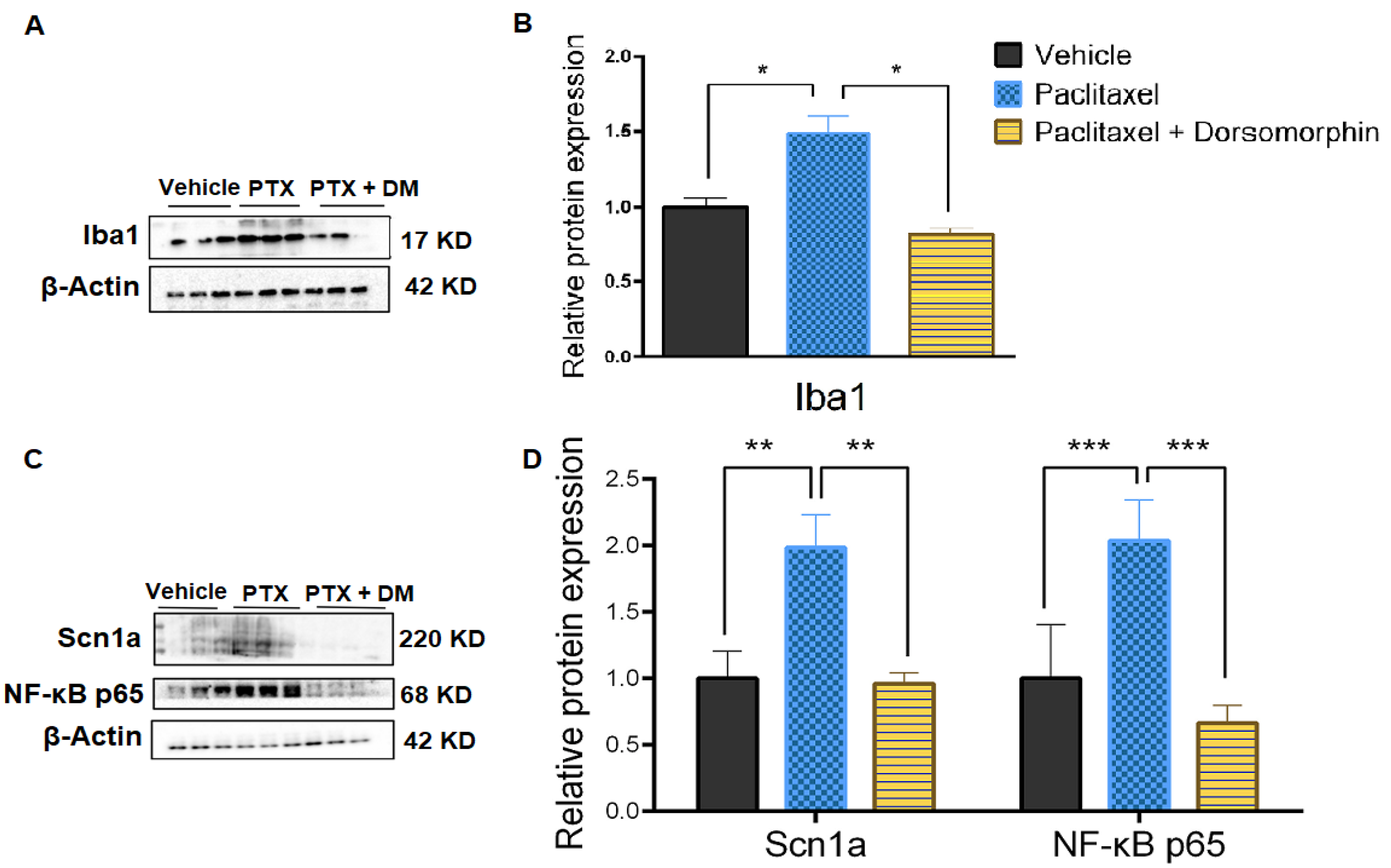
Figure 5.
Dorsomorphin inhibits paclitaxel-induced upregulation of LPA in the mouse spinal. (A) Representative immunohistochemical staining of LPA in vehicle group; (B) Representative immunohistochemical staining of LPA in the paclitaxel group; (C) Representative immunohistochemical staining of LPA in the paclitaxel + dorsomorphin (8 mg/kg) group; (D) Quantitative analysis of LPA-positive cell density in the spinal cord dorsal horn. Spinal cord tissues were collected upon completion of behavioral testing. Data are expressed as mean ± SEM (n = 4 per group). **** p < 0.0001. (Scale bar: 50 μm).
Figure 5.
Dorsomorphin inhibits paclitaxel-induced upregulation of LPA in the mouse spinal. (A) Representative immunohistochemical staining of LPA in vehicle group; (B) Representative immunohistochemical staining of LPA in the paclitaxel group; (C) Representative immunohistochemical staining of LPA in the paclitaxel + dorsomorphin (8 mg/kg) group; (D) Quantitative analysis of LPA-positive cell density in the spinal cord dorsal horn. Spinal cord tissues were collected upon completion of behavioral testing. Data are expressed as mean ± SEM (n = 4 per group). **** p < 0.0001. (Scale bar: 50 μm).
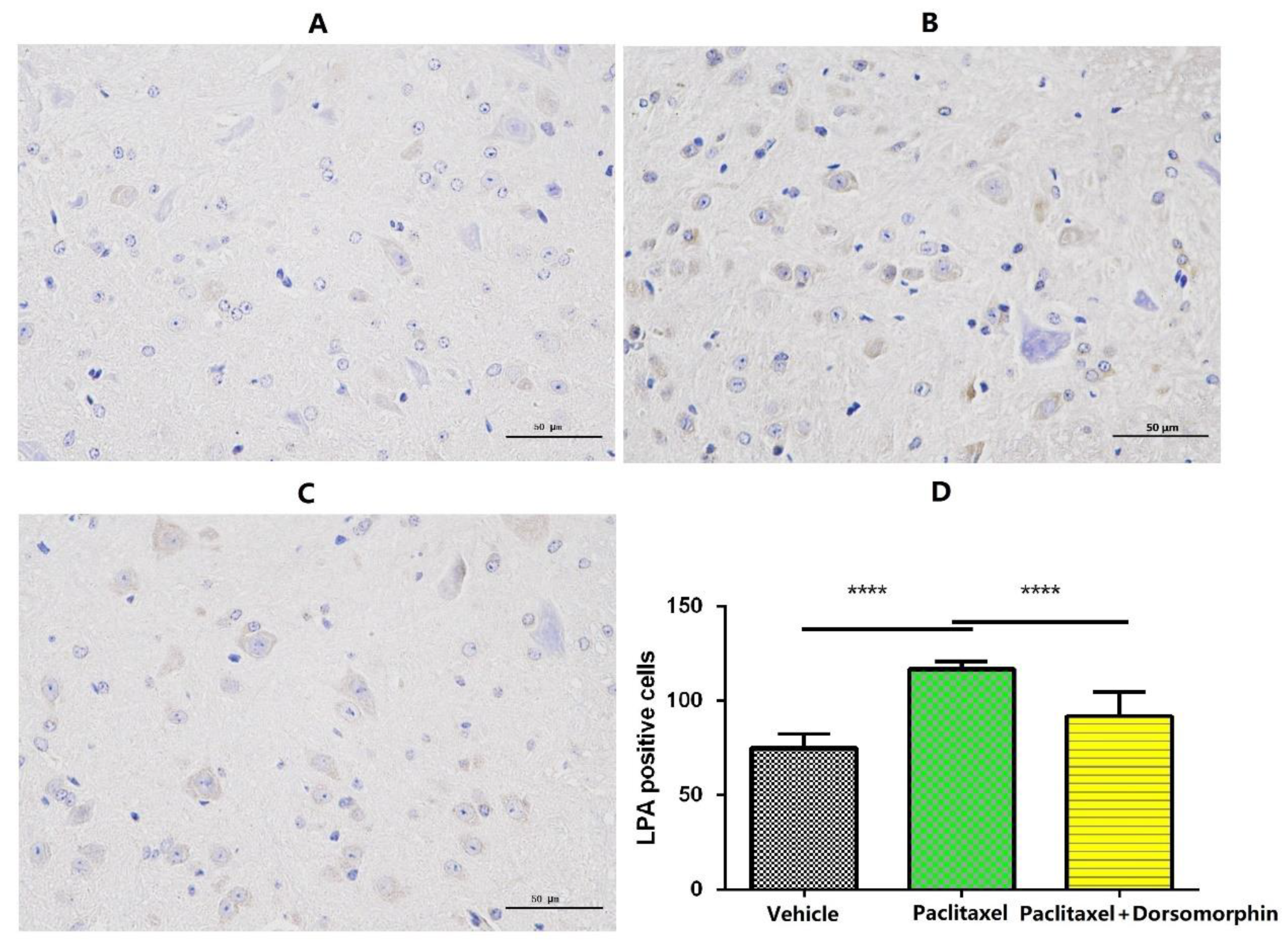
Figure 6.
Dorsomorphin and paclitaxel synergistically inhibit the proliferation of A549 cells. (A) Viability of A549 cells treated with increasing concentrations of paclitaxel (1–20 nM) for 24, 48, and 72 hours. (B) Viability of A549 cells treated with increasing concentrations of dorsomorphin (2.5–10 μM) for 24, 48, and 72 hours. (C–E) Effects of combination therapy compared to paclitaxel monotherapy at (C) low (2.5 nM + 2.5 μM), (D) medium (5 nM + 5 μM), and (E) high (10 nM + 10 μM) concentrations. Data are presented as mean ± SEM (n = 3 biological replicates). *p < 0.05, **p < 0.01, ***p < 0.001 vs. vehicle group; #p < 0.05, ###p < 0.001 vs. corresponding paclitaxel monotherapy group.
Figure 6.
Dorsomorphin and paclitaxel synergistically inhibit the proliferation of A549 cells. (A) Viability of A549 cells treated with increasing concentrations of paclitaxel (1–20 nM) for 24, 48, and 72 hours. (B) Viability of A549 cells treated with increasing concentrations of dorsomorphin (2.5–10 μM) for 24, 48, and 72 hours. (C–E) Effects of combination therapy compared to paclitaxel monotherapy at (C) low (2.5 nM + 2.5 μM), (D) medium (5 nM + 5 μM), and (E) high (10 nM + 10 μM) concentrations. Data are presented as mean ± SEM (n = 3 biological replicates). *p < 0.05, **p < 0.01, ***p < 0.001 vs. vehicle group; #p < 0.05, ###p < 0.001 vs. corresponding paclitaxel monotherapy group.
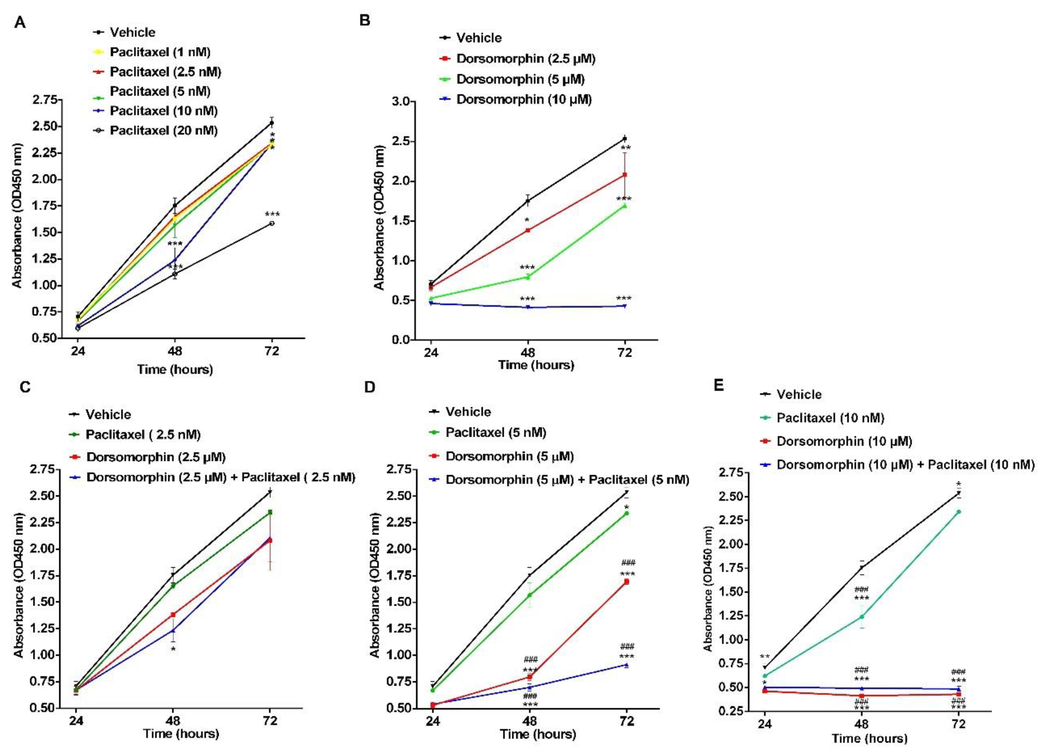
Figure 7.
Combined suppression of A549 cell migration and invasion by paclitaxel (5 nM) and dorsomorphin (5 μM). (A) Representative crystal violet-stained images of migrated (upper) and invaded (lower) cells. Groups: vehicle, 5 nM paclitaxel, 5 μM dorsomorphin, and combination (5 nM paclitaxel + 5 μM dorsomorphin). (B) Quantification of migrated cells. (C) Quantification of invaded cells. Data represent mean ± SEM (n = 3 biological replicates). ***p < 0.001 versus vehicle. Scale bar: 100 μm.
Figure 7.
Combined suppression of A549 cell migration and invasion by paclitaxel (5 nM) and dorsomorphin (5 μM). (A) Representative crystal violet-stained images of migrated (upper) and invaded (lower) cells. Groups: vehicle, 5 nM paclitaxel, 5 μM dorsomorphin, and combination (5 nM paclitaxel + 5 μM dorsomorphin). (B) Quantification of migrated cells. (C) Quantification of invaded cells. Data represent mean ± SEM (n = 3 biological replicates). ***p < 0.001 versus vehicle. Scale bar: 100 μm.
Disclaimer/Publisher’s Note: The statements, opinions and data contained in all publications are solely those of the individual author(s) and contributor(s) and not of MDPI and/or the editor(s). MDPI and/or the editor(s) disclaim responsibility for any injury to people or property resulting from any ideas, methods, instructions or products referred to in the content. |
© 2026 by the authors. Licensee MDPI, Basel, Switzerland. This article is an open access article distributed under the terms and conditions of the Creative Commons Attribution (CC BY) license (http://creativecommons.org/licenses/by/4.0/).
Copyright: This open access article is published under a Creative Commons CC BY 4.0 license, which permit the free download, distribution, and reuse, provided that the author and preprint are cited in any reuse.


