
Submitted:
19 July 2026
Posted:
21 July 2026
You are already at the latest version
Abstract
Background: Posterior spinal fusion (PSF) provides reliable deformity correction for adolescent idiopathic scoliosis (AIS); however, it reduces lumbar mobility because of the fusion of motion segments. Hybrid fixation, which combines thoracic PSF with thoracolumbar/lumbar vertebral body tethering (VBT), aims to preserve lumbar function while maintaining satisfactory deformity correction. Methods: This retrospective comparative cohort study included 66 adolescents with double-major AIS treated with hybrid fixation (n = 30) or posterior long fusion (n = 36). Radiographic outcomes, lumbar flexibility, sagittal range of motion (ROM), lumbar core muscle strength (LCMS), SRS-22r Function scores, complications, and revision surgery were assessed after a minimum follow-up of 24 months. Multivariable regression analyses were used to identify independent predictors of postoperative functional outcomes. Results: Both procedures achieved excellent correction of the main thoracic curve and satisfactory global spinal alignment. Posterior long fusion provided greater thoracolumbar/lumbar curve correction (p < 0.001), whereas hybrid fixation resulted in significantly better lumbar flexibility, sagittal ROM, and LCMS (all p ≤ 0.006). Revision surgery was more frequent after hybrid fixation (20.0% vs. 0%, p = 0.007), primarily because of tether-related complications. Conclusions: Hybrid fixation better preserves objective lumbar function than posterior long fusion while maintaining satisfactory deformity correction in adolescents with double-major AIS. These functional benefits should be balanced against the higher revision burden, supporting individualized treatment selection and shared decision-making when considering motion-preserving surgical strategies.
Keywords:
adolescent idiopathic scoliosis
; hybrid fixation
; vertebral body tethering
; posterior spinal fusion
; lumbar mobility
; patient-reported outcomes
; spinal deformity
; motion preservation
1. Introduction
Adolescent idiopathic scoliosis (AIS) is the most common spinal deformity that requires surgical correction in children and adolescents. Posterior spinal fusion (PSF) remains the gold standard surgical treatment because it provides reliable three-dimensional deformity correction and durable long-term outcomes. However, extending fusion into the lumbar spine inevitably sacrifices mobile motion segments, reducing lumbar flexibility and potentially affecting long-term spinal function. Previous studies have demonstrated that more distal fusion levels are associated with greater postoperative loss of lumbar motion, underscoring the importance of preserving lumbar mobility whenever feasible.[1,2,3]
To overcome this limitation, vertebral body tethering (VBT) has emerged as a fusion-sparing alternative that preserves spinal motion through growth modulation. Compared with PSF, VBT is associated with greater lumbar range of motion, flexibility, trunk muscle endurance, and trunk muscle strength, while facilitating an earlier return to physical activity. However, these functional advantages are accompanied by less predictable deformity correction and higher rates of tether-related complications and revision surgeries. A recent systematic review and meta-analysis including 10 comparative studies and 1,168 patients confirmed this trade-off, demonstrating superior motion preservation but significantly higher revision rates following VBT than PSF. [2,4,5,6]
Hybrid fixation, which combines thoracic PSF with lumbar or thoracolumbar VBT, has recently been introduced as a motion-preserving strategy for patients with double-major AIS. The rationale is to combine the reliable correction achieved with thoracic fusion while preserving lumbar motion through anterior tethering. Early clinical series have demonstrated encouraging radiographic correction and acceptable short-term safety, suggesting that hybrid constructs may represent a balanced alternative to conventional PSF and bilateral VBT. Nevertheless, current evidence remains limited to small retrospective cohorts with relatively short follow-up. Furthermore, most published studies have focused primarily on radiographic correction and complication profiles, whereas objective assessments of lumbar mobility, trunk muscle performance, functional capacity, and patient-reported outcomes remain scarce.[7]
Therefore, the present study compared radiographic correction, lumbar flexibility, trunk muscle performance, patient-reported outcomes, and complications between hybrid fixation and posterior long fusion in adolescents with double-major AIS. We hypothesized that hybrid fixation would better preserve lumbar function while maintaining satisfactory deformity correction, whereas posterior long fusion would provide greater coronal correction with a lower risk of revision surgery.
2. Materials and Methods
2.1. Study Design, Setting and Patient Selection
This retrospective comparative cohort study was conducted at a single tertiary referral spine deformity center. Patients with Lenke type 3 or 6 double-major adolescent idiopathic scoliosis who underwent surgical correction between January 2016 and December 2024 were identified from a prospectively maintained institutional spine database. Patient selection is summarized in Figure 1. Seventy-eight patients met the initial eligibility criteria. After exclusion of 12 patients because of incomplete clinical records (n = 5), incomplete EOS radiographic assessment (n = 5), or follow-up shorter than 2 years (n = 2), 66 patients were included in the final analysis.
Eligible patients were 10–18 years of age, had Lenke type 3 or 6 double-major AIS, Risser grade 0–4, and a minimum clinical and radiographic follow-up of 24 months. Treatment allocation was based on curve characteristics, skeletal maturity, surgeon recommendation, and shared decision-making involving the treating surgeons, patients, and their families. Patients with previous spinal surgery, congenital, neuromuscular, or syndromic scoliosis, or incomplete follow-up or EOS radiographic assessment were excluded.
All patients followed the same standardized postoperative rehabilitation protocol, including early mobilization, supervised physiotherapy, progressive return to activities of daily living, and return to unrestricted sports after radiographic confirmation of spinal stability according to institutional practice.
The study was approved by the Institutional Review Board (Approval No. 2025-08-03/ Date: 30.10.2025) and written informed consent was obtained from all participants and their parents or legal guardians. The study was conducted in accordance with the principles of the Declaration of Helsinki.
2.2. Surgical Technique
All procedures were performed by the senior author (A.H.) using standardized surgical techniques. Posterior instrumentation in both groups consisted of an all-pedicle screw construct (Medtronic, Minneapolis, MN, USA) with 5.5-mm cobalt–chromium rods.
In the hybrid fixation group, thoracic deformity correction with posterior spinal fusion was followed by thoracoscopic anterior vertebral body tethering using REFLECT™ Scoliosis Correction System (Globus Medical, Audubon, PA, USA) of the thoracolumbar or lumbar curve. The tether was sequentially tensioned from proximal to distal under fluoroscopic guidance to achieve gradual correction while avoiding overcorrection. Tensioning was individualized according to curve magnitude, flexibility, skeletal maturity, and the desired intraoperative correction while preserving the potential for continued growth modulation.
Patients in the posterior long fusion group underwent conventional posterior deformity correction. Fusion levels and the upper and lower instrumented vertebrae were selected according to established deformity correction principles, considering the stable vertebra, coronal balance, sagittal alignment, and lumbar curve characteristics while preserving distal mobile segments whenever feasible. All procedures were performed with continuous multimodal intraoperative neuromonitoring.
2.3. Radiographic Evaluation
Standing full-length biplanar EOS radiographs were obtained with patients in a standardized upright position preoperatively and at the standardized 24-month follow-up. Coronal parameters included the main thoracic (MT) Cobb angle, thoracolumbar/lumbar (TL/L) Cobb angle, and coronal balance measured as the horizontal distance between the C7 plumb line and the central sacral vertical line (CSVL). Sagittal parameters included thoracic kyphosis (T5–T12) and lumbar lordosis (L1–S1). Curve correction was calculated as the difference between preoperative and postoperative measurements.
Radiographic measurements were independently performed using the institutional PACS software by two fellowship-trained spine surgeons blinded to treatment allocation. Interobserver and intraobserver reliability were assessed using the intraclass correlation coefficient (ICC) and demonstrated excellent agreement (interobserver ICC = 0.94, 95% CI 0.90–0.97; intraobserver ICC = 0.96, 95% CI 0.93–0.98).
2.4. Functional Assessment
All functional evaluations were performed by the same physiotherapist (S.A) at the standardized 24-month follow-up using identical assessment protocols. Lumbar flexibility was evaluated using the modified Schober test according to the standardized technique and recorded in centimeters. Lumbar range of motion (ROM) was measured using a universal goniometer. Each measurement was performed three times, and the mean value was used for analysis. Lumbar flexion and extension were assessed in standing, whereas axial rotation was measured in the seated position to minimize pelvic compensation. Bilateral lateral bending was also recorded. The primary outcome of interest, defined before data extraction, was total sagittal lumbar ROM, defined as the sum of lumbar flexion and extension.
Lumbar core muscle strength was assessed using the Centaur 3D Trunk Muscle Testing System (BfMC GmbH, Leipzig, Germany) at angular velocities of 10°/s and 60°/s. Three maximal efforts were recorded at each testing condition, and the highest normalized peak torque value was used for analysis. Peak torque values for trunk flexion and extension were normalized to body weight (Nm/kg). Posterior, anterior, and lateral trunk muscle performance were analyzed separately.
Health-related quality of life was assessed using the Scoliosis Research Society-22 revised (SRS-22r) questionnaire. The SRS-22r Function domain was selected a priori as the patient-reported outcome measure for multivariable analyses.
2.5. Complications and Reoperations
All perioperative complications and reoperations occurring within 24 months after the index procedure were recorded. Radiographic tether breakage and overcorrection were predefined and assessed at routine follow-up visits. Reoperation was defined as any unplanned secondary surgical procedure related to the index spinal construct.
2.6. Statistical Analysis
Continuous variables are presented as mean ± standard deviation or median (interquartile range), as appropriate, whereas categorical variables are presented as frequencies and percentages. Data distribution was assessed using the Shapiro–Wilk test. Between-group comparisons were performed using the independent-samples t test or Mann–Whitney U test for continuous variables and the chi-square test or Fisher’s exact test for categorical variables, as appropriate. Between-group effect sizes were calculated as Cohen’s d using the pooled standard deviation, with 95% confidence intervals derived from the non-central t distribution.
All consecutive eligible patients meeting the inclusion criteria during the study period were included in the analysis. No imputation was performed for missing data; patients with incomplete datasets were excluded from the final analysis. No a priori sample size calculation was performed because the cohort size was fixed by the number of eligible patients treated during the study period. For the primary outcome, the available sample of 30 and 36 patients provided 80% power at a two-sided alpha of 0.05 to detect a standardized between-group difference of Cohen’s d = 0.70 or greater.
Secondary outcomes were considered exploratory, and multiplicity was controlled using the Holm procedure. Multivariable linear regression models were constructed to identify independent predictors of postoperative functional outcomes, including the SRS-22r Function domain. Variables entered into the models were selected a priori based on clinical relevance and included age at surgery, sex, body mass index, baseline MT Cobb angle, baseline TL/L Cobb angle, and Risser grade. Two-sided p values < 0.05 were considered statistically significant. Statistical analyses were performed using R version 4.5.1 (R Foundation for Statistical Computing, Vienna, Austria).
3. Results
3.1. Study Cohort
A total of 66 patients with adolescent idiopathic scoliosis (AIS) and double-major curves met the eligibility criteria and completed the standardized 24-month follow-up assessment. Thirty patients underwent hybrid fixation consisting of thoracic posterior spinal fusion (PSF) combined with thoracolumbar/lumbar vertebral body tethering (VBT), whereas 36 underwent conventional posterior long fusion. Baseline demographic characteristics, curve morphology, skeletal maturity, and preoperative deformity severity were comparable between groups (Table 1). Operative characteristics differed as expected according to construct design: the hybrid group had fewer thoracic fused levels and demonstrated significantly shorter operative time, lower estimated blood loss, and shorter hospital stay than the posterior long fusion group. Follow-up duration was comparable between the groups (30.7 ± 4.6 vs. 32.5 ± 6.1 months, p = 0.19). (Table 1).
3.2. Radiographic Outcomes
Both surgical strategies achieved substantial correction of the main thoracic deformity and improved coronal balance at 24 months (Table 2). The magnitude of main thoracic curve correction and changes in coronal balance were comparable between groups. Likewise, postoperative thoracic kyphosis and lumbar lordosis did not differ significantly. In contrast, posterior long fusion achieved significantly greater thoracolumbar/lumbar curve correction than hybrid fixation (p < 0.001), representing the principal radiographic difference between the two treatment strategies. Representative preoperative and final EOS radiographs are shown in Figure 2.
3.3. Functional Outcomes
Objective functional outcomes consistently favored hybrid fixation at the 24-month evaluation (Table 3). Lumbar flexibility measured by the Schober test was significantly greater in the hybrid group than in the posterior long fusion group (4.90 ± 1.10 vs. 3.70 ± 1.00 cm, p = 0.002). The primary outcome of interest, total sagittal lumbar range of motion (ROM), was also significantly greater following hybrid fixation (138.0 ± 8.4° vs. 118.2 ± 9.2°, p < 0.001), corresponding to a large effect size (Cohen’s d = 2.24, 95% CI 1.62–2.86). Similar advantages were observed across all individual ROM components, including lumbar flexion, extension, bilateral lateral bending, and axial rotation (all p < 0.001), with large effect sizes throughout (Cohen’s d 1.11–1.55).
Hybrid fixation was also associated with superior lumbar core muscle strength. Posterior, anterior, and lateral lumbar core muscle strength measurements were all significantly greater in the hybrid group than in the posterior long fusion group (all p ≤ 0.006) (Table 3). Representative Centaur testing and postoperative lumbar core muscle strength are presented in Figure 3. Despite these objective functional advantages, patient-reported function assessed using the SRS-22r Function domain did not differ significantly between groups.
3.4. Complications
Overall complication rates within 24 months were comparable between the two treatment groups (20.0% vs. 11.1%, p = 0.33) (Table 4). Perioperative complications were infrequent, with isolated superficial wound complications occurring only after posterior long fusion and a single transient neurological deficit occurring after hybrid fixation. Procedure-specific events in the hybrid cohort included radiographic tether breakage in eight patients (26.7%) and one case of overcorrection (3.3%). Revision surgery was required in six patients (20.0%) after hybrid fixation, whereas no patient in the posterior long fusion group required secondary surgery during the 24-month follow-up (p = 0.007). (Table 4).
3.5. Multivariable Analysis
After adjustment for the a priori selected covariates, posterior long fusion remained independently associated with inferior functional outcomes at 24 months (Table 5). Compared with hybrid fixation, posterior long fusion was associated with significantly lower total sagittal lumbar ROM (adjusted β = −7.8, 95% CI −12.6 to −3.0; p = 0.001), lower SRS-22r Function domain scores (adjusted β = −0.15, 95% CI −0.28 to −0.02; p = 0.024), and lower posterior lumbar core muscle strength (adjusted β = −0.10 Nm/kg, 95% CI −0.19 to −0.01; p = 0.031), confirming that the observed functional differences remained significant after multivariable adjustment.
4. Discussion
The main finding was that hybrid fixation preserved objective lumbar function better than posterior long fusion in patients with double-major adolescent idiopathic scoliosis (AIS), without compromising overall deformity correction. Patients treated with hybrid fixation had significantly greater lumbar flexibility, sagittal range of motion (ROM), and lumbar core muscle strength (LCMS), whereas posterior long fusion achieved greater thoracolumbar/lumbar curve correction and a substantially lower revision rate. Both strategies corrected the main thoracic deformity well and restored global spinal alignment. These findings reflect a trade-off between lumbar motion preservation and long-term construct durability: hybrid fixation is a motion-preserving option for carefully selected patients, not a universal replacement for posterior fusion.[7]
Both strategies corrected the main thoracic curve well, but posterior long fusion achieved significantly greater thoracolumbar/lumbar correction. This is biologically plausible: rigid posterior pedicle screw constructs provide immediate three-column stability and generate greater corrective force than tether-based constructs.[8,9,10,11] Prior comparative studies and systematic reviews report the same pattern — posterior fusion achieves greater coronal correction, while vertebral body tethering trades some correction for preserved motion. [8,9,10,12] Our findings are consistent with these reports: despite less complete lumbar correction, hybrid fixation maintained adequate overall spinal alignment. A small loss of correction may be acceptable when preserving lumbar mobility is a treatment priority.
Beyond radiographic correction, the most clinically relevant finding was better preservation of objective lumbar function after hybrid fixation. The likely mechanism is straightforward: posterior fusion eliminates motion across instrumented lumbar segments, whereas vertebral body tethering preserves controlled segmental mobility while maintaining spinal alignment.[1,4,12,13] Raitio et al. identified motion preservation as a principal theoretical advantage of vertebral body tethering, though they noted the limited clinical evidence for objective functional outcomes.[4] Wong et al., in a recent systematic review, reported that vertebral body tethering provides less predictable radiographic correction than posterior spinal fusion but consistently better postoperative spinal mobility and function.[12] Our findings extend these observations: the functional advantage persists even when tethering is combined with fusion in a hybrid construct rather than used alone.
Hybrid fixation’s advantage was consistent across every functional assessment — flexion, extension, lateral bending, axial rotation, the Schober test, and isokinetic trunk muscle strength. This consistency across independent measures strengthens the biological plausibility of the effect and suggests that preserved lumbar motion improves neuromuscular performance as well as flexibility. Two biomechanical studies support this interpretation. Lee et al. showed that extending posterior fusion into the distal lumbar spine progressively reduces postoperative mobility with each additional fused segment.[1,14] Pahys et al. similarly found decreasing lumbar flexibility with more distal fusion levels, underscoring the value of preserving mobile lumbar segments when feasible.[14] Together, these findings support hybrid fixation as a way to maintain lumbar motion while still achieving adequate deformity correction.
Despite the objective functional gains after hybrid fixation, SRS-22r Function scores did not differ significantly between groups. This gap between objective and patient-reported outcomes has been reported before in motion-preserving scoliosis surgery.[12,15,16] One explanation is that the SRS-22r, though well validated for health-related quality of life in AIS, may lack the sensitivity to detect subtle differences in lumbar mobility among otherwise healthy adolescents.[17] Wong et al. reported a similar pattern: improved postoperative spinal mobility after vertebral body tethering was not consistently matched by better patient-reported outcomes.[12] Carreon et al. established small minimum clinically important difference (MCID) thresholds for the SRS Activity domain, and Kelly et al. later showed that the minimum detectable difference exceeded these thresholds — meaning modest functional gains can fall below what the instrument can reliably detect. [15,16] Objective measures of flexibility, range of motion, and muscle strength may therefore be more sensitive than patient-reported questionnaires for evaluating motion-preserving techniques, particularly during early and mid-term follow-up.
These functional advantages must be weighed against a higher revision burden. Six patients (20.0%) in the hybrid cohort required revision surgery, compared with none after posterior long fusion, mostly for tether-related mechanical complications.[18] Tether breakage, overcorrection, and progressive loss of correction are the leading causes of secondary surgery after vertebral body tethering, and recent reviews consistently report higher complication and revision rates for VBT than PSF despite its better preservation of spinal mobility and function.[8] Long-term studies also show that tether breakage occurs more often in the lumbar than the thoracic spine, though many radiographic failures stay clinically silent and do not require revision.[19,20,21] Radiographic tether failure and clinical failure are therefore not the same thing. Still, the higher reoperation rate in this study confirms that hybrid fixation trades postoperative lumbar function for reduced mechanical durability, and patients and families should be counseled accordingly before surgery.
These findings have practical implications for patient selection. Posterior long fusion remains preferable when maximum correction and construct durability are the priorities. Hybrid fixation suits adolescents who value lumbar mobility and trunk function more highly, provided they accept a higher likelihood of reoperation — a trade-off that should be made explicit during shared decision-making. Successful use of motion-preserving techniques also depends on appropriate patient selection, guided by skeletal maturity, curve characteristics, flexibility, and patient expectations.[12,22,23]
The present study has several strengths that should be considered when interpreting the findings. To our knowledge, this is among the few comparative studies evaluating hybrid fixation consisting of thoracic posterior spinal fusion combined with thoracolumbar/lumbar vertebral body tethering in patients with double-major AIS. Unlike previous reports that primarily focused on radiographic outcomes, the present study incorporated comprehensive objective functional assessments, including lumbar flexibility, sagittal range of motion, Schober test measurements, and isokinetic lumbar core muscle strength, together with patient-reported outcomes. Furthermore, all procedures were performed by the same experienced deformity team using standardized surgical techniques and postoperative rehabilitation protocols, thereby reducing treatment heterogeneity. Nevertheless, several limitations should be acknowledged. First, the retrospective, non-randomized design introduces the possibility of residual selection bias despite comparable baseline characteristics and multivariable adjustment. Second, the study was conducted at a single tertiary referral center with a relatively limited sample size, which may reduce the generalizability of the findings. Third, although a minimum follow-up of 24 months was available for all patients, longer follow-up is necessary because tether-related complications and revision procedures may continue to accumulate over time. Furthermore, functional assessments were performed by a physiotherapist who was not blinded to treatment allocation, introducing the potential for measurement bias despite the use of standardized assessment protocols and objective measurement methods. In addition, preoperative objective functional assessments, including lumbar range of motion, Schober test measurements, and lumbar core muscle strength, were not available. Consequently, postoperative functional outcomes could not be compared with individual preoperative baseline values. Finally, because hybrid fixation combines two distinct surgical strategies within the same construct, the observed functional benefits cannot be attributed exclusively to lumbar tethering.
5. Conclusions
In conclusion, hybrid fixation was associated with better preservation of lumbar flexibility, range of motion, and trunk muscle strength than posterior long fusion, while maintaining adequate deformity correction in patients with double-major adolescent idiopathic scoliosis. These functional advantages came with a significantly higher risk of revision surgery, reflecting a trade-off between motion preservation and long-term construct durability. Hybrid fixation is therefore not a replacement for conventional posterior spinal fusion, but a selective motion-preserving strategy for carefully chosen patients in whom lumbar function is a priority. Future prospective multicenter studies with longer follow-up are needed to confirm the durability of these benefits, refine patient selection, and further define the role of hybrid fixation in managing double-major adolescent idiopathic scoliosis.
Author Contributions
Conceptualization, B.A. M.E., S.A., H.M.M., and I.D.; methodology, B.A. S.A., M.E.; investigation, B.A. and S.A.; resources, S.A., A.H. and I.D.; data curation, B.A., S.A., I.D.; formal analysis, B.A. and S.A.; validation, I.D., H.M.M.; visualization, B.A.; writing—original draft preparation, B.A., S.A., I.D., M.E. and H.M.M.; writing—review and editing, B.A., S.A., M.E. and I.D.; supervision, B.A., M.E.,and A.H.; project administration, B.A., H.M.M. and S.A. All authors have read and agreed to the published version of the manuscript.
Funding
This research received no external funding.
Institutional Review Board Statement
The study was conducted in accordance with the Declaration of Helsinki and approved by the Clinical Research Ethics Committee of Demiroglu Bilim University (Approval No. 2025-08-03/ Date: 30.10.2025).
Informed Consent Statement
Written informed consent to participate and for publication of anonymized clinical and imaging data was obtained from all participants and their parents or guardians. No identifiable patient information or identifiable clinical images are included in this manuscript.
Data Availability Statement
The data presented in this study are available on request from the corresponding author. The data are not publicly available due to ethical and privacy restrictions related to patient confidentiality and institutional approval requirements.
Acknowledgments
During the preparation of this manuscript, the authors used Paperpal for language editing and manuscript formatting assistance. The authors have reviewed and edited the output and take full responsibility for the content of this publication.
Conflicts of Interest
The authors declare no conflicts of interest.
References
- Lee, M.C.; Õunpuu, S.; Solomito, M.; Smith, B.G.; Thomson, J.D. Loss in Spinal Motion From Inclusion of a Single Midlumbar Level in Posterior Spinal Fusion for Adolescent. Scoliosis Spine 2013, 38, E1405–E1410. [Google Scholar] [CrossRef] [PubMed]
- Hammad, A.; Balsano, M.; Ahmad, A.A. Vertebral Body Tethering: An Alternative to Posterior Spinal Fusion in Idiopathic Scoliosis? Front. Pediatr. 2023, 11. [Google Scholar] [CrossRef] [PubMed]
- Marie-Hardy, L.; Besse, M.; Chatelain, L.; Pannier, S.; Glorion, C.; Ferrero, E. Does the Distal Level Really Matter in the Setting of Health-Related Quality of Life? Assessment of a Series of Adolescent Idiopathic Scoliosis Patients at More Than 7 Years Following Surgery. Spine 2022, 47, E545–E550. [Google Scholar] [CrossRef] [PubMed]
- Raitio, A.; Syvänen, J.; Helenius, I. Vertebral Body Tethering: Indications, Surgical Technique, and a Systematic Review of Published Results. J. Clin. Med. 2022, 11, 2576. [Google Scholar] [CrossRef] [PubMed]
- Abdel-AAl, M.; Ghandour, M.; Mert, Ü.; Pishnamaz, M.; Knobe, M.; Hildebrand, F.; Sobottke, R.; Kabir, K.; Mahmoud, M.A. Anterior Vertebral Body Tethering Versus Posterior Spinal Fusion in Adolescent Idiopathic Scoliosis: A Systematic Review and Meta-Analysis of Comparative Outcomes. J. Clin. Med. 2025, 14, 6707. [Google Scholar] [CrossRef] [PubMed]
- Sullivan, M.H.; Jackson, T.J.; Milbrandt, T.A.; Larson, A.N.; Kepler, C.K.; Sebastian, A.S. Evidence-Based Indications for Vertebral Body Tethering in Spine Deformity. Clin. Spine Surg. A Spine Publ. 2023, 37, 82–91. [Google Scholar] [CrossRef] [PubMed]
- Cherian, D.; Samdani, A.F.; Schüpper, A.J.; Stein, A.A.; Naseer, Z.; Pahys, J.M.; Nice, E.; Hwang, S.W. Early Outcomes in Hybrid Fixation for Idiopathic Scoliosis: Posterior Fusion Combined with Anterior Vertebral Body Tethering. Patient Series. J. Neurosurg. Case Lessons 2023, 6, CASE23331. [Google Scholar] [CrossRef] [PubMed]
- Al-Naseem, A.O.; Al-Naseem, A.; Al Balushi, B.; Marwan, Y.; Leong, J.; Shafafy, R. Posterior Spinal Fusion versus Vertebral Body Tethering for Paediatric Scoliosis: A Meta-Analysis of Comparative Studies. Spine Deform 2025, 13, 681–694. [Google Scholar] [CrossRef] [PubMed]
- Stamiris, S.; Sofos, C.; Sarridimitriou, A.; Kakoulidis, P.; Christidis, P.; Stamiris, D.; Anestiadou, E.; Cheva, A.; Chatzianestiadou, C.; Christodoulou, P.; et al. Comparative Meta-Analysis of Vertebral Body Tethering and Posterior Spinal Fusion in Patients with Idiopathic Scoliosis. Evaluation of Radiographic, Perioperative, Clinical, Patient-Reported Outcomes, and Complication Rates. Spine Deform 2025, 13, 1399–1420. [Google Scholar] [CrossRef] [PubMed]
- Mathew, S.E.; Hargiss, J.B.; Milbrandt, T.A.; Stans, A.A.; Shaughnessy, W.J.; Larson, A.N. Vertebral Body Tethering Compared to Posterior Spinal Fusion for Skeletally Immature Adolescent Idiopathic Scoliosis Patients: Preliminary Results from a Matched Case–Control Study. Spine Deform 2022, 10, 1123–1131. [Google Scholar] [CrossRef] [PubMed]
- Shah, S.A.; Kraft, D.B.; Miyanji, F. Anterior Vertebral Body Tethering: A Review of the Available Evidence. J. Am. Acad. Orthop. Surg. 2024, 32, 247–256. [Google Scholar] [CrossRef] [PubMed]
- Wong, D.; Mong, P.T.; Ng, C.Y.; Ong, C.K.; Qian, Z.; Shao, M.H.; Sin, L.K.E.; Wong, B.Y.; Wong, C.M.; Cheung, J.; et al. Can Anterior Vertebral Body Tethering Provide Superior Range of Motion Outcomes Compared to Posterior Spinal Fusion in Adolescent Idiopathic Scoliosis? A Systematic Review. Eur. Spine J. 2023, 32, 3058–3071. [Google Scholar] [CrossRef] [PubMed]
- Romberg, K.; Danielsson, A.; Olsén, M.F.; Kjellby-Wendt, G. Spinal Mobility and Muscle Function in Middle-Aged Patients Treated for Early Onset Idiopathic Scoliosis: Compared with Untreated and Treated Adolescent Onset Patients. Spine Deform 2022, 10, 1085–1095. [Google Scholar] [CrossRef] [PubMed]
- Pahys, J.M.; Hwang, S.W.; McGarry, M.; Quinonez, A.; Grewal, H.; Samdani, A.F. Incidence and Predictors of Growth Modulation and Overcorrection After Anterior Vertebral Body Tethering. Spine 2026, 51, 286–293. [Google Scholar] [CrossRef] [PubMed]
- Carreon, L.Y.; Sanders, J.O.; Diab, M.; Sucato, D.J.; Sturm, P.F.; Glassman, S.D. The Minimum Clinically Important Difference in Scoliosis Research Society-22 Appearance, Activity, and Pain Domains After Surgical Correction of Adolescent. Spine 2010, 35, 2079–2083. [Google Scholar] [CrossRef] [PubMed]
- Kelly, M.P.; Lenke, L.G.; Sponseller, P.D.; Pahys, J.M.; Bastrom, T.P.; Lonner, B.S.; Abel, M.F. The Minimum Detectable Measurement Difference for the Scoliosis Research Society-22r in Adolescent Idiopathic Scoliosis: A Comparison with the Minimum Clinically Important Difference. Spine J. 2019, 19, 1319–1323. [Google Scholar] [CrossRef] [PubMed]
- Stone, L.E.; Ames, C.P.; Pellise, F.; Newton, P.O.; Upasani, V.V.; Harms Study Group; Kelly, M.P. Scoliosis Research Society-22r and Ceiling Effects: Limited Capabilities for Precision Medicine With Adolescent Idiopathic Scoliosis. Spine 2025, 50, 34–39. [Google Scholar] [CrossRef] [PubMed]
- Stein, A.A.; Samdani, A.F.; Schupper, A.J.; Naseer, Z.; Shah, R.V.; Zeller, S.; Pahys, J.M.; Samuel, S.P.; Quinonez, A.; Hwang, S.W. Lumbar Vertebral Body Tethering: Single Center Outcomes and Reoperations in a Consecutive Series of 106 Patients. Spine 2024, 49, 1548–1554. [Google Scholar] [CrossRef] [PubMed]
- Cahill, P.J.; Miyanji, F.; Lullo, B.R.; Samdani, A.F.; Lonner, B.S.; Pahys, J.M.; Hwang, S.W.; Haber, L.L.; Alanay, A.; Shah, S.A.; et al. Incidence of Tether Breakage in Anterior Vertebral Body Tethering. J. Pediatr. Orthop. 2024, 44, e323–e328. [Google Scholar] [CrossRef] [PubMed]
- Nugraha, H.K.; Haber, L.L.; Hoernschemeyer, D.G.; Cahill, P.J.; Samdani, A.F.; Miyanji, F.; Newton, P.O.; Larson, A.N. on behalf of the Harms Study Group Outcomes of Vertebral Body Tethering in Adolescent Idiopathic Scoliosis: A Prospective, Multicenter Study. JBJS Open Access 2026, 11. [Google Scholar] [CrossRef] [PubMed]
- Yang, M.J.; Samdani, A.F.; Pahys, J.M.; Quinonez, A.; McGarry, M.; Grewal, H.; Hwang, S.W. What Happens After a Vertebral Body Tether Break? Incidence, Location, and Progression With Five-Year Follow-Up. Spine 2023, 48, 742–747. [Google Scholar] [CrossRef] [PubMed]
- Shaw, K.A.; Welborn, M.C.; Matsumoto, H.; Parent, S.; Sachwani, N.; El-Hawary, R.; Skaggs, D.; Newton, P.O.; Blakemore, L.; Vitale, M.; et al. To Tether or Fuse? Significant Equipoise Remains in Treatment Recommendations for Idiopathic Scoliosis. Spine Deform 2022, 10, 763–773. [Google Scholar] [CrossRef] [PubMed]
- Oeding, J.F.; Siu, J.; O’Donnell, J.; Wu, H.-H.; Allahabadi, S.; Saggi, S.; Flores, M.; Brown, K.; Baldwin, A.; Diab, M. Combined Anterior Thoracic Vertebral Body Tethering and Posterior Lumbar Tethering Results in Quicker Return to Sport and Activity Compared to Posterior Spinal Instrumented Fusion in Patients with Adolescent Idiopathic Scoliosis. Glob. Spine J. 2025, 15, 1068–1076. [Google Scholar] [CrossRef] [PubMed]
Figure 1.
Patient Flow Chart.
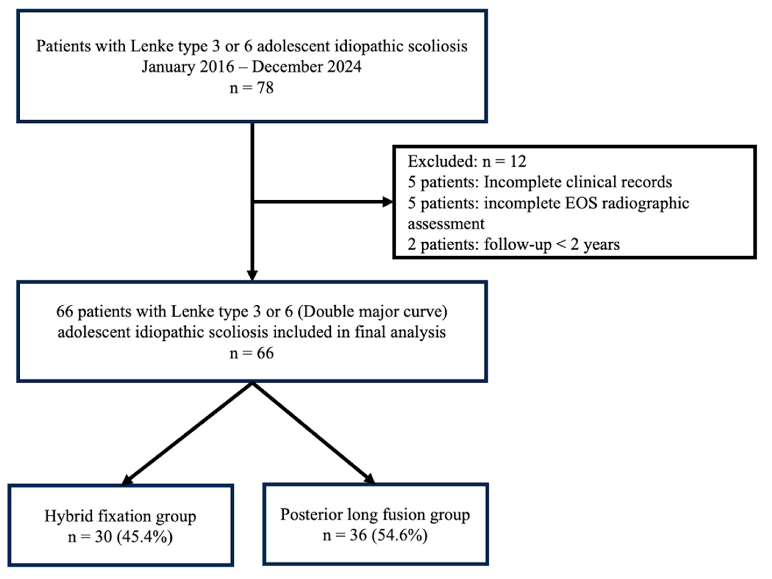
Figure 2.
Representative radiographic outcomes following hybrid fixation and posterior spinal fusion. (A) Standing whole-spine EOS radiographs obtained preoperatively and at final follow-up in representative patients treated with hybrid fixation and posterior spinal fusion. (B) The hybrid construct consists of thoracic posterior spinal fusion combined with thoracolumbar/lumbar vertebral body tethering, whereas the posterior spinal fusion construct extends to the lumbar spine. Both techniques achieved satisfactory coronal deformity correction while demonstrating their respective surgical constructs.
Figure 2.
Representative radiographic outcomes following hybrid fixation and posterior spinal fusion. (A) Standing whole-spine EOS radiographs obtained preoperatively and at final follow-up in representative patients treated with hybrid fixation and posterior spinal fusion. (B) The hybrid construct consists of thoracic posterior spinal fusion combined with thoracolumbar/lumbar vertebral body tethering, whereas the posterior spinal fusion construct extends to the lumbar spine. Both techniques achieved satisfactory coronal deformity correction while demonstrating their respective surgical constructs.
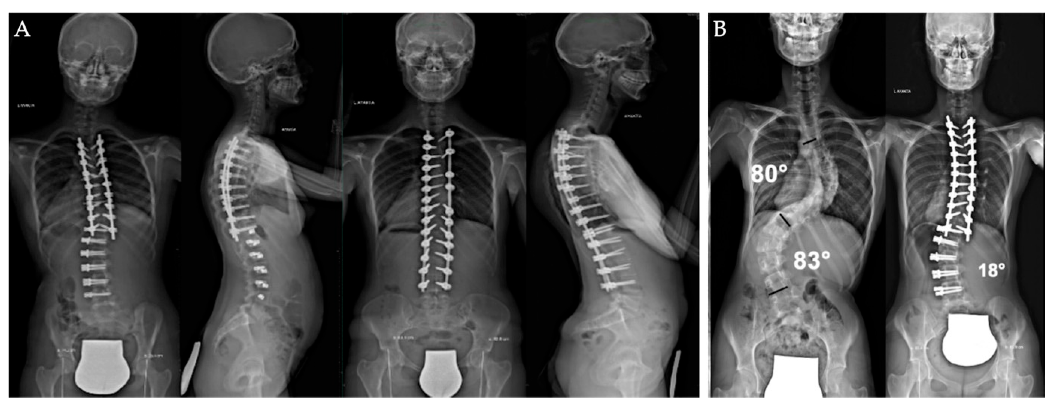
Figure 3.
Postoperative assessment of lumbar core muscle strength using the Centaur 3D Trunk Muscle Testing System. (A) Representative patient positioning during lumbar core muscle strength assessment. (B) Example of the computerized test report generated by the Centaur system. (C) Representative multidirectional testing position during lateral trunk muscle assessment. (D) Comparison of normalized postoperative lumbar core muscle strength (peak torque, Nm/kg) between the hybrid fixation and posterior spinal fusion groups in the posterior, anterior, and lateral directions.
Figure 3.
Postoperative assessment of lumbar core muscle strength using the Centaur 3D Trunk Muscle Testing System. (A) Representative patient positioning during lumbar core muscle strength assessment. (B) Example of the computerized test report generated by the Centaur system. (C) Representative multidirectional testing position during lateral trunk muscle assessment. (D) Comparison of normalized postoperative lumbar core muscle strength (peak torque, Nm/kg) between the hybrid fixation and posterior spinal fusion groups in the posterior, anterior, and lateral directions.
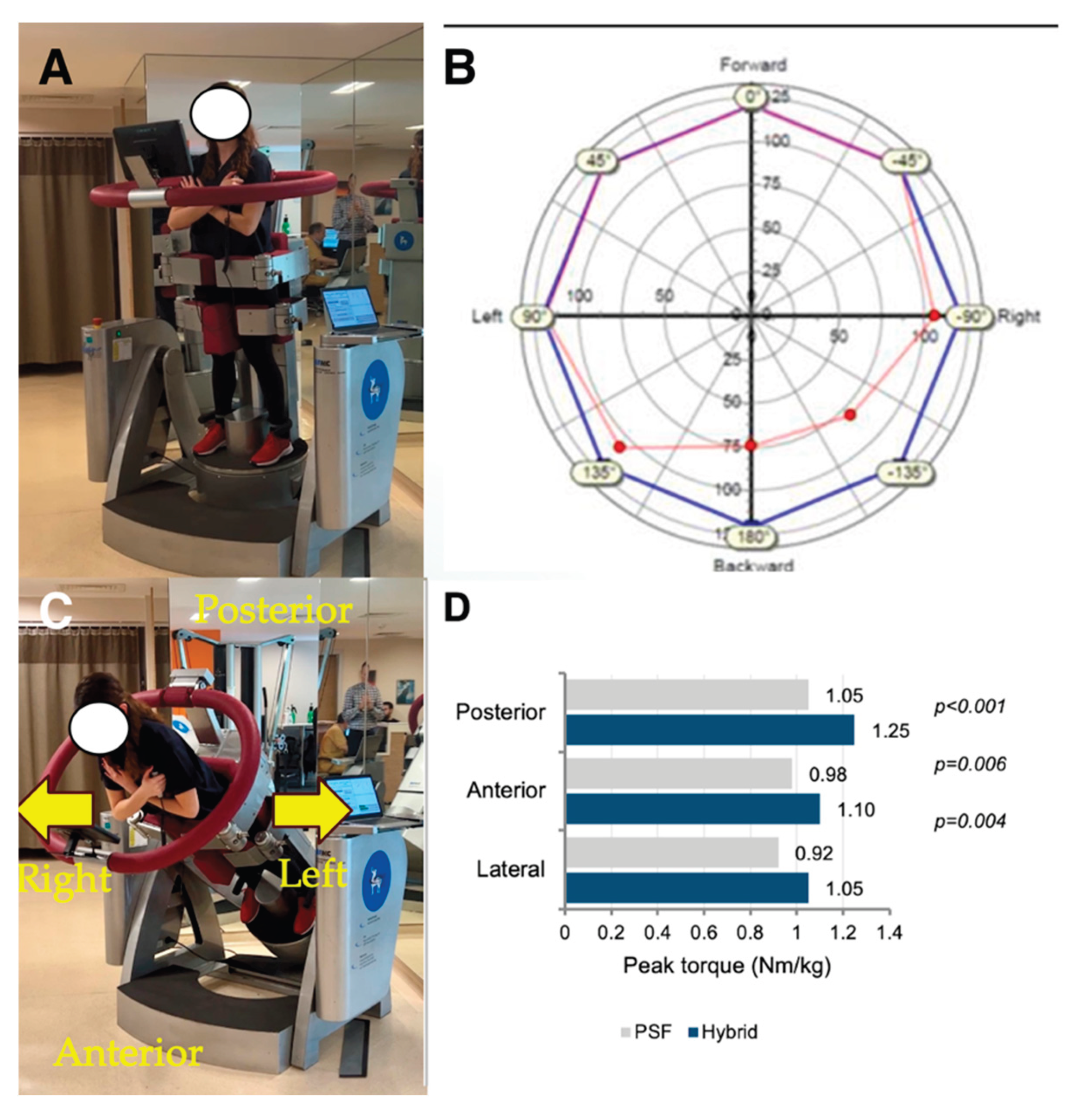

Table 1.
Baseline characteristics and surgical details by group.
| Variable | Hybrid (Thoracic PSF + TL/L VBT) (n=30) | Posterior long fusion (n=36) | p value* |
|---|---|---|---|
| Demographics | |||
| Age at surgery, years | 13.7 ± 1.6 | 14.1 ± 1.7 | 0.31 |
| Sex, female, n (%) | 22 (73.3) | 26 (72.2) | 0.92 |
| BMI, kg/m2 | 20.2 ± 2.3 | 20.6 ± 2.5 | 0.48 |
| Curve characteristics | |||
| Lenke type, n (%) | 0.64 | ||
| 3 | 18 (60.0) | 21 (58.3) | |
| 6 | 12 (40.0) | 15 (41.7) | |
| Skeletal maturity | |||
| Risser grade, median (IQR) | 2 (1–3) | 3 (2–4) | 0.07 |
| Menarche (female only), n (%) | 15/22 (68.2) | 20/26 (76.9) | 0.49 |
| Surgical details | |||
| Distal instrumented vertebra, n (%) | |||
| L3 | 18 (60.0) | 20 (55.6) | |
| L4 | 12 (40.0) | 16 (44.4) | |
| Mean number of thoracic fused levels, n | 8 | 11 | <0.001 |
| TL/L tethered levels, n | 4 | ||
| Operative time, minutes | 275 ± 55 | 310 ± 60 | 0.02 |
| Estimated blood loss, mL | 480 ± 180 | 620 ± 210 | 0.01 |
| Length of stay, days | 4.2 ± 1.1 | 4.8 ± 1.2 | 0.04 |
| Follow-up, months | 30.7 ± 4.6 | 32.5 ± 6.1 | 0.19 |
Footnotes: * p values compare Hybrid versus Posterior long fusion unless otherwise specified. † Reported among female patients only. Values are presented as mean ± standard deviation, median with interquartile range, or number and percentage, as appropriate. Menarche status was analyzed among female patients only. p values compare hybrid fixation and posterior long fusion.
Table 2.
Radiographic outcomes preoperative vs postoperative 24 months by surgical group.
| Parameter | Hybrid Preop | Hybrid 24 mo | Fusion Preop | Fusion 24 mo | p value† |
|---|---|---|---|---|---|
| Coronal plane | |||||
| Main thoracic Cobb (°) | 48.0 ± 9.0 | 18.0 ± 7.5 | 50.5 ± 10.0 | 16.0 ± 6.5 | 0.22 |
| Thoracolumbar/Lumbar Cobb (°) | 42.0 ± 8.5 | 22.0 ± 9.0 | 44.0 ± 9.5 | 12.0 ± 7.5 | <0.001 |
| Coronal balance (C7–CSVL, mm) | 14.0 ± 9.0 | 9.0 ± 7.0 | 12.0 ± 8.0 | 7.0 ± 6.0 | 0.21 |
| Sagittal plane | |||||
| Thoracic kyphosis (T5–T12, °) | 24.0 ± 8.0 | 28.0 ± 9.0 | 23.0 ± 9.0 | 30.0 ± 9.0 | 0.37 |
| Lumbar lordosis (L1–S1, °) | 50.0 ± 10.0 | 52.0 ± 10.0 | 49.0 ± 11.0 | 51.0 ± 10.0 | 0.68 |
Footnotes: † p value for between-group difference in change between Hybrid and Fusion. Coronal balance was defined as the distance between the C7 plumb line and the central sacral vertical line.
Table 3.
Functional outcomes at the postoperative 24-month follow-up.
| Outcome | Hybrid (n=30) | Fusion (n=36) | p value† | Effect size, Cohen’s d (95% CI) |
|---|---|---|---|---|
| Lumbar flexibility | ||||
| Schober test (cm) | 4.90 ± 1.10 | 3.70 ± 1.00 | 0.002 | 1.15 (0.62–1.67) |
| Lumbar ROM, in ° | ||||
| Flexion, ° | 111.6 ± 12.0 | 97.0 ± 11.5 | <0.001 | 1.24 (0.72–1.77) |
| Extension, ° | 26.4 ± 5.2 | 21.2 ± 4.2 | <0.001 | 1.11 (0.59–1.63) |
| Total sagittal lumbar ROM, ° | 138.0 ± 8.4 | 118.2 ± 9.2 | <0.001 | 2.24 (1.62–2.86) |
| Side bending, left, ° | 30.8 ± 6.0 | 21.9 ± 5.5 | <0.001 | 1.55 (1.00–2.10) |
| Side bending, right, ° | 30.0 ± 6.1 | 21.1 ± 5.6 | <0.001 | 1.53 (0.98–2.08) |
| Rotation, left, ° | 44.9 ± 7.5 | 35.1 ± 7.0 | <0.001 | 1.36 (0.82–1.89) |
| Rotation, right, ° | 47.1 ± 7.8 | 37.3 ± 7.2 | <0.001 | 1.31 (0.78–1.84) |
| Lumbar core muscle strength (Nm/kg) | ||||
| Posterior LCMS, Nm/kg | 1.25 ± 0.20 | 1.05 ± 0.18 | <0.001 | 1.06 (0.54–1.57) |
| Anterior LCMS, Nm/kg | 1.10 ± 0.18 | 0.98 ± 0.16 | 0.006 | 0.71 (0.21–1.21) |
| Lateral LCMS, Nm/kg | 1.05 ± 0.17 | 0.92 ± 0.15 | 0.004 | 0.82 (0.31–1.32) |
| Patient reported function | ||||
| SRS 22r Function domain | 4.3 ± 0.4 | 4.1 ± 0.7 | 0.112 | 0.34 (−0.15–0.83) |
Footnotes: † Primary endpoint was total sagittal lumbar ROM (Flexion + Extension) at postoperative 24 months. p values were derived from independent-samples t test or Mann–Whitney U test, as appropriate. Secondary outcomes were exploratory and adjusted using the Holm method. Lumbar core muscle strength was assessed using isokinetic trunk testing. Peak torque for trunk extension (posterior) and flexion (anterior) was recorded in N·m and normalized to body mass (N·m/kg). Cohen’s d was calculated using the pooled standard deviation; values of 0.2, 0.5, and 0.8 correspond to small, medium, and large effects, respectively.
Table 4.
Complications and reinterventions within 24 months.
| Event | Hybrid (n=30) | Fusion (n=36) | p value† |
|---|---|---|---|
| Any complication, n (%) | 6 (20.0) | 4 (11.1) | 0.33 |
| Perioperative (≤30 days), n (%) | 2 (6.7) | 3 (8.3) | 0.79 |
| Radiographic tether breakage, n (%) | 8 (26.7) | — | — |
| Overcorrection, n (%) | 1 (3.3) | — | — |
| Revision surgery, n (%) | 6 (20.0) | 0 (0) | 0.007 |
| Conversion to posterior spinal fusion, n (%) | 1 (3.3) | — | — |
Footnote: P values were calculated using the chi-square test, Fisher’s exact test, or Mann–Whitney U test, as appropriate. Fisher’s exact test was used when expected cell counts were <5. All events were captured within 24 months after index surgery. Tether breakage was defined radiographically as a suspected breakage based on an interscrew angle change of at least 5 degrees between adjacent screws compared with the immediate postoperative radiograph, or a visible discontinuity of the tether when present. Overcorrection was defined as a postoperative coronal curve that crossed neutral to the opposite direction or progressed beyond 10 degrees in the opposite direction on standing radiographs, with or without clinical indication for intervention.
Table 5.
Multivariable linear regression analyses for the selected functional outcomes at 24 months.
Table 5.
Multivariable linear regression analyses for the selected functional outcomes at 24 months.
| Outcome at 24 months | Predictor | Adjusted β | 95% CI | p value |
|---|---|---|---|---|
| Total sagittal lumbar ROM (Flexion plus Extension), ° | Posterior long fusion versus hybrid fixation | −7.8 | −12.6 to −3.0 | 0.001 |
| SRS-22r Function domain | Posterior long fusion versus hybrid fixation | −0.15 | −0.28 to −0.02 | 0.024 |
| Posterior lumbar core muscle strength, Nm/kg | Posterior long fusion versus hybrid fixation | −0.10 | −0.19 to −0.01 | 0.031 |
Footnotes: Adjusted β estimates were derived from multivariable linear regression models. All dependent variables were assessed at the standardized postoperative 24 month visit. Models were adjusted for age at surgery, sex, body mass index, baseline main thoracic Cobb angle, baseline thoracolumbar or lumbar Cobb angle, and Risser grade. Negative coefficients indicate lower 24 month values in the posterior long fusion group relative to the hybrid fixation group.
Disclaimer/Publisher’s Note: The statements, opinions and data contained in all publications are solely those of the individual author(s) and contributor(s) and not of MDPI and/or the editor(s). MDPI and/or the editor(s) disclaim responsibility for any injury to people or property resulting from any ideas, methods, instructions or products referred to in the content. |
© 2026 by the authors. Licensee MDPI, Basel, Switzerland. This article is an open access article distributed under the terms and conditions of the Creative Commons Attribution (CC BY) license (http://creativecommons.org/licenses/by/4.0/).
Copyright: This open access article is published under a Creative Commons CC BY 4.0 license, which permit the free download, distribution, and reuse, provided that the author and preprint are cited in any reuse.