

Submitted:
17 July 2026
Posted:
20 July 2026
You are already at the latest version
Abstract
Environmental stimulation on stroke units (SUs) may affect hemodynamic stability, but the impact of direct and indirect visiting exposure in shared rooms has not been well characterized. This study aimed to assess whether real-time exposure to visits directed to the study patient or to a roommate is associated with blood pressure (BP) variability. We performed a single-center retrospective cohort study. Automated blood pressure measurements were linked to nurse-recorded visitor status in consecutive acute ischemic stroke patients admitted to a university stroke unit over 3 months. Exposure was classified as no visitors, roommate visitors, or own visitors. Mixed-effects models adjusted for time of day and clinical covariates were used. A total of 8,963 blood pressure readings from 87 patients were analyzed: no visitors accounted for 6,607 (73.7%) readings, roommate visitors for 823 (9.2%), and own visitors for 1,533 (17.1%). Visiting exposure showed time-dependent hemodynamic effects. Roommate visits were associated with higher pulse pressure (PP), systolic blood pressure (SBP), and mean blood pressure (MBP) at selected morning, early afternoon, and evening time points, whereas own visits were associated with lower PP at multiple time points. At 09:00, maximal mean BP differences between roommate visitors and own visitors reached 21 mmHg for PP and 27 mmHg for SBP. Visitor presence was associated with short-term, time-dependent BP variability in the early post-stroke period. In shared SU rooms, roommate visitors may represent a modifiable environmental stressor. Ultimately, biobehaviorally informed visiting practices, strategic room allocation, and stimulus-reduction strategies may help optimize hemodynamic stability during acute stroke recovery.
Keywords:
stroke
; blood pressure variability
; visiting hours
; environmental stimulation
; behavioral medicine
; visitor exposure
; stroke unit environment
Introduction
Acute cardiovascular and cerebrovascular events, such as acute ischemic stroke (AIS), trigger profound physiological alterations that are critically modulated by the patient’s immediate psychosocial and behavioral environment ([1,2,3]. Blood pressure (BP) fluctuations and variability are recognized as key hemodynamic factors in the early post-stroke period. Greater variability in systolic BP (SBP), diastolic BP (DBP), mean arterial pressure (MAP), and particularly pulse pressure (PP) has been associated with poorer clinical and functional outcomes after stroke. Early BP control and hemodynamic stability are therefore regarded as important determinants of recovery after acute ischemic stroke (AIS) [3,4,5,6,7,8].
Short-term hemodynamic variability may be influenced by behavioral and environmental factors, ward organization, and patterns of care delivery [9,10].
Blood pressure fluctuations are shaped by both internal and external factors.
Internal mechanisms include sympathetic, humoral, vascular, and cardiac functions, such as sympathetic-adrenal-medullary activation, hypothalamic-pituitary-adrenal axis dysregulation, and the subsequent release of catecholamines and cortisol. These processes increase vascular resistance and arterial stiffness, while altering cardiac performance and output [1,2,11]. External influences include emotional stimuli - such as psychological stress, anxiety, low mood, and perceived isolation (or loneliness) - as well as the presence of social support [12,13,14,15]. In stroke units (SUs), such stimuli may be modified by social interactions, including communication with staff and psychological support [13,14,16]. Some reports suggest that the first minutes of a visit may be associated with a transient stress response accompanied by increased BP [12,17,18,19,20], whereas others have found no meaningful physiological changes or have described a calming effect [21,22,23,24]. In addition, in SUs with multi-bed rooms, the presence of visitors may affect not only the patient being visited but also other patients sharing the room, potentially influencing hemodynamic parameters such as BP.
Most studies have been conducted in intensive care units (ICUs) and cardiac intensive care units (CICUs), with very few undertaken in SUs Importantly, most previous studies have evaluated unit-level visiting policies (e.g., restricted vs. open visiting) rather than the effects of visit timing, so the influence of time of day remains unclear [20,24,25,26,27,28,29].
Moreover, earlier work has tended to focus on general aspects of care rather than on the specific impact of social interactions [30,31].
Visiting practices represent a modifiable, yet still poorly characterized, real-time behavioral exposure. Thus, an important knowledge gap remains regarding the effect of both direct visiting exposure and neighboring-bed visits on BP. There is a need to better understand how organizational aspects of multidisciplinary team care, such as visiting practices, influence BP fluctuations in acute stroke patients.
Aims of the Present Study
The main aim of this study was to evaluate visiting as a modifiable and recurrent factor influencing hemodynamic stability in patients with AIS in SUs. Specifically, we analyzed the presence of visitors—distinguishing visits to the patient from visits to a co-patient in the same room—and its association with BP parameters. We also examined whether the time of day differentiates these relationships, allowing the identification of “time windows” of increased psychological and physiological vulnerability that may be targets for future behavioral interventions.
Methods
This manuscript was prepared in accordance with the Strengthening the Reporting of Observational Studies in Epidemiology (STROBE) statement for observational studies [32].
Setting and Sample
We conducted a single-center retrospective observational study in a stroke unit (SU) of a tertiary university hospital. We analyzed consecutive patients hospitalized over a 3-month period for whom serial automated BP measurements and documentation of visiting were routinely available.
Inclusion Criteria:
We retrospectively reviewed the records of adult patients (age ≥18 years) admitted within 12 hours of acute ischemic stroke onset. All patients underwent an initial non-contrast CT or MRI within one hour of presentation, with follow-up imaging 3–5 days later.
Exclusion Criteria:
Patients were excluded if they: received intravenous thrombolysis (IVT), due to the potential independent influence of reperfusion therapy on hemodynamic parameters; underwent mechanical thrombectomy (MT), for the same reason; received sedative, anxiolytic, or opioid medications that could substantially modify psychological, autonomic, and BP responses; lacked complete documentation of visits or BP measurements (see Table 1).

Table 2.
Association between MBP and PP fluctuations in the stroke unit and the time of day.
| M (IQR) | Mean Blood Pressure | Pulse Pressure | ||||||||
| A | B | C | p-value | post-hoc | A | B | C | p-value | post-hoc | |
| 08:00 | 105.7 (96-119) | 113.7 (100.5-124.3) | 106.7 (94.3-116.3) | 0.498 | 58 (44-73) | 54 (46-74) | 43 (31-52) | 0.003 | a vs c, b vs c | |
| 09:00 | 103.3 (92.5-115.3) | 116.5 (101.3-123) | 101.7 (94-113.3) | 0.012* | a vs b | 58 (44-72) | 68.5 (58-83) | 41 (32-60) | <0.001 | a vs b, a vs c, b vs c |
| 10:00 | 97.7 (88-108.3) | 97.7 (87.3-109) | 101.3 (94.3-115) | 0.037 | a vs c | 55 (43-68) | 49 (40-65) | 48 (33-63) | 0.022 | a vs c |
| 11:00 | 98.3 (89.3-109.3) | 103.3 (90.8-114.2) | 100.3 (91-110.3) | 0.287 | 53 (45-65) | 53.5 (44-70) | 52 (39-65) | 0.237 | ||
| 12:00 | 99.2 (90.3-110.2) | 107 (90.7-117) | 100.5 (90.7-114) | 0.095 | 56 (43-68) | 59 (45-67) | 51 (38-65) | 0.039 | b vs c | |
| 13:00 | 99.7 (88.7-110.3) | 105.3 (92-116.7) | 99.7 (90-111.7) | 0.060 | 55 (43-70) | 59.5 (48-74) | 55 (44-69) | 0.175 | ||
| 14:00 | 97 (88.7-109) | 103.3 (89.3-113.7) | 100.2 (89.3-110) | 0.149 | 55 (41-69) | 59.5 (46-68) | 55 (46-67) | 0.461 | ||
| 15:00 | 99.7 (89.3-111.7) | 105.8 (94.3-116.2) | 100.5 (92.7-111.3) | 0.053 | 55 (43-69) | 58 (47.5-70) | 55 (42-69) | 0.404 | ||
| 16:00 | 99.7 (90.7-111.3) | 103.5 (94.3-113.8) | 104.2 (93.3-113.3) | 0.096 | 58 (46-69) | 56.5 (45-72) | 58 (42.5-71) | 0.867 | ||
| 17:00 | 103.7 (93.3-117) | 108 (95-119.3) | 104.3 (96-114.7) | 0.536 | 58 (46-73) | 60 (51-76) | 59.5 (42-74) | 0.205 | ||
| 18:00 | 101 (91-113.7) | 101.3 (88.3-111.7) | 100.7 (91.3-110) | 0.866 | 59 (46-76) | 57 (46-69) | 52 (42-74) | 0.170 | ||
| 19:00 | 100.3 (91.3-112) | 107.7 (98.7-117.7) | 102.7 (92.3-110.5) | 0.057 | 60 (49-73) | 59 (53-73) | 46 (35-67) | <0.001 | a vs c, b vs c | |
| 20:00 | 104 (93.3-114.7) | 108.2 (96.3-119) | 103.3 (91-109) | 0.393 | 60 (45-74) | 63.5 (48-79) | 53.5 (35.5-69) | 0.040 | b vs c | |
Legend: Type A: no visitors; Type B: visitors present for a co-patient; Type C: visitors present for the patient. *Significant values are shown in bold.
Definitions of Visiting Exposure
Because each patient could experience different visiting conditions during hospitalization, we analyzed exposure states assigned to individual BP measurements rather than fixed patient groups.
Based on the visiting log, each blood pressure (BP) reading was classified into one of three distinct types: (A) no visitors present in the room; (B) visitors at the next bed (co-patient, roommate); (C) visitors for the study patient. The room and bed configuration of the SU intensive section is presented in Figure 1.
During the study period, visits were permitted between 08:00 and 20:00, limited to two visitors. Nurses documented start and end times in the medical records.
Hemodynamic Parameters
- The following parameters were analyzed: systolic blood pressure (SBP) and diastolic blood pressure (DBP). Additionally, the following indices were calculated:
- Mean arterial pressure (MAP): MAP = DBP + (SBP-DBP)
- Pulse pressure (PP): PP = SBP – DBP
BP Measurements and Data Collection Tools and Methods
Patients received treatment during hospitalization based on AIS guidelines, including antihypertensive therapy when indicated [34,35].
We used routinely collected, protocol-based BP recordings as documented in nursing records.
Blood pressure was measured automatically in the supine position on the non-paretic arm using the Ultraview SL2600 monitoring system (Spacelabs Medical Inc., USA), which meets the ANSI/AAMI SP-10 standards.
According to the ward protocol, measurements were taken hourly during the daytime (08:00–20:00; up to 13 readings) and every 4 hours overnight (21:00–07:00; up to 3 readings; i.e., 22:00, 02:00, and 06:00). Although no visits occurred during the nocturnal period, these measurements were used to assess night-time BP. Notably, patients remained continuously connected to monitoring devices during visiting periods. Analyses covered the period from day 1 of hospitalization until discharge from the SU (median length of stay: 7 days).
Data Analysis
Descriptive statistics were presented as medians and interquartile ranges due to non-normal distributions. Comparisons were performed using the Kruskal–Wallis test with appropriate post hoc analysis. Spearman’s rank correlation coefficients were calculated. Statistical significance was set at α=0.05. Analyses were performed using Statistica 13.3 (StatSoft).
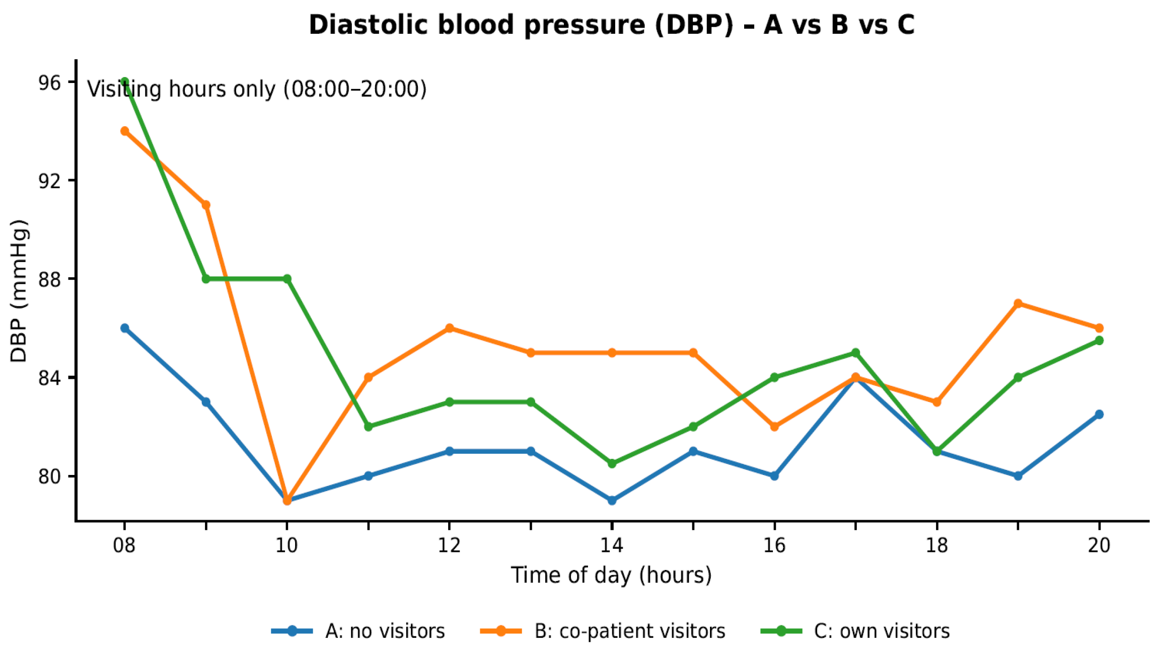
Timestamped ward BP measurements were converted to long format (patient, date, hour, value). For the PP, MAP, DBP and SBP exposure figures, measurements were classified into exposure groups A, B, or C (A: no visitors; B: co-patient visitors; C: own visitors) and restricted to visiting hours (08:00–20:00). Hourly medians and interquartile ranges (IQR; Q1–Q3) were calculated and plotted for each group. The number of observations contributing to each hour was shown below the x-axis (See Figure 3 A and B).
Results
Patient Characteristics and Exposure Profile
The study included 87 patients (median age: 71 years, IQR: 62–78; 48% female). During the first 7 days of hospitalization, a total of 8,963 BP measurements were recorded (see Figure 2 for details). On average, 14 BP measurements per patient per day were obtained, as monitors were temporarily disconnected during rehabilitation, diagnostic procedures, or bathroom use. Sixty-six patients (76%) were receiving antihypertensive treatment.
Visiting Exposure and BP Parameters
The study utilized specific statistical models to account for repeated measurements, the time of day, and patients' key medical characteristics. This analysis revealed that the frequency and type of patient visits (Types A, B, and C) significantly influenced their blood pressure readings over time. Pulse pressure (PP) demonstrated the most frequent and significant differences across exposure categories. Compared to Type A exposure (no visitors), Type B exposure (co-patient visitors) was associated with higher PP, whereas Type C exposure (own visitors) was frequently associated with lower PP at several time points.
Time-of- Day Patterns in BP Variability
Hourly analyses revealed that significant differences between exposure categories were concentrated within three distinct “time windows”: morning (08:00–10:00), early afternoon (12:00–13:00), and evening (19:00–20:00). Additionally, the association between visiting exposure and hemodynamic parameters varied significantly over the course of the day. These effects were most pronounced for pulse pressure (PP) and systolic blood pressure (SBP). Notably, at 09:00, maximal mean BP differences between roommate visitors (Type B) and own visitors (Type C) reached 27 mmHg for PP and 21 mmHg for SBP; while at 19:00, differences reached 13 mmHg and 18.5 mmHg, respectively.
Figure 3B illustrates marked fluctuations in PP, characterized by alternating increases and decreases between the exposure categories. This pattern was driven by greater PP variability during roommate visits (Type B) compared to periods without visitors (Type A), with the most prominent differences observed during the 08:00–10:00 and 19:00–20:00 intervals. Regarding DBP and MAP, Type C exposure was associated with higher values at 10:00 compared to Type A. Detailed adjusted estimates for all BP parameters and estimated marginal means are presented in Table 1A–D, Figure 3A–D, and Supplementary Figures S1–S6.
Figure 3A.
Diurnal profiles of Mean Blood Pressure (MBP/MAP*) across visit types A, B, and C. The time axis is truncated to daytime hours (08:00–20:00). The miniature "n" strip below the x-axis displays the number of measurements recorded per hour (format: n = A/B/C). *MAP - Mean Arterial Pressure.
Figure 3A.
Diurnal profiles of Mean Blood Pressure (MBP/MAP*) across visit types A, B, and C. The time axis is truncated to daytime hours (08:00–20:00). The miniature "n" strip below the x-axis displays the number of measurements recorded per hour (format: n = A/B/C). *MAP - Mean Arterial Pressure.
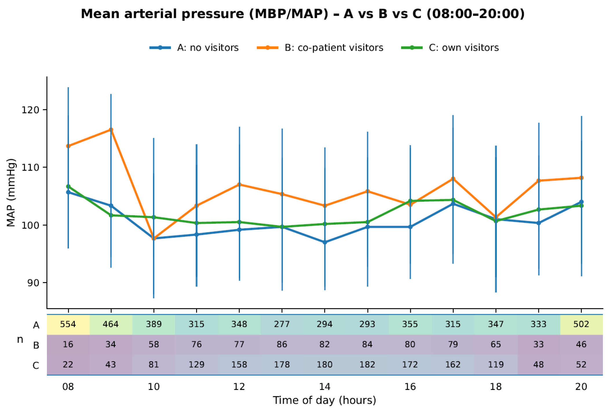
Figure 3B.
Diurnal profiles of pulse pressure (PP) across visit types A, B, and C. The time axis is truncated to daytime hours (08:00–20:00). The miniature "n" strip below the x-axis displays the number of measurements recorded per hour (format: n = A/B/C).
Figure 3B.
Diurnal profiles of pulse pressure (PP) across visit types A, B, and C. The time axis is truncated to daytime hours (08:00–20:00). The miniature "n" strip below the x-axis displays the number of measurements recorded per hour (format: n = A/B/C).
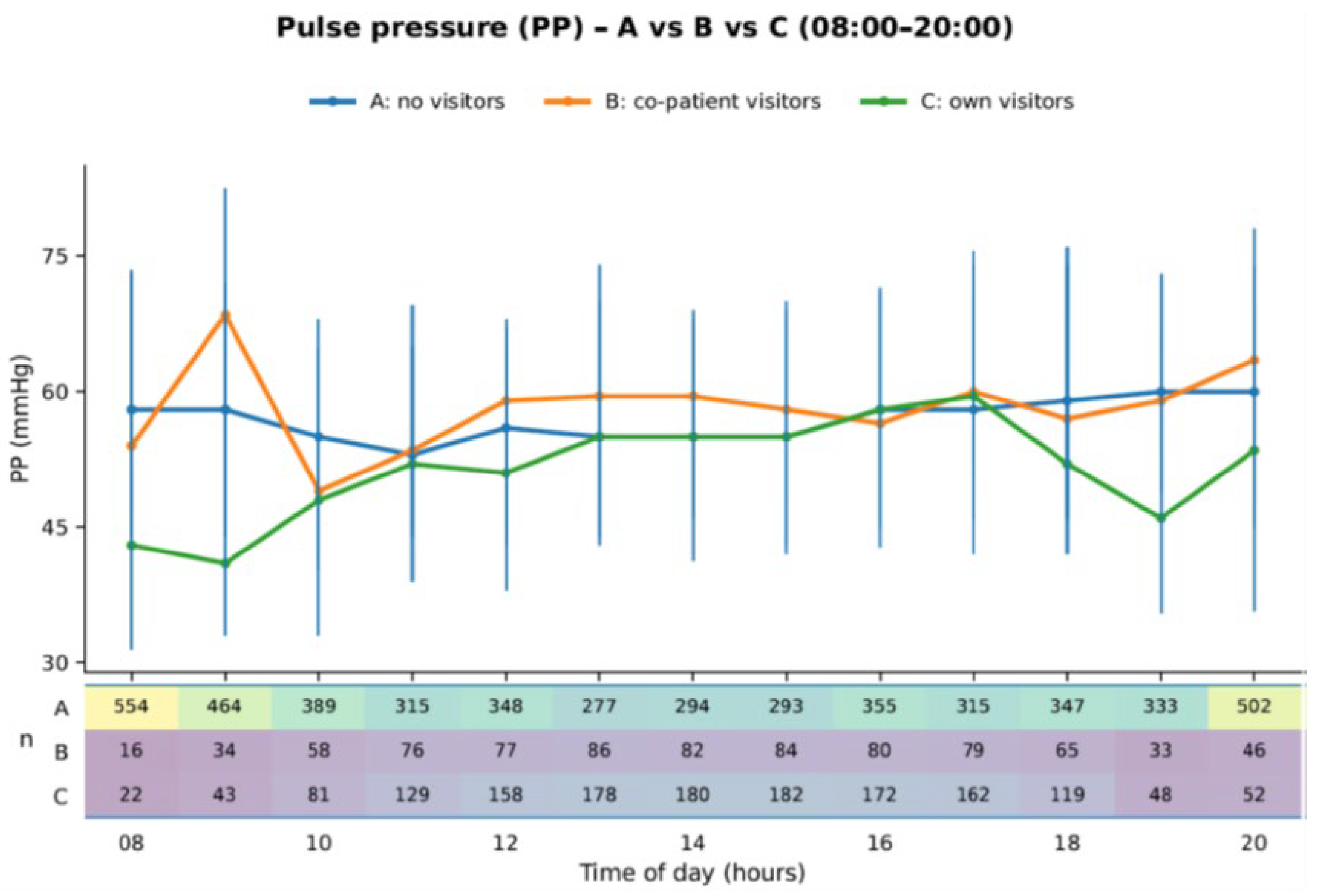
Figure 3C.
Diurnal profiles of Sysstolic Blood Pressure(SBP) across visit types A, B, and C. The time axis is truncated to daytime hours (08:00–20:00); the hourly measurement counts (format: n = A/B/C) are presented in Figure 3A and 3B.
Figure 3C.
Diurnal profiles of Sysstolic Blood Pressure(SBP) across visit types A, B, and C. The time axis is truncated to daytime hours (08:00–20:00); the hourly measurement counts (format: n = A/B/C) are presented in Figure 3A and 3B.
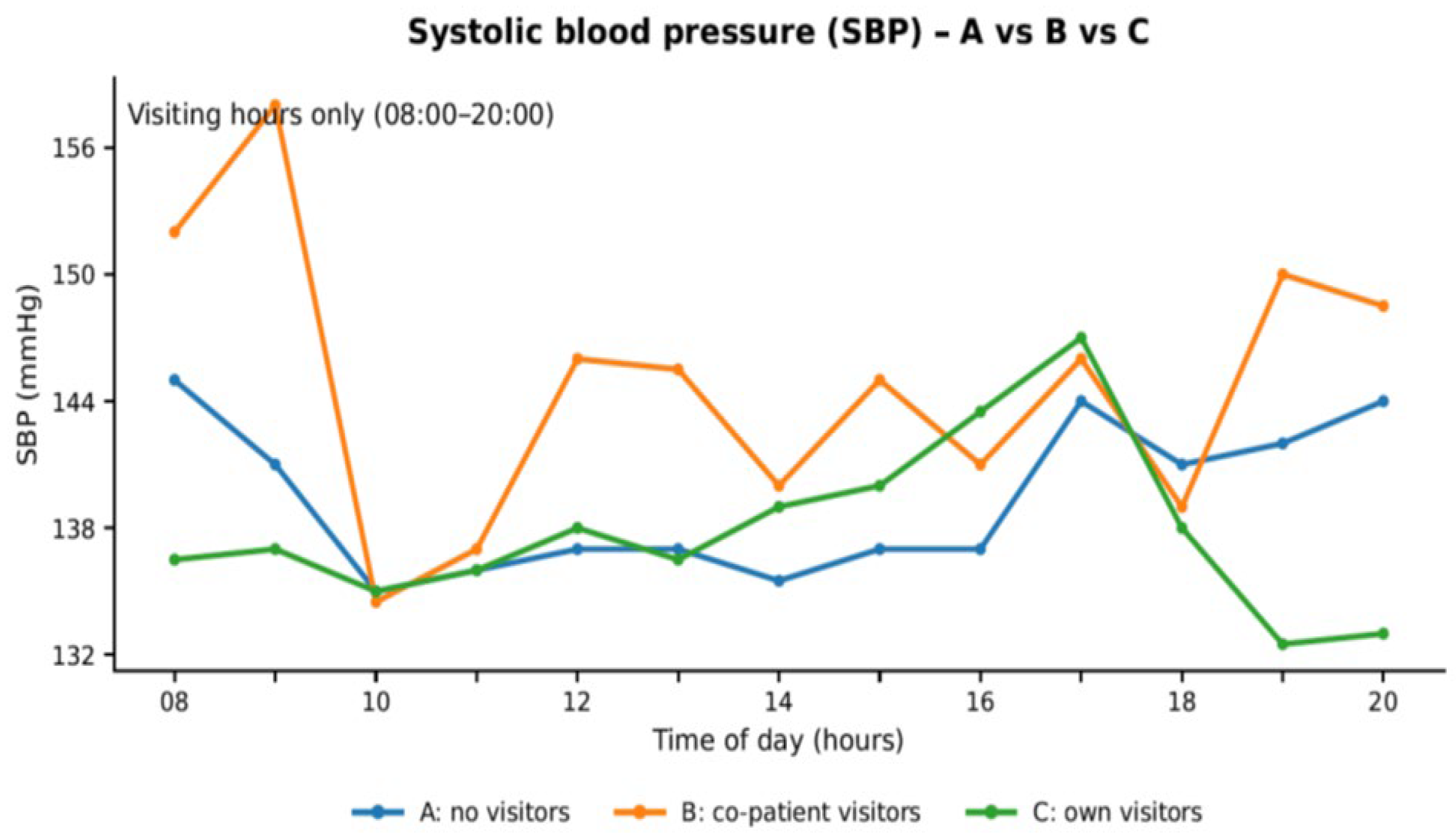
Figure 4C.
Diurnal profiles of diastolic blood pressure (DBP) across visit types A, B, and C. The time axis is truncated to daytime hours (08:00–20:00); the hourly measurement counts. (format: n = A/B/C) are presented in Figure 3A and 3B.
Figure 4C.
Diurnal profiles of diastolic blood pressure (DBP) across visit types A, B, and C. The time axis is truncated to daytime hours (08:00–20:00); the hourly measurement counts. (format: n = A/B/C) are presented in Figure 3A and 3B.
Night-Time BP Observations
Nocturnal BP fluctuations were also determined, and the results are detailed below.
A "dipper" was defined as a patient with a nocturnal BP reduction of 10%–20% relative to the daytime mean. Patients were classified into the following physiological profiles: extreme dippers (>20% BP reduction), n = 1 (1.1%); dippers (10%–20% reduction), n = 5 (5.7%); non-dippers (0% to <10% reduction), n = 38 (43.7%); and reverse dippers (<0% reduction; i.e., night-time BP higher than daytime BP), n = 43 (49.4%) [see also Supplementary Figures S3 A,B-S6 A,B].
Discussion
In this study, we found that morning, early afternoon, and evening BP elevations were observed predominantly during Type B exposure, i.e., when visitors were present for the roommate. Far fewer such reactions were observed during Type C visits (visitors for the study patient), and they were least pronounced during Type A exposure (no visitors present in the room) (see Figure 3A-D and Supplementary Figures S1–S6). Additionally, we found that PP appeared to be the parameter most sensitive to short-term environmental stimuli related to visitor presence. Such “unequal” exposure to social support may intensify negative affect and trigger sympathetic activation, thereby contributing to transient BP elevations and greater BP variability in the acute post-stroke period [19,20,21,22]. Our data suggest that human-environmental interactions on acute wards follow a strict diurnal rhythm. To reduce these BP fluctuations, clinical protocols in multi-bed stroke units should shift from a purely institutional focus toward a biobehavioral management framework that accounts for the collective physiological vulnerability of all patients in a shared space. However, this interpretation remains speculative and should be examined in future studies incorporating organizational, psychological, and behavioral measures.
Clinical and Behavioral Implications for Stroke Unit Management
Any explanation of fluctuations in BP must rely on indirect inferences from clinical psychology and on the authors’ clinical experience with daily routines in stroke units. In the morning—observed in our study as a peak around 09:00, notably for PP, MAP, and SBP during Type B visits—potential stressors include medical and nursing rounds, which may trigger a “white coat effect,” especially when students are present [13]. Additional stressors, such as exposure to unfamiliar individuals during activities like personal hygiene or meals, may cluster during these hours. These situations can be especially distressing due to functional impairments common after stroke. Similar stressors may be present in the early afternoon; however, they appear less pronounced around midday. This pattern may be attributable to a reduction in hygiene-related procedures and to the distracting, yet potentially positive, effect of ward activities, including rehabilitation sessions. Together, these factors may partly explain why differences in BP between visit types are less pronounced at that time [13,27,30].
To the best of our knowledge, this is the first study to demonstrate the impact of visits on blood pressure (BP) at specific times of the day. As this appears to be a novel observation, the underlying mechanisms remain largely unclear. Taken together, our study suggests that blood pressure monitoring during these vulnerable time windows helps optimize the timing and type of behavioral and purely medical interventions, which may ultimately contribute to reducing BP variability.
The Role of Visitors and Patient Loneliness
Another important factor influencing BP variability is the impact of the social environment on cardiovascular health. Loneliness exerts a profound influence on hypertension development over both short- and long-term horizons; cross-lag analyses indicate that it predicts subsequent changes in depressive symptomatology [3,36]. Loneliness, lack of social support, a subjective sense of helplessness, low perceived control, and anger—particularly when chronically suppressed—have all been linked to increased blood pressure and greater BP reactivity to stress in clinical and population studies.
Substantial evidence indicates that loneliness predicts increased BP in middle-aged and older adults over time [14,37,38]. These feelings of social isolation may contribute to the persistent and substantial differences in BP variability observed between Type B and Type C visits, particularly in the evening, when ward activity decreases (see Figure 3A-D and Supplementary Figures S1 and S3). Previously, it was believed that short-term fluctuations in perceived social disconnection among lonely individuals influenced blood pressure only modestly.
However, most existing studies have focused on chronic loneliness, and there is still insufficient data from ecological momentary or ambulatory blood pressure studies that repeatedly sample state loneliness over hours or days. While laboratory research demonstrates that acute social isolation or socially stressful tasks elicit transient increases in systolic and diastolic blood pressure [39], existing field data mainly suggest moderate within-person BP elevations on days with greater social stress or lower perceived support, rather than loneliness per se [30,31,36,37,38]. This supports the hypothesis that repeated, short-lived BP fluctuations during lonely periods may cumulatively contribute to a long-term increase in resting BP over time [37,38]. In terms of mechanisms, the available evidence implicates psychological factors such as stress, depression, atypical physiological reactivity, and neuroendocrine responses—all of which could trigger inflammatory reactions [40]. Crucially, the recent literature confirms that patients with high perceived psychosocial stress demonstrate a statistically significant 1.45- to 1.58-fold higher risk of fatal stroke outcomes [4]. These findings were confirmed by a recent study from Ghana [41].
In the acute stroke setting, however, the presence of the patient’s own visitors (Type C) may reflect a complex interplay between environmental stimulation and the potential mitigation of the physiological stress associated with loneliness. From a behavioral perspective, assessing patients' perceived loneliness is crucial [37]. Therefore, clinical teams should actively incorporate family visitors into the care plan, rather than assuming that all visitors inherently induce stress [20,21,22,23,24].
Sleep Disruption and Stroke-Unit Workflow – An Area for Further Interventions
In our night-time BP observations, we found fewer “dippers” and a slightly higher proportion of “reverse dippers,” with similar proportions of “non-dippers” (5.6% vs 14.5% and 19.2%, respectively) compared with large stroke studies [25,26]. (Table 4). Our study was not designed to assess sleep disturbances; nevertheless, this observation appears noteworthy and warrants further investigation in larger populations [see also Supplemental Table S4)
Implementation Through Team Leadership and Policy
Although not all of these environmental interventions fall strictly within the scope of independent multidisciplinary team practice, team leadership can substantially influence organizational decisions regarding noise control and lighting. For example, implementing naturalistic lighting can further support the regulation of internal clocks in hospitalized patients [42,43,44].
Structured Stroke Unit Policy
To reduce these multi-faceted stressors in a systematic way, we propose a structured SU policy focusing on six key areas: (1) organization of rounds, (2) family involvement, (3) post-round communication, (4) limits on student activity, (5) nighttime noise reduction, and (6) optimization of visiting hours.
Detailed recommendations are provided in Supplementary Table S2.
Strengths, limitations, and future directions
A major strength of this study is its novel focus on the real-time, time-specific impact of ward ecology and social dynamics on cardiovascular autonomic regulation in acute stroke patients. By differentiating between direct social support and indirect environmental exposure (roommates' visitors), this research bridges a critical gap between clinical neurology and behavioral medicine. Furthermore, the use of continuous, high-frequency blood pressure monitoring allowed for the precise identification of diurnal vulnerability windows, providing a detailed insight into human-environmental interactions that retrospective clinical registries typically overlook.
Conversely, several limitations must be acknowledged. This study was designed primarily to examine the workflow of stroke units (SUs) and patient care, with a focus on the organizational, behavioral, and clinical factors—including visiting procedures—that may influence BP variability and functional outcomes [see Supplementary Table S3 for details]. In line with this aim, we did not analyze stroke subtypes, infarct size, lesion location, or symptom-specific profiles. We also did not include additional clinical and biological factors such as lipid status, diabetes, arrhythmias, or formal psychological assessments—variables that are typically evaluated and managed collaboratively by physicians, nurses, and allied health professionals. These factors may have influenced both BP variability and functional outcomes, and they should be incorporated into future trials adopting a comprehensive, team-based approach to stroke care.
The current evidence underscores a critical gap in our understanding of how micro-environmental factors—specifically social interactions and ward ecology—modulate acute stroke recovery. To advance behavioral medicine in acute cerebrovascular care, future research must transition from retrospective observations to prospective, multi-method designs.
Specifically, there is a need to implement real-time tracking of psychological states (e.g., acute stress, perceived social support) alongside physiological biomarkers of recovery, such as cortisol dynamics, heart rate variability, and neuroendocrine profiles. By integrating these psychological and physiological parameters, future longitudinal studies can examine both short- and long-term outcomes.
As an additional future direction, upcoming studies should evaluate how the proposed stroke unit policies work in daily clinical practice. Future research should focus on auditing multidisciplinary team compliance with the new protocols, assessing patient and family satisfaction with visiting hours, and measuring the long-term benefits of environmental changes. Ultimately, these integrated insights will drive the development of precise, biobehaviorally informed visiting policies and environmental interventions within SUs to safeguard hemodynamic stability and improve functional recovery.
Conclusions
- In patients with AIS, exposure to visiting activity in shared stroke-unit rooms was associated with increased short-term BP variability, with these effects significantly moderated by both visitor type and the timing of the encounter.
- Hemodynamic fluctuations were primarily concentrated during the morning, early afternoon, and evening, suggesting that the time of day serves as a critical modifier of cardiovascular reactivity to social environmental stimuli in the acute clinical phase.
- Indirect exposure (roommates’ visits) was more consistently associated with unfavorable hemodynamic patterns than direct social interaction, highlighting the potentially stressful effect of uncontrollable environmental stressors on autonomic system stability.
- Pulse pressure (PP) was the most reliable hemodynamic predictor of early functional improvement, reinforcing its utility as a marker of cardiovascular resilience in behavioral stroke research.
- If confirmed in larger multicenter studies, these findings could inform evidence-based visiting policies and environmental micro-interventions designed to maintain hemodynamic stability in SUs.
Supplementary Materials
The following supporting information can be downloaded at the website of this paper posted on Preprints.org.
Funding
This research received no specific grant from any funding agency in the public, commercial, or not-for-profit sectors.
Acknowledgements
The authors would like to thank Maria Biskupska, MSc, for preparing the graph.
Ethics statement
Ethical and Institutional Approvals. The study was conducted in accordance with the Declaration of Helsinki (2013 revision). Patients and visitors were informed that their presence on the ward (and visiting hours) was being recorded by the nursing staff, and no objections were raised. All patient data were anonymized during transfer to the analysis dataset; patients were assigned unique identifiers, and demographic data were limited to sex and age. To ensure data security and reduce the risk of unauthorized access, the dataset was stored on an encrypted offline external SSD. As the study involved a retrospective analysis of anonymized hospital records, the Institutional Review Board of University of XX determined that formal bioethical approval was not required.
Conflicts of Interest
The authors declare that they have no known competing financial interests or personal relationships that could have appeared to influence the work reported in this paper.
References
- Rozanski, A.; Blumenthal, J.A.; Kaplan, J. Impact of Psychological Factors on the Pathogenesis of Cardiovascular Disease and Implications for Therapy. Circulation 1999, 99(16), 2192–2217. [Google Scholar] [CrossRef] [PubMed]
- Wittstein, I.S.; Thiemann, D.R.; Lima, J.A.C.; Baughman, K.L.; Schulman, S.P.; Gerstenblith, G.; Wu, K.C.; Rade, J.J.; Bivalacqua, T.J.; Champion, H.C. Neurohumoral Features of Myocardial Stunning Due to Sudden Emotional Stress. New. Engl. J. Med. 2005, 352(6), 539–548. [Google Scholar] [CrossRef] [PubMed]
- Hackett, M.L.; Yapa, C.; Parag, V.; Anderson, C.S. Frequency of Depression After Stroke: A Systematic Review of Observational Studies. Stroke 2005, 36(6), 1330–1340. [Google Scholar] [CrossRef] [PubMed]
- Wang, Y.; Shi, X.; Xu, L. The Correlation Between Perceived Psychosocial Stress and Stroke: A Meta-Analysis. Front. Neurol. 2026, 16, 1639076. [Google Scholar] [CrossRef] [PubMed]
- Kamieniarz-Mędrygał, M.; Kaźmierski, R. Significance of Pulse Pressure Variability in Predicting Functional Outcome in Acute Ischemic Stroke: A Retrospective, Single-Center, Observational Cohort Study. Sci. Rep. 2023, 13(1), 3618. [Google Scholar] [CrossRef] [PubMed]
- Appiah, K.O.B.; Patel, M.; Panerai, R.B.; Robinson, T.G.; Haunton, V.J. Increased Blood Pressure Variability Following Acute Stroke Is Associated With Poor Long-Term Outcomes: A Systematic Review. Blood Press. Monit. 2019, 24(2), 67–73. [Google Scholar] [CrossRef] [PubMed]
- Minhas, J.S.; Wang, X.; Lavados, P.M.; Moullaali, T.J.; Arima, H.; Billot, L.; Hackett, M.L.; Olavarria, V.V.; Middleton, S.; Pontes-Neto, O.; et al. Blood Pressure Variability and Outcome in Acute Ischemic and Hemorrhagic Stroke: A Post Hoc Analysis of the HeadPoST Study. J. Hum. Hypertens. 2019, 33, 411–418. [Google Scholar] [CrossRef] [PubMed]
- Stead, L.G.; Enduri, S.; Bellolio, M.F.; Jain, A.R.; Vaidyanathan, L.; Gilmore, R.M.; Kashyap, R.; Weaver, A.L.; Brown, R.D., Jr. The Impact of Blood Pressure Hemodynamics in Acute Ischemic Stroke: A Prospective Cohort Study. Int. J. Emerg. Med. 2012, 5(1), 3. [Google Scholar] [CrossRef] [PubMed]
- Fumagalli, S.; Boncinelli, L.; Lo Nostro, A.; Valoti, P.; Baldereschi, G.; Di Bari, M.; Ungar, A.; Baldasseroni, S.; Geppetti, P.; Masotti, G.; et al. Reduced Cardiocirculatory Complications With Unrestrictive Visiting Policy in an Intensive Care Unit: Results From a Pilot, Randomized Trial. Circulation 2006, 113(7), 946–952. [Google Scholar] [CrossRef] [PubMed]
- Haribhai-Thompson, J.; McBride-Henry, K.; Hales, C.; Rook, H. Understanding of Empathetic Communication in Acute Hospital Settings: A Scoping Review. BMJ Open 2022, 12(9), e063375. [Google Scholar] [CrossRef] [PubMed]
- Atam, I.; Singh, S.; Kumar, S.; Singh, A.K.; Rastogi, D. Neuroendocrine Stress Response in Acute Stroke: Physiological and Clinical Perspectives. Cureus 2026, 18(2), e103236. [Google Scholar] [CrossRef] [PubMed]
- Kleman, M.; Bickert, A.; Karpinski, A.; Wantz, D.; Jacobsen, B.; Lowery, B.; Menapace, F. Physiologic Responses of Coronary Care Patients to Visiting. J. Cardiovasc. Nurs. 1993, 7(3), 52–62. [Google Scholar] [CrossRef] [PubMed]
- Kumpusalo, E.; Teho, A.; Laitila, R.; Takala, J. Janus Faces of the White Coat Effect: Blood Pressure Not Only Rises, It May Also Fall. J. Hum. Hypertens. 2002, 16, 725–728. [Google Scholar] [CrossRef] [PubMed]
- Cacioppo, S.; Grippo, A.J.; London, S.; Goossens, L.; Cacioppo, J.T. Loneliness: Clinical Import and Interventions. Perspect. Psychol. Sci. 2015, 10(2), 238–249. [Google Scholar] [CrossRef] [PubMed]
- Parati, G.; Bilo, G.; Kollias, A.; Pengo, M.; Ochoa, J.E.; Castiglioni, P.; Stergiou, G.S.; Mancia, G.; Asayama, K.; Asmar, R.; et al. Blood Pressure Variability: Methodological Aspects, Clinical Relevance and Practical Indications for Management—A European Society of Hypertension Position Paper. J. Hypertens. 2023, 41(4), 527–544. [Google Scholar] [CrossRef] [PubMed]
- Nembhard, I.M.; David, G.; Ezzeddine, I.; Betts, D.; Radin, J. A Systematic Review of Research on Empathy in Health Care. Heal. Serv. Res. 2023, 58(2), 250–263. [Google Scholar] [CrossRef] [PubMed]
- Brown, A.J. Effect of Family Visits on the Blood Pressure and Heart Rate of Patients in the Coronary-Care Unit. Heart Lung 1976, 5(2), 291–296. [Google Scholar] [PubMed]
- Lazure, L.L.; Baun, M.M. Increasing Patient Control of Family Visiting in the Coronary Care Unit. Am. J. Crit. Care 1995, 4(2), 157–164. [Google Scholar] [CrossRef] [PubMed]
- Mahmoudi, M.; Asgari, P.; Khajeh-Goodari, M.; Hekmatpou, D.; Rafiei, F. The Effect of Increasing Meeting Time on the Physiological Indices of Patients Admitted to the Intensive Care Unit. Jundishapur J. Chronic. Dis. Care 2016, 5(2), e31172. [Google Scholar] [CrossRef]
- Cicek, H.S.; Cambaztepe, N.; Kilic, B. The Effect of Visiting on Vital Signs and Anxiety at Stroke Patients: Blood Pressure Increases while Anxiety is Reduced. Int. J. Caring Sci. 2017, 10(3), 1305–1313. [Google Scholar]
- Simpson, T.; Shaver, J. Cardiovascular Responses to Family Visits in Coronary Care Unit Patients. Heart Lung 1990, 19(4), 344–351. [Google Scholar] [PubMed]
- Simpson, T.; Shaver, J. A Comparison of Hypertensive and Nonhypertensive Coronary Care Patients' Cardiovascular Responses to Visitors. Heart Lung 1991, 20(3), 213–220. [Google Scholar] [PubMed]
- Rezaie, H.; Sadeghi, T.; Abdoli, F. The Effects of Scheduled Visitation on the Physiological Indices of Conscious Patients Admitted at Intensive Care Units. Evid. Based Care. 2016, 5, 33–40. [Google Scholar] [CrossRef]
- Nazari-Ostad, Z.; Namazinia, M.; Hajiabadi, F.; Aghebati, N.; Esmaily, H.; Peivandi Yazdi, A. Effect of Protocol-Based Family Visitation on Physiological Indicators in ICU Patients: A Randomized Controlled Trial. BMC Anesthesiol. 2024, 24(1), 18. [Google Scholar] [CrossRef] [PubMed]
- Kim, B.S.; Kim, J.H.; Kim, W.; Kim, W.S.; Park, S.; Lee, S.J.; Kim, J.Y.; Lee, E.M.; Ihm, S.H.; Pyun, W.B.; et al. Clinical and Life Style Factors Related to the Nighttime Blood Pressure, Nighttime Dipping and Their Phenotypes in Korean Hypertensive Patients. Clin. Hypertens. 2023, 29(1), 21. [Google Scholar] [CrossRef] [PubMed]
- Pandian, J.D.; Wong, A.A.; Lincoln, D.J.; Davis, J.P.; Henderson, R.D.; O'Sullivan, J.D.; Read, S.J. Circadian Blood Pressure Variation After Acute Stroke. J. Clin. Neurosci. 2006, 13(5), 558–562. [Google Scholar] [CrossRef] [PubMed]
- Khaleghparast, S.; Joolaee, S.; Ghanbari, B.; Maleki, M.; Peyrovi, H.; Bahrani, N. A Review of Visiting Policies in Intensive Care Units. Glob. J. Health Sci. 2015, 8(6), 267–276. [Google Scholar] [CrossRef] [PubMed]
- Nassar Junior, A.P.; Besen, B.A.M.P.; Robinson, C.C.; Falavigna, M.; Teixeira, C.; Rosa, R. Flexible Versus Restrictive Visiting Policies in ICUs: A Systematic Review and Meta-Analysis. Crit. Care Med. 2018, 46(7), 1175–1180. [Google Scholar] [CrossRef] [PubMed]
- Rosa, R.G.; Falavigna, M.; da Silva, D.B.; Sganzerla, D.; Santos, M.M.S.; Kochhann, R.; de Moura, R.M.; Eugênio, C.S.; Haack, T.D.S.R.; Barbosa, M.G.; et al. Effect of Flexible Family Visitation on Delirium Among Patients in the Intensive Care Unit: The ICU Visits Randomized Clinical Trial. JAMA 2019, 322(3), 216–228. [Google Scholar] [CrossRef] [PubMed]
- Naef, R.; Ernst, J.; Petry, H.; Carlen, S.; Schmid, F.; Wetli, N. Intensive Care Nurse–Family Engagement From a Global Perspective: A Qualitative Multi-Site Study. Intensive Crit. Care Nurs. 2021, 66, 103070. [Google Scholar] [CrossRef]
- Secunda, K.E.; Kruser, J.M. Patient-Centered and Family-Centered Care in the Intensive Care Unit. Clin. Chest Med. 2022, 43(3), 539–550. [Google Scholar] [CrossRef] [PubMed]
- von Elm, E.; Altman, D.G.; Egger, M.; Pocock, S.J.; Gøtzsche, P.C.; Vandenbroucke, J.P.; STROBE Initiative. The Strengthening the Reporting of Observational Studies in Epidemiology (STROBE) Statement: Guidelines for Reporting Observational Studies. J. Clin. Epidemiol. 2008, 61(4), 344–349. [Google Scholar] [CrossRef] [PubMed]
- European Parliament and Council of the European Union. Regulation (EU) 2016/679 of the European Parliament and of the Council of 27 April 2016 on the protection of natural persons with regard to the processing of personal data and on the free movement of such data, and repealing Directive 95/46/EC (General Data Protection Regulation). Off. J. Eur. Union 2016, 119, 1–88. [Google Scholar] [CrossRef]
- Ringelstein, E.B.; Chamorro, A.; Kaste, M.; Langhorne, P.; Leys, D.; Lyrer, P.; Thijs, V.; Thomassen, L.; Toni, D.; ESO Stroke Unit Certification Committee. European Stroke Organisation Recommendations to Establish a Stroke Unit and Stroke Center. Stroke 2013, 44(3), 828–840. [Google Scholar] [CrossRef] [PubMed]
- Powers, W.J.; Rabinstein, A.A.; Ackerson, T.; Adeoye, O.M.; Bambakidis, N.C.; Becker, K.; Biller, J.; Brown, M.; Demaerschalk, B.M.; Hoh, B.; et al. 2019 Update to the 2018 Guidelines for the Early Management of Acute Ischemic Stroke. Stroke 2018, 50(12), e344–e418. [Google Scholar] [CrossRef] [PubMed]
- Uchino, B.N. Social Support and Health: A Review of Physiological Processes Potentially Underlying Links to Disease Outcomes. J. Behav. Med. 2006, 29(4), 377–387. [Google Scholar] [CrossRef] [PubMed]
- Hawkley, L.C.; Cacioppo, J.T. Loneliness Matters: A Theoretical and Empirical Review of Consequences and Mechanisms. Ann. Behav. Med. 2010, 40(2), 218–227. [Google Scholar] [CrossRef] [PubMed]
- Hawkley, L.C.; Thisted, R.A.; Masi, C.M.; Cacioppo, J.T. Loneliness Predicts Increased Blood Pressure: 5-Year Cross-Lagged Analyses in Middle-Aged and Older Adults. Psychol. Aging 2010, 25(1), 132–141. [Google Scholar] [CrossRef]
- Steptoe, A.; Kivimäki, M. Stress and Cardiovascular Disease. Nat. Rev. Cardiol. 2012, 9(6), 360–370. [Google Scholar] [CrossRef] [PubMed]
- Paul, E.; Bu, F.; Fancourt, D. Loneliness and Risk for Cardiovascular Disease: Mechanisms and Future Directions. Curr. Cardiol. Rep. 2021, 23, 68. [Google Scholar] [CrossRef] [PubMed]
- Darkwah, J.D.; Albert, A.; Bockarie, A.; Ayisi-Boateng, N.K.; Konadu, E.; Adu-Gyamfi, R.; Arthur, A.; Agyenim-Boateng, K.; Neizer, C.; Fiattor, T.; et al. Burden of Perceived Stress Among Stroke Survivors in Ghana: Insights From a Multicentre, Cross-Sectional Study. J. Stroke Cerebrovasc. Dis. 2025, 34(8), 108365. [Google Scholar] [CrossRef] [PubMed]
- Garside, J.; Stephenson, J.; Curtis, H.; Morrell, M.; Dearnley, C.; Astin, F. Are Noise Reduction Interventions Effective in Adult Ward Settings? A Systematic Review and Meta-Analysis. Appl. Nurs. Res. 2018, 44, 6–17. [Google Scholar] [CrossRef] [PubMed]
- Dunn, A.S.; Fastman, B.R.; Weinberg, A.; Condrat, L.; Fraser, A.; Khan, R.; Zambrano Loor, M.P.; Rajda, G.; Octavio, L.; Perez, O.L.; et al. The Impact of Dynamic Lighting on Sleep Timing and Duration for Hospitalised Patients. J. Sleep Res. 2025, 34(6), e70041. [Google Scholar] [CrossRef] [PubMed]
- West, A.; Simonsen, S.A.; Jennum, P.; Cyril Hansen, N.; Schønsted, M.; Zielinski, A.; Sander, B.; Iversen, H.K. An Exploratory Investigation of the Effect of Naturalistic Light on Fatigue and Subjective Sleep Quality in Stroke Patients Admitted for Rehabilitation: A Randomized Controlled Trial. NeuroRehabilitation 2019, 45(2), 187–200. [Google Scholar] [CrossRef] [PubMed]
Figure 1.
Layout of rooms and beds in the acute section of the stroke unit at the study hospital. Figure legend. The definition of roommate (co-patient) visiting (Type B) was adapted to the room layout. In Room 1 (a four-bed room), Type B was defined as visitors being present at least one of the three neighboring beds. In Room 2 (a smaller room separated by sliding doors), Type B was recorded when visitors were present at the directly adjacent bed.
Figure 1.
Layout of rooms and beds in the acute section of the stroke unit at the study hospital. Figure legend. The definition of roommate (co-patient) visiting (Type B) was adapted to the room layout. In Room 1 (a four-bed room), Type B was defined as visitors being present at least one of the three neighboring beds. In Room 2 (a smaller room separated by sliding doors), Type B was recorded when visitors were present at the directly adjacent bed.
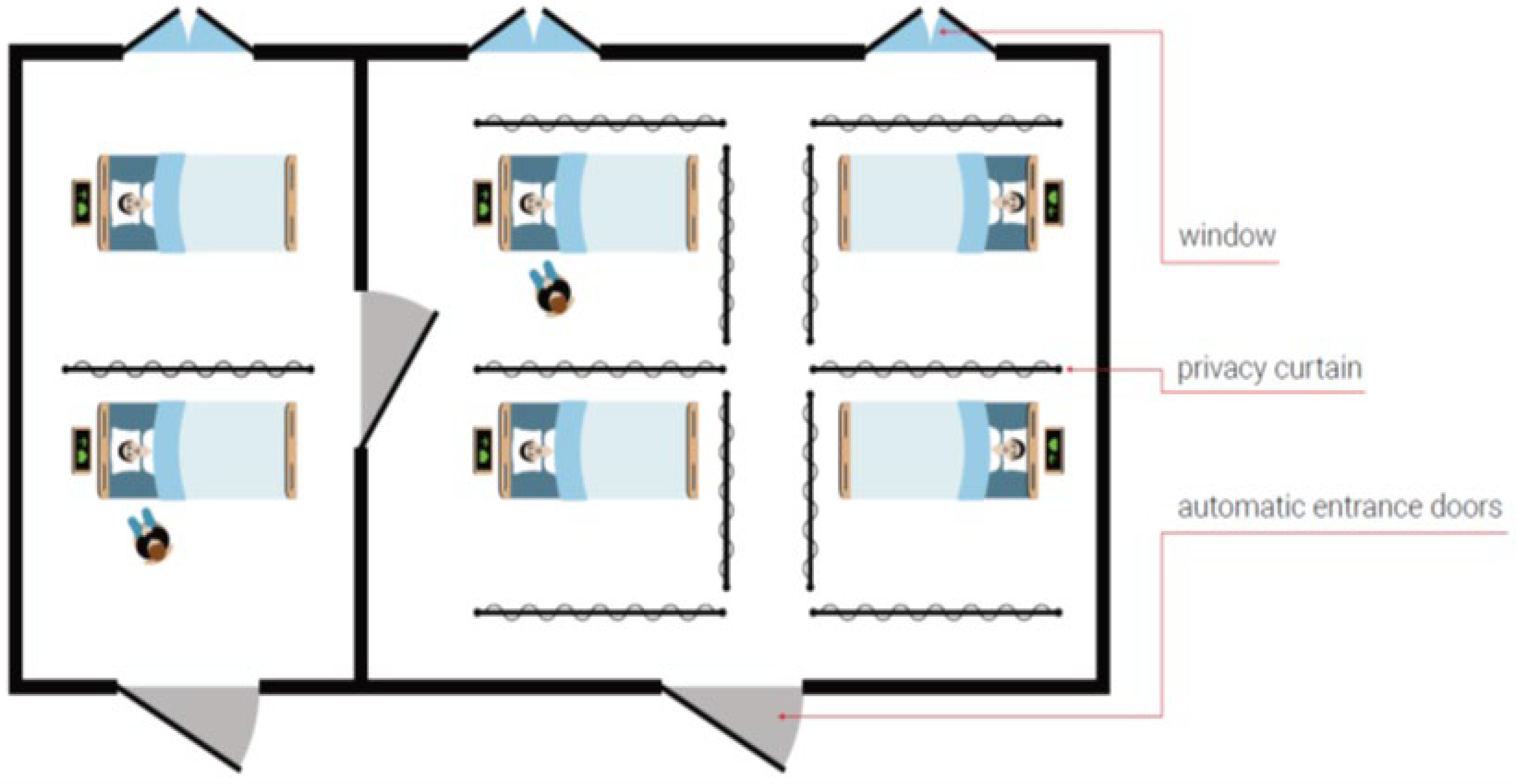
Figure 2.
Flow chart of the recruitment process and total number of BP measurements.
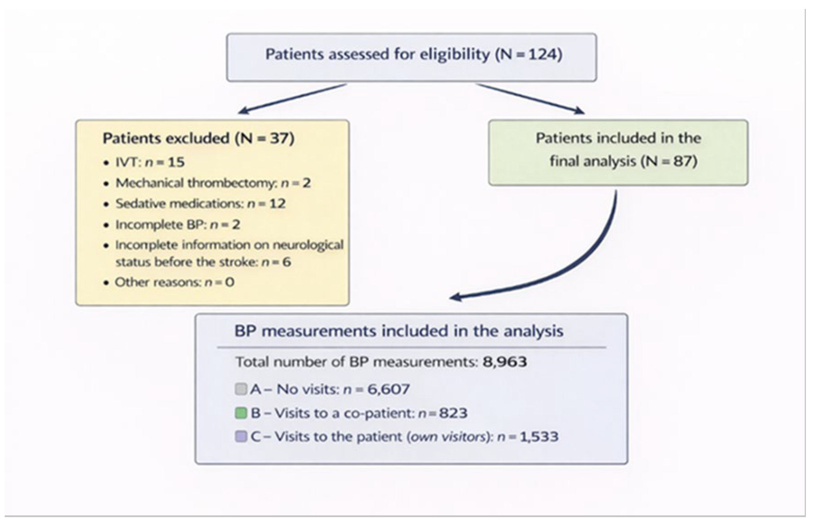
Table 1.
Association between SBP and DBP fluctuations in the stroke unit and the time of day.
| M (IQR) | Systolic Pressure | Diastolic Pressure | ||||||||
| A | B | C | p-value | post-hoc | A | B | C | p-value | post-hoc | |
| 08:00 | 146 (130-164) | 152 (143.5-162.5) | 136.5 (123-145) | 0.053 | 86 (77-98) | 94 (77.5-107) | 96 (80-101) | 0.264 | ||
| 09:00 | 140.5 (127.5-160.5) | 158 (146-175) | 137 (119-150) | <0.001* | a vs b, b vs c | 83 (73-96) | 91 (79-98) | 88 (81-99) | 0.054 | |
| 10:00 | 135 (120-150) | 134.5 (120-148) | 135 (118-154) | 0.979 | 79 (69-90) | 79 (71-90) | 88 (80-96) | <0.001 | a vs c, b vs c | |
| 11:00 | 135.5 (120-151) | 137 (121.5-154) | 136 (118-151) | 0.712 | 80 (71-91) | 84 (72-96) | 82 (75-92) | 0.112 | ||
| 12:00 | 137 (122-153.5) | 146 (121-166) | 138 (118-155) | 0.149 | 81 (72-91) | 86 (73-96) | 83 (73-96) | 0.057 | ||
| 13:00 | 137 (119-155) | 145.5 (129-164) | 136.5 (122-155) | 0.036 | a vs b | 81 (71-90) | 85 (75-92) | 83 (72-92) | 0.170 | |
| 14:00 | 135.5 (118-152) | 140 (127-154) | 139 (120-153) | 0.205 | 79 (70-89) | 85 (72-94) | 80.5 (71-92) | 0.218 | ||
| 15:00 | 137 (120-155) | 145 (128.5-156.5) | 140 (121-154) | 0.079 | 81 (72-91) | 85 (76-95.5) | 82 (73-91) | 0.142 | ||
| 16:00 | 137 (123-158) | 141 (126.5-156.5) | 143.5 (125.5-156) | 0.470 | 80 (72-92) | 82 (75-92.5) | 84 (76-93) | 0.090 | ||
| 17:00 | 144 (126-160) | 146 (132.5-164) | 147 (130-160) | 0.328 | 84 (75-98) | 84 (75-96) | 85 (75-94) | 0.952 | ||
| 18:00 | 141 (125-161) | 139 (126-152) | 138 (121-157) | 0.355 | 81 (72-92) | 83 (71-90) | 81 (73-92) | 0.991 | ||
| 19:00 | 142.5 (125-157) | 150.5 (138-165) | 132 (110-150) | 0.006 | b vs c | 80 (72-92) | 87 (77-97) | 83 (80-90) | 0.054 | |
| 20:00 | 144 (127-161) | 148.5 (140-167) | 133 (121-160) | 0.045 | b vs c | 83 (74-94) | 86 (74-96) | 86 (75-94) | 0.800 | |
Legend: Type A: no visitors; Type B: visitors present for a co-patient; Type C: visitors present for the patient.*Significant values are shown in bold.
Disclaimer/Publisher’s Note: The statements, opinions and data contained in all publications are solely those of the individual author(s) and contributor(s) and not of MDPI and/or the editor(s). MDPI and/or the editor(s) disclaim responsibility for any injury to people or property resulting from any ideas, methods, instructions or products referred to in the content. |
© 2026 by the authors. Licensee MDPI, Basel, Switzerland. This article is an open access article distributed under the terms and conditions of the Creative Commons Attribution (CC BY) license (http://creativecommons.org/licenses/by/4.0/).
Copyright: This open access article is published under a Creative Commons CC BY 4.0 license, which permit the free download, distribution, and reuse, provided that the author and preprint are cited in any reuse.


