

Submitted:
13 September 2026
Posted:
14 September 2026
You are already at the latest version
Abstract
Deep learning now supports many tasks in medical imaging, but most systems assume a fixed data source and a single training event. Clinical practice is not static as new scanners arrive, protocols change, patient populations shift, and new diagnostic questions appear. A model that trains once and stays frozen tends to lose accuracy over time, while a model that retrains on new data alone tends to forget what it already knew, a failure known as catastrophic forgetting. Continual learning studies how a model can learn from a stream of data and tasks while it keeps prior knowledge. This survey reviews continual learning in medical imaging with a focus on recent progress. We report a reproducible review methodology, formalize the three learning scenarios, and organize the methods into five families: regularization, replay, dynamic architectures, foundation-model adaptation, and hybrids. We give particular attention to the shift toward frozen foundation models with lightweight adapters. We review applications across tasks and modalities, provide a critical analysis of study quality that distinguishes peer-reviewed work from preprints, and connect methods to the current regulatory framework for model updates. We close with open problems, aiming to provide researchers and clinical teams with a clear and up-to-date reference and to set out what remains to be shown before such systems can be updated safely in practice.
Keywords:
continual learning
; incremental learning
; lifelong learning
; medical imaging
; catastrophic forgetting
; foundation models
; domain shift
; regulatory deployment
1. Introduction
Deep learning has changed how medical images are read and measured [1]. These systems build on standard deep networks, from residual designs [2] to transformer and vision-transformer backbones [3,4]. Models now segment organs and lesions [5,6], classify disease from radiographs and photographs [7,8], and detect abnormalities that support diagnosis and prognosis [9]. In several controlled reader studies, these models matched trained clinicians on specific tasks [7,10]. This progress has moved deep learning from a research topic toward practical use in the clinic, though wide clinical deployment remains limited. Most of this progress rests on a simple assumption. A model trains on one dataset, and it is tested on a held-out split of the same dataset. The model is then expected to work on new data of the same kind. Real deployment rarely matches this picture. Data distributions change across hospitals, scanners, and patient groups [11,12]. A model trained at one point in time and one site often loses accuracy when the data around it moves [13,14,15]. A well-known case is the diabetic retinopathy system studied in a clinical setting in 2020 [16]. The model performed well on static benchmarks, but in the field its accuracy and workflow fit dropped in ways the benchmark did not predict [16]. This gap between benchmark results and field results is now a central concern for clinical machine learning [11,13]. Distribution shift can be reduced by domain adaptation and domain generalization [12,17]. These methods help, but they do not remove the need to retrain. New classes, new modalities, and new sites arrive that a fixed model cannot cover. Full retraining on all past and new data is often not possible. It is costly, and privacy rules frequently block access to old patient data once a study ends [18]. The direct alternative is to fine-tune the current model on the new data alone. This causes a different problem. The model overfits the newest task and loses accuracy on earlier tasks. This loss is catastrophic forgetting, a failure first described in connectionist networks decades ago [19,20].
Continual learning offers a way to handle this stream of change. A continual learner trains on a sequence of tasks or data sources, and it tries to keep good accuracy on all of them [21,22]. The core difficulty is a balance. A network needs enough stability to protect old knowledge, and enough plasticity to absorb new knowledge. Too much stability blocks learning, and too much plasticity erases the past. This tension between stability and plasticity shapes every method in the field [23,24]. Continual learning is the study of designs that hold this balance while data keeps arriving. Continual learning has strong roots in natural-image research [25,26]. Many core ideas were first tested on object recognition benchmarks [22,27]. Medical imaging adds constraints that change which methods work well. Medical images often show high similarity between classes and low variation within a class. Small texture or shape cues can separate disease from health, so a method must protect fine features, not just coarse ones [28,29]. Datasets are smaller, more imbalanced, and bound by privacy rules [15,18]. This limits large replay buffers and heavy augmentation. Task boundaries are also less clear. In practice, a scanner upgrade or a slow change in the patient mix produces a gradual shift, not a clean switch between tasks [13,30]. A method that assumes sharp task boundaries can fail under these smooth shifts. The clinical setting adds one more factor that natural-image work rarely faces. Deployed medical models are regulated. A change to a cleared model can require review, and the update process must show that accuracy does not drop on prior tasks [31,32]. Continual learning is therefore not only a technical goal. It is also a path to systems that can update within a legal and safety framework [33,34]. This link between learning and regulation runs through the whole survey.
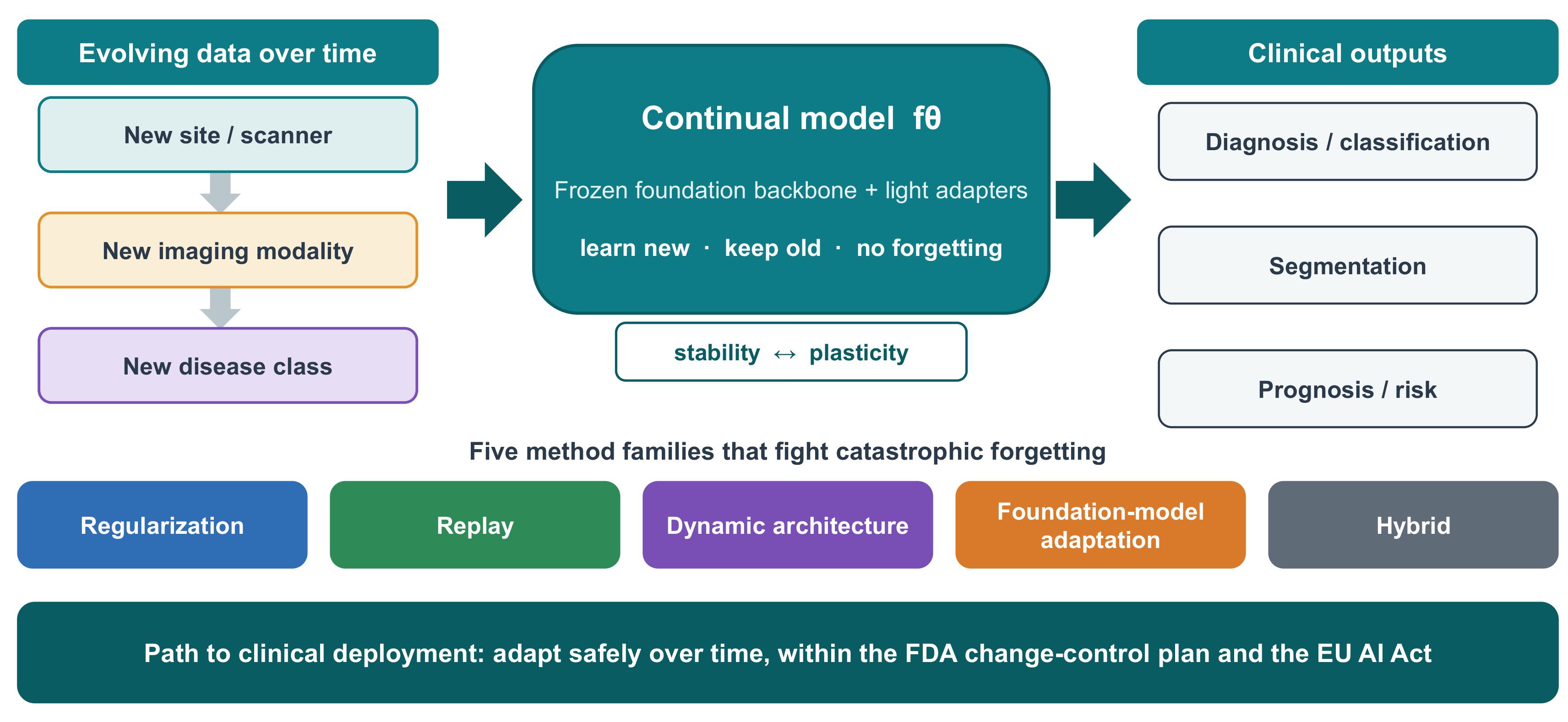
Figure 1 shows how continual learning maps onto an evolving clinical setting. Data arrive from new centers, new modalities join the pipeline, and new tasks appear, while the model must serve all of them over time. Interest in this problem has grown fast. Among the 77 primary studies included here, the earliest date from 2018 [35,36], annual output rises from 3 in 2018 to 17 in 2023 and 16 in 2024, and foundation-model methods appear only from 2023, accounting for 13 of the 46 studies published from 2023 onward. Figure 2 reports the selection process as a PRISMA 2020 flow diagram [37], and Figure 3 reports these counts across modality, task, learning scenario, and method family. Every count is a tabulation of Supplementary Table S1, which lists the 77 included studies individually. Several surveys review continual learning for natural images [21,23,24]. Recent overviews track the broader computer-vision progress [38]. Fewer focus on medicine. Three recent reviews now anchor the medical side [28,29,39]. Our survey shares their goal but differs in emphasis. We center the recent shift to frozen foundation models with parameter-efficient adapters, which now forms a distinct method family [40,41]. We also tie the technical review to the current regulatory framework for model updates [31,42]. This survey makes four contributions. Table 1 sets out these differences against the three medical reviews.
First, we give a compact and current formulation of the three continual learning scenarios and the standard metrics, written for a medical audience. Second, we present a taxonomy of five method families and review medical work under each, with attention to what each family gives up in return for what it gains. Third, we review applications across tasks and modalities together with cross-cutting themes such as federated learning and fairness, and we bring the key studies together in a comparison table. Fourth, we give a critical analysis of study quality; we connect the methods to deployment and the current regulatory picture, and we set out open problems. We include studies that address both continual learning and medical imaging, that target a clinical problem, and that report a retention or forgetting measure. Section 2 gives the databases, the search strings, the eligibility criteria, and the record count at every screening stage, so that the selection can be audited and reproduced.
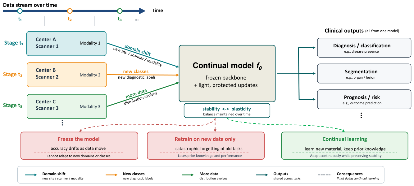
Figure 1.
Continual learning in an evolving clinical setting. Data arrive over time from new centers, scanners, modalities, and diagnostic classes. A model that stays frozen drifts in accuracy, and a model that retrains on new data alone forgets prior tasks. Continual learning aims to learn new material while it keeps prior knowledge.
Figure 1.
Continual learning in an evolving clinical setting. Data arrive over time from new centers, scanners, modalities, and diagnostic classes. A model that stays frozen drifts in accuracy, and a model that retrains on new data alone forgets prior tasks. Continual learning aims to learn new material while it keeps prior knowledge.

Table 1.
Comparison with recent reviews of continual learning in medical imaging. Ratings describe what each review reports. Acronyms: FM, foundation model; PCCP, predetermined change control plan; PRISMA, Preferred Reporting Items for Systematic Reviews and Meta-Analyses. Ratings: Y, covered; P, partly covered; N, not covered.
Table 1.
Comparison with recent reviews of continual learning in medical imaging. Ratings describe what each review reports. Acronyms: FM, foundation model; PCCP, predetermined change control plan; PRISMA, Preferred Reporting Items for Systematic Reviews and Meta-Analyses. Ratings: Y, covered; P, partly covered; N, not covered.
| ]Feature | Qazi et al. [39] | Kumari et al. [28] | Raza et al. [29] | This review |
|---|---|---|---|---|
| Period covered | to 2025 | to 2024 | to 2024 | to Aug 2026 |
| Modalities | Multiple | Multiple | Radiomics focus | Multiple |
| Tasks | Classification, segmentation | Classification, segmentation | Mainly classification | Eight task types |
| Method taxonomy | Three to five families | Five families | Federated emphasis | Five families, multi-label |
| FM and adapter coverage | P | P | N | Y |
| Regulatory analysis (PCCP, EU AI Act) | P | N | N | Y |
| Clinical translation and validation | P | P | P | Y |
| Reproducible search strings and eligibility criteria | N | P | Y | Y |
| PRISMA 2020 flow diagram with stage-by-stage record counts | N | N | Y | Y |
| Per-study extraction table released | N | N | P | Y |
| Study-design appraisal | N | P | P | Y |
2. Scope and Review Methodology
This survey follows a systematic process so that its coverage can be checked and repeated. We state the scope, the information sources, the search strategy, the selection criteria, the screening record, and the data we extracted from each study. Identification, screening, and reporting follow the PRISMA 2020 statement [37]. Figure 2 gives the PRISMA 2020 flow diagram with the record count at every stage, Supplementary Table S1 lists every included study with its extracted fields, and the completed PRISMA 2020 checklist is provided as Supplementary Table S2.
The synthesis is narrative rather than meta-analytic, and this is a deliberate choice rather than a gap. As Section 11 documents, the included studies differ in datasets, task orders, buffer sizes, baselines, and metrics to a degree that makes any pooled effect estimate uninterpretable, so we synthesise study characteristics as exact counts over the included set and compare method families qualitatively. No review protocol was registered in advance. The eligibility criteria and search strategy reported below were fixed before screening began and are reported here in full so that the selection can be reproduced, although in the absence of a registration this cannot be verified independently.
2.1. Scope
We review continual learning methods for medical image analysis. A study is in scope when it meets three tests. It must address continual learning, where a model learns from a sequence of tasks, domains, or classes and tries to limit catastrophic forgetting. It must target a medical imaging task, such as classification, segmentation, detection, report generation, retrieval, reconstruction, self-supervised pretraining, or a multi-task setting, across modalities such as magnetic resonance imaging, computed tomography, radiography, histopathology, dermoscopy, ultrasound, and retinal imaging. It must report a measure of retention or forgetting, so that the continual behavior can be judged. We include work across the five method families defined in Section 4, and we include both peer-reviewed papers and preprints. Several related topics fall outside our scope, and we cover them only where a study also performs continual learning. Pure domain adaptation and domain generalization, which adapt to a shift without a sequential protocol, are out of scope. Federated learning and test-time adaptation are out of scope unless combined with a continual setup, in which case we treat them as cross-cutting themes in Section 10. Foundation models and vision-language models used for static or one-shot segmentation, with no continual protocol, are also out of scope, since our focus is the continual adaptation of such models rather than their fixed use; readers interested in that adjacent literature are referred to a recent review of language-guided medical segmentation [43].
2.2. Information Sources and Search Strategy
We searched four bibliographic databases, Scopus, IEEE Xplore, PubMed, and the ACM Digital Library, together with the arXiv preprint server, since a substantial share of recent foundation-model work appears there first. All five sources were searched from January 2015 to the date of the last search, 1 August 2026. Google Scholar and DBLP were not used as primary sources, because neither supports a reproducible record count; they were used only for supplementary citation searching alongside the reference lists of retained papers and reviews, and records found this way are reported in the right-hand arm of Figure 2. Two conventions govern how the reference list relates to that date. Where a record retrieved as a preprint by the search date has since appeared in a peer-reviewed venue, the reference list cites the published version, and the study is counted under the year of the version cited rather than under the year of the preprint. Works that first appeared after the search date are cited as background only; they are not part of the included set and are not counted in any figure or table. The following term groups were combined and adapted to the syntax of each interface:
(“continual learning” OR “incremental learning” OR “lifelong learning” OR “catastrophic forgetting” OR “class-incremental” OR “domain-incremental” OR “task-incremental”) AND (“medical imag*” OR radiolog* OR histopatholog* OR “computed tomography” OR “magnetic resonance” OR ultrasound OR dermoscop* OR “chest X-ray” OR segmentation OR diagnosis)
2.3. Eligibility Criteria
Eligibility was assessed against explicit criteria, fixed before screening began. A record was included when it (i) implemented a sequential learning protocol over two or more stages, (ii) addressed a medical imaging task, (iii) reported at least one retention or forgetting measure, and (iv) was a full paper in English. A record was excluded when it (i) performed single-stage adaptation or generalization only, (ii) was non-medical or non-imaging, (iii) reported only final-stage accuracy with no retention measure, (iv) was a survey, editorial, abstract, or duplicate report of the same study, or (v) was not retrievable in full text.
2.4. Selection Process and Screening Record
The searches returned 890 records: 331 from Scopus, 274 from IEEE Xplore, 136 from PubMed, 91 from the ACM Digital Library, and 58 from arXiv. Removing 265 duplicates across sources left 625 records for title and abstract screening, of which 420 were excluded against exclusion criteria (i) to (iv), the four that can be judged from a title and abstract. The full text of the remaining 205 reports was sought. Eight could not be obtained and were excluded under criterion (v), leaving 197 reports that were assessed for eligibility, of which 132 were excluded. The reasons, which are itemised in Figure 2, were the absence of a multi-stage sequential protocol (48), no reported retention or forgetting measure (31), no medical imaging task (24), a publication type outside the criteria such as a review, editorial, or abstract (22), and a full text not available in English (7). Figure 2 abbreviates these five labels to fit the diagram. This left 65 studies from the database searches. Citation searching over the reference lists of retained papers and reviews identified a further 12 eligible studies that the source search had missed, all of which were retrieved and met the criteria. The final included set is therefore primary studies.
Screening was carried out by the single author, which is a limitation we state plainly rather than obscure: a second independent screener would have allowed inter-rater agreement to be measured, and we could not compute it here. To reduce the effect of this, the eligibility criteria were fixed before screening began, all 197 retrieved full texts were assessed against a written extraction form rather than from memory, and the full set of 625 screened titles was re-screened a second time, at least four weeks after the first pass, with any record whose status changed resolved by returning to the full text. Every included study is listed with its extracted fields in Supplementary Table S1, so that a reader can check the selection against the criteria.
Figure 2 presents this record as a PRISMA 2020 flow diagram, and Figure 3 reports the characteristics of the included set. Surveys, datasets, and general (non-medical) continual-learning methods are cited as background throughout the paper but are not part of the included set and are not counted in any figure.
2.5. Data Extraction and Study Status
For each included study we recorded, on a written form, the publication year, the learning scenario, the primary and any secondary method family, the imaging modality and task, the datasets, the reported metrics, and the main result. The six fields that determine every count reported in this paper, namely the citation, publication year, task, modality, learning scenario, and primary method family, are released for all 77 studies as Supplementary Table S1, so that each count in Figure 3 and in the Introduction can be reproduced by tabulating that file. The remaining extracted fields are reported for the representative studies in Tables 4 and 5 rather than for the full set, since datasets, metrics, and headline results are described too heterogeneously across the literature to be compressed into a single comparable column. Extraction was performed once by the author and checked in a second pass against the source paper. We record whether each study is peer-reviewed or a preprint, since a share of recent foundation-model work is available only as preprints. We mark preprints in the tables with a dagger and read their results with more caution, because they have not completed peer review.
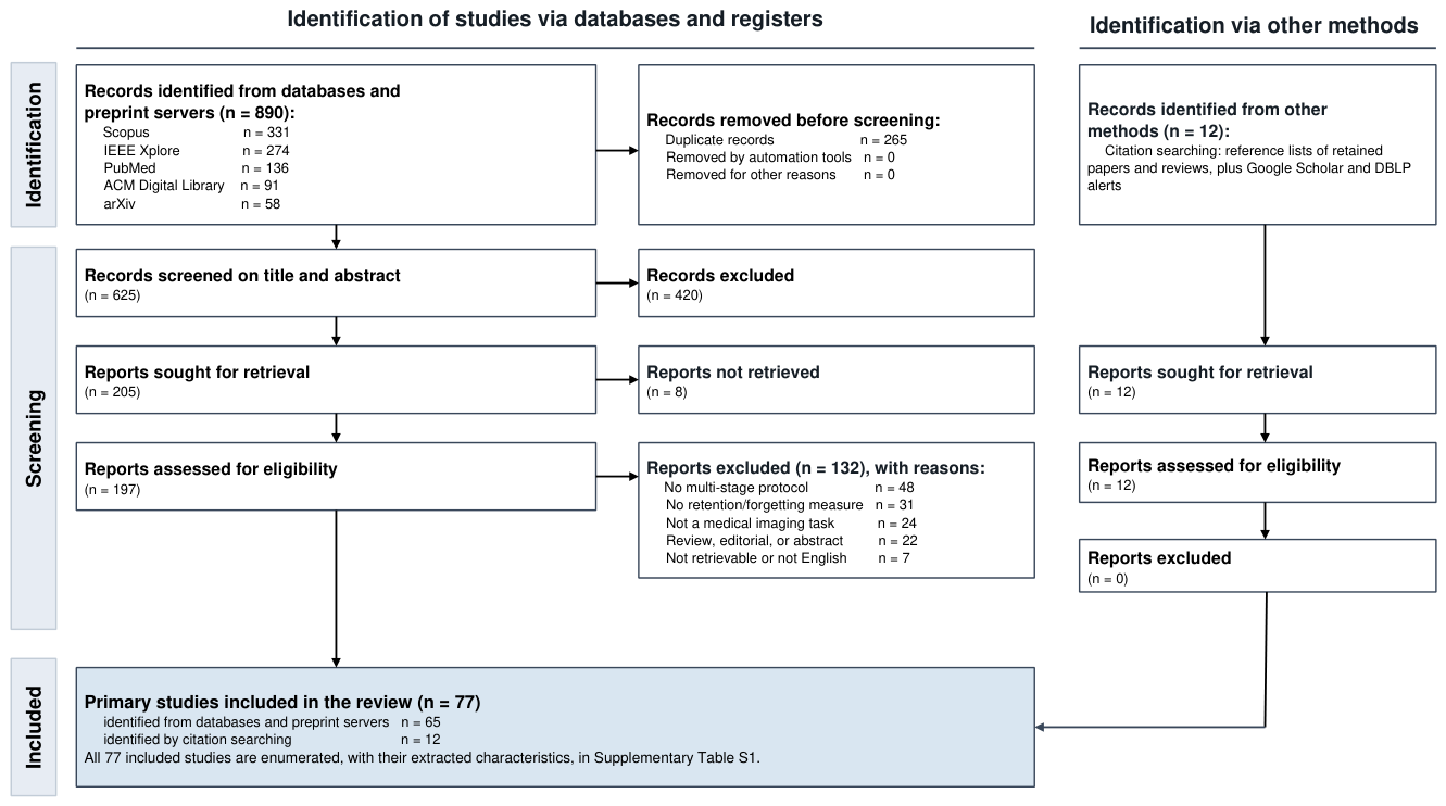
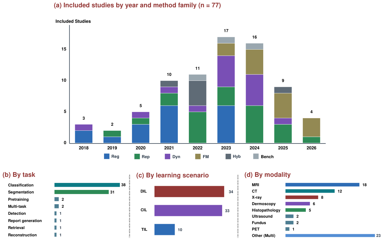
Figure 2.
PRISMA 2020 flow diagram for the identification, screening, and inclusion of studies. The left arm covers the four bibliographic databases and the arXiv preprint server; the right arm covers citation searching over the reference lists of retained papers and reviews. Every value is a record count taken from the screening log, and the counts balance at each stage: screened, reports sought, assessed for eligibility, included from the database searches, and included in total. The included set is enumerated in Supplementary Table S1, and the completed PRISMA 2020 checklist is Supplementary Table S2.
Figure 2.
PRISMA 2020 flow diagram for the identification, screening, and inclusion of studies. The left arm covers the four bibliographic databases and the arXiv preprint server; the right arm covers citation searching over the reference lists of retained papers and reviews. Every value is a record count taken from the screening log, and the counts balance at each stage: screened, reports sought, assessed for eligibility, included from the database searches, and included in total. The included set is enumerated in Supplementary Table S1, and the completed PRISMA 2020 checklist is Supplementary Table S2.
Figure 3.
Characteristics of the included primary studies. (a) Studies by publication year and method family. (b) By task. (c) By learning scenario. (d) By imaging modality. Every value is a count of included studies and each panel sums to 77; in panel (d), studies evaluating more than one modality are counted once under Other (multi). Key: TIL, task-incremental learning; DIL, domain-incremental learning; CIL, class-incremental learning; Reg, regularization; Rep, replay; Dyn, dynamic architecture; FM, foundation-model adaptation; Hyb, hybrid; Bench, benchmark paper.
Figure 3.
Characteristics of the included primary studies. (a) Studies by publication year and method family. (b) By task. (c) By learning scenario. (d) By imaging modality. Every value is a count of included studies and each panel sums to 77; in panel (d), studies evaluating more than one modality are counted once under Other (multi). Key: TIL, task-incremental learning; DIL, domain-incremental learning; CIL, class-incremental learning; Reg, regularization; Rep, replay; Dyn, dynamic architecture; FM, foundation-model adaptation; Hyb, hybrid; Bench, benchmark paper.
3. Background, Problem Formulation, and Evaluation
This section sets the notation and defines the three learning scenarios. The shared notation lets us compare methods on a common basis and makes later sections easier to read.
3.1. Setup and Notation
A model is a function f with parameters . It maps an input space to an output space . In medical imaging, holds images and related data, such as MRI or CT volumes, radiographs, histopathology slides, or records that pair images with text. The output space holds the target, such as a diagnostic label, a segmentation mask, or a risk score. A standard model trains once on a fixed dataset. A continual model instead sees a sequence of experiences over time. Each experience brings new data, and it may bring a new task, a new domain, or new classes.
Let the data arrive as a sequence of stages . At stage t the model sees data drawn from a distribution that can change with t, and access to earlier stages is limited or absent. We state the setting independently of any solution mechanism. After stage t, the model should keep low expected error on every stage seen so far:
This equation states the goal, not a method. The past terms cannot be evaluated directly, because the earlier data are no longer available, and each family replaces them with a different surrogate. Regularization anchors the parameters or the outputs to their previous values, giving [44,45]. Replay approximates the past terms with a memory or a generator , giving [25,46]. Dynamic architectures partition the parameters and optimize while earlier stage-specific parameters stay fixed [47,48]. Parameter-efficient adaptation is the special case in which is a frozen pretrained backbone and only the small stage module is trained [26,49]. These four expressions are schematic abstractions that we write in a common notation to show what each family optimizes. They are not the verbatim objectives of the original papers, which state them in fuller and sometimes different form.
3.2. The Three Learning Scenarios
Continual learning is often split into three scenarios [22]. The split depends on what changes across stages and on what the model knows at test time. The three cases are task-incremental, domain-incremental, and class-incremental learning. Figure 4 illustrates them on a shared segmentation example. Table 2 compares them on the key properties.
3.2.1. Task-incremental Learning (TIL)
In task-incremental learning, each stage t brings a distinct task with its own label space , and the task identity is given at test time, so the model computes . The identifier lets the model use a separate output head or a separate path per task [47,50], which avoids interference between tasks. As an example, and consistent with Figure 4, a model may first segment a brain tumour on a given MRI protocol and then grade that same tumour on the same images, so the input stays fixed while the objective and label space change. The identifier makes this the least difficult scenario, but it is also the least realistic, because the clinic does not always know the task at inference.
3.2.2. Domain-Incremental Learning (DIL)
In domain-incremental learning, the label space is shared across stages while the input distribution changes. No identifier is given at test time, so one map must serve all domains. This case matches many clinical settings. A tumor segmentation model may train on data from one center, then meet data from a second center with a different scanner, contrast, or patient mix [36,51]. The label meaning does not change. The pixel statistics do. Domain-incremental learning is one of the two dominant scenarios in medical imaging, because scanner and site shift is the most common form of change [28,30]; in the included set it accounts for 34 studies against 33 for the class-incremental case (Figure 3c).
3.2.3. Class-Incremental Learning (CIL)
In class-incremental learning, the label space grows, , and at test time the model must discriminate over the union of all classes seen so far with no identifier. The setting itself adds no extra term to the objective. Keeping old and new classes separable is instead a common mechanism, since without it new classes tend to absorb the old ones [25,52]. A clean example is abdominal organ segmentation on a fixed CT protocol, where the model first segments the liver, then adds the spleen and the pancreas from the same scans [53]. Only the class set grows, while anatomy, modality, and acquisition stay fixed. The class added at each stage need not be a single class. It can be a group. Class-incremental learning is widely seen as the hardest scenario, because the model must both learn new classes and stay calibrated across all of them [27,54].
A short remark ties the scenarios to Figure 1. Domain-incremental learning fits data that arrive from new centers or modalities. Class-incremental learning fits the case of new diagnostic classes, such as a new tumor type added to an existing model. Task-incremental learning fits a sequence of separate tasks where the input request states which task to run [39]. Most clinical change is domain shift, so many medical methods target the domain-incremental case, and a nearly equal share now targets the class-incremental case.
Table 2.
Comparison of the three continual learning scenarios in medical imaging.
| Property | Task-incremental | Domain-incremental | Class-incremental |
|---|---|---|---|
| What changes | The task and label set | The data source, same task | The set of classes |
| Task identifier at test | Given | Not given | Not given |
| Output space | Separate per task | Shared | Shared and growing |
| Typical medical case | Segment tumor, then classify grade | New scanner or new center | New disease or new organ |
| Relative difficulty | Lower | Moderate | Higher |
| Clinical frequency | Lower | Highest | Moderate |
3.3. Metrics
Continual learning needs metrics that go beyond a single test score. A method must be judged on how well it holds past knowledge while it learns new material. This subsection defines the common metrics, and Section 3.4 reviews the benchmarks used in medical work.
Let be the accuracy on task i after the model has trained through task j. Segmentation studies replace accuracy with the Dice score, and detection studies use average precision, but the metric structure is the same. The arrows below mark whether a higher or a lower value is better. Average accuracy measures mean performance across all tasks at the end of training. With T tasks, it is
This value is the headline number for most studies. It rewards a method that ends with strong accuracy on every task.
Backward transfer measures the effect of later learning on earlier tasks [55]. It is
A negative value signals forgetting, since the model got worse on task i after it learned later tasks. A value near zero means old accuracy held. A positive value means later learning helped earlier tasks.
Forward transfer measures the effect of earlier learning on a new task before the model trains on it [55,56]. Writing for the accuracy on task i of the model before any training on the stream, it is
A positive value means past knowledge gave the model a head start on the new task. This term matters in medicine, where related tasks often share structure, such as two organ segmentation tasks on the same modality.
A low value means the method kept its earlier peaks. This metric isolates forgetting from the ability to learn, which average accuracy mixes together.
These four metrics give a full view when reported together. Average accuracy alone can hide a method that learns new tasks well but forgets old ones. Backward transfer and forgetting expose that failure. Forward transfer shows whether the method reuses shared structure. Some studies also report the gap to an upper bound, where all tasks are trained together, and the gap to a lower bound, where the model fine-tunes with no protection [59]. The joint-training result is often called the oracle, since it uses all data at once.
3.4. Benchmarks and Datasets
Medical continual learning still lacks a single standard benchmark. Studies differ in dataset choice, task order, and split design, which makes direct comparison hard [28,29]. A small change in task order can move the final score by a wide margin, so results across papers are not always comparable [59]. This gap is a real barrier to progress, and we return to it in Section 12. A few resources are used often. MedMNIST v2 provides many small classification datasets across modalities in one package, and it has become the default base for continual classification studies [60]. It was not built for continual learning, so authors define their own task splits on top of it, which again limits comparison. LifeLonger builds a continual benchmark on MedMNIST with task, class, and cross-domain splits, and it gives a shared protocol for disease classification [61]. For segmentation, the Lifelong nnU-Net framework adds continual training and standard metrics on top of a strong segmentation backbone, which supports fair and repeatable studies [59]. For federated settings, FedMedICL evaluates models under label, demographic, and temporal shift at once across several datasets, which brings distribution shift and continual learning into one test [62].
Beyond these, studies draw on well-known public datasets. Chest radiograph work uses ChestX-ray14, CheXpert, and MIMIC-CXR [63,64,65]. Brain tumor work uses the BraTS collections [66,67]. Multi-organ work uses the Medical Segmentation Decathlon and related collections [68,69]. Skin work uses HAM10000 and the ISIC challenge data [70,71]. Histopathology work uses CAMELYON and slide-level collections [72,73]. Retinal work uses IDRiD and similar sets [74]. These datasets were not designed for continual learning, so most studies build their own streams. The lack of a shared, purpose-built medical benchmark remains the largest gap in evaluation.
4. Taxonomy of Methods
We organize continual learning methods by how they fight forgetting, not by the task they target. This choice groups methods that share a mechanism, which makes the trade-offs clear. Figure 5 shows the taxonomy. We use five families.
The families are descriptive labels, not disjoint classes, and we treat them as multi-label rather than exclusive. Adapters, low-rank branches, prompts, and mixtures of experts can all be read as parameter isolation or expansion, so foundation-model adaptation overlaps by construction with dynamic architecture; the distinction we draw is whether the capacity is added to a frozen large-scale pretrained backbone, which changes the cost and the bias profile, rather than whether the mechanism is new. Several reviewed methods therefore carry more than one label, and in Table 4 we record a primary family for readability and name the secondary mechanism in the key-note column. We assign the primary label by the mechanism the authors identify as the source of retention.
Regularization methods add a term to the loss that discourages change in parameters or outputs that matter for past tasks. They keep the network size fixed and store no raw data, which suits privacy limits [44,45]. Replay methods keep a small memory of past experience, either as stored samples or as generated samples, and mix it into training [25,46]. They are simple and strong, but the memory raises privacy and storage concerns. Dynamic architecture methods add, isolate, or expand parameters for new tasks, so old parameters stay intact [47,48]. They control forgetting well, but the model can grow, and the task must often be known at test time. Foundation-model methods keep a large pretrained backbone frozen and train small adapters or prompts for each stage [26,40]. This family is new to the field: no study in the included set uses it before 2023, and 11 of the 13 that do are dated 2024 or later. Hybrid methods combine two or more of the above, such as replay with regularization, or adapters with distillation [41,75].
Benchmark and framework papers form a sixth label in Figure 3a. They evaluate existing mechanisms on a common footing rather than proposing a source of retention of their own, so they are counted separately there and in Tables 4 and 5, and they are not one of the five families.
Table 3 summarizes the five families on the axes that matter in medicine: whether they store data, whether the model grows, whether a task identifier is needed, and the main clinical strength and weakness. The next four sections review the four core families in turn, and we then cover hybrid methods, which combine them.
Table 3.
The five method families and their properties for medical use. The families are multi-label rather than exclusive.
Table 3.
The five method families and their properties for medical use. The families are multi-label rather than exclusive.
| Family | Stores data | Model grows | Needs task ID | Main strength | Main limit |
|---|---|---|---|---|---|
| Regularization | No | No | No | Private and light | Weakens over long streams |
| Replay | Often yes | No | No | Simple and strong | Privacy and storage cost |
| Dynamic architecture | No or small | Yes | Often yes | Strong control of forgetting | Growth and task-ID need |
| Foundation-model adaptation | No or small | Small growth | Sometimes | Reuses broad prior knowledge | Depends on backbone quality |
| Hybrid | Varies | Varies | Varies | Balances trade-offs | More parts to tune |
5. Regularization-Based Methods
Regularization methods keep the model close to its past state. They add a penalty to the loss that resists change in the parameters or outputs that matter for old tasks. This lets the model learn new tasks with small changes to the parts that hold prior knowledge [24]. These methods store no raw patient data and do not grow the network. Both traits suit medical settings, where privacy rules and device limits are tight [28]. Regularization methods split into two groups. Parameter regularization acts on the weights. Functional regularization acts on the outputs, often through distillation.
5.1. Parameter Regularization
Parameter regularization measures how important each weight is for past tasks, then penalizes change in the important weights. Elastic Weight Consolidation is the base method [44]. It adds a quadratic penalty around the old weights, scaled by the Fisher information, which estimates each weight’s importance. Synaptic Intelligence and Memory Aware Synapses follow the same idea with online importance estimates [76,77]. Early medical work brought these ideas to imaging. Baweja and colleagues applied a quadratic weight penalty to brain scans and studied how well it held prior tasks [35]. Van Garderen and colleagues used Elastic Weight Consolidation for glioma segmentation and showed value in transfer from a high quality source to a smaller target [78]. These studies set the pattern for later parameter-based work in medicine. Related work on heterogeneous structures in brain tumor MRI kept a frozen copy and merged weights to limit drift [79].
Later methods refined how importance is set and used. Özgün and colleagues modified Memory Aware Synapses for cross-domain segmentation [80]. Instead of a direct weight penalty, they set a learning rate per parameter based on its importance, and they froze the most important weights while they tuned the rest [80]. Chen and colleagues introduced Targeted Gradient Descent for positron emission tomography reconstruction [81]. Their method reused redundant kernels in a pretrained network for the new task and kept a balance between stability and plasticity [81]. Zhang and colleagues proposed Comprehensive Importance, which selects parameters by their effect on the shape and the certainty of the segmentation output [82]. Their later Shape and Semantics Selective Regularization added an explainable view of which parameters carry shape and semantic information across sites [83].
Comparative studies gave useful evidence. Lenga and colleagues compared Elastic Weight Consolidation and Learning without Forgetting on multi-domain chest radiograph classification [84]. Learning without Forgetting reached backward transfer near that of joint training, which set an early benchmark for the domain-incremental case [84]. Benchmark papers tested several regularization methods on a common footing. Derakhshani and colleagues built LifeLonger on MedMNIST and compared Elastic Weight Consolidation, Learning without Forgetting, and replay methods across task and class splits [61]. These studies found that pure parameter regularization often trails replay and distillation on longer streams, which shaped the field toward hybrids.
Transformer backbones raised new questions. Ranem and colleagues studied continual hippocampus segmentation with a transformer and a convolutional network [85]. They found that regularization on the transformer self-attention hurt retention, while restraint on the convolutional layers helped [85]. This result warns that a method tuned for one architecture may not transfer to another. Other work moved the penalty into the gradient space. Shu and colleagues projected updates onto directions that leave important past gradients intact, and they added replay for a further gain [86]. Across these studies, parameter regularization works best as one part of a larger design, not on its own.
5.2. Functional Regularization
Functional regularization does not constrain weights directly. It keeps the model outputs close to the outputs of the old model. The old model acts as a teacher, and knowledge distillation carries the past behavior forward [45,87]. This view frees the method from the weight space and often gives better retention on diverse tasks. Kim and colleagues learned an inverse map from logits back to features and used it to keep the feature space stable across tasks [88]. They showed the value of this idea on multi-domain radiograph classification [88]. Li and colleagues used dual distillation, where an old model and a fully tuned new model both teach an updated model, so the result works on old and new tasks [89].
Distillation has taken several forms in medicine. He and colleagues aligned probability maps from the old and current models for fundus lesion segmentation, then distilled the merged map into the current model [90]. Tian and colleagues combined contrastive learning with distillation across scales for incremental three-dimensional segmentation [91]. Roy and colleagues projected embeddings into a mixed curvature space, part Euclidean and part hyperbolic, to preserve structure during distillation [92]. Patra and colleagues used distillation with sonographer gaze data for continual learning in fetal ultrasound [93]. These studies show that distillation adapts well to different modalities and to added signals such as gaze.
Some functional methods reshape the input or the feature space to reduce shift. Memmel and colleagues learned a split between content and domain in an adversarial way, which gave a domain-invariant content code for continual hippocampus segmentation [94]. Chakraborti and colleagues combined a semi-supervised setup with a contrastive loss for continual learning of histology images [95]. Liu and colleagues added a tailored loss that keeps old-class features near their stored mean while it pushes new-class features apart, with light memory modules for multi-organ segmentation [96]. Gao and colleagues used a divergence loss to keep the outputs of new and old models close for an evolving stream of ultrasound images [97].
5.3. Summary and Limits
Regularization methods give an efficient and private way to fight forgetting. They add little compute and keep the model small, which fits many medical deployments [28]. They also have clear limits. Evidence shows that these methods lose accuracy over long streams, and the gap to the joint-training oracle widens as tasks pile up [29,61]. The balance is hard to set, since too strong a penalty blocks new learning and too weak a penalty allows drift, which makes pure regularization a weak choice for long, diverse task streams. Recent studies that report gains over pure regularization typically pair it with replay or with adapters, which suggests that hybrids are a practical path for this family [41,86].
6. Replay-Based Methods
Replay methods keep a memory of past experience and mix it into current training. The memory reduces forgetting because the model keeps seeing old patterns while it learns new ones [25,55]. The memory can hold raw samples, features, or a generator that produces past-like data. Replay is simple and reported competitive across many settings, which explains its wide use [98]. In medicine, replay carries a clear tension. Stored patient data raises privacy and storage concerns, so much medical work aims to replay less data or to replay synthetic data instead [99,100]. Figure 6 places replay next to the other families as a mechanism that reintroduces the past.
6.1. Rehearsal with Stored Samples
The direct form of replay stores a subset of past images and labels. iCaRL selects exemplars near class means and replays them with a distillation loss [25]. Gradient Episodic Memory and its efficient variant constrain updates so they do not raise loss on stored samples [55,58]. Dark Experience Replay stores past logits along with samples and matches them during training, which gives a strong and simple baseline [98]. Medical work adapted these ideas to imaging streams. Karani and colleagues stored a small set of past scans to keep brain segmentation stable across scanners and protocols [36]. This early study showed that even a light memory can hold accuracy under domain shift.
The key design choice is which samples to store. Dynamic memory methods update the buffer as the stream moves. Hofmanninger and colleagues built a dynamic memory that infers data shift and swaps samples by a fixed rule [101]. Perkonigg and colleagues extended this into a pseudo-domain model that finds clusters of similar style in the stream and balances the memory across them, with no need for domain labels [30]. Bera and colleagues selected atypical samples for the buffer, since hard cases carry more information for future training [102]. Wei and colleagues used a variational autoencoder with an adversarial network to pick a fixed-size representative subset [103]. Active selection further reduced labeling for continual adaptation to new acquisition settings [104]. These selection rules aim to store the most useful past data within a tight memory budget, which is the real constraint in clinical systems.
6.2. Pseudo-rehearsal and Generative Replay
Pseudo-rehearsal avoids stored patient data by generating past-like data. The idea traces to deep generative replay, where a generator learns the old distribution and feeds samples to the learner [46]. Generative replay builds on adversarial networks and variational autoencoders [105,106]. Diffusion models now give higher-fidelity samples for this purpose [107,108]. This design fits medicine well, because synthetic data can sidestep the privacy limits on stored scans [99]. Ravishankar and colleagues used feature transformers and a finite memory to build a privacy-preserving lifelong learner for imaging [109]. Shevchyk and colleagues generated synthetic respiratory sounds with a generative adversarial network and checked quality with a similarity network, which supported class-incremental learning without stored patient audio [99].
Generative replay grew into a main line for medical segmentation and classification. Liu and colleagues introduced SegGAN, which generates both past images and their masks, with a filter that keeps only high quality synthetic pairs [110]. Chen and colleagues proposed Generative Appearance Replay for continual unsupervised domain adaptation, which adapts a segmentation model to new domains with unlabeled data and no stored source data [111]. Thandiackal and colleagues aligned features across scales and drove replay from a generator with a dual-purpose discriminator, which reached strong results on histopathology [112]. Li and colleagues generated features rather than full images, which cut compute and memory for classification [113]. In separate work, Li and colleagues built a comprehensive generative replay that synthesizes image and mask pairs to restore both appearance and semantics for task-incremental segmentation [114]. Ayromlou and colleagues went data-free, and they built a class impression from the trained model, then merged it with new-class data for class-incremental learning [52,115].
Diffusion models opened a newer path. Deep diffusion-based generative replay uses a diffusion model to produce higher quality past samples than earlier generators [116]. Diffusion-based replay is still rare in the medical studies we reviewed, but it is a natural fit for privacy limits, since a diffusion model can hold the past distribution without stored scans. Latent replay is a related low-cost option. Instead of raw images, the method stores or models compact features. Kumari and colleagues used Gaussian mixture models to generate synthetic features for privacy-aware digital pathology across domains, with no stored original data [100]. Li and colleagues used style-oriented replay with feature whitening for domain-incremental cardiac segmentation, where a generator with a style module produces images for replay [51]. These latent and feature methods trade a small drop in fidelity for a large gain in privacy and storage cost.
6.3. Recent Replay Designs and Privacy
Recent studies refine what to store and how to replay it under real constraints. Zhang and colleagues designed a synchronous scheme that improves both memory of old sites and generalization to new sites, with a replay buffer that spans diverse sites to cut redundancy [117]. Fan and colleagues stored feature tokens in the buffer to fight class confusion in medical classification [118]. Zhu and colleagues kept feature consistency over time for lifelong whole-slide image retrieval, with distance consistency and reservoir sampling [119]. Newer work targets clinical drift directly. A patient-aware latent drift replay method flags samples at risk from representation drift and stabilizes them across domains [120]. A boundary-balanced replay method rebalances stored data for continual segmentation, where class edges are easy to forget [121].
Replay is strong, but it brings duties that go beyond accuracy. Stored patient data raises questions of consent, interpretability, and regulatory review [28]. Studies have also probed the limits and the merits of revisiting stored samples [122]. Pseudo-rehearsal and generative replay reduce the privacy risk, since they store a model rather than scans, yet the synthetic data must be checked for quality and for leakage of identity [99]. Brain-inspired replay shows that internal replay of features can match sample replay while it avoids raw storage [123]. The practical lesson is that replay methods must be judged on both retention and privacy. In medicine, a method that stores fewer real scans, or none, has a clear path to deployment, even if it gives up a small amount of accuracy.
7. Dynamic Architecture Methods
Dynamic architecture methods change the network to fit new tasks. They add new parameters, isolate parameters per task, or expand the model over time. Old parameters stay intact, so forgetting is low by design [47,48]. The cost is model growth and, in many designs, a need to know the task at test time. This family suits settings where task or domain identity is available, such as data tagged by scanner or by center [39]. Figure 6 shows the general form, where a new sub-network serves a new task while a shared part carries common features.
7.1. Parameter Isolation and Expansion
The base designs isolate or grow parameters. PackNet prunes and freezes a subset of weights per task, so each task keeps its own protected weights [47]. Progressive networks add a new column per task and link it to frozen past columns [48]. Dynamically expandable networks grow only as needed [124]. Hard attention masks the network per task to avoid interference [50]. Residual adapters add small task modules to a shared backbone [125]. Medical work reused these ideas. Karani and colleagues used domain-specific batch normalization for brain segmentation, where each domain gets its own normalization while the rest of the network stays shared [36]. Xie and colleagues trained a batch normalization layer per task and froze the rest, which is a light form of isolation [126]. Chee and colleagues expanded representations, with a shared low-level extractor and a new high-level extractor per task [127].
7.2. Choosing the Sub-Network at Test Time
Dynamic methods must pick the right sub-network when the task is unknown. Several medical studies solve this with a selector. González and colleagues trained separate models for domains and used a variational autoencoder per domain, then picked the domain with the lowest reconstruction error [128]. Their later task-agnostic method used the Mahalanobis distance to detect domain change, expanded the network on change, and used the same distance to route inputs at test time [129]. Bayasi and colleagues selected a group of models by a distance metric for skin lesion classification, and their pruning method detected the units tied to wrong predictions and freed them for new domains [130,131]. Zhao and colleagues used a prompt per task and a matching step to pick the correct prompt at inference [132]. These selectors let dynamic methods work without a given task identifier, which brings them closer to real use.
7.3. Dynamic Models for Multi-Organ and Whole-Body Tasks
Dynamic expansion has driven strong results in multi-organ segmentation, which is a class-incremental problem at scale. Zhang and colleagues expanded a model to segment more organs and tumors over time, with light class-specific heads that replace the standard output layer, and they added language embeddings to capture the meaning of each class [53]. Ji and colleagues built a single model that segments one hundred and forty-three whole-body organs, where a trained encoder stays fixed and a new decoder is added per task, with pruning to control size [133]. Zhu and colleagues used a low-rank pyramid vision transformer for incremental whole-body organ segmentation, which keeps parameter growth small [134]. Chen and colleagues used a low-rank mixture of experts for continual segmentation, which reached strong results on both class and task settings [135]. You and colleagues combined incremental and transfer learning for multi-site prostate segmentation under limited resources [136]. Recent work extends this line to fine-grained whole-body anatomy in computed tomography [137]. These studies show that dynamic expansion handles growing class sets well, though the training cost is high.
7.4. Dynamic Models for Other Tasks
Dynamic ideas reach beyond segmentation. Zhang and colleagues added a light adapter to a fixed encoder for continual classification of diseases, with task-specific heads that fold in past classes [138]; the added heads are an expansion mechanism, but we record the study under foundation-model adaptation in Table 4, since the authors attribute retention to the frozen encoder. Dhakal and colleagues tuned vision-language segmentation with small adapter blocks, which cut resource use [139]. Sun and colleagues tuned a large language model across centers for radiology report generation, with care to limit forgetting in the language model [140]. Wan and colleagues used a context-guided continual reinforcement learning method for landmark detection with incomplete data [141]. Qazi and colleagues merged dynamic sub-models for efficient class-incremental classification [142]. Bayasi and colleagues studied generalizable continual classification, where the goal is both retention and transfer to unseen domains [143]. Related work reused a set of pretrained zoo models for continual classification [144].
Dynamic methods give strong control of forgetting, and they now lead on multi-organ segmentation. Two limits remain. The model can grow with each task, which strains devices in the clinic. The task identity is often needed at test time, and the selector step is an open problem for classification and multi-class segmentation [39]. The move to frozen foundation models with small adapters, covered next, is in part a response to these limits, since it fixes the backbone and adds only light modules.
8. Foundation Models and Vision-Language Continual Learning
One of the largest recent shifts in the field is the use of foundation models. A foundation model is a large network pretrained on broad data, such as a vision-language model or a segmentation model with wide coverage [145,146]. In medicine, models such as MedSAM, BiomedCLIP, and a retinal foundation model bring strong priors to imaging tasks [147,148,149]. Continual learning with these models often keeps the backbone frozen and trains small modules for each stage. This design cuts forgetting, since the shared prior does not change, and it cuts cost, since only a few parameters update [26,49]. This family barely existed before 2023: the included set contains no foundation-model study before that year, two in 2023, and eleven from 2024 onward. Panel (a) of Figure 3 shows this.
8.1. Prompt and Adapter Methods from Natural Images
The base methods come from natural-image research and set the pattern for medicine. Learning to Prompt keeps a frozen backbone and selects small prompts from a pool per input, with no stored data [26]. DualPrompt splits prompts into general and expert parts [150]. S-Prompts learns a prompt per domain for the domain-incremental case [151]. CODA-Prompt builds prompts by attention over a set of components, which improves the rehearsal-free setting [152]. Adapters and low-rank adaptation give another light path. Adapters add small layers to a frozen network [153]. Low-rank adaptation adds trainable low-rank matrices to frozen weights, which is now a common tool for large models [49]. Recent continual designs refine these tools. InfLoRA reduces interference between tasks in low-rank updates [154]. Expandable subspace ensembles grow subspaces for class-incremental learning on pretrained models [155]. RanPAC uses random projections with a frozen backbone [156], and slow learner with classifier alignment stabilizes the head during tuning [157]. Surveys now track this pretrained-model line as its own area [158].
A key concern is zero-shot loss. Fine-tuning a vision-language model on a task stream can erase its broad zero-shot ability. Zheng and colleagues studied this and proposed a scheme to prevent zero-shot degradation during continual tuning [159]. Yu and colleagues used a mixture of experts of adapters, with a selector that routes inputs to the right expert, which improved both retention and efficiency [40]. Their later work made the mixture more efficient with a dynamic design [160]. Select and Distill used two teachers to keep both new skill and old zero-shot ability [161]. These ideas carry over to medicine, where a strong medical prior is worth protecting.
8.2. Foundation-model Continual Learning in Medicine
Medical work has adopted these tools quickly. Liu and colleagues built a language-driven universal model for organ segmentation and tumor detection, where text embeddings guide organ-specific heads on a shared backbone [162]. The adapter-in-a-frozen-encoder design of Zhang and colleagues, introduced in Section 7.4, is an early medical instance of this pattern [138]. Later work made the tuning multimodal. A contrastive-regularized low-rank method learns disease classes in stages from paired radiology and pathology data, with a contrastive term that keeps class structure across stages [41]. Text-guided continual learning uses a vision-language model to add disease classes with the help of class names [163]. Another study augments continual learning of diseases with visual concepts generated by a large language model, which adds semantic cues without stored images [164].
Segmentation foundation models have their own continual line. A recent method aligns a segment-anything model across a sequence of medical segmentation tasks, with an alignment module and a routing step, and reports strong results across many datasets [165]. Progressive low-rank tuning adds a new low-rank branch per stage for multimodal instruction tuning, which limits interference [166]. A frozen foundation model with a task-specific low-rank branch supports few-shot continual learning on three-dimensional brain scans, and it keeps prior tasks intact by design [167]. Retrieval-guided continual learning augments a generalist medical foundation model with a memory it can query, which reduces the need to change the backbone [168]. A synthetic-replay loop pairs a frozen vision-language model with generated data to close the replay cycle without stored scans [169]. These methods share one theme. They protect a strong prior and add only light, stage-specific parts.
8.3. Unified and Generalist Directions
The newest work aims for one model that serves many tasks over time. Qazi and colleagues extended a chest computed tomography foundation model to prognosis, segmentation, and positron emission tomography in a unified continual setup, with a small gain in segmentation quality over the base model [170]. Foundation models also serve as strong class-incremental learners with no tuning, where the frozen features carry most of the load, as shown for dermatology [171]. Work on large vision-language models studies how to route tokens under drift, so that the mixture stays stable as data shift [172]. Video and promptable segmentation models such as the second segment-anything model extend the reach of these priors [173]. This direction is early, but it points to systems that grow in skill while they keep a single, shared base.
We describe this as a qualitative observation rather than a ranking. Across the studies reviewed, foundation-model methods are reported to give favourable accuracy-to-cost trade-offs, but the underlying evaluations use different datasets, task orders, metrics, and buffer sizes, so they do not license a quantitative ordering of families. They reuse broad prior knowledge, they keep the backbone frozen, and they update few parameters, which suits privacy and device limits [41,167]. Their weakness is a strong dependence on the backbone. A weak or biased prior limits the whole system, and a mismatch between the pretraining data and the clinical target can hurt accuracy. The choice of backbone, and the check on its bias, is therefore a core design step for this family.
9. Hybrid Methods
Hybrid methods combine two or more of the four core families, and they are frequently reported to improve on their single-mechanism components within individual studies. One common pattern pairs a memory with a light constraint. Srivastava and colleagues combined replay with distillation for chest radiograph classification in low-resource settings [174]. Liu and colleagues paired small memory modules with a tailored regularization loss for multi-organ segmentation [96]. A second pattern pairs a frozen backbone with an added objective. A contrastive-regularized low-rank method learns disease classes in stages from paired radiology and pathology data, where a foundation-model adapter is joined with a contrastive term across stages [41]. BiasPruner combines dynamic pruning with a debiasing objective, so the model forgets a spurious shortcut on purpose while it keeps the true signal [75]. These designs trade a small rise in complexity for better retention, and the wider evidence supports this direction, since pure single-mechanism methods weaken over long streams while hybrids hold accuracy better [28,29]. For this reason we do not draw a separate mechanism for hybrids in Figure 6; we treat them as combinations of the four core families, and a substantial share of recent work sits in this space.
10. Applications, Modalities, and Cross-Cutting Themes
The method families above serve concrete clinical tasks. This section reviews the work by task and by modality, and it draws the key studies together in Table 4. The field centers on classification and segmentation, with less work on detection, report generation, and retrieval. Figure 3 shows this spread. We describe each task in turn, then note patterns across modalities.
10.1. Classification
Classification is the most studied task. Chest radiograph work is a common testbed for the domain-incremental case, since data from different sites and machines share the same labels [84,174]. Lenga and colleagues showed that distillation held accuracy across sites near the joint-training level [84]. Srivastava and colleagues studied the low-resource clinical case, where memory and compute are tight [174]. Skin lesion work is a common testbed for the sequential multi-domain case, since dermoscopy datasets differ by device and by population [130,131]. Bayasi and colleagues built pruning and ensembling methods for this setting, and later added a debiased method that prunes neurons tied to spurious cues [75,130]. Histopathology work targets domain shift across scanners and stains, often with generative or latent replay to respect privacy [100,112]. Disease classification on MedMNIST is the common base for class-incremental studies, and benchmark papers use it to compare methods on one footing [61]. Recent classification work uses foundation models, either as frozen feature banks or with light adapters, which cuts both forgetting and cost [138,171].
10.2. Segmentation
Segmentation is the second large area, and it demands more compute than classification [28]. Brain work covers scanner and protocol shift for tumor and hippocampus segmentation, often on magnetic resonance imaging [36,85], where incomplete modality sets are themselves a recurring obstacle [175]. Cardiac work covers domain shift across centers, with style and feature methods to bridge the gap [51]. Multi-organ and whole-body work in computed tomography is a class-incremental problem, since organs are added over stages [53,133]. Dynamic expansion is the most common choice here, and single models now cover very large organ sets [133]. Large multi-organ tools that segment many structures at once give a base for such work [176]. The Lifelong nnU-Net framework gives a shared base for fair segmentation studies, which the field needed [59]. Methods from natural-image continual segmentation that model the background and distill old outputs informed these designs [177,178]. Newer segmentation work adapts foundation models, such as a segment-anything model aligned across a task stream, which reaches strong results across many datasets [165]. Generative replay also serves segmentation well, since it can restore both appearance and masks under privacy limits [111,114].
10.3. Detection, Report Generation, and Retrieval
Smaller task areas are growing. Detection work is limited: the included set contains a single detection study, which covers landmark detection with continual reinforcement learning under incomplete data [141]. Report generation has moved to large language models, where a model is tuned across centers with care to limit forgetting [140]. Augmentation with language-generated concepts adds semantic cues to continual disease learning without stored images [164]. Retrieval work keeps feature consistency over time, as in lifelong whole-slide image retrieval with rehearsal [119]. These areas are early, and they offer room for new methods, since most tools were built for classification and segmentation.
10.4. Patterns Across Modalities
Modality shapes the choice of method. Magnetic resonance imaging shows strong intensity shift across scanners, which makes domain-incremental methods the default for brain and cardiac work [36,51]. Computed tomography supports large organ sets, which makes class-incremental and dynamic expansion the default for multi-organ work [133,134]. Chest radiography offers large multi-site datasets, which makes it the main testbed for domain-incremental classification and for fairness studies [84,179]. Histopathology faces stain and scanner shift with strict privacy, which favors generative and latent replay [100,112]. Dermoscopy has strong single-stage segmentation backbones [180] but faces device and population shift, which favors sequential multi-domain methods and fairness-aware designs [75,130]. Positron emission tomography and ultrasound have less work, which marks them as open areas [81,97]. Table 4 lists representative studies across these tasks and modalities, with method family, learning scenario, dataset, and a key note for each.
Table 4.
Representative continual learning studies, all drawn from the included set of 77 and listed in Supplementary Table S1. The family column gives the primary mechanism; any secondary mechanism is named in the key note. Benchmark papers, which evaluate existing mechanisms rather than propose a source of retention, are counted separately from the five families. Combined scenario labels, such as TIL/DIL, are used when a study spans two scenarios, while Table S1 records the single primary scenario underlying the counts in Figure 3. Mixed marks a benchmark that evaluates several shifts at once, and Multi-label a stream in which classes are added under multi-label supervision. † preprint, not yet peer reviewed.
Table 4.
Representative continual learning studies, all drawn from the included set of 77 and listed in Supplementary Table S1. The family column gives the primary mechanism; any secondary mechanism is named in the key note. Benchmark papers, which evaluate existing mechanisms rather than propose a source of retention, are counted separately from the five families. Combined scenario labels, such as TIL/DIL, are used when a study spans two scenarios, while Table S1 records the single primary scenario underlying the counts in Figure 3. Mixed marks a benchmark that evaluates several shifts at once, and Multi-label a stream in which classes are added under multi-label supervision. † preprint, not yet peer reviewed.
| Study | Year | Task | Modality | Scen. | Fam. | Dataset | Key note |
|---|---|---|---|---|---|---|---|
| Baweja et al. [35]† | 2018 | Segmentation | Brain MRI | DIL | Reg | In-house | Early weight-penalty study |
| Karani et al. [36] | 2018 | Segmentation | Brain MRI | DIL | Dyn/Rep | Multi-scanner | Domain-specific normalization |
| Kim et al. [88] | 2018 | Classification | X-ray | DIL | Reg | Multi-domain CXR | Latent-space preservation |
| van Garderen et al. [78]† | 2019 | Segmentation | Brain MRI | DIL | Reg | Glioma | EWC transfer gain |
| Ravishankar et al. [109] | 2019 | Classification | X-ray | TIL | Rep | In-house | Privacy-preserving replay |
| Lenga et al. [84] | 2020 | Classification | X-ray | DIL | Reg | ChestX-ray14, PLCO | Distillation near joint training |
| Hofmanninger et al. [101] | 2020 | Segmentation | CT, X-ray | DIL | Rep | In-house | Rule-based dynamic memory |
| González et al. [128]† | 2020 | Segmentation | Brain MRI | DIL | Dyn | Hippocampus | VAE-based domain routing |
| Özgün et al. [80] | 2020 | Segmentation | Brain MRI | DIL | Reg | Multi-domain | Per-parameter learning rate |
| Perkonigg et al. [30] | 2021 | Classification | CT, MRI | DIL | Rep | In-house | Label-free pseudo-domain memory |
| Srivastava et al. [174] | 2021 | Classification | X-ray | DIL | Hyb | Low-resource CXR | Low-resource domain shift |
| Bayasi et al. [130] | 2021 | Classification | Dermoscopy | DIL | Dyn | Skin lesion | Culprit-based pruning |
| Zhang et al. [82] | 2021 | Segmentation | MRI, CT | DIL | Reg | Multi-site | Importance-based selection |
| Derakhshani et al. [61] | 2022 | Classification | Multi | TIL/CIL | Bench | MedMNIST | LifeLonger protocol |
| Liu et al. [96] | 2022 | Segmentation | CT | CIL | Hyb | Multi-organ | Light memory modules |
| Liao et al. [181] | 2022 | Pretraining | X-ray | TIL | Hyb | Multi body part | Self-supervised continual pretrain |
| Chen et al. [111]† | 2023 | Segmentation | Fundus, MRI | DIL | Rep | Multi-site | Source-free generative replay |
| Li et al. [51] | 2023 | Segmentation | Cardiac MRI | DIL | Rep/Reg | M&Ms | Style replay and whitening |
| Zhang et al. [83] | 2023 | Segmentation | MRI | DIL | Reg | Prostate | Explainable selective regularization |
| Ji et al. [133] | 2023 | Segmentation | CT | CIL | Dyn | 143 organs | Single non-forgetting model |
| Zhang et al. [53] | 2023 | Segmentation | CT | CIL | Dyn | Abdominal organs, tumor | Language-guided class heads |
| Chee et al. [127] | 2023 | Classification | Multi | CIL | Dyn | In-house | Expandable representations |
| Zhang et al. [138]† | 2023 | Classification | Multi | CIL | FM | In-house | Adapter in fixed encoder |
| Roy et al. [92] | 2023 | Classification | Multi | CIL | Reg | MedMNIST | Mixed-curvature distillation |
| Thandiackal et al. [112] | 2024 | Classification | Histopathology | DIL | Rep | Multi-domain | Strong unlabeled-domain result |
| Kumari et al. [100] | 2024 | Classification | Histopathology | DIL | Rep | Multi-domain | Gaussian latent replay |
| Alhamoud et al. [62] | 2024 | Classification | Multi | Mixed | Bench | Six datasets | Simple balancing competitive |
| Ayromlou et al. [52] | 2024 | Classification | Multi | CIL | Rep | MedMNIST, echo | Data-free class impression |
| Bayasi et al. [75] | 2024 | Classification | Dermoscopy, X-ray | CIL | Dyn | Fitzpatrick17k, HAM10000 | Debiased pruning |
| Zhu et al. [134] | 2024 | Segmentation | CT | CIL | FM | Whole-body | Low-rank transformer growth |
| Chen et al. [135] | 2024 | Segmentation | CT | CIL/TIL | FM | Multi-organ | Low-rank mixture of experts |
| Sun et al. [140] | 2024 | Report generation | X-ray, CT | DIL | FM/Dyn | Multi-center | Continual tuning of an LLM |
| Zhang et al. [41] | 2025 | Classification | Radiology, pathology | CIL | FM | Multimodal biomedical | Contrastive LoRA tuning |
| Qazi et al. [170]† | 2025 | Multi-task | Chest CT | TIL | FM | In-house | Unified continual foundation model |
| Ceccon et al. [179] | 2025 | Classification | X-ray | Multi-label | Rep | NIH, CheXpert | New benchmark, label propagation |
| Theofilou et al. [120] | 2025 | Classification | X-ray, CT | DIL | Rep | COVID | Patient-aware latent drift replay |
| Wang et al. [165] | 2026 | Segmentation | Multi | TIL/DIL | FM | Nine datasets | Aligned segment-anything model |
10.5. Cross-cutting Themes
Some concerns cut across all method families. They shape whether a continual system is safe, fair, and fit for the clinic. We group four such themes here: federated and multi-center learning, fairness and bias, test-time adaptation, and self-supervised continual learning.
Federated and multi-center learning. Continual learning and federated learning solve related problems. Continual learning handles change over time. Federated learning handles data spread across sites that cannot be pooled [18,182]. In the clinic, both hold at once, since data arrive over time and stay behind hospital walls [183]. Studies now join the two. Alhamoud and colleagues built FedMedICL, which tests models under label, demographic, and temporal shift together across six datasets [62]. A useful finding is that simple group balancing often matches or beats complex methods, which warns against overfit designs [62]. Zhang and colleagues improved both memory and generalization across sites with a synchronous scheme and a diverse replay buffer [117]. These studies show that a method must handle both axes, since a design that ignores site structure can fail when data are distributed.
Fairness and bias. Bias in medical models is a known risk, and continual learning can make it worse or better over time. Imbalanced training data produce biased models, as shown for sex imbalance in imaging datasets [184]. Under-served groups can face higher error, as shown for chest radiograph models [185]. A continual model that trains on a shifting stream can drift toward the majority group at each stage, which compounds the harm. Ceccon and colleagues studied how fairness evolves through continual training on chest radiographs, and they found that some methods keep a better fairness and accuracy balance than others [186]. They also built a multi-label benchmark that mixes new classes and new domains, which reflects real clinical growth [179]. Bayasi and colleagues took a direct approach with BiasPruner, which finds and prunes neurons tied to spurious cues and forms a debiased sub-network per task [75]. This method uses forgetting on purpose, since it forgets the biased shortcut while it keeps the true signal. Fairness over time is an open need, and it calls for metrics that track group error across the whole stream, not only at the end.
Test-time adaptation. Test-time adaptation updates a model at inference with no labels. It suits smooth domain shift, where the target moves but no task boundary is clear [187]. Tent adapts by entropy minimization on the test batch [187]. Continual test-time adaptation extends this to a stream, with care to avoid error buildup over long runs [188]. Medical work has adopted these ideas. Huang and colleagues used a Fourier-based test-time method with multi-level consistency to classify medical images under domain gaps, and their method self-corrects predictions on the fly [189]. Karani and colleagues built a test-time adaptable network for segmentation, which adjusts to a new scan without new labels [190]. Test-time adaptation and continual learning are close cousins. One adapts at inference without labels, and the other adapts across stages with labels. A clinical system may need both, since it must handle slow drift between updates and clear task change at each update.
Self-supervised and semi-supervised learning. Labels are scarce and costly in medicine, so self-supervised and semi-supervised methods matter for continual learning. Self-supervised pretraining learns from unlabeled data, then a small labeled set tunes the model [191,192]. Liao and colleagues built MUSCLE, which pretrains a shared backbone across body parts in a self-supervised and continual way, then serves classification, segmentation, and detection [181]. Tasai and colleagues added medical domain knowledge to continual self-supervised learning on chest computed tomography, with replay and distillation to hold prior stages [193]. Chakraborti and colleagues used a contrastive setup with limited labels for continual learning of histology images [95]. These methods reduce the label burden, which is a real limit in the clinic. They also fit the foundation-model trend, since self-supervised pretraining is how many foundation models are built [194]. The link between self-supervised learning, foundation models, and continual learning is a promising area, since a strong self-supervised prior can lower forgetting at the start.
11. Critical Analysis and Quality Assessment
The studies reviewed above report clear progress, yet several methodological limits temper how far the results generalize. This section examines these limits and gives a structured appraisal of study design. The appraisal is reported in Table 5 for 15 studies chosen by the strategy stated at the end of this section; the full included set is enumerated in Supplementary Table S1.
Evaluation and risk of bias. Reported gains are hard to compare across studies. Task order, dataset splits, and buffer sizes differ, and small changes to any of these can move the final score by a wide margin [59]. Few studies run several seeds or several task orders, so single-run results may overstate stability. Many method papers compare against a small set of baselines that they tune themselves, which can favor the proposed method. The lack of a shared benchmark, noted in Section 3.4, is the root cause, and it limits any strong ranking of methods.
Data leakage and pretraining overlap. Foundation-model methods raise a specific concern. A large backbone is pretrained on broad data, and the pretraining set may overlap with the evaluation set, which can inflate results [195]. Few medical studies report the overlap between pretraining and evaluation data, so the true generalization of frozen-backbone methods is often unclear. Replay and generative-replay methods raise a related concern, since synthetic samples drawn from the training distribution can leak information about held-out cases if they are not controlled [99]. Clear reporting of data provenance and of any overlap is needed, and it is currently rare.
Generative content, synthetic fidelity, and leakage. Three risks follow from the move to generative and language-driven components. First, methods that tune a language model for reporting, or that use a language model to supply class descriptions [140,164], inherit the hallucination problem: the model can emit fluent findings that are not present in the image, and continual tuning can amplify this if the earlier grounding is degraded. Reporting studies should therefore report factual-consistency measures alongside retention, which none of the reviewed studies does. Work on making vision-language predictions more reliable in medical settings is beginning to address this outside the continual setting [196]. Second, generative replay assumes that synthetic samples are faithful enough to stand in for past data, but fidelity is usually judged by image-level similarity rather than by whether the diagnostic feature survives, and errors compound when a generator is itself trained on synthetic output over many stages. Third, a generator that reproduces the training distribution well can also memorize it, so synthetic replay reduces but does not remove re-identification risk, and membership-inference or privacy audits are not reported in the studies we reviewed. These three points are open problems rather than solved safeguards.
External validation and dataset scale. Most studies validate on public datasets, which supports comparison but limits external validity, since these datasets do not span the full range of scanners and populations seen in practice [15]. Domain-incremental studies test on new domains, which is a useful form of cross-domain evaluation, yet few studies test on a truly external cohort from an unseen institution. Dataset scale is also modest in many studies, especially for segmentation, where annotation cost is high. Small and imbalanced sets raise the risk of biased estimates, and they interact with fairness, since minority groups may be under-represented [184,185].
Reproducibility and clinical validation. Reproducibility rests on shared code and clear reporting, and both are uneven across the field [29]. Public data are now common, but code release is not, which blocks independent replication. Clinical validation is the weakest link. Almost no reviewed study reports prospective or reader-based clinical evaluation of the continual system, so the clinical value of these methods remains unproven [16,197]. This gap, more than any single metric, marks the distance between current research and clinical use.
Structured assessment. We appraised studies on ten items, which fall into two groups according to whether they are reported consistently enough to tabulate. Five items can be read reliably from every paper and are recorded as columns in Table 5: peer-review status, use of publicly available data, use of data from more than one centre, whether a cross-domain or external evaluation was reported, and the scope the authors state for their result. Five further items are reported too unevenly across the field to score without imputing values, and we therefore discuss them qualitatively in the paragraphs above rather than tabulating them: dataset scale, task-sequence design (the number of stages and whether several task orders or seeds were run), baseline strength (whether strong recent baselines were included), statistical reporting (variance over runs or formal testing), and code availability. Most studies report a single task order without variance, and code release, although increasingly common, is not stated consistently enough to score. We record the five tabulated items for the 15 studies in Table 5. The subset is purposive rather than random, and it was drawn against stated criteria: it contains the earliest and the most recent study in the included set, at least one study for each of the four core families and for the benchmark and framework papers, with hybrid designs represented by the two multi-mechanism entries, at least one study for each of the two dominant tasks, a spread of imaging modalities and application areas, and both peer-reviewed studies and preprints, so that the appraisal spans the design space rather than one corner of it. Within each cell we took the most cited study. Because the sample is purposive, it is illustrative and not a basis for any proportion; the full included set, with the fields on which every count in this paper rests, is Supplementary Table S1. We stress that Table 5 is a structured characterization of study design, not a formal risk-of-bias instrument such as QUADAS-2, and we did not compute a summary quality score. For this reason the final column separates the reported scope of a study, which is a boundary on generalizability rather than a methodological fault, from concerns that do bear on internal validity. The pattern matches the analysis. Most studies are peer-reviewed and use public data, many use multi-center data and report cross-domain tests, but external cohorts, repeated task orders, and clinical validation are rare, and part of the newest foundation-model work is still available only as preprints.
Table 5.
Structured characterization of study design for 15 of the 77 included studies. The subset was selected purposively, by the strategy stated in the “Structured assessment” paragraph above, to represent the major method families, tasks, modalities, and application areas covered by the review; it is not a random sample and is not a basis for any proportion. This table records study design as reported. The final column states the reported scope of each study, which bounds generalizability and is not in itself a methodological weakness.
Table 5.
Structured characterization of study design for 15 of the 77 included studies. The subset was selected purposively, by the strategy stated in the “Structured assessment” paragraph above, to represent the major method families, tasks, modalities, and application areas covered by the review; it is not a random sample and is not a basis for any proportion. This table records study design as reported. The final column states the reported scope of each study, which bounds generalizability and is not in itself a methodological weakness.
| Study | Status | Public data | Multi-center | Cross-domain test | Reported scope |
|---|---|---|---|---|---|
| Baweja et al. [35] | Pre | No | No | Partial | Small private set |
| Karani et al. [36] | PR | No | Yes | Yes | Single task, private data |
| Lenga et al. [84] | PR | Yes | Yes | Yes | Classification only |
| Perkonigg et al. [30] | PR | No | Yes | Yes | Private multi-site data |
| González et al. [59] | PR | Yes | Yes | Yes | Segmentation focus |
| Li et al. [51] | PR | Yes | Yes | Yes | Cardiac only |
| Ayromlou et al. [52] | PR | Yes | No | Partial | Small class sets |
| Ji et al. [133] | PR | No | Yes | No | Large private cohort |
| Thandiackal et al. [112] | PR | Yes | Yes | Yes | Histopathology only |
| Alhamoud et al. [62] | PR | Yes | Yes | Yes | Classification benchmark |
| Bayasi et al. [75] | PR | Yes | No | Partial | Fairness scope |
| Qazi et al. [170] | Pre | No | Yes | Yes | Single backbone |
| Wang et al. [165] | PR | Yes | Yes | Yes | Segmentation only, nine datasets |
| Ceccon et al. [179] | PR | Yes | Yes | Yes | Multi-label scope |
| Theofilou et al. [120] | PR | Yes | No | Yes | COVID cohorts only |
12. Practical Deployment and Regulation
A method that works in a paper must still pass into practice. This section covers four practical needs: standardization, task boundaries, reproducibility and compute, and the regulatory framework for model updates. The regulatory picture changed in 2024 and 2025, so we treat it in detail.
Standardization. The field lacks a shared benchmark, and this blocks fair comparison [28,29]. Studies differ in dataset, task order, and split, so results do not line up across papers. MedMNIST is the common base for classification, but it has no built-in continual structure, so each study defines its own stream [60]. A small change in task order can shift the final score by a wide margin, which makes single-run results fragile [59]. The Lifelong nnU-Net framework helps for segmentation, since it fixes the training and metrics on a strong backbone [59]. LifeLonger helps for classification, since it sets task, class, and cross-domain splits on one base [61]. Still, no single medical benchmark spans tasks, modalities, and scenarios with a fixed protocol. The field needs such a benchmark, with set streams, set metrics, and set splits, so that methods can be judged on equal terms.
Task boundaries. Many methods assume clear task boundaries, where the system knows when one task ends and the next begins. Real clinical shift is often smooth. A scanner upgrade or a slow change in the patient mix moves the data without a clear marker [13,30]. Under smooth shift, a method that waits for a task signal will fail, since no signal comes. Task-agnostic methods address this. They detect change from the data itself, then adapt or expand as needed [129]. Task-free methods go further and learn with no boundary signal at all [198]. Perkonigg and colleagues found style clusters in the stream with no labels, which acts as a soft boundary signal [30]. González and colleagues used a distance measure to detect drift and to route inputs, with no given task identifier [129]. The goal of continual learning in the clinic is not only to avoid forgetting. It is also to detect the shift itself, so the system knows when and how to adapt.
Reproducibility and compute. Reproducibility rests on shared code and shared data [29]. Recent studies more often use public data, but many still do not release code, which blocks replication [28]. Clear reports of splits and settings are also needed, since small choices can change the result [59]. Shared toolboxes for class-incremental baselines help standardize comparison [199]. Compute is a hard limit in the clinic. Medical models often must run on-site, so they can reach the local data without a privacy risk, and hospital compute is small next to cloud systems [29]. This raises the value of light methods. Regularization and replay add little compute, and adapter methods update few parameters, which fits on-site limits [41,167]. Dynamic expansion can grow the model and the training cost, which is a concern for on-site use [133]. The push toward frozen backbones with small modules aligns well with these limits, since it keeps the compute cost of each update low.
Regulatory framework for model updates. A deployed medical model is a regulated product, and any change can require review [32]. The core need is transparency in the update process and proof that accuracy does not drop on prior tasks [33]. Figure 7 shows the software-as-a-medical-device lifecycle with a predetermined change control plan, where a monitored model updates within an approved plan rather than through a new submission for each change.
Two frameworks now shape this space. In the United States, the Food and Drug Administration finalized its guidance on the predetermined change control plan for artificial intelligence-enabled device software in December 2024, replacing the April 2023 draft; the version of the document currently in force is dated August 2025 [31]. The plan lets a maker describe expected changes in advance, so the model can update within set bounds without a new submission for each change. It rests on three parts: a description of the planned modifications, a protocol for how the changes are made and checked, and an impact assessment of the risks and controls [31]. Its statutory base is section 515C of the relevant act, added in 2022 [31]. A joint set of guiding principles from the United States, Canada, and the United Kingdom frames such plans as focused, risk-based, and monitored across the life cycle [200].
In the European Union, the Artificial Intelligence Act entered into force in 2024 as Regulation 2024/1689 [42]. It sets a risk-based scheme, and it places most medical device software in the high-risk class. The high-risk obligations phase in over time, with the medical device provisions applying later than the general rules [42]. The Act sets duties on data quality, transparency, human oversight, and post-market monitoring, but it does not yet give device-specific detail for models that update after release [42]. Work in the research community aims to fill this gap. González and colleagues proposed a life-cycle framework for radiology models that evolve after release, with stages that address data drift and update control [33]. Others study continual monitoring of deployed models and the operations needed to run them safely [34,201]. Early clinical writing on continuous learning in radiology set out the same needs years ago [197].
These instruments differ in legal force, and we separate them here. Binding law applies in the European Union, where the AI Act is a regulation with direct effect. In the United States the predetermined change control plan is issued as agency guidance, which is recommendation rather than statute, although it operates under a statutory provision. The trilateral guiding principles from the United States, Canada, and the United Kingdom are a non-binding consensus document, and the life-cycle schemes proposed in the research literature are academic proposals with no regulatory standing. The four mechanisms also occupy different points of one loop rather than competing with each other. Drift detection is the trigger, since it signals that the input distribution has moved; post-deployment monitoring is the ongoing surveillance that supplies that signal and tracks performance by subgroup; the change-control plan is the pre-approved envelope that says which updates may follow without a new submission; and continual learning supplies the update mechanism that must keep prior performance inside that envelope. A continual method is therefore deployable only when it is paired with monitoring that can detect the need to update and with evidence that the update stays within the approved bounds. The predetermined change control plan gives continual learning a legal path, since a model can now evolve within an approved plan. The task for the field is to design update protocols that fit this plan and that show, with evidence, that prior performance holds.
13. Open Challenges
Continual learning in medical imaging has grown from a few early studies to a broad and active field. Progress on catastrophic forgetting is real, yet several problems stay open. We set out the main directions below, then close the survey.
Purpose-built benchmarks and datasets. The field still has no shared continual benchmark that spans tasks, modalities, and scenarios with a fixed protocol [28,29]. Current work builds on MedMNIST and on ad hoc splits, which limits fair comparison [59,60]. A purpose-built benchmark, with set streams, set metrics, and set task orders, would let the field measure real progress. It should cover domain, class, and task shift, and it should include smooth shift, not only clean boundaries.
Continual learning with foundation models. Frozen backbones with light adapters now give strong trade-offs, and this is the fastest-growing family [40,41]. Two problems remain. First, the field needs adaptation that adds new skill with near-zero loss on prior tasks, since some methods already reach this by design [167]. Second, the backbone carries its own bias from pretraining, and a weak or skewed prior limits the whole system [171]. Future work should study how to pick and audit a backbone for a clinical target, and how to update it safely when the prior itself must change.
Privacy-preserving replay. Replay is strong, but stored patient data raises privacy and legal concerns [28]. Generative and diffusion-based replay can hold the past distribution without stored scans, which fits medical limits [116]. The open task is to prove that synthetic replay does not leak patient identity and that its quality holds over long streams. Latent and feature replay offer a low-cost path, and they deserve more study for on-site use [100].
Task-agnostic learning and drift detection. Real clinical shift is often smooth, with no clear task signal [13,30]. Methods that detect drift from the data and adapt without a given identifier are closer to real use [129]. Better drift detection, tied to a decision on when and how to update, is a key need. The goal is a system that senses change, then acts, rather than one that waits for a task label that never comes.
Fairness and bias across the stream. A continual model can drift toward the majority group at each stage, which compounds harm over time [186]. Methods that track and control group error across the whole stream are needed, not only at the final stage [179]. Debiased continual designs show that a model can forget a shortcut while it keeps the true signal, which is a promising idea [75]. Fairness metrics for the continual setting, with a per-stage and per-group view, should become standard.
Explainability and interpretability. Trust in the clinic rests on clear model behavior [28]. A continual model changes over time, so its decisions and its failure modes also change. Methods that explain what a model keeps, what it forgets, and why, would help both clinicians and regulators. Work that ties importance to shape and semantics in segmentation is an early step in this direction [83].
Beyond imaging and toward multimodal data. Most work targets images alone, yet the clinic joins images with text, signals, and genomics [195]. Continual report generation and continual learning from clinical notes are early but growing [140,164]. Multimodal continual learning, where a model adds new data types over time, is an open and high-value area. It matches the direction of medical foundation models, which already join image and text [148].
Update protocols aligned with regulation. The predetermined change control plan gives continual learning a legal path in the United States, and the European framework sets duties for high-risk systems [31,42]. The field should design update protocols that fit these plans, with clear evidence that prior performance holds after each change [33]. This work bridges the gap between a method in a paper and a system a hospital can run and a regulator can accept.
14. Conclusions
Clinical data do not stand still. New scanners, new protocols, new populations, and new questions arrive in a steady stream. A model that trains once and stays fixed will drift, and a model that retrains on new data alone will forget. Continual learning offers a way through, since it lets a model learn from the stream while it keeps prior knowledge. This survey reviewed the field with a focus on recent progress. We set the notation and the three learning scenarios, summarized the metrics and benchmarks, and organized the methods into five families. We gave special attention to the shift toward frozen foundation models with light adapters, which first appears in the included set in 2023 and has grown quickly since 2024. We reviewed applications across tasks and modalities, and we brought the key studies together in a comparison table. We then linked the methods to practice through federated learning, fairness, test-time adaptation, and the current regulatory framework. Two themes stand out. First, many recent methods combine ideas, such as a frozen prior with a light adapter and a small replay memory, rather than rely on one mechanism alone. Second, the field is moving from a pure focus on forgetting toward a broader view that includes privacy, fairness, drift detection, and safe updates under regulation. This broader view is what the clinic needs. The tools now exist to build systems that adapt over time and that respect the limits of medical practice. The task ahead is to prove, with shared benchmarks and clear evidence, that these systems hold their accuracy, stay fair, and update within the rules. We hope this survey helps that work move forward.
Author Contributions
S.Q. is the sole author and was responsible for conceptualization, methodology, the literature search and screening, data extraction, formal analysis, investigation, visualization, writing—original draft preparation, and writing—review and editing. The author has read and agreed to the published version of the manuscript.
Funding
This research received no external funding.
Institutional Review Board Statement
Not applicable. This work is a literature review and did not involve studies on humans or animals.
Informed Consent Statement
Not applicable.
Data Availability Statement
No new patient data were generated in this study. The data underlying the review, namely the search strings, the screening record summarized in Figure 2, and the per-study extraction table for all 77 included primary studies, are provided as Supplementary Tables S1 and S2. No further datasets were created or analyzed.
Conflicts of Interest
The author declares no conflicts of interest.
References
- LeCun, Y.; Bengio, Y.; Hinton, G. Deep learning. Nature 2015, 521, 436–444. [Google Scholar] [CrossRef] [PubMed]
- He, K.; Zhang, X.; Ren, S.; Sun, J. Deep Residual Learning for Image Recognition. In Proceedings of the Proceedings of the IEEE Conference on Computer Vision and Pattern Recognition; IEEE, 2016; pp. 770–778. [Google Scholar] [CrossRef]
- Vaswani, A.; Shazeer, N.; Parmar, N.; Uszkoreit, J.; Jones, L.; Gomez, A.N.; Kaiser; Polosukhin, I. Attention Is All You Need. In Proceedings of the Advances in Neural Information Processing Systems; Curran Associates, Inc., 2017; Volume 30, pp. 5998–6008. [Google Scholar]
- Dosovitskiy, A.; Beyer, L.; Kolesnikov, A.; Weissenborn, D.; Zhai, X.; Unterthiner, T.; Dehghani, M.; Minderer, M.; Heigold, G.; Gelly, S.; et al. An Image Is Worth 16x16 Words: Transformers for Image Recognition at Scale. In Proceedings of the 9th International Conference on Learning Representations (ICLR), 2021. [Google Scholar]
- Ronneberger, O.; Fischer, P.; Brox, T. U-Net: Convolutional Networks for Biomedical Image Segmentation. In Proceedings of the Medical Image Computing and Computer-Assisted Intervention – MICCAI 2015; Springer International Publishing, 2015; Vol. 9351, pp. 234–241. [Google Scholar] [CrossRef]
- Isensee, F.; Jaeger, P.F.; Kohl, S.A.A.; Petersen, J.; Maier-Hein, K.H. nnU-Net: a self-configuring method for deep learning-based biomedical image segmentation. Nat. Methods 2021, 18, 203–211. [Google Scholar] [CrossRef] [PubMed]
- Esteva, A.; Kuprel, B.; Novoa, R.A.; Ko, J.; Swetter, S.M.; Blau, H.M.; Thrun, S. Dermatologist-level classification of skin cancer with deep neural networks. Nature 2017, 542, 115–118. [Google Scholar] [CrossRef] [PubMed]
- Gulshan, V.; Peng, L.; Coram, M.; Stumpe, M.C.; Wu, D.; Narayanaswamy, A.; Venugopalan, S.; Widner, K.; Madams, T.; Cuadros, J.; et al. Development and Validation of a Deep Learning Algorithm for Detection of Diabetic Retinopathy in Retinal Fundus Photographs. JAMA 2016, 316, 2402–2410. [Google Scholar] [CrossRef] [PubMed]
- McKinney, S.M.; Sieniek, M.; Godbole, V.; Godwin, J.; Antropova, N.; Ashrafian, H.; Back, T.; Chesus, M.; Corrado, G.S.; Darzi, A.; et al. International evaluation of an AI system for breast cancer screening. Nature 2020, 577, 89–94. [Google Scholar] [CrossRef] [PubMed]
- De Fauw, J.; Ledsam, J.R.; Romera-Paredes, B.; Nikolov, S.; Tomasev, N.; Blackwell, S.; Askham, H.; Glorot, X.; O’Donoghue, B.; Visentin, D.; et al. Clinically applicable deep learning for diagnosis and referral in retinal disease. Nat. Med. 2018, 24, 1342–1350. [Google Scholar] [CrossRef] [PubMed]
- Finlayson, S.G.; Subbaswamy, A.; Singh, K.; Bowers, J.; Kupke, A.; Zittrain, J.; Kohane, I.S.; Saria, S. The clinician and dataset shift in artificial intelligence. N. Engl. J. Med. 2021, 385, 283–286. [Google Scholar] [CrossRef] [PubMed]
- Guan, H.; Liu, M. Domain Adaptation for Medical Image Analysis: A Survey. IEEE Trans. Biomed. Eng. 2022, 69, 1173–1185. [Google Scholar] [CrossRef] [PubMed]
- Cohen, J.P.; Cao, T.; Viviano, J.D.; Huang, C.W.; Fralick, M.; Ghassemi, M.; Mamdani, M.; Greiner, R.; Bengio, Y. Problems in the deployment of machine-learned models in health care. CMAJ 2021, 193, E1391–E1394. [Google Scholar] [CrossRef] [PubMed]
- Zech, J.R.; Badgeley, M.A.; Liu, M.; Costa, A.B.; Titano, J.J.; Oermann, E.K. Variable generalization performance of a deep learning model to detect pneumonia in chest radiographs: A cross-sectional study. PLoS Med. 2018, 15, e1002683. [Google Scholar] [CrossRef] [PubMed]
- Varoquaux, G.; Cheplygina, V. Machine learning for medical imaging: Methodological failures and recommendations for the future. npj Digit. Med. 2022, 5, 48. [Google Scholar] [CrossRef] [PubMed]
- Beede, E.; Baylor, E.; Hersch, F.; Iurchenko, A.; Wilcox, L.; Ruamviboonsuk, P.; Vardoulakis, L.M. A Human-Centered Evaluation of a Deep Learning System Deployed in Clinics for the Detection of Diabetic Retinopathy. In Proceedings of the Proceedings of the 2020 CHI Conference on Human Factors in Computing Systems, New York, NY, USA, 2020; pp. 1–12. [Google Scholar] [CrossRef]
- Zhou, K.; Liu, Z.; Qiao, Y.; Xiang, T.; Loy, C.C. Domain Generalization: A Survey. IEEE Trans. Pattern Anal. Mach. Intell. 2023, 45, 4396–4415. [Google Scholar] [CrossRef] [PubMed]
- Rieke, N.; Hancox, J.; Li, W.; Milletari, F.; Roth, H.R.; Albarqouni, S.; Bakas, S.; Galtier, M.N.; Landman, B.A.; Maier-Hein, K.; et al. The future of digital health with federated learning. npj Digit. Med. 2020, 3, 119. [Google Scholar] [CrossRef] [PubMed]
- McCloskey, M.; Cohen, N.J. Catastrophic Interference in Connectionist Networks: The Sequential Learning Problem. In Psychology of Learning and Motivation; Bower, G.H., Ed.; Academic Press, 1989; Vol. 24, pp. 109–165. [Google Scholar] [CrossRef]
- French, R.M. Catastrophic forgetting in connectionist networks. Trends Cogn. Sci. 1999, 3, 128–135. [Google Scholar] [CrossRef] [PubMed]
- Parisi, G.I.; Kemker, R.; Part, J.L.; Kanan, C.; Wermter, S. Continual lifelong learning with neural networks: A review. Neural Netw. 2019, 113, 54–71. [Google Scholar] [CrossRef] [PubMed]
- van de Ven, G.M.; Tuytelaars, T.; Tolias, A.S. Three types of incremental learning. Nat. Mach. Intell. 2022, 4, 1185–1197. [Google Scholar] [CrossRef] [PubMed]
- De Lange, M.; Aljundi, R.; Masana, M.; Parisot, S.; Jia, X.; Leonardis, A.; Slabaugh, G.; Tuytelaars, T. A Continual Learning Survey: Defying Forgetting in Classification Tasks. IEEE Trans. Pattern Anal. Mach. Intell. 2022, 44, 3366–3385. [Google Scholar] [CrossRef] [PubMed]
- Wang, L.; Zhang, X.; Su, H.; Zhu, J. A Comprehensive Survey of Continual Learning: Theory, Method and Application. IEEE Trans. Pattern Anal. Mach. Intell. 2024, 46, 5362–5383. [Google Scholar] [CrossRef] [PubMed]
- Rebuffi, S.A.; Kolesnikov, A.; Sperl, G.; Lampert, C.H. iCaRL: Incremental Classifier and Representation Learning. In Proceedings of the Proceedings of the IEEE Conference on Computer Vision and Pattern Recognition; IEEE, 2017; pp. 2001–2010. [Google Scholar] [CrossRef]
- Wang, Z.; Zhang, Z.; Lee, C.Y.; Zhang, H.; Sun, R.; Ren, X.; Su, G.; Perot, V.; Dy, J.; Pfister, T. Learning to Prompt for Continual Learning. In Proceedings of the Proceedings of the IEEE/CVF Conference on Computer Vision and Pattern Recognition; IEEE, 2022; pp. 139–149. [Google Scholar] [CrossRef]
- Masana, M.; Liu, X.; Twardowski, B.; Menta, M.; Bagdanov, A.D.; van de Weijer, J. Class-Incremental Learning: Survey and Performance Evaluation on Image Classification. IEEE Trans. Pattern Anal. Mach. Intell. 2023, 45, 5513–5533. [Google Scholar] [CrossRef] [PubMed]
- Kumari, P.; Chauhan, J.; Bozorgpour, A.; Huang, B.; Azad, R.; Merhof, D. Continual Learning in Medical Image Analysis: A Comprehensive Review of Recent Advancements and Future Prospects. Med. Image Anal. 2025, 106, 103730. [Google Scholar] [CrossRef] [PubMed]
- Raza, A.; Guzzo, A.; Ianni, M.; Lappano, R.; Zanolini, A.; Maggiolini, M.; Fortino, G. Federated Learning in Radiomics: A Comprehensive Meta-Survey on Medical Image Analysis. Comput. Methods Programs Biomed. 2025, 267, 108768. [Google Scholar] [CrossRef] [PubMed]
- Perkonigg, M.; Hofmanninger, J.; Herold, C.J.; Brink, J.A.; Pianykh, O.; Prosch, H.; Langs, G. Dynamic Memory to Alleviate Catastrophic Forgetting in Continual Learning with Medical Imaging. Nat. Commun. 2021, 12, 5678. [Google Scholar] [CrossRef] [PubMed]
- U.S. Food and Drug Administration. Marketing Submission Recommendations for a Predetermined Change Control Plan for Artificial Intelligence-Enabled Device Software Functions: Guidance for Industry and Food and Drug Administration Staff. Technical Report FDA-2022-D-2628, U.S. Food and Drug Administration, Silver Spring, MD, 2025. Final guidance first issued;current document version dated August 2025. 4 December 2024.
- Vokinger, K.N.; Feuerriegel, S.; Kesselheim, A.S. Continual Learning in Medical Devices: FDA’s Action Plan and Beyond. Lancet Digit. Health 2021, 3, e337–e338. [Google Scholar] [CrossRef] [PubMed]
- González, C.; Fuchs, M.; Pinto dos Santos, D.; Matthies, P.; Trenz, M.; Gruning, M.; Chaudhari, A.; Larson, D.B.; Othman, A.; Kim, M.; et al. Regulating Radiology AI Medical Devices That Evolve in Their Lifecycle. arXiv 2024, arXiv:cs. [Google Scholar]
- Feng, J.; Phillips, R.V.; Malenica, I.; Bishara, A.; Hubbard, A.E.; Celi, L.A.; Pirracchio, R. Clinical Artificial Intelligence Quality Improvement: Towards Continual Monitoring and Updating of AI Algorithms in Healthcare. npj Digit. Med. 2022, 5, 66. [Google Scholar] [CrossRef] [PubMed]
- Baweja, C.; Glocker, B.; Kamnitsas, K. Towards Continual Learning in Medical Imaging. arXiv 2018, arXiv:cs. [Google Scholar]
- Karani, N.; Chaitanya, K.; Baumgartner, C.F.; Konukoglu, E. A Lifelong Learning Approach to Brain MR Segmentation Across Scanners and Protocols. In Proceedings of the Medical Image Computing and Computer Assisted Intervention – MICCAI 2018; Springer International Publishing, 2018; Vol. 11070, pp. 476–484. [Google Scholar] [CrossRef]
- Page, M.J.; McKenzie, J.E.; Bossuyt, P.M.; Boutron, I.; Hoffmann, T.C.; Mulrow, C.D.; Shamseer, L.; Tetzlaff, J.M.; Akl, E.A.; Brennan, S.E.; et al. The PRISMA 2020 statement: an updated guideline for reporting systematic reviews. BMJ 2021, 372, n71. [Google Scholar] [CrossRef] [PubMed]
- Qu, H.; Rahmani, H.; Xu, L.; Williams, B.; Liu, J. Recent Advances of Continual Learning in Computer Vision: An Overview. IET Comput. Vis. 2025, 19. [Google Scholar] [CrossRef]
- Qazi, M.A.; Hashmi, A.U.R.; Sanjeev, S.; Almakky, I.; Saeed, N.; González, C.; Yaqub, M. Continual Learning in Medical Imaging: A Survey and Practical Analysis. ACM Comput. Surv. 2026, 58, 197:1–197:25. [Google Scholar] [CrossRef]
- Yu, J.; Zhuge, Y.; Zhang, L.; Hu, P.; Wang, D.; Lu, H.; He, Y. Boosting Continual Learning of Vision-Language Models via Mixture-of-Experts Adapters. In Proceedings of the Proceedings of the IEEE Conference on Computer Vision and Pattern Recognition. CVF, 2024. [Google Scholar]
- Zhang, H.; Liang, Y.; Kuang, H.; Cen, L.; Qu, Z.; Cen, Y.; Zeng, M.; Kan, S. Contrastive Regularization over LoRA for Multimodal Biomedical Image Incremental Learning. In Proceedings of the Proceedings of the 33rd ACM International Conference on Multimedia, New York, NY, USA, 2025; pp. 7500–7509. [Google Scholar] [CrossRef]
- European Parliament; Council of the European Union. Regulation (EU) 2024/1689 of the European Parliament and of the Council of 13 June 2024 Laying Down Harmonised Rules on Artificial Intelligence. J. Eur. Union 2024, L series. [Google Scholar]
- Qamar, S. Language-Guided Segmentation of Medical Images: A Review of Foundation Models. Bioengineering 2026, 13, 803. [Google Scholar] [CrossRef] [PubMed]
- Kirkpatrick, J.; Pascanu, R.; Rabinowitz, N.; Veness, J.; Desjardins, G.; Rusu, A.A.; Milan, K.; Quan, J.; Ramalho, T.; Grabska-Barwinska, A.; et al. Overcoming catastrophic forgetting in neural networks. Proc. Natl. Acad. Sci. 2017, 114, 3521–3526. [Google Scholar] [CrossRef] [PubMed]
- Li, Z.; Hoiem, D. Learning without Forgetting. IEEE Trans. Pattern Anal. Mach. Intell. 2018, 40, 2935–2947. [Google Scholar] [CrossRef] [PubMed]
- Shin, H.; Lee, J.K.; Kim, J.; Kim, J. Continual Learning with Deep Generative Replay. Proc. Adv. Neural Inf. Process. Syst. 2017, 30, 2990–2999. [Google Scholar]
- Mallya, A.; Lazebnik, S. PackNet: Adding Multiple Tasks to a Single Network by Iterative Pruning. In Proceedings of the Proceedings of the IEEE Conference on Computer Vision and Pattern Recognition; IEEE, 2018; pp. 7765–7773. [Google Scholar] [CrossRef]
- Rusu, A.A.; Rabinowitz, N.C.; Desjardins, G.; Soyer, H.; Kirkpatrick, J.; Kavukcuoglu, K.; Pascanu, R.; Hadsell, R. Progressive Neural Networks. arXiv 2016, arXiv:cs. [Google Scholar] [CrossRef]
- Hu, E.J.; Shen, Y.; Wallis, P.; Allen-Zhu, Z.; Li, Y.; Wang, S.; Wang, L.; Chen, W. LoRA: Low-Rank Adaptation of Large Language Models. In Proceedings of the 10th International Conference on Learning Representations (ICLR), 2022. [Google Scholar]
- Serra, J.; Suris, D.; Miron, M.; Karatzoglou, A. Overcoming Catastrophic Forgetting with Hard Attention to the Task. Proc. Proc. 35th Int. Conf. Mach. Learn. PMLR 2018, Vol. 80, 4548–4557. [Google Scholar]
- Li, K.; Yu, L.; Heng, P.A. Domain-Incremental Cardiac Image Segmentation with Style-Oriented Replay and Domain-Sensitive Feature Whitening. IEEE Trans. Med. Imaging 2023, 42, 570–581. [Google Scholar] [CrossRef]
- Ayromlou, S.; Tsang, T.; Abolmaesumi, P.; Li, X. CCSI: Continual Class-Specific Impression for Data-Free Class Incremental Learning. Med. Image Anal. 2024, 97, 103239. [Google Scholar] [CrossRef] [PubMed]
- Zhang, Y.; Li, X.; Chen, H.; Yuille, A.L.; Liu, Y.; Zhou, Z. Continual Learning for Abdominal Multi-Organ and Tumor Segmentation. In Proceedings of the Medical Image Computing and Computer Assisted Intervention – MICCAI 2023; Springer Nature Switzerland, 2023; pp. 35–45. [Google Scholar] [CrossRef]
- Zhou, D.W.; Wang, Q.W.; Qi, Z.H.; Ye, H.J.; Zhan, D.C.; Liu, Z. Deep Class-Incremental Learning: A Survey. IEEE Trans. Pattern Anal. Mach. Intell. 2024, 46, 9851–9873. [Google Scholar] [CrossRef] [PubMed]
- Lopez-Paz, D.; Ranzato, M. Gradient Episodic Memory for Continual Learning. Proc. Adv. Neural Inf. Process. Syst. 2017, 30, 6467–6476. [Google Scholar]
- Díaz-Rodríguez, N.; Lomonaco, V.; Filliat, D.; Maltoni, D. Don’t Forget, There Is More than Forgetting: New Metrics for Continual Learning. In Proceedings of the Continual Learning Workshop at NeurIPS 2018, 2018. [Google Scholar]
- Chaudhry, A.; Dokania, P.K.; Ajanthan, T.; Torr, P.H.S. Riemannian Walk for Incremental Learning: Understanding Forgetting and Intransigence. In Proceedings of the Computer Vision – ECCV 2018; Springer International Publishing, 2018; Vol. 11215, pp. 556–572. [Google Scholar] [CrossRef]
- Chaudhry, A.; Ranzato, M.; Rohrbach, M.; Elhoseiny, M. Efficient Lifelong Learning with A-GEM. In Proceedings of the 7th International Conference on Learning Representations (ICLR), 2019. [Google Scholar]
- González, C.; Ranem, A.; Pinto dos Santos, D.; Othman, A.; Mukhopadhyay, A. Lifelong nnU-Net: A Framework for Standardized Medical Continual Learning. Sci. Rep. 2023, 13, 9381. [Google Scholar] [CrossRef] [PubMed]
- Yang, J.; Shi, R.; Wei, D.; Liu, Z.; Zhao, L.; Ke, B.; Pfister, H.; Ni, B. MedMNIST v2: A large-scale lightweight benchmark for 2D and 3D biomedical image classification. Sci. Data 2023, 10, 41. [Google Scholar] [CrossRef] [PubMed]
- Derakhshani, M.M.; Najdenkoska, I.; van Sonsbeek, T.; Zhen, X.; Mahapatra, D.; Worring, M.; Snoek, C.G.M. LifeLonger: A Benchmark for Continual Disease Classification. In Proceedings of the Medical Image Computing and Computer Assisted Intervention (MICCAI); Springer Nature Switzerland, 2022; pp. 314–324. [Google Scholar]
- Alhamoud, K.; Ghunaim, Y.; Alfarra, M.; Hartvigsen, T.; Torr, P.H.S.; Ghanem, B.; Bibi, A.; Ghassemi, M. FedMedICL: Towards Holistic Evaluation of Distribution Shifts in Federated Medical Imaging. In Proceedings of the Medical Image Computing and Computer Assisted Intervention (MICCAI); Springer Nature Switzerland, 2024; pp. 383–393. [Google Scholar] [CrossRef]
- Wang, X.; Peng, Y.; Lu, L.; Lu, Z.; Bagheri, M.; Summers, R.M. ChestX-Ray8: Hospital-Scale Chest X-Ray Database and Benchmarks on Weakly-Supervised Classification and Localization of Common Thorax Diseases. In Proceedings of the Proceedings of the IEEE Conference on Computer Vision and Pattern Recognition; IEEE, 2017; pp. 2097–2106. [Google Scholar] [CrossRef]
- Irvin, J.; Rajpurkar, P.; Ko, M.; Yu, Y.; Ciurea-Ilcus, S.; Chute, C.; Marklund, H.; Haghgoo, B.; Ball, R.; Shpanskaya, K.; et al. CheXpert: A Large Chest Radiograph Dataset with Uncertainty Labels and Expert Comparison. Proc. Proc. AAAI Conf. Artif. Intell. 2019, Vol. 33, 590–597. [Google Scholar] [CrossRef]
- Johnson, A.E.W.; Pollard, T.J.; Berkowitz, S.J.; Greenbaum, N.R.; Lungren, M.P.; Deng, C.Y.; Mark, R.G.; Horng, S. MIMIC-CXR, a De-Identified Publicly Available Database of Chest Radiographs with Free-Text Reports. Sci. Data 2019, 6, 317. [Google Scholar] [CrossRef] [PubMed]
- Menze, B.H.; Jakab, A.; Bauer, S.; Kalpathy-Cramer, J.; Farahani, K.; Kirby, J.; Burren, Y.; Porz, N.; Slotboom, J.; Wiest, R.; et al. The Multimodal Brain Tumor Image Segmentation Benchmark (BRATS). IEEE Trans. Med. Imaging 2015, 34, 1993–2024. [Google Scholar] [CrossRef] [PubMed]
- Bakas, S.; Akbari, H.; Sotiras, A.; Bilello, M.; Rozycki, M.; Kirby, J.S.; Freymann, J.B.; Farahani, K.; Davatzikos, C. Advancing The Cancer Genome Atlas Glioma MRI Collections with Expert Segmentation Labels and Radiomic Features. Sci. Data 2017, 4, 170117. [Google Scholar] [CrossRef] [PubMed]
- Antonelli, M.; Reinke, A.; Bakas, S.; Farahani, K.; Kopp-Schneider, A.; Landman, B.A.; Litjens, G.; Menze, B.; Ronneberger, O.; Summers, R.M.; et al. The Medical Segmentation Decathlon. Nat. Commun. 2022, 13, 4128. [Google Scholar] [CrossRef] [PubMed]
- Kavur, A.E.; Gezer, N.S.; Baris, M.; Aslan, Y.; Conze, P.H.; Groza, V.; Pham, D.D.; Chatterjee, S.; Ernst, P.; Ozkan, S.I.; et al. CHAOS Challenge: Combined (CT-MR) Healthy Abdominal Organ Segmentation. Med. Image Anal. 2021, 69, 101950. [Google Scholar] [CrossRef] [PubMed]
- Tschandl, P.; Rosendahl, C.; Kittler, H. The HAM10000 Dataset, a Large Collection of Multi-Source Dermatoscopic Images of Common Pigmented Skin Lesions. Sci. Data 2018, 5, 180161. [Google Scholar] [CrossRef] [PubMed]
- Codella, N.C.F.; Rotemberg, V.; Tschandl, P.; Celebi, M.E.; Dusza, S.; Gutman, D.; Helba, B.; Kalloo, A.; Liopyris, K.; Marchetti, M.; et al. Skin Lesion Analysis Toward Melanoma Detection 2018: A Challenge Hosted by the International Skin Imaging Collaboration (ISIC), 2019. arXiv arXiv:cs.
- Campanella, G.; Hanna, M.G.; Geneslaw, L.; Miraflor, A.; Werneck Krauss Silva, V.; Busam, K.J.; Brogi, E.; Reuter, V.E.; Klimstra, D.S.; Fuchs, T.J. Clinical-Grade Computational Pathology Using Weakly Supervised Deep Learning on Whole Slide Images. Nat. Med. 2019, 25, 1301–1309. [Google Scholar] [CrossRef] [PubMed]
- Litjens, G.; Bandi, P.; Ehteshami Bejnordi, B.; Geessink, O.; Balkenhol, M.; Bult, P.; Halilovic, A.; Hermsen, M.; van de Loo, R.; Vogels, R.; et al. 1399 H&E-Stained Sentinel Lymph Node Sections of Breast Cancer Patients: The CAMELYON Dataset. GigaScience 2018, 7, giy065. [Google Scholar] [CrossRef] [PubMed]
- Porwal, P.; Pachade, S.; Kamble, R.; Kokare, M.; Deshmukh, G.; Sahasrabuddhe, V.; Meriaudeau, F. Indian Diabetic Retinopathy Image Dataset (IDRiD): A Database for Diabetic Retinopathy Screening Research. Data 2018, 3, 25. [Google Scholar] [CrossRef]
- Bayasi, N.; Fayyad, J.; Bissoto, A.; Hamarneh, G.; Garbi, R. BiasPruner: Debiasing Continual Learning in Medical Image Classification. In Proceedings of the Medical Image Computing and Computer Assisted Intervention (MICCAI); Springer Nature Switzerland, 2024; Vol. 15010, pp. 90–101. [Google Scholar] [CrossRef]
- Zenke, F.; Poole, B.; Ganguli, S. Continual Learning Through Synaptic Intelligence. Proc. Proc. 34th Int. Conf. Mach. Learn. PMLR 2017, Vol. 70, Proceedings of Machine Learning Research, 3987–3995. [Google Scholar]
- Aljundi, R.; Babiloni, F.; Elhoseiny, M.; Rohrbach, M.; Tuytelaars, T. Memory Aware Synapses: Learning What (Not) to Forget. In Proceedings of the Computer Vision – ECCV 2018; Springer International Publishing, 2018; Vol. 11207, pp. 139–154. [Google Scholar] [CrossRef]
- van Garderen, K.; van der Voort, S.R.; Incekara, F.; Smits, M.; Klein, S. Towards Continuous Learning for Glioma Segmentation with Elastic Weight Consolidation, 2019. arXiv arXiv:cs.
- Liu, X.; Shih, H.A.; Xing, F.; Santarnecchi, E.; El Fakhri, G.; Woo, J. Incremental Learning for Heterogeneous Structure Segmentation in Brain Tumor MRI. In Proceedings of the Medical Image Computing and Computer Assisted Intervention – MICCAI 2023; Springer Nature Switzerland, 2023; pp. 46–56. [Google Scholar]
- Özgün, S.; Rickmann, A.M.; Roy, A.G.; Wachinger, C. Importance Driven Continual Learning for Segmentation Across Domains. In Proceedings of the Machine Learning in Medical Imaging; Springer International Publishing, 2020; Vol. 12436, pp. 423–433. [Google Scholar] [CrossRef]
- Chen, J.; Asma, E.; Chan, C. Targeted Gradient Descent: A Novel Method for Convolutional Neural Networks Fine-Tuning and Online-Learning. In Proceedings of the Medical Image Computing and Computer Assisted Intervention – MICCAI 2021; Springer International Publishing, 2021; Vol. 12903, pp. 25–35. [Google Scholar] [CrossRef]
- Zhang, J.; Gu, R.; Wang, G.; Gu, L. Comprehensive Importance-Based Selective Regularization for Continual Segmentation Across Multiple Sites. In Proceedings of the Medical Image Computing and Computer Assisted Intervention – MICCAI 2021; Springer International Publishing, 2021; Vol. 12901, pp. 389–399. [Google Scholar] [CrossRef]
- Zhang, J.; Gu, R.; Xue, P.; Liu, M.; Zheng, H.; Zheng, Y.; Ma, L.; Wang, G.; Gu, L. S3R: Shape and Semantics-Based Selective Regularization for Explainable Continual Segmentation Across Multiple Sites. IEEE Trans. Med. Imaging 2023, 42, 2539–2551. [Google Scholar] [CrossRef] [PubMed]
- Lenga, M.; Schulz, H.; Saalbach, A. Continual Learning for Domain Adaptation in Chest X-ray Classification. Proceedings of the Proceedings of Machine Learning Research: Medical Imaging with Deep Learning. PMLR 2020, Vol. 121, Proceedings of Machine Learning Research, 413–423. [Google Scholar]
- Ranem, A.; González, C.; Mukhopadhyay, A. Continual Hippocampus Segmentation with Transformers. In Proceedings of the Proceedings of the IEEE/CVF Conference on Computer Vision and Pattern Recognition Workshops, 2022; IEEE/CVF; pp. 3711–3720. [Google Scholar]
- Shu, K.; Li, H.; Cheng, J.; Guo, Q.; Leng, L.; Liao, J.; Hu, Y.; Liu, J. Replay-Oriented Gradient Projection Memory for Continual Learning in Medical Scenarios. In Proceedings of the IEEE International Conference on Bioinformatics and Biomedicine (BIBM); IEEE, 2022; pp. 1724–1729. [Google Scholar]
- Hinton, G.; Vinyals, O.; Dean, J. Distilling the Knowledge in a Neural Network. In Proceedings of the NIPS Deep Learning and Representation Learning Workshop, 2015. [Google Scholar]
- Kim, H.E.; Kim, S.; Lee, J. Keep and Learn: Continual Learning by Constraining the Latent Space for Knowledge Preservation in Neural Networks. In Proceedings of the Medical Image Computing and Computer Assisted Intervention – MICCAI 2018; Springer International Publishing, 2018; Vol. 11070, pp. 520–528. [Google Scholar] [CrossRef]
- Li, Z.; Zhong, C.; Wang, R.; Zheng, W.S. Continual Learning of New Diseases with Dual Distillation and Ensemble Strategy. In Proceedings of the Medical Image Computing and Computer Assisted Intervention – MICCAI 2020; Springer International Publishing, 2020; Vol. 12261, pp. 169–178. [Google Scholar] [CrossRef]
- He, W.; Wang, X.; Wang, L.; Huang, Y.; Yang, Z.; Yao, X.; Zhao, X.; Ju, L.; Wu, L.; Wu, L.; et al. Incremental Learning for Exudate and Hemorrhage Segmentation on Fundus Images. Inf. Fusion 2021, 73, 157–164. [Google Scholar] [CrossRef]
- Tian, M.; Yang, Q.; Gao, Y. Multi-Scale Multi-Task Distillation for Incremental 3D Medical Image Segmentation. In Proceedings of the Computer Vision (ECCV Workshops); Springer, 2023; Vol. 13803, pp. 369–384. [Google Scholar] [CrossRef]
- Roy, K.; Moghadam, P.; Harandi, M. L3DMC: Lifelong Learning Using Distillation via Mixed-Curvature Space. In Proceedings of the Medical Image Computing and Computer Assisted Intervention (MICCAI); Springer Nature Switzerland, 2023; pp. 123–133. [Google Scholar]
- Patra, A.; Cai, Y.; Chatelain, P.; Sharma, H.; Drukker, L.; Papageorghiou, A.T.; Noble, J.A. Multimodal Continual Learning with Sonographer Eye-Tracking in Fetal Ultrasound. In Proceedings of the Simplifying Medical Ultrasound; Springer International Publishing, 2021; pp. 14–24. [Google Scholar]
- Memmel, M.; González, C.; Mukhopadhyay, A. Adversarial Continual Learning for Multi-Domain Hippocampal Segmentation. In Proceedings of the Domain Adaptation and Representation Transfer, and Affordable Healthcare and AI for Resource Diverse Global Health; Springer International Publishing, 2021; pp. 35–45. [Google Scholar] [CrossRef]
- Chakraborti, T.; Gleeson, F.; Rittscher, J. Contrastive Representations for Continual Learning of Fine-Grained Histology Images. In Proceedings of the Machine Learning in Medical Imaging; Springer International Publishing, 2021; Vol. 12966, pp. 1–9. [Google Scholar] [CrossRef]
- Liu, P.; Wang, X.; Fan, M.; Pan, H.; Yin, M.; Zhu, X.; Du, D.; Zhao, X.; Xiao, L.; Ding, L.; et al. Learning Incrementally to Segment Multiple Organs in a CT Image. In Proceedings of the Medical Image Computing and Computer Assisted Intervention – MICCAI 2022; Springer Nature Switzerland, 2022; Vol. 13434, pp. 714–724. [Google Scholar] [CrossRef]
- Gao, J.; Xu, L.; Wan, M. Incremental Learning for an Evolving Stream of Medical Ultrasound Images via Counterfactual Thinking. Comput. Med. Imaging Graph. 2023, 109, 102290. [Google Scholar] [CrossRef] [PubMed]
- Buzzega, P.; Boschini, M.; Porrello, A.; Abati, D.; Calderara, S. Dark Experience for General Continual Learning: A Strong, Simple Baseline. Proc. Adv. Neural Inf. Process. Syst. 2020, 33, 15920–15930. [Google Scholar]
- Shevchyk, A.; Hu, R.; Thandiackal, K.; Heizmann, M.; Brunschwiler, T. Privacy Preserving Synthetic Respiratory Sounds for Class Incremental Learning. Smart Health 2022, 23, 100232. [Google Scholar] [CrossRef]
- Kumari, P.; Reisenbuchler, D.; Luttner, L.; Schaadt, N.S.; Feuerhake, F.; Merhof, D. Continual Domain Incremental Learning for Privacy-Aware Digital Pathology. In Proceedings of the Medical Image Computing and Computer Assisted Intervention (MICCAI); Springer Nature Switzerland, 2024; pp. 34–44. [Google Scholar] [CrossRef]
- Hofmanninger, J.; Perkonigg, M.; Brink, J.A.; Pianykh, O.; Herold, C.; Langs, G. Dynamic Memory to Alleviate Catastrophic Forgetting in Continuous Learning Settings. In Proceedings of the Medical Image Computing and Computer Assisted Intervention – MICCAI 2020; Springer International Publishing, 2020; Vol. 12262, pp. 359–368. [Google Scholar] [CrossRef]
- Bera, S.; Ummadi, V.; Sen, D.; Mandal, S.; Biswas, P.K. Memory Replay for Continual Medical Image Segmentation Through Atypical Sample Selection. In Proceedings of the Medical Image Computing and Computer Assisted Intervention – MICCAI 2023; Springer Nature Switzerland, 2023; pp. 513–522. [Google Scholar]
- Wei, B.Q.; Chen, J.J.; Tseng, Y.C.; Kuo, P.T.P. Representative Data Selection for Efficient Medical Incremental Learning. In Proceedings of the 2023 45th Annual International Conference of the IEEE Engineering in Medicine and Biology Society (EMBC); IEEE, 2023; pp. 1–4. [Google Scholar] [CrossRef] [PubMed]
- Perkonigg, M.; Hofmanninger, J.; Langs, G. Continual Active Learning for Efficient Adaptation of Machine Learning Models to Changing Image Acquisition. In Proceedings of the Information Processing in Medical Imaging; Springer, 2021; Vol. 12729, pp. 649–660. [Google Scholar] [CrossRef]
- Goodfellow, I.J.; Pouget-Abadie, J.; Mirza, M.; Xu, B.; Warde-Farley, D.; Ozair, S.; Courville, A.; Bengio, Y. Generative Adversarial Nets. In Proceedings of the Advances in Neural Information Processing Systems; Curran Associates, Inc., 2014; Volume 27, pp. 2672–2680. [Google Scholar]
- Kingma, D.P.; Welling, M. Auto-Encoding Variational Bayes. In Proceedings of the 2nd International Conference on Learning Representations (ICLR), 2014. [Google Scholar]
- Ho, J.; Jain, A.; Abbeel, P. Denoising Diffusion Probabilistic Models. In Proceedings of the Advances in Neural Information Processing Systems 33, 2020; Curran Associates, Inc.; pp. 6840–6851. [Google Scholar]
- Rombach, R.; Blattmann, A.; Lorenz, D.; Esser, P.; Ommer, B. High-Resolution Image Synthesis with Latent Diffusion Models. In Proceedings of the Proceedings of the IEEE/CVF Conference on Computer Vision and Pattern Recognition; IEEE, 2022; pp. 10684–10695. [Google Scholar] [CrossRef]
- Ravishankar, H.; Venkataramani, R.; Anamandra, S.; Sudhakar, P.; Annangi, P. Feature Transformers: Privacy Preserving Lifelong Learners for Medical Imaging. In Proceedings of the Medical Image Computing and Computer Assisted Intervention – MICCAI 2019; Springer International Publishing, 2019; Vol. 11767, pp. 347–355. [Google Scholar] [CrossRef]
- Liu, M.; Xiao, L.; Jiang, H.; He, Q. A New Generative Replay Approach for Incremental Class Learning of Medical Image for Semantic Segmentation. In Proceedings of the Proceedings of the 2022 International Conference on Intelligent Medicine and Health, 2022; Association for Computing Machinery; pp. 51–56. [Google Scholar] [CrossRef]
- Chen, B.; Thandiackal, K.; Pati, P.; Goksel, O. Generative Appearance Replay for Continual Unsupervised Domain Adaptation, 2023. arXiv arXiv:cs.
- Thandiackal, K.; Piccinelli, L.; Gupta, R.; Pati, P.; Goksel, O. Multi-Scale Feature Alignment for Continual Learning of Unlabeled Domains. IEEE Trans. Med. Imaging 2024, 43, 2599–2609. [Google Scholar] [CrossRef] [PubMed]
- Li, X.; Li, H.; Ma, L. Continual Learning of Medical Image Classification Based on Feature Replay. In Proceedings of the 16th IEEE International Conference on Signal Processing (ICSP); IEEE, 2022; pp. 426–430. [Google Scholar]
- Li, W.; Zhang, J.; Heng, P.A.; Gu, L. Comprehensive Generative Replay for Task-Incremental Segmentation with Concurrent Appearance and Semantic Forgetting. In Proceedings of the Medical Image Computing and Computer Assisted Intervention (MICCAI); Springer Nature Switzerland, 2024; Vol. 15008, pp. 80–90. [Google Scholar] [CrossRef]
- Ayromlou, S.; Abolmaesumi, P.; Tsang, T.; Li, X. Class Impression for Data-Free Incremental Learning. In Proceedings of the Medical Image Computing and Computer Assisted Intervention (MICCAI); Springer Nature Switzerland, 2022; pp. 320–329. [Google Scholar]
- Gao, R.; Liu, W. DDGR: Continual Learning with Deep Diffusion-Based Generative Replay. Proc. Proc. 40th Int. Conf. Mach. Learn. PMLR 2023, Vol. 202, Proceedings of Machine Learning Research, 10744–10763. [Google Scholar]
- Zhang, J.; Xue, P.; Gu, R.; Gu, Y.; Liu, M.; Pan, Y.; Cui, Z.; Huang, J.; Ma, L.; Shen, D. Learning Towards Synchronous Network Memorizability and Generalizability for Continual Segmentation Across Multiple Sites. In Proceedings of the Medical Image Computing and Computer Assisted Intervention (MICCAI); Springer Nature Switzerland, 2022; pp. 380–390. [Google Scholar]
- Fan, Z.; Chen, W.; Fan, J.; Yu, R. Buffer with Feature Tokens: Combating Confusion in Medical Image Continual Learning. In Proceedings of the 2024 IEEE International Conference on Bioinformatics and Biomedicine (BIBM); IEEE, 2024; pp. 3192–3195. [Google Scholar] [CrossRef]
- Zhu, X.; Jiang, Z.; Wu, K.; Shi, J.; Zheng, Y. Lifelong Histopathology Whole Slide Image Retrieval via Distance Consistency Rehearsal. In Proceedings of the Medical Image Computing and Computer Assisted Intervention (MICCAI); Springer Nature Switzerland, 2024; pp. 267–277. [Google Scholar] [CrossRef]
- Theofilou, P.A.; Thota, A.; Kollias, S.; Thota, M. Stable-Drift: A Patient-Aware Latent Drift Replay Method for Stabilizing Representations in Continual Learning. In Proceedings of the Proceedings of the IEEE/CVF International Conference on Computer Vision (ICCV) Workshops. IEEE/CVF, 2025; pp. 7399–7408. [Google Scholar] [CrossRef]
- Ullah, Z.; Choi, S.; Kim, J. BBR-Net: Boundary-Balanced Replay for Continual Medical Image Segmentation. arXiv 2026, arXiv:cs. [Google Scholar]
- Verwimp, E.; De Lange, M.; Tuytelaars, T. Rehearsal Revealed: The Limits and Merits of Revisiting Samples in Continual Learning. In Proceedings of the Proceedings of the IEEE International Conference on Computer Vision; IEEE, 2021; pp. 9385–9394. [Google Scholar]
- van de Ven, G.M.; Siegelmann, H.T.; Tolias, A.S. Brain-Inspired Replay for Continual Learning with Artificial Neural Networks. Nat. Commun. 2020, 11, 4069. [Google Scholar] [CrossRef] [PubMed]
- Yoon, J.; Yang, E.; Lee, J.; Hwang, S.J. Lifelong Learning with Dynamically Expandable Networks. In Proceedings of the International Conference on Learning Representations, 2018; OpenReview. [Google Scholar]
- Rebuffi, S.A.; Bilen, H.; Vedaldi, A. Learning Multiple Visual Domains with Residual Adapters. Proc. Adv. Neural Inf. Process. Syst. 2017, 30, 506–516. [Google Scholar]
- Xie, X.; Xu, J.; Hu, P.; Zhang, W.; Huang, Y.; Zheng, W.S.; Wang, R. Task-Incremental Medical Image Classification with Task-Specific Batch Normalization. In Proceedings of the Pattern Recognition and Computer Vision (PRCV); Springer Nature Singapore, 2023; pp. 309–320. [Google Scholar]
- Chee, E.; Lee, M.L.; Hsu, W. Leveraging Old Knowledge to Continually Learn New Classes in Medical Images. In Proceedings of the Proceedings of the AAAI Conference on Artificial Intelligence; AAAI Press, 2023; Vol. 37, pp. 14178–14186. [Google Scholar] [CrossRef]
- González, C.; Sakas, G.; Mukhopadhyay, A. What Is Wrong with Continual Learning in Medical Image Segmentation? arXiv 2020, arXiv:cs. [Google Scholar]
- González, C.; Ranem, A.; Othman, A.; Mukhopadhyay, A. Task-Agnostic Continual Hippocampus Segmentation for Smooth Population Shifts. In Proceedings of the Domain Adaptation and Representation Transfer; Springer Nature Switzerland, 2022; Vol. 13543, pp. 108–118. [Google Scholar] [CrossRef] [PubMed]
- Bayasi, N.; Hamarneh, G.; Garbi, R. Culprit-Prune-Net: Efficient Continual Sequential Multi-Domain Learning with Application to Skin Lesion Classification. In Proceedings of the Medical Image Computing and Computer Assisted Intervention – MICCAI 2021; Springer International Publishing, 2021; pp. 165–175. [Google Scholar]
- Bayasi, N.; Du, S.; Hamarneh, G.; Garbi, R. Continual-GEN: Continual Group Ensembling for Domain-Agnostic Skin Lesion Classification. In Proceedings of the ISIC Skin Image Analysis Workshop at MICCAI 2023; Springer Nature Switzerland, 2023; pp. 3–13. [Google Scholar] [CrossRef]
- Zhao, D.; Ye, Z.; Zheng, W.S.; Wang, R. Class-Specific Prompts in Vision Transformer for Continual Learning of New Diseases. In Proceedings of the 2023 IEEE International Conference on Bioinformatics and Biomedicine (BIBM); IEEE, 2023; pp. 994–999. [Google Scholar] [CrossRef]
- Ji, Z.; Guo, D.; Wang, P.; Yan, K.; Lu, L.; Xu, M.; Wang, Q.; Ge, J.; Gao, M.; Ye, X.; et al. Continual Segment: Towards a Single, Unified and Non-Forgetting Continual Segmentation Model of 143 Whole-Body Organs in CT Scans. In Proceedings of the Proceedings of the IEEE/CVF International Conference on Computer Vision. IEEE/CVF, 2023; pp. 21140–21151. [Google Scholar] [CrossRef]
- Zhu, V.; Ji, Z.; Guo, D.; Wang, P.; Xia, Y.; Lu, L.; Ye, X.; Zhu, W.; Jin, D. Low-Rank Continual Pyramid Vision Transformer: Incrementally Segment Whole-Body Organs in CT with Light-Weighted Adaptation. In Proceedings of the Medical Image Computing and Computer Assisted Intervention – MICCAI 2024; Springer Nature Switzerland, 2024; Vol. 15008, pp. 371–381. [Google Scholar] [CrossRef]
- Chen, Q.; Zhu, L.; He, H.; Zhang, X.; Zeng, S.; Ren, Q.; Lu, Y. Low-Rank Mixture-of-Experts for Continual Medical Image Segmentation. In Proceedings of the Medical Image Computing and Computer Assisted Intervention – MICCAI 2024; Springer Nature Switzerland, 2024; Vol. 15008, pp. 382–392. [Google Scholar] [CrossRef]
- You, C.; Xiang, J.; Su, K.; Zhang, X.; Dong, S.; Onofrey, J.; Staib, L.; Duncan, J.S. Incremental Learning Meets Transfer Learning: Application to Multi-Site Prostate MRI Segmentation. In Proceedings of the Distributed, Collaborative, and Federated Learning, and Affordable AI and Healthcare for Resource Diverse Global Health; Springer Nature Switzerland, 2022; pp. 3–16. [Google Scholar]
- Guo, D.; Ji, Z.; Su, Y.; Zheng, D.; Guo, H.; Wang, P.; Yan, K.; Wang, Y.; Yu, Q.; Li, Z.; et al. A Continual Learning-Driven Model for Accurate and Generalizable Segmentation of Clinically Comprehensive and Fine-Grained Whole-Body Anatomies in CT, 2025. arXiv arXiv:cs.
- Zhang, W.; Huang, Y.; Zhang, T.; Zou, Q.; Zheng, W.S.; Wang, R. Adapter Learning in Pretrained Feature Extractor for Continual Learning of Diseases. arXiv 2023, arXiv:cs. [Google Scholar]
- Dhakal, M.; Adhikari, R.; Thapaliya, S.; Khanal, B. VLSM-Adapter: Finetuning Vision-Language Segmentation Efficiently with Lightweight Blocks. In Proceedings of the Medical Image Computing and Computer Assisted Intervention (MICCAI); Springer Nature Switzerland, 2024; pp. 729–739. [Google Scholar] [CrossRef]
- Sun, Y.; Khor, H.G.; Wang, Y.; Wang, Z.; Zhao, H.; Zhang, Y.; Ma, L.; Zheng, Z.; Liao, H. Continually Tuning a Large Language Model for Multi-Domain Radiology Report Generation. In Proceedings of the Medical Image Computing and Computer Assisted Intervention (MICCAI); Springer Nature Switzerland, 2024; pp. 177–187. [Google Scholar] [CrossRef]
- Wan, K.; Wang, B.; Wu, F.; Gong, H.; Zhuang, X. Context-Guided Continual Reinforcement Learning for Landmark Detection with Incomplete Data. In Proceedings of the Medical Image Computing and Computer Assisted Intervention (MICCAI); Springer Nature Switzerland, 2024; Vol. 15011, pp. 157–166. [Google Scholar] [CrossRef]
- Qazi, M.A.; Almakky, I.; Hashmi, A.U.R.; Sanjeev, S.; Yaqub, M. DynaMMo: Dynamic Model Merging for Efficient Class Incremental Learning for Medical Images. In Proceedings of the Medical Image Understanding and Analysis; Springer, 2024; Vol. 14859, pp. 245–257. [Google Scholar] [CrossRef]
- Bayasi, N.; Hamarneh, G.; Garbi, R. GC2: Generalizable Continual Classification of Medical Images. IEEE Trans. Med. Imaging 2024, 43, 3767–3779. [Google Scholar] [CrossRef] [PubMed]
- Bayasi, N.; Du, S.; Hamarneh, G.; Garbi, R. Continual-Zoo: Leveraging Zoo Models for Continual Classification of Medical Images. In Proceedings of the Proceedings of the IEEE Conference on Computer Vision and Pattern Recognition Workshops; IEEE, 2024. [Google Scholar]
- Radford, A.; Kim, J.W.; Hallacy, C.; Ramesh, A.; Goh, G.; Agarwal, S.; Sastry, G.; Askell, A.; Mishkin, P.; Clark, J.; et al. Learning Transferable Visual Models From Natural Language Supervision. In Proceedings of the Proceedings of the 38th International Conference on Machine Learning. PMLR Proceedings of Machine Learning Research, 2021; Vol. 139, pp. 8748–8763. [Google Scholar]
- Kirillov, A.; Mintun, E.; Ravi, N.; Mao, H.; Rolland, C.; Gustafson, L.; Xiao, T.; Whitehead, S.; Berg, A.C.; Lo, W.Y.; et al. Segment Anything. In Proceedings of the Proceedings of the IEEE/CVF International Conference on Computer Vision; IEEE, 2023; pp. 4015–4026. [Google Scholar] [CrossRef]
- Ma, J.; He, Y.; Li, F.; Han, L.; You, C.; Wang, B. Segment anything in medical images. Nat. Commun. 2024, 15, 654. [Google Scholar] [CrossRef] [PubMed]
- Zhang, S.; Xu, Y.; Usuyama, N.; Bagga, J.; Tinn, R.; Preston, T.; Rao, R.; Wei, M.; Valluri, N.; Wong, C.; et al. A Multimodal Biomedical Foundation Model Trained from Fifteen Million Image-Text Pairs. NEJM AI 2025, 2. [Google Scholar] [CrossRef]
- Zhou, Y.; Chia, M.A.; Wagner, S.K.; Ayhan, M.S.; Williamson, D.J.; Struyven, R.R.; Liu, T.; Xu, M.; Lozano, M.G.; Woodward-Court, P.; et al. A foundation model for generalizable disease detection from retinal images. Nature 2023, 622, 156–163. [Google Scholar] [CrossRef] [PubMed]
- Wang, Z.; Zhang, Z.; Ebrahimi, S.; Sun, R.; Zhang, H.; Lee, C.Y.; Ren, X.; Su, G.; Perot, V.; Dy, J.; et al. DualPrompt: Complementary Prompting for Rehearsal-Free Continual Learning. In Proceedings of the Computer Vision – ECCV 2022; Springer Nature Switzerland, 2022; Vol. 13686, pp. 631–648. [Google Scholar] [CrossRef]
- Wang, Y.; Huang, Z.; Hong, X. S-Prompts Learning with Pre-trained Transformers: An Occam’s Razor for Domain Incremental Learning. Proc. Adv. Neural Inf. Process. Syst. 2022, 35, 5682–5695. [Google Scholar] [CrossRef]
- Smith, J.S.; Karlinsky, L.; Gutta, V.; Cascante-Bonilla, P.; Kim, D.; Arbelle, A.; Panda, R.; Feris, R.; Kira, Z. CODA-Prompt: Continual Decomposed Attention-Based Prompting for Rehearsal-Free Continual Learning. In Proceedings of the Proceedings of the IEEE/CVF Conference on Computer Vision and Pattern Recognition; IEEE, 2023; pp. 11909–11919. [Google Scholar] [CrossRef]
- Houlsby, N.; Giurgiu, A.; Jastrzebski, S.; Morrone, B.; de Laroussilhe, Q.; Gesmundo, A.; Attariyan, M.; Gelly, S. Parameter-Efficient Transfer Learning for NLP. Proc. Proc. 36th Int. Conf. Mach. Learn. PMLR 2019, Vol. 97, Proceedings of Machine Learning Research, 2790–2799. [Google Scholar]
- Liang, Y.S.; Li, W.J. InfLoRA: Interference-Free Low-Rank Adaptation for Continual Learning. In Proceedings of the Proceedings of the IEEE Conference on Computer Vision and Pattern Recognition. IEEE, 2024. [Google Scholar]
- Zhou, D.W.; Sun, H.L.; Ye, H.J.; Zhan, D.C. Expandable Subspace Ensemble for Pre-Trained Model-Based Class-Incremental Learning. In Proceedings of the Proceedings of the IEEE/CVF Conference on Computer Vision and Pattern Recognition. IEEE/CVF, 2024; pp. 23554–23564. [Google Scholar]
- McDonnell, M.D.; Gong, D.; Parvaneh, A.; Abbasnejad, E.; van den Hengel, A. RanPAC: Random Projections and Pre-Trained Models for Continual Learning. Proc. Adv. Neural Inf. Process. Syst. 36 2023, Vol. 36, 12022–12053. [Google Scholar] [CrossRef]
- Zhang, G.; Wang, L.; Kang, G.; Chen, L.; Wei, Y. SLCA: Slow Learner with Classifier Alignment for Continual Learning on a Pre-Trained Model. In Proceedings of the Proceedings of the IEEE/CVF International Conference on Computer Vision. IEEE/CVF, 2023; pp. 19148–19158. [Google Scholar] [CrossRef]
- Zhou, D.W.; Sun, H.L.; Ning, J.; Ye, H.J.; Zhan, D.C. Continual Learning with Pre-Trained Models: A Survey. In Proceedings of the Proceedings of the Thirty-Third International Joint Conference on Artificial Intelligence. International Joint Conferences on Artificial Intelligence Organization, 2024; pp. 8363–8371. [Google Scholar] [CrossRef] [PubMed]
- Zheng, Z.; Ma, M.; Wang, K.; Qin, Z.; Yue, X.; You, Y. Preventing Zero-Shot Transfer Degradation in Continual Learning of Vision-Language Models. In Proceedings of the Proceedings of the IEEE/CVF International Conference on Computer Vision. IEEE/CVF, 2023; pp. 19125–19136. [Google Scholar] [CrossRef]
- Yu, J.; Huang, Z.; Zhuge, Y.; Zhang, L.; Hu, P.; Wang, D.; Lu, H.; He, Y. MoE-Adapters++: Toward More Efficient Continual Learning of Vision-Language Models via Dynamic Mixture-of-Experts Adapters. IEEE Trans. Pattern Anal. Mach. Intell. 2025, 47, 11912–11928. [Google Scholar] [CrossRef] [PubMed]
- Yu, Y.C.; Huang, C.P.; Chen, J.J.; Chang, K.P.; Lai, Y.H.; Yang, F.E.; Wang, Y.C.F. Select and Distill: Selective Dual-Teacher Knowledge Transfer for Continual Learning on Vision-Language Models. In Proceedings of the Computer Vision (ECCV); Springer Nature Switzerland, 2024; pp. 214–231. [Google Scholar] [CrossRef]
- Liu, J.; Zhang, Y.; Chen, J.N.; Xiao, J.; Lu, Y.; Landman, B.A.; Yuan, Y.; Yuille, A.L.; Tang, Y.; Zhou, Z. CLIP-Driven Universal Model for Organ Segmentation and Tumor Detection. In Proceedings of the Proceedings of the IEEE International Conference on Computer Vision. IEEE, 2023. [Google Scholar]
- Zhang, W.; Zhao, D.; Zheng, W.S.; Wang, R. TexCIL: Text-Guided Continual Learning of Disease with Vision-Language Model. In Proceedings of the 2024 IEEE International Conference on Bioinformatics and Biomedicine (BIBM); IEEE, 2024; pp. 2866–2873. [Google Scholar] [CrossRef]
- Tan, J.; Ma, P.; Dai, Z.; Chen, K.; Wang, R. Augmenting Continual Learning of Diseases with LLM-Generated Visual Concepts. In Proceedings of the 2025 IEEE International Conference on Bioinformatics and Biomedicine (BIBM); IEEE, 2025; pp. 1831–1836. [Google Scholar] [CrossRef]
- Wang, J.; Dai, W.; Wang, H.; Yang, S.; Bi, H.; Sun, J. Continual Alignment for SAM: Rethinking Foundation Models for Medical Image Segmentation in Continual Learning. In Proceedings of the Proceedings of the IEEE Conference on Computer Vision and Pattern Recognition (CVPR); IEEE, 2026; pp. 7520–7529. [Google Scholar]
- Yu, Y.; Zhang, D.; Ren, Y.; Zhao, X.; Chen, X.; Chu, C. Progressive LoRA for Multimodal Continual Instruction Tuning. Proceedings of the Findings of the Association for Computational Linguistics: ACL 2025. Association for Computational Linguistics 2025, 2779–2796. [Google Scholar] [CrossRef]
- Chen, C.S.; Zhang, X.; Chen, G.Y.; Xie, Q.; Zhang, F.; Kuo, E.J. Few-Shot Continual Learning for 3D Brain MRI with Frozen Foundation Models, 2026. arXiv arXiv:cs.
- Chen, Z.; Gao, Y.; Han, M.; Liu, Y.; Chen, Z.; Yang, D.; Zhang, L. Forging a Dynamic Memory: Retrieval-Guided Continual Learning for Generalist Medical Foundation Models. In Proceedings of the Proceedings of the IEEE Conference on Computer Vision and Pattern Recognition (CVPR); IEEE, 2026; pp. 32309–32321. [Google Scholar]
- Wang, K.; Kim, D.; Betke, M. LoRA-Loop: Closing the Synthetic Replay Cycle for Continual VLM Learning. In Proceedings of the Proceedings of the IEEE International Conference on Computer Vision (ICCV) Workshops. IEEE, 2025; pp. 456–465. [Google Scholar]
- Qazi, M.A.; Nwadike, M.S.; Almakky, I.; Yaqub, M.; Saeed, N. UNICON: Unified Continual Learning for Medical Foundation Models. arXiv 2025, arXiv:cs. [Google Scholar]
- Elkhayat, M.; Mahmoud, M.; Fayyad, J.; Bayasi, N. Foundation Models as Class-Incremental Learners for Dermatological Image Classification. In Proceedings of the MICCAI Student Board EMERGE Workshop, 2025; OpenReview. [Google Scholar]
- Zhao, C.; Li, M.; Lu, H.; Gong, D. On Token’s Dilemma: Dynamic MoE with Drift-Aware Token Assignment for Continual Learning of Large Vision Language Models. In Proceedings of the Proceedings of the IEEE/CVF Conference on Computer Vision and Pattern Recognition (CVPR), 2026; IEEE/CVF; pp. 3941–3952. [Google Scholar]
- Ravi, N.; Gabeur, V.; Hu, Y.T.; Hu, R.; Ryali, C.; Ma, T.; Khedr, H.; Radle, R.; Rolland, C.; Gustafson, L.; et al. SAM 2: Segment Anything in Images and Videos. In Proceedings of the International Conference on Learning Representations, 2025; OpenReview. [Google Scholar]
- Srivastava, S.; Yaqub, M.; Nandakumar, K.; Ge, Z.; Mahapatra, D. Continual Domain Incremental Learning for Chest X-Ray Classification in Low-Resource Clinical Settings. In Proceedings of the Domain Adaptation and Representation Transfer, and Affordable Healthcare and AI for Resource Diverse Global Health; Springer International Publishing, 2021; pp. 226–238. [Google Scholar]
- Qamar, S.; Fazil, M.; Ashraf, Z. Bridging Annotation Gaps: Hierarchical Self-Support Learning for Brain Tumor Segmentation. Diagnostics 2026, 16, 1588. [Google Scholar] [CrossRef] [PubMed]
- Wasserthal, J.; Breit, H.C.; Meyer, M.T.; Pradella, M.; Hinck, D.; Sauter, A.W.; Heye, T.; Boll, D.T.; Cyriac, J.; Yang, S.; et al. TotalSegmentator: Robust Segmentation of 104 Anatomic Structures in CT Images. Radiol. Artif. Intell. 2023, 5, e230024. [Google Scholar] [CrossRef] [PubMed]
- Douillard, A.; Chen, Y.; Dapogny, A.; Cord, M. PLOP: Learning without Forgetting for Continual Semantic Segmentation. In Proceedings of the Proceedings of the IEEE/CVF Conference on Computer Vision and Pattern Recognition; IEEE, 2021; pp. 4040–4050. [Google Scholar] [CrossRef]
- Cermelli, F.; Mancini, M.; Bulo, S.R.; Ricci, E.; Caputo, B. Modeling the Background for Incremental Learning in Semantic Segmentation. In Proceedings of the Proceedings of the IEEE/CVF Conference on Computer Vision and Pattern Recognition; IEEE, 2020; pp. 9233–9242. [Google Scholar] [CrossRef]
- Ceccon, M.; Dalle Pezze, D.; Fabris, A.; Susto, G.A. Multi-Label Continual Learning for the Medical Domain: A Novel Benchmark. In Proceedings of the Proceedings of the IEEE/CVF Winter Conference on Applications of Computer Vision. IEEE/CVF, 2 2025; pp. 7163–7172. [Google Scholar]
- Qamar, S.; Furqan Qadri, S.; Alroobaea, R.; Alshmrani, G.M.; Fazil, M.; Jiang, R. ScaleFusionNet: Transformer-Guided Multi-Scale Feature Fusion for Skin Lesion Segmentation. Sci. Rep. 2025, 15, 34393. [Google Scholar] [CrossRef] [PubMed]
- Liao, W.; Xiong, H.; Wang, Q.; Mo, Y.; Li, X.; Liu, Y.; Chen, Z.; Huang, S.C.; Dou, D. MUSCLE: Multi-Task Self-Supervised Continual Learning to Pre-Train Deep Models for X-Ray Images of Multiple Body Parts. In Proceedings of the Medical Image Computing and Computer Assisted Intervention (MICCAI); Springer Nature Switzerland, 2022; pp. 151–161. [Google Scholar]
- McMahan, B.; Moore, E.; Ramage, D.; Hampson, S.; y Arcas, B.A. Communication-Efficient Learning of Deep Networks from Decentralized Data. In Proceedings of the Proceedings of the 20th International Conference on Artificial Intelligence and Statistics. PMLR Proceedings of Machine Learning Research, 2017; Vol. 54, pp. 1273–1282.
- Li, X.; Gu, Y.; Dvornek, N.C.; Staib, L.H.; Ventola, P.; Duncan, J.S. Multi-Site fMRI Analysis Using Privacy-Preserving Federated Learning and Domain Adaptation: ABIDE Results. Med. Image Anal. 2020, 65, 101765. [Google Scholar] [CrossRef] [PubMed]
- Larrazabal, A.J.; Nieto, N.; Peterson, V.; Milone, D.H.; Ferrante, E. Gender Imbalance in Medical Imaging Datasets Produces Biased Classifiers for Computer-Aided Diagnosis. Proc. Natl. Acad. Sci. 2020, 117, 12592–12594. [Google Scholar] [CrossRef] [PubMed]
- Seyyed-Kalantari, L.; Zhang, H.; McDermott, M.B.A.; Chen, I.Y.; Ghassemi, M. Underdiagnosis Bias of Artificial Intelligence Algorithms Applied to Chest Radiographs in Under-Served Patient Populations. Nat. Med. 2021, 27, 2176–2182. [Google Scholar] [CrossRef]
- Ceccon, M.; Dalle Pezze, D.; Fabris, A.; Susto, G.A. Fairness Evolution in Continual Learning for Medical Imaging. IFAC-PapersOnLine 2025, 59, 145–150. [Google Scholar] [CrossRef]
- Wang, D.; Shelhamer, E.; Liu, S.; Olshausen, B.; Darrell, T. Tent: Fully Test-Time Adaptation by Entropy Minimization. In Proceedings of the International Conference on Learning Representations, 2021; OpenReview. [Google Scholar]
- Wang, Q.; Fink, O.; Van Gool, L.; Dai, D. Continual Test-Time Domain Adaptation. In Proceedings of the Proceedings of the IEEE Conference on Computer Vision and Pattern Recognition; IEEE, 2022; pp. 7201–7211. [Google Scholar] [CrossRef]
- Huang, Y.; Yang, X.; Huang, X.; Zhou, X.; Chi, H.; Dou, H.; Hu, X.; Wang, J.; Deng, X.; Ni, D. Fourier Test-Time Adaptation with Multi-Level Consistency for Robust Classification, 2023. arXiv arXiv:cs.
- Karani, N.; Erdil, E.; Chaitanya, K.; Konukoglu, E. Test-Time Adaptable Neural Networks for Robust Medical Image Segmentation. Med. Image Anal. 2021, 68, 101907. [Google Scholar] [CrossRef] [PubMed]
- Chen, T.; Kornblith, S.; Norouzi, M.; Hinton, G. A Simple Framework for Contrastive Learning of Visual Representations. In Proceedings of the Proceedings of the 37th International Conference on Machine Learning. PMLR Proceedings of Machine Learning Research, 2020; Vol. 119, pp. 1597–1607.
- He, K.; Fan, H.; Wu, Y.; Xie, S.; Girshick, R. Momentum Contrast for Unsupervised Visual Representation Learning. In Proceedings of the Proceedings of the IEEE/CVF Conference on Computer Vision and Pattern Recognition; IEEE, 2020; pp. 9729–9738. [Google Scholar] [CrossRef]
- Tasai, R.; Li, G.; Togo, R.; Tang, M.; Yoshimura, T.; Sugimori, H.; Hirata, K.; Ogawa, T.; Kudo, K.; Haseyama, M. Continual Self-Supervised Learning Considering Medical Domain Knowledge in Chest CT Images. In Proceedings of the IEEE International Conference on Acoustics, Speech and Signal Processing (ICASSP); IEEE, 2025. [Google Scholar] [CrossRef]
- Oquab, M.; Darcet, T.; Moutakanni, T.; Vo, H.V.; Szafraniec, M.; Khalidov, V.; Fernandez, P.; Haziza, D.; Massa, F.; El-Nouby, A.; et al. DINOv2: Learning Robust Visual Features without Supervision. Transactions on Machine Learning Research, 2024. [Google Scholar]
- Moor, M.; Banerjee, O.; Abad, Z.S.H.; Krumholz, H.M.; Leskovec, J.; Topol, E.J.; Rajpurkar, P. Foundation models for generalist medical artificial intelligence. Nature 2023, 616, 259–265. [Google Scholar] [CrossRef] [PubMed]
- Qamar, S.; Alshmrani, G.M.M. Adaptive Diffusion Vision-Language Models for Reliable Medical Image Understanding. Technologies 2026, 14, 464. [Google Scholar] [CrossRef]
- Pianykh, O.S.; Langs, G.; Dewey, M.; Enzmann, D.R.; Herold, C.J.; Schoenberg, S.O.; Brink, J.A. Continuous Learning AI in Radiology: Implementation Principles and Early Applications. Radiology 2020, 297, 6–14. [Google Scholar] [CrossRef] [PubMed]
- Aljundi, R.; Kelchtermans, K.; Tuytelaars, T. Task-Free Continual Learning. In Proceedings of the Proceedings of the IEEE/CVF Conference on Computer Vision and Pattern Recognition. IEEE/CVF, 2019; pp. 11254–11263. [Google Scholar] [CrossRef]
- Zhou, D.W.; Wang, F.Y.; Ye, H.J.; Zhan, D.C. PyCIL: A Python Toolbox for Class-Incremental Learning. Sci. China Inf. Sci. 2023, 66, 197101. [Google Scholar] [CrossRef]
- U.S. Food and Drug Administration. Health Canada.; Medicines and Healthcare products Regulatory Agency. Predetermined Change Control Plans for Machine Learning-Enabled Medical Devices: Guiding Principles. Technical report; U.S. Food and Drug Administration; Health Canada; Medicines and Healthcare products Regulatory Agency, 2023. [Google Scholar]
- Moskalenko, V.; Kharchenko, V. Resilience-Aware MLOps for AI-Based Medical Diagnostic Systems. Front. Public Health 2024, 12, 1342937. [Google Scholar] [CrossRef] [PubMed]
Figure 4.
The three continual-learning scenarios. Each panel is constructed so that exactly one factor changes between stages, while the remaining factors are held fixed. Task-incremental learning changes the task and its label space on the same images and gives a task identifier at test time. Domain-incremental learning keeps the task and the label space fixed while the data source changes, with no identifier at test time. Class-incremental learning keeps the images and the acquisition protocol fixed while the class set grows, again with no identifier at test time.
Figure 4.
The three continual-learning scenarios. Each panel is constructed so that exactly one factor changes between stages, while the remaining factors are held fixed. Task-incremental learning changes the task and its label space on the same images and gives a task identifier at test time. Domain-incremental learning keeps the task and the label space fixed while the data source changes, with no identifier at test time. Class-incremental learning keeps the images and the acquisition protocol fixed while the class set grows, again with no identifier at test time.
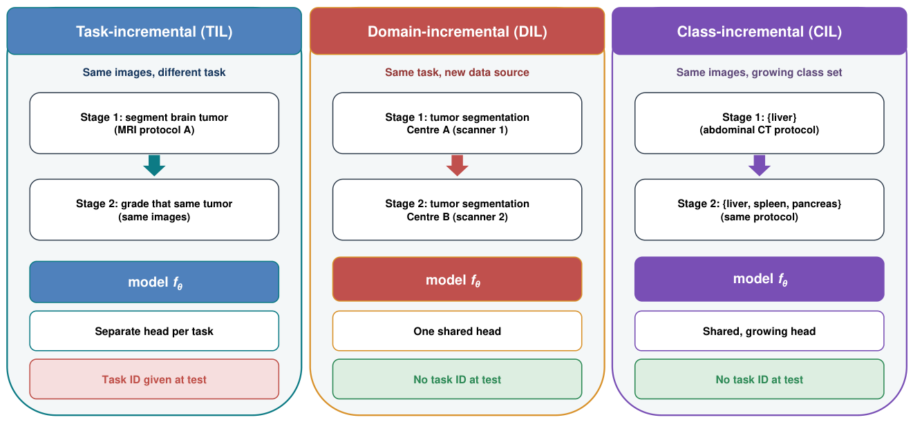
Figure 5.
A taxonomy of continual-learning methods for medical imaging. Methods are grouped by how they resist forgetting into five families, each shown with representative mechanisms.
Figure 5.
A taxonomy of continual-learning methods for medical imaging. Methods are grouped by how they resist forgetting into five families, each shown with representative mechanisms.
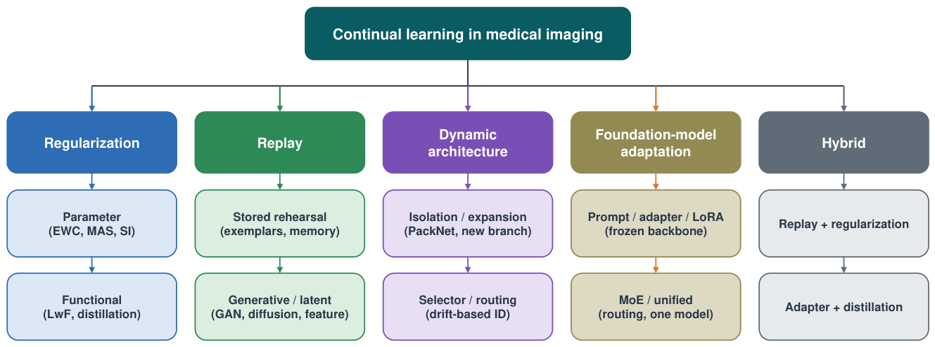
Figure 6.
Mechanisms of the four core method families. Regularization anchors important weights to their old values. Replay reintroduces past data from a buffer or a generator. Dynamic architectures add a new branch for a new task. Foundation-model methods keep a pretrained backbone frozen and train small modules per stage.
Figure 6.
Mechanisms of the four core method families. Regularization anchors important weights to their old values. Replay reintroduces past data from a buffer or a generator. Dynamic architectures add a new branch for a new task. Foundation-model methods keep a pretrained backbone frozen and train small modules per stage.
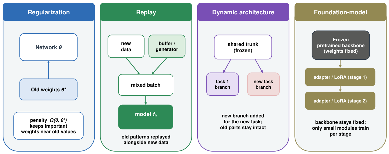
Figure 7.
Deployment lifecycle for a continually updated medical model. Updates run within a predetermined change control plan and under post-market oversight, rather than through a new submission for each change. The plan sets out the planned modifications, the protocol to make and check them, and an impact assessment.
Figure 7.
Deployment lifecycle for a continually updated medical model. Updates run within a predetermined change control plan and under post-market oversight, rather than through a new submission for each change. The plan sets out the planned modifications, the protocol to make and check them, and an impact assessment.
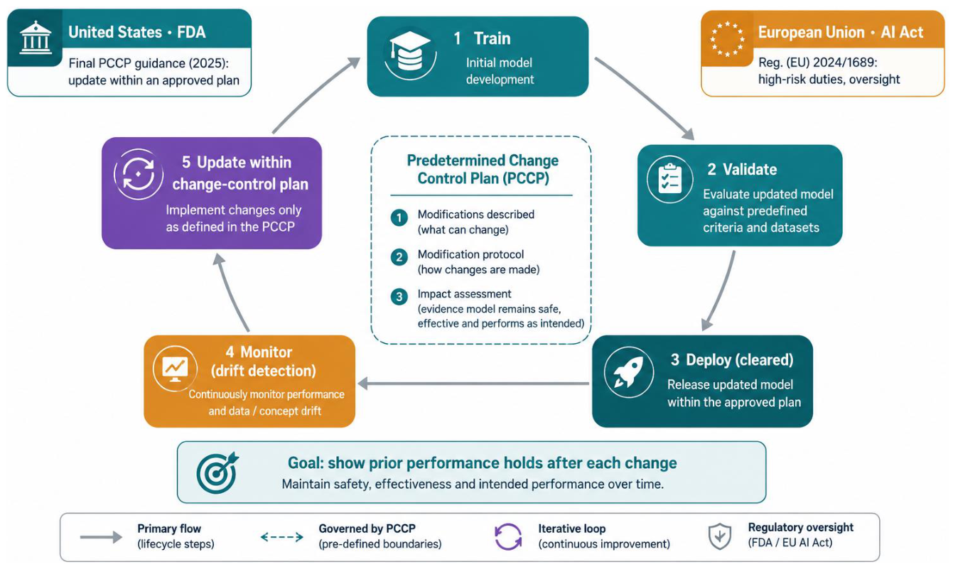
Disclaimer/Publisher’s Note: The statements, opinions and data contained in all publications are solely those of the individual author(s) and contributor(s) and not of MDPI and/or the editor(s). MDPI and/or the editor(s) disclaim responsibility for any injury to people or property resulting from any ideas, methods, instructions or products referred to in the content. |
© 2026 by the authors. Licensee MDPI, Basel, Switzerland. This article is an open access article distributed under the terms and conditions of the Creative Commons Attribution (CC BY) license (http://creativecommons.org/licenses/by/4.0/).
Copyright: This open access article is published under a Creative Commons CC BY 4.0 license, which permit the free download, distribution, and reuse, provided that the author and preprint are cited in any reuse.