

Submitted:
15 July 2026
Posted:
17 July 2026
You are already at the latest version
Abstract
Fms related receptor tyrosine kinase 3-internal tandem duplication (FLT3-ITD) mutations occur in approximately 20–25% of patients with acute myeloid leukemia (AML) and are associated with increased relapse risk and inferior survival outcomes. Although measurable residual disease (MRD) has become a key prognostic tool for guiding post-remission treatment decisions, FLT3-ITD was historically considered a suboptimal MRD marker because of its subclonal nature, structural heterogeneity and dynamic behavior during disease evolution. Despite its strong diagnostic and prognostic relevance, FLT3-ITD has not yet been fully integrated into routine MRD monitoring also due to methodological limitations, and lack of standardized workflows. Recent evidence supports the use of ultra-high sensitivity (UHS) next-generation sequencing (NGS) approaches to detect FLT3-ITD MRD with improved precision, enabling reliable longitudinal tracking of patient-specific clones at very low variant allele frequencies (VAF), and potentially informing clinical decision-making. Indeed, although MRD evaluation is complicated by clonal heterogeneity and molecular instability, advances in high-sensitivity detection techniques have allowed more accurate monitoring of residual leukemic burden. The present review summarizes the biological underpinnings of FLT3-ITD mutated (FLT3-ITDmut) AML, discusses the methodological challenges of MRD detection, and critically evaluates the evolving role of MRD in refining relapse prediction, supporting post-remission therapy tailoring, and contributing to a harmonized framework for FLT3-ITDmut AML management.
Keywords:
acute myeloid leukemia
; FLT3-ITD
; measurable residual disease
; MRD
; next generation sequencing
; NGS
; allogeneic stem-cell transplantation
; allo-HSCT
; FLT3 inhibitors
; FLT3i
1. Introduction
Acute myeloid leukemia (AML) is a clonal hematopoietic neoplasm characterized by a broad spectrum of genetic alterations that drive classification, risk stratification and therapeutic decisions [1,2,3]. This plasticity of the mutational pattern of AML is the result of a multistep process of clonal evolution in which a founder mutation -most commonly affecting genes encoding epigenetic regulators- can be preceded by pre-leukemic events and acquire additional subclonal abnormalities that accumulate over time [4,5]. Acquisition of leukemia-defining lesions, including NPM1 (nucleophosmin 1) [6] mutations and activating signaling pathway alterations, has been shown to drive overt leukemic transformation. An outstanding example of clonal evolution is represented by fms related receptor tyrosine kinase 3-internal tandem duplication (FLT3-ITD) mutations [7,8], which may become undetectable at relapse, with the acquisition of additional mutations accountable for therapy resistance [9]. Internal tandem duplications (ITDs) in the FLT3 gene, affecting the juxtamembrane domain (JM), represent a clinically and biologically distinct entities and the most common types of FLT3 mutation, as they occur in about 10% to 30% of AML cases [10,11]. FLT3-ITD mutations involve insertion of a variable number of bases, resulting in a constitutively active aberrant protein, and leading to uncontrolled proliferation of leukemic blasts, self-renewal, and resistance to differentiation and apoptosis signals [12]. Clinically, ITD mutations of FLT3 significantly increase the risk of relapse, even in patients who initially achieve complete remission (CR), leading to chemotherapy resistance, and decreased overall survival (OS) [13].
While 2017 European LeukemiaNet (ELN) guidelines recommended the use of allelic ratio (AR) values for risk stratification based on NPM1 co-mutations [14], the updated 2022 ELN recommendations [15] substantially revised the classification of FLT3-ITD, categorizing patients as intermediate-risk regardless of AR and NPM1 mutational status [8,16]. This revision reflects current treatment algorithms for FLT3-ITD mutated (FLT3-ITDmut) AML, emphasizing rapid FLT3 mutation detection at diagnosis to enable early integration of FLT3 inhibitors (FLT3i) with intensive induction chemotherapy in fit patients. In the frontline setting, FLT3i include midostaurin, a first-generation multikinase inhibitor with activity against both FLT3-ITD and FLT3-TKD (tyrosine kinase domain) mutations, added to conventional chemotherapy as established by the RATIFY trial [17]. More recently, the type II FLT3 inhibitor quizartinib in the phase III QuANTUM-First study improved survival outcomes in patients with newly diagnosed FLT3-ITDmut AML treated with 3+7 chemotherapy, followed by allogeneic stem-cell transplantation (allo-HSCT) in eligible patients [18]. In contrast, gilteritinib is approved as single agent therapy in relapsed/refractory AML, based on the results of the ADMIRAL trial [19].
In this context, the concept of measurable, residual disease (MRD) has gained increasing importance as a tool to detect submicroscopic levels of leukemic cells that persist after therapy and ultimately drive disease progression or recurrence [20]. Across validated molecular and immunophenotypic MRD studies, MRD positivity after induction, consolidation, or before allo-HSCT, is consistently associated with an increased risk of relapse and inferior survival, leading to its incorporation into post-remission risk stratification algorithms [21,22,23]. FLT3-ITD mutations have been shown to occur at subclonal levels at diagnosis, and may be acquired or lost over the course of disease progression or under therapeutic pressure, reflecting a high degree of clonal instability [9]. Moreover, FLT3-ITD mutations are highly patient-specific, displaying considerable heterogeneity in length, insertion site, and genomic sequence [24,25,26,27]. This behavior poses a major challenge for MRD assessment, as the clearance of FLT3 mutations does not necessarily correspond to the eradication of leukemia burden. Indeed, relapse may arise from ancestral FLT3 wild-type clones or from newly emerging microclones that can persist below the detection threshold of conventional assays, thereby limiting the reliability of FLT3 as a standalone MRD marker [28,29]. As a result of the biological complexity of ITD clones, 2022 ELN MRD recommendations [30] considered FLT3-ITD suboptimal for routine MRD monitoring when assessed using conventional next-generation sequencing (NGS) , as compared to more stable molecular markers, such as NPM1 mutations or recurrent fusion transcripts (e.g., RUNX1/RUNX1T1, CBFB/MYH11, PML/RARA, BCR/ABL1) which are recommended for MRD monitoring by real-time PCR (qRT-PCR ) [31].
However, recent advances in ultra-high sensitivity (UHS) NGS technologies have substantially challenged this paradigm, leading to the 2025 update on MRD in AML, by the ELN-DAVID MRD working party [20]. Dedicated NGS-based assays and specialized bioinformatic pipelines, such as the getITD protocol [32], have substantially improved detection and quantification of FLT3-ITD mutations, enabling the monitoring of patient-specific FLT3-ITD clones [20].
Our review addresses an unmet and highly relevant clinical need in the field of FLT3-ITDmut AML: turning FLT3-ITD from a diagnostic/prognostic marker into a reliable target for MRD assessment, allowing the refinement of AML management.
2. Biology and Clonal Dynamics of FLT3-ITD in AML
Recent advances in NGS technologies have provided a comprehensive characterization of FLT3 alterations as key drivers of leukemogenesis, while refining our understanding of the biological role of FLT3 receptor in AML. FLT3 is a member of the class III receptor tyrosine kinase (RTK) family, which also includes KIT, PDGFRα/β (platelet derived growth factor receptor alpha/beta), and CSF1R (colony stimulating factor 1 receptor) [33]. These receptors play essential roles in hematopoietic development, cellular proliferation, survival, and differentiation. Following post-translational processing, FLT3 is expressed as an immature intracellular isoform and a mature fully glycosylated form localized at the cell surface, where it mediates ligand-dependent signaling [34]. Structurally, FLT3 comprises an extracellular ligand-binding region, a transmembrane domain, an intracellular juxtamembrane domain (JMD), and a tyrosine kinase domain (TKD) [35].
FLT3 mutations are detected in approximately 30% of newly diagnosed AML, with FLT3-ITD and FLT3-TKD point mutations representing the most common variants included in routine diagnostic assessment [36,37]. Less frequent non-canonical FLT3 variants have also been reported in approximately 2% of AML cases, although their biological and clinical significance remains unclear [38,39]. Internal tandem duplications in the FLT3 gene, which typically affect the cytoplasmic JM domain (exons 14-15), represent in-frame mutations induced by duplication of a variable number of bases (ranging from 3 to more than 400), all of which are multiples of three [12]. Therefore, the insertion, while not altering the normal reading frame, results in the translation of an abnormal protein that dimerizes independently of the ligand stimulation, promoting leukemic cell survival and impaired differentiation [40].
Despite its clear prognostic relevance, the role of FLT3-ITD in MRD assessment has long been debated. This is largely due to the intrinsic heterogeneity and patient-specific nature of ITD mutations, as well as their dynamic behavior during disease evolution [9,28]. In contrast to more stable marker such as NPM1, FLT3-ITD are generally considered late-event mutations in leukemogenesis [41], often subclonal and unstable over time in up to 25% of patients AML [41,42,43]. Indeed, longitudinal analyses have shown that ITD mutational status may change over the course of disease, with the mutation either being lost or newly acquired at relapse. These dynamics likely reflect the expansion of pre-existing FLT3-ITD-mutated microclones that were below the limit of detection at diagnosis, or the persistence of clones that survive therapy and subsequently acquire additional genetic lesions [29]. Consistent with this clonal evolution model, approximately 15–30% of patients harboring FLT3-ITD at diagnosis, relapse with FLT3 wild-type disease, whereas a smaller subset of patients initially lacking FLT3-ITD acquire the mutation at relapse [9,44].
The introduction of FLT3i further highlighted the evolutionary plasticity of FLT3-ITDmut AML. Correlative analyses from the RATIFY trial demonstrated that in approximately 46% of patients relapsing after midostaurin-based therapy FLT3-ITD mutations became undetectable at relapse, suggesting expansion of FLT3-independent leukemic populations [8]. Likewise, molecular studies of gilteritinib-treated patients revealed loss of FLT3-mutated clones in approximately 20% of cases, whereas other patients developed resistant disease through emergence of RAS/mitogen-activated protein kinase (MAPK)-driven subclones or secondary FLT3 kinase-domain mutations [45]. In contrast, resistance to quizartinib more commonly involves on-target evolution through acquisition of FLT3-TKD mutations, particularly affecting D835 and F691 residues [46]. These observations indicate that relapse after FLT3i therapy frequently reflects clonal remodeling rather than persistence of the dominant diagnostic clone.
Altogether, these findings establish FLT3-ITD as a highly dynamic molecular lesion whose behavior mirrors the evolutionary trajectory of AML. In the era of FLT3 targeted therapies, understanding this clonal complexity is essential for interpreting molecular response, mechanisms of resistance, and underscores the challenges of using FLT3-ITD as a reliable MRD marker.
3. Methodologies for FLT3-ITD MRD Detection and the Updated 2025 ELN MRD Guidelines
Despite genetic profiling improved the definition of prognostic risk in AML, disease relapse remains the leading cause of treatment failure. Although available treatments induce morphologic complete remission in about 80% of AML patients, 30-40% of them eventually relapse due to the survival of leukemic cells resistant to conventional therapies, which are capable of re-expanding and causing disease relapse [47,48,49]. In this setting, MRD assessment is a cornerstone of AML management, providing prognostic information beyond conventional morphology and enabling the identification of patients at increased risk of relapse [50]. The most recent ELN-DAVID 2025 international guidelines [20] have further strengthened the clinical relevance of MRD monitoring by integrating molecular subtype, treatment phase, and disease kinetics into risk-adapted treatment planning.
Before the introduction of high-sensitivity sequencing technologies, MRD monitoring employed multiparameter flow cytometry (MFC) and molecular assays targeting recurrent genetic abnormalities. Although not particularly sensitive, flow cytometry can be used in the majority of AML patients (>90%), contributing to risk stratification at different stages of treatment. MFC-based MRD assessment relies on the identification of leukemia-associated immunophenotype (LAIP), defined as an antigen expression pattern characteristic of myeloid blasts and informative during patients’ follow-up [51,52]. In the absence of a suitable LAIP, the ELN 2025 guidelines endorse the “different from normal” (DfN) approach, which identifies immunophenotypic deviations from normal hematopoietic maturation [20].
qRT-PCR remains the reference method for molecular MRD assessment, whenever a validated molecular target is available. Among these, NPM1 mutations represent the most extensively validated target because of their biological stability and close correlation with leukemic burden [21,53,54].
In the context of FLT3-ITDmut AML, MRD monitoring represents a challenging and constantly evolving field, in which conventional and more innovative techniques coexist, albeit with different limitations and potential (Figure 1) [55].
Historically, FLT3-ITD detection relied on DNA fragment analysis by capillary electrophoresis (CE) [56], which, although highly effective and routinely used for diagnostic purposes, exhibits limited analytical sensitivity (approximately 10−1-10−2) and is prone to amplification bias due to preferential amplification of shorter fragments. The limited sensitivity of this traditional diagnostic approach has long hindered the detection of low-level FLT3-ITD-positive clones, contributing to underestimate its persistence over time, especially in a context characterized by high molecular heterogeneity. As a result, residual FLT3-ITD detection has traditionally been considered of limited prognostic value, particularly when compared with more stable leukemic mutations [20]. The well-known intrinsic complexity of ITD variants and, above all, the historical lack of standardization across laboratories also impaired the comparison of results across different trials.
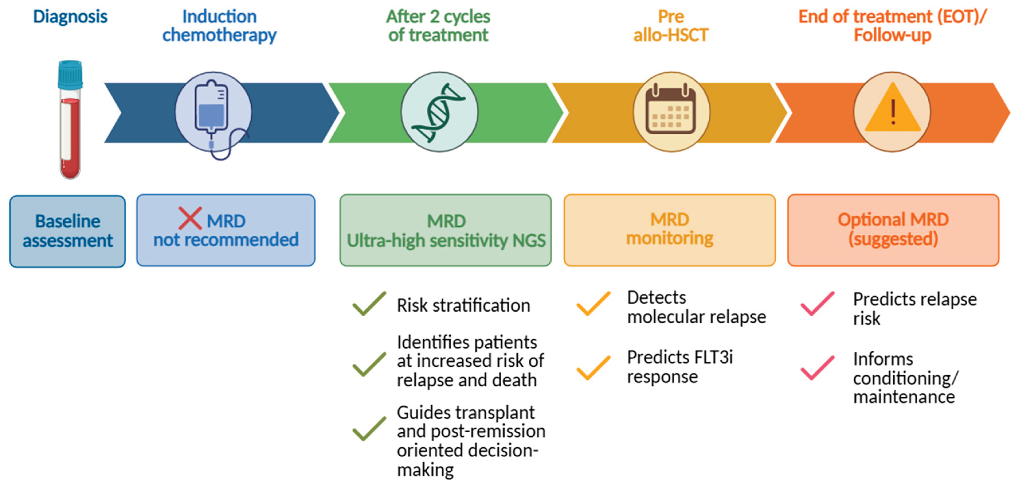
The ELN-DAVID 2025 recommendations have therefore introduced several key changes to improve the accuracy and clinical role of FLT3-ITD MRD assessment in AML. Among these, particular emphasis has been placed on the recommendation to use UHS-NGS methods for the detection of FLT3-ITD mutational burden after two cycles of chemotherapy and prior to allo-HSCT, as these time-points provide the strongest prognostic information. Although assessment at the end of treatment (EOT) and during follow-up may also be informative, supporting evidence remains less compelling (Figure 2) [20,57,58].
From a technical point of view, FLT3-ITD sequencing can be performed using either a single-step or a two-step PCR-based NGS, with specific primers amplifying exons 14-15[59,60,61]. The targeted deep sequencing technique enabled a significant improvement in analytical sensitivity, achieving detection limits (LOD) as low as 10−4–10−5, thus allowing the identification of very low-frequency residual clones. Specifically, it has been established that the cut-off for MRD positivity for FLT3-ITD must correspond to a LOD of at least 0.01%[20]. Despite its extreme sensitivity in detecting mutations, MRD assay by ultra-sensitive NGS is largely affected by the size and the DNA amount of samples [23,62]. NGS detects up to 228 bp FLT3-ITDs [63] and requires at least 500 ng DNA input (700 ng for increased confidence in detecting false negatives), a minimum number of identical reads to establish positivity, and a dedicated bioinformatics pipeline to analyze the results, which is crucial for the correct data interpretation [20]. A major advance was represented by the introduction of the getITD algorithm [32], which enabled standardized NGS-based FLT3-ITD detection, improving accuracy and sensitivity, and facilitated subsequent clinical investigations of FLT3-ITD MRD. In order to ascertain the extent of the disease in patients presenting with multiple ITD clones, it is required to report the VAF values of all individual ITD clones identified, along with their sequences. Furthermore, although it has been established that the major clone reflects the level of MRD, it is strongly recommended to also provide the total VAF, calculated by summing the individual VAFs of the different clones [20]. Although the UHS-NGS method allows for the detection of clones with very low VAF, in cases of AML relapses that are negative for the FLT3-ITD mutation, it is necessary to rely on conventional MRD methods at each FU time-point. Indeed, as pointed out several times, a patient may have a FLT3-ITD negative relapse, or FLT3-ITD mutation may drive disease relapse. The expert panel further suggests performing these analyses preferentially on bone marrow (BM) rather than peripheral blood (PB) samples, as it is well known that MRD levels are higher in BM [20].
Summing up, while the biological stability of the NPM1 mutation makes it a reliable marker reflecting the leukemic burden, the subclonal and dynamic nature of FLT3-ITD requires an integrated approach that considers the timing of testing, the clonal history of the disease, and current therapeutic strategies, including exposure to FLT3 inhibitors. Thus, FLT3-ITD helps refine risk stratification, particularly at key stages of the therapeutic path, as in the pre-transplant phase.
Overall, the ELN-DAVID 2025 guidelines underline the need for an integrated and standardized approach to MRD assessment that accounts for the biological characteristics of the molecular target, in which highly sensitive NGS technologies are playing an increasingly central role, despite technical challenges.
4. Clinical Evidence of FLT3-ITD MRD
Measurable residual disease is crucial information for allo-HSCT allocation [21]. In this setting, FLT3-ITD detection represents a challenging application of NGS, as these structural variants exhibit highly variable sizes and generate complex alignment patterns that are not optimally handled by standard variant-calling pipelines. However, some of these technical caveats have been overcome by updated NGS-based assays [64,65]. Indeed, recent data showed that FLT3-ITD microclones, detectable only by UHS-NGS approaches, are independently associated with an increased risk of relapse in AML patients [66,67]. Interestingly, using an NGS-based analysis and a panel covering the entire FLT3 coding region, Duployez et al. showed that approximately 17% of patients (n=1380) who tested negative (mutation not detectable or with an AR below the threshold) for ITD mutation by fragment analysis had at least one microclone, with a median AR of 3.9×10⁻³. Moreover, authors demonstrated that microclones, albeit found at very low levels at diagnosis, were clinically relevant and were associated with an increased incidence of relapse compared with ITD-negative cases (P=0.001), whereas there were no significant differences in terms of CR following intensive chemotherapy, even when compared to patients presenting with macroclones (AR ≥ 0.05). Of note, 2- and 5-year cumulative incidence of relapse (CIR) rates were higher in patients with FLT3-ITD microclones (45% and 48%) compared with other patient groups. Similarly, microclones exhibited a clear impact on relapse-free survival (RFS), but their effect on OS was less pronounced and did not reach statistical significance. Of particular interest, analysis of 306 paired diagnosis-relapse samples also showed that, in most cases, FLT3-ITD microclones present at diagnosis may expand and become the dominant clone at relapse, suggesting that they act as a reservoir of residual disease capable of spreading and causing relapse. Indeed, in nearly 40% of AML cases treated with intensive chemotherapy without midostaurin (23 out of 55 patients), the FLT3-ITDmut clone at relapse matched one of the microclones already present at diagnosis, particularly in cases with higher baseline AR, reflecting a pattern of clonal selection.
Grob et al. reported the results from a cohort of 161 FLT3-ITD AML, showing that 29% of patients are FLT3-ITD+ after two cycles of induction chemotherapy, despite achieving morphological remission and that FLT3-ITDmut was associated with increased risk of relapse and reduced OS [68]. Specifically, NGS analysis revealed a more than double incidence of relapse at 4 years in patients with FLT3-ITDmut AML who had detectable MRD (75% vs 33%, P<0.001). Correspondingly, OS was around 30% in the FLT3-ITD MRD+ group versus 57% among the 114 patients who were FLT3-ITD MRD- (P<0.001). Notably, MRD clones were identical to those at diagnosis and their size correlated with recurrence rate. In addition to FLT3-ITD+, only white blood cell count (WBC) and late CR were associated with an increased risk of relapse and reduced OS, but not the presence of NPM1 mutation and FLT3-ITD AR. Similarly, a recent study by Lee and colleagues demonstrated the feasibility of highly sensitive longitudinal monitoring of FLT3-ITD clones by NGS-based assays, providing the technical foundation for the clinical validation of FLT3-ITD MRD as a prognostic and potentially predictive biomarker in AML [69].
Overall, detection of FLT3-ITD MRD, assessed using highly sensitive NGS technologies, identifies a subset of at high risk of treatment failure. The implications for clinical practice are significant, as they suggest the need to integrate MRD assessment into clinical decision processes to select patients who are candidates for more intensive strategies, such as transplantation and/or maintenance with FLT3i.
5. FLT3-ITD MRD and FLT3 Inhibitor–Based Therapies
Several clinical trials have proved the role of FLT3 inhibitors at different treatment phases, reshaping the therapeutic landscape of FLT3-ITDmut AML and introducing new dimensions to MRD-guided management [15,21]. To date, the first-generation approved inhibitors include midostaurin (PKC412) and sorafenib (BAY43-9006), administered in combination with first-line chemotherapy, while among second-generation inhibitors are gilteritinib and quizartinib, having a relevant single-agent activity in both newly diagnosed and relapsed/refractory settings [70,71]. These agents not only reduce leukemic burden, but also influence clonal composition, potentially altering the dynamics of MRD. The impact of FLT3 inhibitors on MRD kinetics is heterogeneous and often fails to result in prolonged suppression of the leukemic clone.
In the AMLSG 16-10 trial, Rücker and colleagues emphasized the importance of using NGS-based assays to evaluate the mutational burden of FLT3-ITD MRD in a cohort of patients treated with intensive chemotherapy plus midostaurin, followed by midostaurin as maintenance therapy [72]. This study enrolled 157 FLT3-ITDmut AML patients for whom BM samples were available at three distinct time-points: after 2 cycles of intensive chemotherapy (Cy2, n=142), at the end of treatment (EOT, n=116), and during follow-up (FU, n=148). This controlled environment ensured the strength of the data obtained, combined with use of ultra-sensitive sequencing techniques, according to getITD protocol. Interestingly, prior to investigating the prognostic impact of FLT3-ITD MRD, the authors compared VAF values in paired BM and PB samples and demonstrated that, whereas there are no significant differences at the time of diagnosis, they increase after Cy2 (P<0.001), underscoring the need to use BM as the preferred source for molecular analyses. Moreover, using NGS-based assessment at diagnosis, they identified 465 ITDs in the AML population, with a median VAF of 0.312%, underlining the effectiveness of this method in detecting mutations with very low VAF. They found that persistence of MRD positivity could help identify candidates for more aggressive treatment strategies, such as HSCT in first remission, or for more intensive therapy, including the use of FLT3i. Of note, pre-transplant MRD positivity has consistently been associated with a significantly higher risk of post-transplant relapse and reduced survival. Accordingly, Rücker et al. proved that 15 out of 21 patients with FLT3-ITD MRD+ before HSCT cleared ITD mutations after transplantation [72].
In some cases, patients with persistent MRD may benefit from additional therapy aimed at achieving deeper remission prior to transplantation. Randomized studies demonstrated that the addition of this FLT3i to chemotherapy [73] or its use as post-HSCT maintenance significantly reduced the risk of relapse, with a particularly pronounced effect in patients with MRD positivity after allogeneic transplantation [74,75]. In particular, in the SORMAIN trial 83 patients (median age 54 years) were randomly assigned to receive either sorafenib or placebo over a period of 60 to 100 days following HSCT [74]. Two years of maintenance therapy with sorafenib resulted in a substantially reduced rate of relapse in patients in CR (85% of RFS in the sorafenib group vs 53.3% in the placebo group), suggesting that prolonged inhibition of FLT3 may help manage residual disease, despite not fully eliminating it. Likewise, 24 months OS was significantly higher in the sorafenib arm (90.5% vs 66.2% for placebo). The authors also assessed MRD status pre- and post-HSCT and underlined the benefit of adding sorafenib in patients who were MRD- prior to transplantation (P=0.028) and in patients with post-HSCT MRD+ (P=0.015). Despite its benefits, clinical use of sorafenib is limited by off-target toxicities, frequent dose modifications and treatment discontinuations in real-world practice. Indeed, the study documented a higher number of cases of drug-related toxicity compared to the control group (22% vs 5%), with higher incidence of infections (26% vs 23%) and graft-versus-host disease (GvHD) in the sorafenib group (77%), whereas the incidence of gastrointestinal toxicity was comparable between the two arms of the trial.
The ADMIRAL trial demonstrated that gilteritinib single agent improves OS in patients with relapsed/refractory FLT3-ITDmut AML compared to standard chemotherapy, but it also highlighted that the median duration of response is 4.9 months, with frequent relapses, suggesting that FLT3 inhibition alone is not sufficient to eradicate residual disease [19,76]. Conversely, the use of FLT3i in the maintenance setting, particularly after HSCT, represents a promising strategy to suppress residual disease and prevent relapse [77]. Data from the MORPHO trial, which included patients who achieved CR following transplantation, randomly assigned to receive gilteritinib or placebo, revealed that maintenance therapy may prolong remission duration [77] with 77% RFS vs 70% in placebo (cumulative incidence, CI 62.4 to 76.2), whereas OS was similar for the two arms. The RFS benefit was significant in MRD+ patients, whether assessed before or after transplantation [77,78]. Overall, this study showed that gilteritinib represents a promising strategy for post-HSCT maintenance therapy, but there were no benefits in MRD- patients, suggesting that MRD status should be used to select elegible patients for treatment [77].
The QuANTUM-First trial showed that adding quizartinib to intensive first-line chemotherapy improved survival in FLT3-ITDmut AML, but failed to overcome the persistence of MRD, which still predicts relapse in this setting [57]. MRD status was evaluated at specific time-points (after induction, after consolidation before maintenance and pre-HSCT) in a cohort of patients, comprising both those achieving CR and CR with partial hematologic recovery (CRi), defining the subset of composite CR (CRc) [57]. In detail, the analysis showed that, among the 368 patients in CRc and the 297 patients in CR for whom MRD samples were available, those who were assigned to the quizartinib arm experienced an increased rate and depth of FLT3-ITD clearance compared to placebo. This finding was observed during first and/or second induction cycle following administration of quizartinib, with a significant reduction in the FLT3-ITD VAF, by 2.8-fold (P=0.0439) and 8-fold (P=0.1345), respectively. A similar trend applied when considering the presence of the NPM1 co-mutation. Indeed, unlike previous studies in which NPM1 mutation was considered a condicio sine qua non for MRD negativity [79], the present study demonstrates that quizartinib can induce deep remission regardless of the presence of NPM1 co-mutation in patients with CRc [57]. Importantly, as suggested by the most recent guidelines [20], the authors addressed the importance of choosing a specific cut-off value to achieve RFS, as the absence of detectable mutant alleles below a predefined sensitivity threshold was strongly associated with improved clinical outcomes. Indeed, the use of a 10-4 cut-off in patients in CRc and CR after induction including quizartinib was associated with improved RFS (hazard ratio, HR: 0.791), whereas no significant differences in the choice of cut-offs (10-4 vs 0) were observed if the measurement was performed prior to maintenance therapy. Similarly, treatment with quizartinib was effective in converting MRD status from positive to negative after consolidation or maintenance, regardless of transplantation. Furthermore, the addition of the FLT3i conferred a survival advantage in patients undergoing transplantation in CR1, but not in CRc1, irrespective of pre-transplant MRD status [57].
These data support a dynamic model in which MRD represents not only a prognostic marker, but also as a guide to the use of FLT3 inhibitors along the therapeutic continuum.
6. Integration of FLT3-ITD MRD Into Clinical Decision-Making
Post-induction and peri-HSCT FLT3-ITD MRD, tested via UHS-NGS (able to achieve sensitivity <0.1%), has been recently implemented in the ELN25 MRD guidelines [20].. However, as previously described, several lines of evidence correlated FLT3-ITD MRD status with clinical outcomes [63,68,78] and few studies, leveraging different molecular targeting for monitoring, explored the possibility of molecular MRD-directed decision process. In particular, the analysis of NCRI (National Cancer Research Institute) AML17 and AML19 phase III trials showed that in patients harboring both NPM1 mutations and FLT3-ITD, MRD monitoring (using established molecular biomarkers including NPM1 mutations, core binding factor translocations or KMT2A rearrangements) was associated with a significant reduction in mortality (3-years OS: 69% vs 58%, HR 0.53). In particular, the early identification of molecular relapse, allowed for a pre-emptive treatment in 45% of MRD+ patients [80]. In the same line, the Italian GIMEMA AML1310 trial, incorporated MRD assessment through both PCR (in NPM1-mutated and core-binding factor AML) and MFC in the remaining cases for decision-making [81]. Patients were stratified according to National Comprehensive Cancer Network (NCCN) risk categories and MRD status, and assigned to allogenic or autologous HSCT. In the overall cohort, 2-year OS and relapse-free survival (RFS) were 56% and 54%, respectively. Notably, 2-year OS and disease-free survival (DFS) were 74% and 61% in the favourable-risk group, 42% and 45% in the poor-risk group, 79% and 61% in the intermediate-risk MRD-negative group, and 70% and 67% in the intermediate-risk MRD-positive group. Importantly, outcomes in MRD-positive intermediate-risk patients undergoing allogeneic HSCT were comparable to those observed in the favourable-risk category, supporting the clinical benefit of MRD-driven allocation to transplant in this subgroup.
These papers emphasize the key importance of pre-HSCT MRD monitoring since its positivity, irrespective of the specific marker used, identifies patients at high risk of relapse and inferior outcome, and should prompt consideration of pre-emptive or intensified therapeutic strategies, including the use of FLT3 inhibitors in patients with a known FLT3-mutated disease history [82,83].
7. Conclusions and Future Directions
The increasing body of evidence supporting FLT3-ITD MRD assessment has underscored its potential as a key biomarker for refining risk stratification in FLT3-ITDmut AML, particularly with the advent of FLT3 targeted therapies [57,68,72,74,76,77]. Despite these advances, several issues remain to be addressed before FLT3-ITD MRD can be routinely implemented in clinical practice and integrated into prospective therapeutic algorithms. A critical step toward clinical implementation is the standardization of laboratory methodologies. To date, considerable heterogeneity in NGS platforms, amplification strategies, bioinformatic pipelines, and reporting practices has limited the comparability of findings across studies. Therefore, harmonization of pre-analytical procedures, sequencing strategies, data analysis pipelines, and MRD reporting standards will be critical to ensure the reproducibility and comparability of results across institutions [20,32,68].
Further efforts should focus on integrating FLT3-ITD MRD assessment with established molecular biomarkers, particularly NPM1, which currently represents the benchmark for molecular response assessment [21,48]. It is indeed well recognized that molecular clearance of FLT3-ITD mutations is generally less deep and less durable than that observed for other MRD markers, reflecting both the clonal heterogeneity of the disease and the potential emergence of therapy-resistant microclones under pharmacologic selective pressure [9]. However, FLT3-ITD may provide complementary information, especially in NPM1 wild-type AML and in patients receiving FLT3i-based therapies.
The optimal source for FLT3-ITD MRD monitoring also requires further investigation. Although BM is currently considered the preferred specimen due to its higher analytical sensitivity, recent studies have reported a good concordance between BM and PB MRD assessments, suggesting that PB may represent a feasible alternative for serial monitoring in selected clinical settings [57,72]. The potential implications of using PB as a substitute for BM sampling are yet to be validated.
On a clinical level, the most important future application of FLT3-ITD MRD lies in the development of MRD-guided therapeutic strategies. Indeed, MRD not only represents a powerful prognostic factor but also serves as an essential tool to identify patients who may derive the greatest benefit from allo-HSCT, post-transplant maintenance, or pre-emptive FLT3i therapy at molecular relapse [20].
Taken together, ongoing technological advances and accumulating clinical evidence suggest that FLT3-ITD MRD is transitioning from an investigational biomarker to a clinically actionable tool for post-remission treatment strategies and monitoring disease dynamics.
Author Contributions
Conceptualization and writing—review and editing: G.S., S.T., L.G., N.L., M.D., E. C., S. C., G.F., T.O. and M.T.V. All authors have read and agreed to the published version of the manuscript.
Funding
This work was funded by Fondo Italiano per la Scienza, (FIS3), under grant Agreement N. FIS-2024-06156 to M.T.V., and “Amici di Beat Leukemia Dr. Alessandro Cevenini” - ONLUS to G.S..
Conflicts of Interest
The authors declare no conflicts of interest.:.
Abbreviations
The following abbreviations are used in this manuscript:
| AML | Acute myeloid leukemia |
| AR | Allelic ratio |
| BM | Bone marrow |
| CE | Capillary electrophoresis |
| CIR | Cumulative incidence of relapse |
| CR | Complete remission |
| CRc | Composite complete remission |
| CRi | Complete remission with partial hematologic recovery |
| CSF1R | Colony stimulating factor 1 receptor |
| DfN | Different from normal |
| DFS | Disease-free survival |
| EBMT | European Society for Blood and Marrow Transplantation |
| ELN | European LeukemiaNet |
| EOT | End of treatment |
| FLT3 | Fms related receptor tyrosine kinase 3 |
| FLT3i | FLT3 inhibitors |
| FU | Follow-up |
| GIMEMA | Gruppo Italiano Malattie EMatologiche dell’Adulto |
| GvHD | Graft-versus-host disease |
| HSCT | Hematopoietic stem cell transplantation |
| ITD | Internal tandem duplication |
| JM | Juxtamembrane domain |
| LAIP | Leukemia associated immunophenotype |
| LOD | Limit of detection |
| MAPK | Mitogen-activated protein kinase |
| MFC | Multiparameter flow cytometry |
| MRD | Measurable residual disease |
| NCCN | National Comprehensive Cancer Network |
| NCRI | National Cancer Research Institute |
| NGS | Next-generation sequencing |
| NPM1 | Nucleophosmin |
| OS | Overall survival |
| PB | Peripheral blood |
| PDGFRα/β | Platelet derived growth factor receptor alpha/beta |
| qRT-PCR | Quantitative Reverse Transcription-Polymerase Chain Reaction |
| RFS | Relapse-free survival |
| RTK | Receptor tyrosine kinase |
| TKD | Tyrosine kinase domain |
| TKIs | Tyrosine kinase inhibitors |
| UHS | Ultra-high sensitivity |
| VAF | Variant allele frequency |
| WBC | White blood cell count |
References
- Lowenberg, B.; Downing, J.R.; Burnett, A. Acute Myeloid Leukemia. N. Engl. J. Med. 1999, 341, 1051–1062. [Google Scholar] [CrossRef]
- Papaemmanuil, E.; Gerstung, M.; Bullinger, L.; Gaidzik, V.I.; Paschka, P.; Roberts, N.D.; Potter, N.E.; Heuser, M.; Thol, F.; Bolli, N.; et al. Genomic Classification and Prognosis in Acute Myeloid Leukemia. N. Engl. J. Med. 2016, 374, 2209–2221. [Google Scholar] [CrossRef] [PubMed]
- Levine, R.L. Molecular Pathogenesis of AML: Translating Insights to the Clinic. Best Pract. Res. Clin. Haematol. 2013, 26, 245–248. [Google Scholar] [CrossRef] [PubMed]
- Graubert, T.A.; Brunner, A.M.; Fathi, A.T. New Molecular Abnormalities and Clonal Architecture in AML: From Reciprocal Translocations to Whole-Genome Sequencing. Am. Soc. Clin. Oncol. Educ. B. 2014, e334–e340. [Google Scholar] [CrossRef]
- Di Nardo, C.D.; Cortes, J.E. Mutations in AML: Prognostic and Therapeutic Implications. Hematology 2016, 2016, 348–355. [Google Scholar] [CrossRef] [PubMed]
- Falini, B.; Mecucci, C.; Tiacci, E.; Alcalay, M.; Rosati, R.; Pasqualucci, L.; La Starza, R.; Diverio, D.; Colombo, E.; Santucci, A.; et al. Cytoplasmic Nucleophosmin in Acute Myelogenous Leukemia with a Normal Karyotype. N. Engl. J. Med. 2005, 352, 254–266. [Google Scholar] [CrossRef]
- Travaglini, S.; Angelini, D.F.; Alfonso, V.; Guerrera, G.; Lavorgna, S.; Divona, M.; Nardozza, A.M.; Consalvo, M.I.; Fabiani, E.; De Bardi, M.; et al. Characterization of FLT3-ITDmut Acute Myeloid Leukemia: Molecular Profiling of Leukemic Precursor Cells. Blood Cancer J. 2020, 10, 85. [Google Scholar] [CrossRef] [PubMed]
- Rücker, F.G.; Du, L.; Luck, T.J.; Benner, A.; Krzykalla, J.; Gathmann, I.; Voso, M.T.; Amadori, S.; Prior, T.W.; Brandwein, J.M.; et al. Molecular Landscape and Prognostic Impact of FLT3-ITD Insertion Site in Acute Myeloid Leukemia: RATIFY Study Results. Leukemia 2022, 36, 90–99. [Google Scholar] [CrossRef] [PubMed]
- Schmalbrock, L.K.; Dolnik, A.; Cocciardi, S.; Sträng, E.; Theis, F.; Jahn, N.; Panina, E.; Blätte, T.J.; Herzig, J.; Skambraks, S.; et al. Clonal Evolution of Acute Myeloid Leukemia with FLT3-ITD Mutation under Treatment with Midostaurin. Blood 2021, 137, 3093. [Google Scholar] [CrossRef]
- Bullinger, L.; Döhner, K.; Dohner, H. Genomics of Acute Myeloid Leukemia Diagnosis and Pathways. J. Clin. Oncol. 2017, 35, 934–946. [Google Scholar] [CrossRef] [PubMed]
- Daver, N.; Schlenk, R.F.; Russell, N.H.; Levis, M.J. Targeting FLT3 Mutations in AML: Review of Current Knowledge and Evidence. Leukemia 2019, 33, 299. [Google Scholar] [CrossRef] [PubMed]
- Kiyoi, H.; Ohno, R.; Ueda, R.; Saito, H.; Naoe, T. Mechanism of Constitutive Activation of FLT3 with Internal Tandem Duplication in the Juxtamembrane Domain. Oncogene 2002, 21, 2555–2563. [Google Scholar] [CrossRef]
- Reville, P.K.; Sasaki, K.; Kantarjian, H.M.; Daver, N.G.; Yilmaz, M.; Dinardo, C.D.; Short, N.J.; Borthakur, G.; Pemmaraju, N.; Mehta, R.S.; et al. Improved Outcomes among Newly Diagnosed Patients with FMS-like Tyrosine Kinase 3 Internal Tandem Duplication Mutated Acute Myeloid Leukemia Treated with Contemporary Therapy: Revisiting the European LeukemiaNet Adverse Risk Classification. Am. J. Hematol. 2022, 97, 329–337. [Google Scholar] [CrossRef] [PubMed]
- Döhner, H.; Estey, E.; Grimwade, D.; Amadori, S.; Appelbaum, F.R.; Ebert, B.L.; Fenaux, P.; Larson, R.A.; Levine, R.L.; Lo-coco, F.; et al. Diagnosis and Management OfAML in Adults: 2017 ELN Recommendations from an International Expert Panel. Blood 2017, 129, 424–448. [Google Scholar] [CrossRef]
- Döhner, H.; Wei, A.H.; Appelbaum, F.R.; Craddock, C.; DiNardo, C.D.; Dombret, H.; Ebert, B.L.; Fenaux, P.; Godley, L.A.; Hasserjian, R.P.; et al. Diagnosis and Management of AML in Adults: 2022 Recommendations from an International Expert Panel on Behalf of the ELN. Blood 2022, 140, 1345–1377. [Google Scholar] [CrossRef] [PubMed]
- Döhner, K.; Thiede, C.; Jahn, N.; Panina, E.; Gambietz, A.; Larson, R.A.; Prior, T.W.; Marcucci, G.; Jones, D.; Krauter, J.; et al. Impact of NPM1/FLT3-ITD Genotypes Defined by the 2017 European LeukemiaNet in Patients with Acute Myeloid Leukemia. Blood 2020, 135, 371–380. [Google Scholar] [CrossRef] [PubMed]
- Stone, R.M.; Mandrekar, S.J.; Sanford, B.L.; Laumann, K.; Geyer, S.; Bloomfield, C.D.; Thiede, C.; Prior, T.W.; Döhner, K.; Marcucci, G.; et al. Midostaurin plus Chemotherapy for Acute Myeloid Leukemia with a FLT3 Mutation. N. Engl. J. Med. 2017, 377, 454–464. [Google Scholar] [CrossRef]
- Erba, H.P.; Montesinos, P.; Kim, H.J.; Patkowska, E.; Vrhovac, R.; Žák, P.; Wang, P.N.; Mitov, T.; Hanyok, J.; Kamel, Y.M.; et al. Quizartinib plus Chemotherapy in Newly Diagnosed Patients with FLT3-Internal-Tandem-Duplication-Positive Acute Myeloid Leukaemia (QuANTUM-First): A Randomised, Double-Blind, Placebo-Controlled, Phase 3 Trial. Lancet 2023, 401, 1571–1583. [Google Scholar] [CrossRef] [PubMed]
- Perl, A.E.; Martinelli, G.; Cortes, J.E.; Neubauer, A.; Berman, E.; Paolini, S.; Montesinos, P.; Baer, M.R.; Larson, R.A.; Ustun, C.; et al. Gilteritinib or Chemotherapy for Relapsed or Refractory FLT3-Mutated AML. N. Engl. J. Med. 2019, 381, 1728–1740. [Google Scholar] [CrossRef] [PubMed]
- Cloos, J.; Valk, P.J.M.; Thiede, C.; Döhner, K.; Roboz, G.J.; Wood, B.L.; Walter, R.B.; Wang, S.A.; Wierzbowska, A.; Wei, A.H.; et al. 2025 Update on MRD in Acute Myeloid Leukemia: A Consensus Document from the ELN-DAVID MRD Working Party. Blood 2025. [Google Scholar] [CrossRef] [PubMed]
- Heuser, M.; Freeman, S.D.; Ossenkoppele, G.J.; Buccisano, F.; Hourigan, C.S.; Ngai, L.L.; Tettero, J.M.; Bachas, C.; Baer, C.; Béné, M.C.; et al. 2021 Update on MRD in Acute Myeloid Leukemia: A Consensus Document from the European LeukemiaNet MRD Working Party. Blood 2021, 138, 2753–2767. [Google Scholar] [CrossRef] [PubMed]
- Short, N.J.; Zhou, S.; Fu, C.; Berry, D.A.; Walter, R.B.; Freeman, S.D.; Hourigan, C.S.; Huang, X.; Nogueras Gonzalez, G.; Hwang, H.; et al. Association of Measurable Residual Disease With Survival Outcomes in Patients With Acute Myeloid Leukemia: A Systematic Review and Meta-Analysis. JAMA Oncol. 2020, 6, 1890–1899. [Google Scholar] [CrossRef] [PubMed]
- Hourigan, C.S.; Beugelink, L.; Othman, J.; Gui, G.; Ivey, A.; Dillon, R.J.; Thiede, C.; Levis, M.J.; Dillon, L.W.; Wei, A.H.; et al. ELN-DAVID Recommendations for NGS-Based FLT3-ITD MRD Testing in Patients with Acute Myeloid Leukemia. Blood 2026. [Google Scholar] [CrossRef] [PubMed]
- Thiede, C.; Steudel, C.; Mohr, B.; Schaich, M.; Schäkel, U.; Platzbecker, U.; Wermke, M.; Bornhäuser, M.; Ritter, M.; Neubauer, A.; et al. Analysis of FLT3-Activating Mutations in 979 Patients with Acute Myelogenous Leukemia: Association with FAB Subtypes and Identification of Subgroups with Poor Prognosis. Blood 2002, 99, 4326–4335. [Google Scholar] [CrossRef] [PubMed]
- Schnittger, S.; Schoch, C.; Dugas, M.; Kern, W.; Staib, P.; Wuchter, C.; Löffler, H.; Sauerland, C.M.; Serve, H.; Büchner, T.; et al. Analysis of FLT3 Length Mutations in 1003 Patients with Acute Myeloid Leukemia: Correlation to Cytogenetics, FAB Subtype, and Prognosis in the AMLCG Study and Usefulness as a Marker for the Detection of Minimal Residual Disease. Blood 2002, 100, 59–66. [Google Scholar] [CrossRef]
- Levis, M.; Small, D. FLT3: ITDoes Matter in Leukemia. Leukemia KWRD. 2003, 17, 1738–1752. [Google Scholar] [CrossRef] [PubMed]
- Stirewalt, D.L.; Kopecky, K.J.; Meshinchi, S.; Engel, J.H.; Pogosova-Agadjanyan, E.L.; Linsley, J.; Slovak, M.L.; Willman, C.L.; Radich, J.P. Size of FLT3 Internal Tandem Duplication Has Prognostic Significance in Patients with Acute Myeloid Leukemia. Blood 2006, 107, 3724–3726. [Google Scholar] [CrossRef] [PubMed]
- Ottone, T.; Zaza, S.; Divona, M.; Hasan, S.K.; Lavorgna, S.; Laterza, S.; Cicconi, L.; Panetta, P.; Di Giandomenico, J.; Cittadini, M.; et al. Identification of Emerging FLT3 ITD-Positive Clones during Clinical Remission and Kinetics of Disease Relapse in Acute Myeloid Leukaemia with Mutated Nucleophosmin. Br. J. Haematol. 2013, 161, 533–540. [Google Scholar] [CrossRef]
- Angelini, D.F.; Ottone, T.; Guerrera, G.; Lavorgna, S.; Cittadini, M.; Buccisano, F.; De Bardi, M.; Gargano, F.; Maurillo, L.; Divona, M.; et al. A Leukemia-Associated CD34/CD123/CD25/CD99+ Immunophenotype Identifies FLT3-Mutated Clones in Acute Myeloid Leukemia. Clin. Cancer Res. 2015, 21, 3977–3985. [Google Scholar] [CrossRef] [PubMed]
- Schuurhuis, G.J.; Heuser, M.; Freeman, S.; Béne, M.C.; Buccisano, F.; Cloos, J.; Grimwade, D.; Haferlach, T.; Hills, R.K.; Hourigan, C.S.; et al. Minimal/Measurable Residual Disease in AML: A Consensus Document from the European LeukemiaNet MRD Working Party. Blood 2018, 131, 1275. [Google Scholar] [CrossRef] [PubMed]
- Gabert, J.; Beillard, E.; van der Velden, V.H.J.; Bi, W.; Grimwade, D.; Pallisgaard, N.; Barbany, G.; Cazzaniga, G.; Cayuela, J.M.; Cavé, H.; et al. Standardization and Quality Control Studies of ‘Real-Time’ Quantitative Reverse Transcriptase Polymerase Chain Reaction of Fusion Gene Transcripts for Residual Disease Detection in Leukemia – A Europe Against Cancer Program. Leuk. 2003 1712 2003, 17, 2318–2357. [Google Scholar] [CrossRef] [PubMed]
- Blätte, T.J.; Schmalbrock, L.K.; Skambraks, S.; Lux, S.; Cocciardi, S.; Dolnik, A.; Döhner, H.; Döhner, K.; Bullinger, L. GetITD for FLT3-ITD-Based MRD Monitoring in AML. Leuk. 2019 3310 2019, 33, 2535–2539. [Google Scholar] [CrossRef] [PubMed]
- Markovic, A.; MacKenzie, K.L.; Lock, R.B. FLT-3: A New Focus in the Understanding of Acute Leukemia. Int. J. Biochem. Cell Biol. 2005, 37, 1168–1172. [Google Scholar] [CrossRef] [PubMed]
- Grafone, T.; Palmisano, M.; Nicci, C.; Storti, S. An Overview on the Role of FLT3-Tyrosine Kinase Receptor in Acute Myeloid Leukemia: Biology and Treatment. Oncol. Rev. 2012, 6, 64–74. [Google Scholar] [CrossRef] [PubMed]
- Nakao, M.; Yokota, S.; Iwai, T.; Kaneko, H.; Horiike, S.; Kashima, K.; Sonoda, Y.; Fujimoto, T.; Misawa, S. Internal Tandem Duplication of the Flt3 Gene Found in Acute Myeloid Leukemia. Leukemia 1996, 10, 1911–1918. [Google Scholar] [PubMed]
- Abaza, Y.; McMahon, C.; Garcia, J.S. Advancements and Challenges in the Treatment of AML. Am. Soc. Clin. Oncol. Educ. book. Am. Soc. Clin. Oncol. Annu. Meet. 2024, 44. [Google Scholar] [CrossRef]
- Della Porta, M.G.; Martinelli, G.; Rambaldi, A.; Santoro, A.; Voso, M.T. A Practical Algorithm for Acute Myeloid Leukaemia Diagnosis Following the Updated 2022 Classifications. Crit. Rev. Oncol. Hematol. 2024, 198, 104358. [Google Scholar] [CrossRef] [PubMed]
- Ge, S.S.; Qiu, Q.C.; Dai, H.P.; Shen, X.D.; Wu, T.M.; Du, J.H.; Wan, C.L.; Shen, H.J.; Wu, D.P.; Xue, S.L.; et al. Mutation Spectrum of FLT3 and Significance of Non-Canonical FLT3 Mutations in Haematological Malignancy. Br. J. Haematol. 2023, 202, 539–549. [Google Scholar] [CrossRef] [PubMed]
- Travaglini, S.; Gurnari, C.; Antonelli, S.; Marchesi, F.; De Angelis, G.; Ottone, T.; Divona, M.; Cristiano, A.; Hajrullaj, H.; Mengarelli, A.; et al. Functional Characterization and Response to FLT3 Inhibitors in Acute Myeloid Leukaemia with a Non-Canonical FLT3 Mutation: A Proof of Concept. Br. J. Haematol. 2023, 203, 327–330. [Google Scholar] [CrossRef]
- Parcells, B.W.; Ikeda, A.K.; Simms-Waldrip, T.; Moore, T.B.; Sakamoto, K.M. FMS-like Tyrosine Kinase 3 in Normal Hematopoiesis and Acute Myeloid Leukemia. Stem Cells 2006, 24, 1174–1184. [Google Scholar] [CrossRef] [PubMed]
- Krönke, J.; Bullinger, L.; Teleanu, V.; Tschürtz, F.; Gaidzik, V.I.; Kühn, M.W.M.; Rücker, F.G.; Holzmann, K.; Paschka, P.; Kapp-Schwörer, S.; et al. Clonal Evolution in Relapsed NPM1-Mutated Acute Myeloid Leukemia. Blood 2013, 122, 100–108. [Google Scholar] [CrossRef] [PubMed]
- Kottaridis, P.D.; Gale, R.E.; Langabeer, S.E.; Frew, M.E.; Bowen, D.T.; Linch, D.C. Studies of FLT3 Mutations in Paired Presentation and Relapse Samples from Patients with Acute Myeloid Leukemia: Implications for the Role of FLT3 Mutations in Leukemogenesis, Minimal Residual Disease Detection, and Possible Therapy with FLT3 Inhibitors. Blood 2002, 100, 2393–2398. [Google Scholar] [CrossRef] [PubMed]
- Garg, M.; Nagata, Y.; Kanojia, D.; Mayakonda, A.; Yoshida, K.; Keloth, S.H.; Zang, Z.J.; Okuno, Y.; Shiraishi, Y.; Chiba, K.; et al. Profiling of Somatic Mutations in Acute Myeloid Leukemia with FLT3-ITD at Diagnosis and Relapse. Blood 2015, 126, 2491–2501. [Google Scholar] [CrossRef]
- Kottaridis, P.D.; Gale, R.E.; Frew, M.E.; Harrison, G.; Langabeer, S.E.; Belton, A.A.; Walker, H.; Wheatley, K.; Bowen, D.T.; Burnett, A.K.; et al. The Presence of a FLT3 Internal Tandem Duplication in Patients with Acute Myeloid Leukemia (AML) Adds Important Prognostic Information to Cytogenetic Risk Group and Response to the First Cycle of Chemotherapy: Analysis of 854 Patients from the United Kin…. Blood 2001, 98, 1752–1759. [Google Scholar] [CrossRef]
- McMahon, C.M.; Ferng, T.; Canaani, J.; Wang, E.S.; Morrissette, J.J.D.; Eastburn, D.J.; Pellegrino, M.; Durruthy-Durruthy, R.; Watt, C.D.; Asthana, S.; et al. Clonal Selection with RAS Pathway Activation Mediates Secondary Clinical Resistance to Selective FLT3 Inhibition in Acute Myeloid Leukemia. Cancer Discov. 2019, 9, 1050–1063. [Google Scholar] [CrossRef] [PubMed]
- Smith, C.C.; Wang, Q.; Chin, C.S.; Salerno, S.; Damon, L.E.; Levis, M.J.; Perl, A.E.; Travers, K.J.; Wang, S.; Hunt, J.P.; et al. Validation of ITD Mutations in FLT3 as a Therapeutic Target in Human Acute Myeloid Leukaemia. Nature 2012, 485, 260–263. [Google Scholar] [CrossRef] [PubMed]
- Döhner, H.; Weisdorf, D.J.; Bloomfield, C.D. Acute Myeloid Leukemia. N. Engl. J. Med. 2015, 373, 1136–1152. [Google Scholar] [CrossRef]
- Hourigan, C.S.; Gale, R.P.; Gormley, N.J.; Ossenkoppele, G.J.; Walter, R.B. Measurable Residual Disease Testing in Acute Myeloid Leukaemia. Leukemia 2017, 31, 1482–1490. [Google Scholar] [CrossRef] [PubMed]
- Burnett, A.K.; Russell, N.H.; Hills, R.K.; Hunter, A.E.; Kjeldsen, L.; Yin, J.; Gibson, B.E.S.; Wheatley, K.; Milligan, D. Optimization of Chemotherapy for Younger Patients with Acute Myeloid Leukemia: Results of the Medical Research Council AML15 Trial. J. Clin. Oncol. 2013, 31, 3360–3368. [Google Scholar] [CrossRef] [PubMed]
- Short, N.J.; Dillon, R. Measurable Residual Disease Monitoring in AML: Prospects for Therapeutic Decision-Making and New Drug Development. Am. J. Hematol. 2025, 100 Suppl 2, 5–15. [Google Scholar] [CrossRef] [PubMed]
- Buccisano, F.; Maurillo, L.; Del Principe, M.I.; Del Poeta, G.; Sconocchia, G.; Lo-Coco, F.; Arcese, W.; Amadori, S.; Venditti, A. Prognostic and Therapeutic Implications of Minimal Residual Disease Detection in Acute Myeloid Leukemia. Blood 2012, 119, 332–341. [Google Scholar] [CrossRef] [PubMed]
- Kern, W.; Bacher, U.; Haferlach, C.; Schnittger, S.; Haferlach, T. The Role of Multiparameter Flow Cytometry for Disease Monitoring in AML. Best Pract. Res. Clin. Haematol. 2010, 23, 379–390. [Google Scholar] [CrossRef] [PubMed]
- Ivey, A.; Hills, R.K.; Simpson, M.A.; Jovanovic, J. V.; Gilkes, A.; Grech, A.; Patel, Y.; Bhudia, N.; Farah, H.; Mason, J.; et al. Assessment of Minimal Residual Disease in Standard-Risk AML. N. Engl. J. Med. 2016, 374, 422–433. [Google Scholar] [CrossRef] [PubMed]
- Dillon, R.; Potter, N.; Freeman, S.; Russell, N. How We Use Molecular Minimal Residual Disease (MRD) Testing in Acute Myeloid Leukaemia (AML). Br. J. Haematol. 2021, 193, 231–244. [Google Scholar] [CrossRef] [PubMed]
- Bibault, J.-E.; Figeac, M.; Hélevaut, N.; Rodriguez, C.; Quief, S.; Sebda, S.; Renneville, A.; Nibourel, O.; Rousselot, P.; Gruson, B.; et al. Nextgeneration Sequencing of FLT3 Internal Tandem Duplications for Minimal Residual Disease Monitoring in Acute Myeloid Leukemia. Oncotarget 2015, 6, 22812–22821. [Google Scholar] [CrossRef] [PubMed]
- Noguera, N.I.; Ammatuna, E.; Zangrilli, D.; Lavorgna, S.; Divona, M.; Buccisano, F.; Amadori, S.; Mecucci, C.; Falini, B.; Lo-Coco, F. Simultaneous Detection of NPM1 and FLT3-ITD Mutations by Capillary Electrophoresis in Acute Myeloid Leukemia. Leukemia 2005, 19, 1479–1482. [Google Scholar] [CrossRef] [PubMed]
- Levis, M.J.; Erba, H.P.; Montesinos, P.; Kim, H.-J.; Vrhovac, R.; Zbieta Patkowska, E.; Pavelž, P.P.; Pavelžák, P.; Wang, P.-N.; Rohrbach, J.E.C.; et al. FLT3-ITD Measurable Residual Disease from the QuANTUM-First Trial. Blood Adv. 2026, 10, 917–928. [Google Scholar] [CrossRef] [PubMed]
- Ley, T.J.; Mardis, E.R.; Ding, L.; Fulton, B.; McLellan, M.D.; Chen, K.; Dooling, D.; Dunford-Shore, B.H.; McGrath, S.; Hickenbotham, M.; et al. DNA Sequencing of a Cytogenetically Normal Acute Myeloid Leukaemia Genome. Nat. 2008 4567218 2008, 456, 66–72. [Google Scholar] [CrossRef] [PubMed]
- Othman, J.; Potter, N.; Mokretar, K.; Taussig, D.; Khan, A.; Krishnamurthy, P.; Latif, A.L.; Cahalin, P.; Aries, J.; Amer, M.; et al. FLT3 Inhibitors as MRD-Guided Salvage Treatment for Molecular Failure in FLT3 Mutated AML. Leukemia 2023, 37, 2066–2072. [Google Scholar] [CrossRef] [PubMed]
- Thol, F.; Kölking, B.; Damm, F.; Reinhardt, K.; Klusmann, J.H.; Reinhardt, D.; von Neuhoff, N.; Brugman, M.H.; Schlegelberger, B.; Suerbaum, S.; et al. Next-Generation Sequencing for Minimal Residual Disease Monitoring in Acute Myeloid Leukemia Patients with FLT3-ITD or NPM1 Mutations. Genes Chromosom. Cancer ISSUE:ISSUE:DOI. 2012, 51, 689–695. [Google Scholar] [CrossRef] [PubMed] [PubMed Central]
- Russell, N.; Othman, J.; Cumming, O.; Thomas, A.; Tedjaseputra, A.; Potter, N.; Jovanovic, J.; Gilkes, A.; Batten, L.; Canham, J.; et al. Safety and Efficacy of Combining Midostaurin and Gemtuzumab Ozogamicin with Induction Chemotherapy in FLT3-Mutated AML. Blood Adv. 2025, 9, 6455–6466. [Google Scholar] [CrossRef] [PubMed]
- Ghannam, J.; Dillon, L.W.; Hourigan, C.S. Next-Generation Sequencing for Measurable Residual Disease Detection in Acute Myeloid Leukaemia. Br. J. Haematol. 2020, 188, 77–85. [Google Scholar] [CrossRef] [PubMed]
- Dillon, L.W.; Gui, G.; Ravindra, N.; Andrew, G.; Mukherjee, D.; Wong, Z.C.; Huang, Y.; Gerhold, J.; Holman, M.; D’Angelo, J.; et al. Measurable Residual FLT3 Internal Tandem Duplication Before Allogeneic Transplant for Acute Myeloid Leukemia. JAMA Oncol. 2024, 10, 1104–1110. [Google Scholar] [CrossRef] [PubMed]
- Loo, S.; Dillon, R.; Ivey, A.; Anstee, N.S.; Othman, J.; Tiong, I.S.; Potter, N.; Jovanovic, J.; Runglall, M.; Chong, C.C.; et al. Pretransplant FLT3-ITD MRD Assessed by High-Sensitivity PCR-NGS Determines Posttransplant Clinical Outcome. Blood 2022, 140, 2407–2411. [Google Scholar] [CrossRef] [PubMed]
- Levis, M.J.; Perl, A.E.; Altman, J.K.; Gocke, C.D.; Bahceci, E.; Hill, J.; Liu, C.; Xie, Z.; Carson, A.R.; McClain, V.; et al. A Next-Generation Sequencing-Based Assay for Minimal Residual Disease Assessment in AML Patients with FLT3-ITD Mutations. Blood Adv. 2018, 2, 825–831. [Google Scholar] [CrossRef] [PubMed]
- Duployez, N.; Joudinaud, R.; Boudry, A.; Hunault, M.; Fenwarth, L.; Tavernier, E.; Pautas, C.; Bertoli, S.; Tavitian, S.; Raffoux, E.; et al. Prognostic Impact of FLT3-ITD Microclones in Young Adults with Acute Myeloid Leukemia Treated with Intensive Chemotherapy. Blood 2026. [Google Scholar] [CrossRef] [PubMed]
- Gurnari, C.; Ottone, T.; Piciocchi, A.; Späth, D.; Meid, A.; Marsili, G.; Fabiani, E.; Romano, F.; Pascale, M.R.; Chiusolo, P.; et al. Diagnostic Evaluation Methods and Prognostic Impact of FLT3-ITD Microclones in Acute Myeloid Leukemia (AML): A Retrospective Multicenter Study on Behalf of the EHA AML-Specialized Working Group (SWG). Blood 2025, 146, 217–217. [Google Scholar] [CrossRef]
- Grob, T.; Sanders, M.A.; Vonk, C.M.; Kavelaars, F.G.; Rijken, M.; Hanekamp, D.W.; Gradowska, P.L.; Cloos, J.; Fløisand, Y.; Van Marwijk Kooy, M.; et al. Prognostic Value of FLT3-Internal Tandem Duplication Residual Disease in Acute Myeloid Leukemia. J. Clin. Oncol. 2023, 41, 756–765. [Google Scholar] [CrossRef] [PubMed]
- Lee, J.M.; Park, S.; Hwang, I.; Kang, D.; Cho, B.S.; Kim, H.J.; Ahn, A.; Kim, M.; Kim, Y. FLT3-ITD Measurable Residual Disease Monitoring in Acute Myeloid Leukemia Using Next-Generation Sequencing. Cancers . 2022, 14. [Google Scholar] [CrossRef] [PubMed]
- Mohebbi, A.; Shahriyary, F.; Farrokhi, V.; Bandar, B.; Saki, N. A Systematic Review of Second-Generation FLT3 Inhibitors for Treatment of Patients with Relapsed/Refractory Acute Myeloid Leukemia. Leuk. Res. 2024, 141. [Google Scholar] [CrossRef] [PubMed]
- Zhao, Y.; Zhang, X.; Ding, X.; Wang, Y.; Li, Z.; Zhao, R.; Cheng, H.E.; Sun, Y. Efficacy and Safety of FLT3 Inhibitors in Monotherapy of Hematological and Solid Malignancies: A Systemic Analysis of Clinical Trials. Front. Pharmacol. 2024, 15. [Google Scholar] [CrossRef]
- Rücker, F.G.; Bullinger, L.; Cocciardi, S.; Skambraks, S.; Luck, T.J.; Weber, D.; Krzykalla, J.; Pozek, E.; Schneider, I.; Corbacioglu, A.; et al. Measurable Residual Disease Monitoring in AML with FLT3-ITD Treated with Intensive Chemotherapy plus Midostaurin. Blood Adv. 2024, 8, 6067–6080. [Google Scholar] [CrossRef] [PubMed]
- Serve, H.; Brunnberg, U.; Ottmann, O.; Brandts, C.; Steffen, B.; Krug, U.; Wagner, R.; Müller-Tidow, C.; Berdel, W.E.; Cristina Sauerland, M.; et al. Sorafenib in Combination with Intensive Chemotherapy in Elderly Patients with Acute Myeloid Leukemia: Results from a Randomized, Placebo-Controlled Trial. J. Clin. Oncol. PAGE:STRING:ARTICLE/CHAPTER. 2013, 31, 3110–3118. [Google Scholar] [CrossRef] [PubMed]
- Burchert, A.; Bug, G.; Fritz, L. V.; Finke, J.; Stelljes, M.; Röllig, C.; Wollmer, E.; Wäsch, R.; Bornhäuser, M.; Berg, T.; et al. Sorafenib Maintenance After Allogeneic Hematopoietic Stem Cell Transplantation for Acute Myeloid Leukemia With FLT3-Internal Tandem Duplication Mutation (SORMAIN). J. Clin. Oncol. 2020, 38, 2993–3002. [Google Scholar] [CrossRef] [PubMed]
- Xuan, L.; Wang, Y.; Huang, F.; Fan, Z.; Xu, Y.; Sun, J.; Xu, N.; Deng, L.; Li, X.; Liang, X.; et al. Sorafenib Maintenance in Patients with FLT3-ITD Acute Myeloid Leukaemia Undergoing Allogeneic Haematopoietic Stem-Cell Transplantation: An Open-Label, Multicentre, Randomised Phase 3 Trial. Lancet Oncol. 2020, 21, 1201–1212. [Google Scholar] [CrossRef] [PubMed]
- Daver, N.; Perl, A.E.; Maly, J.; Levis, M.; Ritchie, E.; Litzow, M.; McCloskey, J.; Smith, C.C.; Schiller, G.; Bradley, T.; et al. Venetoclax Plus Gilteritinib for FLT3-Mutated Relapsed/Refractory Acute Myeloid Leukemia. J. Clin. Oncol. 2022, 40, 4048–4059. [Google Scholar] [CrossRef] [PubMed]
- Levis, M.J.; Hamadani, M.; Logan, B.; Jones, R.J.; Singh, A.K.; Litzow, M.; Wingard, J.R.; Papadopoulos, E.B.; Perl, A.E.; Soiffer, R.J.; et al. Gilteritinib as Post-Transplant Maintenance for AML with Internal Tandem Duplication Mutation of FLT3. J. Clin. Oncol. WEBSITE:WEBSITE:ASCO-SITE;ISSUE:ISSUE:DOI. 2024, 42, 1766–1775. [Google Scholar] [CrossRef] [PubMed]
- Levis, M.J.; Hamadani, M.; Logan, B.R.; Jones, R.J.; Singh, A.K.; Litzow, M.R.; Wingard, J.R.; Papadopoulos, E.B.; Perl, A.E.; Soiffer, R.J.; et al. Measurable Residual Disease and Posttransplantation Gilteritinib Maintenance for Patients with FLT3-ITD–Mutated AML. Blood 2025, 145, 2138–2148. [Google Scholar] [CrossRef] [PubMed]
- Othman, J.; Tiong, I.S.; O’Nions, J.; Dennis, M.; Mokretar, K.; Ivey, A.; Austin, M.; Latif, A.L.; Amer, M.; Chan, W.Y.; et al. Molecular MRD Is Strongly Prognostic in Patients with NPM1-Mutated AML Receiving Venetoclax-Based Nonintensive Therapy. Blood 2024, 143, 336–341. [Google Scholar] [CrossRef] [PubMed]
- Potter, N.; Jovanovic, J.; Ivey, A.; Othman, J.; Thomas, A.; Gilkes, A.; Runglall, M.; Kanda, A.; Thomas, I.; Johnson, S.; et al. Molecular Monitoring versus Standard Clinical Care in Younger Adults with Acute Myeloid Leukaemia: Results from the UK NCRI AML17 and AML19 Randomised, Controlled, Phase 3 Trials. Lancet Haematol. 2025, 12, e346–e356. [Google Scholar] [CrossRef] [PubMed]
- Venditti, A.; Piciocchi, A.; Candoni, A.; Melillo, L.; Calafiore, V.; Cairoli, R.; De Fabritiis, P.; Storti, G.; Salutari, P.; Lanza, F.; et al. GIMEMA AML1310 Trial of Risk-Adapted, MRD-Directed Therapy for Young Adults with Newly Diagnosed Acute Myeloid Leukemia. Blood 2019, 134, 935–945. [Google Scholar] [CrossRef] [PubMed]
- Sanz, J.; Bug, G.; Ciceri, F.; Craddock, C.; Dillon, R.; Esteve, J.; Porta, M.G. Della; Heuser, M.; de Leeuw, D.C.; Nagler, A.; et al. Measurable Residual Disease-Guided Interventions in Patients with Acute Myeloid Leukaemia Undergoing Allogeneic Haematopoietic Cell Transplantation: Best Practice Recommendations from the European Society for Blood and Marrow Transplantation Practice Har…. Lancet Oncol. 2025, 26, e586–e596. [Google Scholar] [CrossRef] [PubMed]
- Olivieri, J.; Angelucci, E.; Cairoli, R.; Martelli, M.P.; Martino, M.; Papayannidis, C.; Sica, S.; Voso, M.T.; Venditti, A. A Modified-Delphi Consensus on the Management of Patients with FLT3-Mutated AML. Cancers . 2026, 18. [Google Scholar] [CrossRef] [PubMed]
Figure 1.
Comparison of MRD detection methods in FLT3-ITDmut AML. The panels highlight the main features of the three principal methods currently employed for MRD assessment in FLT3-ITDmut AML, emphasizing their complementary roles in MRD monitoring and risk stratification in AML. MRD: measurable residual disease; UHS-NGS: ultra-high sensitivity next generation sequencing; qPCR: quantitative PCR; MFC: multiparameter flow cytometry; LOD: limit of detection; VAF: variant allele frequency; PB: peripheral blood; BM: bone marrow; CE: capillary electrophoresis; LAIP: leukemia associated immunophenotype; DfN: different from normal. Created with BioRender.com.
Figure 1.
Comparison of MRD detection methods in FLT3-ITDmut AML. The panels highlight the main features of the three principal methods currently employed for MRD assessment in FLT3-ITDmut AML, emphasizing their complementary roles in MRD monitoring and risk stratification in AML. MRD: measurable residual disease; UHS-NGS: ultra-high sensitivity next generation sequencing; qPCR: quantitative PCR; MFC: multiparameter flow cytometry; LOD: limit of detection; VAF: variant allele frequency; PB: peripheral blood; BM: bone marrow; CE: capillary electrophoresis; LAIP: leukemia associated immunophenotype; DfN: different from normal. Created with BioRender.com.
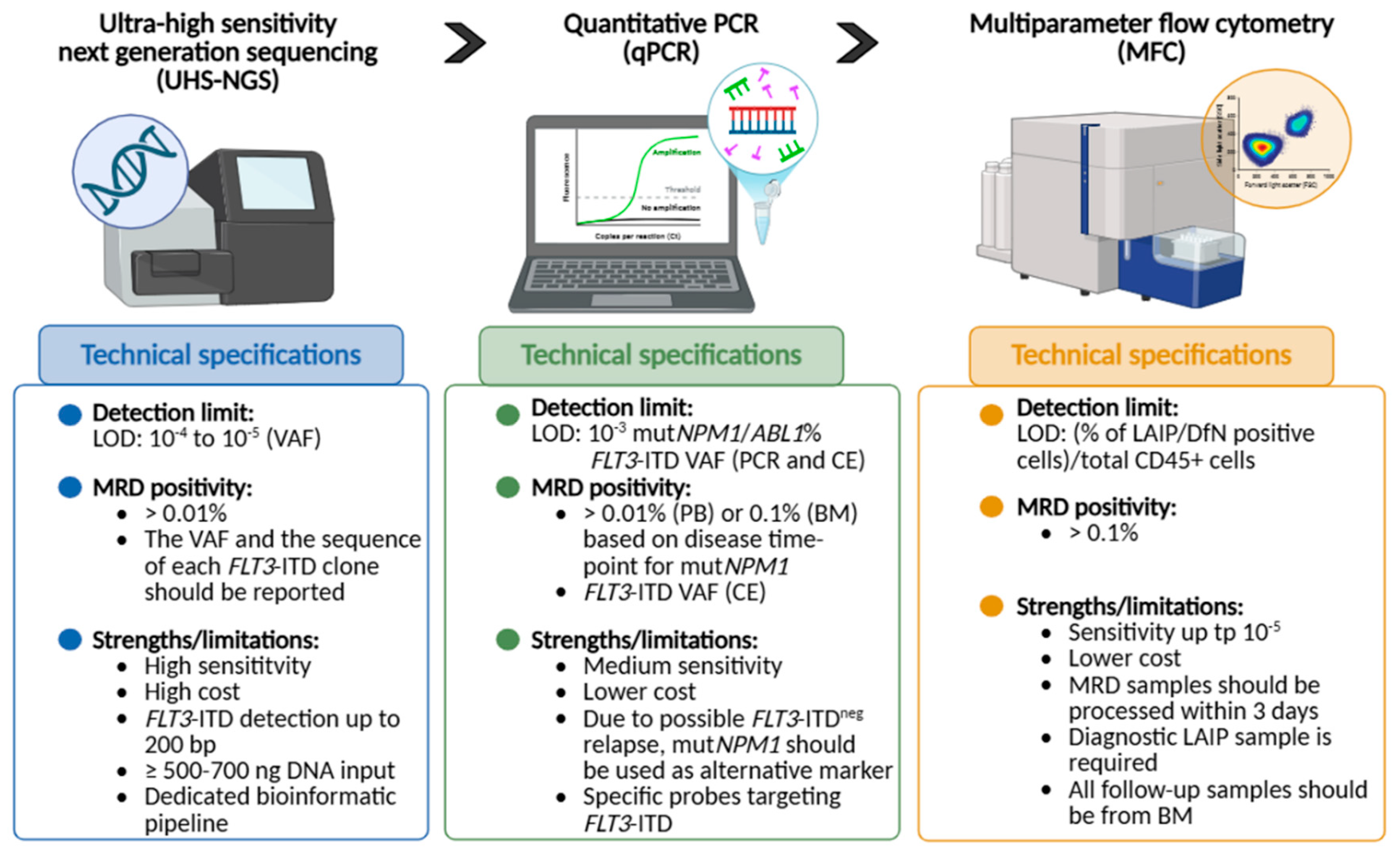
Figure 2.
Timeline of MRD assessment in FLT3-ITDmut AML. Overview of the informative time-points for the detection of FLT3-ITD mutational burden. MRD: measurable residual disease; NGS: next generation sequencing; HSCT: hematopoietic stem cell transplantation; FLT3i: FLT3 inhibitors. Created with BioRender.com.
Figure 2.
Timeline of MRD assessment in FLT3-ITDmut AML. Overview of the informative time-points for the detection of FLT3-ITD mutational burden. MRD: measurable residual disease; NGS: next generation sequencing; HSCT: hematopoietic stem cell transplantation; FLT3i: FLT3 inhibitors. Created with BioRender.com.
Disclaimer/Publisher’s Note: The statements, opinions and data contained in all publications are solely those of the individual author(s) and contributor(s) and not of MDPI and/or the editor(s). MDPI and/or the editor(s) disclaim responsibility for any injury to people or property resulting from any ideas, methods, instructions or products referred to in the content. |
© 2026 by the authors. Licensee MDPI, Basel, Switzerland. This article is an open access article distributed under the terms and conditions of the Creative Commons Attribution (CC BY) license (http://creativecommons.org/licenses/by/4.0/).
Copyright: This open access article is published under a Creative Commons CC BY 4.0 license, which permit the free download, distribution, and reuse, provided that the author and preprint are cited in any reuse.