

Submitted:
16 July 2026
Posted:
17 July 2026
You are already at the latest version
Abstract
Background: Respiratory epithelial adenomatoid hamartoma (REAH) and seromucinous hamartoma (SH) are uncommon benign epithelial lesions of the sinonasal tract. Their clinical and endoscopic appearance may overlap with inflammatory nasal polyps, and definitive diagnosis usually requires histopathologic examination. Case presentation: A 37-year-old man presented with several years of nasal obstruction, hyposmia, and facial pressure over the paranasal sinus regions. He had no history of asthma or previous sinonasal surgery. Nasal endoscopy revealed bilateral polypoid lesions involving the middle meatus. Computed tomography showed extensive chronic sinonasal inflammatory disease, including complete opacification of the right maxillary sinus, bilateral frontal sinus involvement, and bilateral ethmoidal opacification. The patient underwent endoscopic sinonasal surgery with bilateral uncinectomy, bilateral antrostomy, and anterior and posterior ethmoidectomy. Polypoid tissue was excised and submitted for histopathologic examination. The final diagnosis was a hybrid epithelial hamartomatous lesion with features of both REAH and SH. At the 4 and 8 week postoperative visit, the patient reported clear symptomatic improvement, mainly in nasal obstruction, smell and nasal breathing. Conclusions: Hybrid REAH and SH may clinically resemble bilateral inflammatory nasal polyposis, especially in patients with chronic rhinosinusitis. This case highlights the importance of routine histopathologic examination of sinonasal polypoid lesions after endoscopic surgery.
Keywords:
case report
; respiratory epithelial adenomatoid hamartoma (REAH)
; seromucinous hamartoma (SH)
; nasal polyp
; chronic rhinosinusitis
; endoscopic sinus surgery
; histopathologic examination
1. Introduction
Hamartomas are benign, tumor-like lesions composed of a disorganized proliferation of mature tissue elements normally present at the affected anatomical site. Unlike true neoplasms, they have traditionally been considered developmental or malformative processes rather than autonomous clonal proliferations. Hamartomas are frequently described in organs such as the lung, gastrointestinal tract, kidney, and liver, whereas involvement of the upper aerodigestive tract is uncommon [1,2]. Within the sinonasal tract, several histologically distinct hamartomatous lesions have been recognized, including respiratory epithelial adenomatoid hamartoma (REAH), seromucinous hamartoma (SH), chondro-osseous respiratory epithelial adenomatoid hamartoma, and nasal chondromesenchymal hamartoma. REAH and SH are of uncertain etiology and can occur in isolation or in chronic rhinosinusitis, as they are frequently associated with inflammatory sinonasal disorders and allergy, including multiple varieties of rhinosinusitis and inflammatory polyps [2].
REAH and SH are composed predominantly of epithelial or glandular structures and may show considerable clinical and histopathological overlap. Their etiology and pathogenesis remain incompletely understood. Although the term “hamartoma” implies a non-neoplastic developmental proliferation, the frequent association of REAH with chronic inflammatory sinonasal disease has led to the hypothesis that at least some lesions may represent an acquired reactive process induced by prolonged mucosal inflammation [2]. Conversely, molecular alterations identified in a proportion of REAH and SH lesions have raised the possibility that some may have a neoplastic component. At present, however, both entities are regarded as benign lesions with indolent clinical behavior.
REAH is currently considered the most frequently recognized epithelial hamartoma of the sinonasal tract [2]. REAH has been reported over a wide age range but predominantly affects middle-aged and older adults, with a clear male predominance in most published series [1,2,3,4]. It can occur as an isolated localized mass or as a more diffuse lesion associated with chronic rhinosinusitis and inflammatory nasal polyposis. In localized cases, the posterior-superior nasal septum and olfactory cleft are characteristic sites of involvement [2,5,6]. In a recent series, 56% of patients had olfactory cleft involvement. These patients were normally older and had higher rates of comorbid allergic rhinitis and asthma [6].
SH is considerably less common. Since its initial recognition, only a limited number of cases and small clinicopathological series have been published. SH generally occurs in adults and most often involves the nasal septum or posterior nasal cavity, although lesions arising in the ethmoid region, lateral nasal wall, and paranasal sinuses have also been reported [2].
The clinical presentation of REAH and SH is nonspecific. Patients may report progressive nasal obstruction, hyposmia or anosmia, rhinorrhea, epistaxis, headache, facial pressure, or other symptoms indistinguishable from those of chronic rhinosinusitis [3,4,7,8]. When the lesions arise in the olfactory clefts, olfactory dysfunction may be particularly prominent.
Endoscopically, REAH usually appears as a polypoid, nodular, or fleshy lesion with a pink, white, or yellowish surface. SH may also present as a polypoid or nodular mass. Neither entity has a sufficiently specific endoscopic appearance to permit a reliable preoperative diagnosis [2,6].
Radiological findings are likewise variable. Sinonasal computed tomography may show a localized soft-tissue mass or diffuse mucosal opacification associated with chronic inflammatory disease. Widening of the olfactory clefts has been described as a useful imaging sign of REAH, particularly when the lesion is centered in the superior nasal cavity [5,9]. Nevertheless, this finding is not universally present and is less useful in extra-olfactory cleft or diffuse disease. Moreover, patients with extensive chronic rhinosinusitis may exhibit widespread sinonasal opacification that obscures the underlying hamartomatous component. Consequently, the radiological interpretation is frequently inflammatory nasal polyposis, and the definitive diagnosis is established only after histopathological examination of surgical specimens.
The histological diagnosis of REAH is based on a proliferation of round to oval glands lined by ciliated pseudostratified respiratory epithelium. The glands frequently appear to invaginate directly from the surface epithelium and are usually separated by inflamed or edematous stroma. One of the most characteristic features is the presence of a thick eosinophilic basement membrane-like material surrounding the glands [2]. Cytological atypia, increased mitotic activity, necrosis, and destructive stromal invasion are absent.
In contrast, SH is composed of crowded, variably sized seromucinous glands and tubules lined by a single layer of bland cuboidal or low columnar epithelial cells [2,10,11]. An important feature is the absenceof an identifiable myoepithelial cell layer around the glands [10]. Although loss of myoepithelial cells may ordinarily raise concern for an invasive glandular malignancy, the overall circumscribed architecture, cytological blandness, absence of destructive infiltration, and lack of other malignant features support the diagnosis of SH.
A close morphological relationship exists between REAH and SH. Some lesions contain unequivocal areas of both respiratory epithelial glandular proliferation and seromucinous glandular proliferation and have been described as hybrid epithelial hamartomas [2,11]. However, focal seromucinous differentiation may occur within REAH, and respiratory epithelial invaginations can be identified in SH. Therefore, the boundaries between these entities are not always sharply defined. Some authors have suggested that REAH, SH, and hybrid lesions may represent points along a spectrum of benign sinonasal epithelial proliferations and that classification according to the predominant morphological component may be more appropriate than rigid separation into distinct categories [2].
The recognition of these lesions is clinically important because they may mimic both benign inflammatory conditions and malignant epithelial neoplasms. RH and SH can mimic nasal polyposis, inverted papilloma or adenocarcinoma, squamous cell carcinoma, olfactory neuroblastoma, and lymphoma [3,4,8]. Inverted papillomas, while commonly displaying respiratory type epithelium, tend to have a predominance of squamotransitional epithelium, with a more irregular inverted growth pattern than is seen in the well-spaced glands characteristic of REAH. Additionally, REAH most commonly have less intraepithelial neutrophilic inflammation and thicker basement membrane-like material surrounding the glands [2].
Hybrid lesions containing well-developed REAH and SH components remain exceptionally uncommon. Their presentation within bilateral polypoid sinonasal disease creates an additional diagnostic challenge because the clinical and radiological findings strongly suggest conventional chronic rhinosinusitis with nasal polyps. This report describes a hybrid epithelial hamartomatous lesion with features of both REAH and SH in a young adult who underwent endoscopic sinus surgery for presumed bilateral inflammatory nasal polyposis. The case emphasizes the clinicopathological overlap between sinonasal hamartomas and chronic inflammatory disease, the importance of systematic histopathological examination, and the need to distinguish these benign proliferations from malignant glandular lesions.
2. Detailed Case Description
A 37-year-old man presented with several years of nasal obstruction, hyposmia, and facial pressure over the paranasal sinus regions. He denied mucopurulent rhinorrhea. He had no history of asthma or previous sinonasal surgery.
Nasal endoscopy revealed bilateral polypoid lesions involving the middle meatus. Computed tomography of the paranasal sinuses (Figure 1) showed complete opacification of the right maxillary sinus, associated thickening of the bony walls, obliteration of the right maxillary drainage infundibulum, and partial opacification of the right middle meatus. Complete opacification of the left frontal sinus with internal calcifications was also observed, together with near-complete opacification of the right frontal sinus and extensive bilateral opacification of the ethmoid air cells. These findings were considered compatible with chronic rhinosinusitis with nasal polyps.
The patient underwent functional endoscopic sinus surgery (FESS) under general anesthesia. Bilateral uncinectomy and antrostomy were performed, together with anterior and posterior ethmoidectomy. Polypoid tissue was removed from the anterior and posterior ethmoid cavities and mucopurulent material from the right maxillary sinus was suctioned. The excised tissue and polyps were submitted for routine histopathological examination.
Histopathologic analysis (Figure 2) revealed a hybrid epithelial hamartomatous lesion composed of features of both seromucinous hamartoma and respiratory epithelial adenomatoid hamartoma. The seromucinous component showed a crowded proliferation of small seromucinous glands lined by bland cuboidal epithelial cells, without cytologic atypia. The background stroma is edematous and periglandular hyalinization can be noted. The REAH component showed a proliferation of benign glands lined by ciliated respiratory-type columnar epithelium and surrounded by a thick eosinophilic hyalinized basement membrane. The glandular lumina contain mucinous or amorphous material. The stroma surrounding gland is edematous and contains a mixed inflammatory infiltrate resembling the stroma of inflammatory polyps. No histologic features of malignancy were identified. No immunohistochemistry was performed.
At the 4-week and 8-week postoperative visit, the patient reported clear clinical improvement, especially in smell, with complete recovering, and in nasal obstruction, with better nasal breathing. Postoperative nasal endoscopy at 8 weeks showed patent, well-healed sinonasal cavities without residual polypoid lesions (Figure 3 and Figure 4).
3. Discussion
REAH and SH are benign epithelial proliferations of the sinonasal tract that remain unfamiliar to many clinicians because of their rarity, nonspecific clinical manifestations, and frequent association with inflammatory disease. The present case illustrates several diagnostically relevant features: the lesion occurred in a relatively young adult, presented as bilateral polypoid disease, was associated with extensive radiological findings of chronic rhinosinusitis, and was diagnosed unexpectedly after routine histopathological examination of tissue removed during endoscopic sinus surgery.
The epidemiological profile of the present patient differs partially from that reported in most REAH series. Although REAH can occur from early adulthood to advanced age, the typical patient is a middle-aged or older man [1,2,3,4]. SH likewise predominantly affects adults, with most reported cases occurring in middle or late adulthood [2,10,11]. The age of 37 years is therefore younger than the usual age at diagnosis but remains within the reported spectrum. Male sex is consistent with the demographic predominance described for REAH, whereas SH has not shown an equally consistent sex distribution across the limited available series.
The anatomical distribution also deserves consideration. REAH is characteristically associated with the posterior-superior nasal septum and olfactory clefts [2,5,9]. In a recent series comparing olfactory cleft and extra-olfactory cleft disease, olfactory cleft involvement was identified in a substantial proportion of patients, but lesions outside this location were also recognized [6]. Extra-olfactory cleft lesions may arise in the middle meatus, ethmoid cavities, lateral nasal wall, or paranasal sinuses and may be more difficult to distinguish clinically from ordinary inflammatory polyps. In the present case, no olfactory cleft lesion was specifically described on computed tomography, preoperative endoscopy, or the operative report. The polypoid tissue was principally identified in the middle meatuses and ethmoid cavities. Nevertheless, the absence of documented olfactory cleft involvement should not be interpreted as definitive proof that this region was entirely unaffected, because the available imaging was originally evaluated in the context of chronic rhinosinusitis rather than specifically for REAH. In addition, the histological specimens were obtained from polypoid ethmoidal tissue and were not mapped systematically according to individual sinonasal subsites. It is therefore more accurate to describe the case as clinically dominated by middle meatal and ethmoidal disease rather than to conclude categorically that the olfactory clefts were uninvolved.
The radiological appearance of REAH is variable and depends on whether the lesion is isolated or associated with diffuse inflammatory disease. Localized REAH may appear as a homogeneous soft-tissue mass, whereas diffuse REAH associated with nasal polyposis may be indistinguishable from inflammatory mucosal thickening. Hawley et al. identified olfactory cleft widening as a potentially useful radiological marker, particularly when the cleft width reaches or exceeds approximately 10 mm [9]. However, this sign is primarily applicable to olfactory cleft-centered REAH and cannot exclude REAH arising elsewhere. Moreover, extensive chronic rhinosinusitis may conceal localized architectural changes. Thus, radiology can raise suspicion but does not replace histopathological diagnosis.
The extensive bilateral ethmoidal, maxillary, and frontal sinus opacification observed in our patient strongly favored a preoperative diagnosis of chronic rhinosinusitis with nasal polyps. Complete right maxillary sinus opacification, bony wall thickening, and infundibular obstruction indicated longstanding inflammatory disease. Frontal sinus opacification and internal calcifications within the left frontal sinus were likewise interpreted in an inflammatory context. None of these findings specifically suggested an epithelial hamartoma. This emphasizes that REAH and SH may be microscopic or incidental components within tissue that is clinically and radiologically dominated by chronic inflammation rather than discrete masses identifiable before surgery.
The relationship between REAH and chronic inflammation remains unresolved. REAH has been reported both as an isolated lesion and in association with chronic rhinosinusitis, allergic rhinitis, asthma, and inflammatory nasal polyposis [2,5,6]. Its frequent coexistence with inflammatory polyps has prompted the hypothesis that chronic mucosal irritation may induce glandular proliferation and respiratory epithelial invagination. Under this model, REAH could represent an exaggerated reactive response rather than a congenital malformation. An alternative possibility is that REAH represents a true benign neoplasm or a heterogeneous group containing both reactive and neoplastic lesions. Nevertheless, the absence of aggressive clinical behavior, destructive invasion, significant atypia, and metastatic potential supports their continued classification as benign lesions. From a practical perspective, whether they are considered reactive proliferations, hamartomas, or indolent neoplasms does not currently alter management, which is based on complete conservative endoscopic excision when symptomatic.
Histologically, the REAH component in this case demonstrated the characteristic combination of respiratory epithelial-lined glands, ciliation, thick eosinophilic basement membrane-like material, and an inflamed edematous stroma. Recognition of the thick periglandular basement membrane is particularly helpful because inflammatory polyps may contain entrapped or hyperplastic glands but generally lack the regular adenomatoid proliferation and prominent hyalinized basement membrane characteristic of REAH [2].
The SH component creates a separate but overlapping diagnostic problem. SH is characterized by a proliferation of small seromucinous glands lined by uniform cuboidal or low columnar cells. The glands may be closely packed and can lack an apparent myoepithelial cell layer [10]. Because loss of myoepithelial cells is commonly associated with invasive epithelial neoplasia in salivary-type tissues, this finding may produce concern for low-grade adenocarcinoma. However, the absence of myoepithelial cells should not be interpreted in isolation. In SH, the glands maintain bland cytology and generally lack a destructive, infiltrative, or desmoplastic pattern.
The lesion in our patient showed unequivocal areas with the morphological characteristics of SH together with separate glands diagnostic of REAH. This supports classification as a hybrid epithelial hamartomatous lesion. Hybrid lesions were recognized in early descriptions of SH and have continued to be discussed in subsequent reviews [2,11]. Their existence raises questions regarding the histogenetic relationship between the two entities. Both arise within the same specialized sinonasal mucosa, both may be associated with inflammation, and both exhibit benign glandular differentiation. Respiratory epithelial invaginations may transition into smaller seromucinous structures, creating a morphological continuum. Hahn and Weinreb emphasized the substantial morphological overlap between REAH and SH and suggested that lesions containing both patterns may be named according to their predominant component rather than necessarily being regarded as a separate biological entity [2]. In the current case, both components were sufficiently represented to support the descriptive term “hybrid REAH and SH.”
Immunohistochemistry was not performed in the present case. This is a reasonable approach when hematoxylin and eosin morphology is characteristic and no malignant features are identified.
The treatment of symptomatic REAH and SH is conservative surgical excision, most commonly through an endoscopic approach [11]. Published series indicate that endoscopic removal is usually curative, and recurrence is uncommon when the lesion has been adequately excised [11]. Cases occurring in diffuse chronic rhinosinusitis may require surgery primarily to restore sinus ventilation and remove inflammatory disease rather than to achieve wide margins around the hamartoma.
In the present case, bilateral uncinectomy, antrostomy, and anterior and posterior ethmoidectomy provided adequate treatment of both the inflammatory sinonasal disease and the hamartomatous proliferation. The patient experienced substantial early improvement in nasal obstruction, nasal airflow, and olfactory function. At 8 weeks, endoscopy demonstrated patent, healed cavities without residual polypoid tissue.
The follow-up period remains short, and longer surveillance is necessary to document persistent symptom control and exclude recurrent inflammatory or hamartomatous disease. Nevertheless, no histological feature suggested an increased risk of aggressive recurrence. Follow-up should therefore be guided primarily by the extent and phenotype of the patient’s chronic rhinosinusitis rather than by concern for malignant behavior of the hamartoma.
A central clinical implication of this case concerns the role of routine histopathological examination after sinonasal surgery. The value and cost-effectiveness of routinely examining bilateral nasal polyps has been debated because the majority demonstrate conventional inflammatory changes [12]. Some studies have suggested selective submission when the lesion is unilateral, atypical, recurrent, or associated with concerning clinical or radiological features. However, the current case demonstrates that rare epithelial lesions may be clinically indistinguishable from ordinary bilateral polyposis. Without histopathological evaluation, the hybrid hamartomatous component would not have been recognized. Although detection of a benign hamartoma did not require additional oncological treatment, obtaining the correct diagnosis has several benefits. It prevents future misinterpretation of the lesion as a low-grade malignancy, contributes to accurate documentation of rare pathological entities, improves communication between otolaryngologists, radiologists, and pathologists, and expands knowledge of their true prevalence. It may also prompt more focused radiological review if recurrent superior nasal cavity or olfactory cleft disease develops.
The case has several limitations. First, the follow-up was limited to 8 weeks, precluding conclusions regarding long-term recurrence. Second, the pathological specimen was obtained during surgery for diffuse inflammatory disease and was not prospectively mapped according to exact sinonasal subsite. The precise anatomical distribution of the hamartomatous components therefore cannot be reconstructed with certainty. Third, olfactory function was assessed clinically according to patient-reported recovery and was not measured using a validated psychophysical olfactory test. Fourth, immunohistochemical or molecular studies were not performed. Finally, as a single case, the report cannot establish the pathogenesis, prevalence, or optimal terminology of hybrid REAH–SH lesions.
Despite these limitations, the case is relevant because it documents the coexistence of characteristic REAH and SH components in bilateral polypoid sinonasal tissue from a patient treated for presumed chronic rhinosinusitis with nasal polyps. It illustrates how extensive inflammatory disease can conceal a rare epithelial proliferation and reinforces the importance of correlating histological glandular architecture with clinical and radiological findings. Greater awareness of REAH, SH, and their hybrid forms is essential to avoid both under-recognition as nonspecific polyposis and overdiagnosis as a malignant glandular neoplasm.
4. Conclusions
Hybrid sinonasal epithelial hamartomas containing features of both REAH and SH are rare benign lesions that may be clinically, endoscopically, and radiologically indistinguishable from chronic rhinosinusitis with bilateral nasal polyps. Definitive diagnosis relies on recognition of the characteristic histopathological components: respiratory epithelial-lined glands with thick eosinophilic basement membrane material in REAH and bland seromucinous glandular proliferation in SH.
The absence of cytological atypia, destructive invasion, desmoplasia, necrosis, and other malignant features is essential for distinguishing these lesions from low-grade sinonasal adenocarcinoma and other neoplasms. Conservative endoscopic excision is generally adequate, although continued follow-up is appropriate, particularly when the lesion occurs in the setting of extensive chronic inflammatory sinonasal disease.
This case supports the value of histopathological examination of surgically removed sinonasal polypoid tissue, even when the preoperative presentation is entirely compatible with bilateral inflammatory nasal polyposis. Accurate recognition promotes appropriate conservative management and prevents both misclassification as nonspecific inflammatory disease and overdiagnosis as malignancy.
Author Contributions
Conceptualization, Raquel Muñoz-Andrés, Carlos Carazo-Casas, Mayte Herrera; methodology, Raquel Muñoz-Andrés, Carlos Carazo-Casas, Mayte Herrera, Alfonso Santamaría-Gadea; software, Raquel Muñoz-Andrés, Carlos Carazo Casas.; investigation, Raquel Muñoz-Andrés, Carlos Carazo Casas, Mayte Herrera; resources, Raquel Muñoz-Andrés, Carlos Carazo Casas, Mónica García-Cosio Piqueras, Kadir Jesús Quintero-Gil.; writing—original draft preparation, Raquel Muñoz-Andrés, Carlos Carazo-Casas; writing—review and editing, Raquel Muñoz-Andrés, Carlos Carazo-Casas, Alfonso Santamaría-Gadea.; supervision, Mayte Herrera, Alfonso Santamaría-Gadea. All authors have read and agreed to the published version of the manuscript.
Funding
This research received no external funding.
Institutional Review Board Statement
Not applicable.
Informed Consent Statement
Written informed consent has been obtained from the patients to publish this paper.
Data Availability Statement
No new data were created.
Conflicts of Interest
The authors declare no conflicts of interest.
References
- Liao, J.; Zhang, X.; Xu, W.; Li, F.; Qin, G. Nasal Respiratory Epithelial Adenomatoid Hamartoma: Three Case Reports and Review of the Literature. Ear Nose Throat J. 2024, 103, 696–701. [Google Scholar] [CrossRef] [PubMed]
- Hahn, E.; Weinreb, I. Sinonasal Hamartomas. Surg. Pathol. Clin. 2024, 17, 577–585. [Google Scholar] [CrossRef] [PubMed]
- Fernandes Lopes, P.; Ruas Morgado, F.; Canavarros, F.; Santos, A.; Macor, C. Respiratory Epithelial Adenomatoid Hamartoma, a Different Presentation. Clin. Case Rep. 2025, 13, e8345. [Google Scholar] [CrossRef] [PubMed]
- Houas, J.; Ghammam, M.; Farhani, S.; Naouar, M.; Mokni, W.; Sriha, B.; Abdelkefi, M. Respiratory Epithelial Adenomatoid Hamartoma: Expanding the Spectrum of Rare and Misleading Nasopharyngeal Lesions. Ear Nose Throat J. 2025, 01455613251329672. [Google Scholar] [CrossRef] [PubMed]
- Shanbag, R.; Patil, P.; Rani, S.H.; Kulkarni, S. Respiratory Epithelial Adenomatoid Hamartoma (REAH) in the Olfactory Cleft: Often Masked by Bilateral Nasal Polyps. Indian J. Otolaryngol. Head. Neck Surg. 2019, 71, 2121–2126. [Google Scholar] [CrossRef] [PubMed]
- Fischer, J.L.; Oh, E.; Sajed, D.P.; Beswick, D.M.; Lee, J.T.; Wang, M.B.; Suh, J.D. Respiratory Epithelial Adenomatoid Hamartoma Characteristics of Extra-Olfactory Cleft Disease. The Laryngoscope 2026, 136, 658–668. [Google Scholar] [CrossRef] [PubMed]
- Liu, S.; Bai, B.; Feng, Y.; Li, M.; Li, L.; Gong, L. A Case Report of Epithelial Adenomatoid Hamartoma Misdiagnosed as a Nasal Polyp. Medicine 2025, 104, e41424. [Google Scholar] [CrossRef] [PubMed]
- Nefzaoui, S.; Zoghlami, I.; Gharsalli, J.; Sabehi, E.; Romdhane, N.; Helal, I.; Chiboub, D.; Hariga, I.; Mbarek, C. Respiratory Epithelial Adenomatoid Hamartoma: A Rare Differential Diagnosis of Sinonasal Mass. Rare Tumors 2024, 16, 20363613241255567. [Google Scholar] [CrossRef] [PubMed]
- Hawley, K.A.; Ahmed, M.; Sindwani, R. CT Findings of Sinonasal Respiratory Epithelial Adenomatoid Hamartoma: A Closer Look at the Olfactory Clefts. AJNR Am. J. Neuroradiol. 2013, 34, 1086–1090. [Google Scholar] [CrossRef] [PubMed]
- Weinreb, I.; Gnepp, D.R.; Laver, N.M.; Hoschar, A.P.; Hunt, J.L.; Seethala, R.R.; Barnes, E.L.; Chetty, R.; Perez-Ordoñez, B. Seromucinous Hamartomas: A Clinicopathological Study of a Sinonasal Glandular Lesion Lacking Myoepithelial Cells. Histopathology 2009, 54, 205–213. [Google Scholar] [CrossRef] [PubMed]
- Khan, R.A.; Chernock, R.D.; Lewis, J.S. Seromucinous Hamartoma of the Nasal Cavity: A Report of Two Cases and Review of the Literature. Head. Neck Pathol. 2011, 5, 241. [Google Scholar] [CrossRef] [PubMed]
- Wong, J.S.; Hoffbauer, S.; Yeh, D.H.; Rotenberg, B.; Gupta, M.; Sommer, D.D. The Usefulness of Routine Histopathology of Bilateral Nasal Polyps – a Systematic Review, Meta-Analysis, and Cost Evaluation. J. Otolaryngol.-Head. Neck Surg. 2015, 44, 46. [Google Scholar] [CrossRef] [PubMed]
Figure 1.
Coronal computed tomography of the paranasal sinuses. (A) Coronal CT image showing opacification of the right maxillary sinus and ethmoid air cells. (B) Coronal CT image showing opacification of the left frontal sinus with internal calcifications.
Figure 1.
Coronal computed tomography of the paranasal sinuses. (A) Coronal CT image showing opacification of the right maxillary sinus and ethmoid air cells. (B) Coronal CT image showing opacification of the left frontal sinus with internal calcifications.
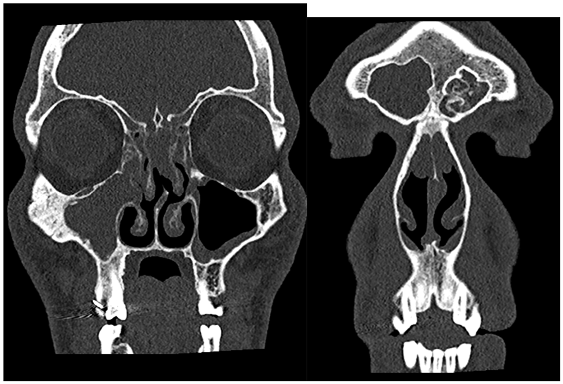
Figure 2.
Histopathologic features of the hybrid epithelial hamartomatous lesion. (A) Seromucinous hamartoma component showing a crowded proliferation of small seromucinous glands lined by bland cuboidal epithelial cells, without cytologic atypia. Not surrounded by a basement membrane. Hematoxylin and eosin, objective magnification. (B,C) Respiratory epithelial adenomatoid hamartoma component showing benign glands lined by ciliated respiratory-type columnar epithelium and surrounded by a thick eosinophilic hyalinized basement membrane. Hematoxylin and eosin, objective magnification.
Figure 2.
Histopathologic features of the hybrid epithelial hamartomatous lesion. (A) Seromucinous hamartoma component showing a crowded proliferation of small seromucinous glands lined by bland cuboidal epithelial cells, without cytologic atypia. Not surrounded by a basement membrane. Hematoxylin and eosin, objective magnification. (B,C) Respiratory epithelial adenomatoid hamartoma component showing benign glands lined by ciliated respiratory-type columnar epithelium and surrounded by a thick eosinophilic hyalinized basement membrane. Hematoxylin and eosin, objective magnification.
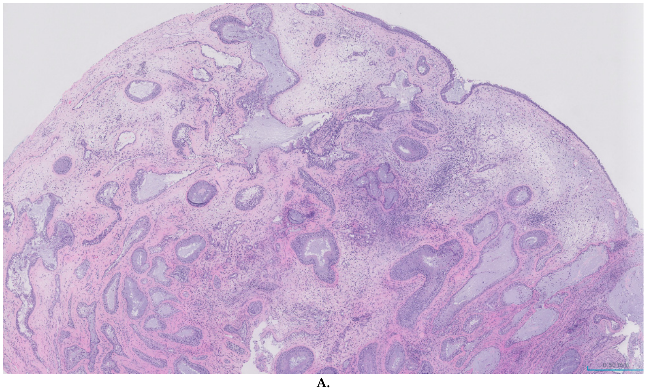
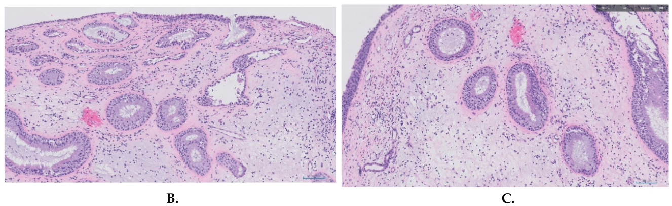
Figure 3.
Postoperative results after 8 weeks. Right maxillary sinus (A) and right ethmoidectomy (B). Left maxillary sinus (C).
Figure 3.
Postoperative results after 8 weeks. Right maxillary sinus (A) and right ethmoidectomy (B). Left maxillary sinus (C).
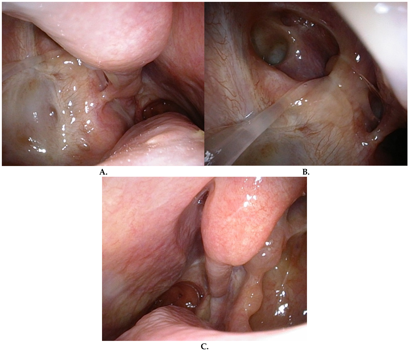
Figure 4.
QR code to access the video showing the nasal endoscopy in the 8th week follow-up.

Disclaimer/Publisher’s Note: The statements, opinions and data contained in all publications are solely those of the individual author(s) and contributor(s) and not of MDPI and/or the editor(s). MDPI and/or the editor(s) disclaim responsibility for any injury to people or property resulting from any ideas, methods, instructions or products referred to in the content. |
© 2026 by the authors. Licensee MDPI, Basel, Switzerland. This article is an open access article distributed under the terms and conditions of the Creative Commons Attribution (CC BY) license (http://creativecommons.org/licenses/by/4.0/).
Copyright: This open access article is published under a Creative Commons CC BY 4.0 license, which permit the free download, distribution, and reuse, provided that the author and preprint are cited in any reuse.