
Submitted:
15 July 2026
Posted:
16 July 2026
You are already at the latest version
Abstract
Background: Maxillary sinus floor elevation is a predictable procedure for implant re-habilitation in the posterior maxilla with reduced residual bone height. Schneiderian membrane perforation is the most frequent intraoperative complication during sinus lift surgery and may compromise graft stability and treatment outcomes. Stabilized collagen membrane techniques have been proposed to manage extensive perforations.
Objective: To describe the management of a large Schneiderian membrane perforation (>10 mm) using a stabilized collagen membrane technique and to evaluate the clinical outcome.
Case Description: A 67-year-old male patient (ASA I, non-smoker) with insufficient posterior maxillary bone height underwent lateral window sinus augmentation. During membrane elevation, a large perforation (>10 mm) occurred. A resorbable collagen membrane was adapted and stabilized to isolate the defect and create a contained compartment for graft placement. A xenograft was inserted, and an additional collagen membrane was used to cover the lateral window and stabilized with fixation pins. Pri-mary closure was achieved with tension-free sutures.
Results: Healing was uneventful, without sinus pathology or postoperative complica-tions. Radiographic evaluation confirmed graft stability and adequate volume mainte-nance. Implant placement was successfully performed after 6 months, achieving satis-factory primary stability and favorable clinical outcomes after one year of loading.
Conclusion: The stabilized collagen membrane technique allowed predictable man-agement of a large Schneiderian membrane perforation, enabling successful graft con-tainment and implant rehabilitation.
Keywords:
sinus floor augmentation
; sinus membrane perforation
; dental implants
; bone transplantation
; case reports
1. Introduction
The rehabilitation of the atrophic posterior maxilla with dental implants remains a clinical challenge due to the progressive reduction of alveolar bone volume and height following tooth loss, often resulting in insufficient bone availability for implant placement.[1] In these cases, maxillary sinus floor elevation using the lateral window approach has become a predictable surgical procedure to achieve vertical bone augmentation and enable implant placement. [1]
The lateral window technique has demonstrated high long-term implant survival rates – 95.6% after 5 years and 93.1% after 10 years. [2] However, the procedure remains technique-sensitive and may be influenced by several anatomical and surgical factors, including sinus septa, membrane thickness, residual ridge height, sinus pathology, and operator experience. [3,4]
Among the reported complications associated with sinus floor elevation procedures, Schneiderian membrane perforation is considered the most common intraoperative event, with incidence rates reported around 20-25% depending on the surgical approach and anatomical conditions. [4] Large membrane perforations may compromise graft stability, increase the risk of sinus contamination and postoperative complications, and potentially affect treatment outcomes if not adequately managed. [4]
Several techniques have been proposed for the management of Schneiderian membrane perforations, including direct suturing, use of resorbable collagen membranes, and stabilization approaches designed to isolate the defect and maintain graft containment.[4] The use of stabilized collagen membranes has shown promising clinical outcomes in the management of extensive perforations, allowing continuation of the augmentation procedure without interruption. [5,6]
This case report describes the management of a large Schneiderian membrane perforation (>10 mm) occurring during lateral window sinus augmentation using a stabilized collagen membrane technique, as well as the clinical and radiographic outcomes following 1 year after the implant rehabilitation.
2. Case Presentation
A 67-year-old male patient presented to a private clinic seeking an implant-supported rehabilitation of the maxilla. The patient expressed a strong preference for a full fixed rehabilitation, as he had previously experienced difficulties adapting to a removable prosthesis and was dissatisfied with its comfort, stability and function.
The patient was classified as ASA I, with no relevant systemic diseases and reported being a non-smoker. No known drug allergies or ongoing medications were reported. The patient presented with a fully edentulous maxilla, with a history of tooth loss attributed to dental caries and periodontal disease.
2.1. Diagnostic Assessment
Initial radiographic assessment included panoramic radiography and cone-beam computed tomography (CBCT). Three-dimensional evaluation demonstrated severe vertical bone deficiency in the posterior maxilla (second quadrant), with a residual bone height of approximately 5 mm (Figure 1), precluding predictable implant placement without prior sinus augmentation. A sinus retention cyst was also detected during the preoperative assessment.
2.2. Therapeutic Intervention
A lateral window sinus augmentation procedure was planned and performed under local anesthesia using articaine with epinephrine (Artinibsa® 40 mg/mL + 0.01 mg/mL, Inibsa®, Llissá de Vall, Spain). Following anesthesia, a mid-crestal incision with buccal releasing incisions was performed, and a full-thickness mucoperiosteal flap was elevated to expose the lateral wall of the maxillary sinus. The dimensions and location of the lateral window were determined preoperatively using CBCT evaluation. After preparation of the lateral window with a piezoelectric surgical device (VarioSurg™, NSK, Tokyo, Japan), careful elevation of the Schneiderian membrane was initiated using sinus curettes (Hu-Friedy®, Chicago, IL, USA) (Figure 2).
During membrane elevation, a large perforation (>10 mm) of the Schneiderian membrane occurred (Figure 3). Instead of aborting the procedure, Schneiderian membrane elevation was cautiously completed.
Following the technique described by Testori et al. [6], a resorbable collagen membrane (Bio-Gide®, Geistlich®, Wolhusen, Switzerland) was used to reconstruct the perforated Schneiderian membrane. The membrane was stabilized externally and folded inward to create a protected compartment for graft containment. A xenogeneic bone substitute (Bio-Oss® S, Geistlich®, Wolhusen, Switzerland) was placed within this compartment, and a second collagen membrane was positioned over the lateral window and secured with fixation pins (Figure 4). Tension-free primary closure was obtained using interrupted sutures (Supramid®, B. Braun®, Melsungen, Germany).
The postoperative regimen included amoxicillin/clavulanic acid (875/125 mg) twice daily for 8 days, a systemic corticosteroid (60 mg/day) for 3 days, paracetamol (500 mg) combined with codeine phosphate hemihydrate (30 mg) twice daily for 3 days for pain control, and a 0.12% chlorhexidine mouthwash three times daily for 10 days.
2.3. Follow-up and Outcomes
Postoperative healing was uneventful, with no signs of infection, sinus complications, or graft exposure. Clinical follow-up visits were conducted at 1 week, 2 weeks and 1 month, with sutures being removed at 2 weeks.
CBCT evaluation after a 6-month healing period (Figure 4) confirmed successful graft containment and volume stability (Figure 5).
Implant placement was subsequently performed, with six Nobel Biocare Replace (Nobel Biocare®, Kloten, Switzerland) implants inserted in the augmented maxilla according to the prosthetically driven treatment plan. The implants were distributed to maximize anteroposterior spread and provide adequate support for the planned full-arch rehabilitation. (Figure 6). All implants achieved satisfactory primary stability at the time of placement, allowing progression to the prosthetic rehabilitation phase.
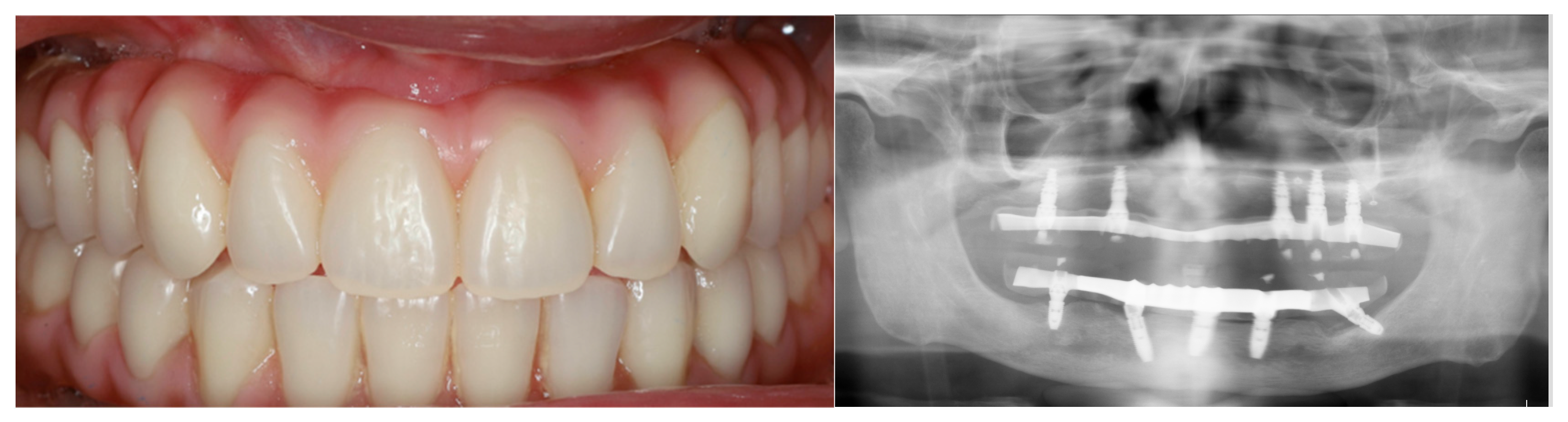
At 1-year follow-up after successful osseointegration, a full-arch fixed implant-supported metalacyclic rehabilitation was performed, restoring function and aesthetics (Figure 7).
At the 1-year follow-up after the rehabilitation, the patient reported a high level of satisfaction with the rehabilitation, expressing confidence during mastication and the absence of pain or discomfort. In the patient's own words, “I am very happy with the rehabilitation.”.
Figure 6.
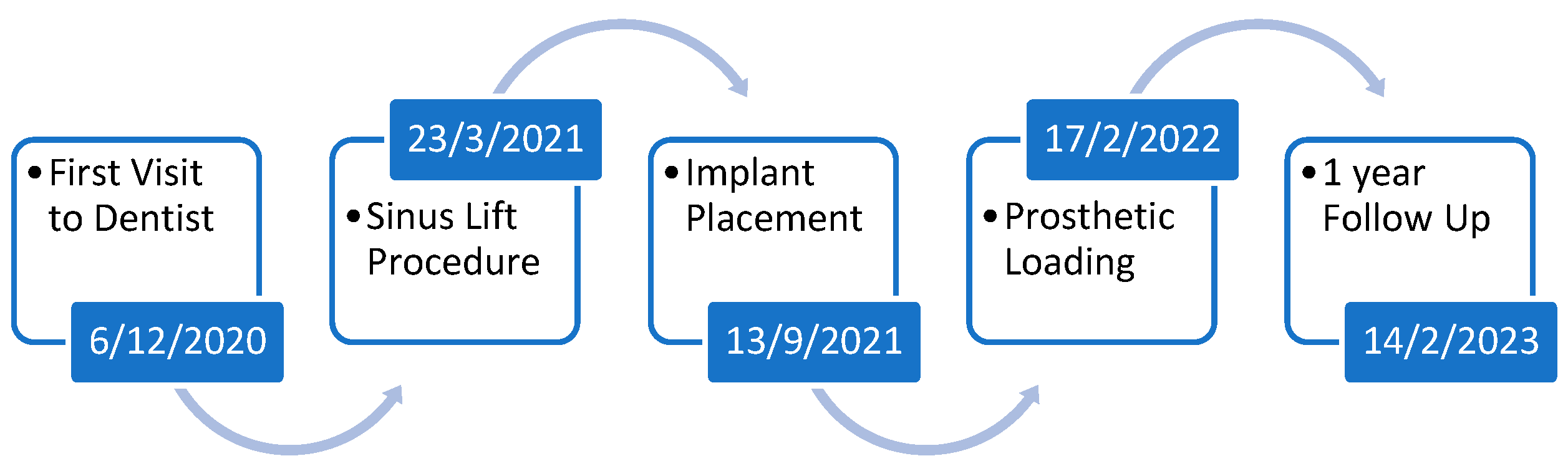
Case Report Timeline.
3. Discussion
Maxillary sinus floor augmentation using the lateral window technique is considered a predictable and well-documented procedure for the rehabilitation of the atrophic posterior maxilla, particularly in patients with limited residual bone height [7]. In the present case, the residual bone height was approximately 3 mm, which justified the indication for sinus augmentation before implant placement. Despite the high success rates reported for this procedure, perforation of the Schneiderian membrane remains the most common intraoperative complication associated with sinus lift surgery. [4]
The incidence of membrane perforation reported in the literature varies considerably, ranging from 6% to 42%, although most studies describe rates between 20% and 25%. Several factors have been associated with an increased risk of perforation, including sinus anatomy, membrane thickness, the presence of septa, retention cyst, and reduced residual bone height [8]. In particular, residual bone heights below 3.5 mm appear to increase the likelihood of membrane perforation during elevation [9,10].
The management of Schneiderian membrane perforations depends largely on the size and extent of the defect. Small perforations can often be managed conservatively, whereas larger defects usually require additional measures to stabilize the membrane and protect the grafted area [4]. The use of resorbable collagen membranes has been widely described as a reliable option for repairing large perforations and maintaining graft containment. However, in extensive defects, some authors still recommend interrupting the procedure and performing a second surgery after healing of the membrane [4].
In the present case, although a large perforation (>10 mm) occurred during membrane elevation, the procedure was not interrupted. Instead, careful continuation of the membrane elevation was performed, followed by stabilization with a resorbable collagen membrane. By partially adapting the membrane externally and folding it internally, it was possible to create a contained compartment capable of supporting and isolating the graft material from the sinus cavity. This approach is consistent with the technique originally described by Testori et al. 2008 [6] for the management of large Schneiderian membrane perforations, which was subsequently refined by Tian et al. 2026 [5]. This strategy appeared to provide sufficient stability for the xenogeneic graft material and helped maintain the augmented space during healing. In addition, the placement of a second collagen membrane over the lateral window may have contributed to graft protection and stabilization, in line with guided bone regeneration principles.
Although membrane perforations have been associated with increased risks of postoperative complications such as sinusitis, graft contamination, and graft instability, these complications were not observed in the present case [10]. Healing progressed uneventfully, with no signs of infection, graft exposure, or sinus-related symptoms during follow-up. Radiographic evaluation demonstrated maintenance of graft volume, allowing implant placement after 6 months with satisfactory primary stability. Previous studies evaluating similar techniques have also reported favorable histologic outcomes, demonstrating new bone formation and successful graft integration despite the occurrence of large Schneiderian membrane perforations [6]. Furthermore, successful osseointegration and definitive full-arch implant-supported rehabilitation were achieved after 1 year.
The present case reinforces the idea that large Schneiderian membrane perforations do not necessarily compromise the outcome of sinus augmentation procedures when appropriately managed. Careful handling of the membrane and adequate stabilization of the grafted compartment appear to be critical factors for achieving favorable healing and successful implant rehabilitation, even in challenging clinical situations with severe maxillary atrophy.
Nevertheless, the limitations inherent to a single case report should be acknowledged, including the limited follow-up period. Further clinical studies with larges samples of patients are necessary to better clarify the predictability of different management approaches for extensive Schneiderian membrane perforations during sinus augmentation procedures.
4. Conclusions
The stabilized collagen membrane technique appears to be a predictable option for the management of large Schneiderian membrane perforations (>10 mm), allowing successful graft containment, continuation of the sinus augmentation procedure, and subsequent implant rehabilitation. Nevertheless, this is a technically demanding surgical approach that requires adequate clinical experience and meticulous execution.
Author Contributions
F. Correia contributed to the study conception, surgical procedures, data acquisition, data analysis, manuscript drafting, and critical revision of the manuscript. S.M. Azevedo contributed to the study conception, manuscript drafting, and critical revision of the manuscript. M. Costa contributed to the study conception, manuscript drafting, and critical revision of the manuscript. R. Faria-Almeida contributed to the study conception and critical revision of the manuscript. All authors gave final approval of the manuscript and agreed to be accountable for all aspects of the work.
Funding
This work received financial support from the PT national funds (FCT/MECI, Fundação para a Ciência e Tecnologia and Ministério da Educação, Ciência e Inovação) through the project UID/50006/2025 DOI 10.54499/UID/50006/2025 -Laboratório Associado para a Química Verde - Tecnologias e Processos Limpos..
Institutional Review Board Statement
Ethical review and approval were waived for this clinical case report due to its descriptive nature and the absence of experimental interventions beyond routine clinical treatment.
Informed Consent Statement
Written informed consent was obtained from the patient and the healthcare proxy/decision-maker for the publication of this clinical case report and the accompanying clinical and radiographic images.
Data Availability Statement
The data presented in this clinical case report are not publicly available due to patient privacy and confidentiality considerations but may be obtained from the corresponding author upon reasonable request.
Acknowledgments
The authors would like to thank the patient for consenting to the publication of this clinical case report and the accompanying clinical and radiographic documentation. During the preparation of this manuscript, the authors used Grammarly (Grammarly Inc.) and ChatGPT (OpenAI) exclusively for limited language editing and grammar correction.
Conflicts of Interest
The authors declare no conflicts of interest.”.
Abbreviations
The following abbreviations are used in this manuscript:
| CBCT | cone-beam computed tomography |
References
- Avila-Ortiz, G.; Vegh, D.; Mukaddam, K.; Galindo-Moreno, P.; Pjetursson, B.; Payer, M. Treatment alternatives for the rehabilitation of the posterior edentulous maxilla. Periodontol. 2000 2023, 93(1), 183–204. [Google Scholar] [CrossRef]
- Pjetursson, B. E.; Thoma, D.; Jung, R.; Zwahlen, M.; Zembic, A. A systematic review of the survival and complication rates of implant-supported fixed dental prostheses (FDPs) after a mean observation period of at least 5 years. Clin. Oral Implant. Res. From NLM. 2012, 23 Suppl 6, 22–38. [Google Scholar] [CrossRef] [PubMed]
- Wang, H. L.; Katranji, A. ABC sinus augmentation classification. Int. J. Periodontics Restor. Dent. From NLM. 2008, 28(4), 383–389. [Google Scholar]
- Molina, A.; Sanz-Sánchez, I.; Sanz-Martín, I.; Ortiz-Vigón, A.; Sanz, M. Complications in sinus lifting procedures: Classification and management. Periodontol 2000 2022, 88(1), 103–115. [Google Scholar] [CrossRef]
- Tian, J.; Jiang, X.; Zhang, Y.; Di, P.; Testori, T.; Lin, Y. The Repair of Large Maxillary Sinus Membrane Perforations with a Collagen Membrane Stabilized with Tacks on the Mesial and Lateral Walls: A Technical Note. Int. J. Periodontics Restor. Dent. 2026, 46(2), 232–243. [Google Scholar] [CrossRef]
- Testori, T.; Wallace, S. S.; Del Fabbro, M.; Taschieri, S.; Trisi, P.; Capelli, M.; Weinstein, R. L. Repair of large sinus membrane perforations using stabilized collagen barrier membranes: surgical techniques with histologic and radiographic evidence of success. Int. J. Periodontics Restor. Dent. From NLM. 2008, 28(1), 9–17. [Google Scholar]
- Starch-Jensen, T.; Jensen, J. D. Maxillary Sinus Floor Augmentation: a Review of Selected Treatment Modalities. J. Oral Maxillofac. Res. 2017, 8(3), e3. [Google Scholar] [CrossRef]
- Wang, W.; Jin, L.; Ge, H.; Zhang, F. Analysis of the Prevalence, Location, and Morphology of Maxillary Sinus Septa in a Northern Chinese Population by Cone Beam Computed Tomography. Comput Math. Methods Med. 2022, 2022, 1644734. [Google Scholar] [CrossRef]
- Yilmaz, H. G.; Tözüm, T. F. Are gingival phenotype, residual ridge height, and membrane thickness critical for the perforation of maxillary sinus? J. Periodontol. From NLM. 2012, 83(4), 420–425. [Google Scholar] [CrossRef] [PubMed]
- Schwarz, L.; Schiebel, V.; Hof, M.; Ulm, C.; Watzek, G.; Pommer, B. Risk Factors of Membrane Perforation and Postoperative Complications in Sinus Floor Elevation Surgery: Review of 407 Augmentation Procedures. J. Oral Maxillofac. Surg. From NLM. 2015, 73(7), 1275–1282. [Google Scholar] [CrossRef] [PubMed]
Figure 1.
CBCT evaluation.
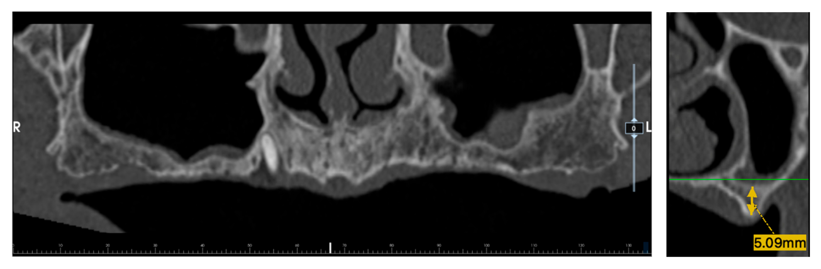
Figure 2.
a - Lateral Window Design; b – Lateral Window Elevation.
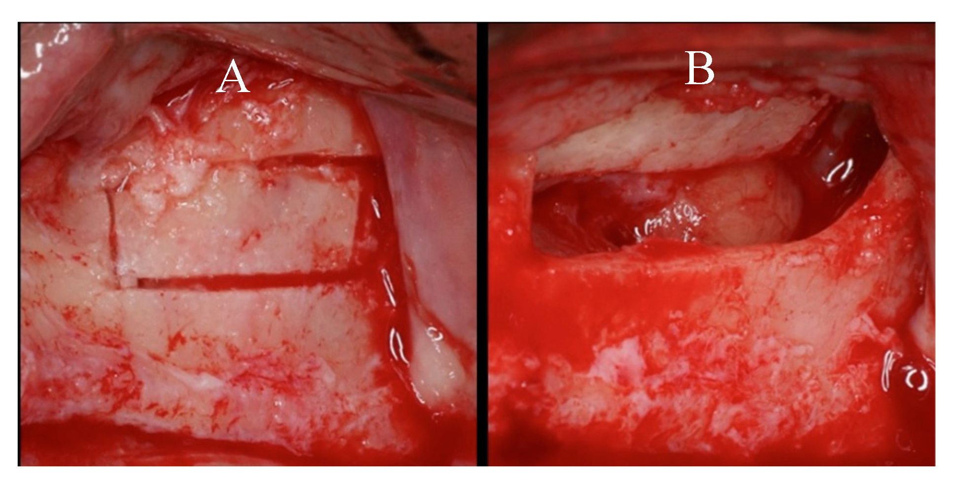
Figure 3.
Schneiderian Membrane Perforation.
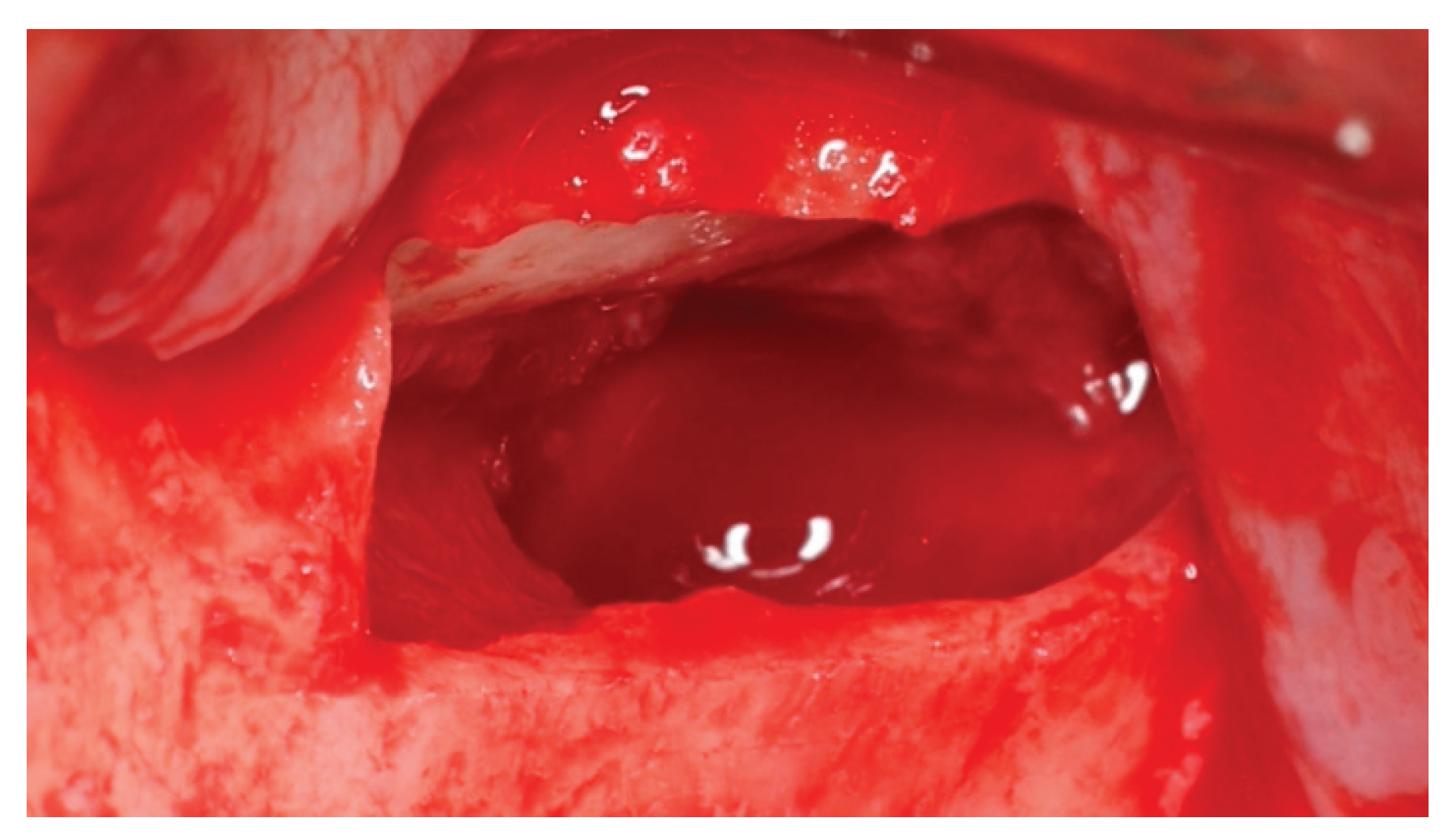
Figure 4.
a - Membrane Stabilization; b – Membrane Fixation; c – Xenograft Placement.
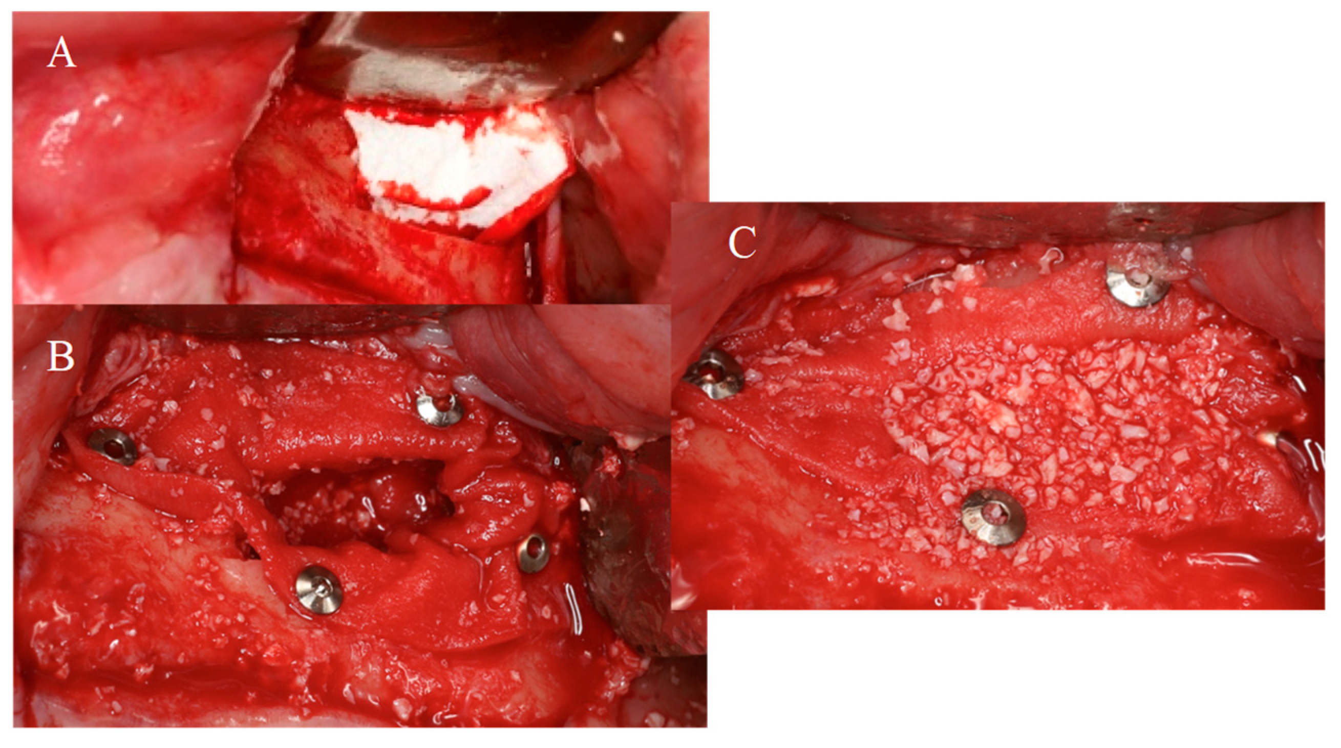
Figure 5.
Bone Volume Gained.
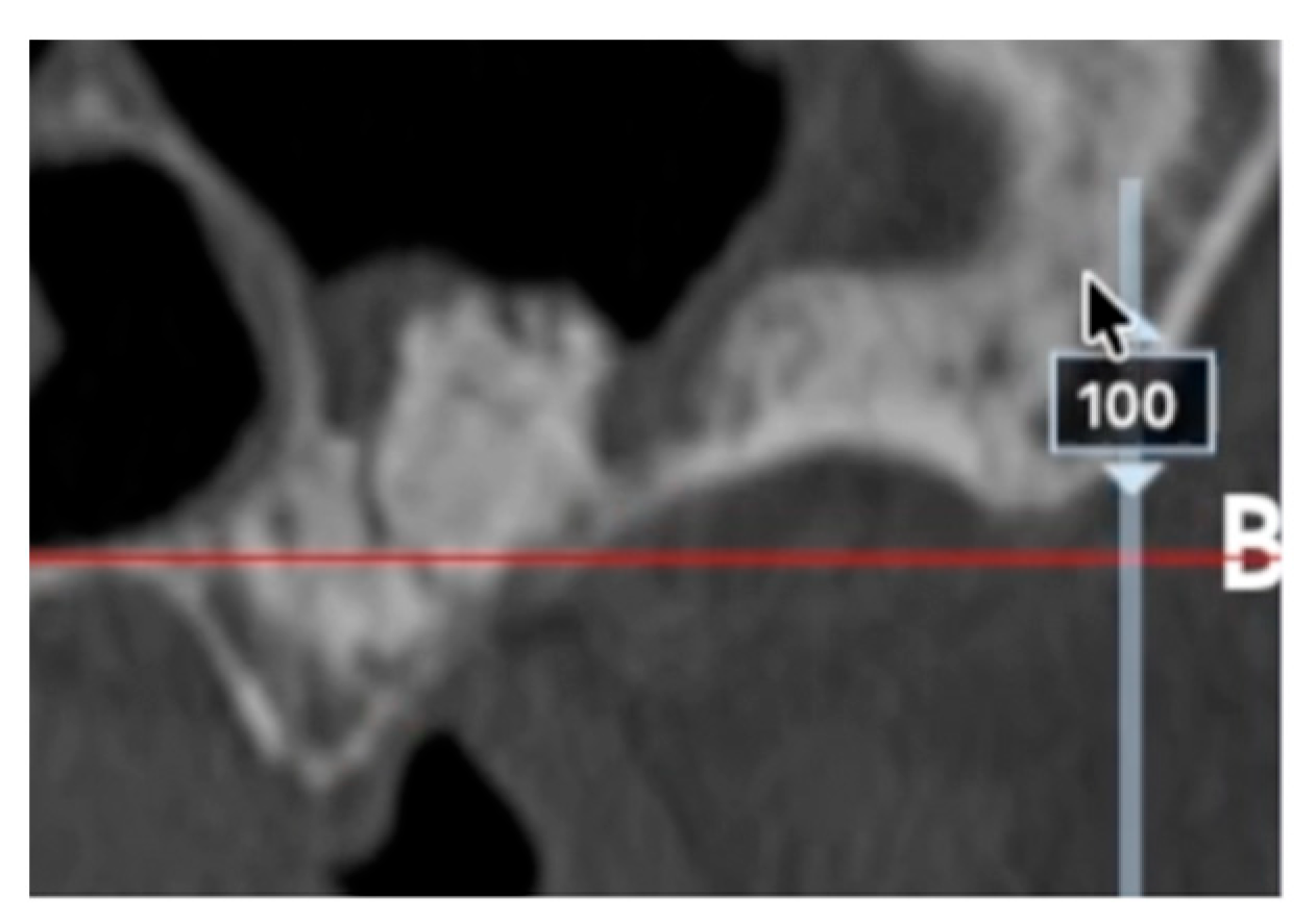
Figure 6.
a - Implant Placement 1st Quadrant; b – Implant Placement 2nd Quadrant.
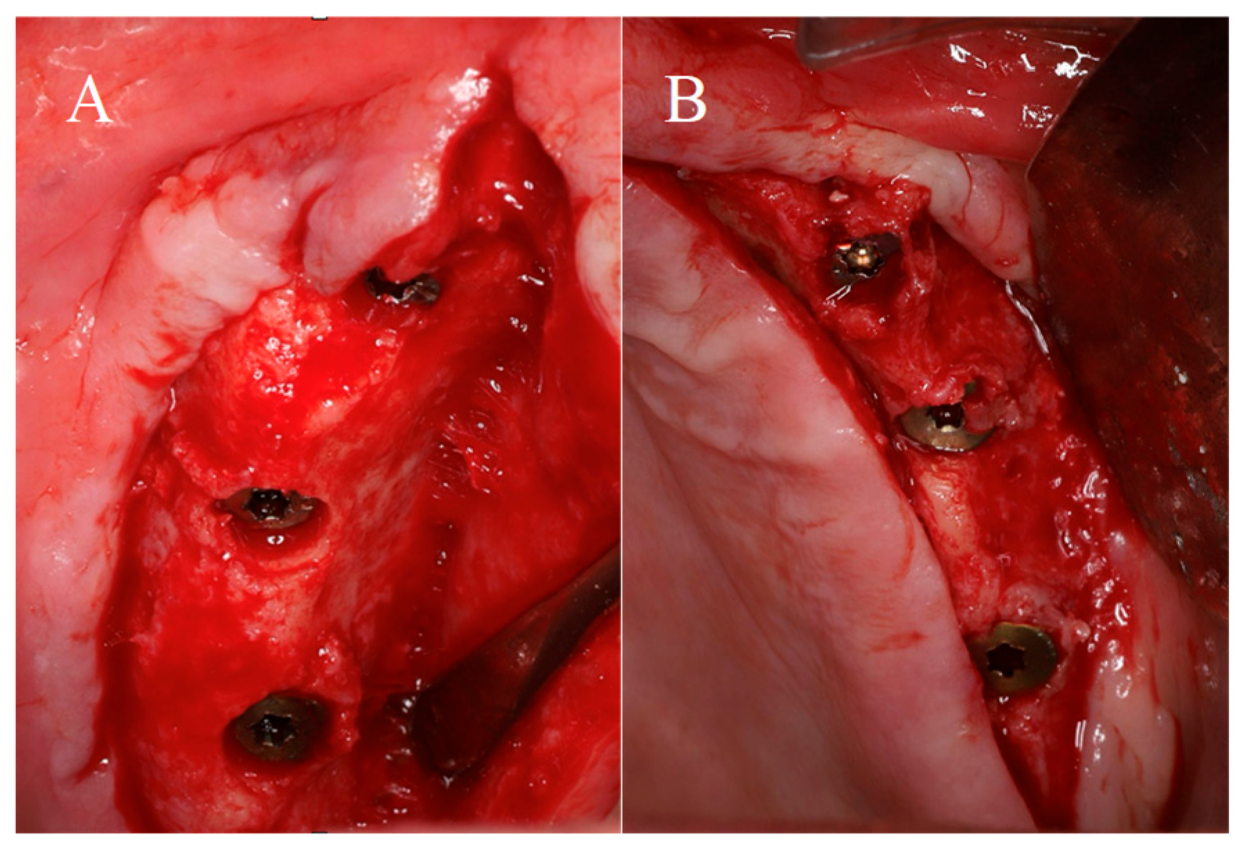
Figure 7.
a- Fixed Implant-supported Rehabilitation; b – Orthopantomography 1 year follow-up.
Disclaimer/Publisher’s Note: The statements, opinions and data contained in all publications are solely those of the individual author(s) and contributor(s) and not of MDPI and/or the editor(s). MDPI and/or the editor(s) disclaim responsibility for any injury to people or property resulting from any ideas, methods, instructions or products referred to in the content. |
© 2026 by the authors. Licensee MDPI, Basel, Switzerland. This article is an open access article distributed under the terms and conditions of the Creative Commons Attribution (CC BY) license (http://creativecommons.org/licenses/by/4.0/).
Copyright: This open access article is published under a Creative Commons CC BY 4.0 license, which permit the free download, distribution, and reuse, provided that the author and preprint are cited in any reuse.


