
Submitted:
11 July 2026
Posted:
15 July 2026
You are already at the latest version
Abstract
Polycystic ovary syndrome (PCOS) is a heterogeneous endocrine–metabolic disorder fre-quently associated with insulin resistance (IR) and increased cardiovascular risk. Soluble CD40 ligand (sCD40L) and soluble E-selectin (sE-selectin) are circulating biomarkers re-flecting immune activation and endothelial dysfunction, respectively. However, their dif-ferential associations with IR in PCOS, particularly in the context of central obesity, re-main incompletely understood. This cross-sectional study included 80 women with PCOS stratified according to waist-to-height ratio (WHtR > 0.50 vs. ≤ 0.50). Clinical, metabolic, hormonal, inflammatory, and endothelial parameters were evaluated. Correlation and multivariable regression analyses were performed to identify independent determinants of circulating sCD40L and sE-selectin. Women with central obesity exhibited significantly higher fasting insulin, HOMA-IR, triglycerides, non-HDL cholesterol, systolic blood pres-sure, and sE-selectin concentrations, together with lower HDL cholesterol. No significant differences were observed in TNF-α, IL-6, or sCD40L. In adjusted regression models, fast-ing glucose independently predicted sCD40L (β = 0.27, p = 0.02), whereas fasting insulin emerged as the strongest determinant of sE-selectin (β = 0.41, p < 0.001). These findings suggest distinct associations of immune and endothelial biomarkers with IR in PCOS. As-sessment of sCD40L and sE-selectin may provide complementary information for early cardiometabolic risk stratification in affected women.
Keywords:
polycystic ovary syndrome
; insulin resistance
; sCD40L
; sE-selectin
; endothelial dysfunction
; immune activation
1. Introduction
Polycystic ovary syndrome (PCOS) represents far more than a reproductive disorder; it is a multifaceted metabolic and endocrine condition that connects gynecologic, metabolic, and cardiovascular domains. Affecting approximately 5–20% of women of reproductive age, PCOS is increasingly recognized as a lifelong disorder with consequences extending beyond menstrual irregularities and infertility [1,2]. The syndrome’s heterogeneity—spanning hyperandrogenism, ovarian dysfunction, and variable metabolic phenotypes—reflects complex interactions among genetic predisposition, environmental factors, and lifestyle influences [3]. Among its metabolic features, insulin resistance (IR) stands out as a central and unifying abnormality, contributing to both hyperandrogenism and heightened cardiometabolic risk [4,5]. Yet, the nature of the link between IR and vascular health in PCOS remains only partially understood.
Mounting evidence suggests that immune dysregulation and endothelial activation form a biological interface between metabolic stress and cardiovascular disease in PCOS [6,7]. The chronic, low-grade inflammatory state typical of the condition involves cytokines such as tumor necrosis factor-α (TNF-α) and interleukin-6 (IL-6), which impair insulin signaling and disturb endothelial homeostasis [8]. In this context, soluble immune and endothelial biomarkers have emerged as valuable tools to capture subclinical processes that precede overt cardiovascular pathology.
Among these, soluble CD40 ligand (sCD40L) and soluble E-selectin (sE-selectin) are of particular interest due to their dual role as mediators and markers of vascular-immune cross-talk. sCD40L, primarily released by activated platelets and T lymphocytes, is a potent amplifier of inflammation, stimulating endothelial cells to express adhesion molecules, cytokines, and tissue factors [9,10]. Elevated sCD40L levels have been linked to IR, thrombosis, and atherosclerosis in a range of metabolic conditions [11]. Conversely, sE-selectin, a soluble form of the endothelial adhesion molecule E-selectin, reflects direct endothelial activation and facilitates leukocyte rolling and adhesion to the vascular wall [12]. Increased circulating sE-selectin has been reported in obesity, metabolic syndrome, and early vascular dysfunction [13,14]. Importantly, these molecules - though both linked to inflammation - capture distinct biological processes: sCD40L reflecting immune and platelet activation, and sE-selectin denoting endothelial stress. Their combined evaluation could thus provide a nuanced picture of how IR differently affects immune and vascular compartments in PCOS.
Although both biomarkers have been individually associated with metabolic and cardiovascular abnormalities, their relative relationships with IR and central obesity within PCOS have not been systematically compared.
Central obesity, typically assessed via waist-to-height ratio (WHtR) or waist circumference, further modulates these pathways [15]. The pattern of fat distribution, rather than total adiposity, may critically determine the balance between metabolic inflammation and endothelial integrity [16]. However, whether immune activation (indexed by sCD40L) and endothelial dysfunction (indexed by sE-selectin) are similarly or divergently influenced by IR in PCOS remains unclear.
Therefore, this study aimed to investigate whether biomarkers of immune activation (sCD40L) and endothelial activation (sE-selectin) exhibit differential associations with IR in women with PCOS stratified by central obesity. We hypothesized that these biomarkers would display distinct relationships with metabolic risk factors, reflecting different pathophysiological pathways linking adiposity, IR, and vascular risk in PCOS.
2. Results
2.1. Age, Anthropometric and Clinical Characteristics According to VAI
Eighty women with PCOS were included in the study and stratified according to central obesity (waist-to-height ratio, WHtR > 0.50, n = 40; WHtR ≤ 0.50, n = 40). The two groups were comparable in age (p = 0.074). As expected, women with central obesity exhibited significantly higher body weight, BMI (body mass index), waist circumference, hip circumference, WHR (waist-to-hip ratio), and WHtR values (all p < 0.001), confirming marked differences in overall and central adiposity. Systolic blood pressure (SBP) was also higher in the centrally obese group (p = 0.004), whereas diastolic blood pressure (DBP) did not differ significantly (Table 1).
2.2. Metabolic and Atherogenic Parameters
Central obesity was associated with a more adverse metabolic profile (Table 2). Fasting insulin (IRI 0’) and homeostatic model assessment for insulin resistance (HOMA-IR) were markedly increased in the centrally obese group (p < 0.001). Fasting glucose (GLU 0’) showed a modest upward trend but did not reach statistical significance. Serum triglycerides (TG) were significantly higher in women with central obesity (p < 0.01), while HDL-cholesterol (HDL-C) was reduced (p < 0.01). Total and LDL-cholesterol (LDL-C) did not differ significantly between groups. Non-HDL cholesterol (non-HDL-C) values were significantly higher in women with central obesity compared with those without (p = 0.030), further indicating an atherogenic lipid pattern in this subgroup (Table 2).
2.3. Hormonal Parameters
Hormonal profiles were broadly comparable between women with and without central obesity (Table 3). Luteinizing hormone (LH), follicle-stimulating hormone (FSH), and the LH/FSH ratio showed no significant differences between groups (p > 0.05). Estradiol (E2), total testosterone (T), androstenedione (A4), and dehydroepiandrosterone sulfate (DHEA-S) levels were also similar. SHBG tended to be lower in centrally obese women, but the difference was not significant (p = 0.313). Overall, no substantial hormonal differences were observed, suggesting that the observed metabolic and vascular differences were not accompanied by substantial differences in gonadotropic or androgenic hormone profiles.
2.4. Inflammatory and Endothelial markers; Leukocyte and Platelet Counts
Serum levels of TNF-α and IL-6 were comparable between groups (p > 0.05), suggesting that systemic inflammation was not substantially different. However, divergent patterns were observed for the vascular-immune markers (Table 4). Mean sCD40L concentrations did not differ significantly by obesity status (p = 0.56). sE-selectin levels were significantly higher in the centrally obese subgroup (p < 0.01) compatible with increased endothelial activation. The two groups exhibited comparable leukocyte (WBC) and platelet (PLT) counts, with no statistically significant differences (Table 4).
2.5. Correlations with Metabolic Parameters
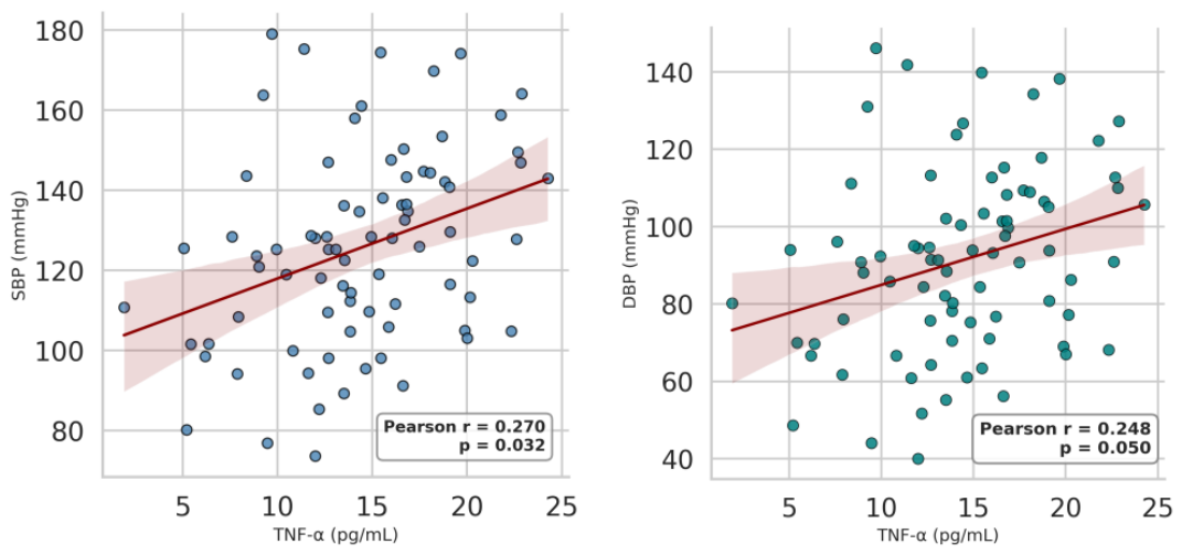
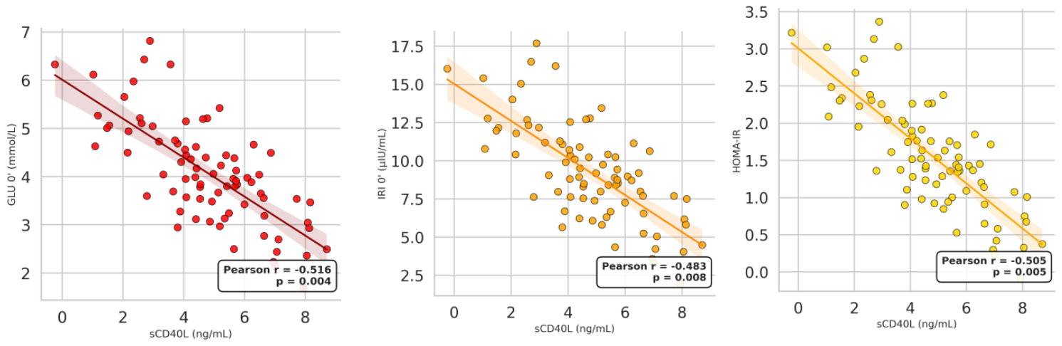
In the overall cohort, TNF-α was found to correlate positively with SBP (r = 0.270, p = 0.032), as well as with DBP (r = 0.240, p = 0.050) (Figures 1 and 2). sCD40L was inversely correlated with GLU 0’ (r = −0.516, p = 0.004), IRI 0’ (r = −0.483, p = 0.008), and HOMA-IR (r = −0.505, p = 0.005) (Figures 3, 4 and 5). In contrast, sE-selectin demonstrated positive correlations with IRI 0’ (r = 0.540, p = 0.002) and HOMA-IR (r = 0.503, p = 0.005) (Figures 6 and 7). These findings suggest distinct patterns of association between immune and endothelial biomarkers and indices of IR. The correlations should be interpreted in the context of the cross-sectional design and therefore do not imply causality.
2.6. Multivariable Regression Analyses
To identify independent determinants of circulating biomarker concentrations, multivariable linear regression analyses were performed after adjustment for age, BMI, and waist circumference. GLU 0’ remained independently and inversely associated with sCD40L concentrations (β = −0.27, p = 0.020), whereas IRI 0’ emerged as the strongest independent predictor of sE-selectin levels (β = 0.41, p < 0.001). TNF-α and IL-6 were not independently associated with either biomarker (Table 5).
3. Discussion
The present study investigated the relationships between endothelial and immune activation markers, insulin resistance, and central obesity in women with PCOS. Women with elevated WHtR exhibited a less favorable cardiometabolic profile characterized by higher IRI 0’, HOMA-IR, TG, non-HDL-C, and SBP, together with lower HDL-C. These findings are consistent with previous evidence indicating that visceral adiposity is a major determinant of metabolic risk in PCOS [17,18,19], while anthropometric indices reflecting abdominal adiposity may provide clinically relevant information beyond BMI alone [20,21]. The absence of significant differences in gonadotropic and androgenic hormone concentrations between groups further suggests that the observed metabolic and vascular alterations were primarily associated with adiposity-related metabolic disturbances rather than endocrine differences.
A major finding of this study was the association between sE-selectin and insulin resistance. Women with central obesity exhibited significantly higher circulating sE-selectin concentrations, while fasting insulin emerged as the strongest independent predictor of sE-selectin levels after adjustment for age, BMI, and waist circumference. These observations are consistent with previous reports demonstrating increased circulating sE-selectin concentrations in obesity and metabolic disorders [13,14,22]. In women with PCOS, insulin resistance has been associated with oxidative stress, enhanced leukocyte adhesion, and endothelial activation, providing a potential mechanistic link between metabolic dysfunction and vascular alterations [23,24]. Experimental and clinical studies suggest that insulin resistance and compensatory hyperinsulinemia may contribute to endothelial activation through mechanisms involving oxidative stress, impaired endothelial insulin signaling, and inflammatory pathways, leading to altered leukocyte–endothelial interactions [25,26,27]. Collectively, these findings support the concept that endothelial activation represents an early vascular manifestation of metabolic dysfunction in PCOS.
Interestingly, circulating TNF-α and IL-6 concentrations did not differ significantly between groups and were not independently associated with either sE-selectin or sCD40L. This observation is consistent with previous evidence showing that circulating inflammatory cytokines may display heterogeneous associations with metabolic and vascular alterations in PCOS [28]. This apparent dissociation suggests that endothelial activation in PCOS may precede detectable elevations in circulating inflammatory cytokines. Local adipose tissue inflammation, oxidative stress, and endothelial-specific signaling pathways may therefore contribute to endothelial dysfunction even when systemic inflammatory markers remain within comparable ranges. In addition, circulating TNF-α and IL-6 may not adequately reflect tissue-level inflammatory activity, particularly in relatively young women without overt cardiometabolic disease.
Unlike sE-selectin, sCD40L demonstrated inverse associations with fasting glucose, fasting insulin, and HOMA-IR. This finding differs from several studies reporting elevated sCD40L concentrations in obesity, IR, and PCOS [29,30,31,32]. However, the available evidence remains heterogeneous. Although increased circulating sCD40L concentrations have frequently been described in obesity and PCOS, accumulating evidence suggests that platelet activation, insulin signaling, and the metabolic milieu collectively influence sCD40L release, indicating that its relationship with IR may be context-dependent rather than uniformly linear. Supporting this concept, Gateva et al. reported that circulating sCD40L was associated with IR, but not with glucose tolerance, in obese nondiabetic individuals [33]. Furthermore, experimental studies have shown that acute hyperglycemia and euglycemic hyperinsulinemia may transiently suppress circulating sCD40L concentrations [34]. In addition, acute hyperinsulinemia has been shown to modulate several prothrombotic and vascular biomarkers, supporting the concept that insulin exerts direct effects on vascular homeostasis beyond glucose regulation [35]. Together, these observations suggest that circulating sCD40L may reflect dynamic metabolic and platelet-related responses rather than a simple linear consequence of worsening IR. Consistent with this interpretation, fasting glucose remained independently and inversely associated with sCD40L concentrations in the present study after adjustment for potential confounding factors.
Activated platelets are the principal source of circulating sCD40L, and CD40/CD40L signaling plays an important role in inflammatory and thrombotic processes that may be influenced by metabolic stress [36,37]. One possible explanation for the observed inverse associations is altered platelet activation or changes in sCD40L release under conditions of chronic metabolic stress. Although this hypothesis remains speculative, it may partly account for the heterogeneous findings reported across different metabolic populations and warrants further mechanistic investigation.
Our findings further emphasize the importance of central adiposity in shaping the cardiometabolic phenotype of PCOS. Women with increased WHtR exhibited a cluster of metabolic abnormalities, including hyperinsulinemia, dyslipidemia, and elevated systolic blood pressure, supporting previous evidence that visceral fat accumulation contributes to insulin resistance and endothelial dysfunction through mechanisms involving increased free fatty acid release, oxidative stress, and adipokine dysregulation [16,38]. Furthermore, anthropometric indices reflecting abdominal adiposity, such as WHtR and waist circumference, have been shown to provide better discrimination of cardiometabolic risk than BMI in women with PCOS [20,21]. Consistent with these observations, our results suggest that central obesity and insulin resistance, rather than differences in androgen status, are major determinants of metabolic and vascular alterations in PCOS [39].
From a clinical perspective, fasting insulin emerged as the principal independent determinant of circulating sE-selectin concentrations, emphasizing the close relationship between insulin resistance and endothelial activation in PCOS. The complementary behavior of sE-selectin and sCD40L suggests that endothelial and immune biomarkers may reflect different biological pathways involved in cardiometabolic risk. Whether these biomarkers improve early cardiovascular risk stratification in women with PCOS should be evaluated in prospective longitudinal studies.
The strengths of this study include the use of WHtR as a marker of central obesity, comprehensive metabolic and hormonal characterization, simultaneous assessment of endothelial and immune biomarkers, and adjustment for major anthropometric confounders in multivariable analyses. Several limitations should also be acknowledged. First, the cross-sectional design precludes causal inference. Second, the moderate sample size may have limited the statistical power to detect weaker associations. Third, the absence of a non-PCOS control group prevents discrimination between alterations specifically related to PCOS and those attributable to obesity or insulin resistance. Finally, endothelial function was evaluated using circulating biomarkers rather than vascular imaging or functional endothelial assessments.
In conclusion, women with PCOS and central obesity exhibited a more adverse metabolic profile together with higher circulating sE-selectin concentrations, supporting a close relationship between insulin resistance and endothelial activation. In contrast, sCD40L demonstrated inverse associations with indices of insulin resistance, suggesting more complex regulation of immune–vascular signaling pathways. These findings emphasize the heterogeneous mechanisms linking central adiposity, metabolic dysfunction, and vascular alterations in PCOS and support further investigation of circulating endothelial and immune biomarkers as potential tools for early cardiometabolic risk assessment.
4. Materials and Methods
4.1. Study Design and Participants
This cross-sectional study included 80 women with polycystic ovary syndrome (PCOS), aged 18–35 years, who were enrolled at the Clinic of Endocrinology and Metabolic Diseases, “Sv. Georgy” University Hospital of Plovdiv, from June 2020 to June 2023. The study was designed as an internal comparison between women with PCOS stratified according to central obesity status; no non-PCOS control group was included. All participants were diagnosed with PCOS according to the Rotterdam criteria [40], requiring at least two of the following: (i) oligo/anovulation, (ii) clinical and/or biochemical hyperandrogenism, and (iii) polycystic ovarian morphology on ultrasound, after exclusion of related disorders (e.g., congenital adrenal hyperplasia, Cushing’s syndrome, thyroid dysfunction, or hyperprolactinemia). Furthermore, individuals with diabetes mellitus, chronic inflammatory or autoimmune conditions, or those using insulin-sensitizing or lipid-lowering medications (such as metformin or statins), hormonal contraceptives, or currently pregnant were excluded. All women in the final study population were non-smokers.
Participants were stratified according to central obesity using the waist-to-height ratio (WHtR), applying the established cut-off value of 0.50 [15]. Accordingly, 40 women were classified as having central obesity (WHtR > 0.50) and 40 as not having central obesity (WHtR ≤0.50).
All clinical, biochemical, and hormonal assessments were performed during the early follicular phase of the menstrual cycle (days 2–5) or following progesterone-induced withdrawal bleeding in women with oligo- or amenorrhea.
The study protocol was approved by the institutional Ethics Committee of Medical University of Plovdiv, Bulgaria, and all procedures adhered to the Declaration of Helsinki (2013 revision). Written informed consent was obtained from all participants before enrollment.
4.2. Anthropometric and Clinical Assessment
Body weight and height were measured using standardized calibrated instruments, with participants wearing light clothing and no shoes. Body mass index (BMI) was calculated as weight (kg) divided by height squared (m²). Waist circumference (W) was measured midway between the lower rib margin and the iliac crest, and hip circumference (H) at the widest point over the buttocks. The waist-to-hip ratio (WHR) and waist-to-height ratio (WHtR) were calculated accordingly.
Blood pressure was measured twice in the seated position after a 10-minute rest using a validated automatic sphygmomanometer, and the mean of two readings was recorded for both systolic (SBP) and diastolic (DBP) pressure.
4.3. Bioghemical Analysis; Leukocyte and Platelet Counts
Fasting plasma glucose was measured using the glucose oxidase–peroxidase (GOD–POD) method, and fasting serum insulin concentrations were determined by chemiluminescent immunoassay (CLIA; Beckman Coulter, Brea, CA, USA). The analytical sensitivity of the insulin assay was 0.03 μIU/mL, with intra- and inter-assay coefficients of variation below 6%. Insulin resistance was estimated using the homeostasis model assessment for insulin resistance (HOMA-IR), calculated as: HOMA-IR = fasting glucose (mmol/L) × fasting insulin (μIU/mL) / 22.5.
Serum total cholesterol (TC), triglycerides (TG), and high-density lipoprotein cholesterol (HDL-C) were determined by standard enzymatic methods using commercial reagents (Schneiders Medizintechnik, Zwolle, The Netherlands) on a Delta Kone automated analyzer (Kone Instruments, Espoo, Finland). Low-density lipoprotein cholesterol (LDL-C) was calculated using the Friedewald equation, and non-HDL cholesterol was calculated as total cholesterol minus HDL cholesterol.
Leukocyte (WBC) and platelet (PLT) counts were determined using an automated hematology analyzer based on the Coulter principle. Samples showing abnormal cell morphology or platelet aggregation were verified manually.
4.4. Hormonal Assays
Circulating concentrations of reproductive hormones and androgen-related parameters were assessed by automated chemiluminescent immunoassays (CLIA) using commercially available platforms and reagents, following the manufacturers’ validated protocols. Serum LH and FSH levels were determined using sandwich-type immunochemical assays (Beckman Coulter, Inc., Brea, CA, USA), with analytical sensitivities of 0.2 IU/L and 0.2 mIU/mL, respectively. Estradiol (E2) concentrations were obtained using a competitive immunochemical CLIA method (Beckman Coulter, Inc., Ireland), with an assay sensitivity of 73 pmol/L.
Total testosterone (Т) and DHEA-S were analyzed using CLIA-based assays provided by Beckman Coulter, Inc. (Brea, CA, USA). The reported analytical sensitivities were 0.35 ng/mL for T and <2 μg/dL for DHEA-S. SHBG measurements were performed using the Access 2 Immunoassay System (Beckman Coulter, Inc.), with a sensitivity threshold of 0.33 nmol/L and an analytical measuring range extending to 180 nmol/L. Androstenedione (A4) levels were determined by a CLIA assay (catalog no. L2KAO2; Siemens Healthcare Diagnostics, Tarrytown, NY, USA), with an analytical sensitivity of 1.0 nmol/L.
The analytical reliability of the assays was evaluated according to the manufacturers’ quality specifications. Intra- and inter-assay coefficients of variation (CVs) were generally below 10%; however, higher variability was observed for E2 and A4 measurements, consistent with the reported assay performance characteristics. Internal quality controls, calibration procedures, and assay validation were performed throughout the analytical process in accordance with the manufacturers’ recommendations.
All hormonal assessments were performed during the early follicular phase of the menstrual cycle..
4.5. Inflammatory and Endothelial Markers
Serum tumor necrosis factor-α (TNF-α) and interleukin-6 (IL-6) concentrations were determined using commercially available enzyme-linked immunosorbent assay (ELISA) kits (DRG Instruments GmbH, Marburg, Germany) according to the manufacturers' instructions. The analytical sensitivities were 3.0 pg/mL for TNF-α and 2.0 pg/mL for IL-6. The intra-assay coefficient of variation for the IL-6 assay was <7.7%.
Serum soluble CD40 ligand (sCD40L) and soluble E-selectin (sE-selectin) concentrations were measured using commercially available ELISA kits (Bender MedSystems, Vienna, Austria) following the manufacturers' protocols. Both assays demonstrated acceptable analytical performance, with intra-assay coefficients of variation <8% and inter-assay coefficients of variation <10%.
4.6. Statistical Analysis
Statistical analyses were performed using IBM SPSS Statistics for Windows, version 21.0 (IBM Corp., Armonk, NY, USA). Continuous variables are presented as mean ± standard deviation (SD). Data normality was assessed using the Shapiro–Wilk test.
Comparisons between women with and without central obesity (WHtR > 0.50 vs. ≤ 0.50) were performed using the independent-samples Student's t-test. Pearson's correlation coefficients (r) were calculated to evaluate associations between inflammatory and endothelial biomarkers and metabolic or clinical parameters.
Multivariable linear regression analyses were performed to identify independent predictors of circulating sCD40L and sE-selectin concentrations. Variables showing significant correlations and considered biologically relevant were entered into the models, with adjustment for age, body mass index (BMI), and waist circumference. Because fasting insulin and HOMA-IR are highly correlated measures of insulin resistance, they were not included simultaneously in the same model. Standardized regression coefficients (β) are presented.
No correction for multiple comparisons was applied because the analyses were based on a limited number of pre-specified hypotheses. All statistical tests were two-sided, and p < 0.05 was considered statistically significant.
5. Conclusions
In conclusion, this study demonstrates that central obesity in PCOS is associated with pronounced insulin resistance and endothelial activation, manifested by elevated sE-selectin levels. Fasting insulin emerged as the strongest independent determinant of sE-selectin, while fasting glucose predicteds CD40L levels. Systemic inflammation did not independently influence either marker. These findings highlight the dominant role of metabolic disturbances, rather than inflammatory or hormonal variations, in driving early vascular dysfunction in PCOS. Targeting insulin resistance and central adiposity may therefore represent key strategies for mitigating cardiovascular risk in this population.
Author Contributions
Conceptualization: D.K.-T., M.I.-G., E.B., A.G. and P.K.; methodology: D.K.-T., P.N, and A.G.; software: D.K.-T.; validation: D.K.-T., M.I.-G. and M.O.; formal analysis: T.D.; investigation: T.D.; resources: M.O., A.G. and D.K.; data curation: M.I.-G., E.B. and P.N.; writing—original draft preparation: D.K.-T., P.K. and D.K.; writing—review and editing: M.O.; visualization: T.D.; supervision: M.O.; project administration: D.K.-T. All authors have read and agreed to the published version of the manuscript.
Funding
This research received no external funding.
Institutional Review Board Statement
The study was conducted in accordance with the Declaration of Helsinki and approved by the Ethics Committee of the Medical University of Plovdiv (protocol code 2444/26 October 2020).
Informed Consent Statement
Informed consent was obtained from all subjects involved in the study.
Data Availability Statement
The data that support the findings of this study are available from the corresponding author upon reasonable request. Due to ethical restrictions and participant confidentiality, the dataset is not publicly available.
Acknowledgments
The authors would like to thank the laboratory staff and clinical personnel at “Sv. Georgy” University Hospital of Plovdiv, Bulgaria, for their assistance with sample collection and processing.
Conflicts of Interest
The authors declare no conflicts of interest.
Abbreviations
The following abbreviations are used in this manuscript:
| PCOS | Polycystic Ovary Syndrome |
| IR | Insulin Resistance |
| BMI | Body Mass Index |
| WHR | Waist-to-Hip Ratio |
| WHtR | Waist-to-Height Ratio |
| HOMA-IR | Homeostatic Model Assessment for Insulin Resistance |
| SBP | Systolic Blood Pressure |
| DBP | Diastolic Blood Pressure |
| GLU 0’ | Fasting Glucose |
| IRI 0’ | Fasting Insulin |
| TC | Total Cholesterol |
| HDL-C | High-Density Lipoprotein Cholesterol |
| LDL-C | Low-Density Lipoprotein Cholesterol |
| TG | Triglycerides |
| LH | Luteinizing Hormone |
| FSH | Follicle-Stimulating Hormone |
| E2 | Estradiol |
| SHBG | Sex Hormone-Binding Globulin |
| A4 | Androstenedione |
| DHEA-S | Dehydroepiandrosterone Sulfate |
| sCD40L | Soluble CD40 Ligand |
| IL-6 | Interleukin 6 |
| TNF- a | Tumor Necrosis Factor alpha |
| WBC | Leukocytes |
| PLT | Platelets |
| ELISA | Enzyme-Linked Immunosorbent Assay |
References
- Azziz, R.; Carmina, E.; Chen, Z.; Dunaif, A.; Laven, J.S.E.; Legro, R.S.; Lizneva, D.; Natterson-Horowitz, B.; Teede, H.J.; Yildiz, B.O. Polycystic ovary syndrome. Nat. Rev. Dis. Prim. 2016, 2, 16057. [Google Scholar] [CrossRef] [PubMed]
- Bozdag, G.; Mumusoglu, S.; Zengin, D.; Karabulut, E.; Yildiz, B.O. The prevalence and phenotypic features of polycystic ovary syndrome: A systematic review and meta-analysis. Hum. Reprod. 2016, 31, 2841–2855. [Google Scholar] [CrossRef] [PubMed]
- Escobar-Morreale, H.F. Polycystic ovary syndrome: definition, aetiology, diagnosis and treatment. Nat. Rev. Endocrinol. 2018, 14, 270–284. [Google Scholar] [CrossRef] [PubMed]
- Dunaif, A. Insulin resistance and the polycystic ovary syndrome: mechanism and implications for pathogenesis. Endocr. Rev. 1997, 18, 774–800. [Google Scholar] [CrossRef] [PubMed]
- Rojas, J.; Chávez, M.; Olivar, L.; Rojas, M.; Morillo, J.; Mejías, J.; Calvo, M.; Bermúdez, V. Polycystic ovary syndrome, insulin resistance, and obesity: navigating the pathophysiologic labyrinth. Int. J. Reprod. Med. 2014, 2014, 719050. [Google Scholar] [CrossRef] [PubMed]
- González, F. Inflammation in PCOS: underpinning of insulin resistance and obesity. Steroids 2012, 77, 300–305. [Google Scholar] [CrossRef] [PubMed]
- Orio, F.; Vuolo, L.; Palomba, S.; Lombardi, G.; Colao, A. Metabolic and cardiovascular consequences of polycystic ovary syndrome. Minerva Ginecol. 2008, 60, 39–51. [Google Scholar] [PubMed]
- Kelly, C.C.; Lyall, H.; Petrie, J.R.; Gould, G.W.; Connell, J.M.; Sattar, N. Low grade chronic inflammation in women with polycystic ovarian syndrome. J. Clin. Endocrinol. Metab. 2001, 86, 2453–2455. [Google Scholar] [CrossRef] [PubMed]
- Аndré, P. Platelet-derived CD40L: the switch-hitting player of cardiovascular disease. Circulation 2002, 106, 896–899. [Google Scholar] [CrossRef] [PubMed]
- San Miguel Hernández, A.; Inglada-Galiana, L.; García Iglesias, R.; Alonso Castillejos, N.; Martín Gil, F.J. Soluble CD40 ligand: a potential marker of cardiovascular risk. Rev. Clin. Esp. 2007, 207, 418–421. [Google Scholar] [CrossRef] [PubMed]
- Urbich, C.; Dimmeler, S. CD40 and vascular inflammation. Can. J. Cardiol. 2004, 20, 681–683. [Google Scholar] [PubMed]
- Galkina, E.; Ley, K. Immune and inflammatory mechanisms of atherosclerosis. Annu. Rev. Immunol. 2009, 27, 165–197. [Google Scholar] [CrossRef] [PubMed]
- Adamska, A.; Karczewska-Kupczewska, M.; Nikołajuk, A.; Otziomek, E.; Górska, M.; Kowalska, I.; Strączkowski, M. Relationships of serum soluble E-selectin concentration with insulin sensitivity and metabolic flexibility in lean and obese women. Endocrine 2014, 45, 422–429. [Google Scholar] [CrossRef] [PubMed]
- Matsumoto, K.; Sera, Y.; Abe, Y.; Tominaga, T.; Horikami, K.; Hirao, K.; Ueki, Y.; Miyake, S. High serum concentrations of soluble E-selectin correlate with obesity but not fat distribution in patients with type 2 diabetes mellitus. Metabolism 2002, 51, 932–934. [Google Scholar] [CrossRef] [PubMed]
- Ashwell, M.; Gunn, P.; Gibson, S. Waist-to-height ratio is a better screening tool than waist circumference and BMI for adult cardiometabolic risk factors: systematic review and meta-analysis. Obes. Rev. 2012, 13, 275–286. [Google Scholar] [CrossRef] [PubMed]
- Bril, F.; Ezeh, U.; Amiri, M.; Hatoum, S.; Pace, L.; Chen, Y.-H.; Bertrand, F.; Gower, B.; Azziz, R. Adipose Tissue Dysfunction in Polycystic Ovary Syndrome. J. Clin. Endocrinol. Metab. 2023, 109, 10–24. [Google Scholar] [CrossRef] [PubMed]
- Jena, D.; Choudhury, A.K.; Mangaraj, S.; Singh, M.; Mohanty, B.K.; Baliarsinha, A.K. Study of visceral and subcutaneous abdominal fat thickness and its correlation with cardiometabolic risk factors and hormonal parameters in polycystic ovary syndrome. Indian J. Endocrinol. Metab. 2018, 22, 321–327. [Google Scholar] [CrossRef] [PubMed]
- Dumesic, D.A.; Abbott, D.H.; Sanchita, S.; Chazenbalk, G.D. Endocrine-metabolic dysfunction in polycystic ovary syndrome: an evolutionary perspective. Curr. Opin. Endocr. Metab. Res. 2020, 12, 41–48. [Google Scholar] [CrossRef] [PubMed]
- Akkus, C.; Oner, O.; Kilic, A.O.; Duran, C. Visceral Adiposity Index (VAI) levels and metabolic risk across phenotypes of polycystic ovary syndrome (PCOS). Medicina 2025, 61, 1673. [Google Scholar] [CrossRef] [PubMed]
- Zhu, M.; Wang, K.; Feng, J.; Liu, Y.; Guan, M.; Wang, Y.; Wu, X. The waist-to-height ratio is a good predictor for insulin resistance in women with polycystic ovary syndrome. Front. Endocrinol. 2024, 15, 1502321. [Google Scholar] [CrossRef] [PubMed]
- Kałuzna, M.; Czlapka-Matyasik, M.; Bykowska-Derda, A.; Moczko, J.; Ruchała, M.; Ziemnicka, K. Indirect predictors of visceral adipose tissue in women with polycystic ovary syndrome: a comparison of methods. Nutrients 2021, 13, 2494. [Google Scholar] [CrossRef] [PubMed]
- Lee, C.-H.; Kuo, F.-C.; Tang, W.-H.; Lu, C.-H.; Su, S.-C.; Liu, J.-S.; Hsieh, C.-H.; Hung, Y.-J.; Lin, F.-H. Serum E-selectin concentration is associated with risk of metabolic syndrome in females. PLoS ONE 2019, 14, e0222815. [Google Scholar] [CrossRef] [PubMed]
- Diamanti-Kandarakis, E.; Paterakis, T.; Alexandraki, K.; Piperi, C.; Aessopos, A.; Katsikis, I.; Katsilambros, N.; Kreatsas, G.; Panidis, D. Indices of low-grade chronic inflammation in polycystic ovary syndrome and the beneficial effect of metformin. Hum. Reprod. 2006, 21, 1426–1431. [Google Scholar] [CrossRef] [PubMed]
- Victor, V.M.; Rovira-Llopis, S.; Bañuls, C.; Díaz-Morales, N.; Martínez de Marañón, A.; Ríos-Navarro, C.; Álvarez, A.; Gómez, M.; Rocha, M.; Hernández-Mijares, A. Insulin resistance in PCOS patients enhances oxidative stress and leukocyte adhesion: role of myeloperoxidase. PLoS ONE 2016, 11, e0151960. [Google Scholar] [CrossRef] [PubMed]
- Kampoli, A.-M.; Tousoulis, D.; Briasoulis, A.; Latsios, G.; Papageorgiou, N.; Stefanadis, C. Potential pathogenic inflammatory mechanisms of endothelial dysfunction induced by type 2 diabetes mellitus. Curr. Pharm. Des. 2011, 17, 4147–4158. [Google Scholar] [CrossRef] [PubMed]
- Rathjen, T.; Kunkemoeller, B.; Cederquist, C.T.; Wang, X.; Lockhart, S.M.; Patti, J.C.; Willenbrock, H.; Olsen, G.S.; Povlsen, G.K.; Beck, H.C.; et al. Endothelial cell insulin signaling regulates CXCR4 (C-X-C motif chemokine receptor 4) and limits leukocyte adhesion to endothelium. Arterioscler. Thromb. Vasc. Biol. 2022, 42, e217–e227. [Google Scholar] [CrossRef] [PubMed]
- Dube, R. Does endothelial dysfunction correlate with endocrinal abnormalities in patients with polycystic ovary syndrome? Avicenna J. Med. 2016, 6, 91–102. [Google Scholar] [CrossRef] [PubMed]
- Escobar-Morreale, H.F.; Luque-Ramírez, M.; González, F. Circulating inflammatory markers in polycystic ovary syndrome: a systematic review and meta-analysis. Fertil. Steril. 2011, 95, 1048–1058.e1–e2. [Google Scholar] [CrossRef] [PubMed]
- Oktem, M.; Ozcimen, E.E.; Uckuyu, A.; Esinler, I.; Pamuk, B.; Bayraktar, N.; Kulaksizoglu, S.; Zeyneloglu, H.B. Polycystic ovary syndrome is associated with elevated plasma soluble CD40 ligand, a marker of coronary artery disease. Fertil. Steril. 2009, 91, 2545.e1–2545.e4. [Google Scholar] [CrossRef] [PubMed]
- El-Mesallamy, H.O.; Abd El-Razek, R.S.; El-Refaie, T.A. Circulating high-sensitivity C-reactive protein and soluble CD40 ligand are inter-related in a cohort of women with polycystic ovary syndrome. Eur. J. Obstet. Gynecol. Reprod. Biol. 2013, 168, 178–182. [Google Scholar] [CrossRef] [PubMed]
- Kalı, Z.; Çağıran, F.T. Soluble CD40 ligand (sCD40L) may be a predictive marker of vascular endothelial damage in lean and obese women with PCOS. Eurasian Clin. Anal. Med. 2023, 11, 47–50. [Google Scholar] [CrossRef]
- Unek, I.T.; Bayraktar, F.; Solmaz, D.; Ellidokuz, H.; Sisman, A.R.; Yuksel, F.; Yesil, S. The levels of soluble CD40 ligand and C-reactive protein in normal weight, overweight and obese people. Clin. Med. Res. 2010, 8, 89–95. [Google Scholar] [CrossRef] [PubMed]
- Gateva, A.; Assyov, Y.; Tsakova, A.; Kamenov, Z. Soluble CD40L is associated with insulin resistance, but not with glucose tolerance in obese nondiabetic patients. Arch. Physiol. Biochem. 2016, 122, 161–165. [Google Scholar] [CrossRef] [PubMed]
- Oliver, S.R.; Flores, R.L.; Pontello, A.M.; Rosa, J.S.; Zaldivar, F.P.; Galassetti, P.R. Acute suppression of circulating sCD40L during hyperglycemia and euglycemic-hyperinsulinemia in healthy young males. J. Investig. Med. 2008, 56, 902–910. [Google Scholar] [CrossRef] [PubMed]
- Muscelli, E.; Camastra, S.; Santini, E.; Casolaro, A.; Pinnola, S.; Astiarraga, B.D.; Solini, A. Short-term acute hyperinsulinemia and prothrombotic factors in subjects with normal glucose tolerance. Horm. Metab. Res. 2009, 41, 568–572. [Google Scholar] [CrossRef] [PubMed]
- Jin, R.; Yu, S.; Song, Z.; Zhu, X.; Wang, C.; Yan, J.; Wu, F.; Nanda, A.; Granger, D.N.; Li, G. Soluble CD40 ligand stimulates CD40-dependent activation of the β2 integrin Mac-1 and protein kinase C ζ (PKCζ) in neutrophils: implications for neutrophil-platelet interactions and neutrophil oxidative burst. PLoS ONE 2013, 8, e64631. [Google Scholar] [CrossRef] [PubMed]
- Antoniades, C.; Bakogiannis, C.; Tousoulis, D.; Antonopoulos, A.S.; Stefanadis, C. The CD40/CD40 ligand system: linking inflammation with atherothrombosis. J. Am. Coll. Cardiol. 2009, 54, 669–677. [Google Scholar] [CrossRef] [PubMed]
- Chait, A.; den Hartigh, L.J. Adipose tissue distribution, inflammation and its metabolic consequences, including diabetes and cardiovascular disease. Front. Cardiovasc. Med. 2020, 7, 22. [Google Scholar] [CrossRef] [PubMed]
- Dokras, A.; Jagasia, D.H.; Maifeld, M.; Sinkey, C.A.; VanVoorhis, B.J.; Haynes, W.G. Obesity and insulin resistance but not hyperandrogenism mediates vascular dysfunction in women with polycystic ovary syndrome. Fertil. Steril. 2006, 86, 1702–1709. [Google Scholar] [CrossRef] [PubMed]
- Rotterdam ESHRE/ASRM-Sponsored PCOS Consensus Workshop Group. Revised 2003 consensus on diagnostic criteria and long-term health risks related to polycystic ovary syndrome. Fertil. Steril. 2004, 81, 19–25. [Google Scholar] [CrossRef] [PubMed]
Figures 1 and 2.
Positive Relationship of TNF-α with Systolic Blood Pressure (SBP) and Diastolic Blood Pressure (DBP) in the Whole Group of PCOS Women.
Figures 1 and 2.
Positive Relationship of TNF-α with Systolic Blood Pressure (SBP) and Diastolic Blood Pressure (DBP) in the Whole Group of PCOS Women.
Figures 3, 4 and 5.
Negative Associations of sCD40L with GLU 0’, IRI 0’ and HOMA-IR in the Whole Group of PCOS Women.
Figures 3, 4 and 5.
Negative Associations of sCD40L with GLU 0’, IRI 0’ and HOMA-IR in the Whole Group of PCOS Women.
Figures 6 and 7.
Positive Correlations of sE-selectin with IRI 0’and HOMA-IR in the Studied Group of PCOS Women.
Figures 6 and 7.
Positive Correlations of sE-selectin with IRI 0’and HOMA-IR in the Studied Group of PCOS Women.
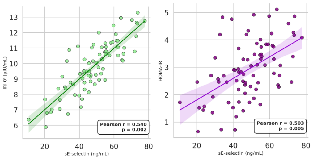

Table 1.
Age, anthropometric and clinical parameters in the two studied groups of women with PCOS.
| Parameter | PCOS with WHtR ≤ 0.50 (n = 40) |
PCOS with WHtR > 0.50 (n = 40) |
|---|---|---|
| Age (years) | 23.10 ± 4.05 | 25.20 ± 5.44 NS |
| Height (cm) | 166.44 ± 7.55 | 166.37 ± 5.48 NS |
| Weight (kg) | 59.66 ± 9.62 | 85.52 ± 11.26 *** |
| BMI (kg/m2) | 21.46 ± 2.60 | 30.88 ± 3.80 *** |
| Waist (cm) | 70.26 ± 6.38 | 97.09 ± 7.79 *** |
| Hip (cm) | 94.87 ± 7.35 | 111.49 ± 7.98 ** |
| WHR | 0.74 ± 0.06 | 0.87 ± 0.06 *** |
| WHtR | 0.42 ± 0.04 | 0.58 ± 0.05 *** |
| SBP (mmHg) | 110.64 ± 11.71 | 118.68 ± 11.50 ** |
| DBP (mmHg) | 71.15 ± 8.39 | 75.15 ± 10.33 NS |
NS—not significant (p > 0.05); **—p < 0.01; ***—p < 0.001.
Table 2.
Metabolic and atherogenic parameters in the two studied groups of women with PCOS.
| Parameter | PCOS with WHtR ≤ 0.50 (n = 40) |
PCOS with WHtR > 0.50 (n = 40) |
|---|---|---|
| GLU 0’ (mmol/L) | 4.73 ± 0.45 | 4.99 ± 0.75 NS |
| IRI 0’ (μIU/mL) | 6.56 ± 3.09 | 11.39 ± 5.87 *** |
| HOMA-IR | 1.41 ± 0.76 | 2.64 ± 1.68 *** |
| TC (mmol/L) | 4.36 ± 0.97 | 4.57 ± 0.78 NS |
| LDL-C (mmol/L) | 2.60 ± 0.99 | 2.85 ± 0.73 NS |
| HDL-C (mmol/L) | 1.47 ± 0.49 | 1.17 ± 0.25 ** |
| TG (mmol/L) | 0.75 ± 0.32 | 1.22 ± 0.49 *** |
| Non-HDL-C | 2.95 ± 1.01 | 3.40 ± 0.77 * |
NS—not significant (p > 0.05); *—p < 0.05; **—p < 0.01; ***—p < 0.001.
Table 3.
Hormonal parameters in the two studied groups of women with PCOS.
| Parameter | PCOS with WHtR ≤ 0.50 (n = 40) |
PCOS with WHtR > 0.50 (n = 40) |
|---|---|---|
| LH (IU/L) | 7.91 ± 3.33 | 8.59 ± 4.34 NS |
| FSH (mIU/mL) | 5.78 ± 1.78 | 5.43 ± 1.92 NS |
| LH/FSH | 1.38 ± 0.76 | 1.88 ± 1.40 NS |
| E2 (pg/mL) | 251.31 ± 99.11 | 244.56 ± 101.11 NS |
| Total testosterone (ng/mL) | 0.67 ± 0.17 | 0.70 ± 0.21 NS |
| Androstenedione (ng/mL) | 3.84 ± 1.06 | 3.77 ± 1.66 NS |
| DHEA-S (μg/dL) | 285.85 ± 101.62 | 278.52 ± 104.37 NS |
| SHBG (nmol/L) | 44.50 ± 20.01 | 35.04 ± 18.43 NS |
NS—not significant (p > 0.05).
Table 4.
Inflammatory and endothelial markers; leukocyte and platelet counts in the two studied groups of women.
Table 4.
Inflammatory and endothelial markers; leukocyte and platelet counts in the two studied groups of women.
| Parameter | PCOS with WHtR ≤ 0.50 (n = 40) |
PCOS with WHtR > 0.50 (n = 40) |
|---|---|---|
| TNF-α (pg/mL) | 7.43 ± 4.50 | 8.39 ± 5.28 NS |
| IL-6 (pg/mL) | 1.45± 0.91 | 1.36 ± 0.88 NS |
| sCD40L (ng/mL) | 10.37 ± 4.17 | 9.43 ± 4.29 NS |
| sE-selectin (ng/mL) | 41.70 ± 18.06 | 72.93 ± 24.64 ** |
| Leucocytes (×10⁹/L) | 6.11± 1.72 | 6.78 ± 1.27 NS |
| PLT (×10⁹/L) | 271.11± 67.24 | 294.85 ± 78.32 NS |
NS—not significant (p > 0.05); **—p < 0.01.
Table 5.
Independent predictors of circulating sCD40L and sE-selectin concentrations in women with PCOS (multivariable linear regression analysis).
Table 5.
Independent predictors of circulating sCD40L and sE-selectin concentrations in women with PCOS (multivariable linear regression analysis).
| Dependent variable | Predictor | Standardized β | p-Value |
|---|---|---|---|
| sCD40L (ng/mL) | GLU 0’(mmol/L) | -0.27 | 0.020 |
| sE-selectin (ng/mL) | IRI 0’(μIU/mL) | 0.41 | < 0.001 |
Models adjusted for age, BMI, and waist circumference. Variables that did not reach statistical significance (TNF-α and IL-6) are not shown.
Disclaimer/Publisher’s Note: The statements, opinions and data contained in all publications are solely those of the individual author(s) and contributor(s) and not of MDPI and/or the editor(s). MDPI and/or the editor(s) disclaim responsibility for any injury to people or property resulting from any ideas, methods, instructions or products referred to in the content. |
© 2026 by the authors. Licensee MDPI, Basel, Switzerland. This article is an open access article distributed under the terms and conditions of the Creative Commons Attribution (CC BY) license (http://creativecommons.org/licenses/by/4.0/).
Copyright: This open access article is published under a Creative Commons CC BY 4.0 license, which permit the free download, distribution, and reuse, provided that the author and preprint are cited in any reuse.


