

Submitted:
07 July 2026
Posted:
09 July 2026
You are already at the latest version
Abstract
Background: Tooth auto-transplantation (TAT) is a surgical procedure in which a person’s own tooth is extracted and repositioned in the recipient site. The aim of the study is to evaluate the clinical outcomes, treatment strategies, and methodological characteristics, and to propose a preliminary management algorithm of TAT in patients with cleft lip and/or palate (CLP). Methods: A search of the literature was conducted in PubMed, Google Scholar, ClinicalKey, Web of Science, and Cochrane Library databases until 21 June 2026. The systematic review was written according to Preferred Reporting Items for Systematic Reviews and Meta-analyses (PRISMA) guidelines. Risk-of -bias was assessed by the Joanna Briggs Institute (JBI) tool. Results: Five case series and three case reports were included, with four “low” and four “moderate” risk-of-bias ratings. They presented data about 23 patients and 27 TAT cases. The majority of patients (n=16) received secondary alveolar bone graft with iliac bone. The most common donor teeth were mandibular premolars (n=20). The most frequent recipient sites were the maxillary 2nd premolar (n=9) and maxillary incisors (n=15). Twenty-six teeth (96.3%) survived, and one tooth (3.7%) was extracted. Conclusion: TAT appears to be a promising treatment option in carefully selected CLP patients; however, further high-quality studies are needed with the application of our proposed algorithm.
Keywords:
tooth auto-transplantation
; cleft lip and/or palate
; secondary alveolar bone graft
; surgery
; orthodontics
; endodontics
; periodontics
; systematic review
1. Introduction
Cleft lip is defined as a facial deformity with mal-fusion of the upper lip occurring either uni- or bi-laterally as morphologically complete or incomplete. In most cases, cleft palate is present in combination with cleft lip, which is a failure of the hard palate fusion during embryological development [1]. The most common craniofacial congenital anomaly in newborns is cleft lip and/or palate (CLP), with an estimated prevalence of 0.1% in the general population [2]. Despite the worldwide ethnic group variations, the approximate number of people born with orofacial clefts is about 1 in 600 to 800 live births [3]. Cleft lip with or without palate affects males approximately twice as often as females [4]. The etiology of CLP is categorized into genetic and non-genetic (e.g., smoking and alcohol) causes during embryological facial development between weeks 4 and 8 [5,6]. The anomaly is associated with a variety of functional and aesthetic consequences, most commonly aplasia of the maxillary lateral incisors. Other dental anomalies such as supernumerary teeth, decayed teeth, microdontia, etc., often occur alongside [6].
In the 1950s, tooth auto-transplantation (TAT) was introduced for the management of tooth hypodontia in cases of CLP. This is a surgical procedure in which a person’s own tooth (usually premolars or third molars) is extracted from its original location while sustaining the periodontal ligament and is repositioned in the recipient site, which is usually the area of a missing or damaged tooth [7,8,9,10]. The procedure should be completed within 15 minutes to maintain the viability of the periodontal cells and followed by suturing or splinting of the auto-transplanted tooth [9,11].
However, patients with CLP require a pre-operative bone augmentation procedure, which often involves an autologous bone graft. The most reliable and consistent method of augmentation of the bony alveolar cleft area is to harvest and utilize iliac bone, as it is a large source of osteogenic cancellous bone [12]. After TAT, orthodontic and endodontic interventions are often required as part of post-operative treatment, such as orthodontic appliances to stabilize the auto-transplanted teeth and root canal treatment to stimulate further root development or prevent endodontic complications [13,14].
To the best of the authors’ knowledge, no systematic review has been published to date that focuses on the use of TAT in patients with CLP. The aim of this study is to gather available studies and critically evaluate patient characteristics, treatment strategies, and clinical outcomes, and to propose a preliminary expert-informed clinical algorithm for patients with CLP undergoing TAT. Analysis of this treatment modality based on existing cases would significantly improve patient treatment outcomes and success.
2. Materials and Methods
2.1. The Protocol for the Systematic Review
The present systematic review was conducted according to the guidelines of Preferred Reporting Items for Systematic Reviews and Meta-Analyses (PRISMA). The review protocol was registered in the International Prospective Register of Systematic Reviews (PROSPERO) (Registration number CRD420251183772).
After the review had been registered in PROSPERO, the search was expanded from the originally planned databases (PubMed, Google Scholar, ClinicalKey) to include Web of Science and Cochrane Library. This ensured a comprehensive and reproducible methodological quality. No other changes from the registered protocol occurred.
2.2. Eligibility Criteria
The study was constructed according to the PRISMA guidelines; hence, the Population, Intervention, Comparison, Outcome, and Study design (PICOS) framework was applied. Randomized, retrospective, and prospective studies, case reports and series (S) on human patients diagnosed with CLP regardless of age, gender, and ethnicity (P), were included. The included intervention for the population was TAT performed in relation to the alveolar cleft area, with or without prior alveolar cleft reconstruction or bone grafting procedures (I). The outcomes (O) considered for the analysis were survival rates and radiological examination findings (e.g., root resorption/development, pulp obliteration, and ankylosis), tooth mobility test, percussion test, gingival examinations (e.g., health, plaque, depth, bleeding), and aesthetic evaluation indicating the success of the provided treatment. In addition, clinical treatment methods and their choice were observed, namely, alveolar cleft closure method, donor tooth type and root maturity, TAT recipient site, orthodontic treatment application and initiation time point, endodontic treatment application, age at alveolar cleft closure and TAT, time from bone graft to TAT, and follow-up duration. No comparison (C) was available, because all included studies were single-arm case reports or case series. Therefore, outcomes were synthesized descriptively across studies. The developed focus question was: What are the treatment strategies, methodological characteristics, and the clinical outcomes of TAT in patients with CLP, and what preliminary clinical treatment algorithm can be proposed based on the available evidence?
2.3. Search Strategy, Information Sources, and Study Selection
An advanced literature search was conducted across PubMed, Google Scholar, ClinicalKey, Web of Science, and Cochrane Library databases with no lower date limit, up to 21 June 2026. The databases were scanned using keyword-Boolean combinations as provided in Supplementary Table 1. Supplementary database Google Scholar results were sorted by relevance, and 100 accessible pages (1000 articles) were screened by title and abstract. Google Scholar is acknowledged as less reproducible due to the lack of support for field tags and limited retrievability. ClinicalKey was searched through the Lithuanian University of Health Sciences institutional access with the application of the “Full Text Only” filter and sorted by relevance. All retrieved records were screened by title and abstract.
Before the initial database search, seven authors reviewed the search strategy. Further, searching, pooling, and scanning of the studies were done to exclude duplicates. Then, the study titles and abstracts were screened for relevance to the review’s criteria. For the remaining studies, full texts were carefully reviewed to conform with the requirements, and in case of exclusion, the reason was noted. Lastly, the reference lists of the eligible studies were manually scanned to detect possible additional articles (Supplementary Table 1). The whole procedure was completed independently by two authors (M.A.S. and B.H.). Inquiries during the process were resolved by two authors (G.J. and A.V.).
2.4. Inclusion and Exclusion Criteria
Inclusion and exclusion criteria are presented in Table 1.
2.5. Data Extraction
Data extraction was performed independently by two authors (M.A.S., B.H.) using a standardized data extraction form developed for this review. The following outcome measures were extracted from the included studies.
Survival was defined as retention of the auto-transplanted tooth in situ at the last reported follow-up, regardless of the presence or absence of other clinical findings. Success was considered a broader outcome requiring favourable clinical and radiographic findings, including tooth retention, absence of major complications (e.g., root resorption, ankylosis, pathological mobility or extraction), and satisfactory periodontal, functional and aesthetic outcomes when reported. Any disagreements were resolved through discussion with two additional authors (G.J., A.V.).
When outcome data were not provided in the publication, they were recorded as “not reported” (NR). No assumptions were made regarding the presence or absence of complications when information was unavailable.
2.6. Risk-of-Bias/Critical Appraisal Assessment
The risk-of-bias of the included case series and reports [15,16,17,18,19,20,21,22] was evaluated in accordance with the Joanna Briggs Institute (JBI) Critical Appraisal Checklist for Case Reports and Case Series [23]. The JBI risk-of-bias scale for case series and reports consists of ten and eight domains, respectively. Each domain is deemed as “yes” (green), “no” (red), “unclear” (yellow), or “not applicable” (gray). The greater the number of domains graded as “yes”, the lower the risk-of-bias.
3. Results
3.1. Study Selection
The literature review was conducted using five databases: PubMed, Google Scholar, ClinicalKey, Web of Science, and Cochrane Library. Specific keyword-strings were used, and a total of 3442 articles were identified, of which 1598 were eliminated as duplicates and 850 based on Google Scholar’s 1000 articles limitation, leaving 994 studies for screening. After preliminary screening, 980 studies were excluded according to the inclusion and exclusion criteria, resulting in 14 articles eligible for full-text review. Following full-text assessment, 6 articles were excluded (detailed reasons in Supplementary Table 2), leaving 8 studies that met the inclusion criteria. Publications were released between 1998 and 2024. The search process and study selection are illustrated in the PRISMA flow diagram (Figure 1).
3.2. Risk-of-Bias/Critical Appraisal of the Included Studies
Based on JBI critical appraisal tools for case reports and case series, the domain-level assessments for case reports and series are presented in Figure 2 and Figure 3, respectively.
Overall, all studies adequately described patient characteristics, clinical history, intervention procedures, and treatment outcomes [19,20,21,22]. However, several studies demonstrated limitations related to incomplete or unclear inclusion of case reports or participants into case series [15,17,18,22]. All three case reports [19,20,21] exhibited a low risk-of-bias. Regarding case series, four [15,17,18,22] were deemed as moderate risk-of-bias, and one [16] as low risk-of-bias. Nevertheless, it should be acknowledged that all included studies were case reports or case series, which are inherently susceptible to selection bias, reporting bias and limited generalizability. Despite the fact that the studies fulfilled most of the JBI appraisal criteria and the inability to apply Grading of Recommendations, Assessment, Development, and Evaluation Analysis (GRADE) to case reports and series, the certainty of evidence should be interpreted as “very low” due to study design, small sample size, indirectness, heterogeneity, selective reporting, and lack of comparative data for clinical decision-making.
3.3. Characteristics of Included Studies
The systematic review included eight studies, consisting of five case series [15,16,17,18,22] and three case reports [19,20,21]. In total, the studies comprised 23 CLP patients with an overall of 27 auto-transplanted teeth. Gender-wise, 15 of the patients were males and the remaining 8 were females. The age at alveolar cleft closure by a bone graft ranged from 9 years and 1 month to 23 years and 8 months. Time from bone graft until TAT was from 0 to 52 months. Patient age at TAT varied from 10 years and 5 months to 27 years and 3 months.
Methodologically, 16 out of 23 patients underwent an iliac crest bone graft for the alveolar cleft closure, specifically: secondary alveolar bone graft (SABG) with iliac crest particulate cancellous bone and marrow (PCBM) (n=6); SABG with iliac crest cancellous bone (n=5); SABG with autogenous iliac bone (n=3); SABG with iliac crest PCBM + platelet-rich plasma (n=1); and SABG with iliac crest cancellous bone + premaxillary osteotomy (n=1), and the rest were resolved conservatively without a graft [15,16,17,18,19,20,21,22]. Among all cases [15,16,17,18,19,20,21,22], mandibular 1st (n=7) and 2nd (n=13) premolars were the most commonly auto-transplanted, followed by maxillary 1st (n=2) and 2nd (n=2) premolars, maxillary 3rd molar (n=1), mandibular central incisor (n=1), and supernumerary tooth (n=1). As for the recipient sites, maxillary 2nd premolar was the most common (n=9), followed by maxillary central incisors (n=8), lateral incisors (n=7), 1st premolar (n=2), and canine (n=1). Five studies [16,17,18,19,22] auto-transplanted teeth with immature roots (n=23), while three studies [15,20,21] used teeth with mature roots (n=4).
Additionally, all studies [15,16,17,18,19,20,21,22] applied orthodontic movements of the auto-transplanted teeth. However, this was not universal for all individual cases, as one patient (Case #21) in the study by Naros et al. did not undergo orthodontic treatment [22]. The application of orthodontic forces post TAT ranged from 15 days to 1 year, or was initiated after healing. Endodontically, treatment implementation ranged from immediately before TAT up until 1 year post-operatively in five studies [15,17,19,21,22]. Follow-up among all studies varied from 1 month to 12 years and 9 months post TAT [15,16,17,18,19,20,21,22].
To accurately evaluate the strength of current clinical evidence, the completeness of the reported data across the eight included studies was assessed. Eight cases from the Naros et al. study failed to report the exact timing of root canal treatment. Nine cases from the same series vaguely stated that orthodontic treatment began after healing without specifying the exact days, weeks or months [22].
3.3.1. Survival of the Auto-Transplanted Teeth
Among the included studies [15,16,17,18,19,20,21,22], with a total of 27 auto-transplanted teeth, 26 teeth remained in situ during the reported follow-up period (descriptive survival proportion: 96.3%). Only one auto-transplanted tooth (3.7%) failed and had to be extracted after 27 months due to external root resorption [22]. Functional, aesthetic, and occlusal outcomes were variably reported across studies and therefore could not be consistently synthesized.
3.3.2. Radiological Examination Findings
Upon radiological follow-up evaluation, all studies [15,16,17,18,19,20,21,22] reported a variety of positive diagnostic outcomes: periodontal space regeneration; presence of continuous lamina dura; a crown-root ratio >1; continued root length development; and bone graft radiopacification. However, Czochrowska et al., Aizenbud et al., and eight out of ten auto-transplanted teeth in Naros et al. demonstrated pulp obliteration [16,18,22].
More specifically, in the Naros et al. study, one auto-transplanted tooth did not undergo any root development and showed pathological periodontal space progression with external root resorption, while another auto-transplanted tooth demonstrated moderate root development [22].
3.3.3. Tooth Mobility/Stability
3.3.4. Percussion Test
Naros et al. performed a percussion test on the auto-transplanted teeth, in which one tooth responded positively with a dull sound [22].
3.3.5. Gingival Examination
Gingival observations of 22 auto-transplanted teeth were reported post follow-up in a total of five studies [16,18,20,21,22]. Aizenbud et al. and Miura et al. reported normal gingival contour, with no infections, disease, or redness around all auto-transplanted teeth [18,20]. In Naros et al., probing depth of auto-transplanted teeth did not exceed 3 mm [22].
Czochrowska et al. noted plaque accumulation on three out of five auto-transplanted teeth, yet no difference was observed in the gingival index score compared with the control group teeth. No periodontal pockets deeper than 4 mm were observed. However, in one case, the interproximal gingival papilla adjacent to the auto-transplanted tooth did not reach the contact point of the adjacent tooth. Nevertheless, the remaining auto-transplanted teeth had well-preserved papillae [16].
Additionally, Kokai et al. detected gingival recession of the buccal gingiva [21].
3.3.6. Aesthetics Appraisal
Czochrowska et al. performed a structured comparative aesthetic evaluation of their five TAT cases by a professional and the involved patient in relation to the contralateral central incisor [16]. The professional rated three cases as a match and the other two as a deviation. Meanwhile, four patients were satisfied, and one was dissatisfied.
Other studies [18,19,21] qualitatively mentioned that appropriate facial and smile aesthetics of the auto-transplanted teeth had been achieved and agreed upon by the corresponding authors and/or patients.
A concise overview of the characteristics and outcomes of the included studies is presented in Table 2.
Additionally, a detailed overview of characteristics and clinical outcomes is available in Supplementary tables 3 and 4, respectively.
4. Discussion
The present systematic review aimed to analyse the existing literature on TAT in patients with CLP, focusing on patient characteristics, treatment strategies, and clinical outcomes, and to suggest a preliminary expert-informed clinical algorithm for their management. The results suggest that TAT may be a viable option for CLP patients, with an acceptable prognosis.
4.1. Preliminary Expert-Informed Clinical Algorithm for the Treatment of Cleft Lip and/or Palate Patients with Tooth Auto-Transplantation
Based on the findings of the present systematic review and supported by evidence from related literature, we propose a preliminary, expert-informed clinical algorithm for the management of patients with CLP undergoing TAT, which requires prospective validation. It is illustrated in Figure 4.
Separate comprehensive sections about each stage of the treatment algorithm are available below in the discussion section.
To ensure clinical safety and academic transparency, it is critical to distinguish between findings directly observed in our cleft cohort, recommendations extrapolated from broader adjacent literature, and expert clinical interpretations by the authors. Table 3 outlines the explicit source derivation and evidence level for each core element of the proposed preliminary algorithm.
4.2. Secondary Alveolar Bone Graft
Given this complexity, the success of TAT in patients with CLP is multi-factorial, where interdisciplinary decision-making and patient-specific factors converge into a definitive outcome. Among these factors, the choice of alveolar cleft closure is one of the earliest determinants of success, as all the included studies [15,16,17,18,19,20,21,22] performed the gold standard SABG using the iliac crest as the donor site for patients requiring alveolar cleft closure pre-operatively. Notably, Miura et al. used platelet-rich plasma (PRP) as an adjuvant to the iliac bone graft, aiming to promote new bone formation [20]. However, a systematic review by Vishva et al. examining the effect of PRP on bone volume following SABG in alveolar cleft patients found no statistical significance between PRP and non-PRP groups [24]. In addition, a similar systematic review by Thangarajah et al. concluded that PRP, platelet-rich fibrin, and control groups had comparable bone height, density, and volume [25]. Furthermore, Kokai et al. performed a premaxillary osteotomy in combination with iliac bone graft in an adult patient aged 26 years and 6 months, with a bilateral CLP [21]. According to the literature, the surgical management of the premaxilla is recommended between the ages of 8 and 12 due to numerous advantages (e.g., bone height, stability) [26]. Despite the limited evidence supporting this procedure in adult patients, Kokai et al. noted an optimal outcome [21].
4.3. Timing of Alveolar Cleft Closure
The timing of alveolar cleft closure remains debatable to this today, as agreed by many review studies [27,28,29,30,31]. For instance, Kaura et al. determined that no chronological age had a superior outcome; however, SABG during the mixed dentition period is the consensus [27]. Further, Elhaddaoui et al. and Kim et al. stated that SABG between the ages of 8 and 12, before or just after canine eruption is the traditional gold standard for an optimal outcome, based on the original inventors [28,29,32]. More recently, Kim et al., Fahradyan et al., and Mundra et al. noted that early mixed dentition SABG around 6 and up until 8 years of age is gaining interest; however, it may only be applied in the presence of lateral incisor germ or just before the eruption of the central incisor. Conversely, in the case of the absence of a lateral incisor, there will be no benefits, as the grafted bone may only be preserved through the tooth eruption physiology, and alternatively the timing should be just before the eruption of the canine [29,30,31]. If the alveolar cleft management is indicated during early mixed dentition, based on their clinical experience, Mundra et al. suggest a pre-operative and a 6-month post-operative orthodontic treatment [31]. Lastly, the authors acknowledge that existing data are not sufficient for definitive conclusions, and success is more related to peri-operative protocols, surgical technique, and multidisciplinary co-operation [27,28,29,30,31]. Comparatively, according to Table 2, TAT #1, #2, #16, #19, and #20, the alveolar clefts of these patients were closed at >12 years of age [15,21,22]. Despite that, these cases were successful, which may be explained by the fact that chronological age is not a definitive determinant of success, but is based on multiple factors as mentioned previously.
4.4. Timing of Tooth Auto-Transplantation Post Secondary Alveolar Bone Graft
Further, the waiting time from SABG to the TAT varied across the included studies [15,16,17,18,19,20,21,22]. While the current literature lacks evidence regarding the optimal timing for TAT post SABG, there is only adjacent evidence regarding the usage of cone-beam computed tomography (CBCT) for the assessment of bone quality post-grafting of alveolar clefts [33,34,35,36]. Zhang et al. and Kumar et al. observed equivalent bone height and mineral bone density to the normal anterior maxillary bone at 3 months follow-up [33,34]. Later, from 3 to 6 months, Zhang et al. noticed a sustained bone density, but a decline in bone height both labially and palatally [34]. Further, studies with longer follow-up noted a periodic gradual increase in bone resorption at 6 months, 1 to 2 years, and more than 2 years [35,36]. Based on the SABG findings, a 3-month post-grafting interval may represent a biologically plausible time point for performing TAT. Among the included studies, TAT #19 and #20 had a 3-month waiting time, while the others had either less or more than the recommendation [15,16,17,18,19,20,21,22]. Regardless, all cases were successful, except for TAT #21, possibly indicating the multifactorial concept of success.
4.5. Age at Tooth Auto-Transplantation
TAT has been performed at a wide age range [15,16,17,18,19,20,21,22]. While there is no strict age limit, the operation is usually performed in patients under the age of eighteen. According to the modern literature, success rates may be associated with the age of the patient [37,38,39]. In a study by Tsukiboshi et al., a higher success rate (~92%) was observed in patients younger than 30 years, compared with patients older than 30 years (~80%) [37]. A systematic review of children/adolescents, including patients aged 8–18 years, observed an ~85% success rate and ~94% survival rate [38]. In general, the younger the patient, the better the prognosis, as supported by data from individual studies [37,38,39]. This may be explained by the superior periodontal healing potential of younger developing patients, and high pulp vascularization in immature teeth [39].
Regarding our cases, most patients underwent TAT in adolescence, and the follow-up results were successful [15,16,17,18,19,20,22]. In contrast, in patients #17 and #20, TAT was done at 27 and 23 years of age, respectively, and successful outcomes were achieved despite being outside the paediatric/adolescent window, possibly attributable to other favourable factors for success (e.g., aseptic, atraumatic surgical technique) [21,22].
4.6. Donor Tooth Type, Root Maturity, and Recipient Site of Auto-Transplanted Teeth
Another factor which may determine the success of TAT is the donor tooth type. Accordingly, numerous systematic reviews [13,40,41] consistently deemed premolars as donors with the highest success rate and predictability, followed by canines, molars, and 3rd molars. Regardless of tooth type, root maturity is a crucial element of TAT. Based on that, teeth with immature roots (open apex) may have a greater chance of success and survival, in comparison to mature roots (closed apex), as they are associated with a higher risk of ankylosis, root resorption, and pulp necrosis [38,40,41,42,43,44,45,46]. Additionally, several debates exist as to whether the recipient site is a prognostic factor for TAT. Taken together, the evidence suggests that anterior recipient sites may have slightly higher success and survival rates than posterior sites [40,41,42,43,47]. This could be explained by the reduced occlusal loads, easy surgical access, and ease of hygiene maintenance in the anterior region. Overall, the authors emphasize that such findings are not consistent or well isolated and should be interpreted with caution [40,41,42,43,47]. The predominant use of premolars as donor teeth, immature roots, and the natural anterior region cleft recipient sites may explain the 96.3% survival proportion in the present study.
4.7. Endodontic Treatment – Application and Timing
As a precaution against complications in the auto-transplanted teeth, root canal treatment is usually done. In this regard, a systematic review by Yang et al. on the efficacy of root canal treatment in auto-transplanted third molars noted a reduced complication rate and an improvement in long-term survival [48]. Therefore, the authors proposed a protocol based on pulpal regeneration potential [48]. In the case of mature roots, an immediate intra-operative or ex vivo root canal treatment could be performed, and if an adequate ex vivo management environment is not possible (e.g., moisture, atraumatic handling), the root canal treatment may be performed 3 to 6 months post TAT, during initial osseointegration [48]. Conversely, for immature roots, root canal therapy may be avoided, with careful vitality observation at 1, 3, 6, and 12 months [48]. For such teeth, endodontic management is indicated in the case of no pulp vitality recovery at six months and radiographic presence of periapical pathology [48]. Based on the proposed protocol, only the study by Kokai et al. [21] appears consistent, while other studies do not, which may be explained by the outdated nature of the protocol [15,17,19,22]. Nevertheless, successful results were observed.
4.8. Splinting of Auto-Transplanted Teeth
Hypothetically, the splinting method may significantly impact the success of auto-transplanted teeth. The type and/or duration of splinting were not specified in most of the included studies, with only a few studies reporting this [17,18,19,20,22]. Tanimoto et al., Luvizuto et al., and Miura et al. applied orthodontic brackets with a wire [17,19,20]. Aizenbud et al. used silk 3/0 sutures up to 2 weeks [18]. Naros et al. splinted using either titanium-trauma-splints or sutures, for 10 to 26 days [22]. The evidence suggests that rigid and prolonged splinting is associated with a higher risk of ankylosis and pulp necrosis; on the contrary, flexible, short-term splinting (7-14 days) may lead to more desirable outcomes and lower failure rates [42,49]. This could be explained by the fact that rigid splinting may prevent physiological movements of the auto-transplanted tooth that allow periodontal ligament healing and pulp revitalization [49,50]. Thus, in the case of poor stability of the auto-transplanted tooth, longer splinting (~4 weeks) may be needed [50]. Overall, the findings appear to be consistent with the included studies’ splinting methods [17,18,19,20,22].
4.9. Orthodontic Treatment – Application and Timing
A crucial factor for the success of auto-transplanted teeth is the early application of orthodontic forces, which may be associated with higher success rates and lower ankylosis risk [51,52]. However, the timing may differ depending on the root formation; i.e., for teeth with mature roots, orthodontic forces may be applied 4 to 8 weeks after TAT [51]. By contrast, for teeth with immature roots, the ideal timing could be 3 to 9 months post TAT, allowing periodontal healing and pulp revascularization, just before pulp obliteration [53,54]. Additionally, delayed orthodontic treatment may predispose the auto-transplanted teeth to external root resorption [55]. In the present review, there was great variability in the timing of orthodontic treatment for the auto-transplanted teeth (15 days to 1 year or after healing) [15,16,17,18,19,20,21,22]. Only Czochrowska et al., Miura et al., and Kokai et al. had adequate timing of orthodontic forces based on the root development [16,20,21]. Yet, the other included studies did not have a strict protocol according to the literature, and despite that, successful outcomes were observed [15,17,18,19,22].
4.10. Follow-Up – Duration and Examination
In the present review, among the included articles, there was great variability in follow-up durations and observations [15,16,17,18,19,20,21,22]. A retrospective cohort study of 134 auto-transplanted teeth by Marton et al. [56] noted that half of all complications may occur during the first 18 months, with the initial diagnosis at ~12–22 months:
- -
- Inflammatory root resorption at ~12 months;
- -
- Cervical resorption at ~18 months;
- -
- Apical pathology at ~20 months;
- -
- Replacement resorption at ~21 months.
Therefore, the authors [56] recommend the follow-up of auto-transplanted teeth, post-operatively at:
- -
- 1 week;
- -
- 1 month;
- -
- 3 months;
- -
- Every 3 months until 2 years;
- -
- Continuous long-term monitoring in case of relapse or delayed complications.
It is assumed that clinical and radiological assessments should be performed at each follow-up visit [18,19,22,56]. In our study, only Luvizuto et al. had adequate periodic follow-up examinations, while the others reported the follow-up at a long-term timepoint [15,16,17,18,19,20,21,22]. Hence, none of the studies documented a thorough evaluation of the cases [15,16,17,18,19,20,21,22]. Combining all the parameters that were observed in the included studies [15,16,17,18,19,20,21,22], we prepared a list of recommended assessments as illustrated in Table 4.
4.11. Radiographic and Clinical Follow-Up Determinants
TAT literature data on pulp canal obliteration indicate it is a common radiographic finding of successful pulp recovery, which may be observed partially at 6 months [57,58,59,60]. Favourable periodontal ligament healing is crucial, as it may be completed approximately after 8 weeks [60]. Radiographically, the regeneration is characterized by the presence of normal periodontal ligament space, a continuous lamina dura, and the absence of root resorption [61]. Overall, the radiographic findings of the present review are consistent with the existing literature, except for Naros et al. in TAT case #21, which exhibited an external root resorption with no root development [22]. The prevalence of these complications is ~4.4% and ~4%, respectively [45,61].
Further, most of the successful auto-transplanted teeth exhibit normal physiological mobility and stability levels at the long-term follow-up, comparable to natural teeth [56,57]. Thus, it is highly dependent on the periodontal ligament regeneration [57]. In the present study, only TAT #5 manifested increased mobility, which was deemed to be due to ~3 mm root resorption during orthodontic treatment [16].
Another key success criterion is the percussion test. A favourable outcome would be the absence of pain to percussion and the production of a low-pitch dull sound, due to the periodontal ligament cushioning effect [62]. However, in case of ankylosis, a high-pitch metallic sound would be noted, which occurs in 4.4–6.7% of auto-transplanted teeth [45,62]. Consequently, only TAT #21’s percussion test was faintly positive, indicating pain due to peri-apical root pathology and not ankylosis, as the authors stated [22].
In successfully auto-transplanted teeth, the expected probing depth varies from 2.7 to 3.0 mm, which is similar to natural teeth [63,64]. Thus, minimal gingival recession and clinical attachment level changes may be expected, yet are insignificant to consider clinically [64]. Notably, TAT #3 – 7 had a probing depth of no more than 4 mm, slightly exceeding the 3 mm threshold [16]. Additionally, in TAT #5, the papilla did not reach the interproximal contact point, which could be explained by the slightly increased probing depth threshold, and therefore possibly indicates a distance increase from the contact point to the bony crest of more than 5 mm, leading to a significant decrease in complete papilla fill [16,65,66]. Moreover, TAT #17 had a recession in the buccal gingiva at the 3-year follow-up. Although the authors did not mention the cause, it could be due to various reasons, such as thin buccal bone, thin gingival phenotype, and more [67,68].
Nevertheless, the most important outcome for patients is aesthetics. The current evidence highlights that most cases attain a satisfactory–excellent appraisal, with 65% to 75% of auto-transplanted teeth matching or almost matching the adjacent teeth, and 71% to 89% of patients reporting a satisfactory outcome [63,69,70]. Our findings are consistent with the data from previous studies [16,18,19,21].
In total, only one TAT case was unsuccessful (patient #21), and no clear reason for failure was observed when analysing the results of clinical follow-up [22]. Therefore, we suspect a possible methodological inconsistency, where unusual observations may include the absence of endodontic and orthodontic treatment of the auto-transplanted tooth. Since it has an immature root, spontaneous revascularization may occur, but pulp necrosis may still develop, which can easily become complicated if an appropriate monitoring protocol is not followed [48,56]. Thus, the absence of orthodontic intervention may also be a partial reason for failure; as mentioned before, early timing of orthodontic forces in accordance with root formation may increase success rates significantly [51,52].
Barber et al. presented important prognostic factors and outcomes for TAT [71]. After synthesizing the data from available studies [57,58,59,60,61,62,63,64,65,66,67,68,69,70,71] and the results of this review, we developed a list of favourable, sub-favourable, and unfavourable outcomes, which are presented in Table 5.
4.12. Findings of Similar Excluded Studies
Four studies reporting TAT in patients with CLP were detected during the literature search but did not qualify for our inclusion criteria [72,73,74,75]. The first original study published in 1987 by Hillerup et al. reported TAT in four CLP patients [72]. The methodology of the authors is comparable to the preliminary expert-informed clinical algorithm illustrated above. The time from SABG to TAT was more than 3 months, and a pulpectomy with calcium hydroxide deposit was performed to induce apical closure two months post TAT. During ≥1.5-year follow-up, three of the cases were successful and one case exhibited external inflammatory root resorption [72]. Further, the study by De Muynck et al. assessed mandibular premolar TAT in a CLP patient and reported a successful outcome after one year [73]. Consistently, Pichelmayer et al. observed favourable TAT of the mandibular 2nd premolars into the bilateral CLP, following segmental distraction osteogenesis, and deemed it a viable option [74]. Lastly, Edetanlen et al. noted a successful TAT of a maxillary central incisor in a 26-year-old patient with a cleft, evidencing that optimal results may be achieved in adult patients with mature roots [75].
4.13. Histological Findings of the Secondary Alveolar Bone Graft in Cleft Lip and/or Palate Patients
In a study by Hamamoto et al., a histological examination of the autogenous iliac bone graft was performed post TAT. The six-month specimen had a typical alveolar bone structure composed of proper cortical bone, cancellous bone, and bone marrow with regenerated blood vessels. Osteoblasts lined the bony surface, lacunae were filled with osteocytes, and tartrate-resistant acid phosphatase (TRAP)-positive osteoclasts were detected along the bone surface, while TRAP-negative multinucleated giant cells (foreign body giant cells) were not detected. The twelve-month specimen had similar characteristics, but TRAP-positive osteoclasts were seldom observed and the ultrastructure of the bone-lining cells had poorly developed organelles.
Overall, these autogenous iliac bone findings are consistent with previous histological studies describing bone healing after TAT. One study noted that osteoclastic activity begins early and increases gradually over the course of bone healing. TRAP expression was prevalent at 28 and 60 days [76]. Osteoclastic activity transitions from active graft resorption to low-level physiological remodelling within bone multicellular units (BMUs). At 12 months, the number of active BMUs on the transplant side normalizes to levels similar to those of native alveolar bone, as confirmed by CBCT showing no significant differences in bone dimensions between transplant and contralateral control sites [77,78].
4.14. Limitations and Recommendations for Future Research
This systematic review has several limitations that should be considered when interpreting the results. Firstly, the included studies were case reports and series, some of which lacked clearly defined and standardized treatment protocols for TAT, particularly regarding surgical, orthodontic, and endodontic management. In addition, follow-up protocols were insufficiently reported in some studies, with variability in the duration and frequency of assessments. This heterogeneity limits direct comparison of treatment outcomes, as the absence of complications during short-term follow-up cannot be interpreted as equivalent to successful long-term outcomes. Important adverse effects, including root resorption, ankylosis, pulp necrosis, and periodontal deterioration, may develop months or years after transplantation. Therefore, the reported survival and success outcomes should be interpreted in the context of follow-up duration. Furthermore, small sample sizes and the absence of randomized clinical trials reduce the level of evidence and increase the risk-of-bias.
The authors acknowledge that a formal meta-analysis, as well as subgroup and sensitivity analyses, were not suitable due to the small number of single arm case reports and series, with heterogeneity in graft materials, orthodontic and endodontic timing, follow-up intervals, outcome interpretations, as well as incomplete reporting of some variables. Hence, the 96.3% descriptive survival proportion denotes a simple summary that does not account for the variability or missing data, rather than pooled data.
These limitations highlight the need for future research with standardized protocols, larger samples, and higher-quality study designs.
5. Conclusions
After synthesis of available scientific data, a preliminary expert-informed clinical algorithm for patients with cleft lip and/or palate undergoing tooth auto-transplantation was proposed, which may become a useful tool in planning and performing this treatment procedure.
Tooth auto-transplantation may be a viable option in appropriately selected patients with cleft lip and/or palate, demonstrating a high reported survival rate, while functional and aesthetic outcomes were generally favourable in studies that assessed them. Accordingly, to achieve optimal treatment success, the present review suggests: iliac crest secondary alveolar bone graft at ≤12 years of age and 3 months of healing (if indicated); premolar donors with immature roots; anterior recipient sites; timely root maturity-based endodontic and orthodontic treatment; ≤18 years of age for tooth auto-transplantation; consistent and thorough follow-up evaluation.
However, the available evidence is limited and heterogeneous due to small sample sizes, and the results should be interpreted with caution. Future high-quality prospective studies with long follow-ups and the application of the proposed preliminary expert-informed clinical algorithm are recommended.
Supplementary Materials
The following supporting information can be downloaded at the website of this paper posted on Preprints.org. Table S1: Database search strategy; Table S2: Excluded studies reasoning; Table S3: Detailed characteristics overview; Table S4: Detailed clinical outcomes overview.
Acknowledgments
The authors declare no conflict of interest, and the study received no external funding.
Abbreviations
| TAT – Tooth auto-transplantation PICOS – Population, Intervention, Comparison, Outcome, and Study design GRADE – Grading of Recommendations, Assessment, Development, and Evaluation Analysis JBI - Joanna Briggs Institute PRISMA – Preferred Reporting Items for Systematic Reviews and Meta-Analyses PROSPERO - International Prospective Register of Systematic Reviews NR – Not reported SABG – Secondary Alveolar Bone Graft CLP – Cleft Lip and/or Palate PRP – Platelet-rich plasma PCBM - Particulate cancellous bone and marrow CBCT – Cone-beam computed tomography TRAP - Tartrate-resistant acid phosphatase BMU – Bone multicellular unit |
References
- Hammond NL, Dixon MJ. Revisiting the embryogenesis of lip and palate development. Oral Dis. 2022;28(5):1306-1326. [CrossRef]
- Howe LJ, Lee MK, Sharp GC, et al. Investigating the shared genetics of non-syndromic cleft lip/palate and facial morphology. PLoS Genet. 2018;14(8):e1007501. Published 2018 Aug 1. [CrossRef]
- Vyas T, Gupta P, Kumar S, Gupta R, Gupta T, Singh HP. Cleft of lip and palate: A review. J Family Med Prim Care. 2020;9(6):2621-2625. Published 2020 Jun 30. [CrossRef]
- Pool SMW, der Lek LMV, de Jong K, Vermeij-Keers C, Mouës-Vink CM. Embryologically Based Classification Specifies Gender Differences in the Prevalence of Orofacial Cleft Subphenotypes. Cleft Palate Craniofac J. 2021;58(1):54-60. [CrossRef]
- Worley ML, Patel KG, Kilpatrick LA. Cleft Lip and Palate. Clin Perinatol. 2018;45(4):661-678. [CrossRef]
- Haque S, Alam MK. Common dental anomalies in cleft lip and palate patients. Malays J Med Sci. 2015;22(2):55-60.
- Dokova AF, Lee JY, Mason M, Moretti A, Reside G, Christensen J. Advancements in tooth autotransplantation. J Am Dent Assoc. 2024;155(6):475-483. [CrossRef]
- Cross D, El-Angbawi A, McLaughlin P, et al. Developments in autotransplantation of teeth. Surgeon. 2013;11(1):49-55. [CrossRef]
- Wu Y, Chen J, Xie F, Liu H, Niu G, Zhou L. Autotransplantation of mature impacted tooth to a fresh molar socket using a 3D replica and guided bone regeneration: two years retrospective case series. BMC Oral Health. 2019;19(1):248. Published 2019 Nov 14. [CrossRef]
- Kim E, Jung JY, Cha IH, Kum KY, Lee SJ. Evaluation of the prognosis and causes of failure in 182 cases of autogenous tooth transplantation. Oral Surg Oral Med Oral Pathol Oral Radiol Endod. 2005;100(1):112-119. [CrossRef]
- Ong D, Itskovich Y, Dance G. Autotransplantation: a viable treatment option for adolescent patients with significantly compromised teeth. Aust Dent J. 2016;61(4):396-407. [CrossRef]
- Dasari MR, Babu VR, Apoorva C, Allareddy S, Devireddy SK, Kanubaddy SR. Correction of Secondary Alveolar Clefts with Iliac Bone Grafts. Contemp Clin Dent. 2018;9(Suppl 1):S100-S106. [CrossRef]
- Lucas-Taulé E, Bofarull-Ballús A, Llaquet M, Mercade M, Hernández-Alfaro F, Gargallo-Albiol J. Does Root Development Status Affect the Outcome of Tooth Autotransplantation? A Systematic Review and Meta-Analysis. Materials (Basel). 2022;15(9):3379. Published 2022 May 8. [CrossRef]
- Tan BL, Tong HJ, Narashimhan S, Banihani A, Nazzal H, Duggal MS. Tooth autotransplantation: An umbrella review. Dent Traumatol. 2023;39 Suppl 1:2-29. [CrossRef]
- Hamamoto N, Hamamoto Y, Kobayashi T. Tooth autotransplantation into the bone-grafted alveolar cleft: report of two cases with histologic findings. J Oral Maxillofac Surg. 1998;56(12):1451-1456. [CrossRef]
- Czochrowska EM, Semb G, Stenvik A. Nonprosthodontic management of alveolar clefts with 2 incisors missing on the cleft side: a report of 5 patients. Am J Orthod Dentofacial Orthop. 2002;122(6):587-592. [CrossRef]
- Tanimoto K, Yanagida T, Tanne K. Orthodontic treatment with tooth transplantation for patients with cleft lip and palate. Cleft Palate Craniofac J. 2010;47(5):499-506. [CrossRef]
- Aizenbud D, Zaks M, Abu-El-Naaj I, Rachmiel A, Hazan-Molina H. Mandibular premolar autotransplantation in cleft affected patients: the replacement of congenital missing teeth as part of the cleft patient’s treatment protocol. J Craniomaxillofac Surg. 2013;41(5):371-381. [CrossRef]
- Luvizuto ER, Faco EF, Faco RS, et al. Bone augmentation and autogenous transplantation of premolar to the site of the fissure in a cleft palate patient. Dent Traumatol. 2013;29(6):483-488. [CrossRef]
- Miura K, Yoshida M, Asahina I. Secondary bone grafting with simultaneous auto-tooth transplantation to the alveolar cleft. J Oral Maxillofac Surg. 2015;73(6):1050-1057. [CrossRef]
- Kokai S, Fukuyama E, Sato Y, et al. Comprehensive treatment approach for bilateral cleft lip and palate in an adult with premaxillary osteotomy, tooth autotransplantation, and 2-jaw surgery. Am J Orthod Dentofacial Orthop. 2015;147(1):114-126. [CrossRef]
- Naros A, Schulz M, Finke H, Reinert S, Krimmel M. Autologous Tooth Transplantation in Craniofacial Malformations. Cleft Palate Craniofac J. 2024;61(9):1429-1438. [CrossRef]
- Barker TH, Stone JC, Sears K, et al. Revising the JBI quantitative critical appraisal tools to improve their applicability: an overview of methods and the development process. JBI Evid Synth. 2023;21(3):478-493. Published 2023 Mar 1. [CrossRef]
- Vishva P, R N, Harikrishnan S. The Effect of Platelet-Rich Plasma on Bone Volume in Secondary Alveolar Bone Grafting in Alveolar Cleft Patients: A Systematic Review. Cureus. 2023;15(9):e46245. Published 2023 Sep 30. [CrossRef]
- Thangarajah S, Nordin R, Tan HL, Soh HY, Nabil S. The Effect of Platelet-Rich Fibrin and Platelet-Rich Plasma in Secondary Alveolar Bone Grafting in Cleft Lip and Palate Patients: A Systematic Review. J Clin Med. 2024;13(7):1875. Published 2024 Mar 24. [CrossRef]
- Bittermann GK, de Ruiter AP, Janssen NG, et al. Management of the premaxilla in the treatment of bilateral cleft of lip and palate: what can the literature tell us?. Clin Oral Investig. 2016;20(2):207-217. [CrossRef]
- Kaura AS, Srinivasa DR, Kasten SJ. Optimal Timing of Alveolar Cleft Bone Grafting for Maxillary Clefts in the Cleft Palate Population. J Craniofac Surg. 2018;29(6):1551-1557. [CrossRef]
- Elhaddaoui R, Bahije L, Zaoui F, Rerhrhaye W. Calendrier de la greffe osseuse et séquences d’éruption canine dans les cas de fentes labio-alvéolo-palatines : revue systématique [Timing of alveolar bone graft and sequences of canine eruption in cases of cleft lip and palate: a systematic review]. Orthod Fr. 2017;88(2):193-198. [CrossRef]
- Kim J, Jeong W. Secondary bone grafting for alveolar clefts: surgical timing, graft materials, and evaluation methods. Arch Craniofac Surg. 2022;23(2):53-58. [CrossRef]
- Fahradyan A, Tsuha M, Wolfswinkel EM, Mitchell KS, Hammoudeh JA, Magee W 3rd. Optimal Timing of Secondary Alveolar Bone Grafting: A Literature Review. J Oral Maxillofac Surg. 2019;77(4):843-849. [CrossRef]
- Mundra LS, Lowe KM, Khechoyan DY. Alveolar Bone Graft Timing in Patients With Cleft Lip & Palate. J Craniofac Surg. 2022;33(1):206-210. [CrossRef]
- Boyne PJ, Sands NR. Secondary bone grafting of residual alveolar and palatal clefts. J Oral Surg. 1972;30(2):87-92.
- Zhang DZ, Xiao WL, Zhou R, Xue LF, Ma L. Evaluation of Bone Height and Bone Mineral Density Using Cone Beam Computed Tomography After Secondary Bone Graft in Alveolar Cleft. J Craniofac Surg. 2015;26(5):1463-1466. [CrossRef]
- Kumar A, Batra P, Sharma K, et al. A Three-Dimensional Scale for the Qualitative and Quantitative Assessments of Secondary Alveolar Bone Grafting (SABG) in Unilateral Cleft Lip and Palate Patients Using Cone-Beam Computed Tomography (CBCT). Indian J Plast Surg. 2022;56(2):138-146. Published 2022 Oct 26. [CrossRef]
- Doucet K, Shaheen E, Danneels M, et al. Three-dimensional evaluation of secondary alveolar bone grafting in patients with unilateral cleft lip and palate: A 2-3 year post-operative follow-up. Orthod Craniofac Res. 2024;27 Suppl 1:100-108. [CrossRef]
- Jahanbin A, Kamyabnezhad E, Raisolsadat MA, Farzanegan F, Bardideh E. Long-Term Stability of Alveolar Bone Graft in Cleft Lip and Palate Patients: Systematic Review and Meta-Analysis. J Craniofac Surg. 2022;33(2):e194-e200. [CrossRef]
- Tsukiboshi M, Yamauchi N, Tsukiboshi Y. Long-term outcomes of autotransplantation of teeth: A case series. Dent Traumatol. 2019;35(6):358-367. [CrossRef]
- García-Miralles E, Marqués-Martínez L, Borrell-García C, Boo-Gordillo P, Aura-Tormos JI, Guinot-Barona C. Tooth Autotransplantation with Immature Donors in Children and Adolescents: A Systematic Review with Quality-Assessed Evidence. J Clin Med. 2025;14(23):8387. Published 2025 Nov 26. [CrossRef]
- Meto A, Çota K, Meto A, Bara S, Boschini L. Tooth Autotransplantation in Contemporary Dentistry: A Narrative Review of Its Clinical Applications and Biological Basis. J Clin Med. 2025;14(17):6249. Published 2025 Sep 4. [CrossRef]
- Atala-Acevedo C, Abarca J, Martínez-Zapata MJ, Díaz J, Olate S, Zaror C. Success Rate of Autotransplantation of Teeth With an Open Apex: Systematic Review and Meta-Analysis. J Oral Maxillofac Surg. 2017;75(1):35-50. [CrossRef]
- Baxmann M, Huth KC, Kárpáti K, Baráth Z. Autogenous Transplantation of Teeth Across Clinical Indications: A Systematic Review and Meta-Analysis. J Clin Med. 2025;14(14):5126. Published 2025 Jul 18. [CrossRef]
- Chung WC, Tu YK, Lin YH, Lu HK. Outcomes of autotransplanted teeth with complete root formation: a systematic review and meta-analysis. J Clin Periodontol. 2014;41(4):412-423. [CrossRef]
- Cremona M, Bister D, Sherriff M, Abela S. Prognostic factors, outcomes, and complications for dental autotransplantation: an umbrella review. Eur J Orthod. 2024;46(1):cjad067. [CrossRef]
- Rohof ECM, Kerdijk W, Jansma J, Livas C, Ren Y. Autotransplantation of teeth with incomplete root formation: a systematic review and meta-analysis. Clin Oral Investig. 2018;22(4):1613-1624. [CrossRef]
- Rowland HG, Ferrer Molina M, Hijazi Alsadi T, Muwaquet Rodriguez S. Outcomes of dental autotransplantation in relation to dental root formation. Systematic review and meta-analysis. Biomed Eng Online. 2025;24(1):153. Published 2025 Dec 30. [CrossRef]
- Rohof ECM, Kerdijk W, Jansma J, Livas C, Ren Y. Autotransplantation of teeth with incomplete root formation: a systematic review and meta-analysis. Clin Oral Investig. 2018;22(4):1613-1624. [CrossRef]
- Akhlef Y, Schwartz O, Andreasen JO, Jensen SS. Autotransplantation of teeth to the anterior maxilla: A systematic review of survival and success, aesthetic presentation and patient-reported outcome. Dent Traumatol. 2018;34(1):20-27. [CrossRef]
- Yang X, Yin L, Guo D, et al. Effectiveness of root canal therapy in auto-transplanted third molars: a systematic review, meta-analysis and case series report. Clin Oral Investig. 2025;29(4):209. Published 2025 Mar 27. [CrossRef]
- Bauss O, Schilke R, Fenske C, Engelke W, Kiliaridis S. Autotransplantation of immature third molars: influence of different splinting methods and fixation periods. Dent Traumatol. 2002;18(6):322-328. [CrossRef]
- Isa-Kara M, Sari F, Emre-Coşkun M, et al. Stabilization of autotransplanted teeth using thermoplastic retainers. Med Oral Patol Oral Cir Bucal. 2011;16(3):e369-e375. Published 2011 May 1. [CrossRef]
- Kokai S, Kanno Z, Koike S, et al. Retrospective study of 100 autotransplanted teeth with complete root formation and subsequent orthodontic treatment. Am J Orthod Dentofacial Orthop. 2015;148(6):982-989. [CrossRef]
- Yang Y, Bai Y, Li S, Li J, Gao W, Ru N. Effect of early orthodontic force on periodontal healing after autotransplantation of permanent incisors in beagle dogs. J Periodontol. 2012;83(2):235-241. [CrossRef]
- Paulsen HU, Andreasen JO, Schwartz O. Pulp and periodontal healing, root development and root resorption subsequent to transplantation and orthodontic rotation: a long-term study of autotransplanted premolars. Am J Orthod Dentofacial Orthop. 1995;108(6):630-640. [CrossRef]
- Denys D, Shahbazian M, Jacobs R, et al. Importance of root development in autotransplantations: a retrospective study of 137 teeth with a follow-up period varying from 1 week to 14 years. Eur J Orthod. 2013;35(5):680-688. [CrossRef]
- Declerck E, EzEldeen M, Wyatt J, et al. Application of orthodontic force in autotransplanted teeth: a longitudinal study. Clin Oral Investig. 2025;29(1):69. Published 2025 Jan 20. [CrossRef]
- Marton J, Žižka R, Kučerová L, Krejčí P, Starosta M, Pokorný Z. Prevalence of Post-Operative Complications in Autotransplanted Teeth: A Long-Term Retrospective Cohort. Dent Traumatol. Published online December 2, 2025. [CrossRef]
- Kafourou V, Tong HJ, Day P, Houghton N, Spencer RJ, Duggal M. Outcomes and prognostic factors that influence the success of tooth autotransplantation in children and adolescents. Dent Traumatol. 2017;33(5):393-399. [CrossRef]
- Rugani P, Kirnbauer B, Mischak I, Ebeleseder K, Jakse N. Extraoral Root-End Resection May Promote Pulpal Revascularization in Autotransplanted Mature Teeth-A Retrospective Study. J Clin Med. 2022;11(23):7199. Published 2022 Dec 3. [CrossRef]
- Plakwicz P, Cudziło D, Czochrowska EM, Gawron K, Kuc-Michalska M, Kukuła KT. Pulp Revascularization After Autotransplantation of the Mandibular Canines With Partially Resected Roots: Report of 5 Cases With Follow-ups Between 26 and 80 Months. J Endod. 2023;49(5):478-486. [CrossRef]
- Andreasen JO, Paulsen HU, Yu Z, Bayer T, Schwartz O. A long-term study of 370 autotransplanted premolars. Part II. Tooth survival and pulp healing subsequent to transplantation. Eur J Orthod. 1990;12(1):14-24. [CrossRef]
- Harzer W, Rüger D, Tausche E. Autotransplantation of first premolar to replace a maxillary incisor - 3D-volume tomography for evaluation of the periodontal space. Dent Traumatol. 2009;25(2):233-237. [CrossRef]
- Campbell KM, Casas MJ, Kenny DJ, Chau T. Diagnosis of ankylosis in permanent incisors by expert ratings, Periotest and digital sound wave analysis. Dent Traumatol. 2005;21(4):206-212. [CrossRef]
- de Freitas Coutinho NB, Nunes FC, Gagno Intra JB, et al. Success, Survival Rate, and Soft Tissue Esthetic of Tooth Autotransplantation. J Endod. 2021;47(3):391-396. [CrossRef]
- Lucas-Taulé E, Llaquet M, Muñoz-Peñalver J, Nart J, Hernández-Alfaro F, Gargallo-Albiol J. Mid-term outcomes and periodontal prognostic factors of autotransplanted third molars: A retrospective cohort study. J Periodontol. 2021;92(12):1776-1787. [CrossRef]
- Prato GP, Rotundo R, Cortellini P, Tinti C, Azzi R. Interdental papilla management: a review and classification of the therapeutic approaches. Int J Periodontics Restorative Dent. 2004;24(3):246-255.
- Joshi K, Baiju CS, Khashu H, Bansal S, Maheswari IB. Clinical assessment of interdental papilla competency parameters in the esthetic zone. J Esthet Restor Dent. 2017;29(4):270-275. [CrossRef]
- Restrepo-Restrepo FA, Uribe-Jaramillo DF, Villa-Machado PA, et al. Retrospective Follow-up Assessment of Risk Variables Influencing the Outcome of Autologous Tooth Transplantation. J Endod. 2024;50(6):747-757. [CrossRef]
- Cortellini P, Bissada NF. Mucogingival conditions in the natural dentition: Narrative review, case definitions, and diagnostic considerations. J Clin Periodontol. 2018;45 Suppl 20:S190-S198. [CrossRef]
- Akhlef Y, Hosseini M, Schwartz O, Andreasen JO, Gerds TA, Jensen SS. Autotransplantation of Premolars to the Anterior Maxilla: A Long-Term Retrospective Cohort Study of Survival, Success, Esthetic, and Patient-Reported Outcome With up to 38-Year Follow-Up. Dent Traumatol. 2025;41(3):322-337. [CrossRef]
- Czochrowska EM, Stenvik A, Zachrisson BU. The esthetic outcome of autotransplanted premolars replacing maxillary incisors. Dent Traumatol. 2002;18(5):237-245. [CrossRef]
- Barber SK, Kenny K, Czochrowska E, Plakwicz P, Houghton NY, Day PF. Identifying important prognostic factors and outcomes for autotransplantation of developing teeth: Clinicians’ perspectives. Dental Traumatology. 2023;39(Suppl. 1):30–39. https://doi-org.ezproxy.dbazes.lsmuni.lt/10.1111/edt.12843.
- Hillerup S, Dahl E, Schwartz O, Hjørting-Hansen E. Tooth transplantation to bone graft in cleft alveolus. Cleft Palate J. 1987;24(2):137-141.
- De Muynck S, Verdonck A, Schoenaers J, Carels C. Combined surgical/orthodontic treatment and autotransplantation of a premolar in a patient with unilateral cleft lip and palate. Cleft Palate Craniofac J. 2004;41(4):447-455. [CrossRef]
- Pichelmayer M, Mossböck R, Droschl H. Maxillary segmental distraction in a patient with bilateral cleft lip and alveolus with subsequent tooth transplantation: a preliminary case report. Cleft Palate Craniofac J. 2008;45(4):446-451. [CrossRef]
- Edetanlen BE, Azodo CC, Egbor PE, Akpata O. Autogenous Tooth Transplantation In Adult Orofacial Cleft Deformity: A Case Report. Benin J Postgrad Med. 2009;11(1). [CrossRef]
- Panzarini SR, Okamoto R, Poi WR, et al. Histological and immunohistochemical analyses of the chronology of healing process after immediate tooth replantation in incisor rat teeth. Dent Traumatol. 2013;29(1):15-22. [CrossRef]
- Ryser MD, Nigam N, Komarova SV. Mathematical modeling of spatio-temporal dynamics of a single bone multicellular unit. J Bone Miner Res. 2009;24(5):860-870. [CrossRef]
- Saiter Assis Beltrame L, Delatorre Bronzato J, Jacy da Silva Almeida T, et al. Evaluation of Bone Growth around Autotransplanted Teeth Using Cone-Beam Computed Tomographic Images. J Endod. 2024;50(5):590-595. [CrossRef]
Figure 1.
PRISMA flow diagram.
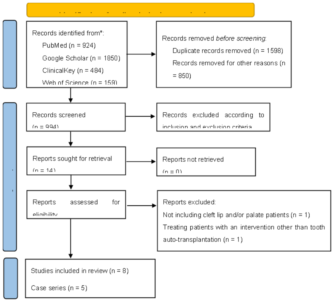
Figure 2.
JBI critical appraisal tool evaluation for case reports.
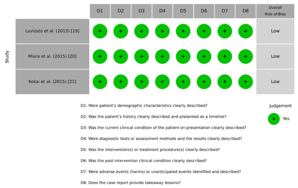
Figure 3.
JBI critical appraisal tool evaluation for case series.
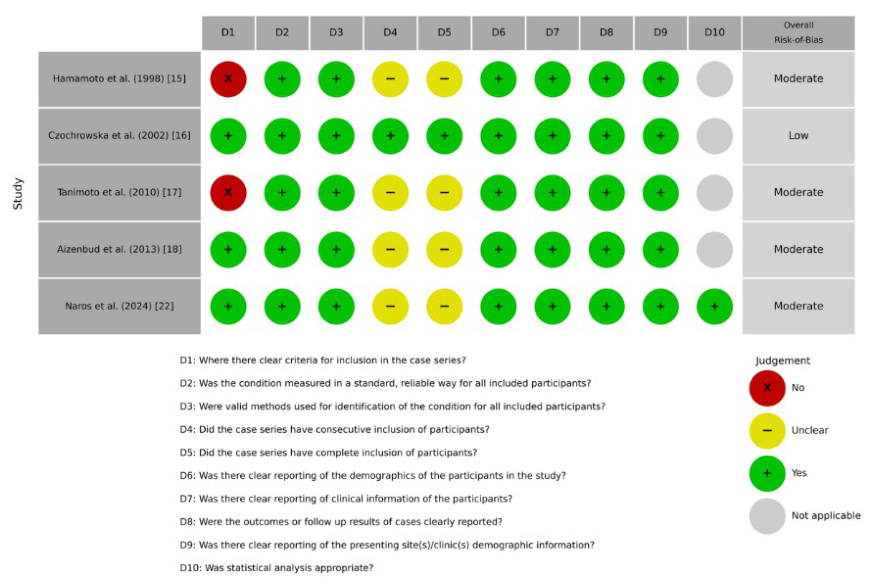
Figure 4.
A preliminary expert-informed clinical algorithm for the management of cleft lip and/or palate patients with tooth auto-transplantation. CLP = Cleft lip and/or palate; SABG = Secondary alveolar bone graft; RCT = Root canal treatment.
Figure 4.
A preliminary expert-informed clinical algorithm for the management of cleft lip and/or palate patients with tooth auto-transplantation. CLP = Cleft lip and/or palate; SABG = Secondary alveolar bone graft; RCT = Root canal treatment.
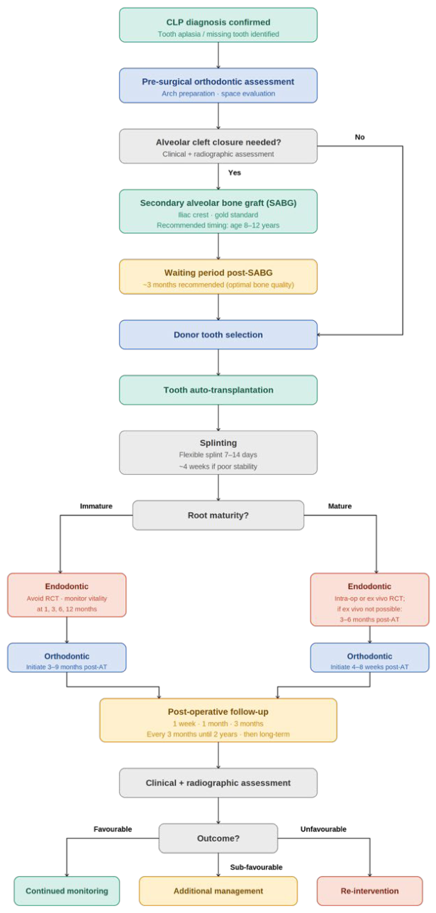

Table 1.
Inclusion and exclusion criteria.
| Inclusion criteria | Exclusion criteria |
|
|
Table 2.
A concise table presenting the characteristics and outcomes of the included studies.
| Study | Study design | Patients/Teeth | Cleft type | Donor tooth | Root maturity | Grafting approach | Graft → TAT timing | Follow-up | Tooth survival | Major complications |
| Hamamoto et al. (1998) [15] | Case series | 2/2 | UCLP | Mandibular central incisor Mandibular 2nd premolar |
Mature | SABG with Iliac PCBM | 6 - 12m. | 3 - 20m. | 2/2 | NR |
| Czochrowska et al. (2002) [16] | Case series | 5/5 | UCLP | Mandibular 1st premolar (n = 4) Mandibular 2nd premolar (n = 1) |
Immature | SABG with Iliac cancellous (n = 3) None (n = 2) |
14 - 26m. (n = 3) N/A (n = 2) |
2.5 - 7.7y. | 5/5 | Pulp obliteration Increased mobility (n = 1) Deficient papilla (n = 1) Patient dissatisfied (n = 1) |
| Tanimoto et al. (2010) [17] | Case series | 2/2 | UCLP | Maxillary 1st premolar Mandibular 1st premolar |
Immature | SABG with autogenous Iliac | 15 - 21m. | 10m. - 2.1y. | 2/2 | NR |
| Aizenbud et al. (2013) [18] | Case series | 4/5 | UCLP (n = 2) BCLP (n = 2) |
Mandibular 2nd premolar | Immature | SABG with Iliac PCBM | 12 - 52m. | 2.1 - 6.2y. | 5/5 | Pulp obliteration |
| Luvizuto et al. (2013) [19] | Case report | 1/1 | UCLP | Mandibular 2nd premolar | Immature | SABG with autogenous Iliac | 6m. | 1m. – 5y. | 1/1 | NR |
| Miura et al. (2015) [20] | Case report | 1/1 | BCL + UCA | Supernumerary tooth | Mature | SABG with Iliac PCBM + PRP | 0m. | 1.2 - 2.3y. | 1/1 | NR |
| Kokai et al. (2015) [21] | Case report | 1/1 | BCLP | Maxillary 2nd premolar | Mature | SABG with Iliac cancellous + Premaxillary osteotomy | 9m. | 2.8 - 3y. | 1/1 | Buccal gingival recession |
| Naros et al. (2024) [22] | Case series | 7/10 | UCLP (n = 6) CP (n = 1) |
Mandibular 2nd premolar (n = 5) Mandibular 1st premolars (n = 2) Maxillary 1st premolars (n = 1) Maxillary 2nd premolar ( n = 1) Maxillary 3rd molar (n = 1) |
Immature | SABG with Iliac cancellous (n = 2) None (n = 5) |
2 - 3m. (n = 2) N/A (n = 5) |
2.3 – 12.8y. | 9/10 | Pulp obliteration (n = 8) External root resorption, Periodontal pathology, No root development, Dull percussion (n = 1; tooth extraction at 27m.) Moderate root development (n = 1) |
| Total (1998 – 2024) [15,16,17,18,19,20,21,22] | Case series (n = 5) Case report (n = 3) |
23/27 | UCLP (n = 18) BCLP (n = 3) BCL + UCA (n = 1) CP (n = 1) |
Mandibular 2nd premolar (n = 13) Mandibular 1st premolars (n = 7) Maxillary 1st premolars (n = 2) Maxillary 2nd premolar ( n = 2) Maxillary 3rd molar (n = 1) Mandibular central incisor (n = 1) Supernumerary tooth (n = 1) |
Immature (n = 23) Mature (n = 4) |
SABG with Iliac PCBM (n = 6) SABG with Iliac cancellous (n = 5) SABG with autogenous Iliac (n = 3) SABG with Iliac PCBM + PRP (n = 1) SABG with Iliac cancellous + Premaxillary osteotomy (n = 1) None (n = 7) |
0 - 52m. N/A (n = 5) |
1m. - 12.8y. | 26/27 | Pulp obliteration (n = 13) Increased mobility (n = 1) Deficient papilla (n = 1) Patient dissatisfied (n = 1) Buccal gingival recession (n = 1) External root resorption, Periodontal pathology, No root development, Dull percussion (n = 1; tooth extraction at 27m.) Moderate root development (n = 1) |
SABG = Secondary alveolar bone graft; UCLP = Unilateral cleft lip and palate; BCLP = Bilateral cleft lip and palate; BCL = Bilateral cleft lip; UCA = Unilateral cleft alveolus; CP = Cleft palate; PCBM = Particulate cancellous bone and marrow; PRP = Platelet-rich plasma; NR = Not reported; y. = Year(s); m. = Month(s).
Table 3.
Detailed source derivation and evidence level for each core element of the preliminary expert-informed clinical algorithm.
Table 3.
Detailed source derivation and evidence level for each core element of the preliminary expert-informed clinical algorithm.
| Algorithm Element | Source Derivation Category | Primary Evidence Basis and References | Clinical Justification and Limitations |
|
SABG timing (mixed dentition, 8-12 years) |
Extrapolated from adjacent literature | Gold-standard cleft literature [28,29,32] | Observed in review: highly variable; several successful cases were treated at >12 years. Justification: extrapolated from established cleft timelines to optimize bone volume before canine eruption. |
|
Post-SABG waiting period (3 months of healing) |
Extrapolated from adjacent literature | Adjacent CBCT bone-density and volume literature [33,34] | Observed in review: gaps in reporting; ranged widely from 0 to 52 months. Justification: derived from adjacent radiographic data showing peak mineral bone density at 3 months post-grafting. |
|
Splinting duration (flexible splint for 7-14 days) |
Extrapolated from adjacent literature | General dental traumatology / TAT literature [49,50] | Observed in review: poorly or inconsistently specified across the included cleft studies. Justification: extrapolated to mitigate ankylosis and pulp necrosis risks associated with rigid or prolonged fixation. |
|
Endodontic timing (immature roots: avoid RCT and observe; indicated in case of complications) (mature roots: intra-op or 3-6 months post-operatively) |
Extrapolated from adjacent literature | General third-molar TAT guidelines [48] | Observed in review: highly inconsistent protocols due to the historical age of older studies. Justification: extrapolated from broader endodontic consensus to ensure systematic management of closed-apex complications. |
|
Orthodontic timing (immature roots: indicated 3-9 months post-op) (mature roots: indicated 4-8 weeks post-op) |
Extrapolated from adjacent literature | Broader biomechanical TAT studies [53,54] | Observed in review: extreme heterogeneity (ranging from 15 days to 1 year). Justification: extrapolated to allow adequate initial periodontal ligament healing and pulp revascularization before moving the tooth. |
|
Post-op follow-up schedule (1w., 1m., 3m., then every 3m. until 2 years) |
Extrapolated from adjacent literature and expert interpretation | General TAT complication timelines [56] + Author consensus | Observed in review: no included study documented a thorough, standardized, periodic tracking timeline. Justification: formulated by mapping standard clinical follow-up intervals onto the known chronological emergence of post-op complications. |
SABG = Secondary alveolar bone graft; CBCT = Cone-beam computed tomography; RCT = Root canal treatment; m. = Month(s); w. = Week(s);.
Table 4.
List of clinical and radiographic examinations for auto-transplanted teeth.
| Clinical examinations | Radiographic examinations |
|
|
Table 5.
Favourable, sub-favourable, and unfavourable follow-up outcomes in auto-transplanted teeth.
Table 5.
Favourable, sub-favourable, and unfavourable follow-up outcomes in auto-transplanted teeth.
| Favourable outcomes | Sub-favourable outcomes* | Unfavourable outcomes |
|
|
|
* = Relatively favourable outcomes with no significant compromise on the survival of auto-transplanted teeth, which could be left as is or managed through additional interventions.
Disclaimer/Publisher’s Note: The statements, opinions and data contained in all publications are solely those of the individual author(s) and contributor(s) and not of MDPI and/or the editor(s). MDPI and/or the editor(s) disclaim responsibility for any injury to people or property resulting from any ideas, methods, instructions or products referred to in the content. |
© 2026 by the authors. Licensee MDPI, Basel, Switzerland. This article is an open access article distributed under the terms and conditions of the Creative Commons Attribution (CC BY) license (http://creativecommons.org/licenses/by/4.0/).
Copyright: This open access article is published under a Creative Commons CC BY 4.0 license, which permit the free download, distribution, and reuse, provided that the author and preprint are cited in any reuse.


