

Submitted:
06 July 2026
Posted:
08 July 2026
You are already at the latest version
Abstract
This review examines the evolving role of muscle biopsy in the diagnosis of neuromuscular disorders in the era of modern genomic medicine. Historically the cornerstone of myopathy diagnosis, muscle biopsy enabled structural, histochemical, and ultrastructural characterization of muscle diseases. However, the introduction of next-generation sequencing and other genomic technologies has shifted the diagnostic paradigm, with genetic testing now serving as the preferred first-line approach for many hereditary myopathies due to its non-invasive nature and high diagnostic yield. However, muscle biopsy remains indispensable in the evaluation of inflammatory, toxic, metabolic, mitochondrial, and certain rare acquired myopathies. Biopsy is also valuable when genetic testing is inconclusive, particularly for interpreting variants of uncertain significance, through histopathological, immunohistochemical, and biochemical analyses. In certain disorders diagnosis may rely primarily on biopsy findings. Emerging technologies, including RNA sequencing, transcriptomics, proteomics, spatial transcriptomics, and artificial intelligence–assisted pathology, are expanding the diagnostic value of muscle tissue beyond traditional morphological assessment. Rather than being replaced by genomic medicine, muscle biopsy is evolving into a complementary component of an integrated diagnostic strategy that combines clinical, pathological, and molecular data to improve diagnostic accuracy and guide precision medicine in neuromuscular disorders.
Keywords:
muscle biopsy
; neuromuscular disorders
; next-generation sequencing
; inflammatory myopathies
; immunohistochemistry
; variants of uncertain significance (VUS)
1. Introduction
The diagnostic landscape for neuromuscular disorders has undergone a profound transformation, moving from a reliance on macroscopic pathology to a sophisticated, multi-tiered approach that incorporates advanced molecular and genetic tools. For decades, the muscle biopsy procedure served as the unequivocal cornerstone of myopathology. Around seventy years ago, the development of techniques such as cryostat sectioning, histochemistry, and electron microscopy cemented the muscle biopsy as the primary method for describing and classifying a wide range of myopathies [1]. This historical foundation established a critical link between clinical symptomatology and observable, microscopic tissue pathology, a paradigm that dominated the field for generations.
The advent of next-generation sequencing (NGS) technology marked a new chapter, enabling the discovery of a rapidly increasing number of inherited myopathies [2]. This non-invasive diagnostic approach, often initiated with a simple blood sample, quickly became the preferred first-line test for many suspected hereditary disorders. For conditions like Duchenne muscular dystrophy (DMD), where the causative gene mutation is well-understood, genetic testing is now considered the "gold standard"[3] This development has modified the traditional role of muscle biopsy, prompting a re-evaluation of its place in the diagnostic algorithm. Despite the efficiency and breadth of modern genetic testing, this review states that muscle biopsy has not been rendered obsolete. While genetic analysis now serves as the primary screening tool for many hereditary conditions, muscle biopsy remains indispensable for many conditions. This review delineates when and why each tool is most effective.
2. History of Muscle Biopsy
In the mid-19th century, the diagnosis of muscle disease relied entirely on clinical observation, with pathological confirmation available only at autopsy. This changed in 1865 when the French neurologist Guillaume Duchenne de Boulogne developed a specialized tissue punch (emporte-pièce histologique) to obtain muscle samples from living patients with what is now known as Duchenne muscular dystrophy (DMD). Microscopic examination revealed that the enlarged calf muscles were largely replaced by fat and connective tissue rather than hypertrophied muscle fibers, leading Duchenne to coin the term paralysie myoscléreuse [4,5]. Despite this pioneering achievement, muscle biopsy was not widely adopted because the procedure was invasive, painful, and associated with a high risk of infection in the pre-antiseptic era. Consequently, for many decades diagnosis continued to rely primarily on autopsy findings, with muscle biopsy used only in selected cases. By the late 19th and early 20th centuries, the development of reliable local anesthetics and antiseptic surgical techniques made open muscle biopsies far safer. The primary objective during this era was to answer whether the patient's weakness was caused by a problem in the muscle itself or in the supplying nerves. Muscle biopsies were used to formalize the distinctions between neuropathic patterns such as "group atrophy" and myopathic patterns such as variation in fiber size, necrotic fibers, and increased connective tissue [6].
Throughout the first half of the 20th century, muscle biopsy remained firmly rooted in this structural paradigm. Tissues were fixed in formalin, embedded in paraffin wax, and stained with standard hematoxylin and eosin (H&E). The true golden age of myology began in the late 1950s and peaked in the 1970s. Two massive technical innovations collided: the widespread adoption of the cryostat (which allowed fresh muscle tissue to be snap-frozen in liquid nitrogen and sliced while frozen, preserving delicate cellular enzymes) and enzyme histochemistry. Biochemical stains were applied directly to frozen muscle slices. Instead of just looking at cell shapes, metabolic and enzymatic activity could be visualized [7,8]. This led to the landmark discovery that human skeletal muscle is composed of distinct fiber types: Type I (“slow-twitch”) fibers, rich in oxidative enzymes and specialized for endurance, and Type II (“fast-twitch”) fibers, enriched in glycolytic pathways, highly reactive for ATPase activity, and adapted for rapid, forceful contractions [1,8].
Equipped with stains like Gömöri trichrome, Periodic acid Schiff, Sudan black, Nicotinamide adenine dinucleotide dehydrogenase-tetrazolium reductase (NADH-TR), Cytochrome c oxidase (COX), succinate dehydrogenase (SDH), and Adenosine triphosphatases (ATPases), myologists began discovering entirely new categories of diseases that looked near normal under standard H&E stains. They identified several diseases that came to be named for their distinctive pathological features. “Central core disease,” described in 1956, was named after histochemical studies revealed central “cores” within muscle fibers that lacked oxidative enzyme activity [9,10]. The term “nemaline myopathy” was coined in 1963 after the modified Gomori trichrome stain demonstrated clusters of tiny thread-like red rods (“nemaline rods”) derived from Z-disk structural proteins [11]. Stains like Periodic acid–Schiff (PAS) and Oil Red O allowed clinicians to diagnose glycogen storage diseases (like Pompe or McArdle disease) and lipid storage myopathies by demonstrating abnormal accumulations of sugar or fat.
The integration of electron microscopy (EM) into the muscle biopsy workflow enabled visualization of the ultrastructural organization of the sarcomere and mitochondria. The combination of histochemical techniques and electron microscopy opened the field of mitochondrial medicine. Using the modified Gomori trichrome stain, the so-called “ragged-red fibers” were identified, leading to the emergence of the field of mitochondrial myopathies [12]. EM is still useful in selected cases to confirm the presence of abnormalities identified by light microscopy. Some myopathies can be diagnosed more readily and with greater accuracy using EM [13].
By the early 1980s, immunohistochemistry (IHC) and Western blotting had been introduced into muscle pathology. These techniques employed custom-engineered antibodies to determine whether specific structural proteins were present or absent in muscle tissue. In 1987, the missing protein in DMD was identified: dystrophin [14]. Muscle biopsies were subsequently stained with anti-dystrophin antibodies and biopsies from boys with DMD showed a complete absence of sarcolemmal staining. Since then, IHC has been used for the diagnoses of muscular dystrophies through detection of absent or abnormally localized structural proteins (dystrophin, dysferlin, caveolin-3, and others) [15]. An immunohistochemical approach may enable clear discrimination between dystrophic myopathies associated with inflammation and primary inflammatory myopathies. In inflammatory myopathies, IHC distinguishes myositis subtypes through characteristic staining patterns, including expression of MHC class I and II, C5b-9, and p62/TDP-43, as well as analysis of the inflammatory infiltrate [15].
3. History of Modern Genetics in the Diagnosis of Muscular Diseases
During the 1990s, polymerase chain reaction (PCR), Sanger sequencing, and Southern blot techniques enabled direct mutation detection in many hereditary myopathies, gradually reducing reliance on invasive muscle biopsy. The discovery of genes encoding sarcolemmal, nuclear, sarcomeric, and mitochondrial proteins revealed the marked genetic heterogeneity of muscular disorders and allowed their classification according to molecular defects rather than solely pathological appearance. The introduction of Sanger sequencing enabled molecular diagnosis through single-gene testing. However, this approach was limited by its sequential nature: clinicians had to analyze genes one at a time based on clinical suspicion, phenotype, histological findings, and presumed inheritance patterns. With more than 170 genes associated with muscle disorders and substantial phenotypic overlaps between conditions, this strategy became increasingly impractical, time-consuming, and costly [16].
The development of NGS fundamentally transformed the diagnostic landscape of inherited neuromuscular diseases. Beginning around 2010, NGS enabled the simultaneous parallel sequencing of multiple genes through targeted gene panels. This technological advance substantially increased diagnostic yield, with gene panels achieving diagnostic rates of approximately 30–70%, while also reducing both cost and turnaround time compared with sequential single-gene testing [17].
Whole-exome sequencing (WES) was introduced shortly thereafter and became particularly valuable for unresolved or atypical neuromuscular cases. WES sequences the protein-coding regions of the genome (the exome), which comprise only 1–2% of the genome but harbor most known disease-causing variants. WES enabled the discovery of numerous novel neuromuscular disease genes and expanded the phenotypic spectrum of previously recognized disorders. It proved especially useful in congenital myopathies and complex pediatric neuromuscular syndromes in which the clinical phenotype did not clearly suggest a single candidate gene [18].
Whole-genome sequencing (WGS) represented the next major technological advance. Unlike WES, WGS sequences both coding and non-coding regions of the genome. WGS improved the detection of structural variants, repeat expansions, copy-number variations, deep intronic mutations, and mitochondrial DNA abnormalities that may be missed by exome sequencing. In neuromuscular disorders, WGS has become increasingly important for patients who remain genetically undiagnosed after conventional testing or WES. It offers comprehensive coverage of coding, non-coding, and intergenic regions, enabling improved detection of structural variants and simultaneous analysis of both nuclear and mitochondrial genomes [19].
More recently, the limitations of short-read sequencing platforms, particularly in detecting structural variants and repeat expansions, have driven the development of long-read sequencing technologies. These methods generate longer contiguous DNA reads and improve the detection of repetitive elements, copy-number alterations, and large expansions [20].
4. Genetic Testing and Muscle Biopsy: Diagnostic Approach
4.1. When to Start with Genetic Testing
Genetic testing is the preferred initial diagnostic approach when a hereditary neuromuscular disorder is suspected, and the phenotype is reasonably well characterized (e.g. “LGMD like”, congenital myopathy, dystrophinopathy, FSHD, OPMD, channelopathies) [21,22]. Key situations to begin with genetics:
Clear or pattern based inherited phenotype (typical Duchenne/Becker, classic LGMD subtype, recognizable congenital myopathy pattern).
Family history suggesting Mendelian transmission.
Chronic course, and no strong red flags for acquired/inflammatory disease.
Availability of high-quality phenotype-driven NGS panels or exome sequencing and a strong expectation of monogenic or oligogenic etiology, such as in muscular dystrophies, many congenital myopathies, and channelopathies.
Situations where biopsy would not change the need for molecular confirmation (e.g. suspected dystrophinopathy in a boy: start with DMD deletion/duplication + sequencing; biopsy only if genetics is negative or discordant).
4.2. When to Start with Muscle Biopsy
Suspected inflammatory myopathy, especially when antibody panel results are delayed, inconclusive, discordant, seronegative, or only weakly positive.
Rapidly progressive weakness accompanied by markedly elevated CK levels, myopathic EMG findings, abnormal muscle MRI, systemic manifestations, or clinical red flags suggestive of vasculitis, immune-mediated necrotizing myopathy, overlap syndromes, or toxic myopathy, in which muscle biopsy findings may immediately guide immunotherapy decisions and alter clinical management [21].
Suspected metabolic or mitochondrial myopathy, particularly when histochemical and respiratory chain studies are required and mtDNA analysis or next-generation sequencing may need to be performed on muscle tissue rather than blood.
Although current guidelines often prioritize genetic testing before muscle biopsy, a pediatric cohort study found that genetic testing performed after biopsy had greater diagnostic utility because histopathological findings helped guide targeted molecular analysis. For example, ragged-red fibers prompted mitochondrial DNA testing, while multiminicores directed evaluation of congenital myopathy genes. In this setting, muscle biopsy provided critical clues that narrowed the differential diagnosis and enabled a more focused and efficient genetic workup [23].
It should be noted that certain dystrophies (dysferlinopathies, facioscapulohumeral muscular dystrophy) can show considerable inflammatory infiltrates on biopsy and even upregulation of major histocompatibility complex class I (MHC-I), complicating the distinction from inflammatory myopathies [24]. Biopsy of patients with calpainopathy due to homozygous pathogenic CAPN3 mutations may reveal infiltrates consisting of eosinophils [25] or macrophages and lymphocytes [26]. Accordingly, the presence of inflammatory infiltrates on biopsy does not exclude an underlying hereditary disorder [27].
4.3. Where Biopsy Adds Unique Value After Genetic Testing
Even in “genetics-first” diagnostic pathways, muscle biopsy often provides information that NGS cannot provide, or it helps interpret the genetic findings [28,29].
Dystrophinopathy with negative or inconclusive DMD testing: biopsy may be used to assess dystrophin expression and to support RNA-based detection of deep intronic or regulatory variants.
LGMD-like phenotypes with negative broad gene panels or exome sequencing: biopsy may reveal specific protein deficiencies (e.g., sarcoglycans, dysferlin, calpain 3, anoctamin-5), thereby guiding targeted re-analysis or additional genetic testing.
Variant interpretation: muscle biopsy may demonstrate protein absence, mislocalization, or characteristic structural changes that support the pathogenicity of a VUS or a candidate gene identified by exome/genome sequencing.
mRNA and cDNA analysis from muscle tissue can provide direct evidence of the functional effects of genetic variants, particularly in cases unresolved by DNA testing [30]. This approach detects splicing defects, exon skipping, deep intronic variants, and transcript loss due to nonsense-mediated decay that may be missed by standard sequencing. Because many neuromuscular genes are not expressed in blood, muscle tissue or myogenic cells derived from fibroblasts offer a more informative source and can improve diagnostic yield. RNA sequencing identifies pathogenic mechanisms in up to 38% of unresolved cases and can help reclassify variants of uncertain significance by demonstrating their functional impact. Emerging technologies, including transcriptomics, proteomics, and multi-omics approaches, are further expanding the diagnostic and research potential of muscle tissue analysis. [28,30].
4.4. When Diagnosis Can Rest Essentially on Muscle Biopsy
There are clinical situations in which the diagnosis is established primarily by muscle biopsy, while genetic testing is either non-contributory or serves an adjunctive rather than decisive role.
Inflammatory myopathies: Polymyositis, dermatomyositis, immune-mediated necrotizing myopathy, or inclusion body myositis may be identified on biopsy, particularly when autoantibody panels are negative, delayed, or nonspecific. In such cases, treatment decisions can often be based on histopathological findings in conjunction with the clinical phenotype [27].
Classic dystrophinopathy with negative or inconclusive genetics: Marked reduction or absence of dystrophin expression on immunohistochemistry in a boy with a Duchenne/Becker muscular dystrophy phenotype remains highly reliable method confirming dystrophin deficiency. Subsequent genetic testing is then used mainly to define the precise variant for genetic counseling and mutation-specific therapies [31].
Metabolic and mitochondrial myopathies with demonstrable biochemical defects: Clear and reproducible respiratory chain deficiencies, ragged-red fibers, COX-negative fibers, or specific enzymatic defects identified in muscle tissue may establish a working diagnosis and guide management, even when the underlying genetic defect has not yet been identified [32].
Rare treatable entities with unresolved or negative genetics: Some disorders, such as lipid storage myopathy, may remain genetically unresolved despite extensive testing. In these cases, the diagnosis and consequently the initiation of targeted treatment may depend entirely on muscle biopsy findings [33].
Rare, acquired entities diagnosable only morphologically: Sporadic late-onset nemaline myopathy (SLONM), amyloid myopathy, and the recently described monoclonal gammopathy-associated glycogen storage myopathy (MGGSM), are potentially treatable disorders that may be clinically suspected, but require muscle biopsy for definitive diagnosis [34].
This diagnostic approach is summarized in Figure 1.
5. Limitations of Muscle Biopsy
A significant proportion of muscle biopsies, up to approximately 20% in routine clinical practice, fail to yield a specific diagnosis, frequently returning "non-informative" or completely normal findings [35]. The limitations include:
Sampling error due to patchy disease distribution: Many myopathies exhibit highly focal or heterogeneous tissue involvement. A biopsy needle or incision can easily harvest entirely unaffected or non-specifically altered muscle tissue right alongside highly pathological lesions, leading to a false-negative result [36]. MRI may help reduce sampling error by identifying muscles with active disease and guiding optimal biopsy site selection. This is particularly valuable in inflammatory myopathies, where pathological changes are often patchy and unevenly distributed.
Freezing artifacts: Skeletal muscle tissue is highly sensitive to cryopreservation techniques. If a sample froze too slowly, the formation of ice crystals causes fine sarcoplasmic holes. This severe artifact can mask subtle structural features, such as metabolic defects or vacuolar changes, rendering the tissue uninterpretable [37].
Small muscle sample: Adequate muscle biopsy specimens should contain at least 200–250 well-oriented transverse muscle fibers. Fine-gauge needle biopsies often provide insufficient tissue, limiting optimal orientation and ancillary biochemical or mitochondrial studies, and increasing the risk of sampling error [38].
Inappropriate site selection: Selecting a muscle with severe, long-standing weakness can result in an "end-stage" biopsy where the primary pathological hallmarks have vanished, replaced entirely by non-specific fibrofatty tissue and marked atrophy.
5.1. When to Consider a Repeat Muscle Biopsy
Repeat muscle biopsy is required in only a small proportion of myopathy evaluations and should be reserved for selected situations. Indications include a non-specific or non-diagnostic initial biopsy despite strong clinical suspicion of a treatable neuromuscular disorder, disease progression or unexpected treatment failure suggesting an incomplete diagnosis, and technically inadequate specimens affected by sampling or processing artefacts. Diagnostic yield may also improve when the repeat biopsy is guided by MRI or ultrasound to target active disease, or when an open surgical biopsy is performed after a non-diagnostic needle biopsy. In a large study, repeat biopsy was performed in 3.6% of cases and established a specific diagnosis in 43%. [39].
6. The Role of Muscle Biopsy in Different Clinical Conditions
6.1. Muscle Biopsy in Rapidly Progressive Weakness
The main cause of rapidly progressive weakness, particularly when associated with markedly elevated CK levels and a myopathic EMG, is inflammatory or immune-mediated myopathy. Muscle biopsy remains essential for the diagnosis and subclassification of inflammatory myopathies. Each subtype of inflammatory myopathy demonstrates characteristic myopathological features that facilitate accurate classification. However, muscle biopsy is no longer mandatory for well-defined clinical-serologic presentations, such as classic dermatomyositis with characteristic rashes, or autoantibody-positive IMNM (anti-SRP or anti-HMGCR) with typical clinical features [27]. In patients with connective tissue disorders presenting with muscle weakness and elevated creatine kinase, biopsy is usually not performed. It nevertheless remains essential in patients without myositis-specific autoantibodies, in cases with delayed or inconclusive serologic results, and is particularly critical for the diagnosis of inclusion body myositis. Myositis-specific autoantibodies are found in only two-thirds of patients with myositis, making muscle biopsy particularly important in seronegative cases [40].
6.2. Dermatomyositis
Dermatomyositis is a disease mainly of skin and muscle but may affect lung and other tissues. The hallmark feature is perifascicular atrophy, characterized by layers of atrophic muscle fibers located at the periphery of fascicles. Inflammatory infiltrates are predominantly perivascular and interfascicular, in contrast to the endomysial pattern observed in other inflammatory myopathy subtypes. Vascular pathology is prominent and includes endothelial hyperplasia with tubuloreticular inclusions, fibrin thrombi, and capillary obliteration. MHC-I immunostaining demonstrates a characteristic perifascicular pattern, with increased expression in atrophic perifascicular fibers [27,40,41].
Recent studies have identified antibody-specific pathological variations. Anti-TIF1-γ dermatomyositis is characterized by vacuolated (“punched-out”) fibers and perifascicular MHC-I enhancement. Anti-Mi-2-associated disease shows prominent muscle damage, perifascicular necrosis, and increased perimysial alkaline phosphatase activity. Anti-MDA5 dermatomyositis demonstrates scattered or diffuse MxA staining with less severe muscle pathology, whereas anti-NXP-2 is associated with microinfarction [42].
6.3. Immune-Mediated Necrotizing Myopathy (IMNM)
IMNM is characterized primarily by widespread, simultaneous muscle fiber necrosis and active regeneration. Unlike other inflammatory myopathies, it features a striking lack of primary lymphocytic inflammation, with immune cells mostly limited to macrophages clearing necrotic debris [Figure 2]. Diagnostic hallmarks include the deposition of the complement membrane attack complex (C5b-9) on the membranes of non-necrotic fibers and localized MHC-I expression. Immune-mediated necrotizing myopathy is associated with two specific antibodies: anti-SRP and anti-HMGCR [27,43]. It should be noted that, rarely, anti-HMGCR IMNM may present with a slowly progressive course and dystrophic biopsy features, thereby mimicking muscular dystrophy [Figure 3].
6.4. Anti-Synthetase Syndrome
Anti-synthetase syndrome is characterized by the presence of anti-aminoacyl-tRNA synthetase antibodies and a constellation of clinical features affecting multiple organ systems. The classic clinical presentation includes myositis, interstitial lung disease, arthritis, mechanic's hands, Raynaud phenomenon, and fever.
Perifascicular necrosis and regeneration are the hallmark findings, present in about half of cases. Increased perimysial alkaline phosphatase activity is a distinctive feature, representing enhanced enzyme activity in the connective tissue surrounding fascicles.
Inflammatory infiltrates are variable in distribution and intensity. Some cases show myofiber necrosis similar to immune-mediated necrotizing myopathy. MHC-I upregulation can be demonstrated, though the pattern and extent vary. The pathological presentation is relatively homogeneous across different anti-synthetase antibodies [44].
6.5. Idiopathic Polymyositis
Idiopathic polymyositis refers to patients whose disease does not fit any of the other inflammatory myopathy subtypes and is therefore often considered a diagnosis of exclusion. This entity is now regarded as relatively rare [40]. Polymyositis shows endomysial inflammation with or without invasion of nonnecrotic fibers by mononuclear cells (usually CD8+ T cells) and with or without perimysial or perivascular inflammation invading nonnecrotic muscle fibers expressing MHC-I.Unlike dermatomyositis, inflammatory infiltrates are more within fascicles rather than perivascular/interfascicular. Necrotic fibers tend to be scattered or isolated rather than grouped [45].
6.6. Metabolic Myopathies
For suspected metabolic myopathies causing rhabdomyolysis muscle biopsy should be deferred until recovery from the acute episode. Rhabdomyolysis can occur in metabolic myopathies including glycogen storage diseases (McArdle disease, Pompe disease) and fatty acid oxidation disorders (carnitine palmitoyltransferase II deficiency). Mitochondrial myopathies can present with exercise intolerance and rhabdomyolysis. In these conditions, muscle biopsy may provide valuable diagnostic information. Gomori trichrome shows ragged red fibers in mitochondrial myopathies, PAS staining reveals glycogen accumulation in glycogen storage diseases, and increased lipid content on Oil-Red O or Sudan black suggests fatty acid oxidation disorders. In addition, muscle biopsy can play a pivotal role in the interpretation of VUS, providing functional and pathological evidence that may help establish pathogenicity [46].
6.7. Critical Illness Myopathy
Critical illness myopathy (CIM) is the most common non-inflammatory cause of rapidly progressive weakness in hospitalized patients, affecting 25–84% of patients requiring prolonged mechanical ventilation and up to 35% of those with status asthmaticus or COPD. CIM typically presents with generalized flaccid weakness and preserved sensation, when sensory examination is feasible. Risk factors include prolonged ICU stay, sepsis, multi-system organ dysfunction, hyperglycemia, and exposure to steroids and neuromuscular blocking agents. Muscle biopsy demonstrates myofiber necrosis and selective loss of myosin filaments. [47].
6.8. Drug-Induced and Toxic Myopathies
Statin-induced myopathy can present acutely with rhabdomyolysis, particularly when combined with other medications (fibrates, azole antifungals, macrolides) or in the setting of hypothyroidism, renal impairment, or vitamin D deficiency. A subset of patients with statin myopathy had histopathologic or electron microscopic evidence for mitochondrial dysfunction in skeletal muscle [48].
Other toxic myopathies are associated with alcohol, colchicine, antimalarial agents (chloroquine and hydroxychloroquine), and certain chemotherapeutic drugs, all of which may cause acute or subacute muscle weakness. In most cases, clinical improvement follows withdrawal of the offending agent. However, if weakness fails to improve within several weeks after drug discontinuation or continues to progress despite cessation of therapy, muscle biopsy should be considered to exclude an alternative or coexisting myopathic process.
The hallmark pathological feature of colchicine-induced myopathy is a vacuolar myopathy characterized by the accumulation of autophagic vacuoles in the absence of significant muscle fiber necrosis. These vacuoles predominantly involve type 1 muscle fibers and stain strongly for acid phosphatase, reflecting their lysosomal origin. Similar autophagic vacuolar changes may also be observed in chloroquine- and hydroxychloroquine-induced myopathies [49,50]. LC3 and p62 are established markers of autophagosome accumulation. Immunohistochemical staining for these proteins demonstrates characteristic punctate sarcoplasmic aggregates, providing evidence of impaired autophagic flux and autophagosome accumulation without the need for electron microscopy [51].
6.9. Endocrine Myopathies
Endocrine myopathies can closely mimic inflammatory myopathies and may lead to misdiagnosis if thyroid and metabolic function tests are not obtained early in the diagnostic evaluation. Patients with severe hypothyroidism may present with proximal muscle weakness, markedly elevated creatine kinase levels, and a necrotizing pattern on muscle biopsy [52]. Consequently, they may be incorrectly diagnosed with IMNM if thyroid dysfunction is not recognized. Importantly, central hypothyroidism can also be associated with necrotizing myopathy despite a normal thyroid-stimulating hormone (TSH) level, underscoring the need to assess free thyroid hormone levels when clinical suspicion persists [53].
Hyperthyroidism may likewise cause profound, rapidly progressive weakness, occasionally manifesting as thyrotoxic periodic paralysis, which is often precipitated by carbohydrate-rich meals or rest following exercise. The pathological findings in hyperthyroid myopathy are nonspecific and variable, ranging from minimal myopathic changes to more pronounced structural abnormalities. Unlike drug-induced vacuolar myopathies, hyperthyroid myopathy lacks characteristic histopathological features, and muscle biopsy is rarely required for diagnosis because the clinical presentation and laboratory findings are usually sufficient to establish the underlying endocrine disorder [54].
6.10. Amyloid Myopathy
Light chain amyloidosis can present as rapidly progressive proximal weakness with elevated creatine kinase, myogenic EMG pattern, and fatty replacement on muscle MRI. Muscle biopsy reveals amyloid deposits in vessel walls, and serum light chain analysis confirms the diagnosis. This may be the initial manifestation of multiple myeloma [55].
6.11. Hereditary Myopathies with Rapidly Progressive Course
Most hereditary myopathies follow a slowly progressive clinical course. However, a subset may present with relatively rapid disease progression. This poses a significant diagnostic challenge, as distinguishing hereditary myopathies from seronegative inflammatory myopathies is crucial for establishing an accurate diagnosis and for initiating appropriate targeted therapy when indicated. Muscle biopsy remains a valuable tool in making this distinction.
An example of a hereditary disease that may progress rapidly is reducing body myopathy (RBM). This is an X-linked dominant muscle disorder caused by a pathogenic variant in the FHL1 gene. Pathologically, it is defined by reducing bodies: intracytoplasmic inclusions in muscle fibers that reduce nitro-blue tetrazolium (NBT) and stain intensely with menadione-NBT. The clinical spectrum varies widely, from mild, asymmetric proximal weakness to severe, progressive myopathy with respiratory failure and cardiac involvement. Presentation in women can be influenced by skewed X-inactivation. A muscle biopsy is essential for promptly differentiating RBM from inflammatory or other hereditary myopathies [56,57] [Figure 4].
7. Muscle Biopsy in Slowly Progressive Myopathies
7.1. Muscular Dystrophies
Muscular dystrophies are a heterogeneous group of inherited muscle disorders characterized by progressive muscle weakness and degeneration. In most cases, muscle biopsy demonstrates a dystrophic pattern; however, these histopathological changes are often nonspecific and insufficient to establish a precise diagnosis.
Immunohistochemistry and immunoblotting can provide valuable diagnostic information by identifying deficiencies or abnormalities of specific muscle proteins. These techniques are particularly useful in autosomal recessive muscular dystrophies, where the causative protein defect is known and reliable antibodies are available for detection.
With the widespread availability of next-generation sequencing, muscle biopsy has shifted from a primary diagnostic tool to a complementary investigation, whereas genetic testing has become the first-line diagnostic approach for most muscular dystrophies. In disorders with characteristic clinical phenotypes, such as myotonic dystrophy, facioscapulohumeral muscular dystrophy (FSHD), and oculopharyngeal muscular dystrophy (OPMD), genetic testing should be performed as the initial diagnostic investigation, without a preceding muscle biopsy [21].
Current indications for muscle biopsy in muscular dystrophies include:
7.1.1. Negative or Inconclusive Genetic Testing
When clinical suspicion for a muscular dystrophy remains high despite nondiagnostic genetic results, muscle biopsy may reveal protein abnormalities that can guide further genetic investigations.
7.1.2. Suspected Dystrophinopathy with Negative Genetic Testing
In patients with a clinical phenotype suggestive of Duchenne muscular dystrophy (DMD) or Becker muscular dystrophy, muscle biopsy with dystrophin immunohistochemistry and Western blot analysis is indicated when routine genetic testing is unrevealing [Figure 5]. Rare deep intronic variants or complex structural rearrangements may escape detection by standard genetic methods [58]. Moreover, distinguishing complete from partial dystrophin deficiency can help differentiate DMD from milder dystrophinopathy phenotypes.
7.1.3. Interpretation of VUS Significance
Muscle biopsy may demonstrate specific protein abnormalities or structural changes that support the pathogenicity of variants identified by next-generation sequencing. Immunohistochemical and immunoblot analyses provide direct evidence of protein expression and integrity that cannot be inferred from DNA sequencing alone [59].
An integrated diagnostic approach that combines clinical phenotype, age at onset, pattern of muscle weakness, serum creatine kinase levels, muscle imaging findings, genetic testing, and the selective use of muscle biopsy remains the optimal strategy for the evaluation of muscular dystrophies.
7.2. Congenital Myopathies
The role of muscle biopsy has shifted from the primary gold standard for diagnosing congenital myopathies to an integrative tool within the modern diagnostic landscape. While advanced genetic testing has fundamentally changed the diagnostic workflow, muscle biopsy remains indispensable for managing the genetic and clinical heterogeneity characteristic of these disorders [29].
Muscle biopsy is critical for resolving the pathogenicity of VUSs identified during genetic testing. Histological and ultrastructural analysis can confirm if a genetic variant correlates with observed structural protein dysfunction [60]. In cases where clinical presentation is ambiguous, morphological and histochemical findings (e.g. nemaline bodies, central cores, or fiber-type disproportion) can help prioritize candidate gene panels, and facilitate interpretation of genetic findings [29]. It remains vital for distinguishing congenital myopathies from other neuromuscular conditions such as inflammatory myopathies, metabolic disorders, or neuropathic processes when genetic results are inconclusive or absent [60].
7.3. Myofibrillar Myopathies
In the pre-genomic era, muscle biopsy was regarded as the diagnostic gold standard for myofibrillar myopathies (MFMs) [61]. Diagnosis relied on the identification of characteristic pathological features by light and electron microscopy, including focal myofibrillar dissolution originating at the Z-disk, progressive disorganization of the myofibrillar network, and abnormal intracellular accumulation of intermediate filaments and chaperone proteins such as desmin, αB-crystallin, and myotilin [62]. However, MFMs are clinically, genetically, and pathologically heterogeneous. No specific histopathological pattern reliably predicts the underlying genetic defect or clinical phenotype, and pathogenic findings in classical MFM-associated genes may occur without typical myofibrillar pathology.
7.4. GNE Myopathy
Genetic testing is now considered the main diagnostic approach representing a shift from the pre-genetic era when muscle biopsy was essential for the diagnosis. Nevertheless, muscle biopsy retains an important role when VUSs are identified on molecular genetic testing. The characteristic histopathologic findings including rimmed vacuoles on modified Gomori trichrome staining, fiber size variation, atrophy, and absence of inflammation, can provide supportive evidence for variant pathogenicity. However, a biopsy performed too early or on an inappropriate muscle (e.g., the heavily spared quadriceps femoris) can lead to false-negative histopathology (e.g. absence of vacuoles). As awareness of GNE myopathy increases among clinicians, the role of muscle biopsy will likely continue to diminish, reserved primarily for cases with uncertain genetic findings [64,65].
7.5. Lipid Storage Myopathies
Multiple acyl-coenzyme A dehydrogenase deficiency (MADD) is a rare autosomal recessive fatty acid oxidation disorder, caused by defects in electron transfer flavoprotein (ETF) genes. As a result, electron transfer from multiple flavoprotein dehydrogenases to the mitochondrial respiratory chain is impaired, leading to defective oxidation of fatty acids and other metabolic substrates. It presents in severe neonatal forms with or without congenital anomalies or as late-onset forms. A subset of patients may present with a pure myopathic form with progressive proximal weakness which may be associated with axonal sensory neuropathy.
Genetic testing may identify pathogenic variants in the ETFDH, ETFA, or ETFB genes. However, several reports have described patients with clinically and biochemically confirmed MADD who carry only a single heterozygous variant or in whom no pathogenic variants are detected. In such cases, the diagnosis has been established by muscle biopsy demonstrating lipid accumulation within muscle fibers, together with supportive findings on acylcarnitine profiling and a favorable response to therapy. Prompt muscle biopsy is particularly important in these patients, as it can facilitate the diagnosis of a potentially treatable disorder and enable the initiation of highly effective therapy [Figure 6]. Recent findings indicate that sertraline (an SSRI antidepressant) can trigger this myopathy [32,66].
7.6. Inclusion-Body Myositis
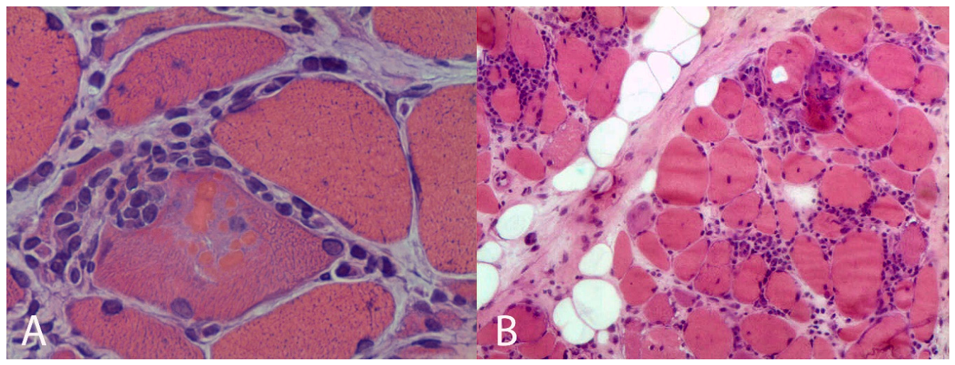
Inclusion-body myositis (IBM) is a slowly progressive, asymmetric myopathy that typically affects the quadriceps and finger flexors. Although its etiology remains unclear, genetic susceptibility has been linked to HLA-DRB1*03:01, which is associated with earlier onset and more severe disease [67]. Pathologically, IBM combines inflammatory and degenerative features that distinguish it from other inflammatory myopathies. The inflammatory component consists of endomysial CD8+ T-cell infiltrates invading MHC-I–expressing non-necrotic fibers, often with additional perivascular inflammation. The degenerative component includes rimmed vacuoles, endomysial fibrosis, fiber size variability, and atrophic angulated fibers, although rimmed vacuoles are absent in up to half of cases. Mitochondrial abnormalities such as ragged-red and COX-negative fibers are common, and electron microscopy may show 15–18 nm tubulofilamentous inclusions. Protein aggregates containing p62, TDP-43, and amyloid deposits further support the mixed inflammatory–degenerative nature of the disease [67,68,69] [Figure 7]. Anti-cN1A antibodies are highly specific (92-98%) but only moderately sensitive (30-70%) for IBM, making them useful for confirming the diagnosis when positive but insufficient to rule it out when negative [27].
7.7. Sporadic Late-Onset Nemaline Myopathy
Sporadic late-onset nemaline myopathy (SLONM) is a rare, acquired, adult-onset muscle disease characterized by subacute progressive proximal and axial weakness, with nearly half of cases associated with monoclonal gammopathy. The disease manifests with prominent neck extensor and paraspinal muscle involvement, often causing head drop or camptocormia, along with dysphagia, dyspnea, and respiratory compromise in many patients. Muscle biopsy is essential and diagnostic, demonstrating the pathologic hallmark of nemaline rods (aggregates of degenerated Z-disk and thin filament proteins) in atrophic muscle fibers. However, diagnosis can be challenging because rod-containing fibers may be sparse and patchily distributed, with a considerable number of patients requiring EM or repeat muscle biopsies to establish the diagnosis. Immunohistochemical staining for α-actinin significantly enhances diagnostic sensitivity when rods are not readily apparent on standard modified Gömöri trichrome staining. Importantly, SLONM is a treatable condition that responds to immunomodulatory therapies including intravenous immunoglobulins, chemotherapy, or autologous stem cell transplantation, making timely biopsy-confirmed diagnosis critical to prevent diagnostic delay and initiate appropriate treatment for this otherwise potentially lethal disease [70,71].
7.8. AL Amyloidosis Myopathy
AL amyloidosis is a rare blood disorder where misfolded immunoglobulin light chains form insoluble fibrils that deposit in organs. When it affects skeletal muscle (AL amyloidosis myopathy), it typically causes gradual or subacute proximal muscle weakness and dysphagia, and may involve facial, neck, or tongue muscles. Peripheral neuropathy occurs in about one-third of patients. Diagnosis requires a muscle biopsy, though Congo red staining is not always routinely done. The biopsy serves both to identify amyloid deposits and to characterize the amyloid subtype through immunohistochemistry or mass spectrometry. This condition should be suspected in patients over 40 presenting with unexplained myopathy, especially alongside neuropathy, cardiomyopathy, dysphagia, or weight loss [33,72].
7.9. Monoclonal Gammopathy-Associated Glycogen Storage Myopathy
Monoclonal gammopathy-associated glycogen storage myopathy (MGGSM) is a recently recognized, treatable acquired myopathy linked to monoclonal gammopathy, first described in 2023 [73]. It presents in adults with subacute proximal and axial weakness, often accompanied by weight loss, muscle stiffness, dysphagia, or head drop. Muscle biopsy is diagnostic, showing PAS-positive glycogen-filled vacuoles, autophagic material, and granular sarcolemmal MAC staining, while genetic testing for inherited glycogen storage diseases is negative. The condition often responds well to immunotherapy or autologous stem cell transplantation [73,74,75].
8. Future Perspectives
Because genetic testing often identifies variants of uncertain significance (VUSs) or does not fully explain the clinical phenotype, advanced analysis of muscle tissue has become increasingly important. Muscle biopsy is evolving from a purely structural examination into a powerful molecular tool. For example, RNA sequencing can provide a diagnosis in 35–40% of cases that remain unsolved after exome sequencing by detecting abnormal splicing, deep intronic variants, and other transcript-level defects not visible in DNA analysis [76]. RNA sequencing of muscle tissue identified a recurrent deep intronic defect in COL6A1 that caused inclusion of a harmful pseudoexon, leading to the diagnosis of previously unexplained cases of COL6-related muscular dystrophy [28].
New technologies such as spatial transcriptomics and proteomics preserve tissue architecture while revealing molecular changes within their anatomical context. Combining genomic, transcriptomic, proteomic, and metabolomic data provides a comprehensive view of disease, linking genetic variants to downstream effects on proteins, cellular pathways, and metabolism [76,77,78,79].
Artificial intelligence (AI) is also transforming muscle pathology. Deep-learning algorithms can analyze digital biopsy slides, quantify pathological features, map inflammatory changes, and even predict underlying molecular signatures. As a result, muscle biopsy is becoming not only a tool for detecting structural abnormalities but also a functional platform that helps interpret genetic findings and guide precision therapies [79].
Traditional histopathology remains essential for evaluating disease pathology. The future of muscle biopsy lies in integrating advanced molecular technologies with morphological assessment to support precision medicine in neuromuscular disorders.
9. Conclusion
The diagnostic approach to myopathies has undergone a profound transformation with the advent of next-generation sequencing and other genomic technologies. Although genetic testing has become the preferred first-line investigation in many inherited muscle disorders, muscle biopsy continues to play a pivotal role in selected clinical settings. Beyond its established value in inflammatory, toxic, metabolic, and mitochondrial myopathies, muscle biopsy remains an important tool for interpreting variants of uncertain significance, guiding targeted molecular investigations, and providing functional evidence of disease mechanisms. Emerging technologies, including transcriptomics, proteomics, and multi-omics approaches, are further expanding the diagnostic and research potential of muscle tissue analysis. Rather than being replaced by genomic medicine, muscle biopsy is evolving into a complementary and increasingly sophisticated component of an integrated diagnostic strategy. The future of neuromuscular diagnosis will likely depend not on choosing between biopsy and genetics, but on combining clinical, pathological, and molecular data to achieve the highest diagnostic accuracy and improve patient care.
References
- Engel, W.K. The essentiality of histo- and cytochemical studies of skeletal muscle in the investigation of neuromuscular disease. Neurology 1962, 12, 778–794. [Google Scholar] [CrossRef]
- Babić Božović, I.; Maver, A.; Leonardis, L.; Meznaric, M.; Osredkar, D.; Peterlin, B. Diagnostic yield of exome sequencing in myopathies: Experience of a Slovenian tertiary centre. PLoS ONE 2021, 16, e0252953. [Google Scholar] [CrossRef] [PubMed]
- Nallamilli, B.R.R.; Guruju, N.; Jump, V.; Liu, R.; Hegde, M. Molecular Diagnosis of Duchenne Muscular Dystrophy Using Single NGS-Based Assay. Curr. Protoc. 2023, 3, e669. [Google Scholar] [CrossRef] [PubMed]
- Nelson, K.R.; Genain, C. Duchenne de Boulogne and the muscle biopsy. J. Child Neurol. 1989, 4, 315. [Google Scholar] [CrossRef] [PubMed]
- Parent, A. Duchenne De Boulogne: a Pioneer in neurology and medical photography. Can. J. Neurol. Sci. 2005, 32, 369–377. [Google Scholar] [CrossRef] [PubMed]
- Fardeau, M. Sur la technique des biopsies musculaires (IV) - L’avènement de l’histochimie et de la cytoenzymologie dans l’analyse des biopsies musculaires. Un bref et personnel survol historique [About the technique of muscle biopsy (IV). The advent of histochemistry and cytoenzymology in the analysis of muscle biopsies. A short and personal historical overview]. Med. Sci. 2017, 33, 7–10. [Google Scholar] [CrossRef]
- Dubowitz, V.; Pearse, A.G. Reciprocal relationship of phosphorylase and oxidative enzymes in skeletal muscle. Nature 1960, 185, 701–702. [Google Scholar] [CrossRef] [PubMed]
- Padykula, H.A.; Herman, E. The specificity of the histochemical methods for adenosine triphosphatases. J. Histochem. Cytochem. 1955, 3, 170–195. [Google Scholar] [CrossRef] [PubMed]
- Magee, K.R.; Shy, G.M. A new congenital non-progressive myopathy. Brain 1956, 79, 610–621. [Google Scholar] [CrossRef] [PubMed]
- Engel, W.K.; Foster, J.B.; Hughes, B.P.; Huxley, H.E.; Mahler, R. Central Core disease—an investigation of a rare muscle cell abnormality. Brain 1961, 84, 167–185. [Google Scholar] [CrossRef] [PubMed]
- Shy, G.M.; Engel, W.K.; Somers, J.E.; Wanko, T. Nemaline Myopathy. Brain 1963, 86, 793–810. [Google Scholar] [CrossRef] [PubMed]
- Reichmann, H.; Vogler, L.; Seibel, P. Ragged red or ragged blue fibers. Eur. Neurol. 1996, 36, 98–102. [Google Scholar] [CrossRef] [PubMed]
- Goebel, H.H.; Stenzel, W. Practical application of electron microscopy to neuromuscular diseases. Ultrastruct. Pathol. 2013, 37, 15–18. [Google Scholar] [CrossRef] [PubMed]
- Hoffman, E.P.; Brown, R.H., Jr.; Kunkel, L.M. Dystrophin: the protein product of the Duchenne muscular dystrophy locus. Cell 1987, 51, 919–928. [Google Scholar] [CrossRef] [PubMed]
- Danielsson, O.; Häggqvist, B. Skeletal muscle immunohistochemistry of acquired and hereditary myopathies. Curr. Opin. Rheumatol. 2021, 33, 529–536. [Google Scholar] [CrossRef] [PubMed]
- Ankala, A.; da Silva, C.; Gualandi, F.; Ferlini, A.; Bean, L.J.; Collins, C.; Tanner, A.K.; Hegde, M.R. A comprehensive genomic approach for neuromuscular diseases gives a high diagnostic yield. Ann. Neurol. 2015, 77, 206–214. [Google Scholar] [PubMed]
- Çavdarlı, B.; Köken, Ö.Y.; Satılmış, S.B.A.; Bilen, Ş.; Ardıçlı, D.; Ceylan, A.C.; Gündüz, C.N.S.; Topaloğlu, H. High diagnostic yield of targeted next-generation sequencing panel as a first-tier molecular test for the patients with myopathy or muscular dystrophy. Ann. Hum. Genet. 2023, 87, 104–114. [Google Scholar] [PubMed]
- Herman, I.; Lopez, M.A.; Marafi, D.; Pehlivan, D.; Calame, D.G.; Abid, F.; Lotze, T.E. Clinical exome sequencing in the diagnosis of pediatric neuromuscular disease. Muscle Nerve 2021, 63, 304–310. [Google Scholar] [PubMed]
- Brashear, A.M.; Gustafson, A.G.; Quitadamo, A.; Evangelista, E.; Quinn, D.; Strom, S.P.; Snyder, H.L.; Longoni, M.; Shazand, K. Improved genomic characterization of a clinically heterogeneous pediatric cohort with NGS vs. WES. Sci. Rep. 2025, 15, 37679. [Google Scholar] [CrossRef] [PubMed]
- Saito, Y.; Nishino, I. Disease-specific genetic diagnostic strategies for muscle diseases unresolved by short-read sequencing. J. Hum. Genet. 2025. [Google Scholar] [CrossRef] [PubMed]
- Nicolau, S.; Milone, M.; Liewluck, T. Guidelines for genetic testing of muscle and neuromuscular junction disorders. Muscle Nerve 2021, 64, 255–269. [Google Scholar] [CrossRef] [PubMed]
- Meyer, A.P.; Ma, J.; Brock, G.; et al. Exome sequencing in the pediatric neuromuscular clinic leads to more frequent diagnosis of both neuromuscular and neurodevelopmental conditions. Muscle Nerve 2023, 68, 833–840. [Google Scholar] [CrossRef] [PubMed]
- Yang, K.; Iannaccone, S.; Burkhalter, L.S.; Reisch, J.; Cai, C.; Schindel, D. Role of Nerve and Muscle Biopsies in Pediatric Patients in the Era of Genetic Testing. J. Surg. Res. 2019, 243, 27–32. [Google Scholar] [CrossRef] [PubMed]
- Fanin, M.; Angelini, C. Muscle pathology in dysferlin deficiency. Neuropathol. Appl. Neurobiol. 2002, 28, 461–470. [Google Scholar] [CrossRef] [PubMed]
- Schröder, T.; Fuchss, J.; Schneider, I.; Stoltenburg-Didinger, G.; Hanisch, F. Eosinophils in hereditary and inflammatory myopathies. Acta Myol. 2013, 32, 148–153. [Google Scholar] [PubMed]
- Schutz, P.W.; Scalco, R.S.; Barresi, R.; Houlden, H.; Parton, M.; Holton, J.L. Calpainopathy with macrophage-rich, regional inflammatory infiltrates. Neuromuscul. Disord. 2017, 27, 738–741. [Google Scholar] [CrossRef] [PubMed]
- Allenbach, Y.; Benveniste, O. Inflammatory Myopathies. N. Engl. J. Med. 2026, 394, 1925–1938. [Google Scholar] [CrossRef] [PubMed]
- Thompson, R.; Spendiff, S.; Roos, A.; Bourque, P.R.; Warman Chardon, J.; Kirschner, J.; Horvath, R.; Lochmüller, H. Advances in the diagnosis of inherited neuromuscular diseases and implications for therapy development. Lancet Neurol. 2020, 19, 522–532. [Google Scholar] [CrossRef] [PubMed]
- Bui, M.T.; Fernández-Eulate, G.; Evangelista, T.; Lacène, E.; Brochier, G.; Labasse, C.; Madelaine, A.; Chanut, A.; Beuvin, M.; Borsato-Levy, F.; Biancalana, V.; Barcia, G.; De Lonlay, P.; Laporte, J.; Böhm, J.; Romero, N.B. Relevance of muscle biopsies in the neonatal and early infantile period: a 52 years retrospective study in the gene-sequencing era. Acta Neuropathol. Commun. 2024, 12, 191. [Google Scholar] [CrossRef] [PubMed]
- Hong, S.E.; Kneissl, J.; Cho, A.; Kim, M.J.; Park, S.; Lee, J.; Woo, S.; Kim, S.; Kim, J.S.; Kim, S.Y.; Jung, S.; Kim, J.; Shin, J.Y.; Chae, J.H.; Choi, M. Transcriptome-based variant calling and aberrant mRNA discovery enhance diagnostic efficiency for neuromuscular diseases. J. Med. Genet. 2022, 59, 1075–1081. [Google Scholar] [CrossRef] [PubMed]
- Bushby, K.; Finkel, R.; Birnkrant, D.J.; Case, L.E.; Clemens, P.R.; Cripe, L.; Kaul, A.; Kinnett, K.; McDonald, C.; Pandya, S.; Poysky, J.; Shapiro, F.; Tomezsko, J.; Constantin, C. DMD Care Considerations Working Group. Diagnosis and management of Duchenne muscular dystrophy, part 1: diagnosis, and pharmacological and psychosocial management. Lancet Neurol. 2010, 9, 77–93. [Google Scholar] [CrossRef] [PubMed]
- Ng, Y.S.; Bindoff, L.A.; Gorman, G.S.; Klopstock, T.; Kornblum, C.; Mancuso, M.; McFarland, R.; Sue, C.M.; Suomalainen, A.; Taylor, R.W.; Thorburn, D.R.; Turnbull, D.M. Mitochondrial disease in adults: recent advances and future promise. Lancet Neurol. 2021, 20, 573–584. [Google Scholar] [CrossRef] [PubMed]
- Sadeh, M.; Dory, A.; Lev, D.; Yosovich, K.; Dabby, R. Riboflavin-responsive lipid-storage myopathy in elderly patients. J. Neurol. Sci. 2024, 456, 122808. [Google Scholar] [CrossRef] [PubMed]
- Soontrapa, P.; Jones, F.J.S.; Milone, M.; Pinto, M.V.; Malfatti, E.; Muchtar, E.; Liewluck, T. Clinicopathologic Features, Pathogenesis, and Treatment of Monoclonal Gammopathy-Associated Myopathies. Neurology 2025, 105, e214101. [Google Scholar] [CrossRef] [PubMed]
- Kleinveld, V.E.A.; Wanschitz, J.; Hotter, A.; Mayr, J.A.; Höftberger, R.; Löscher, W.N.; Horlings, C.G.C. Diagnostic Value of Muscle Biopsy for the Evaluation of Adult Myopathy in Daily Clinical Practice. Diagnostics 2025, 15, 3102. [Google Scholar] [CrossRef] [PubMed]
- Goutman, S.A.; Prayson, R.A. Role of repeat skeletal muscle biopsy: how useful is it? Muscle Nerve 2013, 47, 835–839. [Google Scholar] [CrossRef] [PubMed]
- Nix, J.S.; Moore, S.A. What Every Neuropathologist Needs to Know: The Muscle Biopsy. J. Neuropathol. Exp. Neurol. 2020, 79, 719–733. [Google Scholar] [CrossRef] [PubMed]
- Ross, L.; McKelvie, P.; Reardon, K.; Wong, H.; Wicks, I.; Day, J. Muscle biopsy practices in the evaluation of neuromuscular disease: A systematic literature review. Neuropathol. Appl. Neurobiol. 2023, 49, e12888. [Google Scholar] [CrossRef] [PubMed]
- Khoo, T.; Saxon, S.; Koszyca, B.; Gutschmidt, B.; Limaye, V. The Role of Repeat Skeletal Muscle Biopsy: Indications, Yield and Outcomes. Muscle Nerve 2025, 72, 217–223. [Google Scholar] [CrossRef] [PubMed]
- Raaphorst, J.; van der Kooi, A.J.; Mecoli, C.A.; Weihl, C.C.; Tas, S.W.; Schmidt, J.; de Visser, M. Advances in the classification and management of idiopathic inflammatory myopathies. Lancet Neurol. 2025, 24, 776–788. [Google Scholar] [CrossRef] [PubMed]
- Connolly, C.M.; Gupta, L.; Fujimoto, M.; Machado, P.M.; Paik, J.J. Idiopathic inflammatory myopathies: current insights and future frontiers. Lancet Rheumatol. 2024, 6, e115–e127. [Google Scholar] [CrossRef] [PubMed]
- Tanboon, J.; Inoue, M.; Saito, Y.; Tachimori, H.; Hayashi, S.; Noguchi, S.; Okiyama, N.; Fujimoto, M.; Nishino, I. Dermatomyositis: Muscle Pathology According to Antibody Subtypes. Neurology 2022, 98, e739–e749. [Google Scholar] [CrossRef] [PubMed]
- Dalakas, M.C. Inflammatory muscle diseases. N. Engl. J. Med. 2015, 372, 1734–1747. [Google Scholar] [CrossRef] [PubMed]
- Noguchi, E.; Uruha, A.; Suzuki, S.; Hamanaka, K.; Ohnuki, Y.; Tsugawa, J.; Watanabe, Y.; Nakahara, J.; Shiina, T.; Suzuki, N.; Nishino, I. Skeletal Muscle Involvement in Antisynthetase Syndrome. JAMA Neurol. 2017, 74, 992–999. [Google Scholar] [CrossRef] [PubMed]
- Chahin, N.; Engel, A.G. Correlation of muscle biopsy, clinical course, and outcome in PM and sporadic IBM. Neurology 2008, 70, 418–424. [Google Scholar] [CrossRef] [PubMed]
- Bhai, S.F.; Vissing, J. Diagnosis and management of metabolic myopathies. Muscle Nerve 2023, 68, 250–256. [Google Scholar] [CrossRef] [PubMed]
- Huang, A.; Salazar, M.; Weber, H.; Gozum, N.; Yang, J.; Henson, T.; Badjatia, N.; Harrison, T.B.; Mayer, S.A. ICU-acquired weakness: Critical illness myopathy and polyneuropathy. J. Crit. Care 2025, 88, 155074. [Google Scholar] [CrossRef] [PubMed]
- Hou, T.; Li, Y.; Chen, W.; Heffner, R.R.; Vladutiu, G.D. Histopathologic and Biochemical Evidence for Mitochondrial Disease Among 279 Patients with Severe Statin Myopathy. J. Neuromuscul. Dis. 2017, 4, 77–87. [Google Scholar] [CrossRef] [PubMed]
- Fernandez, C.; Figarella-Branger, D.; Alla, P.; Harlé, J.R.; Pellissier, J.F. Colchicine myopathy: a vacuolar myopathy with selective type I muscle fiber involvement. An immunohistochemical and electron microscopic study of two cases. Acta Neuropathol. 2002, 103, 100–106. [Google Scholar] [CrossRef] [PubMed]
- Naddaf, E.; Paul, P.; AbouEzzeddine, O.F. Chloroquine and Hydroxychloroquine Myopathy: Clinical Spectrum and Treatment Outcomes. Front. Neurol. 2021, 11, 616075. [Google Scholar] [CrossRef] [PubMed]
- Lee, H.S.; Daniels, B.H.; Salas, E.; Bollen, A.W.; Debnath, J.; Margeta, M. Clinical utility of LC3 and p62 immunohistochemistry in diagnosis of drug-induced autophagic vacuolar myopathies: a case-control study. PLoS ONE 2012, 7, e36221. [Google Scholar] [CrossRef] [PubMed]
- Scott, K.R.; Simmons, Z.; Boyer, P.J. Hypothyroid myopathy with a strikingly elevated serum creatine kinase level. Muscle Nerve 2002, 26, 141–144. [Google Scholar] [CrossRef] [PubMed]
- Tiniakou, E.; Mammen, A.L. Necrotizing myopathy caused by central hypothyroidism. Muscle Nerve 2015, 52, 152–153. [Google Scholar] [CrossRef] [PubMed]
- Korényi-Both, A.; Korényi-Both, I.; Kayes, B.C. Thyrotoxic myopathy. Pathomorphological observations of human material and experimentally induced thyrotoxicosis in rats. Acta Neuropathol. 1981, 53, 237–248. [Google Scholar] [CrossRef] [PubMed]
- Kaminskiene, P.; Stojkovic, T.; Roos-Weil, D.; Reimbold, P.; Chanut, A.; Lacene, E.; Evangelista, T. Rapidly progressive myopathy: unveiling light chain amyloidosis as an initial manifestation of multiple myeloma: a case report and literature review. Neuromuscul. Disord. 2024, 41, 51–55. [Google Scholar] [CrossRef] [PubMed]
- Cowling, B.S.; Cottle, D.L.; Wilding, B.R.; D’Arcy, C.E.; Mitchell, C.A.; McGrath, M.J. Four and a half LIM protein 1 gene mutations cause four distinct human myopathies: a comprehensive review of the clinical, histological and pathological features. Neuromuscul. Disord. 2011, 21, 237–251. [Google Scholar] [CrossRef] [PubMed]
- Severa, G.; Barnerias, C.; Gitiaux, C.; Laforet, P.; Desguerre, I.; Souvannanorath, S.; Periou, B.; et al. Reducing body myopathy in female patients with FHL1 variants showing rapid and severe evolution mimicking inflammatory myopathy: A case series. Neurol. Genet. 2026, 12, e200392. [Google Scholar] [CrossRef] [PubMed]
- Pickart, A.M.; Martin, A.S.; Gross, B.N.; Dellefave-Castillo, L.M.; McCallen, L.M.; Nagaraj, C.B.; Rippert, A.L.; Schultz, C.P.; Ulm, E.A.; Armstrong, N. Genetic counseling for the dystrophinopathies-Practice resource of the National Society of Genetic Counselors. J. Genet. Couns. 2025, 34, e1892. [Google Scholar] [PubMed]
- Hofer, M.; Brady, S. Clinicopathological collaboration in adult muscle disease: a pragmatic pathway to approach diagnostic dilemmas. Pathology 2025, 57, 220–229. [Google Scholar] [CrossRef] [PubMed]
- Veneruso, M.; Fiorillo, C.; Broda, P.; Baratto, S.; Traverso, M.; Donati, A.; Savasta, S.; Falsaperla, R.; Mancardi, M.M.; Pedemonte, M.; Panicucci, C.; Piatelli, G.; Pacetti, M.; Moscatelli, A.; Ramenghi, L.A.; Nobili, L.; Minetti, C.; Bruno, C. The Role of Muscle Biopsy in Diagnostic Process of Infant Hypotonia: From Clinical Classification to the Genetic Outcome. Front. Neurol. 2021, 12, 735488. [Google Scholar] [CrossRef] [PubMed]
- Selcen, D. Myofibrillar myopathies. Neuromuscul. Disord. 2011, 21, 161–171. [Google Scholar] [CrossRef] [PubMed]
- Wannarong, T.; Milone, M.; Selcen, D.; Dyck, P.J.B.; Liewluck, T. Natural History and Phenotypic Spectrum of Myofibrillar Myopathies and Myopathies Associated With MFM-Related Genes. Neurology 2025, 105, e214255. [Google Scholar] [CrossRef] [PubMed]
- Bortolani, S.; Savarese, M.; Vattemi, G.; Bonanno, S.; Falzone, Y.M.; Pugliese, A.; Primiano, G.; Sancricca, C.; Lopergolo, D.; Greco, G.; Gemelli, C.; Ravaglia, S.; Bencivenga, R.P.; Velardo, D.; Magri, F.; Valentino, M.L.; Cheli, M.; Torchia, E.; Lucchini, M.; Petrucci, A.; Ricci, G.; Garibaldi, M.; Astrea, G.; Rubegni, A.; Angelini, C.I.; Ariatti, A.; Santorelli, F.M.; Ruggieri, A.; Antonini, G.; Siciliano, G.; Filosto, M.; Mirabella, M.; Liguori, R.; Comi, G.P.; Ruggiero, L.; Grandis, M.; Massa, R.; Malandrini, A.; Servidei, S.; Mongini, T.E.; Rodolico, C.; Toscano, A.; Previtali, S.C.; Tonin, P.; Diaz-Manera, J.; Monforte, M.; Ricci, E.; Maggi, L.; Tasca, G. Clinical, Histopathologic, and Genetic Features of Patients With Myofibrillary and Distal Myopathies: Experience From the Italian Network. Neurology 2024, 103, e209697. [Google Scholar] [CrossRef] [PubMed]
- Carrillo, N.; Malicdan, M.C.; Huizing, M. GNE Myopathy: Etiology, Diagnosis, and Therapeutic Challenges. Neurotherapeutics 2018, 15, 900–914. [Google Scholar] [CrossRef] [PubMed]
- Mitrani-Rosenbaum, S. GNE myopathy. In Principles and Practice of the Muscular Dystrophies; Narayanaswami, P., Liewluck, T., Eds.; Springer Nature Switzerland: Cham, Switzerland, 2023; pp. 147–161. [Google Scholar]
- Lupica, A.; Oteri, R.; Volta, S.; Ghezzi, D.; Drago, S.F.A.; Rodolico, C.; Musumeci, O.; Toscano, A. Diagnostic Challenges in Late Onset Multiple Acyl-CoA Dehydrogenase Deficiency: Clinical, Morphological, and Genetic Aspects. Front. Neurol. 2022, 13, 815523. [Google Scholar] [CrossRef] [PubMed]
- Needham, M.; Mastaglia, F.L. Sporadic inclusion body myositis: A review of recent clinical advances and current approaches to diagnosis and treatment. Clin. Neurophysiol. 2016, 127, 1764–1773. [Google Scholar] [CrossRef] [PubMed]
- Machado, P.; Miller, A.; Holton, J.; Hanna, M. Sporadic inclusion body myositis: an unsolved mystery. Acta Reum.> Port. 2009, 34, 161–182. [Google Scholar]
- Weihl, C.C.; Mammen, A.L. Sporadic inclusion body myositis - a myodegenerative disease or an inflammatory myopathy. Neuropathol. Appl. Neurobiol. 2017, 43, 82–91. [Google Scholar] [CrossRef] [PubMed]
- Nicolau, S.; Milone, M. Sporadic Late-Onset Nemaline Myopathy: Current Landscape. Curr. Neurol. Neurosci. Rep. 2023, 23, 777–784. [Google Scholar] [CrossRef] [PubMed]
- Lauletta, A.; Forcina, F.; Merlonghi, G.; Fionda, L.; Leonardi, L.; Costanzo, R.; Tufano, L.; Rossini, E.; Marando, D.; Vera, V.; Antonini, G.; Morino, S.; Garibaldi, M. Sporadic late-onset nemaline myopathy (SLONM): Data from a case series and literature review of 144 patients. Autoimmun. Rev. 2026, 25, 103960. [Google Scholar] [CrossRef] [PubMed]
- Pinto, M.V.; Dyck, P.J.B.; Liewluck, T. Neuromuscular amyloidosis: Unmasking the master of disguise. Muscle Nerve 2021, 64, 23–36. [Google Scholar] [CrossRef] [PubMed]
- Soontrapa, P.; Tracy, J.A.; Gonsalves, W.I.; Liewluck, T. Treatment-responsive glycogen storage myopathy in a patient with POEMS syndrome: A new monoclonal gammopathy-associated myopathy. Eur. J. Neurol. 2023, 30, 3404–3406. [Google Scholar] [CrossRef] [PubMed]
- Staedler, K.; Allenbach, Y.; Salort-Campana, E.; Malfatti, E.; Rigolet, A.; Attarian, S.; Maues de Paula, A.; Léonard-Louis, S.; Benveniste, O.; Stojkovic, T. Vacuolar myopathy with monoclonal gammopathy and stiffness (VAMMGAS). Eur. J. Neurol. 2025, 32, e70026. [Google Scholar] [CrossRef] [PubMed]
- Jones, F.J.S.; Cheema, I.; Laughlin, R.S.; Muchtar, E.; Liewluck, T. Concurrent Sporadic Late-Onset Nemaline Myopathy and an Excessive Glycogen Accumulation Associated With Monoclonal Gammopathy. Eur. J. Neurol. 2026, 33, e70557. [Google Scholar] [CrossRef] [PubMed]
- Hong, S.E.; Kneissl, J.; Cho, A.; Kim, M.J.; Park, S.; Lee, J.; Woo, S.; Kim, S.; Kim, J.S.; Kim, S.Y.; Jung, S.; Kim, J.; Shin, J.Y.; Chae, J.H.; Choi, M. Transcriptome-based variant calling and aberrant mRNA discovery enhance diagnostic efficiency for neuromuscular diseases. J. Med. Genet. 2022, 59, 1075–1081. [Google Scholar] [CrossRef] [PubMed]
- Virtanen, L.; D'Ercole, C.; Giordani, L. Across the space: applications of spatial transcriptomic technology in healthy and diseased muscle. Front. Cell Dev. Biol. 2025, 13, 1656918. [Google Scholar] [CrossRef] [PubMed]
- Syntakas, A.E.; Kartawinata, M.; Evans, N.M.L.; Nguyen, H.D.; Papadopoulou, C.; Obaidi, M.A.; Pilkington, C.; Glackin, Y.; Mahony, C.B.; Croft, A.P.; Eaton, S.; Cortina-Borja, M.; Ogunbiyi, O.; Merve, A.; Wedderburn, L.R.; Wilkinson, M.G.L.; JDCBS. Spatial transcriptomic analysis of muscle biopsy from patients with treatment-naive juvenile dermatomyositis reveals mitochondrial abnormalities despite disease-related interferon-driven signature. Ann. Rheum. Dis. 2025, 84, 1706–1720. [Google Scholar] [CrossRef] [PubMed]
- Alem, L.; de Abreu Pereira, D.; Carneiro, K. The Molecular Diagnosis of Myopathies: Integrating Genomic, Proteomic, and Pathological Insights Toward Precision Medicine. Clin. Genet. 2026, 110, 15–28. [Google Scholar] [CrossRef] [PubMed]
Figure 1.
Diagnostic algorithm for the evaluation of myopathy.
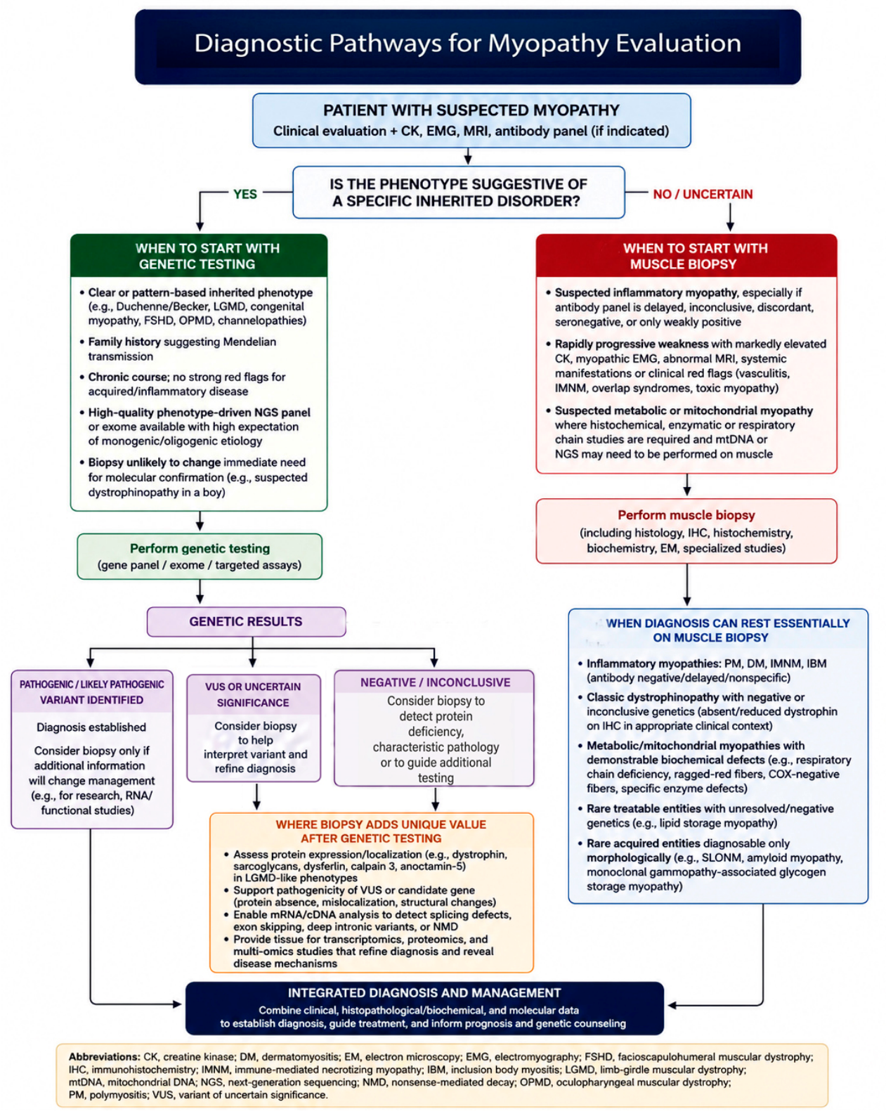
Figure 2.
Immune-mediated necrotizing myopathy. Necrotic muscle fibers are infiltrated by macrophages. H&E.
Figure 2.
Immune-mediated necrotizing myopathy. Necrotic muscle fibers are infiltrated by macrophages. H&E.
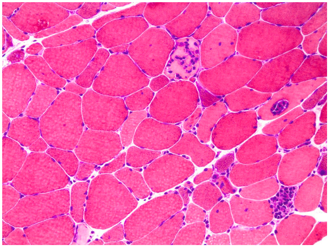
Figure 3.
Dystrophin immunostaining in anti-HMGCR IMNM demonstrating dystrophy-like changes, including marked fiber size variation, rounded fibers, increased fibrous tissue, and scattered necrotic fibers.
Figure 3.
Dystrophin immunostaining in anti-HMGCR IMNM demonstrating dystrophy-like changes, including marked fiber size variation, rounded fibers, increased fibrous tissue, and scattered necrotic fibers.
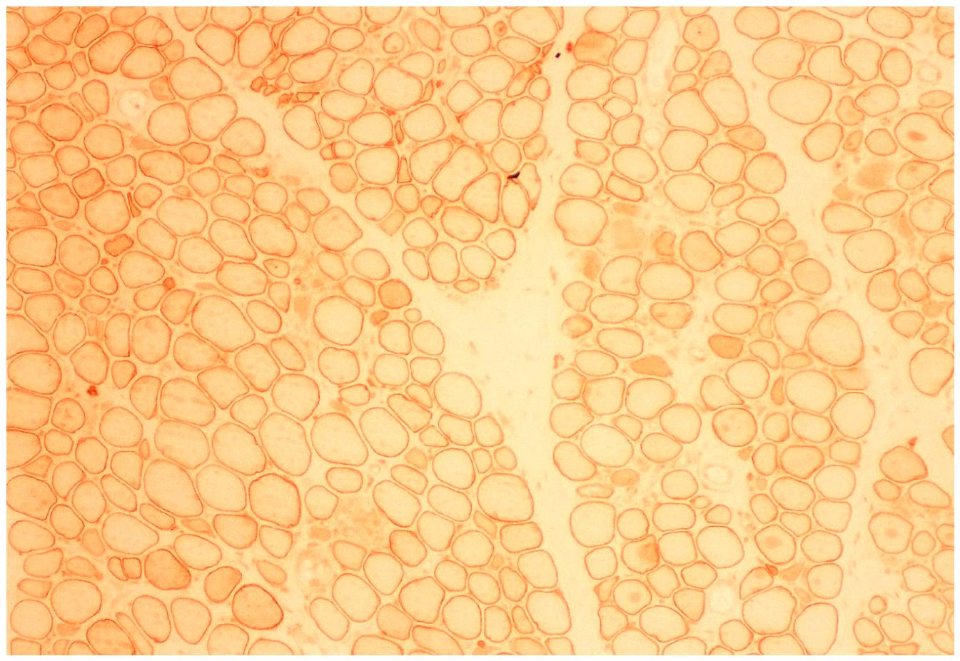
Figure 4.
Reducing body myopathy. Myotilin immunostaining highlights irregularly shaped cytoplasmic inclusions within muscle fibers of variable sizes.
Figure 4.
Reducing body myopathy. Myotilin immunostaining highlights irregularly shaped cytoplasmic inclusions within muscle fibers of variable sizes.
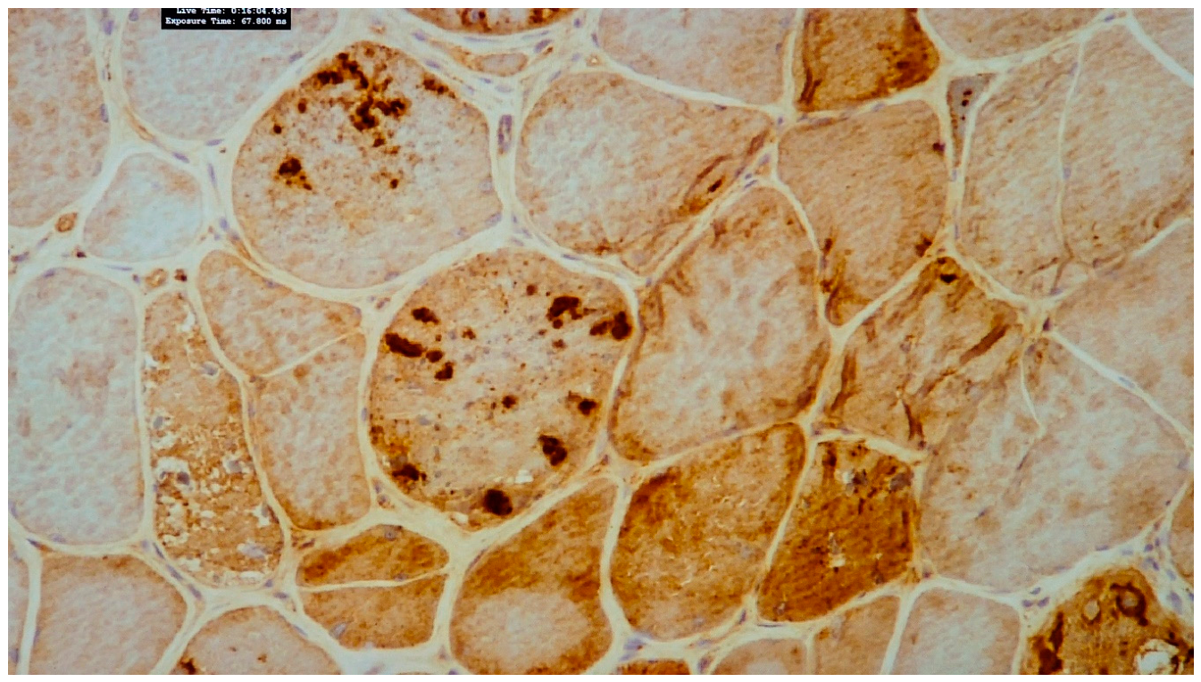
Figure 5.
Dystrophin-2 immunostaining of a muscle biopsy from a patient with Becker muscular dystrophy demonstrating reduced and patchy sarcolemmal expression of dystrophin.
Figure 5.
Dystrophin-2 immunostaining of a muscle biopsy from a patient with Becker muscular dystrophy demonstrating reduced and patchy sarcolemmal expression of dystrophin.
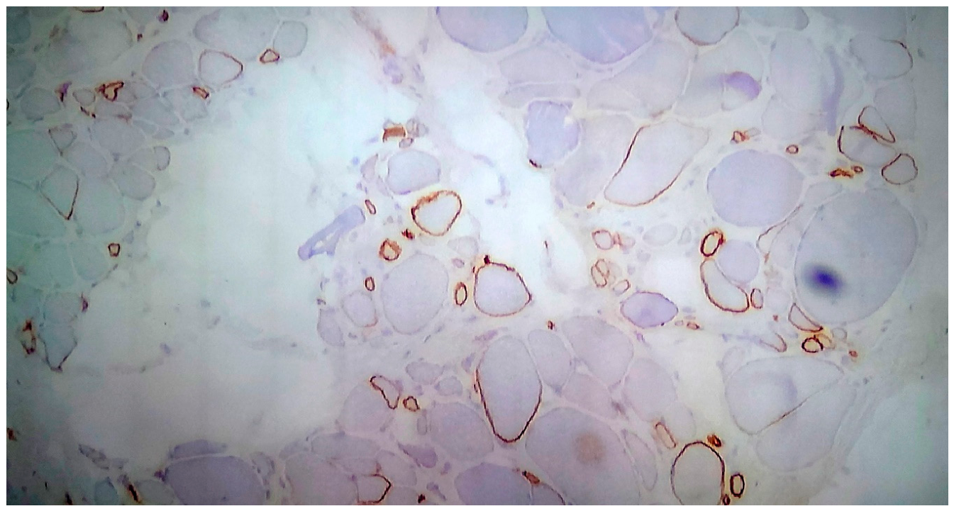
Figure 6.
Oil Red O stain of a muscle biopsy showing numerous small and large lipid-filled vacuoles predominantly within type 1 muscle fibers.
Figure 6.
Oil Red O stain of a muscle biopsy showing numerous small and large lipid-filled vacuoles predominantly within type 1 muscle fibers.
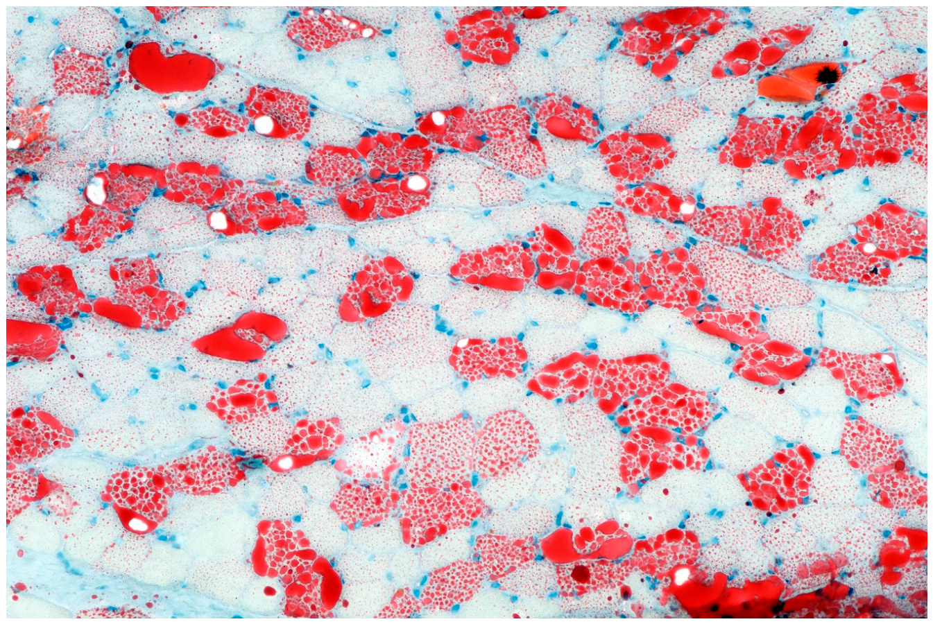
Figure 7.
Inclusion body myositis (IBM). A. Endomysial inflammatory infiltrates surrounding non-necrotic muscle fibers, with focal invasion of individual fibers. B. Cytoplasmic aggregates within muscle fibers in the absence of rimmed vacuoles. H&E.
Figure 7.
Inclusion body myositis (IBM). A. Endomysial inflammatory infiltrates surrounding non-necrotic muscle fibers, with focal invasion of individual fibers. B. Cytoplasmic aggregates within muscle fibers in the absence of rimmed vacuoles. H&E.
Disclaimer/Publisher’s Note: The statements, opinions and data contained in all publications are solely those of the individual author(s) and contributor(s) and not of MDPI and/or the editor(s). MDPI and/or the editor(s) disclaim responsibility for any injury to people or property resulting from any ideas, methods, instructions or products referred to in the content. |
© 2026 by the authors. Licensee MDPI, Basel, Switzerland. This article is an open access article distributed under the terms and conditions of the Creative Commons Attribution (CC BY) license.
Copyright: This open access article is published under a Creative Commons CC BY 4.0 license, which permit the free download, distribution, and reuse, provided that the author and preprint are cited in any reuse.


