

Submitted:
07 July 2026
Posted:
08 July 2026
You are already at the latest version
Abstract
This study aimed to identify novel antimicrobial peptides (AMPs) from Lactiplantibacillus plantarum K9 isolated from Protaetia brevitarsis seulensis larvae using genome and transcriptome analyses. L. plantarum K9 showed the strongest antimicrobial activity, with MIC90 values of 22 μl against E. coli and 21 μl against S. aureus. Approximately 54% of the antimicrobial activity was attributed to peptides or proteins. Genome analysis revealed a 3-Mb chromosome and three plasmids. Screening of hypothetical proteins using CAMPR4 identified 18 AMP candidates. Transcriptomic analysis showed that most AMP genes were more highly expressed in the stationary phase and were upregulated under low-pH conditions. Synthetic peptide assays demonstrated strong activity of AMP4, 6, 7, 12-2, 14, and 18 against S. aureus, while AMP16 and 18 were effective against E. coli. Among all candidates, AMP12 exhibited the highest activity and strong specificity toward S. aureus. Structural analysis revealed a conserved α-helical region that likely mediates membrane-targeting antimicrobial activity. These results suggest that LP AMP12 is a novel antimicrobial peptide contributing to the antimicrobial properties of L. plantarum K9.
Keywords:
AMP
; antibacterial
; CAMPR4
; genome
; L. plantarum
; transcriptome
1. Introduction
Lactic acid bacteria are included among the most widely used probiotics. Representative genera of lactic acid bacteria include Aerococcus, Carnobacterium, Enterococcus, Lactobacillus, Lactococcus, Leuconostoc, Streptococcus, Tetragenococcus, Vagococcus, and Weissella. In addition, numerous species belonging to the Lactobacillus and Bifidobacterium genera have been commonly used as probiotics [1]. In particular, Lactiplantibacillus plantarum is a widely used probiotic bacterium. As a facultatively heterofermentative species, it is relatively easy to culture and can thrive in a broad range of environments, including fermented products, meat, plants, and the gastrointestinal tract [2]. Due to the diverse adaptability of L. plantarum, several beneficial effects have been reported including cholesterol-lowering effects, improvement of vascular endothelial function, and reduction of postprandial glucose and HbA1c levels in individuals with prediabetes or type 2 diabetes [3,4,5].
L. plantarum is capable of producing antimicrobial peptides (AMPs) such as plantaricin and holins, and these substances, together with organic acids, are expected to play important roles in controlling food spoilage, animal diseases, and antibiotic-resistant bacteria [6]. Recently, antimicrobial peptides (AMPs) derived from diverse sources have been increasingly developed from microorganisms, viruses, fungi, and protozoa [7]. The discovered AMPs are being registered in antimicrobial peptide databases such as the Antimicrobial Peptide Database (APD; http://aps.unmc.edu/AP) and DRAMP (http://dramp.cpu-bioinfor.org/), and the registered AMPs are provided as potential therapeutic candidates [8,9]. However, naturally produced AMPs are generated in low yields; therefore, to improve their production, various approaches such as artificial synthesis, bacterial cloning, and expression in cells have been employed to enhance productivity [10]. On the other hand, various strategies such as unusual amino acid incorporation, cyclization, terminal or side-chain modification, and nanoparticle formulation have been employed to prevent rapid degradation of AMPs [10].
In this study, to isolate lactic acid bacteria with more beneficial functions for humans, Lactiplantibacillus plantarum K9 originated from Protaetia brevitarsis seulensis was screened and isolated for its high antimicrobial activity. The isolated L. plantarum K9 was subjected to genome and transcriptome analyses, which the transcriptome was analyzed depending on pH conditions and growth states. In addition, based on the results of genome analysis, candidate AMPs were predicted using the CAMPR4 program, and their antimicrobial activities were evaluated through oligopeptide synthesis and gene cloning.
2. Results
2.1. Antimicrobial Activity of L. Plantarum K9 Isolated From the Gut of P. Brevitarsis Larvae
The antimicrobial activity of the L. plantarum K9 strain against Escherichia coli (EC) and Staphylococcus aureus (SA) is shown in Figure 1. The cell-free supernatant of L. plantarum K9 exhibited comparable antimicrobial activity against both EC and SA, with MIC90 values of 21.9 and 21.0 µl/ml, respectively (Table 1 and suppl. Figure 1 to 6). When compared with L. plantarum NIBR97 [11], L. plantarum K9 showed slightly superior antimicrobial activity (Figure 1A and Table 1). L. plantarum is generally capable of facultative heterofermentative metabolism; however, when glucose is supplied in a medium, it predominantly produces lactic acid. As a result, lactic acid production is maintained at high levels, and antimicrobial activity derived from this production is typically observed to be high level [12]. Therefore, to determine whether the antimicrobial activity observed in this study was associated with antimicrobial peptides (AMPs), the cell-free supernatant was treated with proteinase K. As shown in Figure 1B, proteinase K treatment resulted in a reduction in antimicrobial activity ranging from 41.5 to 46.3%, which is caused by the active and inactive states of proteinase K although not statistically significant. These results suggest that substances whose activities are affected by proteinase K are present at levels corresponding to the observed reduction in activity, and these substances are presumed to consist of AMPs or proteins, or their derivatives. To investigate the mechanism of antimicrobial action, scanning electron microscopy (SEM) was performed. As shown in Figure 1C, the treated cells exhibited the formation of holes in the cell envelope.
On the other hand, to evaluate its potential as a probiotic based on acid tolerance, acid and bile acid tolerance assays of L. plantarum K9 were performed to identify differences from L. plantarum NIBR97, which has previously been reported to exhibit strong antimicrobial and antiviral activities. In the bile acid tolerance test, no significant difference was observed between the two strains [11; suppl. Figure 7], However, in terms of acid tolerance, L. plantarum K9 was clearly observed to be tolerant at pH 2.0, whereas L. plantarum NIBR97 showed no detectable tolerance under the same condition (Figure 1D).
2.2. Genome Analysis and Amp Prediction Results
Genomic analysis was performed to analyze the potential presence of additional AMPs beyond well-known compounds such as plantaricin and holin in L. plantarum K9, which exhibited relatively strong antimicrobial activity. As a result, L. plantarum K9 was found to consist of four contigs, and contig 1 was assigned to the chromosome with a length of 3,022,717 bp (Figure 2A). Contigs 2 and 3 were composed of 61,375 bp and 32,518 bp, respectively, and were identified as circular plasmids (Figure 2B and C). Contig 4 was composed of 7,922 bp and observed to be a linear plasmid (Figure 2D). Since plasmids are generally circular, the linear structure of contig 4 may have resulted from nucleotide sequence loss during genome assembly, and this possibility cannot be ruled out.
AMPs from L. plantarum K9 were predicted from the hypothetical proteins of the genome by the CAMPR4 program. As a result, a total of 18 candidate AMPs were predicted in hypothetical proteins (Figure 2E and Suppl. Figure 7~48). The predicted AMPs exhibited more than 50% sequence positivity with previously known AMPs (Table 2). The neighboring gene organization of the candidate AMPs were predominantly composed of hypothetical proteins (Figure 2E). AMP9 and AMP10 were arranged with three hypothetical protein genes located between them, whereas AMP12 and AMP13 were positioned adjacent to each other. AMP9 and AMP10 consisted of identical sequences, and the three intervening genes also displayed repetitive patterns with identical amino acid sequences. Among the candidate AMPs, those predicted to retain signal sequences included AMP1, AMP5, AMP6, AMP12, and AMP17. The sequence identity of the candidate AMPs ranged from approximately 32% to 63% with previously known AMPs, while sequence positivity ranged from 50% to 86%. The number of amino acids varied from 14 to 62, and the predicted isoelectric point (pI) ranged from 4.9 to 12.61; however, a greater number of candidate AMPs were observed to fall within the alkaline range.
2.3. Results of Transcriptome Analysis
The transcriptome analysis results of the candidate AMPs under different conditions are summarized in Table 3, Table 4 and Table 5. For all genes, a total of 228 genes exhibited expression changes of two-fold or greater in response to pH variation. Among them, up-regulated genes were observed more frequently than down-regulated genes at the decreased pH. When compared to pH 3.5 (non-treated), the marked transcriptional changes were particularly observed at pH 3.0 and 2.5, whereas no significant differences were detected between pH 2.5 and 3.0 (Table 3). These results suggest that numerous genes associated with acid tolerance undergo expression changes in response to the decreased pH. In contrast, gene expression changes associated with growth phase were markedly less pronounced than those induced by pH variation, which identified by only four up-regulated and seven down-regulated genes (Table 3). Observation of AMP expression changes according to growth phase revealed that AMP1, AMP4, AMP11, AMP12, and AMP15 were up-regulated during the exponential growth phase, whereas the remaining AMPs exhibited the opposite trend (Table 4). The gene to show the highest level of differential expression was AMP18, with a 2.29-fold change, while AMP3 and AMP6 exhibited very low expression changes of 1.14- and 1.10-fold, respectively. On the other hand, AMP10 expression was not detected during the exponential phase, and AMP13 was not expressed at all.
Changes in AMP gene expression in response to pH variation were observed to be much more pronounced than those associated with growth phase (Table 5). In particular, expression of the AMP18 gene increased by 15.51-fold at pH 2.5 compared to the non-treated condition. At pH 3.0, AMP18 expression also increased by 4.03-fold relative to the non-treated condition, and a relatively high difference of 3.85-fold was observed when comparing pH 2.5 to pH 3.0. Overall, larger expression changes were observed when comparing the non-treated condition with pH 2.5 than with pH 3.0. Although relatively small differences were observed between pH 2.5 and 3.0, substantial expression changes were detected for AMP2, AMP12, and AMP18. Notably, AMP9 and AMP10 expression was not detected under non-treated conditions, and AMP10 expression was observed only at pH 3.0. AMP13 according to pH changes, consistent with the growth phase–dependent expression analysis, was not expressed under any tested condition, suggesting that it may not represent a functional gene.
2.4. Evaluation of the Antimicrobial Activity of Synthesized Amps
Peptide synthesis was performed for regions exhibiting homology to known AMPs. As a result, seven AMPs, AMP1~3, AMP5, AMP8, AMP10, and AMP15, could not be synthesized (Table 6). AMP11-1 and AMP11-2 showed very low solubility, whereas AMP12-1, AMP16-1, and AMP16-2 exhibited slightly reduced solubility. In contrast, AMP13 was observed to precipitate rapidly after dissolution.
Antimicrobial activity was evaluated at a concentration of 2.5 mg/ml, the highest concentration previously used in studies on Nibribacter [13]. As shown in Figure 3A, very low antimicrobial activity was observed against E. coli. Only AMP16-1 and AMP18 exhibited activities of 50.7% and 38.6%, respectively, compared to the control. In contrast, against S. aureus, AMP4, AMP7, and AMP18 showed relatively high antimicrobial activities of 73.4%, 55.7%, and 69.7%, respectively, compared to the control. In addition, AMP6, AMP12-2, and AMP14 displayed activities ranging from 37.5% to 46.8%. These results indicate that the AMPs predicted from L. plantarum K9 act more specifically against S. aureus than against E. coli.
Based on these characteristics, MIC50 values against S. aureus were evaluated. AMP4 exhibited the best MIC50 value of 1.53 mg/ml; however, no statistically significant differences were observed when compared with AMP7, AMP12-2, and AMP18. AMP6 showed the worst MIC50 value among the analyzed peptides at 11.9 mg/ml, while AMP14 exhibited an MIC50 value of 8.27 mg/ml.
2.5. Antimicrobial Activity of Cloned Amps
To examine whether the candidate AMPs exhibit antimicrobial activity after expression in E. coli, oligonucleotides for PCR amplification were designed to include the upstream promoter and downstream terminator regions, followed by PCR amplification and cloned into a T-vector. After transformation into E. coli, antimicrobial activity was analyzed using the T-vector alone as a control. As shown in Figure 4A and B, antimicrobial activity against E. coli was very low, similar to that observed with the synthetic AMPs. Among the tested AMPs, AMP4 exhibited the highest MIC50 value at 194.2 µl/ml, followed by AMP12 and AMP14 (Figure 4A and B). AMP16, which showed antimicrobial activity in the synthetic AMP assay, exhibited no detectable activity, whereas AMP18 showed very low activity with an MIC50 value of 558.8 µl/ml. These results suggest that these AMPs do not function properly against E. coli when expressed in E. coli.
Antimicrobial activity against S. aureus was much higher than that against E. coli, consistent with the results obtained using synthetic AMPs (Figure 4C and D). AMP12 exhibited the highest activity with an MIC50 value of 45.2 µl/ml, followed by AMP6, AMP8, AMP14, and AMP15, which showed relatively high and comparable activities. In the synthesized peptides, AMP4, AMP7, AMP12, and AMP18 exhibited strong antimicrobial activity against S. aureus. However, among the cloned genes, only AMP7 and AMP12 consistently showed high antimicrobial activity. These results suggest that AMP7 and 12 have high potential for application as Gram-positive-specific antimicrobial peptides. In particular, AMP12 is proposed to have high industrial applicability if its antimicrobial activity can be further enhanced through peptide sequence modification.
2.6. Characterization of the Amp12 Gene
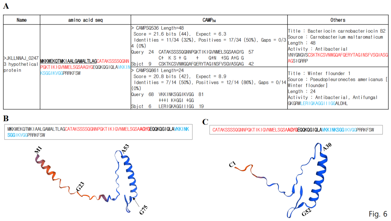
The full-length peptide of AMP12 consists of 88 amino acids and contains two regions homologous to previously reported AMPs carnobacteriocin B2 and Winter flounder 1 (Table 2 and Figure 5A). AMP12 was predicted to retain a signal sequence. The signal sequence of AMP12 predicted by SignalP 5.0 was identified as MKKWEKQTMKIAALGAMALTLAG, with a cleavage site at G23, which occurs immediately before the region homologous to carnobacteriocin B2. The antimicrobial activities of the homologous regions of carnobacteriocin B2 and winter flounder 1 were confirmed using the synthetic peptides AMP12-1 and AMP12-2, as well as the cloned AMP12 (Figs. 3 and 4). However, further studies are required to determine whether the antimicrobial activity of cloned AMP12 results from the removal of the signal sequence followed by proteolytic cleavage, with each fragment exhibiting activity individually, or from the combined effect of the full-length sequence.
To predict structural changes in AMP12 following various cleavage events, three-dimensional structural modeling was performed, and the results are shown in Figure 5B and C. The signal sequence region was predicted to form a distinct α-helix (Figure 6A). In addition, an extra α-helical structure was predicted to span residues A53 to G75, partially overlapping the sequences homologous to both carnobacteriocin B2 and Winter flounder 1. When the signal sequence was cleaved at G23, the α-helix corresponding to residues A53–G75 of the original sequence was predicted to remain unchanged (Figure 5B and C). When comparing the antimicrobial activities of the synthesized peptides and cloned gene constructs of AMP4, AMP7, and AMP18 with those of the synthesized peptides and cloned gene construct of AMP12, we suggest that the antimicrobial activity of the cloned AMP12 has higher value than that of the individual synthesized peptides, AMP12-1 and AMP12-2, due to the presence of both active regions within a single peptide.
3. Discussion
To isolate a novel lactic acid bacterial strain, only lactic acid bacteria were isolated by plating the gut microbiota of Protaetia brevitarsis larvae on MRS medium. Among the strains that grow on MRS medium, further analysis of ten strains exhibiting strong antimicrobial activity revealed that LAB K9 showed the most superior activity (Suppl. Figs. 1 and 2). Based on 16S rDNA sequence analysis of the LAB K9 strain (Suppl. Figure 3), it was identified as Lactiplantibacillus plantarum, which was named as Lactiplantibacillus plantarum K9.
To evaluate the industrial applicability of this novel lactic acid bacterium, its antimicrobial activity was compared with that of L. plantarum NIBR97, a strain previously isolated in our laboratory [11]. As a result, L. plantarum K9 was observed to exhibit superior antimicrobial activity compared to L. plantarum NIBR97 (Figure 1A and Table 1). Moreover, in contrast to L. plantarum NIBR97, which showed no responsiveness to proteinase K treatment [11], the antibacterial activity of L. plantarum K9 was reduced by 41.5–46.3% following treatment with proteinase K (Figure 1B). The differential responses exhibited by these closely related strains highlight significant strain-specific characteristics. However, previous results showed that the antimicrobial activity of Lactiplantibacillus taiwanensis was sensitive to proteinase K treatment, suggesting that L. plantarum K9 exhibits a similar trend. [14]. The antimicrobial substances produced by L. plantarum K9 were observed to exert their antimicrobial effects by acting on the cell membrane and inducing hole formation, consistent with findings from previous studies by L. plantarum NIBR97 and L. taiwanensis (Figure 1C).
On the other hand, L. plantarum K9, isolated from the gut of Protaetia brevitarsis larvae, exhibited higher acid tolerance than L. plantarum NIBR97 (Figure 1D), which was isolated from kimchi. This result is presumed to reflect the adaptive characteristics of lactic acid bacteria inhabiting the gastrointestinal environment, and it is further suggested that this strain may possess additional functional properties that facilitate adaptation to the gut environment. When L. plantarum K9 genome (Figure 2A to D) was compared to the genome of L. plantarum NIBR97 analyzed in a previous study, all contigs (1 to 4) were similarly conserved; however, an additional contig (contig 5) was uniquely present in NIBR97 [11]. L. plantarum K9 genome exhibited overall high homology in genomic sequence with L. plantarum NIBR97 [data not shown]. However, additional studies were considered necessary for the following reasons. First, L. plantarum NIBR97 was isolated from kimchi, a traditional Korean fermented food [data not shown], whereas L. plantarum K9 was isolated from the gut of Protaetia brevitarsis larvae. The differential responses to proteinase K treatment indicate that the antimicrobial substances produced by these strains are likely distinct in nature. In addition, L. plantarum K9 AMP12 was present exclusively in L. plantarum K9 but was not detected in L. plantarum NIBR97 [data not shown].
A total of 18 AMPs using the CAMPR4 program were predicted in L. plantarum K9 (Figure 2E). In Nibribacter radioresistens, 11 AMPs were predicted using the same CAMPR4 program, and signal sequences were identified in NB AMP3, NB AMP6, and NB AMP9 [13]. The NB AMPs ranged from 9 to 30 amino acids in length, with predicted isoelectric points (pI) ranging from 3.8 to 10.85. In addition, the neighboring genes of NB-AMPs were predominantly composed of hypothetical proteins, showing a similar pattern to that observed in this study. Among the L. plantarum K9 AMPs (LP AMPs), the presence of signal sequences was predicted in LP AMP1, LP AMP5, LP AMP6, LP AMP12, LP AMP16, and LP AMP17. The LP AMPs exhibited a wide range of amino acid lengths, varying from 14 to 62 residues, and their predicted pI values ranged from 4.9 to 12.61. Similar to the NB AMPs, the neighboring genes of LP AMPs were also densely populated with hypothetical proteins.
Based on the transcriptomic analysis of L. plantarum K9, the number of genes exhibiting more than two-fold changes in expression increased with increasing acidity compared with the non-treated condition (Table 2 and Table 3). The differentially expressed genes were predominantly involved in transcriptional regulation, heat shock proteins (HSPs), and transporter functions, indicating that they may play important roles in acid tolerance. On the other hand, in the transcriptomic analysis according to growth phase, in contrast to pH changes, there were very few genes whose expression increased sharply. Proteins exhibiting more than a two-fold change were identified only during the stationary phase, with four proteins detected, including peptidoglycan binding domain–containing proteins (three types) and an extracellular membrane anchor (one type), whereas no proteins showing more than a two-fold increase were observed during the exponential growth phase. The increased expression of these proteins is presumed to be associated with antimicrobial activity, immune recognition, host interaction, or biofilm formation, which are required for cell survival during the stationary phase [15,16].
Among LP-AMPs, AMP1, 4, 11, 12, and 15 showed increased expression during the exponential growth phase, whereas the remaining AMPs exhibited the opposite trend (Table 3). In the transcriptomic analysis according to growth stage in N. radioresistens, the expressions of NB AMPs were also observed to follow diverse patterns [13]. Therefore, the LP AMPs identified in this study are presumed to be genes potentially associated with diverse functional properties. On the other hand, in response to pH changes, LP AMP18 exhibited a very high expression level compared with the non-treated condition, whereas LP AMP13 showed no detectable expression, suggesting that it may not be a gene or may be a gene that is expressed only under specific conditions (Table 4). Transcriptomic analysis of pH reduction caused by Lactiplantibacillus plantarum VAL6 revealed that changes in pH induced alterations in the expression of genes associated with exopolysaccharides and various other factors [17,18]. Therefore, it is suggested that the pH changes applied in this study may induce alterations in the expression patterns of genes associated with diverse functional elements.
The synthetic LP AMPs identified in this study were found to exhibit lower antimicrobial activity, as MIC₅₀ values of 1.53-11.94 mg/ml against S. aureus (Figure 3), when compared to previous studies, in which the MIC₅₀ values against E. coli and S. aureus were reported to be 2.2–128 μg/mL and 32–256 μg/mL, respectively [13,19,20]. The synthetic LP AMPs, when analyzed at a concentration of 2.5 mg/mL, were estimated to exhibit much lower antimicrobial activity against E. coli. Therefore, it is suggested that the antimicrobial activity of the predicted LP AMP candidates is substantially lower than that expected based on sequence homology. Among the NB AMP clones of N. radioresistens, those that showed relatively high activity against E. coli were AMP9, AMP10, and AMP11, with MIC₅₀ values observed to be 153.9, 131.0, and 154 μL/mL, respectively [13; data not shown]. These results showed slightly higher activity compared to LP AMP4 (Figure 4). Among the NB AMP clones of N. radioresistens, those exhibiting higher activities against S. aureus were NB AMP2, 3, 4, 5, 6, 7, 10, and 11, and among these, NB AMP6 showed the highest activity with an MIC₅₀ value of 123.1 μL/mL [13; data not shown]. Based on these results, the value for LP AMP12 (45.2 ul) in this study was observed by 2.7-fold higher than those of NB AMPs. Therefore, when comparing the antimicrobial activities of the synthesized and cloned LP AMP12 with that of previously reported synthetic and cloned AMPs, it was found to be highly distinctive, which NB AMPs exhibited high activity only in the synthetic AMPs. On the other hand, LP AMP12 not only originates from a gene that is absent in L. plantarum NIBR97, but also retains a signal sequence and, in particular, exhibits very high antimicrobial activity against S. aureus. Thus, it is suggested that LP AMP12 has potential for use as a Gram-positive–specific antimicrobial agent.
In three-dimensional structural modeling of synthetic NB AMPs from N. radioresistens, AMPs exhibiting superior antimicrobial activity were shown to predominantly maintain an α-helix structure [13]. In addition, EcDBS1R6 has been reported to be secreted after its signal sequence is cleaved by a signal peptidase and subsequently acts on the bacterial membrane [21]. Because LP AMP12 also retains a signal sequence, it is presumed to act via a mechanism similar to that of EcDBS1R6; however, it is suggested that LP AMP12 may act specifically against Gram-positive bacteria.
4. Materials and Methods
4.1. Evaluation of the Antimicrobial Efficacy of Cell-Free Extracts
Antimicrobial activity was evaluated using the Gram-negative bacterium Escherichia coli (EC, ATCC 25922) and the Gram-positive bacterium Staphylococcus aureus (SA, ATCC 6538) by either the disc diffusion method or a microtiter plate assay. For antimicrobial analysis, strains pre-cultured by overnight (O/N) were adjusted to a concentration of 10⁶ CFU/mL and used in the experiments. After adding the reaction mixtures to microtiter plates or spotting them onto discs, the plates were incubated at 37 °C overnight while observing the reaction outcomes. Lactiplantibacillus plantarum K9 was cultured in MRS medium at 37 °C for 24 h, followed by centrifugation at 12,000 × g for 10 min. The supernatant was collected and filtered through a 0.2 μm filter to obtain a cell-free extract. For the antimicrobial assays, the cell-free extract was used at different concentrations, and the pre-cultured target strains were added after being adjusted to 10⁶ CFU/mL. The final reaction volume was adjusted to 200 μL with LB broth.
4.2. Acid Tolerance
L. plantarum K9 and NIBR97 were cultured to the exponential growth phase and used for the experiments. Each strain was centrifuged at 3,500 × g for 20 min, after which the supernatant was removed. The bacterial cells were washed twice with PBS buffer (pH 7.2) and then used for subsequent experiments. Fresh MRS broth was adjusted to pH 2.0, 2.5, 3.0, and 3.5 using HCl and used for the experiments. The cell pellets of each strain were resuspended in 10 mL of pH-adjusted MRS broth and incubated at 37 °C for 3 h. After incubation, the resulting suspensions were appropriately diluted with sterile distilled water, and viable cell counts were determined.
4.3. Scanning Electron Microscope (Sem)
E. coli was incubated with the cell-free supernatant from L. plantarum K9 culture for 24 h. The treated E. coli was incubated with the cell-free supernatant from L. plantarum K9 culture. The treated E. coli cells were fixed with one volume of 2.5% glutaraldehyde (Sigma-Aldrich, St. Louis, MO, USA) for 24 h at 4 °C. Then, the samples were rinsed with sterile PBS buffer thrice and sequentially dehydrated with graded ethanol (30%, 50%, 70%, 80%, 90%, and 100% (v/v); 15 min incubation for each concentration). Finally, the samples were dried at room temperature and sputter-coated with gold for SEM. Cells were fixed with one volume of 2.5% glutaraldehyde (Sigma-Aldrich, St. Louis, MO, USA) for 24 h at 4 °C. Then, the samples were rinsed with sterile PBS buffer thrice and sequentially dehydrated with graded ethanol (30%, 50%, 70%, 80%, 90%, and 100% (v/v); 15 min incubation for each concentration). Finally, the samples were dried at room temperature and sputter-coated with gold for SEM.
4.4. Discovery of Bioactive Compounds Through Genome Analysis
Genome analysis of L. plantarum K9 was performed using a genome preparation kit (Wizard Genomic DNA Purification Kit, Promega). A total of 5 μg for each sample was used as input into library preparation. The SMRTbell library was constructed with SMRTbell™ Template Prep Kit 1.0 (PN 100-259-100) following manufacture’s instructions (Pacific Biosciences). The small fragments lower than 20 kb of SMRTbell template were removed using Blue Pippin Size selection system for large-insert library. The constructed library was validated by Agilent 2100 Bioanalyzer. After a sequencing primer is annealed to the SMRTbell template, DNA polymerase is bound to the complex using DNA/Polymerase Binding kit P6. This polymerase-SMRTbell-adaptor complex is then loaded into SMRT cells. The SMRTbell library was sequenced using 1 SMRT cells (Pacific Biosciences) using C4 chemistry (DNA sequencing Reagent 4.0) and 240 min movies were captured for each SMRT cell using the PacBio RS II (Pacific Biosciences) sequencing platform [22].
L. plantarum K9 was prepared by P6-C4 chemistry and sequenced using 1 SMRT cell with MagBead OneCellPerWell v1 Protocol (Insert Sizes 20 kb, movie time 1x240 min). We produced 117,132 of long reads and 913,773,737 base pairs after subreads filtering. De novo assembly was conducted using the hierarchical genome assembly process (HGAP, Version 2.3) workflow, including consensus polishing with Quiver [23]. As the estimated genome size was 3,244,413bp and average coverage was 103X, we performed error correction based the longest about 30X (487,589,278bp) seed bases with rest shorter reads, and then assembled with error corrected reads. As a result of HGAP process, we got the results 3,037,359 bp N50 contig and 4,382,491 bp total contig lengths by polish process. Finally, since bacterial genomes and plasmids are typically circular, we checked the forms for each of contigs using MUMmer 3.5 and trimmed one of the self-similar ends for manual genome closure [24].
Putative gene coding sequences (CDSs) from the assembled contigs were identified using Glimmer v3.02 [25] and open reading frames (ORFs) were obtained. These ORFs were searched using Blastall alignment (http://www.ncbi.nlm.nih.gov/books/NBK1762/) against the NCBI Non-redundant protein database (nr) for all species. GO annotation was assigned to each of ORFs by Blast2GO software [26] analyzing the best hits of the BLAST results. Additionally, ribosomal RNAs and transfer RNAs were predicted using RNAmmer 1.2 and tRNAscan-SE 1.4 [26,27].
4.5. Bioinformatic Analysis for Identification of Amps From the L. Plantarum K9 Genome
Peptides with a similarity (identities/positives) of more than 50% to existing AMPs and possessing a signal sequence cleaved by an endopeptidase were selected as AMP candidates using CAMPR4 (http://www.camp.bicnirrh.res.in/campHelp.php) (accessed on 15 April 2024) and SignalP 5.0 (https://services.healthtech.dtu.dk/services/SignalP-5.0/), respectively. Their amino acid and nucleotide sequences were subsequently used for peptide synthesis and gene cloning, respectively.
4.6. Identification of Bioactive Compound Expression Through Transcriptomic Analysis for Transcriptomic Analysis, L. Plantarum K9 Was Cultured to the Exponential Growth Phase (A₆₀₀ = 0.5) and the Stationary Phase (A₆₀₀ ≥ 1.5), After Which Rna Was Isolated From Each Condition. on the Other Hand, to Observe Changes in Mrna Expression in Response to Ph Variation, Cultured L. Plantarum K9 Cells Were Exposed to Ph-Adjusted Conditions of 2.5 and 3.0 and Incubated at 37 °C for 3 H, Followed by Rna Isolation From Each Condition. Total Rna Extraction Was Performed Using the Accuprep® Bacterial Rna Extraction Kit (Bioneer). Rna Purity Was Measured Using 1 Μl of Total Rna Extract with A Nanodrop 1000 Spectrophotometer. the Quality of Total Rna Was Verified by Confirming the Rna Integrity Number (Rin) Values Measured Using an Agilent Technologies 2100 Bioanalyzer. for Total Rna Sequencing Library Preparation, the Nugen Universal Prokaryotic Rna-Seq Kit (Part Number 0363-32) Was Used, and All Procedures Were Performed According to the Manufacturer’s Instructions. A Total of 300 Ng of Total Rna Was Used to Synthesize First- and Second-Strand Cdna Using Selective Primers. After Applying Parameters Specific to the Ovation Protocol, the Cdna Was Fragmented to an Average Size of 200 Bp by Sonication Using A Covaris S220 System in Microtubes. All Subsequent Steps, Including Cdna Purification, End Repair, Adaptor Ligation, First-Strand Selection, and First-Strand Purification, Were Performed According to the Manufacturer’s Official Protocol. During Strand Selection Ii, Rrna Was Removed in A Bacterial Species–specific Manner Using the Anydeplete Technique with Anydeplete Probes. the Libraries Were Amplified by Pcr, and the Quantity and Quality of the Amplified Products Were Assessed by Capillary Electrophoresis (Bioanalyzer, Agilent). Following Qpcr Performed with Sybr Green Pcr Master Mix (Applied Biosystems), Equimolar Amounts of Index-Tagged Libraries Were Combined Into A Single Pool. Rna Sequencing Was Carried Out Using the Illumina Novaseq 6000 System.
4.7. Expression Analysis Between Samples and Identification of Degs
At first, reads for each sample were mapped to the reference genome by Tophat (v2.0.13). The aligned results were added to Cuffdiff (v2.2.0) to report differentially expressed genes. For library normalization and dispersion estimation, geometric and pooled (“blind” when each condition has single replicates, or “pooled” when multiple replicates are available) methods were applied.
Cuffdiff provides various output files, and using one of its outputs, “gene_exp.diff”, DEGs (Differentially Expressed Genes) were identified. To detect DEGs between sample1 as control and sample2 as case, two filtering processes were applied. First, using Cuffdiff status code, genes that only have “OK” status were extracted. Status code indicates whether each condition contains enough reads in a locus for a reliable calculation of expression level, and “OK” status means the test is successful to calculate gene expression level. For the second filtering, 2 fold change was calculated and genes belonging to the following range were selected.
Up-regulated:
log2[case]−log2[control]>=log2(2)=1 log2[case]−log2[control]>=log2(2)=1
Down-regulated:
log2[case]−log2[control]<=log2(1/2)=−1 log2[case]−log2[control]<=log2(1/2)=−1
For ontology analysis, genes after 2 fold change were picked (for mouse samples, genes with 2fold&pvalue<0.05&FDR<0.1 were selected for ontology analysis) and applied to DAVID as an input to get a comprehensive set of functional annotation. Disease, Gene ontology, pathway categories were selected, and Ease score was changed from 0.1 to 1 to include more output. Ease score is a conservative adjustment to the Fisher exact probability. It weights significance in favor of the association supported by more genes. DAVID then generated functional annotation chart which lists annotation terms and their associated genes under study.
4.8. L. Plantarum K9 Amps Gene Cloning and Peptide Synthesis
General gene cloning was performed using the method of Sambrook et al. (2001) [28]. Candidate AMPs obtained through the analysis of the genome and transcriptome were designed and synthesized with primers for PCR amplification (Table 1; Genotech, Daejon, Republic of Korea). After amplifying each AMP gene using this primer set and PCR PreMix (AcuPower PCRMix, Bioneer), gel extraction was performed. Eluted DNA fragments were ligated to the T-easy vector (Promega) and transformed into E. coli DH5α (Invitrogen, Carlsbad, CA, USA) using the heat-shock method using calcium chloride. Cloned AMPs were identified using restriction enzyme cleavage and nucleotide sequencing (Applied Biosystems).
Amino acid sequences in regions with homology to predicted AMPs were obtained, and these sequences were synthesized as artificial peptides (Table 2; Cosmogenetech, Seoul, Republic of Korea).
4.9. Evaluation of Antibacterial Activity of Cell-Free Supernatants and Synthetic Peptides
The cell-free supernatant of all E. coli DH5α strains containing cloned AMPs was collected after 24 h of culture and filtered through a 0.2 μm syringe (mixed cellulose esters (MCE), Merck, Darmstadt, Germany), and finally the cell-free supernatant was used for the evaluation of antibacterial activity. The cell-free supernatant from the E. coli DH5α, harboring only the plasmid vector without the LP-AMP genes, was confirmed as a negative control to have very little effect on antibacterial activities. The synthesized AMPs were suspended at a concentration of 10 mg/mL in distilled water and then used for antibacterial activity. The antibacterial activity against the Gram-negative bacterium, E. coli (EC; ATCC 10536), and the Gram-positive bacterium, Staphylococcus aureus (SA, ATCC 6538), was analyzed using the microtiter plate method and expressed as minimal inhibition concentration (MIC); the bacteria pre-cultured overnight (O/N) were cultured at 106 CFU/mL with the cell-free supernatant or the synthetic peptides, the reaction mixtures were added to the microtiter plate, and finally the reactivity was observed by O/N culture at 37 ◦C.
4.10. Statistical analysis
The collected data were analyzed using the PROC ANOVA procedure of the SAS program (ver. 9.2; SAS Institute Inc, Cary, NC, USA). Mean values that differed at the level of 5% significance were verified using Duncan’s multiple range test (DMRT).
Author Contributions
W.Y.B., and K.H.B. conceived and designed the experiments; W.Y.B., K.H.B., A. Y. C., and S. H. K. performed the experiments; W.Y.B., K.H.B., J. H. and S.W.K. analyzed the data; J.H. and S.W.K wrote the paper. All authors have read and agreed to the published version of the manuscript.
Funding
This research was funded by the budget of the Glocal University 30 Regional Coexistence Project of Jeonbuk State.
Institutional Review Board Statement
Not applicable.
Informed Consent Statement
Not applicable.
Data Availability Statement
The original contributions presented in this study are included in the article. Further inquiries can be directed to the corresponding author.
Conflicts of Interest
Author S.W.K. was employed by the company FARMIMs Co., Ltd. The remaining authors declare that the research was conducted in the absence of any commercial or financial relationships that could be construed as a potential conflict of interest.
References
- Akpoghelie, P.O.; Edo, G.I.; Mafe, A.N.; Isoje, E.F.; Igbuku, U.A.; Ali, A.B.M.; Yousif, E.; Owheruo, J.O.; Oberhiri, S.; Essaghah, A.E.A.; Ahmed, D.S.; Umar, H.; Alamiery, A.A. Food, Health, and Environmental Impact of Lactic Acid Bacteria: The Superbacteria for Posterity. Probiotics Antimicrob. Proteins 2025, 17, 2819–25855. [Google Scholar] [CrossRef] [PubMed]
- Chatsirisakul, O.; Leenabanchong, N.; Siripaopradit, Y.; Chang, C.W.; Buhngamongkol, P.; Pongpirul, K. Strain-specific therapeutic potential of Lactiplantibacillus plantarum: A Systematic Scoping Review. Nutrients 2025, 17, 1165. [Google Scholar] [CrossRef] [PubMed]
- Costabile, A.; Buttarazzi, I.; Kolida, S.; Quercia, S.; Baldini, J.; Swann, J.R.; Brigidi, P.; Gibson, G.R. An in vivo assessment of the cholesterol-lowering efficacy of L. plantarum ECGC 13110402 in normal to mildly hypercholesterolaemic adults. PLoS ONE 2025, 12, e0187964. [Google Scholar]
- Malik, M.; Suboc, T.M.; Tyagi, S.; Salzman, N.; Wang, J.; Ying, R.; Tanner, M.J.; Kakarla, M.; Baker, J.E.; Widlansky, M.E. L. plantarum 299v Supplementation improves vascular endothelial function and reduces inflammatory biomarkers in men with stable coronary artery disease. Circ. Res. 2018, 123, 1091–1102. [Google Scholar] [CrossRef] [PubMed]
- Oh, M.-R.; Jang, H.-Y.; Lee, S.-Y.; Jung, S.-J.; Chae, S.-W.; Lee, S.-O.; Park, B.-H. L. plantarum HAC01 Supplementation improves glycemic control in prediabetic subjects: A randomized, double-blind, placebo-controlled trial. Nutrients 2021, 13, 2337. [Google Scholar] [CrossRef] [PubMed]
- Chen, X.; Bai, H.; Mo, W.; Zheng, X.; Chen, H.; Yin, Y.; Liao, Y.; Chen, Z.; Shi, Q.; Zuo, Z.; Liang, Z.; Peng, H. Lactic Acid Bacteria Bacteriocins: Safe and Effective Antimicrobial Agents. Int. J. Mol. Sci. 2025, 26, 4124. [Google Scholar] [CrossRef] [PubMed]
- Datta, S.; Roy, A. Antimicrobial peptides as potential therapeutic agents: A review. Int. J. Pept. Res. Ther. 2021, 27, 555–577. [Google Scholar] [CrossRef]
- Cresti, L.; Cappello, G.; Pini, A. Antimicrobial peptides towards clinical application—A long history to be concluded. Int. J. Mol. Sci. 2024, 25, 4870. [Google Scholar] [CrossRef] [PubMed]
- Wang, G.; Vaisman, I.I.; Van Hoek, M.L. “Machine Learning Prediction of Antimicrobial Peptides,” in Computational Peptide Science: Methods and Protocols; Simonson, T., Ed.; Springer US: New York, NY, 2022; pp. 1–37. [Google Scholar]
- Sadeeq, M.; Li, Y.; Wang, C.; Hou, F.; Zuo, J.; Xiong, P. Unlocking the power of antimicrobial peptides: advances in production, optimization, and therapeutics. Front. Cell. Infect. Microbiol. 2025, 15, 1528583. [Google Scholar] [CrossRef] [PubMed]
- Kim, S.W.; Kang, S.I.; Shin, D.H.; Oh, S.Y.; Lee, C.W.; Yang, Y.; Son, Y.K.; Yang, H.S.; Lee, B.H.; An, H.J.; Jeong, I.S.; Bang, W.Y. Potential of cell-free supernatant from Lactobacillus plantarum NIBR97, including novel bacteriocins, as a natural alternative to chemical disinfectants. Pharmaceuticals 2020, 13, 266. [Google Scholar] [CrossRef] [PubMed]
- Holubchyk, D.S.; Dugan, O.M.; Danylenko, S.G.; Khablenko, A.D.; Yalovenko, O.I.; Potemska, O.I. The probiotic properties of Lactiplantibacillus plantarum isolated from plant material. Biotechnol. Acta 2025, 18, 38–43. [Google Scholar] [CrossRef]
- Kim, S.W.; Bang, W.Y. Identification of antimicrobial peptides from Nibribacter radioresistens, a UV and gamma radiation tolerant bacterium. Genes 2025, 16, 353. [Google Scholar] [CrossRef] [PubMed]
- Kim, S.W.; Ha, Y.J.; Bang, K.H.; Lee, S.; Yeo, J.H.; Yang, H.S.; Kim, T.W.; Lee, K.P.; Bang, W.Y. Potential of bacteriocins from Lactobacillus taiwanensis for producing bacterial ghosts as a next generation Kimaccine. Toxins 2020, 12, 432. [Google Scholar] [CrossRef] [PubMed]
- Kleerebezem, M.; Hols, P.; Bernard, E.; Rolain, T.; Zhou, M.; Siezen, R.J.; Bron, P.A. The extracellular biology of the lactobacilli. FEMS Microbiol. Rev. 2010, 34, 199–230. [Google Scholar] [CrossRef] [PubMed]
- Muscariello, L.; De Siena, B.; Marasco, R. Lactobacillus cell surface proteins involved in interaction with mucus and extracellular matrix components. Curr. Microbiol. 2020, 77, 3831–3841. [Google Scholar] [CrossRef] [PubMed]
- Le, T.S.; Nguyen, P.T.; Nguyen-Ho, S.H.; Nguyen, T.P.; Nguyen, T.T.; Thai, M.N.; Nguyen-Thi, T.U.; Nguyen, M.C.; Hoang, Q.K.; Nguyen, H.T. Expression of genes involved in exopolysaccharide synthesis in Lactiplantibacillus plantarum VAL6 under environmental stresses. Arch. Microbiol. 2021, 203, 4941–4950. [Google Scholar] [CrossRef] [PubMed]
- Peng, L.; Zhao, K.; Chen, S.; Ren, Z.; Wei, H.; Wan, C. Whole genome and acid stress comparative transcriptome analysis of Lactiplantibacillus plantarum ZDY2013. Arch. Microbiol. 2021, 203, 2795–2807. [Google Scholar] [CrossRef] [PubMed]
- Lach, J.; Krupińska, M.; Mikołajczyk, A.; Strapagiel, D.; Stączek, P.; Matera-Witkiewicz, A. Novel antimicrobial peptides from saline environments active against E. faecalis and S. aureus: Identification, characterization and potential usage. Int. J. Mol. Sci. 2023, 24, 11787. [Google Scholar] [CrossRef] [PubMed]
- Zou, R.; Zhu, X.; Tu, Y.; Wu, J.; Landry, P.M. Activity of antimicrobial peptides aggregates decreases with increased cell membrane crossing free energy cost. Biochem. 2018, 57, 2606–2610. [Google Scholar] [CrossRef]
- Porto, W.F.; Irazazabal, L.N.; Humblot, V.; Haney, E.F.; Ribeiro, S.M.; Hancock, R.E.; Ladram, A.; Franco, O.L. EcDBS1R6: A novel cationic antimicrobial peptide derived from a signal peptide sequence. Biochim. Biophys. Acta BBA Gen. Subj. 2020, 1864, 129633. [Google Scholar] [CrossRef]
- Pearson, G.A.; Serrao, E.A.; Procaccini, G.; Duarte, C.; Marba, N. Genomic DNA isolation from green and brown algae (Caulerpales and Fucales) for microsatellite library construction. J. Phycol. 2006, 42, 741–745. [Google Scholar] [CrossRef]
- Chin, C. S.; Alexander, D.H.; Marks, P.; Klammer, A.A.; Drake, J.; Heiner, C.; Clum, A.; Copeland, A.; Huddleston, J.; Eichler, E. E.; Turner, S. W.; Korlach, J. Nonhybrid, finished microbial genome assemblies from long-read SMRT sequencing data. Nat. Methods 2013, 10, 563–569. [Google Scholar] [CrossRef] [PubMed]
- Kurtz, S.; Phillippy, A.; Delcher, A.L.; Smoot, M.; Shumway, M.; Antonescu, C.; Salzberg, S.L. Versatile and open software for comparing large genomes. Genome Biol. 2004, 5, R12. [Google Scholar] [CrossRef] [PubMed]
- Delcher, A.L.; Bratke, K.A.; Powers, E.C.; Salzberg, S.L. Identifying bacterial genes and endosymbiont DNA with Glimmer. Bioinformatics 2007, 23, 673–679. [Google Scholar] [CrossRef] [PubMed]
- Conesa, A.; Götz, S.; Garcia-Gomez, J.M.; Terol, J.; Talon, M.; Robles, M. Blast2GO: a universal tool for annotation, visualization and analysis in functional genomics research. Bioinformatics 2005, 21, 3674–3676. [Google Scholar] [CrossRef] [PubMed]
- Lowe, T.M.; Eddy, S.R. tRNAscan-SE: a program for improved detection of transfer RNA genes in genomic sequence. Nucleic Acids Res. 1997, 25, 955–964. [Google Scholar] [CrossRef] [PubMed]
- Sambrook, J.; Fritsch, E.F.; Maniatis, T. Molecular cloning: a laboratory manual; Cold spring harbor laboratory press, New York, USA, 2001; pp. 1–31~1.43. [Google Scholar]
Figure 1.
Antibacterial activities of Lactiplantibacillus plantarum K9 cell-free supernatants. (A) Antibacterial activities depending on cell-free supernatant amount of L. plantarum K9 and NIBR97, (B, SEM result of E. coli treated with L. plantarum K9 cell-free supernatant, (C) changes of antibacterial activity according to proteinase K treatment, and (D) acid tolerances of L. plantarum K9 and NIBR97. The cell-free supernatants were prepared by centrifugation and filteration of L. plantarum K9 cultural broth. Proteinase K was treated for 1 h at 37℃ with 80 ug/ml concentration. Small and capital letters indicate significant correlations between the variables (p< 0.05). EC and SA indicate E. coli and S. aureus, respectively.
Figure 1.
Antibacterial activities of Lactiplantibacillus plantarum K9 cell-free supernatants. (A) Antibacterial activities depending on cell-free supernatant amount of L. plantarum K9 and NIBR97, (B, SEM result of E. coli treated with L. plantarum K9 cell-free supernatant, (C) changes of antibacterial activity according to proteinase K treatment, and (D) acid tolerances of L. plantarum K9 and NIBR97. The cell-free supernatants were prepared by centrifugation and filteration of L. plantarum K9 cultural broth. Proteinase K was treated for 1 h at 37℃ with 80 ug/ml concentration. Small and capital letters indicate significant correlations between the variables (p< 0.05). EC and SA indicate E. coli and S. aureus, respectively.
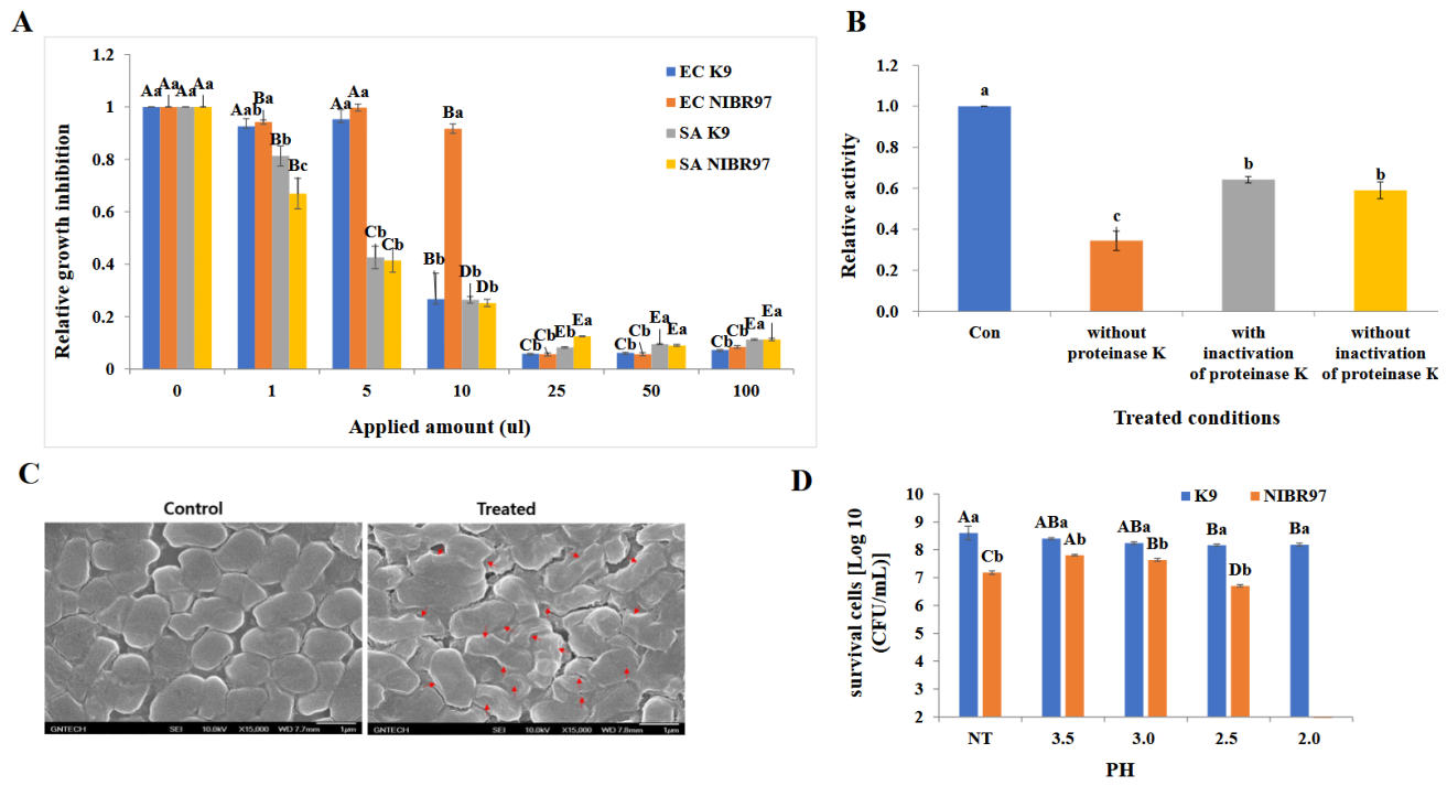
Figure 2.
Classification according to contig of L. plantarum K9. (A) contig 1, (B) contig 2, (C) contig 3, (D) contig 4, (E) arrangements of franking genes for each AMPs. L. plantarum K9 maintains 4 separated contigs, which were composed of 3 circular forms and 1 linear form. Each contig was composed of 3,022,717, 61.375, 32,518, and 7,922 bps. rRNA and tRNA were only expressed at contig 1. HP; hypothetical protein, btuD; Vitamin B12 import ATP-binding protein BtuD, phnD2; putative ABC transporter phosphonate/phosphite binding protein PhnD2, yegS; lipid kinase YegS, cmK; Cytidylate kinase, recQ; ATP-dependent DNA helicase RecQ, yutF; Acid sugar phosphatase, mggB; Mannosylglucosyl-3-phosphoglycerate phosphatase, bmrA; Multidrug resistance ABC transporter ATP-binding/permease protein BmrA, pglH; GalNAc-alpha-(1->4)-GalNAc-alpha-(1->3)-diNAcBac-PP-undecaprenol alpha-1%2C4-N-acetyl-D-galactosaminyltransferase, braC; Leucine-, isoleucine-,C valine-, threonine-, and alanine-binding protein, azoB; NAD(P)H azoreductase, spxA; Regulatory protein Spx, MSMEI; putative oxidoreductase/MSMEI_2347, pln 3; plantaricin 3, folT; Folate transporter FolT, katA; Vegetative catalase, iolT; Major myo-inositol transporter IolT, .
Figure 2.
Classification according to contig of L. plantarum K9. (A) contig 1, (B) contig 2, (C) contig 3, (D) contig 4, (E) arrangements of franking genes for each AMPs. L. plantarum K9 maintains 4 separated contigs, which were composed of 3 circular forms and 1 linear form. Each contig was composed of 3,022,717, 61.375, 32,518, and 7,922 bps. rRNA and tRNA were only expressed at contig 1. HP; hypothetical protein, btuD; Vitamin B12 import ATP-binding protein BtuD, phnD2; putative ABC transporter phosphonate/phosphite binding protein PhnD2, yegS; lipid kinase YegS, cmK; Cytidylate kinase, recQ; ATP-dependent DNA helicase RecQ, yutF; Acid sugar phosphatase, mggB; Mannosylglucosyl-3-phosphoglycerate phosphatase, bmrA; Multidrug resistance ABC transporter ATP-binding/permease protein BmrA, pglH; GalNAc-alpha-(1->4)-GalNAc-alpha-(1->3)-diNAcBac-PP-undecaprenol alpha-1%2C4-N-acetyl-D-galactosaminyltransferase, braC; Leucine-, isoleucine-,C valine-, threonine-, and alanine-binding protein, azoB; NAD(P)H azoreductase, spxA; Regulatory protein Spx, MSMEI; putative oxidoreductase/MSMEI_2347, pln 3; plantaricin 3, folT; Folate transporter FolT, katA; Vegetative catalase, iolT; Major myo-inositol transporter IolT, .
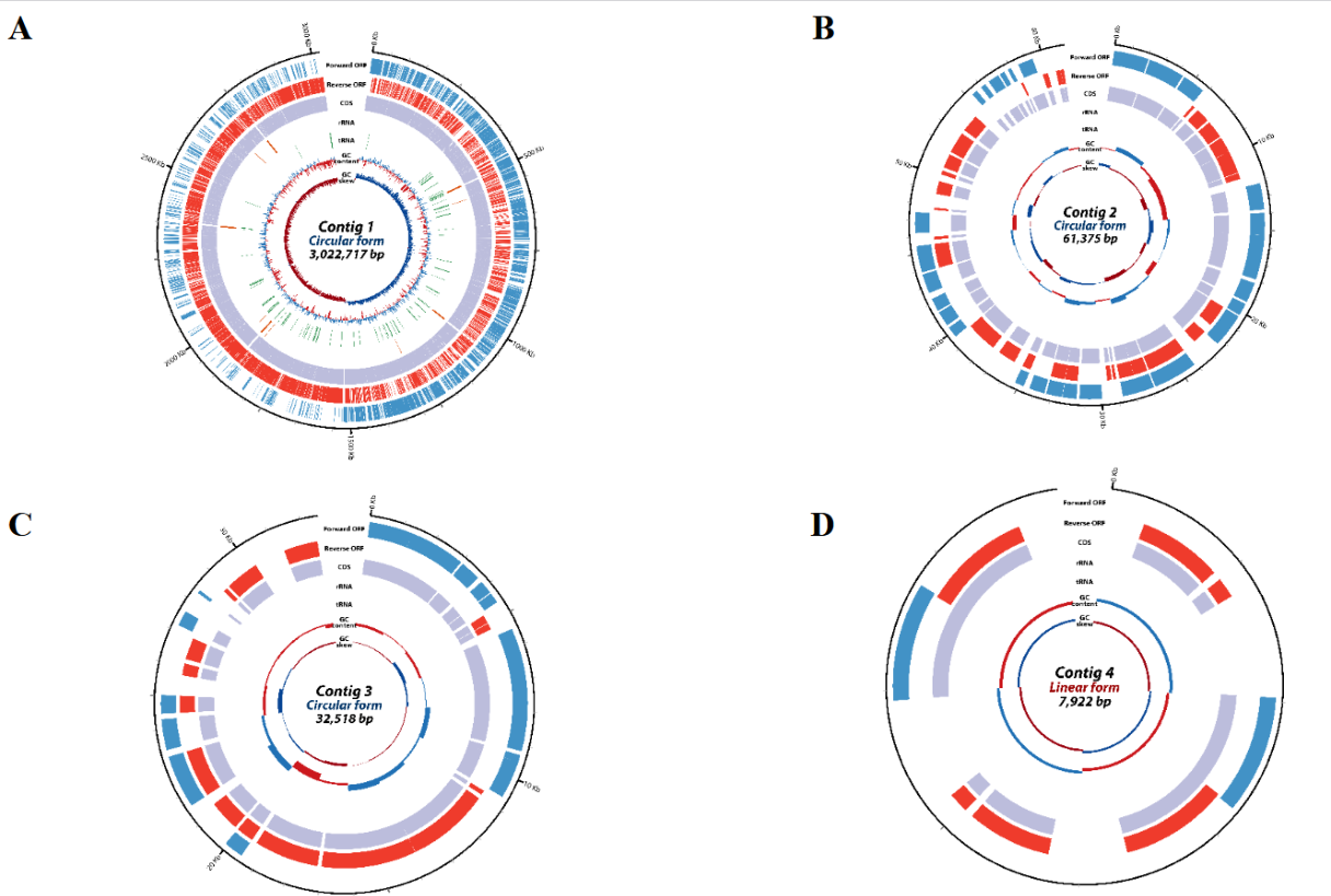
Figure 3.
Antibacterial assays with synthesized AMPs. (A) antibacterial assays of the synthesized AMPs and MIC50 of the synthesized AMPs against S. aureus. The candidate AMPs were synthesized by the predicted oligopeptides with CAMPsign (http://www.campsign.bicnirrh.res.in/). The synthesized peptides were applied for antibacterial assays with 2.5 mg/ml amount. AMP12-2 indicates Winter flounder 1 homolog.
Figure 3.
Antibacterial assays with synthesized AMPs. (A) antibacterial assays of the synthesized AMPs and MIC50 of the synthesized AMPs against S. aureus. The candidate AMPs were synthesized by the predicted oligopeptides with CAMPsign (http://www.campsign.bicnirrh.res.in/). The synthesized peptides were applied for antibacterial assays with 2.5 mg/ml amount. AMP12-2 indicates Winter flounder 1 homolog.
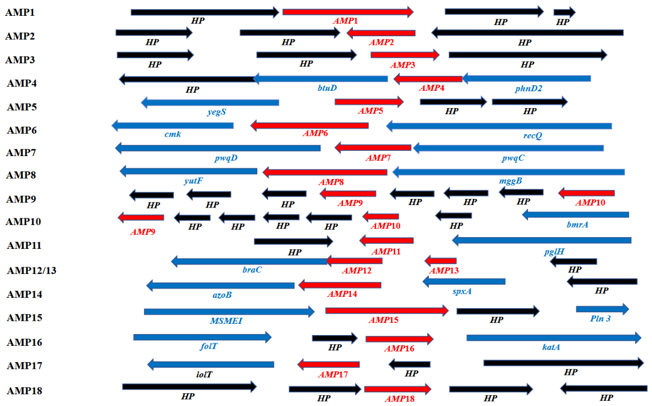
Figure 4.
Antibacterial assays of cloned AMPs. The cloned AMPs were expressed into E. coli, the cell-free supernatants were separated from cells, and then the solutions were applied for antibacterial assays (A) and MIC50 (B) against E. coli (EC) and antibacterial assays (C) and MIC50 (D) against S. aureus (SA). E. coli and S. aureus were adjusted into 106 CFU/ml for microtiter plate assay. Small and large letters indicate significant correlations between the variables (p< 0.05).
Figure 4.
Antibacterial assays of cloned AMPs. The cloned AMPs were expressed into E. coli, the cell-free supernatants were separated from cells, and then the solutions were applied for antibacterial assays (A) and MIC50 (B) against E. coli (EC) and antibacterial assays (C) and MIC50 (D) against S. aureus (SA). E. coli and S. aureus were adjusted into 106 CFU/ml for microtiter plate assay. Small and large letters indicate significant correlations between the variables (p< 0.05).
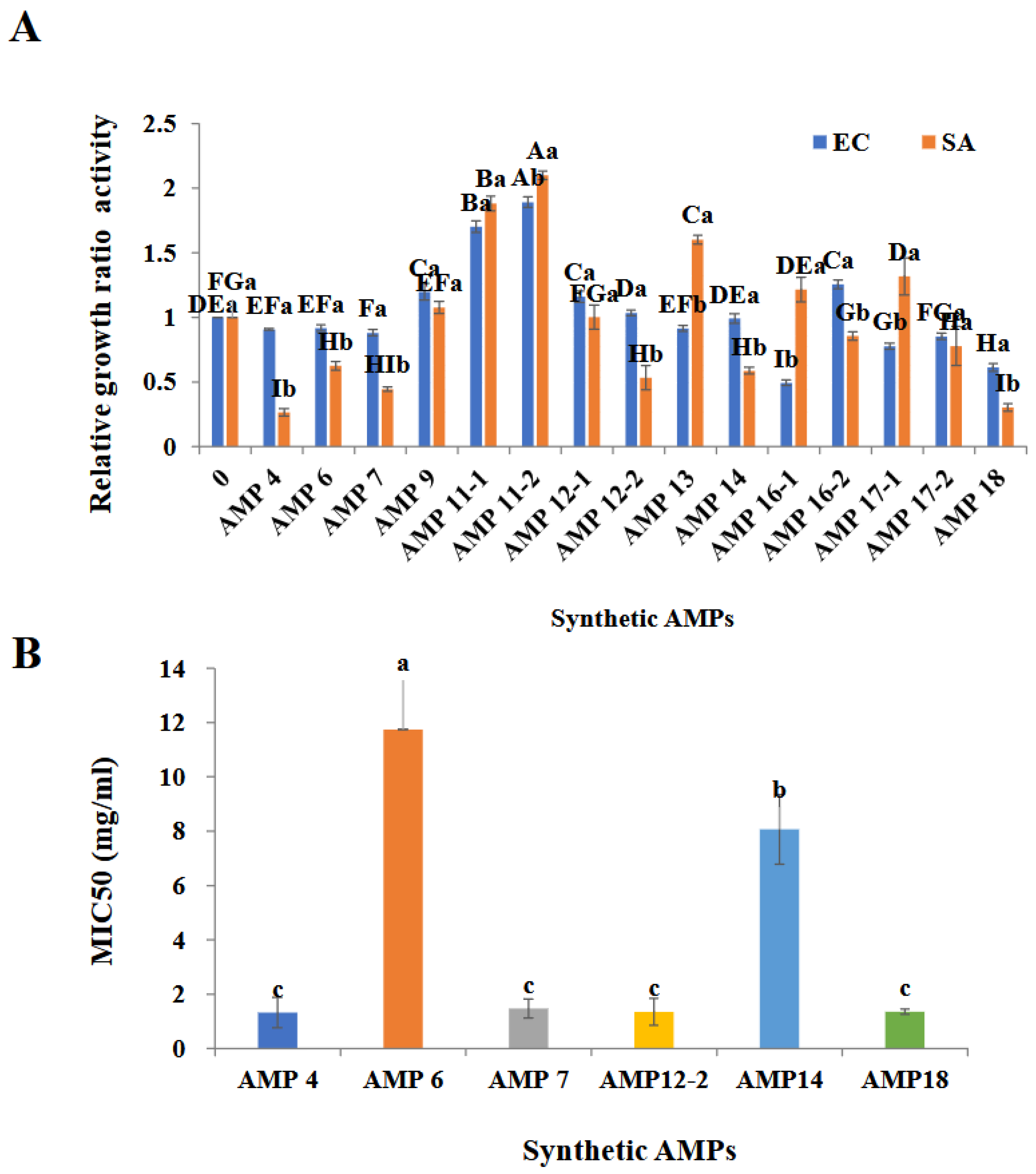
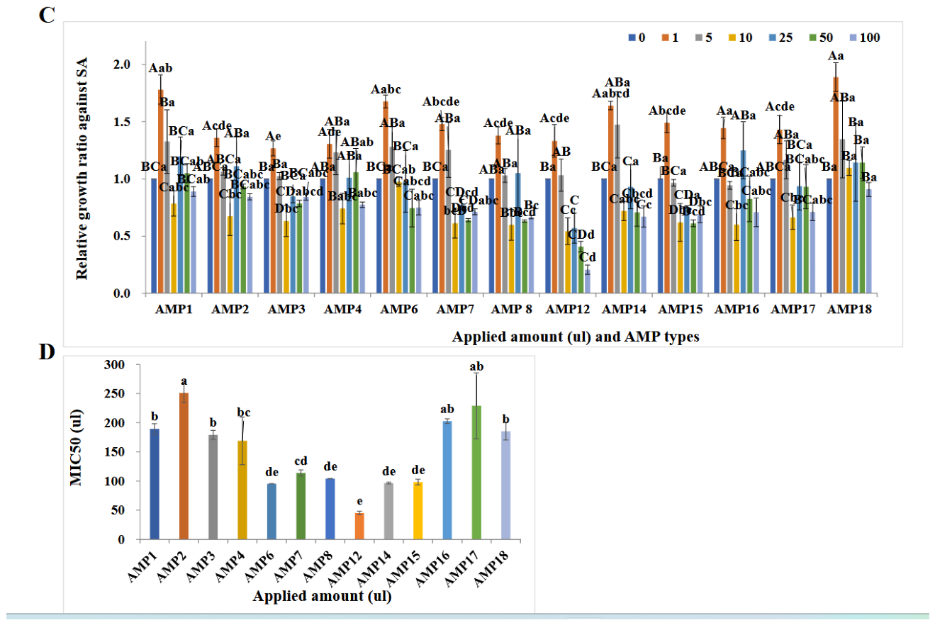
Figure 5.
Properties of AMP12. (A) Prediction of AMP12 by CAMPR4. The red and bule-colored letters indicate homologous regions with CAMPSQ536 and CAMPSQ861, respectively. Prediction of signal sequence was done by SignalP 5.0 (https://services.healthtech.dtu. /services/ SignalP-5.0/). Signal sequence marked with bold and underlined letters was predicted by both Gram positive and negative with SignalP 5.0 (https://swissmodel.expasy.org/interactive). (B) whole amino acid sequence model of AMP12, (C) model without the signal sequence predicted by SignalP 5.0. Bold letters in Figure 6B and C indicate the predictedα-helix structure A53 to G75.
Figure 5.
Properties of AMP12. (A) Prediction of AMP12 by CAMPR4. The red and bule-colored letters indicate homologous regions with CAMPSQ536 and CAMPSQ861, respectively. Prediction of signal sequence was done by SignalP 5.0 (https://services.healthtech.dtu. /services/ SignalP-5.0/). Signal sequence marked with bold and underlined letters was predicted by both Gram positive and negative with SignalP 5.0 (https://swissmodel.expasy.org/interactive). (B) whole amino acid sequence model of AMP12, (C) model without the signal sequence predicted by SignalP 5.0. Bold letters in Figure 6B and C indicate the predictedα-helix structure A53 to G75.
Disclaimer/Publisher’s Note: The statements, opinions and data contained in all publications are solely those of the individual author(s) and contributor(s) and not of MDPI and/or the editor(s). MDPI and/or the editor(s) disclaim responsibility for any injury to people or property resulting from any ideas, methods, instructions or products referred to in the content. |
© 2026 by the authors. Licensee MDPI, Basel, Switzerland. This article is an open access article distributed under the terms and conditions of the Creative Commons Attribution (CC BY) license (http://creativecommons.org/licenses/by/4.0/).
Copyright: This open access article is published under a Creative Commons CC BY 4.0 license, which permit the free download, distribution, and reuse, provided that the author and preprint are cited in any reuse.