

Submitted:
03 July 2026
Posted:
07 July 2026
You are already at the latest version
Abstract
Purpose: To report a child with severe treatment resistant juvenile idiopathic arthritis associated uveitis (JIAU), a condition that typically presents as chronic anterior uveitis, although severe panuveitis with retinal vasculitis may rarely occur.Methods: We describe the clinical course, treatment response, and challenge, dechallenge, and rechallenge sequence in a child with severe refractory JIAU treated with combined infliximab and tofacitinib therapy.Results: The patient showed marked improvement in ocular inflammation, macular edema, and visual acuity following addition of tofacitinib to ongoing infliximab therapy. Inflammation recurred after holding infliximab despite continuation of tofacitinib and improved again following reintroduction of infliximab.Conclusion: This challenge, dechallenge, and rechallenge sequence suggests that neither infliximab nor tofacitinib alone provided adequate disease control, whereas their combination was associated with sustained suppression of ocular inflammation. Combined tumor necrosis factor alpha and Janus kinase inhibition may represent a rescue therapeutic option for carefully selected patients with severe refractory JIAU, although its long term safety and efficacy require further evaluation.
Keywords:
cystoid macular edema
; infliximab
; Janus kinase inhibitor
; juvenile idiopathic arthritis
; optical coherence tomography
; retinal vasculitis
; tofacitinib
; uveitis
Introduction
Uveitis is the most common extraarticular manifestation of juvenile idiopathic arthritis (JIA) [1]. JIA associated uveitis (JIAU) typically presents as chronic anterior uveitis. However, a minority of patients may develop other forms of ocular inflammation, including panuveitis and diffuse retinal vasculitis. Inadequate control of inflammation in JIAU may lead to severe structural complications and permanent visual impairment [2]. Various topical, local, and systemic treatment strategies are used to achieve sustained control of ocular inflammation. Nevertheless, refractory uveitis despite multiple treatments remains a major challenge in some patients.
Topical corticosteroids are the mainstay of initial treatment for anterior JIA associated uveitis. When systemic therapy is required, methotrexate is generally the preferred conventional disease modifying antirheumatic drug. Other conventional immunosuppressive agents, including cyclosporine and mycophenolate mofetil, have also been used. Monoclonal tumor necrosis factor inhibitors, particularly adalimumab and infliximab, are recommended for patients with disease that is refractory or intolerant to conventional therapy. Tumor necrosis factor alpha (TNF-alfa) is an important therapeutic target in JIA associated uveitis [3].
More recently, Janus kinase (JAK) inhibitors have emerged as potential therapeutic options for refractory JIA associated uveitis, particularly in patients with an inadequate response to conventional or biologic disease modifying antirheumatic drugs [4]. Clinical evidence remains limited, but available pediatric data suggest that tofacitinib may be considered as a second line option in selected patients with treatment resistant uveitis [5].
Herein, we describe a patient with refractory JIAU in whom multiple medical and surgical treatment strategies had failed. The patient showed a marked response after combined treatment with infliximab and tofacitinib. Challenge dechallenge and rechallenge sequence suggested that combined treatment was required to achieve sustained inflammatory control in this patient.
Case Presentation
A seven-year-old boy was referred with refractory pauciarticular juvenile idiopathic arthritis associated uveitis, which had been diagnosed at the age of 4. Laboratory evaluation showed positive antinuclear antibodies, whereas human leukocyte antigen B27 testing and a comprehensive evaluation for tuberculosis, syphilis, sarcoidosis, and other systemic and infectious causes of retinal vasculitis was negative. His family history was unremarkable for rheumatologic or immunologic disease.
At the initial ophthalmic examination in 2017, both eyes showed active anterior chamber inflammation, posterior synechiae, keratic precipitates, cataract, and calcific band keratopathy. Posterior segment examination was not possible because of the posterior synechiae and cataracts. B scan ultrasonography showed no evidence of vitritis or retinal detachment. The patient was treated with prednisolone at 0.5 mg/kg/day, methotrexate at 15 mg/m²/week, folic acid at 0.5 mg/day, cyclosporine at 5 mg/kg/day, and etanercept.
After 6 months, etanercept was replaced with adalimumab at 24 mg/m² every 2 weeks, which was continued for 4 years. In 2018, the right eye underwent combined lensectomy, anterior vitrectomy, EDTA assisted superficial keratectomy, and simultaneous intravitreal injection of bevacizumab and triamcinolone acetonide. In 2019, the left eye underwent lensectomy, pars plana vitrectomy with endolaser photocoagulation of the inferior retina, EDTA assisted superficial keratectomy, and simultaneous intravitreal injection of bevacizumab and triamcinolone acetonide.
In 2020, intravitreal methotrexate was administered to the left eye because of severe macular edema. In 2021, mycophenolate mofetil at 500 mg twice daily was added because of an inadequate treatment response. Four months later, adalimumab and cyclosporine were discontinued, and infliximab at 6 mg/kg every 8 weeks was initiated. At this stage, the patient was receiving mycophenolate mofetil, methotrexate, and infliximab.
Five months later, bilateral intravitreal triamcinolone acetonide injections were administered. During the treatment course, the right eye received a total of 5 intravitreal bevacizumab injections, 4 intravitreal triamcinolone acetonide injections, and 2 periocular triamcinolone acetonide injections. The left eye received a total of 5 intravitreal bevacizumab injections, 4 intravitreal triamcinolone acetonide injections, and 1 intravitreal methotrexate injection.
Despite these treatments, uveitis remained severely uncontrolled one month after the injections. Fluorescein angiography showed severe posterior and peripheral retinal vascular leakage, together with intense optic disc and macular leakage. Macular and retinal nerve fiber layer optical coherence tomography showed severe cystoid macular edema and retinal nerve fiber layer thickening (Figure 1). Best corrected visual acuity was 0.70 logMAR in the right eye and 1.00 logMAR in the left eye. Scatter laser photocoagulation was also performed in the left eye during the treatment course to address areas of retinal nonperfusion and improve inflammatory control.
Tofacitinib at 0.25 mg/kg/day was subsequently added to the treatment regimen. One month later, a dramatic treatment response was observed, with improvement in visual acuity, fluorescein angiographic findings, and macular and retinal nerve fiber layer optical coherence tomography findings (Figure 2). This dramatic improvement was initially attributed to tofacitinib. This assumption, together with concerns regarding excessive immunosuppression and safety of the unprecedented use of combined anti-TNF alpha and JAK inhibitor therapy for uveitis, led to the discontinuation of infliximab.
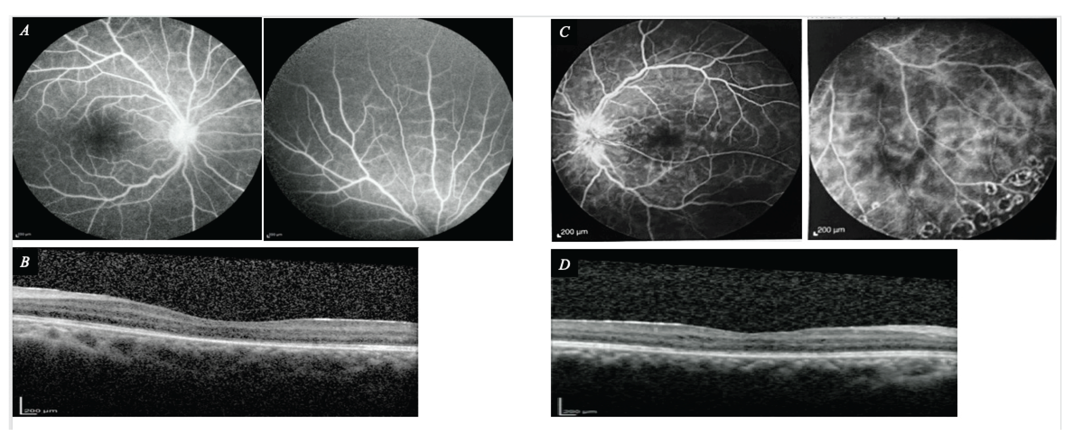
However, 2 months after infliximab withdrawal, inflammation recurred despite continuation of tofacitinib (Figure 3). At that time, best corrected visual acuity was 0.54 logMAR in the right eye and 1.00 logMAR in the left eye. Infliximab at 6 mg/kg every 8 weeks was therefore reintroduced in combination with tofacitinib. This again resulted in a dramatic improvement in ocular inflammation and visual acuity (Figure 4). At the final follow up visit, best corrected visual acuity was 0.22 logMAR in the right eye and 0.52 logMAR in the left eye.
Discussion
Juvenile idiopathic arthritis associated uveitis is an immune mediated disease involving dysregulated cellular immunity and multiple inflammatory cytokine pathways [6]. Studies of aqueous humor from affected patients have demonstrated altered cytokine, chemokine, and matrix metalloproteinase profiles, supporting the involvement of several overlapping inflammatory pathways [7]. Tofacitinib preferentially inhibits JAK1 and JAK3 dependent signaling and consequently modulates cytokines involved in innate and adaptive immune responses, whereas infliximab directly neutralizes tumor necrosis factor alpha, an important inflammatory mediator in JIAU [6,8]. Concurrent inhibition of these distinct pathways provides a biologically plausible explanation for the response observed in our patient, although pharmacologic synergy cannot be established from a single case.
In this patient, inflammation remained uncontrolled during infliximab-based treatment but improved markedly after tofacitinib was added. Inflammation subsequently recurred after infliximab withdrawal despite continuation of tofacitinib and improved again after infliximab was reintroduced. This challenge, dechallenge, and rechallenge sequence suggests that the two agents had a complementary therapeutic effect in this individual patient. To the best of our knowledge, concurrent infliximab and tofacitinib therapy has not previously been reported for the treatment of uveitis.
Evidence supporting JAK inhibition in uveitis remains limited. Improvement with tofacitinib, baricitinib, or upadacitinib has been reported in refractory JIA associated uveitis, pediatric uveitis, and other forms of noninfectious ocular inflammation [4,5,9,10,11]. A recent systematic review found that the available evidence consists predominantly of case reports and small case series, with substantial clinical heterogeneity and limited follow up [10]. Therefore, although the published findings are encouraging, they do not establish comparative efficacy, optimal treatment duration, or long-term safety.
No treatment related adverse event was observed during the available follow up in our patient. However, this observation cannot establish the long-term safety of combined infliximab and tofacitinib therapy. Adverse events reported during pediatric treatment with tofacitinib include elevated liver enzyme levels, hypercholesterolemia, and lymphadenitis [12]. Moreover, current prescribing information does not recommend the use of tofacitinib in combination with biologic disease modifying antirheumatic drugs because of the potential for increased immunosuppression and infection risk [13]. This combination should therefore be regarded as an exceptional rescue strategy for severe, sight threatening, treatment-resistant disease and should be considered only after careful multidisciplinary assessment, with close clinical and laboratory monitoring.
Conclusion
Combined infliximab and tofacitinib therapy led to control of severe refractory JIA associated panuveitis and retinal vasculitis in this patient. The challenge, dechallenge, and rechallenge response suggests a complementary effect between TNF-alpha and JAK inhibition. Larger studies with longer follow up are required to determine the efficacy and safety of this approach.
Ethics Statement
This case report was approved by the Ethics Committee of Tehran University of Medical Sciences (Ethics Code: IR.TUMS.FARABIH.REC.1405.016). Written informed consent was obtained from the patient’s legal guardian for publication of the case report and accompanying images.
Funding Statement: No funding was received for this work.
Data Availability Statement
Data sharing is not applicable to this article because this is a case report and no datasets were generated or analyzed.
Declaration of Interest: The authors report no conflicts of interest.
References
- Clarke SL, Sen ES, Ramanan AV. Juvenile idiopathic arthritis-associated uveitis. Pediatr Rheumatol Online J. 2016;14(1):27. Published 2016 Apr 27. [CrossRef]
- Maleki A, Patel PD, Foster CS. Juvenile idiopathic arthritis and its associated uveitis. Expert Rev Clin Immunol. 2023;19(9):1157-1169. [CrossRef]
- Constantin T, Foeldvari I, Anton J, et al. Consensus-based recommendations for the management of uveitis associated with juvenile idiopathic arthritis: the SHARE initiative. Ann Rheum Dis. 2018;77(8):1107-1117. [CrossRef]
- Miserocchi E, Giuffrè C, Cornalba M, Pontikaki I, Cimaz R. JAK inhibitors in refractory juvenile idiopathic arthritis-associated uveitis. Clin Rheumatol. 2020;39(3):847-851. [CrossRef]
- Dutta Majumder P, Abraham S, Sudharshan S, Janarthanan M, Ramanan AV. Tofacitinib for Refractory Uveitis and Scleritis in Children: A Case Series. Ocul Immunol Inflamm. 2024;32(9):2092-2095. [CrossRef]
- Sen ES, Dick AD, Ramanan AV. Uveitis associated with juvenile idiopathic arthritis. Nat Rev Rheumatol. 2015;11(6):338-348. [CrossRef]
- Bauer D, Kasper M, Walscheid K, et al. Multiplex Cytokine Analysis of Aqueous Humor in Juvenile Idiopathic Arthritis-Associated Anterior Uveitis With or Without Secondary Glaucoma. Front Immunol. 2018;9:708. Published 2018 Apr 5. [CrossRef]
- Ghoreschi K, Jesson MI, Li X, et al. Modulation of innate and adaptive immune responses by tofacitinib (CP-690,550). J Immunol. 2011;186(7):4234-4243. [CrossRef]
- Vitale A, Palacios-Olid J, Caggiano V, et al. Efficacy and safety of Janus kinase inhibitors in non-infectious inflammatory ocular diseases: a prospective cohort study from the international AIDA network registries. Front Med (Lausanne). 2024;11:1439338. Published 2024 Aug 23. [CrossRef]
- Almerri S, Alshatti H, Ali YT, Behbehani R. JAK inhibitors in autoimmune ocular inflammatory diseases - A systematic review of case reports and series. J Ophthalmic Inflamm Infect. 2025;15(1):93. Published 2025 Dec 10. [CrossRef]
- Beckman M, Srivastava SK, Lowder CY, Baynes K, Lowe A, Sharma S. The use of Janus kinase inhibitors to treat noninfectious uveitis. Eye (Lond). 2026;40(1):91-97. [CrossRef]
- Kostik MM, Raupov RK, Suspitsin EN, et al. The Safety and Efficacy of Tofacitinib in 24 Cases of Pediatric Rheumatic Diseases: Single Centre Experience. Front Pediatr. 2022;10:820586. Published 2022 Feb 8. [CrossRef]
- US Food and Drug Administration. Xeljanz, Xeljanz XR, and Xeljanz oral solution (tofacitinib): prescribing information [Internet]. Silver Spring, MD: US Food and Drug Administration; 2026 Mar [cited 2026 Jun 18].
Figure 1.
Optical coherence tomography before tofacitinib initiation while the patient was receiving infliximab-based therapy. (A, C) Peripapillary retinal nerve fiber layer (RNFL) OCT of the right and left eyes, respectively, showing marked RNFL thickening, consistent with active optic disc inflammation. (B, D) Macular OCT of the right and left eyes, respectively, showing severe cystoid macular edema with large intraretinal cystoid spaces and associated subretinal fluid, more pronounced in the left eye.
Figure 1.
Optical coherence tomography before tofacitinib initiation while the patient was receiving infliximab-based therapy. (A, C) Peripapillary retinal nerve fiber layer (RNFL) OCT of the right and left eyes, respectively, showing marked RNFL thickening, consistent with active optic disc inflammation. (B, D) Macular OCT of the right and left eyes, respectively, showing severe cystoid macular edema with large intraretinal cystoid spaces and associated subretinal fluid, more pronounced in the left eye.
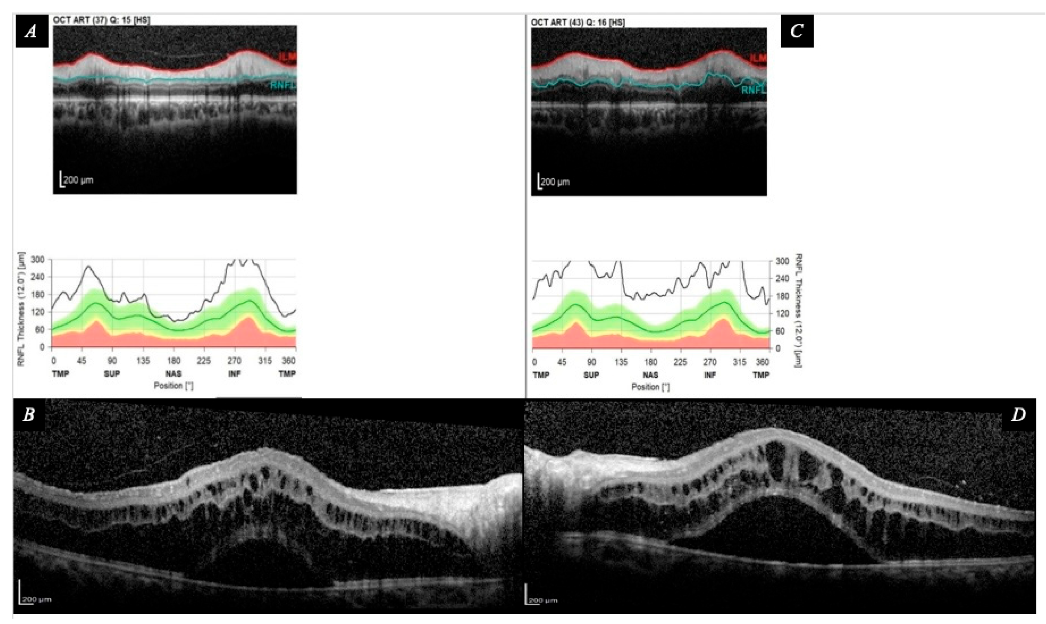
Figure 2.
Structural optical coherence tomography one month after addition of oral tofacitinib to ongoing therapy. (A, C) Peripapillary RNFL OCT of the right and left eyes, respectively, demonstrating reduction of RNFL thickening compared with pretreatment imaging. (B, D) Macular OCT of the right and left eyes, respectively, demonstrating marked anatomic improvement, with substantial reduction of cystoid macular edema and resolution of subretinal fluid. Mild residual intraretinal cystic change persists in both eyes, more pronounced in the right eye.
Figure 2.
Structural optical coherence tomography one month after addition of oral tofacitinib to ongoing therapy. (A, C) Peripapillary RNFL OCT of the right and left eyes, respectively, demonstrating reduction of RNFL thickening compared with pretreatment imaging. (B, D) Macular OCT of the right and left eyes, respectively, demonstrating marked anatomic improvement, with substantial reduction of cystoid macular edema and resolution of subretinal fluid. Mild residual intraretinal cystic change persists in both eyes, more pronounced in the right eye.
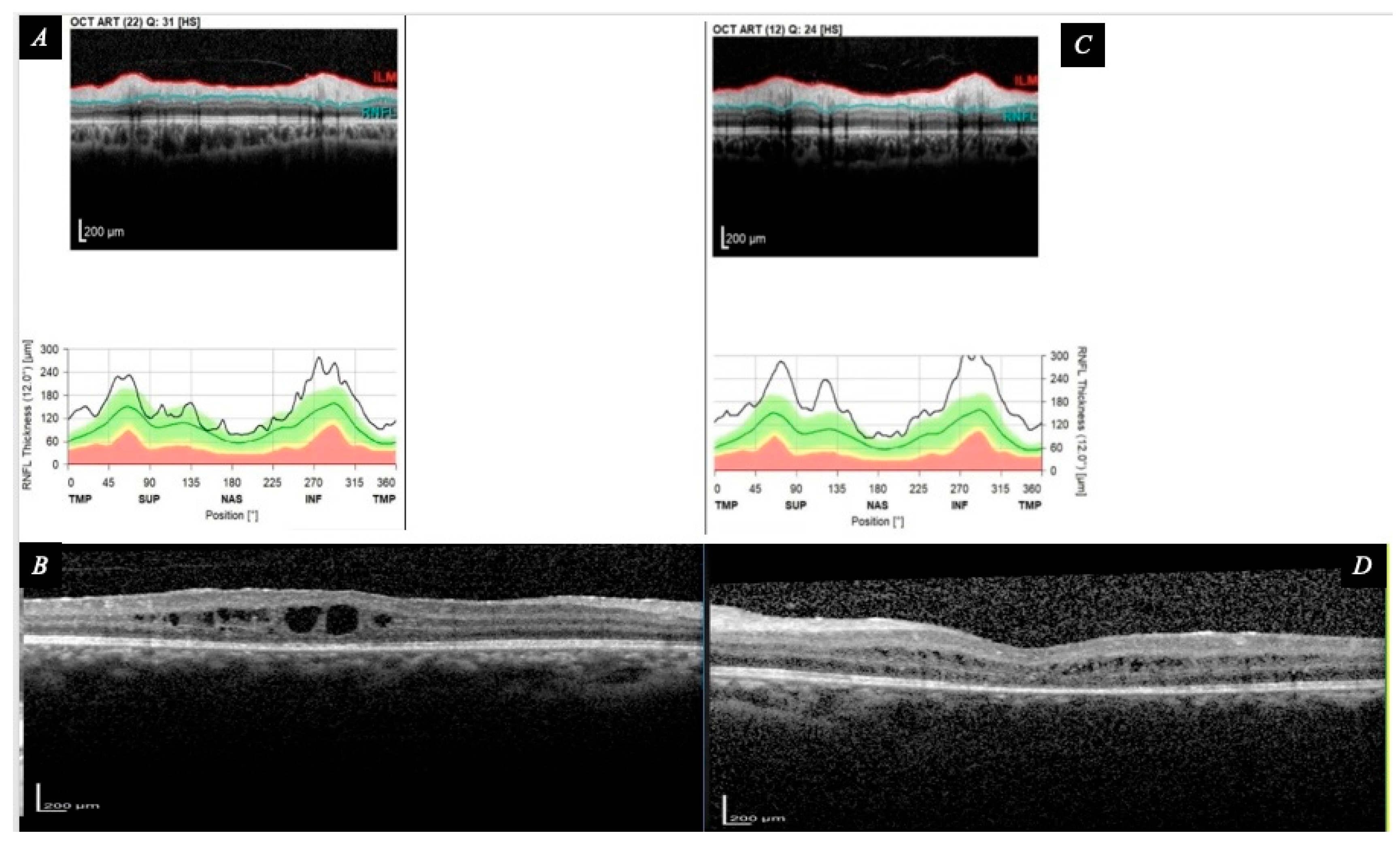
Figure 3.
Recurrence after holding infliximab while tofacitinib was continued. (A, C) Fluorescein angiography of the right and left eyes, respectively, demonstrates active posterior segment inflammation, with marked optic disc leakage, diffuse posterior retinal vascular leakage, and severe peripheral vascular leakage/retinal vasculitis. Prior laser photocoagulation scars are visible in the left peripheral retina. (B, D) Corresponding macular OCT shows recurrent severe cystoid macular edema with subretinal fluid in both eyes.
Figure 3.
Recurrence after holding infliximab while tofacitinib was continued. (A, C) Fluorescein angiography of the right and left eyes, respectively, demonstrates active posterior segment inflammation, with marked optic disc leakage, diffuse posterior retinal vascular leakage, and severe peripheral vascular leakage/retinal vasculitis. Prior laser photocoagulation scars are visible in the left peripheral retina. (B, D) Corresponding macular OCT shows recurrent severe cystoid macular edema with subretinal fluid in both eyes.
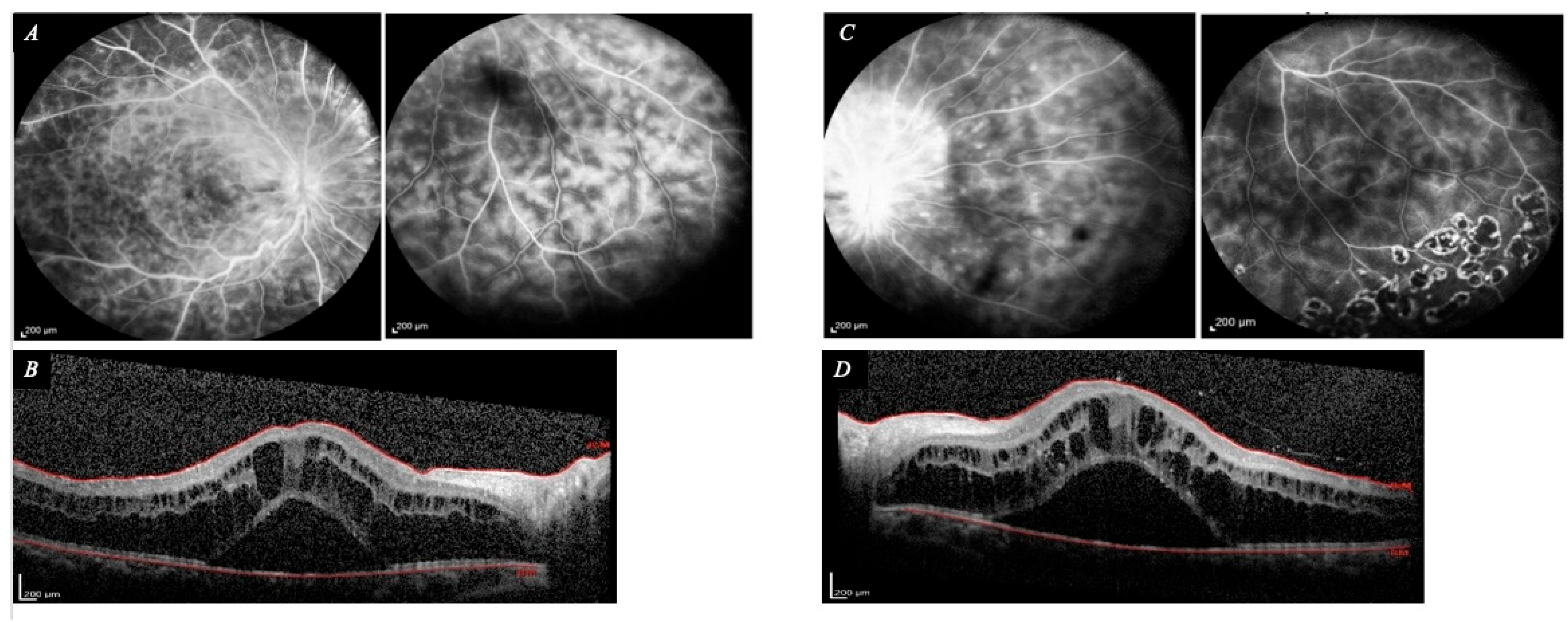
Figure 4.
Response after reintroduction of infliximab while tofacitinib was continued. (A, C) Fluorescein angiography of the right and left eyes, respectively, shows marked reduction in optic disc, macular, and peripheral vascular leakage compared with recurrence imaging. Residual peripheral staining or leakage and prior laser scars are visible in the left eye. (B, D) Macular OCT confirms complete resolution of cystoid macular edema and subretinal fluid in both eyes.
Figure 4.
Response after reintroduction of infliximab while tofacitinib was continued. (A, C) Fluorescein angiography of the right and left eyes, respectively, shows marked reduction in optic disc, macular, and peripheral vascular leakage compared with recurrence imaging. Residual peripheral staining or leakage and prior laser scars are visible in the left eye. (B, D) Macular OCT confirms complete resolution of cystoid macular edema and subretinal fluid in both eyes.
Disclaimer/Publisher’s Note: The statements, opinions and data contained in all publications are solely those of the individual author(s) and contributor(s) and not of MDPI and/or the editor(s). MDPI and/or the editor(s) disclaim responsibility for any injury to people or property resulting from any ideas, methods, instructions or products referred to in the content. |
© 2026 by the authors. Licensee MDPI, Basel, Switzerland. This article is an open access article distributed under the terms and conditions of the Creative Commons Attribution (CC BY) license (http://creativecommons.org/licenses/by/4.0/).
Copyright: This open access article is published under a Creative Commons CC BY 4.0 license, which permit the free download, distribution, and reuse, provided that the author and preprint are cited in any reuse.