

Submitted:
04 July 2026
Posted:
06 July 2026
You are already at the latest version
Abstract
Background/Objectives: Diabetic retinopathy (DR) is a leading cause of visual im-pairment in United States adults. Artificial intelligence (AI) screening systems using non-dilated fundus photography in primary care settings offer a convenient alternative to dilated screening, but image quality remains a critical limitation. No studies have as-sessed how primary care photographer experience affects gradability of non-dilated images in AI-assisted screening. We hypothesized that medical assistant (MA) photog-raphers’ gradability rates (GR) would improve with experience.
Methods: We retrospectively reviewed 1354 DR screenings performed from 05/12/2021-12/28/2022 across 10 Temple Primary Care Clinics using the Canon CR-2 AF camera and EyeNuk’s EyeArt system. 30 MA photographers captured images; MAs with < 10 screenings were excluded. GR were calculated for each photographer. Linear and polynomial regression analyses assessed associations between number of photographs and GR. For MA photographers with >90 screenings, GR was further evaluated over time in blocks of ten consecutive screenings.
Results: Eleven MA photographers met inclusion criteria. Linear regression showed no significant association between number of photographs and percentage of gradable images (F = 0.259, p = 0.7784). Three MAs conducted at least 91 screenings. Linear re-gression analysis of successive screenings showed no significant association with average GR (F = 0.684, p = 0.4323).
Conclusions: Image gradability did not improve with increasing MA photographer experience. Repeated practice alone appears insufficient to achieve high GR. Standard-ized competency-based training, refresher sessions, or robotic fundus photography may be needed to optimize image quality in AI-assisted DR screening programs in a primary care setting.
Keywords:
artificial intelligence
; diabetic retinopathy screening
; fundus photography
; image gradability
; primary care
1. Introduction
Diabetic retinopathy (DR) is a leading cause of visual impairment among adults in the United States. Of an estimated 38.4 million people with diabetes mellitus (DM) [1], 9.6 million are diagnosed with DR [2], with roughly 5% suffering from vision-threatening DR (VTDR) [2]. With the increasing lifespan of patients with DM and a growing global aging population, a high burden of DR and demand for eye care is projected to continue through 2045 [3]. Routine screening for DR after an initial diagnosis of diabetes is a valuable method of preventing the development of VTDR [4].
Dilation remains the standard of care for DR screening in ophthalmology settings considering the difficulty in obtaining a wide field of view of the retina while non-dilated [5]. Artificial intelligence (AI)-based screening systems such as EyeNuk’s (Los Angeles, CA) EyeArt, Digital Diagnostics’ IDx-DR, and AEYE Health’s AEYE-DS have high sensitivity and specificity rates, proving to be valid alternatives to traditional dilated screening approaches and increasing efficiency of screenings [6,7,8,9,10,11,12]. As of 2023, United States Food and Drug Administration (FDA) clearance has enabled EyeArt to be used with multiple camera models, expanding its access in primary care settings [13].
AI-based screening through digital fundus photography of non-dilated pupils in primary care settings is a convenient, highly reliable alternative to standard-of-care dilated screening at an eye-care provider that can improve DR screening rates by providing diabetes care and diabetic eye disease care in the same location [14,15]. The implementation of precise AI screening for DR using non-dilated photographs across primary care settings can decrease clinic burden for ophthalmologists, improve workflow, and avoid potential risks of dilation by non-ophthalmic personnel including acute angle-closure glaucoma [16].
Despite these benefits, image quality proves to be a critical limitation of AI-based systems [14]. Successful grading by AI screening programs is dependent on high quality fundus photographs. A gradable fundus photograph requires a clear and in-focus view of the fundus. Poor image quality remains common in non-dilated AI-based DR screening in primary care settings, leading to high rates of false positives [14].
Photographers must actively mitigate issues such as incorrect focus, dirty lens reflections, uneven light exposure, patient motion, and eyelid obstruction. Additionally, fundus photographs must be labeled and uploaded correctly to provide AI-gradable images. Since the quality of photographs determines the effectiveness of DR screening, attempts have been made to improve gradability rates (GR) such as offering multiple camera options [17,18] and using dilating agents when appropriate [19,20].
Importantly, one study that focused on smartphone-based retinal photography in primary care found that medical assistants (MAs) can obtain high-quality retinal photography of dilated pupils within minutes of first-time operation [21], underscoring the promise of training non-ophthalmic personnel to take fundus photography and expand screening access. This finding is limited to photographs of dilated pupils using smartphone-based photography but supports the idea that photographers with limited experience can obtain high quality photographs. Alternatively, a 2004 study evaluating whether fundus photograph quality improves with operator experience found no significant difference in fundus image quality among photographers with differing levels of training when using a nonmydriatic camera [22]. While this study provides insight of image quality obtained with more traditionally-used nonmydriatic cameras operated by inexperienced personnel, its experimental design relied on subjective human grader evaluation of image quality based on a 3-level scale [22] unlike the objective algorithms used in current AI-based systems.
There are no studies so far that have assessed the impact of a primary care-based photographer's experience on the quality of non-dilated fundus photographs using an AI-based DR screening program. We hypothesized that the non-dilated, AI-assisted fundus photograph GR of MA photographers at Temple Primary Care Clinics improve with the number of photographs taken.
2. Materials and Methods
We performed a retrospective chart review of 1354 DR screenings of diabetic patients 18 years and older between 05/12/2021 – 12/28/2022. The protocol was approved by the Temple Institutional Review Board. A DR screening consisted of fundus photographs of both eyes taken with Canon CR-2 AF camera model accompanied by EyeNuk. Photographs were read by EyeArt. Fundus photographs were captured by 30 different MA photographers at 10 Temple Primary Care Clinics. All MA photographers received photography training by an ophthalmic photographer prior to initiating screening.
Screening results with at least one uninterpretable eye photograph were reported as “ungradable” by EyeArt.
MA photographers who conducted fewer than 10 screenings were excluded due to the low sample size. GR were calculated by dividing the number of gradable screenings by the total number of screenings conducted by each photographer.
We examined whether photographer experience, defined as the total number of photographs taken, was associated with the proportion of gradable images. A simple linear regression analysis was used to determine if a linear relationship exists. A simple polynomial expansion was used to evaluate potential nonlinear effects. For each model, overall significance was assessed using the F-statistic; a p-value of 0.05 or less was considered significant.
For a longitudinal learning-curve analysis, MA photographers who conducted greater than 90 DR screenings were identified and had their GR assessed over time in intervals of 10 consecutive screenings for the first 91-100 screenings. MA GRs were averaged at each interval of 10 photographs taken. A 95% confidence interval was used at each interval. Any association between photographer experience, using successive screenings as a proxy, and average image gradability was assessed using simple linear regression; overall model significance was assessed using the F-statistic; p-values of 0.05 or less were considered significant.
3. Results
Out of the 30 MA photographers, only 11 conducted at least 10 screenings so only those 11 were included in the analysis.
The simple linear regression analysis showed no significant association between number of photographs and percentage of gradable images (F statistic = 0.187, p = 0.6754) (Figure 1, Table 1). The simple polynomial expansion analysis also showed no evidence of association (F statistic = 0.259, p = 0.7784) (Figure 1, Table 1).
Three MA photographers conducted at least 91 screenings. The linear regression analysis showed no significant association between photographer experience, using successive screenings as a proxy, and average image gradability (F statistic = 0.684, p = 0.4323) (Figure 2).
4. Discussion
For a successful screening program to function, photo gradability should be high because high rates of ungradable images by MA photographers would wind up placing the patients back in the traditional in-person exam system. This adds cost and time when it already very hard to get an appointment, resulting in more suboptimal care. An MA just learning to take photographs would not be expected to have the same gradability rate as an experienced ophthalmic photographer, but with time and practice, the MA should show improvement with the goal of achieving the rate of the photographer. However, contrary to our hypothesis, image gradability did not improve with increased photographer experience. The data demonstrates no correlation between the number of photographs taken by a MA photographer and his/her gradability rate.
Our findings match a previous study which found that photographic training and experience do not influence fundus image quality for images taken with a nonmydriatic camera. Similar to the study that reported 79% of images captured by minimally trained photographers of nonmydriatic pupils were of poor or moderate quality [22], we found similar results.
Our findings are limited by a small sample size. The performance of only 11 MA photographers was evaluated in this study, some of which had taken as few as 12 photographs (Figure 1, Table 1). Additionally, only 3 MA photographers took a large volume of over 90 photographs, limiting the strength of our findings of individual photographer performance over successive sets of photographs (Figure 2). The results are specific to an urban health center that provides care to medically underserved patients and may not be generalizable.
Not all ungradable images should be viewed as purely detrimental and the fault of the photographer. Some are due to media opacities such as cataracts, which may lead to surgical referrals and significant visual improvement. These incidental findings, although unrelated to DR, represent a potential secondary benefit of regular DR screening.
Repeated practice alone did not improve performance. Given the lack of improvement in gradability of images over time, it is reasonable to conclude that teaching MAs one time to operate a fundus camera is ineffective in achieving a high gradability rate. One avenue worth exploring is the development of a standardized competency-based training program for MA photographers with training completion marked by certification. Refresher photographer training supervised by experienced photographers may be necessary to enhance performance in DR screening initiatives. Further research is necessary to identify why MA photographer performance does not improve with experience.
Aside from dilation, which is not practical in a primary care setting, another way to potentially improve photo gradability is to use fully automated robotic fundus photography. This might remove the barrier of training medical professionals to obtain high quality photographs. We are investigating this approach now.
Author Contributions
Conceptualization, C.H.; methodology, C.H.; software, O.S.; formal analysis, J.G. and S.A.; investigation, O.S.; writing—original draft preparation, J.G., S.A., J.H., and Y.Z.; writing—review and editing, J.G., J.H., and Y.Z.; visualization, J.G.; supervision, J.H. and Y.Z.; project administration, J.G., J.H., and Y.Z. All authors have read and agreed to the published version of the manuscript.
Funding
This research received no external funding.
Institutional Review Board Statement
The study was conducted in accordance with the Declaration of Helsinki, and approved by the Institutional Review of Temple University (IRB number 28368 and date of approval 06/01/2021).
Informed Consent Statement
Patient consent was waived due to minimal risk to human subjects as determined by the IRB.
Data Availability Statement
The original contributions presented in this study are included in the article. Further inquiries can be directed to the corresponding author.
Acknowledgments
We thank the medical assistants and staff at Temple Primary Care Clinics for their contributions to diabetic retinopathy screening. We also acknowledge the support of the Temple Ophthalmology Department and Oleg Shum, BS, the ophthalmic photographer who trained the medical assistants.
Conflicts of Interest
The authors declare no conflicts of interest.
Abbreviations
The following abbreviations are used in this manuscript:
| DR | Diabetic retinopathy |
| DM | Diabetes mellitus |
| VTDR | Vision-threatening diabetic retinopathy |
| AI | Artificial intelligence |
| GR | Gradability rates |
| MA | Medical assistant |
References
- CDC. Diabetes. 2024 [cited 2025 June 11]. National Diabetes Statistics Report. Available from: https://www.cdc.gov/diabetes/php/data-research/index.html.
- Lundeen EA, Burke-Conte Z, Rein DB, Wittenborn JS, Saaddine J, Lee AY, et al. Prevalence of Diabetic Retinopathy in the US in 2021. JAMA Ophthalmol. 2023 Aug 1;141(8):747.
- Teo ZL, Tham YC, Yu M, Chee ML, Rim TH, Cheung N, et al. Global Prevalence of Diabetic Retinopathy and Projection of Burden through 2045: Systematic Review and Meta-analysis. Ophthalmology. 2021 Nov;128(11):1580–91.
- Tozer K, Woodward MA, Newman-Casey PA. Telemedicine and Diabetic Retinopathy: Review of Published Screening Programs. J Endocrinol Diabetes. 2015;2(4).
- 12. Retinopathy, Neuropathy, and Foot Care: Standards of Care in Diabetes—2025 | Diabetes Care | American Diabetes Association [Internet]. [cited 2025 July 29]. Available from: https://diabetesjournals.org/care/article/48/Supplement_1/S252/157552/12-Retinopathy-Neuropathy-and-Foot-Care-Standards.
- Bellemo V, Lim G, Rim TH, Tan GSW, Cheung CY, Sadda S, et al. Artificial Intelligence Screening for Diabetic Retinopathy: the Real-World Emerging Application. Curr Diab Rep. 2019 July 31;19(9):72.
- Grzybowski A, Jin K, Zhou J, Pan X, Wang M, Ye J, et al. Retina Fundus Photograph-Based Artificial Intelligence Algorithms in Medicine: A Systematic Review. Ophthalmol Ther. 2024 Aug 1;13(8):2125–49.
- Grzybowski A, Brona P, Lim G, Ruamviboonsuk P, Tan GSW, Abramoff M, et al. Artificial intelligence for diabetic retinopathy screening: a review. Eye. 2020 Mar;34(3):451–60.
- Nanegrungsunk O, Ruamviboonsuk P, Grzybowski A. Prospective studies on artificial intelligence (AI)-based diabetic retinopathy screening. Ann Transl Med. 2022 Dec;10(24):1297–1297.
- EyeArt [Internet]. Eyenuk, Inc. ~ Artificial Intelligence Eye Screening. 2020 [cited 2025 June 23]. Available from: https://www.eyenuk.com/us-en/products/eyeart/.
- Grzybowski A, Rao DP, Brona P, Negiloni K, Krzywicki T, Savoy FM. Diagnostic Accuracy of Automated Diabetic Retinopathy Image Assessment Softwares: IDx-DR and Medios Artificial Intelligence. Ophthalmic Res. 2023 Sept 27;66(1):1286–92.
- AEYE Diagnostic Screening | AEYE Health [Internet]. [cited 2025 July 1]. Available from: https://www.aeyehealth.com/aeye-diagnostic-screening.
- New FDA Clearance Makes Eyenuk the First Company with Multiple Cameras for Autonomous AI Detection of Diabetic Retinopathy [Internet]. Eyenuk, Inc. ~ Artificial Intelligence Eye Screening. 2023 [cited 2025 June 23]. Available from: https://www.eyenuk.com/us-en/articles/news/eyenuk-fda-multiple-cameras.
- Kanagasingam Y, Xiao D, Vignarajan J, Preetham A, Tay-Kearney ML, Mehrotra A. Evaluation of Artificial Intelligence–Based Grading of Diabetic Retinopathy in Primary Care. JAMA Netw Open. 2018 Sept 28;1(5):e182665.
- Wintergerst MWM, Bejan V, Hartmann V, Schnorrenberg M, Bleckwenn M, Weckbecker K, et al. Telemedical Diabetic Retinopathy Screening in a Primary Care Setting: Quality of Retinal Photographs and Accuracy of Automated Image Analysis. Ophthalmic Epidemiol. 2022 June 1;29(3):286–95.
- Yang MC, Lin KY. Drug-induced Acute Angle-closure Glaucoma: A Review. J Curr Glaucoma Pract. 2019;13(3):104–9.
- Rajalakshmi R, Prathiba V, Arulmalar S, Usha M. Review of retinal cameras for global coverage of diabetic retinopathy screening. Eye. 2021 Jan;35(1):162–72.
- Vujosevic S, Aldington SJ, Silva P, Hernández C, Scanlon P, Peto T, et al. Screening for diabetic retinopathy: new perspectives and challenges. Lancet Diabetes Endocrinol. 2020 Apr;8(4):337–47.
- Murgatroyd H, Ellingford A, Cox A, Binnie M, Ellis JD, MacEwen CJ, et al. Effect of mydriasis and different field strategies on digital image screening of diabetic eye disease. Br J Ophthalmol. 2004 July 1;88(7):920.
- Deb-Joardar N, Germain N, Thuret G, Manoli P, Garcin AF, Millot L, et al. Screening for Diabetic Retinopathy by Ophthalmologists and Endocrinologists With Pupillary Dilation and a Nonmydriatic Digital Camera. Am J Ophthalmol. 2005 Nov;140(5):814-821.e2.
- Li P, Paulus Y, Davila J, Gosbee J, Margolis T, Fletcher D, et al. Usability testing of a smartphone-based retinal camera among first-time users in the primary care setting. BMJ Innov. 2019 Oct;5(4):120–6.
- Maberley D, Morris A, Hay D, Chang A, Hall L, Mandava N. A comparison of digital retinal image quality among photographers with different levels of training using a non-mydriatic fundus camera. Ophthalmic Epidemiol. 20040701;11(3):191–7.
Figure 1.
Figure 1 shows the lack of a statistically significant association between photographer experience and image gradability. Scatterplot showing the relationship between number of photographs taken (a proxy for photographer experience) and GRs. Data point labels 1-11 correspond to individual MA photographers. A simple linear regression analysis and quadratic regression analysis show no statistically significant association between number of photographs taken and image gradability (linear regression: F statistic = 0.187, p = 0.6754; quadratic regression: F statistic = 0.259, p = 0.7784).
Figure 1.
Figure 1 shows the lack of a statistically significant association between photographer experience and image gradability. Scatterplot showing the relationship between number of photographs taken (a proxy for photographer experience) and GRs. Data point labels 1-11 correspond to individual MA photographers. A simple linear regression analysis and quadratic regression analysis show no statistically significant association between number of photographs taken and image gradability (linear regression: F statistic = 0.187, p = 0.6754; quadratic regression: F statistic = 0.259, p = 0.7784).
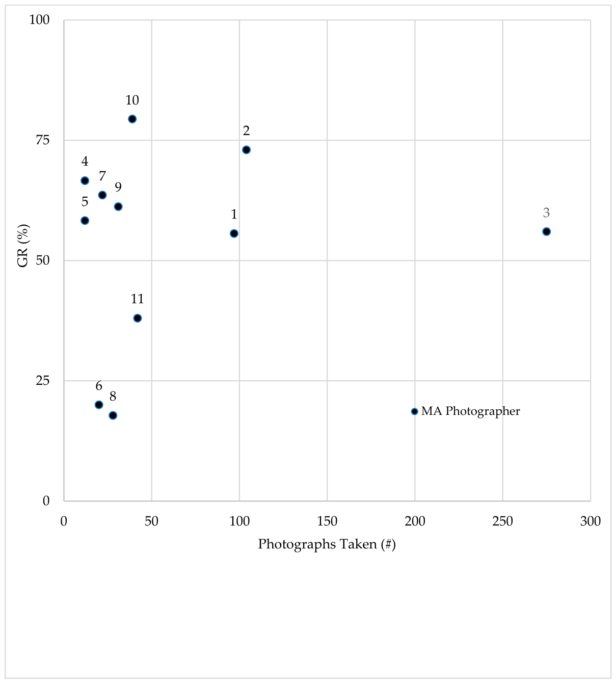
Figure 2.
Figure 2 shows average GRs of MA photographers #1-3 across successive sets of 10 screenings for >90 photographs taken. X-axis labels correspond to the upper limit of screenings performed in each interval. 95% confidence intervals are displayed with double-sided whiskers. A simple linear regression analysis shows no statistically significant association between successive number of photographs taken and average image gradability (linear regression: F statistic = 0.684, p = 0.4323).
Figure 2.
Figure 2 shows average GRs of MA photographers #1-3 across successive sets of 10 screenings for >90 photographs taken. X-axis labels correspond to the upper limit of screenings performed in each interval. 95% confidence intervals are displayed with double-sided whiskers. A simple linear regression analysis shows no statistically significant association between successive number of photographs taken and average image gradability (linear regression: F statistic = 0.684, p = 0.4323).
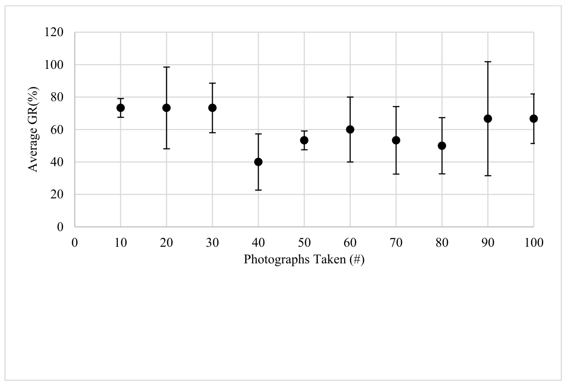

Table 1.
MA photographer photographs taken (#) and photographs gradable (# and %).
| MA | Photographs Taken (#) | Photographs Gradable (#) | Photographs Gradable (%) | |
|---|---|---|---|---|
| 1 | 97 | 54 | 55.6 | |
| 2 | 104 | 76 | 73.0 | |
| 3 | 275 | 154 | 56.0 | |
| 4 | 12 | 8 | 66.6 | |
| 5 | 12 | 7 | 58.3 | |
| 6 | 20 | 4 | 20.0 | |
| 7 | 22 | 14 | 63.6 | |
| 8 | 28 | 5 | 17.8 | |
| 9 | 31 | 19 | 61.2 | |
| 10 | 39 | 31 | 79.4 | |
| 11 | 42 | 16 | 38.0 | |
Disclaimer/Publisher’s Note: The statements, opinions and data contained in all publications are solely those of the individual author(s) and contributor(s) and not of MDPI and/or the editor(s). MDPI and/or the editor(s) disclaim responsibility for any injury to people or property resulting from any ideas, methods, instructions or products referred to in the content. |
© 2026 by the authors. Licensee MDPI, Basel, Switzerland. This article is an open access article distributed under the terms and conditions of the Creative Commons Attribution (CC BY) license (http://creativecommons.org/licenses/by/4.0/).
Copyright: This open access article is published under a Creative Commons CC BY 4.0 license, which permit the free download, distribution, and reuse, provided that the author and preprint are cited in any reuse.