

Submitted:
03 July 2026
Posted:
06 July 2026
You are already at the latest version
Abstract
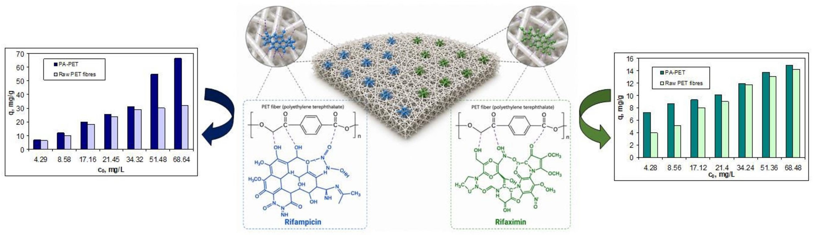
This study examines the potential of physically activated PET fibers (PA-PET) as an adsorbent for the removal of two antibiotics (rifampicin (RIF) and rifaximin (RIX)) from aqueous solution. To evaluate the performance of PA-PET, the effects of the initial antibiotic concentration and the contact time were experimentally investigated at pH 2.0 and an adsorbent dose of 0.4 g/L. If in the case of contact time there are no notable differences (equilibrium being reached after 180 min., similar to raw PET fibers), in the case of the initial concentration of antibiotics a significant increase in adsorption capacity was observed. The highest increases in adsorption capacity were obtained at RIF concentrations of 68 mg/L, where the adsorption capacity increased by more than 107%, and at RIX concentrations of 4.28 mg/L, where the increase reached approximately 84%, compared with the raw PET fibers. To further highlight the performance of PA-PET in the adsorption of RIF and RIX, the isotherm and kinetic data were modeled. Desorption of RIF and RIX from PA-PET was carried out using a medical-grade polyelectrolyte solution (SL) as the desorbing agent. Although the desorption efficiency is relatively modest (below 33% for RIF and 26% for RIX, after 360 min), the molecular structure of RIF and RIX does not change after desorption, and antibiotics can be reintroduced into the technological circuit. These results demonstrate that PA-PET is as a promising, low-cost, and efficient adsorbent for antibiotic removal, offering a practical solution for pollution reduction and more sustainable waste management.
Keywords:
antibiotics retention
; adsorption
; PA-PET
; isotherm modeling
; kinetic modeling
1. Introduction
The expansion of the medical sector and the pharmaceutical industry in recent years has made antibiotics an important new category of pollutants that affect the quality of environmental ecosystems [1,2]. The discharge of industrial effluents that do not meet quality standards, or the irrational and uncontrolled use of antibiotics, are just two of the causes that lead to the accumulation of these pollutants in the environment, thus contributing to the degradation of ecosystem quality [2,3]. This is because once they reach water sources, antibiotics, even at low concentrations, inhibit the growth of cells in biological organisms, can eliminate microbes, can seriously affect the health of fish and aquatic plants, and can have severe consequences on the health of humans and animals [4,5]. Rifampicin (RIF) and rifaximin (RIX) are two examples of broad-spectrum antibiotics that are successfully used in the treatment of mycobacterial infections both in humans and animals [6,7]. Although these two antibiotics are used in the treatment of different pathologies (RIF – respiratory system infections, RIX – gastrointestinal tract infections), both are considered drugs with minimal systemic absorption [8,9]. Therefore, wherever they are used in medical treatment, the risk that significant amounts of these antibiotics reach wastewater, and subsequently the environment is substantial, and this was one of the criteria for selecting RIF and RIX for the experimental studies.
To date, the literature reports a wide variety of methods that can be used for the removal of antibiotics from aqueous media [10,11,12]. Biological treatments, photocatalytic degradation, advanced oxidation processes, biodegradation, membrane filtration, etc. [13,14,15], are some examples of such methods that have proved their technological efficiency and applicability on an industrial scale [15,16]. However, the high consumption of chemical reagents and energy, the difficulties of long-term operational and for variable wastewater volumes, as well as the generation of secondary wastes (in some cases in large quantities and with biological risk) [17,18], have led to the continuous search for alternative methods for removing antibiotics from aqueous media that minimize these drawbacks.
Compared with the examples mentioned ab—ove, adsorption is a simple method, easily adaptable to various operating conditions, which can be efficiently managed (both in terms of material requirements and equipments) and which have proven its effectiveness in environmental remediation processes [19,20]. Moreover, if the material used as an adsorbent is a waste derived from other industrial activities, adsorption can be included in the category of ecological method that comply with the principles of the circular economy and sustainable development [20,21].
The selection of the polyethylene terephthalate fibers (PET fibers) for the experimental studies was made precisely taking into account these considerations. It is well known that obtaining PET fibers is one of the most widespread ways of valorizing PET waste, and their production continues to increase due to the enormous quantities of PET waste that need to be recycled [22,23]. Therefore, their availability is not an impediment. Moreover, obtaining PET fibers is simple and cost-effective, being carried out through a hot mechanical extrusion process (180-220 °C), which does not require additional chemical reagents or generate secondary waste [22,24]. Therefore, the PET fibers obtained in this way also exhibit high chemical and mechanical resistance, are stable over time, and do not degrade at ordinary temperatures (up to 250 °C) [25], just like the plastic material from which they are made. All these characteristics are important for the adsorption processes of antibiotics from aqueous media and represent the main arguments underlying the use of PET fibers for this purpose.
However, previous studies [26,27] have shown that PET fibers have modest efficiency in retaining RIF and RIX from aqueous solutions. The adsorption of antibiotic molecules onto PET fibers occurs predominantly through electrostatic, hydrogen bonding and π-π interactions, which cause the RIF and RIX molecules to „fold” around the fibers [26]. Consequently, the limited number of active sites on the PET fibers surface and the rigidity of the antibiotic molecules are the main reasons why the adsorption efficiency is quite modest, regardless of the experimental conditions. Therefore, the only way to increase the efficiency of the adsorption process is to increase the number of active sites, and this can be achieved through activation.
Although the activation of PET fibers can be achieved by chemical or physical processes [28,29,30], physical activation offers several advantages that should be considered when designing environmental decontamination applications [30]. Thus, unlike chemical activation, the physical activation of PET fibers is carried out easily (through hot pressing), does not involve the use of chemical reagents or exceptional experimental conditions, and can be readily scaled up with minimal investment. Moreover, the physical activation of PET fibers keeps the cost of the obtained adsorbent low, and it is much easier to use in experimental studies due to its robustness and ease of manipulation. These considerations provide both the scientific and practical justification for selecting the physical activation of PET fibers as an approach to enhance their performance in antibiotic adsorption processes.
In this study, physical activated PET fibers (PA-PET) were used for the adsorption of the antibiotics RIF and RIX from aqueous media. The adsorptive performances of PA-PET were examined in batch systems, at different values of initial antibiotics concentration (RIF and RIX) and contact time, compared to raw PET fibers. The isotherm and kinetic data were analyzed using various adsorption models, and the obtained results were used to outline the adsorption mechanism of antibiotics on PA-PET. All these aspects are important because they provide relevant information about the adsorption behaviors and practical potential of this low-cost material.
2. Materials and Methods
2.1. Materials
Rifampicin (RIF) and rifaximin (RIX) solutions with a concentration of 214 mg/L (stock solutions) were obtained by dissolving a given amount of solid antibiotic (purity > 99.9%, purchased from Sigma Aldrich) in 96% ethanol (Chemical Company, Iași, Romania), followed by vigorous stirring. Each stock solution was freshly prepared prior to use and stored in the refrigeration for maximum 2-3 days. All working solutions were prepared from these stock solutions by dilution with distilled water. For adjusting the pH of the working solutions, a 1 mol/L HNO3 solution (Chemical Company, Iași, Romania) was used.
Raw PET fibers (6.7 × 64 SD) were obtained by hot mechanical processing of PET waste and were supplied by a local company (GreenFiber International Company, Iași, Romania). The physically activated PET fibers (PA-PET) were produced from raw PET fibers, also within GreenFiber International Company, Iași, Romania. The technological process for obtaining PA-PET involves carding, followed by mechanical consolidation (needle perforation) and thermal pressing (heated-roll calenders, up to 120 °C) of PET fibers. These processes result in a cloth-like agglomerated of PET fibers that do not fall apart during use, which is why they have been called physically activated PET fibers. For the experimental studies, raw PET fibers and PA-PET were cut to acceptable sizes (approximately 1 cm for raw PET fibers and about 1 cm2 for PA-PET), washed with a HNO3 solution (0.1 mol/L), rinsed several times with distilled water, and dried at room temperature. All PET fibers samples (raw or physically activated) were stored in desiccators at constant humidity.
2.2. Characterization of PA-PET adsorbent
The structural characteristics of PA-PET were revealed by FTIR spectroscopy (Bio-Rad Spectrometer (Berlin, Germany), KBr pellet technique, resolution of 4 cm-1, spectral range between 400 and 4000 cm-1) and SEM microscopy (SEM Hitachi S3000N (Tokyo, Japan), 20 kV), compared to raw PET fibers. In addition, thermal decomposition curves of the raw PET fibers were recorded to evaluate the potential degradation of the PET fibers during the PA-PET production process.
2.3. Batch adsorption/desorption studies
All adsorption studies were carried out at pH = 2.0, an adsorbent dose of 4 g/L, and room temperature (22 ± 1 °C), previously established as the optimal conditions [26]. To evaluate the adsorption performance of PA-PET compared with the raw PET fibers, the experiments were performed in a mono-component system, at various initial antibiotic concentrations (4.29—68.69 mg/L, and constant contact time (1440 min)) and contact time (5—1440 min, and constant initial concentration of RIF and RIX (21.45 mg/L)). After completing the adsorption experiments, the solid fibers (raw PET and PA-PET) were removed from the solution (using a tweezers), and the equilibrium concentration of RIF and RIX was analyzed spectrophotometrically (Carry 60 UV-VIS spectrophotometer (Agilent, Santa Clara, CA, USA), experimental conditions: pH = 7.0 (phosphate buffer), 1 cm glass cell, the maximum wavelength at 470 nm for RIF and 444 nm for RIX, against distilled water). All experiments were repeated three times, and the average values were used for the calculations and graphical representations. The efficiency of the adsorption processes was evaluated using the adsorption capacity (q, mg/g) and the removal percent (R, %), calculated from the experimental data using equations (1) and (2).
where: c0, c are the initial and equilibrium concentration of antibiotics, (mg/L); m is the mass of adsorbent, (g); and V is the volume of solution, (L).
In the case of desorption experiments, in the first step, 1 g of PA-PET was mixed with 250 mL of antibiotic (RIF and RIX) solution (75 mg/L) (at pH 2, with a contact time of 24 h and a temperature of 22 ± 1 °C), removed from the solution and air-dried at room temperature. In the second step, 0.1 g of antibiotic-loaded PA-PET were treated with 10 mL of a medical-grade polyelectrolyte solution (SL) (hydration salt solution, purchased from Biomega Natural Nutrients S.L., Madrid, Spain), whose composition is provided in Table S1 (Supplementary Materials). The phases were separated after well-defined time intervals (ranging from 10 to 360 min), and the antibiotic (RIF and RIX) concentration in the solution was determined spectrophotometrically, as described above. Desorption efficiency (Desorption, %) was calculated using the relation:
where: cd is the concentration of antibiotic (RIF and RIX) in the solution after desorption, (mg/L), qe is the adsorption capacity at equilibrium, for each antibiotic, (mg/g ), and m is the mass of loaded PA-PET, (g).
2.4. Isotherm and kinetic modelling of the experimental data
The equilibrium experimental data were analyzed using the Langmuir (eq. 4), Freundlich (eq. 5), and Temkin (eq. 6) models. These models allow the determination of the adsorption mode (mono- or multi-layer) and provide insight into the nature of the interactions involved (through the calculation of the adsorption energy) [31,32,33]. Using these models, it is possible to evaluate how the adsorption of antibiotics onto PA-PET occurs and to determine the nature of the interactions governing these processes. These aspects are important for evaluating the efficiency of the adsorption processes.
where: q—adsorption capacity (mg/g), qmax—maximum adsorption capacity (mg/g), KL—Langmuir constant (L/g), c—equilibrium concentration of antibiotics (RIF and RIX) (mg/L), n—heterogeneity factor, KF—Freundlich constant (L/g), KT—Temkin constant (L/g), B—Temkin isotherm constant, defined by: BT = R⋅T/bT, (J/mol), bT—constant related to the heat of adsorption process, R—the universal gas constant (8.314 J mol/K), T—absolute temperature (K).
The Langmuir constant (KL, L/g) was used to calculate the Gibbs free energy (ΔG, kJ/mol), according to the relation:
where all notations have the meanings mentioned above.
The models used for the analysis of kinetic data were: the pseudo-first order model (PFO) (eq. 8), the pseudo-second order model (PSO) (eq. 9) and intra-particle diffusion model (IPD) (eq. 10) [34,35]. The use of these kinetic models allows the identification of the essential elementary steps involved in the adsorption processes, which represents the starting point for selecting an appropriate desorption agent.
where: qe, qt—the adsorption capacities at equilibrium and at time t (mg/g), k1—the rate constant of pseudo-first order model (1/min), k2—the rate constant of pseudo-second order model (g/mg min), kdiff—the intra-particle diffusion rate constant (mg/g min1/2), c—the concentration of antibiotics (RIF and RIX) in solution at equilibrium (mg/L).
The most appropriate isotherm and kinetic models for describing the experimental adsorption data were selected by evaluating the regression coefficients (R2) and chi-square (eq. (11)), obtained from the statistical assessment performed by ANOVA.
where: qexp and qcalc are the experimental and calculated values of the adsorption capacity, at equilibrium (mg/g).
3. Results and Discussion
3.1. Comparative study of antibiotic (RIF and RIX) adsorption on raw PET fibers and PA-PET
Previous results have shown that RIF and RIX can be retained on raw PET fibers, and that the experimental conditions in which adsorption occurs with maximum efficiency are: pH = 2.0, an adsorbent dose of 0.4 g/L, and ambient temperature (22 ± 1 °C). However, the removal percents do not exceed 75-85%, even at low concentrations (up to 5 mg/L) [26]. Therefore, activating this adsorbent through a physical process, already applied at industrial scale, may be a viable solution for improving the performance of these adsorption processes.
In this study, physically activated PET fibers (PA-PET) were used for the adsorption of RIF and RIX from aqueous solutions, and the experimental investigations were carried out for different initial antibiotic concentrations and various contact times. The adsorption capacities obtained for PA-PET, compared with those of raw PET fibers, for both antibiotics (RIF and RIX) are illustrated in Figure 1 and Figure 2.
The experimental results presented in Figure 1 show that increasing the initial antibiotic concentration leads to an increase in adsorption capacity for both antibiotics (RIF and RIX) and for both adsorbents. However, when using PA-PET, an improvement in the adsorption efficiency is observed across the entire initial concentration range. In the case of RIF, this improvement in adsorption efficiency becomes much more evident at high concentrations (above 50 mg/L), where the adsorption capacities are more than 80% higher for PA-PET compared with raw PET fibers, while at low concentrations (below 35 mg/L) the adsorption capacities show comparable values for both adsorbents (Figure 1a). In the case of RIX, an improvement in adsorption efficiency is observed in the low concentration range (below 10 mg/L), where the adsorption capacities are more than 75% higher for PA-PET compared with raw PET fibers (Figure 1b). At initial concentrations higher than 10 mg/L, the adsorption capacities obtained for PA-PET becomes increasingly closer to those obtained for raw PET fibers, as the initial RIX concentration increases (Figure 1b).
These encouraging results suggest that the agglomeration of PET fibers following physical activation probably generates a larger number of available active sites capable of interacting with antibiotic molecules in the aqueous solution, thereby improving adsorption efficiency. The increase in adsorption efficiency when using PA-PET is also important from an applicative point of view, because at initial antibiotic concentrations below 5 mg/L the removal percentages exceed 90% (92.15% for RIF and 90.46% for RIX), compared with 86.05% for RIF and 75.95% for RIX in the case of raw PET fibers, and the adsorption process can be considered quantitative. All these observations broaden the applicability of the adsorption processes and support the prospect of using PA-PET adsorbent on a large scale.
The influence of contact time on the adsorption of RIF and RIX onto PA-PET and raw PET fibers is illustrated in Figure 2. As can be observed, the time required to reach adsorption equilibrium does not differ significantly for the two adsorbents, for both antibiotics. In the case of both PA-PET and raw PET fibers, the minimum time needed for the adsorption processes to reach equilibrium is 180 min (Figure 2 a and b), although in the case of RIF adsorption onto PA-PET, this value can also be considered as 120 min (Figure 2a).
This insignificant variation in the minimum time required to reach equilibrium suggests that the adsorption kinetics do not change when using PA-PET compared with raw PET fibers, but only the number of available active sites increases, which leads to higher adsorption capacities for PA-PET than for raw PET fibers for both antibiotics (Figure 2a,b).
Considering these experimental results, it can be stated that PA-PET is a cost-effective adsorbent (its preparation is based on simple mechanical and thermal processes) which improves the efficiency of RIF and RIX adsorption from aqueous solutions. Consequently, PA-PET can be considered an economically and technologically advantageous adsorbent, with clear application potential. For this reason, a detailed study of the adsorption processes is required, and this will be presented in the following sections.
3.2. Characterization of PA-PET adsorbent
To determine whether the improvement in the adsorption performance of PA-PET is due to the increase in the number of active sites as a results of the ordering of PET fibers (during the PA-PET manufacturing process), or the formation of new active sites, characterization of the adsorbent is necessary.
The thermal analysis of the raw PET fibers (Figure 3a) shows that no thermal degradation processes occur up to 350 °C (DTG curve), but only some phase transformation processes, in the range 200-250 °C (DTA curve), characteristic of PET fiber softening. Therefore, the formation of new active sites as a result of thermal degradation of PET fibers during PA-PET fabrication is unlikely, since the temperature required for PA-PET manufacturing (up to 120 °C) is much lower than the temperature at which degradation occurs (above 350 °C) (Figure 3a). Moreover, the high thermal stability of PET fibers is one of their well-known advantages, consistently reported in the literature [36,37].
The absence of new functional groups as a result of the preparation of PA-PET (through physical processes) is also supported by the FTIR spectra and SEM images. As shown in Figure 3b, the FTIR spectra of the two materials are similar, and no additional bands appear in the PA-PET spectrum. The higher number of absorption maxima in the PA-PET spectrum compared with the raw PET fibers is mainly due to splitting of the absorption bands, but also to experimental difficulties encountered in obtaining a sufficiently transparent pellet for experimental recording.
The SEM images confirm the smooth morphology of the PET fibers surface after physical activation. As shown in Figure 3c, regardless of the magnification, the PET fibers exhibit a uniform surface without defects (breaks or cracks), which rules out the occurrence of degradation processes during the preparation of PA-PET.
However, it should be noted that in PA-PET the fibers are ordered (first image—Figure 3c), and the network formed could be responsible for the easier retention of antibiotic molecules and, therefore, for the increase in experimental adsorption capacities. To demonstrate this, the adsorption processes of RIF and RIX on PA-PET must be quantitatively analyzed, which can be achieved by modeling the experimental data.
3.3. Modeling of equilibrium and kinetic data for RIF and RIX adsorption on PA-PET
The equilibrium data were quantitatively evaluated using Langmuir, Freundlich, and Temkin models. These models allow the determination of how antibiotic molecules interact with the PA-PET surface and the nature of the interactions occurring during the adsorption process [21,32], under the given experimental conditions. The linear representations of these models for the adsorption of RIF and RIX onto PA-PET are illustrated in Figure 4, and the characteristic parameters of these models are summarized in Table 1.
The data presented in Table 1 show that the Langmuir model provides the best description of RIF and RIX adsorption onto PA-PET, since for this model the highest regression coefficients (R2) and the lowest values of the chi-square are obtained. However, these values of statistical parameter do not differ significantly from those obtained for the Freundlich model. This allows us to say that the retention of RIF and RIX occurs on the PA-PET surface up to the formation of a monolayer (according to the Langmuir model). But, once the concentration of RIF and RIX on the adsorbent surface becomes sufficiently high, the antibiotic molecules may interact with each other (due to their large molecular volume and polar structure), which is similar to a Freundlich-type behavior of the adsorption processes.
This hypothesis is also supported by the values of the parameter n (from the Freundlich model), which are greater than unity (Table 1) and indicate that the adsorption process is favorable, both in the case of RIF and RIX. However, the values of this parameter differ significantly for the two antibiotics. This indicates that, although both adsorption processes are favorable, the retention of RIX is efficient only at low concentrations and decreases rapidly as the initial concentration increases (n = 3.31). In contrast, the adsorption efficiency of RIF decreases more gradually with increasing concentration (n = 1.35). This behavior is consistent with the experimental data presented in Figure 1, which clearly highlight these particularities of RIF and RIX adsorption on PA-PET.
The Langmuir-type behavior of RIF and RIX adsorption onto PA-PET is also suggested by the values of the maximum adsorption capacities (qmax, mg/g). The similar values of the maximum adsorption capacities (Table 1) and those obtained experimentally (66.46 mg/g for PIF and 14.88 mg/g for RIX) clearly indicate that the adsorption of RIF and RIX onto PA-PET occurs until the adsorbent surface is occupied, at which point the adsorption processes reach equilibrium. Moreover, the negative ΔG values (Table 1) indicate that the adsorption of RIF and RIX onto PA-PET is spontaneous, while the low adsorption energy values (bt, kJ/mol) (Table 1) suggest that the retention of the antibiotic molecules on the adsorbent surface involves relatively weak physicochemical interactions.
Much more important from an application point of view are the maximum adsorption capacities (qmax, mg/g) obtained when using PA-PET as an adsorbent. As shown in Table 1, the maximum adsorption capacity obtained in the case of RIF is more than 4 times higher than that obtained in the case of RIX, indicating that RIF has a much higher availability to bind to the PA-PET surface, compared to RIX. This higher adsorption efficiency of RIF was also observed in the case of using PET fibers as an adsorbent, and is mainly attributed to the greater ability (flexibility) of RIF molecules to interact with functional groups [26]. However, compared to PET fibers, the maximum adsorption capacities obtained for PA-PET are more than 60% higher in the case of RIF (from 44.84 mg/g for PET fibers to 71.94 mg/g for PA-PET) and more than 8% higher in the case of RIX (from 15.63 mg/g for PET fibers to 16.95 mg/g for PA-PET). These increases in the maximum adsorption capacities indicate that the formation of an ordered network of PET fibers during PA-PET preparation (Figure 3c) significantly improves the geometric (spatial) arrangement of the active sites on the fiber surface, thereby allowing them to interact much more efficiently with the antibiotic molecules. If the structure of the antibiotic molecule allows these interactions to occur, the adsorption process becomes efficient within a specific range of initial concentrations (up to 10 mg/L for RIF and below 5 mg/L for RIX) (Figure 1).
To provide a comprehensive picture of how the adsorption of RIF and RIX on PA-PET occurs, and to identify the elementary steps that control the rate of the adsorption process, kinetic modeling of the experimental data is required. The linear representations of the pseudo-first order, pseudo-second order, and intra-particle diffusion kinetic models are illustrated in Figure 5, while the parameters of these models are presented in Table 2.
The pseudo-first order kinetic model shows limited ability to describe the experimental data obtained for the adsorption of RIF and RIX onto PA-PET (Figure 5a). The calculated values of the adsorption capacities differ significantly from the experimental values (high chi-square values) for both antibiotics (Table 2). This indicates that for the retention of RIF and RIX on the PA-PET surface, the interaction of antibiotic molecules with a single active site is not sufficient. However, the regression coefficients higher than 0.89 (Table 2) allow us to state that this type of interaction most likely represents one of the elementary steps of the adsorption mechanism.
In contrast, the pseudo-second order kinetic model (Figure 5b) provides a much better description of the experimental data. The high regression coefficients (R2 > 0.999), the low chi-square values, and the close values of the calculated and experimental adsorption capacities (Table 2) clearly indicate that the adsorption of RIF and RIX on PA-PET follows pseudo-second order kinetics. Therefore, the retention of RIF and RIX on the PA-PET surface requires two active sites, which interact successively with the antibiotic molecules in the aqueous solution. In the first stage, the interactions between the antibiotic molecules and the functional groups of PA-PET are most likely of electrostatic type (as the adsorption efficiency depends on pH [26]), followed by a second stage in which physical interactions (π-π interactions, hydrogen bonding, etc.) contribute to the fixation of the antibiotic molecules onto the PA-PET surface. The achievement of these interactions does not depend on the nature of the antibiotic molecule (RIF or RIX), as the rate constants (k2, g/mg min) have almost identical values (Table 2).
However, it should be noted that the values of the rate constant k2 obtained for the adsorption of RIF and RIX on PA-PET are higher than those obtained for the raw PET fibers. Thus, for RIF, the increase in the rate constant is more than 2 times (from 0.0019 g/mg min for raw PET fibers [26] to 0.0046 g/mg min for PA-PET (Table 2)), while for RIX this increase is more than 1.5 times (from 0.0031 g/mg min for raw PET fibers [26] to 0.0047 g/mg min for PA-PET (Table 2)). These differences in the rate constants indicate that, regardless of the nature of the antibiotic (RIF or RIX), the agglomeration of PET fibers in PA-PET facilitates the interaction of antibiotic molecules with the adsorbent. This is most likely due to the much more appropriate geometric positioning of the active sites which, as a result of mechanical pressing, are located at appropriate distances and no longer force the RIF and RIX molecules to fold in order to stabilize on the adsorbent surface (as occurs in the case of raw PET fibers).
The elementary diffusion steps also contribute to the adsorption of RIF and RIX on PA-PET, but they are not the rate-controlling steps. As shown in Figure 5c, none of the linear representations pass through the origin, and the two distinguishable regions correspond to: (1) the diffusion of antibiotic molecules from the bulk solution to the external surface of the adsorbent (external diffusion), and (2) the diffusion of antibiotic molecules into the liquid film around the PET fibers that form the PA-PET (internal diffusion). It can also be observed from Figure 5c and Table 2 that, while the first diffusion step is a rapid one that depends on the nature of the antibiotic, the second diffusion step is much slower (by at least one order of magnitude), and the nature of the antibiotic influences this process much less. This allows us to say that when the adsorbent (PA-PET) is introduced into the aqueous solution containing RIF and RIX, the antibiotic molecules spontaneously migrate toward the PA-PET surface (first diffusion step: kdiff,1—high; c1—low (Table 2)), and one there, they interact with the most geometrically favorable functional groups. These interactions occur gradually, which makes the rate of the second diffusion step much lower, while the concentration of RIF and RIX in the film surrounding the adsorbent remains high (second diffusion step: kdiff,2—low; c2—high (Table 2)).
All these particularities allow the design of an adsorption mechanism and highlight the practical utility of the adsorption processes of RIF and RIX onto PA-PET, aspects that will be discussed in the following sections.
3.4. Insights of the adsorption mechanism
The experimental results presented in the previous sections have shown that the adsorption of RIF and RIX onto PA-PET exhibits the following distinctive characteristics:
(i) The adsorption takes place in a strongly acidic media (pH = 2.0, previously established as optimal [26]), which ensures the protonation of the functional groups of polyethylene terephthalate (pKa = 3.54 [38]) and the first dissociation step of the antibiotic molecules (RIF: pKa1 = 1.80; RIX: pKa1 = 2.08 [39,40]), suggesting the presence of electrostatic interactions during the adsorption processes.
(ii) The adsorption of RIF and RIX onto PA-PET is best described by the Langmuir model (Table 1), which means that the antibiotic molecules are retained on the adsorbent surface up to the formation of a monolayer, after which the adsorption processes reach equilibrium. The fairly good agreement of the experimental data with the Freundlich model suggests that RIF and RIX interact with functional groups located in different geometric planes on the PA-PET surface (originating from different PET fibers), and these interactions, according to the parameters of the Temkin model [41], are predominantly physicochemical in nature.
(iii) The agreement between the experimental data and the pseudo-second order kinetic model (Table 2) indicates that, during the adsorption processes, RIF and RIX molecules must interact with two active sites on the PA-PET surface. Therefore, even if only one of these interactions is electrostatic (at pH = 2.0, RIF and RIX are ionized only in their first dissociation step), the second interaction may be physical in nature (such as hydrogen bonding, π-π interactions, etc.), and it plays a key role in stabilizing the antibiotic molecules on the PA-PET surface [42].
(iv) The adsorption of RIF and RIX onto PA-PET through physicochemical interactions (electrostatic forces, hydrogen bonding, π-π interactions, etc.) is also supported by the FTIR spectra recorded for PA-PET before and after RIF and RIX adsorption (data not shown). The shifts of the absorption maxima of the most relevant bands by up to 10 cm−1 (Table 3) indicate that the functional groups of PA-PET do not participate in the formation of new chemical bonds (no additional bands appear), but only the chemical environment surrounding these groups is changed.
(v) The elementary diffusion processes only serve to transport the RIF and RIX molecules into the proximity of the functional groups of PA-PET. Consequently, diffusion from the bulk solution to the adsorbent surface occurs rapidly, whereas diffusion within the liquid film surrounding the fibers that make up PA-PET is a much slower process (Table 2). These differences allow us to say that RIF and RIX molecules easily reach the PA-PET surface, where they accumulate until find the available active sites.
Based on these considerations, a schematic illustration of the adsorption mechanism of RIF and RIX onto PA-PET is presented in Figure 6.
3.5. Desorption studies
In the case of RIF and RIX adsorption onto PA-PET, the main objective of desorption studies is the recovery of the antibiotic molecules. This is because PA-PET is an inexpensive, readily available, and easy-to-prepare adsorbent, and therefore its regeneration is less important, from a practical point of view. Therefore, when designing desorption process, it is essential to consider conditions that do not alter the structure of the antibiotic molecules.
The experimental results presented in the previous sections have shown that the adsorption of RIF and RIX onto PA-PET occurs in a strongly acidic medium (pH = 2.0) through physicochemical interactions (electrostatic, hydrogen bonding and π-π interactions). Under these conditions, desorption of the antibiotic molecules can be most easily achieved by increasing the pH of the aqueous solution [43,44]. Increasing the pH leads to the ionization of the antibiotic molecules to superior dissociation steps (more negative charges), as well as to the deprotonation of the functional groups of PA-PET. This generates electrostatic repulsions that release RIF and RIX molecules into the aqueous solution.
Unfortunately, bases (such as NaOH, KOH, etc.) cannot be used to increase the pH of the aqueous solution. This is because their alkaline nature can lead to degradation of the structure of antibiotic molecules [45] and loss of biological activity. Much more suitable for this purpose is the use of salt solutions, whose weakly acidic/ basic pH is sufficient to create the electrostatic repulsions required for desorption.
In this study, a medical-grade polyelectrolyte solution (SL) was used for desorption of RIF and RIX from PA-PET, which due to its high concentration of Na(I) and K(I) ions and its weakly acidic pH (6.79) (see Table S1, Supplementary Materials), meets the required conditions for desorbing agents.
The experimental results obtained for desorption of RIF and RIX from PA-PET upon treatment with 10 mL of the polyelectrolyte solution, at different contact times (10-360 min), are presented in Figure 7.
It can be observed from Figure 7 that, as the contact time increases, the amount of antibiotic desorbed from the PA-PET surface also increases, for both RIF and RIX. The release of antibiotic molecules into the polyelectrolyte solution (used as the desorption agent) is more efficient during the initial stages, when in the first 10 minutes 28.10% of RIF and 18.54% of RIX are desorbed. After this, desorption rate decreases, and the processes reach equilibrium after 60 minutes in the case of RIF and 180 minutes in the case of RIX. However, it should be noted that at the end of the 360 min., desorption percent of the two antibiotics remains relatively low, not exceeding 33% for RIF and 26% for RIX.
This variation in the desorption efficiency of RIF and RIX from the PA-PET surface is a consequence of the specific characteristics of the adsorption processes, mentioned previously. In the initial stage, the rapid release of both RIF and RIX is governed by desorption of molecules located near the outer surface of the adsorbent. When the desorption agent comes into contact with the loaded adsorbent, electrostatic repulsions are generated, leading to the release of antibiotic molecules into the solution. As the contact time increases, the driving force of desorption process decreases, on the one hand due to the significant reduction of antibiotic molecules on the external surface of PA-PET (which have already been desorbed), and on the other hand due to the accumulation of desorbed RIF and RIX molecules in the solution. RIF and RIX molecules that are trapped between the PET fibers of the adsorbent, even though they are released through desorption, reach the bulk solution more slowly due to electrostatic repulsions. This explains the modest values of the efficiency of desorption process. Under these conditions, a possible approach to increase the efficiency of desorption process is to increase the volume of the desorption agent, or to use several successive desorption steps. These alternatives require a more detailed discussion, and therefore they will be presented in a subsequent study.
Even though the desorption efficiency of RIF and RIX from PA-PET is relatively modest, it is important to determine whether any structural modifications occur in the antibiotic molecules after their release into the solution. This is because, as previously mentioned, the recovery of the two antibiotics is much more important from an application point of view than the regeneration of an inexpensive and readily available adsorbent (such as PA-PET).
To highlight any structural changes of the RIF and RIX molecules after desorption, the visible spectrum (400–600 nm) of the solutions obtained after desorption was recorded and compared with that of the standard solutions (Figure 8). Both in the standard solution and in the solution obtained after desorption, the antibiotic concentrations were adjusted to 25.0 mg/L for RIF and 12.7 mg/L for RIX.
As can be seen in Figure 8, in the VIS spectra recorded for the standard solutions and for those obtained after desorption (and concentration adjustment) there are no major differences for both antibiotics. The overlap of the spectra, both in terms of absorption maximum and spectral band width (Table 4), indicates that after desorption, the RIF and RIX molecules are not involved in secondary processes that could lead to modifications of the chromophore structure. The only notable difference is the maximum absorbance, which in the case of the standard solutions is higher than in the solutions obtained after desorption (Figure 8), for both antibiotics. Nevertheless, these differences are not significant (0.039 a.u. for RIF and 0.014 a.u. for RIX (Table 4)) and can be attributed to experimental errors associated with adjusting the antibiotic concentrations in the solutions obtained after desorption.
The chromophore responsible for the presence of these absorption bands in the VIS spectra is the strongly conjugated naphthoquinone system, which influences the biological activity of RIF and RIX, contributing to the way these molecules interact with bacterial enzymes [46,47].
All these experimental results show that after desorption from PA-PET, the structure of RIF and RIX molecules does not change (they retain their biological activity) and can be reintroduced into the technological circuit. This is particularly important from a practical point of view, as it highlights the usefulness of the adsorption process of RIF and RIX onto PA-PET in the context of the circular economy. However, finding environmentally friendly solutions that could improve the adsorption efficiency of the two antibiotics onto PET-derived adsorbents remains a challenge, which will be addressed in future studies.
5. Conclusions
In this study, physically activated PET fibers (PA-PET) were used as adsorbents for the retention of rifampicin (RIF) and rifaximin (RIX) from aqueous solutions. The performance of PA-PET was evaluated at different initial antibiotic concentrations and various contact times, under the previously established optimal conditions (pH = 2.0, adsorbent dose = 0.4 g/L). If, in the case of contact time, no notable differences are observed, the variation of the initial concentration of RIF and RIX leads to a significant increase in the adsorption capacity of PA-PET compared with the raw PET fibers. This increase in the adsorption capacity of PA-PET is notable for high RIF concentrations (above 50 mg/L) and for low RIX concentrations (up to 10 mg/L). The use of PA-PET is also important from an applicative point of view, because at initial antibiotic concentrations below 5 mg/L the removal percentages exceed 90% (92.15% for RIF and 90.46% for RIX), and the adsorption process can be considered quantitative. This improvement in the adsorption performance of PA-PET is most likely due to the formation of the PET fiber network, which allows a much easier retention of the RIF and RIX molecules. The modeling of the experimental isotherms and kinetic curves showed that the adsorption of RIF and RIX onto PA-PET is best described by the Langmuir model and by the pseudo-second order kinetic model. Therefore, the adsorption of RIF and RIX onto PA-PET occurs until a monolayer is formed (qmax = 71.94 mg/g for RIF and 16.95 mg/g for RIX), and the retention of the antibiotic molecules on the PA-PET surface involves physicochemical interactions (at least one of which is electrostatic). For desorption of RIF and RIX from PA-PET, a medical polyelectrolyte solution was used as the desorbing agent. Although the desorption efficiency of RIF and RIX from PA-PET is relatively modest (after 360 minutes desorption percent remains below 33% for RIF and 26% for RIX), much more important is the fact that the molecular structure of RIF and RIX does not change, and the desorbed antibiotics can be reintroduced into the technological circuit. This is particularly important from a practical point of view, as it highlight the usefulness of the adsorption process of RIF and RIX onto PA-PET in the context of the circular economy. All the experimental results indicate that PA-PET is as a promising, low-cost, and efficient adsorbent for RIF and RIX removal, offering a practical solution for pollution control and more sustainable waste management.
Supplementary Materials
The following supporting information can be downloaded at: https://www.mdpi.com/article/doi/s1, Figure S1: Physically activated PET fibers (PA-PET); Table S1: Chemical composition of the medical-grade electrolyte solution (SL) used as the desorbing agent.
Author Contributions
Conceptualization, E.F.P. and L.B.; methodology, E.F.P. and L.B.; validation, E.F.P. and L.B.; formal analysis, E.F.P. and L.B.; investigation, E.F.P. and L.B.; resources, E.F.P. and L.B.; writing—original draft preparation, L.B.; writing—review and editing, L.B.; visualization, E.F.P.; supervision, L.B. All authors have read and agreed to the published version of the manuscript.
Funding
This research received no external funding.
Data Availability Statement
The original contributions presented in the study are included in the article; further inquiries can be directed to the corresponding author.
Conflicts of Interest
The authors declare no conflicts of interest.
References
- Palacio, D.A.; Aranda, F.L.; Rivas, B.L. Removal of antibiotic emerging pollutants: an overview. J. Chil. Chem. Soc. 2022, 67(3), 5547–5561. [Google Scholar] [CrossRef]
- Ma, J.; Cui, Y.; Li, A.; Zou, X.; Ma, C.; Chen, Z. Antibiotics and antibiotic resistance genes from wastewater treated in constructed wetlands. Ecol. Eng. 2022, 177, 106548. [Google Scholar] [CrossRef]
- Wu, D.; Bai, H.; He, L.Y.; He, L.X.; Gao, F.Z.; Liu, C.X.; Van den Brink, P.J.; Smith, H.; Ying, G.G. From river to groundwater: Antibiotics pollution, resistance prevalence, and source tracking. Environ. Int. 2025, 196, 109305. [Google Scholar] [CrossRef] [PubMed]
- Aydin, S.; Aydin, M.E.; Ulvi, A.; Kilic, H. Antibiotics in hospital effluents: Occurrence, contribution to urban wastewater, removal in a wastewater treatment plant, and environmental risk assessment. Environ. Sci. Pollut. Res. 2018, 26, 544–558. [Google Scholar] [CrossRef]
- Samrot, A.V.; Wilson, S.; Preeth, R.S.S.; Prakash, P.; Sathiyasree, M.; Saigeetha, S.; Shobana, N.; Pachiyappan, S.; Rajesh, V.V. Sources of Antibiotic Contamination in Wastewater and Approaches to Their Removal-An Overview. Sustainability 2023, 15, 12639. [Google Scholar] [CrossRef]
- Hardie, K.R.; Fenn, S.J. JMM profile: rifampicin: a broad-spectrum antibiotic. J. Med. Microbiol. 2022, 71, 001566. [Google Scholar] [CrossRef]
- Piccin, A.; Gulotta, M.; di Bella, S.; Martingano, P.; Crocè, L.S.; Giuffrè, M. Diverticular Disease and Rifaximin: An Evidence-Based Review. Antibiotics 2023, 12, 443. [Google Scholar] [CrossRef] [PubMed]
- Khadka, P.; Dummer, J.; Hill, P.C.; Katare, R.; Das, S.C. A review of formulations and preclinical studies of inhaled rifampicin for its clinical translation. Drug Deliv. Transl. Res. 2023, 13, 1246–1271. [Google Scholar] [PubMed]
- Tsakiridou, G.; Papanastasiou, A.M.; Efentakis, P.; Angelerou, M.F.G.; Kalantzi, L. Challenges in the Investigation of Therapeutic Equivalence of Locally Applied/Locally Acting Drugs in the Gastrointestinal Tract: The Rifaximin Case. Pharmaceutics 2025, 17, 839. [Google Scholar] [CrossRef] [PubMed]
- Azanu, D.; Adu-Poku, D.; Saah, S.A.; Appaw, W.O. Prevalence of pharmaceuticals in surface water samples in Ghana. J. Chem. 2021, 2021, 1–11. [Google Scholar] [CrossRef]
- Bai, X.; Chen, W.; Wang, B.; Sun, T.; Wu, B.; Wang, Y. Photocatalytic degradation of some typical antibiotics: Recent advances and future outlooks. Int. J. Mol. Sci. 2022, 23, 8130. [Google Scholar] [CrossRef] [PubMed]
- Wang, N.; Peng, L.; Gu, Y.; Liang, C.; Pott, R.W.M.; Xu, Y. Insights into biodegradation of antibiotics during the biofilm-based wastewater treatment processes. J. Clean. Prod. 2023, 393, 136321. [Google Scholar] [CrossRef]
- Cuerda-Correa, E.M.; Alexandre-Franco, M.F.; Fernández-González, C. Advanced oxidation processes for the removal of antibiotics from water. An overview. Water 2020, 12, 102. [Google Scholar] [CrossRef]
- Wang, X.; Jing, J.; Zhous, M.; Dewil, R. Recent advances in H2O2-based advanced oxidation processes for removal of antibiotics from wastewater. Chin. Chem. Lett. 2023, 34(3), 107621. [Google Scholar] [CrossRef]
- Wang, N.; Peng, L.; Gu, Y.; Liang, C.; Pott, R.W.M.; Xu, Y. Insights into biodegradation of antibiotics during the biofilm-based wastewater treatment processes. J. Clean. Prod. 2023, 393, 136321. [Google Scholar] [CrossRef]
- Mangla, D.; Annu; Sharma, A.; Ikram, S. Critical review on adsorptive removal of antibiotics: Present situation, challenges and future perspective. J. Hazard. Mater. 2022, 425, 127946. [Google Scholar] [CrossRef] [PubMed]
- Maged, A.; Dissanayake, P.D.; Yang, X.; Pathirannahalage, C.; Bhatnagar, A.; Ok, Y.S. New mechanistic insight into rapid adsorption of pharmaceuticals from water utilizing activated biochar. Environ. Res. 2021, 202, 111693. [Google Scholar] [CrossRef] [PubMed]
- Du., C.; Zhang, Z.; Yu, G.; Wu, H.; Chen, H.; Zhou, L.; Zhang, Y.; Su, Y.; Tan, S.; Yang, L.; Song, J.; Wang, S. A Review of Metal Organic Framework (MOFs)-Based Materials for Antibiotics Removal via Adsorption and Photocatalysis. Chemosphere 2021, 272, 129501. [Google Scholar] [CrossRef] [PubMed]
- Ahmed, Y.; Rahman, M.M.; Alam, M.S.; Miah, M.I.; Choudhury, M.S.H.; Alharbi, O.A.; Akhtar, P.; Rahman, S.M. Harnessing neural network model with optimization for enhanced ciprofloxacin antibiotic adsorption from contaminated water: A transparent and objective framework. J. Water. Proc. Eng. 2024, 65, 105724. [Google Scholar] [CrossRef]
- Khazaie, A.; Kia, H.; Moniri, E.; Hassani, A.H.; Miralinaghi, M. Adsorption modeling of tetracycline removal by multi-walled carbon nanotube functionalized with aspartic acid and poly-pyrrole using Bayesian optimized artificial neural network. J. Taiwan Inst. Chem. Eng. 2023, 144, 104743. [Google Scholar] [CrossRef]
- Rangabhashiyam, S.; Anu, N.; Nandagopal Giri, M.S.; Selvaraju, N. Relevance of isotherm models in biosorption of pollutants by agricultural by-products. J. Environ. Chem. Eng. 2014, 2(1), 398–414. [Google Scholar] [CrossRef]
- Ernazarova, S.; Juraev, A.; Ernazarov, S.; Alimuhamedov, M. Obtaining polyester fiber based on various secondary polyethylene terephthalate raw materials. AIP Conf. Proc. 2025, 3304, 040074. [Google Scholar] [CrossRef]
- Gnoffo, C.; Arrigo, R.; Frache, A. An Upcycling Strategy for Polyethylene Terephthalate Fibers: All-Polymer Composites with Enhanced Mechanical Properties. J. Compos. Sci. 2024, 8, 527. [Google Scholar] [CrossRef]
- Singh, A.K.; Bedi, R.; Kaith, B.S. Composite materials based on recycled polyethylene terephthalate and their properties-A comprehensive review. Compos. Part B Eng. 2021, 219, 108928. [Google Scholar] [CrossRef]
- Slezák, E.; Ronkay, F.; Bocz, K. Development of an Engineering Material with Increased Impact Strength and Heat Resistance from Recycled PET. J. Polym. Environ. 2023, 31, 5296–5308. [Google Scholar] [CrossRef]
- Fasniuc-Pereu, E.; Drăgoi, E.N.; Bulgariu, D.; Popescu, M.C.; Bulgariu, L. Removal of Rifampicin and Rifaximin Antibiotics on PET Fibers: Optimization, Modeling, and Mechanism Insight. Polymers 2025, 17, 2089. [Google Scholar] [CrossRef] [PubMed]
- Fasniuc-Pereu, E.; Bulgariu, L. Retention of Rifaximin from Aqueous Solution by Adsorption on PET Fibers. Proceedings of 2024 E-Health and Bioengineering Conference (EHB), 2024. [Google Scholar] [CrossRef]
- Yu, J.; Bai, L.; Feng, Z.; Chen, L.; Xu, S.; Wang, Y. Waste treats waste: Facile fabrication of porous adsorbents from recycled PET and sodium alginate for efficient dye removal. Chemosphere 2024, 355, 141738. [Google Scholar] [CrossRef] [PubMed]
- Lu, C.; Hao, Z.; Chu, H.; Lu. Investigation on performance of engineered cementitious composites (ECC) based on surface modification of PET fibers using graphene oxide (GO) and polydopamine (PDA). Constr. Build. Mater. 2023, 368, 130343. [Google Scholar] [CrossRef]
- Ahmed, F.M.; El-Shahat, E.S.; Othman, H.A.; Mohamed, A.L. Chemical and Physical Modification Strategies of Synthetic Fibers to Enhance Dye Uptake and Color Performance. Egypt. J. Chem. 2026, 69, 341–365. [Google Scholar] [CrossRef]
- Lima, E.C.; Gomes, A.A.; Tran, H.N. Comparison of the nonlinear and linear forms of the van’t Hoff equation for calculation of adsorption thermodynamic parameters (ΔS° and ΔH°). J. Molec. Liq. 2020, 311, 113315. [Google Scholar] [CrossRef]
- Kern, M.; Skulj, S.; Rozman, M. Adsorption of a wide variety of antibiotics on graphene-based nanomaterials: A modelling study. Chemosphere 2022, 296, 134010. [Google Scholar] [CrossRef] [PubMed]
- Bıyıkoğlu, M. Innovative approaches in wastewater treatment: kinetic and isotherm investigation of dye adsorption on sulfur-modifed PET fbers. Res. Chem. Intermed. 2025, 51, 3281–3299. [Google Scholar] [CrossRef]
- Tan, K.L.; Hameed, B.H. Insight into the adsorption kinetics models for the removal of contaminants from aqueous solutions. J. Taiwan Inst. Chem. Eng. 2017, 74, 25–48. [Google Scholar] [CrossRef]
- Wang, J.; Guo, X. Rethinking of the intraparticle diffusion adsorption kinetics model: Interpretation, solving methods and applications. Chemosphere 2022, 309, 136732. [Google Scholar] [CrossRef] [PubMed]
- Thiounn, T.; Smith, R.C. Advances and approaches for chemical recycling of plastic waste. J. Polym. Sci. 2020, 58, 1347–1364. [Google Scholar] [CrossRef]
- Wu, W.J.; Sun, X.L.; Chen, Q.; Qian, Q. Recycled Poly(Ethylene Terephthalate) from Waste Textiles with Improved Thermal and Rheological Properties by Chain Extension. Polymers 2022, 14, 510. [Google Scholar] [CrossRef] [PubMed]
- Damayanti; Wu, H.S. Strategic Possibility Routes of Recycled PET. Polymers 2021, 13, 1475. [Google Scholar] [CrossRef] [PubMed]
- Erdem, H. The application of different technologies for removal of rifampicin from aquatic environments. A recent review. Dicle Univ. J. Eng. 2023, 14(1), 145–163. [Google Scholar] [CrossRef]
- Kogawa, A.C.; Salgado, H. Status of Rifaximin: A review of characteristics, uses and analytical methods. Crit. Rev. Anal. Chem. 2018, 48(6), 1–8. [Google Scholar] [CrossRef]
- Ali, I.; ALOthman, Z.A.; Mbianda, X.Y.; Basheer, A.A. Preparation and characterization of nanoporous carbon for removal of amoxicillin antibiotic from water: Modelling, kinetics and thermodynamic studies. Inorg. Chem. Comm. 2023, 155, 111006. [Google Scholar] [CrossRef]
- Bouaziz, N.; Kouira, O.; Aouaini, F.; Bukhari, L.; Knani, S.; Znaidia, S.; Lamine, A.B. Adsorption of antibiotics by bentonite-chitosan composite: Phenomenological modeling and physical investigation of the adsorption process. Int. J. Biol. Macromolec. 2023, 242(4), 125156. [Google Scholar] [CrossRef]
- Sadabad, H.R.; Coleman, H.M.; Dooley, J.S.G.; Snelling, W.J.; O’Hagan, B.; Ganin, A.Y.; Arnscheidt, J. Desorption of antibiotics from granular activated carbon during water treatment by adsorption. Environ. Proc. 2024, 11(4), 64. [Google Scholar] [CrossRef]
- Zhang, C.; Li, R.; Ke, D.; Suo, H.; Wang, S.; Ma, E.; Chen, Y.; Liu, C. Intraparticle sorption and desorption of antibiotics. J. Hazard. Mater. 2024, 465, 133311. [Google Scholar] [CrossRef] [PubMed]
- Bai, X.; Chen, W.; Wang, B.; Sun, T.; Wu, B.; Wang, Y. Photocatalytic Degradation of Some Typical Antibiotics: Recent Advances and Future Outlooks. Int. J. Mol. Sci. 2022, 23, 8130. [Google Scholar] [CrossRef] [PubMed]
- Skrzypczak, N.; Przybylsk, P. Modifications, biological origin and antibacterial activity of naphthalenoid ansamycins. Nat. Prod. Rep. 2022, 39, 1653. [Google Scholar] [CrossRef] [PubMed]
- Stephanie, F.; Tambunan, U.S.F.; Siahaan, T.J. M. tuberculosis Transcription Machinery: A Review on the Mycobacterial RNA Polymerase and Drug Discovery Efforts. Life 2022, 12, 1774. [Google Scholar] [CrossRef] [PubMed]
Figure 1.
Variation of the adsorption capacity (q, mg/g) as a function of the initial concentration of RIF (a) and RIX (b). (Experimental conditions: pH = 2.0; adsorbent dose = 0.4 g/L, contact time = 1440 min, temperature = 22 ± 1 °C).
Figure 1.
Variation of the adsorption capacity (q, mg/g) as a function of the initial concentration of RIF (a) and RIX (b). (Experimental conditions: pH = 2.0; adsorbent dose = 0.4 g/L, contact time = 1440 min, temperature = 22 ± 1 °C).
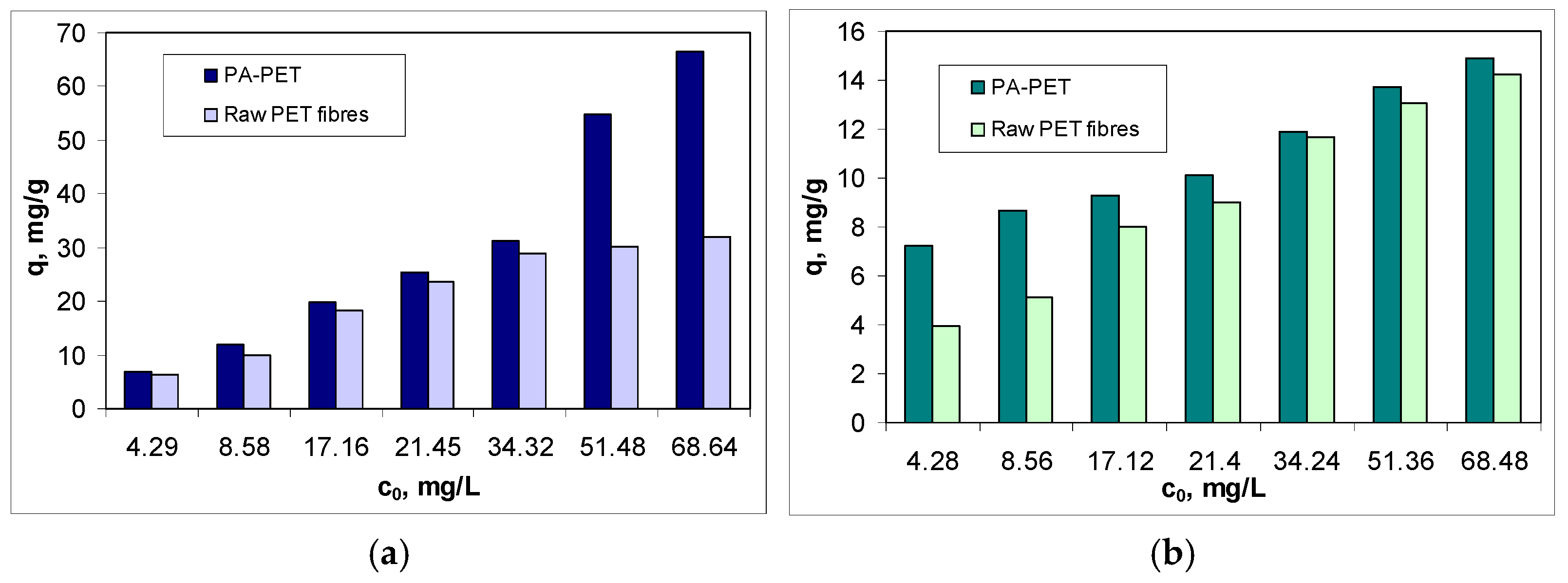
Figure 2.
Variation of the adsorption capacity (q, mg/g) as a function of contact time for RIF (a) and RIX (b). (Experimental conditions: pH = 2.0; adsorbent dose = 0.4 g/L, c0 = 21.45 mg RIF/L and 21.2 mg RIX/L, temperature = 22 ± 1 °C).
Figure 2.
Variation of the adsorption capacity (q, mg/g) as a function of contact time for RIF (a) and RIX (b). (Experimental conditions: pH = 2.0; adsorbent dose = 0.4 g/L, c0 = 21.45 mg RIF/L and 21.2 mg RIX/L, temperature = 22 ± 1 °C).
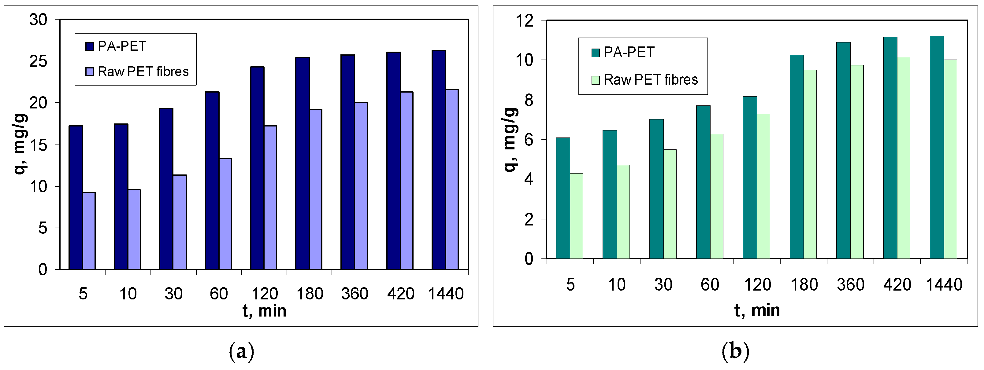
Figure 3.
(a) Thermal degradation curves of raw PET fibers. (b) FTIR spectra of PA-PET and raw PET fibers. (c) SEM images of PA-PET at different magnification orders.
Figure 3.
(a) Thermal degradation curves of raw PET fibers. (b) FTIR spectra of PA-PET and raw PET fibers. (c) SEM images of PA-PET at different magnification orders.
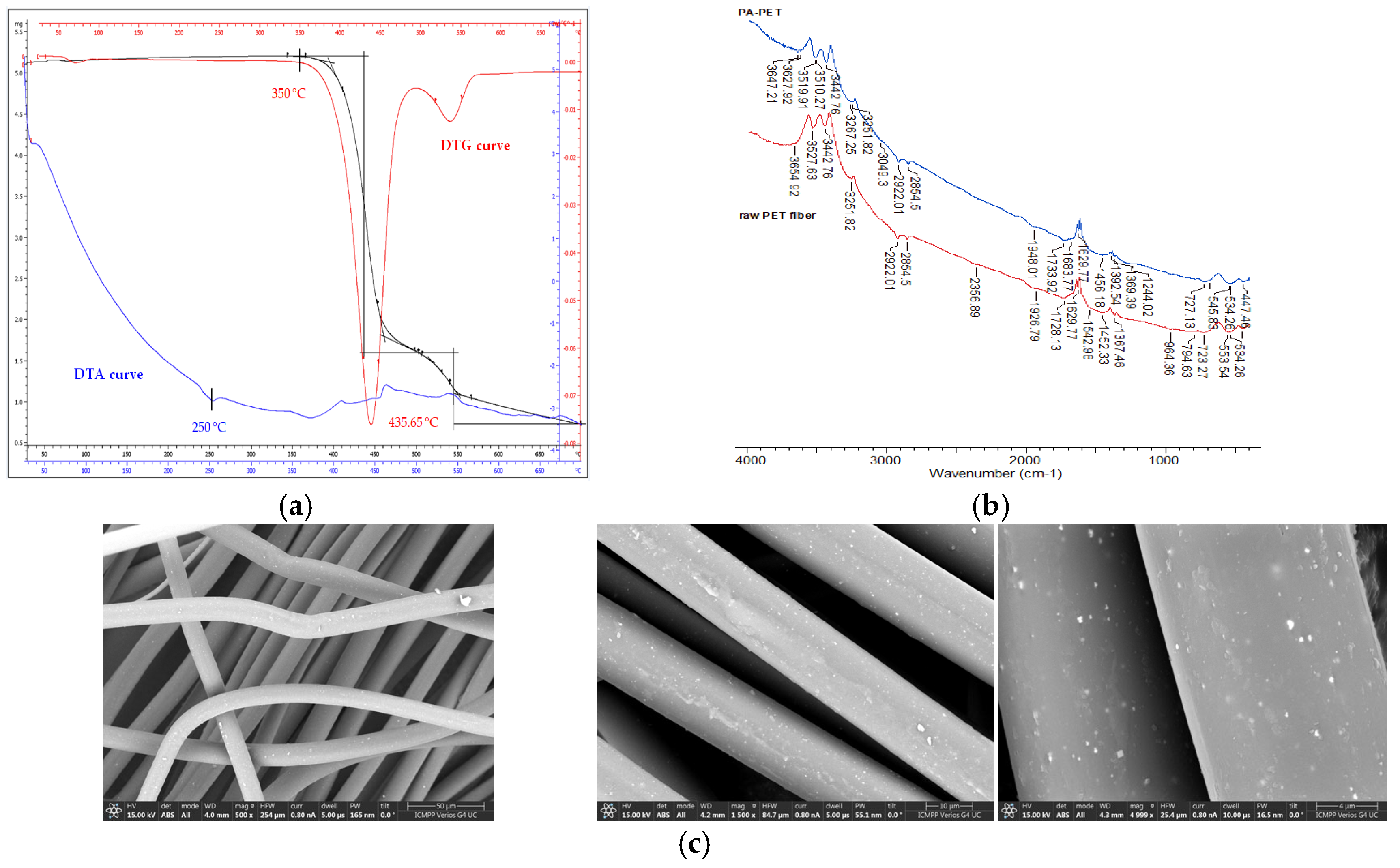
Figure 4.
Linear representation of Langmuir (a), Freundlich (b) and Temkin (c) models for the adsorption of RIF and RIX on PA-PET. (Experimental conditions: pH = 2.0; adsorbent dose = 0.4 g/L, temperature = 22 ± 1 °C).
Figure 4.
Linear representation of Langmuir (a), Freundlich (b) and Temkin (c) models for the adsorption of RIF and RIX on PA-PET. (Experimental conditions: pH = 2.0; adsorbent dose = 0.4 g/L, temperature = 22 ± 1 °C).
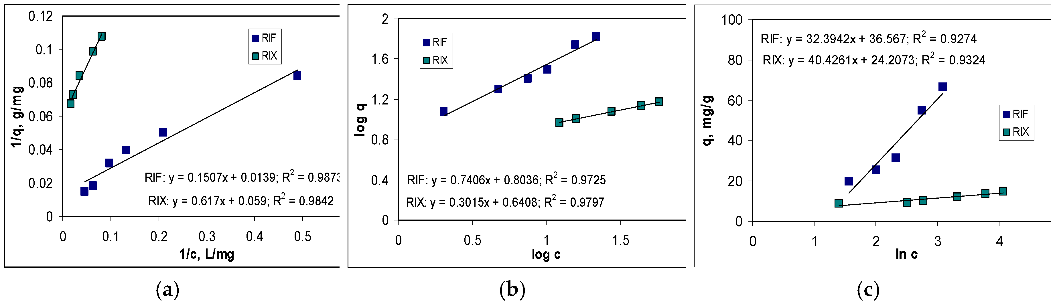
Figure 5.
Linear representation of pseudo-first order (a), pseudo-second order (b) and intra-particle diffusion (c) kinetic models for the adsorption of RIF and RIX on PA-PET. (Experimental conditions: pH = 2.0; adsorbent dose = 0.4 g/L, temperature = 22 ± 1 °C).
Figure 5.
Linear representation of pseudo-first order (a), pseudo-second order (b) and intra-particle diffusion (c) kinetic models for the adsorption of RIF and RIX on PA-PET. (Experimental conditions: pH = 2.0; adsorbent dose = 0.4 g/L, temperature = 22 ± 1 °C).
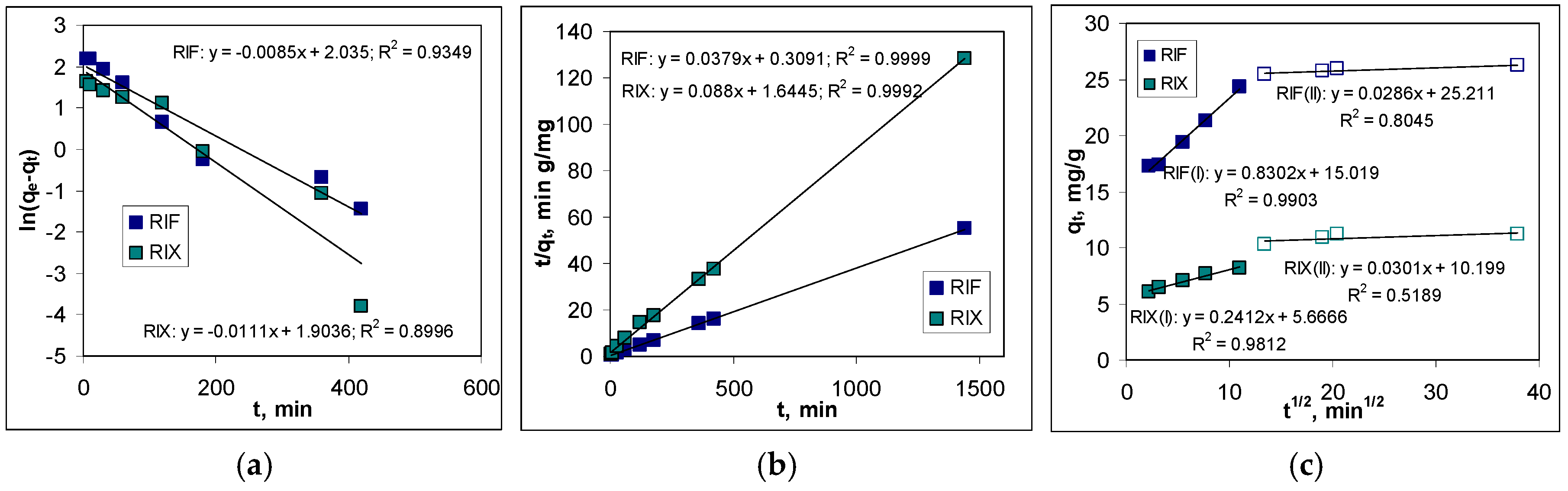
Figure 6.
Schematic illustration of RIF and RIX adsorption on PA-PET.
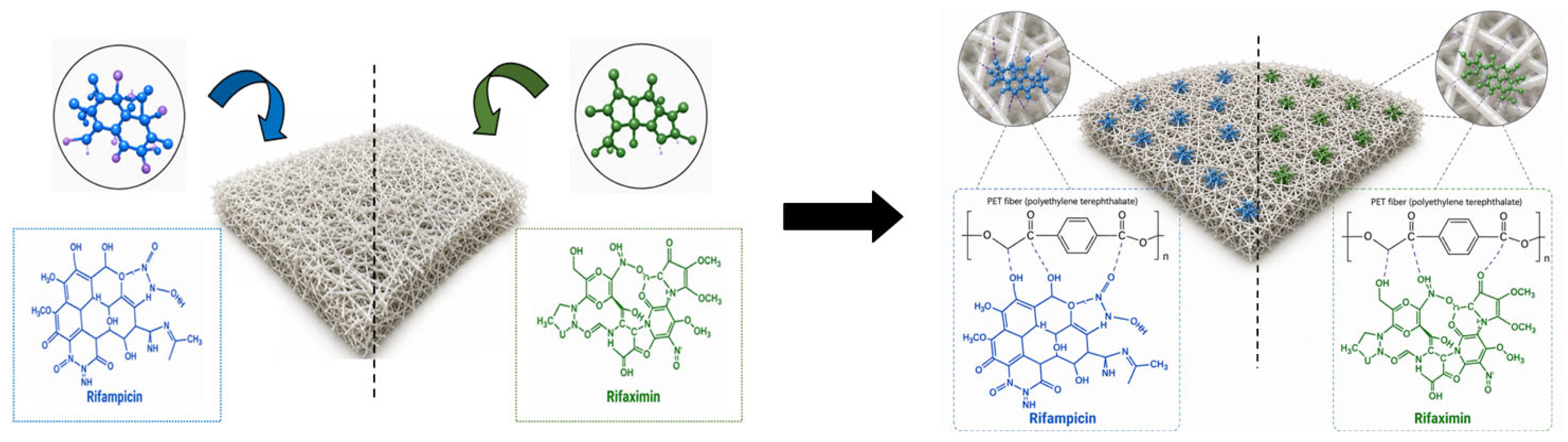
Figure 7.
Variation of desorption efficiency of RIF and RIX from PA-PET as a function of contact time. (Experimental conditions: pH = 6.79; 10 mL desorption agent; q = 31.68 mg RIF/g PA-PET and 16.32 mg RIX/g PA-PET, temperature = 22 ± 1 °C).
Figure 7.
Variation of desorption efficiency of RIF and RIX from PA-PET as a function of contact time. (Experimental conditions: pH = 6.79; 10 mL desorption agent; q = 31.68 mg RIF/g PA-PET and 16.32 mg RIX/g PA-PET, temperature = 22 ± 1 °C).
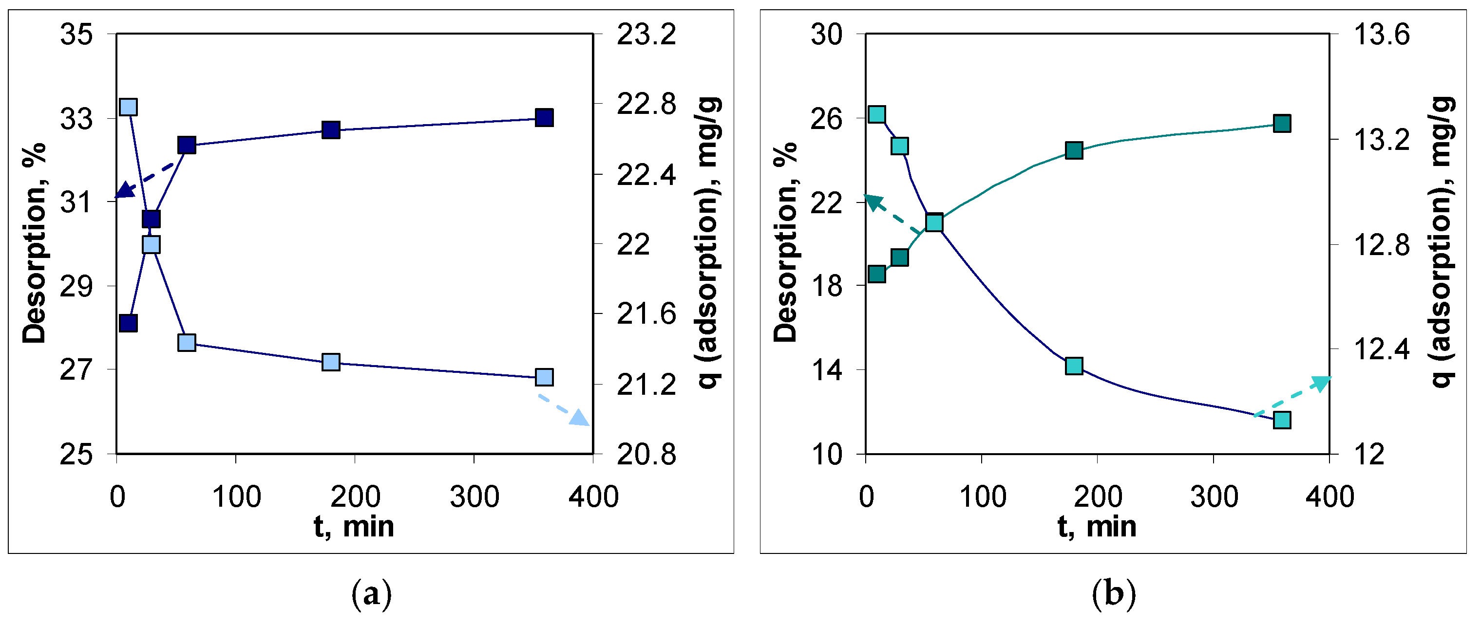
Figure 8.
VIS spectra of RIF (a) and RIX (b) (Experimental conditions: pH = 7.0, phosphate buffer; c = 25.0 mg/L for RIF and 12.7 mg/L for RIX).
Figure 8.
VIS spectra of RIF (a) and RIX (b) (Experimental conditions: pH = 7.0, phosphate buffer; c = 25.0 mg/L for RIF and 12.7 mg/L for RIX).
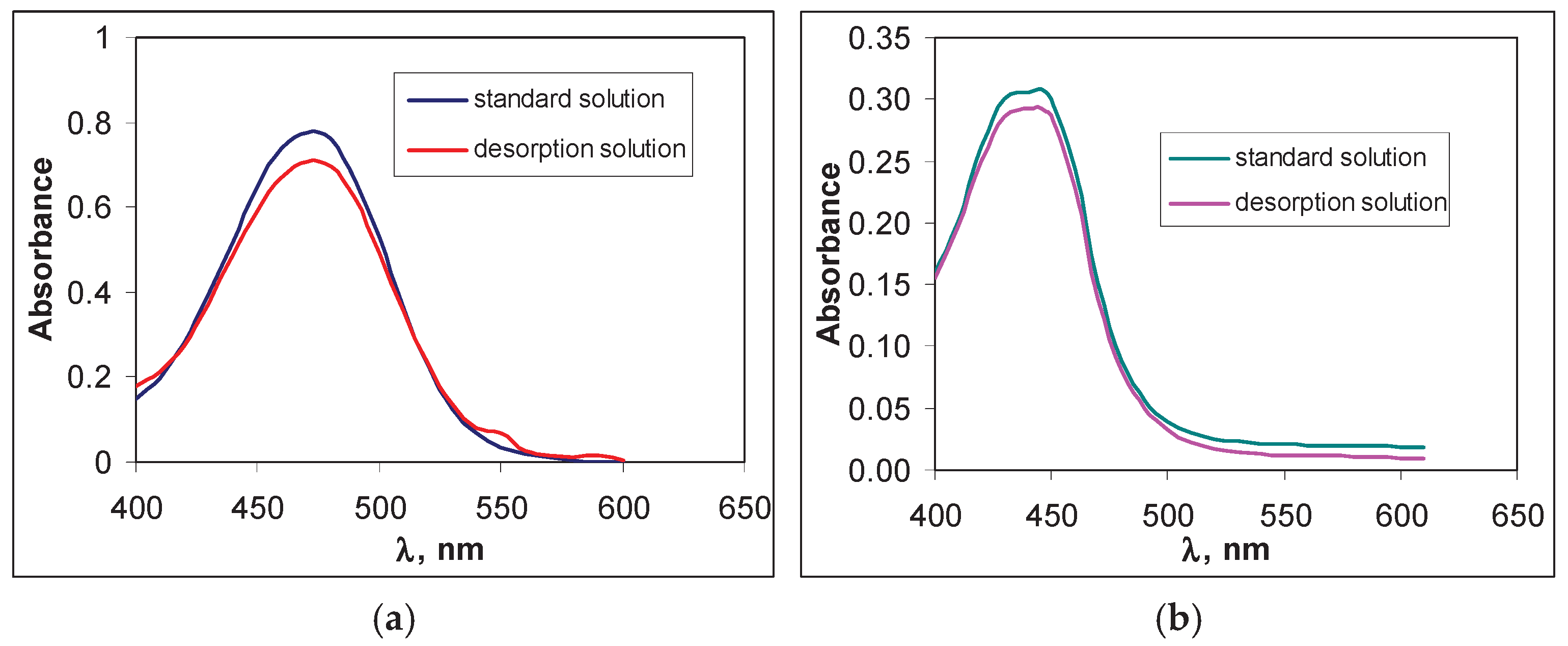

Table 1.
Langmuir, Freundlich and Temkin parameters for RIF and RIX adsorption on PA-PET.
| Isotherm model | Parameter | RIF | RIX |
|---|---|---|---|
| Langmuir | R2 | 0.9873 | 0.9842 |
| Chi-square | 0.2044 | 0.2104 | |
| qmax, mg/g | 71.94 | 16.95 | |
| KL, L/g | 0.0922 | 0.0956 | |
| ΔG, kJ/mol | -5.85 | -5.76 | |
| Freundlich | R2 | 0.9725 | 0.9797 |
| Chi-square | 0.2532 | 0.2809 | |
| n | 1.35 | 3.31 | |
| KF, L/g | 6.3621 | 4.3732 | |
| Temkin | R2 | 0.9274 | 0.9324 |
| Chi-square | 0.3203 | 0.2895 | |
| AT, L/g | 1.6207 | 1.4903 | |
| bT, J/mol | 75.71 | 60.67 |
Table 2.
Pseudo-first order, pseudo-second order and intra-particle diffusion parameters for RIF and RIX adsorption on PA-PET.
Table 2.
Pseudo-first order, pseudo-second order and intra-particle diffusion parameters for RIF and RIX adsorption on PA-PET.
| Kinetic model | Parameter | RIF | RIX |
|---|---|---|---|
| qe,exp., mg/g | 26.24 | 11.21 | |
| Pseudo-first order model | R2 | 0.9349 | 0.8996 |
| Chi-square | 0.7276 | 0.6971 | |
| qe, mg/g | 7.65 | 6.71 | |
| k1, 1/min | 0.0085 | 0.0111 | |
| Pseudo-second order model | R2 | 0.9999 | 0.9992 |
| Chi-square | 0.0678 | 0.1274 | |
| qe, mg/g | 26.38 | 11.36 | |
| k2, g/mg min | 0.0046 | 0.0047 | |
| Intra-particle diffusion model | R2 | 0.9903 | 0.9812 |
| c1, mg/L | 15.02 | 5.67 | |
| kdiff, 1, mg/g min1/2 | 0.8302 | 0.2412 | |
| R2 | 0.8045 | 0.5189 | |
| c2, mg/L | 25.21 | 10.20 | |
| kdiff, 2, mg/g min1/2 | 0.0286 | 0.0301 |
Table 3.
Wavenumbers of absorption maxima in FT-IR spectra of PA-PET before and after RIF and RIX adsorption.
Table 3.
Wavenumbers of absorption maxima in FT-IR spectra of PA-PET before and after RIF and RIX adsorption.
| Functional groups | Before adsorption | After adsorption | |
|---|---|---|---|
| RIF | RIX | ||
| Hydroxyl groups (water; alcohols) | 3647 cm-1 | 3654 cm-1 | 3651 cm-1 |
| Carbonyl groups (esters) | 1733 cm-1 | 1737 cm-1 | 1728 cm-1 |
| Carbonyl groups (carboxylic acids; ketones) | 1683 cm-1 | 1691 cm-1 | 1687 cm-1 |
| Deformation vibration of C–H bond (-CH2) in polymer chain | 1456 cm-1 | 1452 cm-1 | 1455 cm-1 |
| Vibration of C–H bond in therephthalic ring | 727 cm-1 | 723 cm-1 | 728 cm-1 |
Table 4.
Spectrophotometric parameters of VIS spectra illustrated in Figure 8.
Table 4.
Spectrophotometric parameters of VIS spectra illustrated in Figure 8.
| Parameter | RIF spectra | RIX spectra | ||
|---|---|---|---|---|
| Standard solution |
Desorption solution | Standard solution |
Desorption solution | |
| Absorbance (max) | 0.776 | 0.737 | 0.308 | 0.294 |
| λ, nm | 470 | 470 | 444 | 444 |
| Δλ1/2, nm | 68.0 | 65.0 | 74.0 | 76.0 |
| ε, L/mol cm | 2.74 104 | 2.57 104 | 1.91 104 | 1.82 104 |
Notations: λ—the wavelength corresponding to the absorption maximum; Δλ1/2—the spectral band width; ε—the molar absorption coefficient.
Disclaimer/Publisher’s Note: The statements, opinions and data contained in all publications are solely those of the individual author(s) and contributor(s) and not of MDPI and/or the editor(s). MDPI and/or the editor(s) disclaim responsibility for any injury to people or property resulting from any ideas, methods, instructions or products referred to in the content. |
© 2026 by the authors. Licensee MDPI, Basel, Switzerland. This article is an open access article distributed under the terms and conditions of the Creative Commons Attribution (CC BY) license (http://creativecommons.org/licenses/by/4.0/).
Copyright: This open access article is published under a Creative Commons CC BY 4.0 license, which permit the free download, distribution, and reuse, provided that the author and preprint are cited in any reuse.


