

Submitted:
01 July 2026
Posted:
03 July 2026
You are already at the latest version
Abstract
Background: Hemophagocytic lymphohistiocytosis is a life-threatening hyperinflammatory syndrome that can mimic sepsis in adults, delaying diagnosis and treatment. Methods: We reviewed three adult patients admitted to a high-complexity referral hospital in Colombia, all initially managed as sepsis, analyzing clinical findings, laboratory results, bone marrow studies, diagnostic approach, and outcomes. Results: All patients had persistent fever, cytopenias, organomegaly, hyperferritinemia, and hypertriglyceridemia, with ongoing inflammation despite appropriate antimicrobial therapy. Bone marrow examination showed hemophagocytosis in all cases. One case was associated with acute myeloid leukemia and had a poor outcome, while two infection-related cases, triggered by disseminated histoplasmosis and visceral leishmaniasis, improved with targeted therapy and supportive care. Conclusions: Persistent inflammation, cytopenias, and markedly elevated ferritin despite standard sepsis treatment should prompt early consideration of hemophagocytic lymphohistiocytosis and investigation of the underlying trigger.
Keywords:
hemophagocytic lymphohistiocytosis
; hyperinflammation
; cytokine storm
; sepsis
; hyperferritinemia
; histoplasmosis
; visceral leishmaniasis
; acute myeloid leukemia
1. Introduction
Hemophagocytic lymphohistiocytosis (HLH) is a rare and life-threatening condition where the immune system basically goes into overdrive. Instead of protecting the body, immune cells become hyperactive, triggering severe inflammation and elevating cytokine levels, leading to organ damage. In adults, HLH usually happens because of something else going on, like certain cancers of the blood, severe infections, autoimmune disorders, or problems with the immune system. These days, doctors see HLH as part of a broader family of “cytokine storm” illnesses, since they all involve a similar immune system overreaction. [1,2]
Figuring out if an adult has secondary HLH isn’t easy, mainly because the symptoms often look like those seen in sepsis and similar inflammatory conditions. People with HLH or a bad infection might have ongoing fever, low blood counts, problems with various organs, blood clotting issues, and high ferritin levels. Since these signs can show up in both situations, doctors sometimes miss the real cause behind the inflammation. Lately, some studies have pointed out that HLH is often overlooked in adults who show up very sick with what seems like sepsis or stubborn inflammation that doesn’t get better. [3,4]
The HLH-2004 criteria and HScore are still the main tools doctors use to diagnose HLH in adults, but both have their drawbacks. This is especially true for patients who are seriously ill or in hospitals with fewer resources, where it's hard to get tests like soluble CD25 or NK-cell function. Plus, some of the things doctors look for with the HLH-2004 criteria can also show up in people with sepsis or other critical illnesses, which makes diagnosing HLH in adults even trickier. [4,5,6]
Certain blood cancers are some of the main causes of HLH in adults, and these cases often have worse outcomes. Some infections, like those caused by Histoplasma capsulatum or Leishmania species, can also keep the immune system too active, leading to more inflammation, especially in people living in areas where these diseases are common or in those with weakened immune systems. [7,8,9] Spotting ongoing inflammation early, even when the patient is already getting the right antibiotics, is key for making the diagnosis and starting treatment on time.
In this report, we share the cases of several adult patients with secondary HLH who were first treated as if they had sepsis at a specialized hospital in Colombia. These cases show just how much HLH and sepsis-like illnesses can look alike, especially when patients have blood cancers or infections common in certain regions.
2. Case presentation
2.1. Case 1
A 54-year-old man with end-stage chronic kidney disease on hemodialysis, hypertension, and hypothyroidism was admitted with persistent fever, dry cough, lumbar pain, constitutional symptoms, and pancytopenia. Initial evaluation suggested high-risk febrile neutropenia, and broad-spectrum antimicrobial therapy was started. Blood cultures yielded Streptococcus gallolyticus, prompting targeted antibiotic treatment after susceptibility testing. Despite microbiological control and supportive management, the patient remained febrile with progressive clinical deterioration and persistent cytopenias.
Further evaluation revealed marked hyperferritinemia (8,975 ng/mL), hypertriglyceridemia (789 mg/dL), and splenomegaly, raising suspicion for an underlying hyperinflammatory syndrome beyond isolated sepsis. The patient fulfilled 5 of 8 HLH-2004 criteria, with an HScore of 222, corresponding to a high probability of secondary HLH. Bone marrow aspirate demonstrated hemophagocytosis (Figure 1A–B), while flow cytometry identified 22.8% myeloblasts with immunophenotypic features consistent with acute myeloid leukemia (AML-M2) with aberrant CD7 and CD56 expression.
The clinical picture was interpreted as malignancy-associated HLH occurring in the setting of concomitant bacteremia. Given the patient’s comorbidity burden and functional status, treatment with azacitidine was initiated with palliative intent in addition to supportive care and infection control measures. The patient showed unfavorable clinical evolution during hospitalization.
2.2. Case 2
A 34-year-old man with newly diagnosed advanced HIV infection (stage C3) and severe immunosuppression (CD4 count: 38 cells/mm³) was admitted with prolonged fever, weight loss, chronic diarrhea, jaundice, and progressive dyspnea. Initial evaluation focused on opportunistic infections, particularly after thoracic imaging revealed a right pulmonary mass with mediastinal extension. During hospitalization, the patient developed diffuse pulmonary infiltrates and hypoxemia, prompting empiric treatment for Pneumocystis jirovecii pneumonia.
Despite broad antimicrobial therapy, the patient showed persistent systemic inflammation associated with hepatosplenomegaly, bicytopenia, hyperferritinemia (3,551 ng/mL), hypertriglyceridemia, and progressive clinical deterioration, raising suspicion for secondary HLH. Bone marrow aspirate demonstrated hemophagocytosis (Figure 2A–B), and diagnostic assessment yielded an HScore of 246 and fulfillment of 7 of 8 HLH-2004 criteria.
Etiologic investigation identified disseminated Histoplasma capsulatum infection, supported by positive urinary antigen testing and histopathologic findings demonstrating intracellular fungal organisms within pulmonary and mediastinal tissue samples. Additional viral reactivation markers, including Cytomegalovirus and Epstein–Barr virus serologies, were detected; however, disseminated histoplasmosis was considered the principal trigger of the hyperinflammatory syndrome.
Treatment included liposomal amphotericin B, dexamethasone, and subsequent initiation of antiretroviral therapy after exclusion of central nervous system infection. The patient showed progressive clinical and hematologic improvement and was discharged with outpatient follow-up.
2.3. Case 3
A 28-year-old previously healthy man was admitted with prolonged fever, weight loss, malaise, and progressive cytopenias. Initial laboratory evaluation revealed anemia, leukopenia, and thrombocytopenia, while abdominal imaging demonstrated marked hepatosplenomegaly. Blood cultures were negative, although urine culture yielded Enterococcus faecium. Despite antimicrobial therapy, the patient developed worsening pancytopenia and persistent systemic inflammation.
Further evaluation showed marked hyperferritinemia (8,856 ng/mL), hypertriglyceridemia, massive splenomegaly, and nodular splenic lesions initially raising suspicion for a lymphoproliferative disorder. Bone marrow examination demonstrated a hypercellular marrow without evidence of hematologic malignancy; however, subsequent marrow evaluation identified intracellular amastigote forms compatible with Leishmania spp. (Figure 3A–B), together with histiocytic hemophagocytosis. Serologic testing by indirect immunofluorescence was positive for visceral leishmaniasis.
In the context of persistent fever, cytopenias, hepatosplenomegaly, hyperinflammation, and bone marrow findings, the clinical presentation was interpreted as secondary HLH associated with visceral leishmaniasis. Treatment with liposomal amphotericin B was initiated, leading to progressive clinical improvement and complete hematologic recovery.
The main demographic, clinical, laboratory, and outcome characteristics of the patients included in this series are summarized in Table 1.
4. Discussion
Secondary HLH in adults remains a major diagnostic challenge because its clinical presentation frequently overlaps with sepsis and other hyperinflammatory syndromes encountered in critical care settings. In the present series, all patients initially presented with persistent systemic inflammation interpreted as severe infection, delaying recognition of the underlying hyperinflammatory disorder. Despite distinct etiologic triggers, the cases shared a common phenotype characterized by prolonged fever, cytopenias, organomegaly, hyperferritinemia, hypertriglyceridemia, and progressive clinical deterioration despite antimicrobial therapy. These findings are consistent with the current understanding of HLH as part of a broader spectrum of cytokine storm syndromes driven by dysregulated immune activation and maladaptive inflammatory responses. [1,2,3,4]
Current evidence suggests that impaired cytotoxic activity of NK cells and cytotoxic T lymphocytes results in persistent antigenic stimulation, uncontrolled macrophage activation, and excessive cytokine release, ultimately leading to tissue injury and multiorgan dysfunction. Interferon-γ, interleukin (IL)-1, IL-6, IL-18, and JAK/STAT signaling pathways appear to play central roles in the amplification of systemic inflammation. [1,2] Because similar inflammatory pathways may also occur in sepsis, differentiation between HLH and overwhelming infection remains particularly difficult in critically ill adults. Recent studies have emphasized that HLH and sepsis may represent overlapping hyperferritinemic inflammatory syndromes rather than completely distinct entities. Key clinical and laboratory features that may assist in differentiating HLH from sepsis are summarized in Table 2. [3,4,10,11]
One of the most clinically relevant findings in our series was the persistence of systemic inflammation despite broad antimicrobial therapy. Persistent fever associated with worsening cytopenias and marked hyperferritinemia prompted reevaluation beyond isolated infection in all patients. Although elevated ferritin lacks specificity and may occur in malignancy, liver dysfunction, and critical illness, markedly increased levels in the appropriate clinical context strongly support consideration of HLH. Ferritin has also been increasingly recognized as a potential mediator of immune dysregulation within hyperferritinemic syndromes. [1,10] Nevertheless, ferritin alone cannot reliably distinguish HLH from severe sepsis and should always be interpreted together with clinical evolution and additional inflammatory features. [3,6]
The diagnosis of HLH in adults continues to rely on integration of clinical, laboratory, and histopathologic findings. Although the HLH-2004 criteria and HScore remain the most used diagnostic tools in adult practice, important limitations persist, particularly in critically ill patients and resource-limited settings where soluble CD25 levels and NK-cell functional assays are not routinely available. [3,4,5,6,12]
In this regard, Figure 4 presents the HScore as a practical probability-based diagnostic approach, whereas Figure 5 summarizes the HLH-2004 criteria traditionally used to support case identification and diagnostic classification [6,14]. Furthermore, several diagnostic variables included in HLH-2004 criteria may also be altered in sepsis and critical illness. Recent multicenter validation studies have demonstrated important limitations of currently available diagnostic criteria in adult populations, reinforcing the need for dynamic clinical assessment and repeated diagnostic reassessment. [5,6]
Bone marrow hemophagocytosis represented an important supportive finding in our cases; however, current evidence indicates that hemophagocytosis is neither sufficiently sensitive nor specific to establish HLH independently. Hemophagocytosis may also occur in severe infection, autoimmune disease, malignancy, and transfusion-related inflammatory states, and may be absent during early phases of HLH. Accordingly, contemporary diagnostic recommendations emphasize that bone marrow findings should always be interpreted within the broader clinical and laboratory context. [4,5,6,12]
The etiologic triggers identified in this series reflect the heterogeneous nature of adult secondary HLH. One patient developed malignancy-associated HLH in the setting of acute myeloid leukemia (AML). Malignancy-associated HLH is recognized as one of the most severe forms of adult HLH and is associated with particularly poor outcomes among patients with hematologic neoplasms. Persistent cytokine dysregulation, bone marrow infiltration, immune escape, and treatment-related immunosuppression may contribute to the aggressive clinical course observed in these patients. [2,7]
The remaining cases were associated with disseminated histoplasmosis and visceral leishmaniasis, both recognized intracellular infectious triggers of HLH. Intracellular pathogens promote sustained macrophage activation and reticuloendothelial inflammation, generating substantial overlap with hematologic malignancies and sepsis. Disseminated histoplasmosis has increasingly been recognized as an important trigger of HLH in immunocompromised patients, particularly in advanced HIV infection. [8,13] Similarly, visceral leishmaniasis may induce profound immune activation characterized by pancytopenia, hepatosplenomegaly, hyperferritinemia, and bone marrow hemophagocytosis, frequently mimicking lymphoproliferative disorders and other hematologic diseases. [9] In endemic regions, these infections should therefore be actively considered in adults presenting with persistent febrile syndromes and hyperferritinemic inflammation.
Therapeutic management of secondary HLH requires both suppression of the hyperinflammatory response and prompt treatment of the underlying trigger. Corticosteroids remain a central component of therapy; however, increasing interest has emerged regarding targeted immunomodulatory strategies directed against interferon-γ, IL-1, and JAK/STAT signaling pathways. [2] In infection-associated HLH, early etiologic treatment may substantially improve outcomes, as observed in our patients with disseminated histoplasmosis and visceral leishmaniasis. In contrast, malignancy-associated HLH continues to carry particularly high mortality despite therapeutic advances. [2,7]
This report has limitations, including its retrospective design, small sample size, and incomplete availability of advanced immunologic testing. Nevertheless, the cases presented illustrate diagnostic challenges frequently encountered in adult secondary HLH and reinforce the importance of maintaining a high index of suspicion in patients with persistent sepsis-like inflammatory syndromes and unexplained hyperferritinemia.
4. Conclusions
Secondary HLH should be considered in adults with persistent systemic inflammation, cytopenias, organ dysfunction, and hyperferritinemia despite appropriate antimicrobial therapy. Early recognition of hyperinflammatory patterns and identification of underlying triggers remain essential to improve outcomes, particularly in malignancy-associated HLH and intracellular infections.
Author Contributions
Conceptualization, B.D.J.R.P., D.O.G. and J.P.V.G.; methodology, B.D.J.R.P., D.O.G. and J.P.V.G.; formal analysis, B.D.J.R.P., D.O.G., J.P.V.G. and R.T.B.; investigation, B.D.J.R.P., D.O.G., J.P.V.G., E.T.A. and R.T.B.; data curation, B.D.J.R.P., D.O.G., J.P.V.G., E.T.A. and R.T.B.; writing—original draft preparation, B.D.J.R.P., D.O.G., J.P.V.G., E.T.A. and R.T.B.; visualization, D.O.G; writing—review and editing, A.A.H.; supervision, A.A.H.; project administration, A.A.H. All authors have read and agreed to the published version of the manuscript. The authors confirm that this manuscript has not been submitted to any other journal.
Funding
This research received no external funding.
Institutional Review Board Statement
Ethical review and approval were waived for this study because it is a retrospective case series based on anonymized clinical data. Written informed consent was obtained from all patients for the use of their clinical information for scientific and publication purposes. All data were de-identified prior to analysis, and the study was conducted in accordance with the principles of the Declaration of Helsinki.
Informed Consent Statement
Informed consent was obtained from all subjects involved in the study to publish this paper.
Data Availability Statement
The original contributions presented in this study are included in the article. Further inquiries can be directed to the corresponding author.
Acknowledgments
The authors have reviewed and edited the output and take full responsibility for the content of this publication.
Conflicts of Interest
The authors declare no conflicts of interest.
Abbreviations
The following abbreviations are used in this manuscript:
| HLH | Hemophagocytic lymphohistiocytosis |
| AML | Acute myeloid leukemia |
| HIV | Human immunodeficiency virus |
| CKD | Chronic kidney disease |
| NK | Natural killer |
References
- Cron, R.Q.; Goyal, G.; Chatham, W.W. Cytokine storm syndrome. Annu. Rev. Med. 2023, 74, 321–337. [CrossRef]
- Wu, Y.; Sun, X.; Kang, K.; Yang, Y.; Li, H.; Zhao, A.; et al. Hemophagocytic lymphohistiocytosis: current treatment advances, emerging targeted therapy and underlying mechanisms. J. Hematol. Oncol. 2024, 17, 106. [CrossRef]
- Hsu, J.I.; Nikiforow, S.; Berliner, N. Hemophagocytic lymphohistiocytosis in adults. Blood 2026, 147, 1037–1047. [CrossRef]
- Bauchmüller, K.; Clark, S.; Manson, J.J.; Tattersall, R.S. Haemophagocytic lymphohistiocytosis in critical care. BJA Educ. 2025, 25, 107–114. [CrossRef]
- Lachmann, G.; Heeren, P.; Schuster, F.S.; Nyvlt, P.; Spies, C.; Feinkohl, I.; et al. Multicenter validation of secondary hemophagocytic lymphohistiocytosis diagnostic criteria. J. Intern. Med. 2025, 297, 312–327. [CrossRef]
- Henter, J.I.; Sieni, E.; Eriksson, J.; Bergsten, E.; Hed Myrberg, I.; Canna, S.W.; et al. Diagnostic guidelines for familial hemophagocytic lymphohistiocytosis revisited. Blood 2024, 144, 2308–2318. [CrossRef]
- Setiadi, A.; Zoref-Lorenz, A.; Lee, C.Y.; Jordan, M.B.; Chen, L.Y.C. Malignancy-associated haemophagocytic lymphohistiocytosis. Lancet Haematol. 2022, 9, e217–e227. [CrossRef]
- Rolsdorph, L.Å.; Mosevoll, K.A.; Helgeland, L.; Reikvam, H. Concomitant hemophagocytic lymphohistiocytosis and cytomegalovirus disease: a case based systemic review. Front. Med. 2022, 9, 819465. [CrossRef]
- Burza, S.; Croft, S.L.; Boelaert, M. Leishmaniasis. Lancet 2018, 392, 951–970. [CrossRef]
- Schulert, G.S.; Canna, S.W. Convergent pathways of the hyperferritinemic syndromes. Int. Immunol. 2018, 30, 195–203. [CrossRef]
- Karki, R.; Netea, M.G.; Diorio, C.; et al. Cytokine storm. Nat. Rev. Dis. Primers 2026, 12, 1. [CrossRef]
- Hines, M.R.; von Bahr Greenwood, T.; Beutel, G.; Beutel, K.; Hays, J.A.; Horne, A.; et al. Consensus-based guidelines for the recognition, diagnosis, and management of hemophagocytic lymphohistiocytosis in critically ill children and adults. Crit. Care Med. 2022, 50, 860–872. [CrossRef]
- Wheat, L.J.; Azar, M.M.; Bahr, N.C.; Spec, A.; Relich, R.F.; Hage, C. Histoplasmosis. Infect. Dis. Clin. North Am. 2016, 30, 207–227. [CrossRef]
- Fardet, L.; Galicier, L.; Lambotte, O.; Marzac, C.; Aumont, C.; Chahwan, D.; et al. Development and validation of the HScore, a score for the diagnosis of reactive hemophagocytic syndrome. Arthritis Rheumatol. 2014, 66, 2613–2620. [CrossRef]
Figure 1.
Bone marrow aspirate showing hemophagocytosis: (A) Large histiocytic cell with abundant pale basophilic cytoplasm containing phagocytosed hematopoietic elements, consistent with hemophagocytosis. (B) Bone marrow aspirate field showing a macrophage-like cell with cytoplasmic vacuolization and engulfed cellular material, supporting marrow hemophagocytosis in the setting of secondary HLH. .
Figure 1.
Bone marrow aspirate showing hemophagocytosis: (A) Large histiocytic cell with abundant pale basophilic cytoplasm containing phagocytosed hematopoietic elements, consistent with hemophagocytosis. (B) Bone marrow aspirate field showing a macrophage-like cell with cytoplasmic vacuolization and engulfed cellular material, supporting marrow hemophagocytosis in the setting of secondary HLH. .
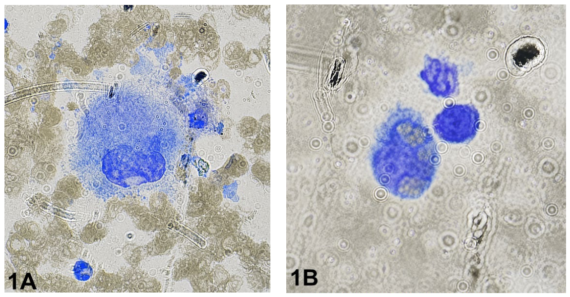
Figure 2.
Bone marrow aspirate showing hemophagocytosis in secondary HLH associated with disseminated histoplasmosis: (A) Large histiocytic cell with abundant basophilic cytoplasm containing engulfed hematopoietic elements, consistent with hemophagocytosis. (B) Macrophage-like cell with vacuolated cytoplasm and phagocytosed cellular material, supporting active marrow hemophagocytosis.
Figure 2.
Bone marrow aspirate showing hemophagocytosis in secondary HLH associated with disseminated histoplasmosis: (A) Large histiocytic cell with abundant basophilic cytoplasm containing engulfed hematopoietic elements, consistent with hemophagocytosis. (B) Macrophage-like cell with vacuolated cytoplasm and phagocytosed cellular material, supporting active marrow hemophagocytosis.
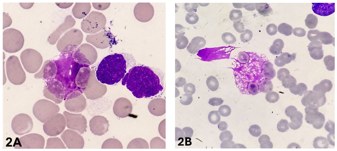
Figure 3.
Bone marrow aspirate findings in visceral leishmaniasis-associated secondary HLH: (A) Bone marrow aspirate showing scattered mononuclear cells and clusters of small basophilic bodies compatible with Leishmania spp. amastigotes. (B) Additional marrow field demonstrating multiple clustered amastigote-like organisms, supporting visceral leishmaniasis as the infectious trigger of secondary HLH. .
Figure 3.
Bone marrow aspirate findings in visceral leishmaniasis-associated secondary HLH: (A) Bone marrow aspirate showing scattered mononuclear cells and clusters of small basophilic bodies compatible with Leishmania spp. amastigotes. (B) Additional marrow field demonstrating multiple clustered amastigote-like organisms, supporting visceral leishmaniasis as the infectious trigger of secondary HLH. .
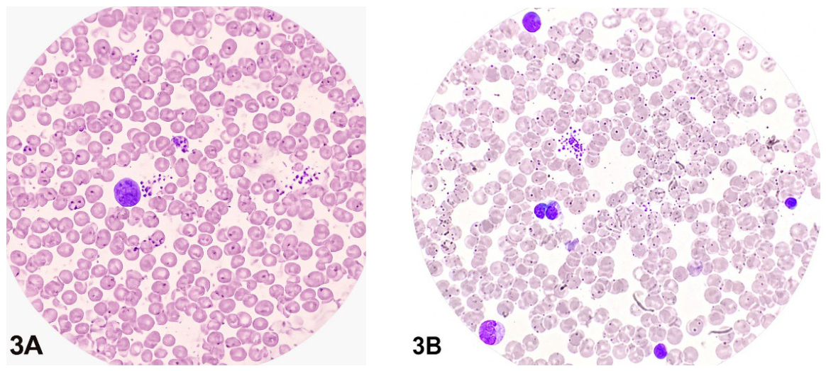
Figure 4.
HScore. Note: Weighted scores for clinical features of HLH and the probability of HLH based on total points. Adapted from Fardet et al. [14]
Figure 4.
HScore. Note: Weighted scores for clinical features of HLH and the probability of HLH based on total points. Adapted from Fardet et al. [14]
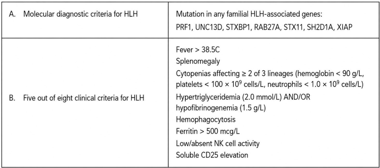
Figure 5.
HLH-2004 Diagnostic Criteria: The diagnosis FHL can be established if at least 1 of either 1 or 2 below is fulfilled. Adapted from Henter et al. [6].
Figure 5.
HLH-2004 Diagnostic Criteria: The diagnosis FHL can be established if at least 1 of either 1 or 2 below is fulfilled. Adapted from Henter et al. [6].
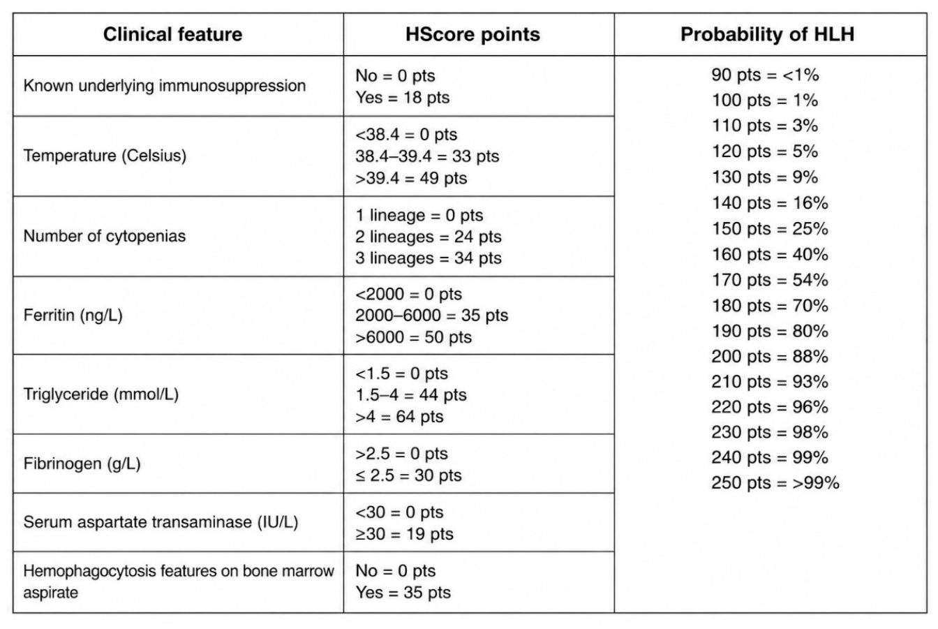

Table 1.
Characteristics of the patients.
| Variable | Case 1 | Case 2 | Case 3 |
|---|---|---|---|
| Age, Sex | 54, Male | 34, Male | 28, Male |
| Underlying condition | CKD on hemodialysis | Advanced HIV infection (C3) | Previously health |
| Initial clinical impression | Febrile neutropenia/sepsis | Opportunistic infection/sepsis | Prolonged febrile syndrome/sepsis |
| Main trigger associated with HLH | Acute myeloid leukemia | Disseminated histoplasmosis | Visceral leishmaniasis |
| Persistent fever | Yes | Yes | Yes |
| Cytopenias | Pancytopenia | Bicytopenia | Pancytopenia |
| Hepatosplenomegaly/Splenomegaly | Splenomegaly | Hepatosplenomegaly | Massive splenomegaly |
| Ferritin (ng/mL) | 8,975 | 3,551 | 8,856 |
| Triglycerides (mg/dL) | 789 | 284–347 | 230 |
| Fibrinogen (mg/dL) | 593 | 291 | Not available |
| Bone marrow hemophagocytosis | Present | Present | Present |
| HLH-2004 criteria fulfilled | 5/8 | 7/8 | Compatible clinical criteria |
| HScore | 222 | 246 | Not available |
| Additional bone marrow findings | AML M2 infiltration | Intracellular fungal structures | Amastigotes compatible with Leishmania spp. |
| Initial antimicrobial treatment | Broad-spectrum antibiotics | TMP-SMX and broad antimicrobial coverage | Empirical antimicrobial therapy |
| HLH-directed therapy | Corticosteroids/supportive care | Dexamethasone | Supportive care |
| Etiologic treatment | Azacitidine | Liposomal amphotericin B | Liposomal amphotericin B |
| Clinical outcome | Unfavorable | Clinical improvement | Complete recovery |
Table 2.
Distinctive characteristics of HLH vs sepsis.
| Feature | HLH | Sepsis |
|---|---|---|
| Ferritin | Very high | Moderate |
| Cytopenias | Severe/multilineage | Variable |
| Splenomegaly | Common | Uncommon |
| Persistent fever despite antibiotics | Frequent | Variable |
| Triglycerides | Elevated | Mild |
| Hemophagocytosis | Possible | Possible |
Disclaimer/Publisher’s Note: The statements, opinions and data contained in all publications are solely those of the individual author(s) and contributor(s) and not of MDPI and/or the editor(s). MDPI and/or the editor(s) disclaim responsibility for any injury to people or property resulting from any ideas, methods, instructions or products referred to in the content. |
© 2026 by the authors. Licensee MDPI, Basel, Switzerland. This article is an open access article distributed under the terms and conditions of the Creative Commons Attribution (CC BY) license (http://creativecommons.org/licenses/by/4.0/).
Copyright: This open access article is published under a Creative Commons CC BY 4.0 license, which permit the free download, distribution, and reuse, provided that the author and preprint are cited in any reuse.