
Submitted:
01 July 2026
Posted:
02 July 2026
You are already at the latest version
Abstract
With the rapid expansion of nuclear medicine, the safe and sustainable management of medical radioactive wastewater has become a critical challenge at the interface of radiation protection and environmental engineering. This review systematically synthesises the full lifecycle of medical radioactive wastewater, encompassing generation, source reduction, collection, treatment, and discharge. The characteristics of key radionuclides and major contamination pathways are first clarified. Strategies for source control are then critically evaluated, including advances in imaging equipment, reconstruction algorithms, personalised dosing, radiopharmaceutical optimisation, water-saving practices, and source-separated collection. At the treatment stage, the evolution and limitations of decay tank systems are analysed alongside emerging decontamination technologies, with particular emphasis on adsorption, ion exchange, and membrane-based processes for process intensification. Furthermore, a comparative analysis of international management and discharge standards reveals a fundamental divergence between China’s concentration-based regulatory approach and the risk-informed, dose-based frameworks adopted in most other countries. Building on these insights, an end-to-end, closed-loop management framework is proposed, integrating source reduction, classified collection, adaptive treatment, and risk-based discharge. This framework provides a systematic pathway to optimise radiation safety, treatment efficiency, and economic feasibility, offering theoretical and practical guidance for the sustainable development of nuclear medicine wastewater management.
Keywords:
medical radioactive wastewater
; nuclear medicine
; source reduction
; decay tank
; discharge control
1. Introduction
Clinical nuclear medicine is a clinical medical discipline that utilises radionuclides and their tracers for the diagnosis and treatment of diseases, comprising diagnostic nuclear medicine and therapeutic nuclear medicine [1]. Diagnostic nuclear medicine utilises radiopharmaceuticals, which selectively concentrate in tissues and organs and decay to emit γ-photons or characteristic X-rays, to perform organ function testing and radionuclide imaging. It primarily consists of in vivo diagnostics, mainly involving radionuclide imaging and organ function testing, and in vitro diagnostics, mainly involving the testing of biological samples. Therapeutic nuclear medicine involves the internal irradiation of lesions using short-range β-particles or α-particles emitted by highly selectively concentrated radionuclides or their tracers.
As of 2023, there were 1,237 departments in China engaged in nuclear medicine. In terms of equipment, the installed base of positron emission tomography/ computed tomography (PET/CT) systems stood at approximately 2,800 units, single-photon emission computed tomography/ computed tomography (SPECT/CT) systems totalled 1,044 units, and there were 148 medical cyclotrons in 2023. In terms of clinical service volume, approximately 1.382 million PET/CT scans and 2.717 million SPECT scans were performed nationwide in 2023. The total number of radionuclide therapy cases exceeded 528,000 as early as 2022 and continues to rise [2]. Radioisotopes commonly used in nuclear medicine diagnostics and therapy include 131I, 99mTc, 18F, 125I, 177Lu and 89Sr, among others [3]. These radioisotopes are discharged into the environment via wastewater. Medical radioactive wastewater is a direct source of external radiation exposure for hospital staff and the public. At the same time, improper treatment poses radiation safety risks to water bodies, soil, the atmosphere and biological communities. Furthermore, radionuclides can accumulate and amplify through the food chain, gradually entering the human body and creating a cumulative effect of radiation hazards [4,5].Consequently, countries worldwide attach great importance to the control of hospital radioactive wastewater. The discharge standard of water pollutants for medical organizations of China [6] requires that hospital radioactive wastewater undergo pre-treatment to ensure that the concentration of radioactive substances complies with limit values before it can be discharged into the municipal sewage treatment system.Currently, there are numerous review articles on the treatment of radioactive wastewater [7,8,9,10,11,12,13,14,15], which propose a variety of treatment methods, such as traditional natural decay tanks [16,17], as well as chemical precipitation [18], ion exchange [19,20,21,22], adsorption [23], membrane treatment [24,25,26,27], and biological treatment [28,29] for the removal of radionuclides. However, these reviews primarily focus on the treatment stage, and there are few reviews addressing the systematic management and control of medical radioactive wastewater.
Radioactive nuclide pollutants are generated during nuclear medicine diagnostic and therapeutic procedures; the methods and doses of radioactive nuclides and their tracers used in these procedures directly determine the volume of radioactive nuclide emissions [17,30,31]. Hospital water usage patterns and radioactive wastewater collection methods determine the volume of radioactive wastewater discharged, as well as the treatment processes and investment costs [31]. The selection of treatment processes is closely linked to the investment and operating costs of treatment plants, as well as treatment efficacy [8]. How radioactive wastewater is managed and how discharge standards are formulated [32,33] directly influence the methods of wastewater treatment.This review takes a comprehensive view of the entire process of medical radioactive wastewater generation, treatment and discharge, including how to reduce the discharge of radionuclides and the volume of wastewater at source; it conducts a comparative analysis of various radioactive wastewater treatment technologies, evaluating the advantages and disadvantages of each; simultaneously, it compares and discusses the requirements of different countries and organisations regarding radioactive wastewater management standards and discharge standards, and systematically proposes a framework for the treatment of medical radioactive wastewater.
2. Characteristics and Generation Mechanisms of Medical Radioactive Wastewater
2.1. Medical Radionuclides
Medical radionuclides are indispensable core agents in nuclear medicine diagnostics and therapy. They are widely used in the preparation of various radiopharmaceuticals and enable the precise achievement of key diagnostic and therapeutic objectives such as tumour imaging, lesion localisation and targeted therapy; they are therefore essential substances for ensuring the effectiveness of diagnosis and therapy [3]. However, during diagnostic and therapeutic procedures, drug preparation residues, instrument cleaning and patient excretions, these nuclides inevitably enter wastewater, becoming the primary pollutants in medical radioactive wastewater. The radioactivity directly determines the degree of wastewater contamination; if not treated properly, the radioactive wastewater pose potential hazards to the environment and human health [34]. The main physical properties and medical applications of common nuclear medicine radionuclides are shown in Table 1 [35].
As can be seen from Table 1, radioactive isotopes with shorter half-lives, such as ⁹⁹mTc, ¹⁸F, ⁶⁸Ga and ¹²³I, are primarily used for diagnostic purposes. Whilst those with longer half-lives, such as ¹⁷⁷Lu, ⁸⁹Sr, ²²³Ra and ²²⁵Ac, are mainly used for therapeutic purposes; the isotope ¹³¹I, however, has dual diagnostic and therapeutic applications.
2.2. Sources of Medical Radioactive Wastewater
The generation of medical radioactive wastewater is clearly scenario-specific, primarily concentrated in the diagnostic and therapeutic procedures and related operational stages of the Department of Nuclear Medicine. Specifically, it can be divided into the following three core processes. Due to differences in source, the radioactive activity and composition of pollutants in the wastewater generated by each process vary to some extent, directly influencing subsequent reduction and treatment methods:
- In the course of diagnostic and therapeutic procedures in nuclear medicine, excretions produced after patients have ingested or been injected with radioisotopes—such as sweat, saliva, urine, faeces and vomit [36].This type of wastewater is one of the major sources of medical radioactive wastewater; its radioactivity is closely related to the dose of the radionuclide received by the patient, the half-life of the radionuclide, and the metabolic rate—radioactive isotopes not absorbed or utilised by the patient are excreted through metabolic processes and subsequently enter the wastewater system. Table 2 shows the typical proportion of therapeutic radionuclides discharged via metabolic pathways [34].
- 2.
- Wash water generated from the cleaning of patient medication cups, syringes and pipettes used during the dispensing of high-activity radioisotopes [37]. During diagnostic and therapeutic procedures and drug preparation in the Department of Nuclear Medicine, trace amounts of radionuclides remain on the surfaces of various vessels that have come into contact with radioisotopes. To prevent cross-contamination and ensure the safety of diagnosis and treatment, these vessels must be washed repeatedly; the wastewater generated during this process carries residual radioactive substances, forming radioactive washwater. Although the radioactivity of this wastewater is lower than that of wastewater generated from patient excreta, the high frequency of vessel cleaning and the large volume of water used result in a relatively significant volume of wastewater. Furthermore, as the distribution of nuclide residues is uniform, this represents one of the key areas requiring strict control in wastewater reduction efforts.
- 3.
- Radioactive wastewater discharged from the preparation of medical labelled compounds and the disposal of excess radioactive isotopes [38]. During the preparation of radiopharmaceuticals, operations such as the synthesis and purification of medical labelled compounds are required, which generate small quantities of waste liquid containing radionuclides. Furthermore, to ensure precise therapeutic dosing and prevent excessive radionuclides from entering the patient’s body, surplus radioactive isotopes generated during preparation must be disposed of in accordance with regulations; such disposal waste liquid also directly constitutes a component of medical radioactive wastewater. The wastewater generated by this process has relatively high radioactivity, with a single nuclide component and concentrated concentrations. If not subject to targeted control measures, it can easily cause local radioactive contamination and increase the difficulty of subsequent treatment.
3. Source-Oriented Reduction Strategies
Waste reduction forms the foundation of any radioactive waste management strategy. Its core objective is to reduce the radioactivity and volume of waste, thereby optimising storage, treatment and disposal processes, ultimately minimising environmental impact and reducing management costs. Regarding control at the source of radioactive waste generation, the primary measures include implementing source reduction strategies—limiting the generation of radioactive materials whilst simultaneously reducing waste volume. These two approaches complement and reinforce one another, comprehensively enhancing the effectiveness of medical radioactive wastewater management [31].
3.1. Measures for the Reduction of Medical Radionuclides
As the primary pollutants in medical radioactive wastewater, medical radionuclides originate from the use of radionuclides and their tracers in nuclear medicine for the diagnosis and treatment of diseases. Reducing the dosage used, whilst ensuring diagnostic accuracy, therapeutic efficacy and radiation safety, is the preferred approach to controlling the generation of radionuclide pollutants.
In traditional nuclear medicine diagnostics and therapy, radionuclide dosages are often based on fixed standards that do not fully account for patient-specific variations, equipment performance, or technological advancements, resulting in some patients receiving unnecessarily high radiation doses [39]. With improvements in imaging equipment sensitivity, innovations in reconstruction algorithms, and the development of radiopharmaceuticals, the reduction of radionuclide usage has evolved from being ‘theoretically feasible’ to ‘clinically implemented’. In recent years, the widespread adoption of high-sensitivity detectors, Time-of-Flight (TOF) technology, Deep Learning Reconstruction (DLR) and other technologies, alongside the application of personalised dosing regimens, has enabled a reduction in radionuclide dosage of 30%-90% in certain scenarios without compromising diagnostic or therapeutic outcomes [40,41,42,43,44]. This review systematically examines and summarises various strategies for reducing radionuclide dosage in nuclear medicine, drawing on recent domestic and international research findings and clinical case studies.
3.1.1. Innovations in Imaging Equipment and Detection Technology: Enhancing Sensitivity and Reducing Counting Requirements
The detection sensitivity of imaging equipment is one of the key factors determining radiotracer dosage; higher sensitivity yields a greater number of photon counts for the same radiotracer dose, resulting in a higher signal-to-noise ratio (SNR), thereby ensuring image quality whilst reducing dosage. In recent years, hardware innovations in nuclear medicine imaging equipment have primarily focused on three aspects: detector materials, structural design, and technological integration.
a. High-Sensitivity Detector Materials and Structural Optimisation
Traditional PET detectors predominantly utilise BGO crystals, which have low light output and poor temporal resolution, thereby limiting sensitivity improvements [45]. Currently, scintillation crystals such as Lutetium Oxyorthosilicate (LSO) and Lutetium Yttrium Oxyorthosilicate (LYSO) are widely used in PET detectors. These crystals are characterised by high light output, rapid decay, and excellent temporal resolution (reaching the sub-nanosecond range) [45]. When combined with silicon photomultiplier tubes (SiPMs) to replace traditional photomultiplier tubes (PMTs), the sensitivity of the detector can be significantly improved [46]. For example, the uEXPLORER, which utilises LSO crystals and SiPM detectors, achieves an axial field of view of up to 194 cm and a system sensitivity of 174 kcps/MBq—approximately 40 times higher than that of conventional PET scanners. It offers a spatial resolution of ≤3.0mm and supports low-dose, short-duration scans [47]. Using Long Axial Field of View (LAFOV) PET, and in accordance with European standards [48], Liu et al. [49] performed LAFOV PET dynamic imaging on healthy volunteers following injection of a 1/10 dose of 18F-FDG; semi-quantitative analysis revealed no difference in distribution within the body compared to a full dose; Tan et al. [50] performed self-controlled dynamic LAFOV PET scans using 1/10 and half doses in cancer patients, confirming no difference in tumour lesion detection efficiency; Hu et al. [51] demonstrated that an 8-minute scan with a 1/10 dose yielded image quality comparable to a 2-minute scan with a full dose. Due to improvements in detector geometry and detector technology, new TOF-PET/MR systems require significantly lower activity to generate PET images of good to excellent quality; the theoretical activity concentration required is 35% of that required for TOF-PET/CT [52].
For SPECT imaging, structural optimisation of the detector has similarly led to a significant improvement in sensitivity. Dedicated cardiac SPECT cameras employ a dual-probe configuration fixed at a 90° angle, equipped with 40 × 25 cm² NaI(Tl) scintillation crystals (9.5 mm thick) and a 35 mm-thick lightweight, low-energy, high-resolution (LEHR) collimator, combined with a square array of 24 photomultiplier tubes and fused silica light guides, significantly enhancing system sensitivity (79 cps/MBq) and spatial resolution (7.6 mm). These hardware optimisations enable image quality to be restored via deep learning models (such as GANs) even under low-dose conditions, such as half or a quarter of the standard dose, thereby reducing radiation exposure whilst ensuring diagnostic accuracy [53].
b. Clinical Applications of TOF Technology
TOF technology is a core technique for enhancing sensitivity and SNR in PET imaging. Currently, TOF technology has become standard equipment in mid-to-high-end PET systems and is frequently used in conjunction with Ordered Subsets Expectation Maximization (OSEM), further improving image quality and expanding the scope for dose reduction. Sekine et al. [54] noted that PET images without TOF reconstruction exhibit higher artefact scores and reduced clarity compared to those with TOF reconstruction. In clinical practice, when the acceptable noise threshold is set at 10% and Point Spread Function (PSF) + TOF iterative reconstruction is employed, paediatric PET/CT examinations demonstrate a 50% potential for dose reduction [42]. In studies investigating clinically acceptable levels of fluorodeoxyglucose (FDG) dose reduction in TOF-PET/MRI examinations of breast cancer patients, a 90% reduction in the standard F-18-FDG dose was found to yield clinically acceptable PET image quality in TOF-PET/MRI [55].
3.1.2. Optimisation of Image Reconstruction Algorithms: Compensating for Insufficient Counts at Low Doses and Improving Image Quality
Image reconstruction algorithms form the core link between photon counting and clinical imaging. The traditional filtered backprojection (FBP) algorithm is prone to noise and artefacts in low-dose scenarios (where photon counts are low), and is unable to meet diagnostic requirements. In recent years, the rapid development of iterative reconstruction algorithms and deep learning-based reconstruction algorithms has provided crucial support for low-dose nuclear medicine imaging. Through algorithmic optimisation, these approaches compensate for insufficient SNR to achieve image quality comparable to that of standard-dose imaging.
a. Optimisation and Application of Iterative Reconstruction Algorithms
Iterative reconstruction algorithms (such as OSEM, BSREM, and TrueX) are based on statistical models and correct image errors through repeated iterations. Compared to the FBP algorithm, they can significantly reduce noise and improve the SNR. Among these, OSEM is currently the most widely used iterative reconstruction algorithm in clinical practice. By dividing the projection data into ordered subsets and performing iterative cycles, it accelerates convergence whilst preserving statistical properties, thereby further enhancing image quality in low-dose scenarios. Sheehy et al. [56] noted that, compared with the FBP method, the OSEM method for 3D resolution recovery demonstrated superior performance in improving image quality, reducing radiopharmaceutical activity and radiation dose when applied to the reconstruction of paediatric Tc-99m-DMSA renal SPECT data, with radiopharmaceutical activity reduced by 50%. Pretorius et al. [57] reported that, based on human observer assessments, the Area Under the Curve (AUC) of 25% full-dose cardiac perfusion SPECT/CT images reconstructed using OSEM was higher than that of FBP and closer to the AUC of full-dose OSEM; this holds promise for replacing higher-dose examinations in clinical interpretation. Svirydenka et al. [58] applied a new commercially available TOF block sequential regularised expectation maximisation (BSREM) reconstruction algorithm; the tracer dose for a standard 3-minute PET scan using Ga-68-PSMA-11 could be reduced to approximately 10%, representing a dose reduction of about 15 MBq.
b. Breakthroughs in DLR
DLR has been a research focus in low-dose nuclear medicine imaging in recent years. By using neural networks to learn the mapping relationship between standard-dose and low-dose images, it can achieve noise suppression, detail restoration and SNR enhancement in low-dose images, thereby overcoming the physical limitations of traditional iterative reconstruction. Advances in artificial intelligence (AI) technology have further driven the clinical translation of DLR algorithms. Sanaat et al. [59] utilised an improved cyclic consistency generative adversarial network to synthesise clinical full-width FluoroDeoxy (FD) positron emission tomography images from low-dose images at one-eighth the standard injection dose or acquisition time. The predicted FD images performed almost as well as those obtained at standard injection doses in terms of lesion detection rate, qualitative scores, and quantitative bias and variance. Deep Progressive Reconstruction (DPR) Algorithm As an important offshoot of the DLR algorithm, the DPR algorithm can effectively suppress noise in cranial 18F-FDG PET images and improve image quality, whilst still achieving good contrast between grey matter, white matter and cerebrospinal fluid even when the simulated dose is reduced to one-quarter of the standard dose [60].
3.1.3. Individualised Acquisition and Administration Protocols: Precise Matching of Requirements to Avoid Dose Waste
In traditional nuclear medicine diagnostics and therapy, radiotracer dosing typically follows fixed standards without fully accounting for individual differences such as patient weight, body surface area (BSA), age, lesion type, and equipment performance. This results in some patients, such as children and small-statured patients receiving excessively high doses, whilst some obese patients receive insufficient doses requiring repeat scans, thereby increasing the total radiation dose [39,61]. Consequently, the development of individualised acquisition and administration protocols is a key strategy for reducing radiopharmaceutical usage.
Currently, individualised administration primarily involves dose calculations based on body weight, BSA or body mass indices (BMI), replacing traditional fixed-dose regimens. Sekine et al. [54] used TOF-PET/MR imaging with SiPM detectors to determine clinically acceptable reductions in the dose of injectable F-18-FDG for patients with different BMI, noting that patients with a BMI > 25 could receive 60% of the standard dose; whilst patients with a BMI < 25 could receive a 50% dose, with image quality remaining acceptable. In China, Yan et al. [62] established a database of dose conversion coefficients for in-vivo irradiation within a Chinese adult reference phantom, providing a standardised tool for the scientific calculation of radionuclide dosages. Dose assessments based on the Chinese phantom can reduce the administered dose by 10%-20% whilst ensuring that target organ doses meet the required standards. Zhao et al. [63] reconstructed low-dose simulations from raw data of routine doses in paediatric patients, demonstrating that a low dose of 0.37 MBq/kg (one-tenth of the routine dose) acquired over 10 minutes is sufficient for diagnosis; Chen et al. [64] performed half-dose 18F-FDG imaging on 100 paediatric cancer patients aged 1-13 years, reducing the scan time to 1 minute (1/20th of the conventional dose) whilst maintaining image quality that met clinical requirements.
3.1.4. Radiopharmaceutical Modifications: Improving the Target-to-Background Ratio and Increasing Nuclide Utilisation
The target-to-background ratio (TBR) of radiopharmaceuticals is one of the key factors determining the required nuclide dose. A higher TBR results in greater nuclide uptake by the lesion and lower uptake by normal tissue, thereby ensuring lesion detection and therapeutic efficacy whilst reducing the total dose. In recent years, improvements to radiopharmaceuticals have primarily focused on three areas: ligand modification, carrier optimisation and nuclide selection, with the core objectives being to enhance the TBR, prolong retention time in the lesion and reduce uptake in normal tissues.
Ligand modification is the primary method for improving the TBR, with the modification of Evans Blue (EB) being the most widely applied technique. EB is an azo dye with high affinity for albumin; it binds reversibly to plasma albumin, thereby prolonging the plasma half-life of the radiopharmaceutical, increasing lesion uptake and retention time, and consequently improving nuclide utilisation and reducing the administered dose [65]. Zang et al. [66] conducted a dosimetric comparison study in nine patients with metastatic Castration-Resistant Prostate Cancer using low-dose 177Lu-EB-PSMA (0.80-1.11 GBq) and 177Lu-PSMA-617 (1.30-1.42 GBq). The results indicated that177Lu-EB-PSMA were all assessed as having achieved partial remission after a single course of treatment, whereas the 5 patients treated with 177Lu-PSMA-617 showed poor therapeutic efficacy.
In terms of carrier optimisation, nanomaterials have demonstrated significant efficacy in reducing the required radionuclide dose. Taking the single-layer two-dimensional nanosheet ZnFe(CN)₅NO as an example [67], this nanosheet can remain in the tumour site for an extended period following administration, thereby increasing the concentration of the radionuclide at the tumour site and reducing the dose required to compensate for dispersed distribution. Furthermore, the nanosheets can be labelled with the radionuclide 32P without the need for additional chelating agents, simplifying the labelling process and reducing the potential decrease in radionuclide utilisation caused by the introduction of chelating agents, thereby indirectly reducing the total amount of radionuclide required.
Regarding the selection of radionuclides, replacing long-half-life radionuclides with those having shorter half-lives or even non-radioactive alternatives—provided that diagnostic and therapeutic efficacy is not compromised—can alleviate the difficulties associated with subsequent waste disposal [68]. Bell et al. [69] proposed a feasibility study for a new technology called nSPECT, a non-radioisotope nuclear medicine imaging method. This study indicated that if accelerator methods cannot produce sufficient 99mTc, nSPECT may provide an alternative means of medical imaging.
3.2. Measures to Reduce the Volume of Medical Radioactive Wastewater
The reduction of medical radioactive wastewater volume must encompass both the generation and collection processes, focusing on three dimensions: ‘water conservation at source, classified discharge control, and system optimisation’. Through the coordinated implementation of various measures, a significant reduction in wastewater volume can be achieved. The following section details the application methods and effects of these measures, drawing on relevant practical case studies.
3.2.1. Water Conservation at Source: Reducing Unnecessary Water Use and Controlling Volume at the Point of Generation
Water conservation at source is the most direct and cost-effective measure for reducing wastewater volume. Its core lies in optimising water usage by patients and healthcare staff, whilst promoting water-saving equipment to reduce volume at the point of generation. This measure requires no large-scale engineering modifications, making it suitable for rapid implementation across all types of hospitals. It is also the preferred method for reducing wastewater volume in primary care hospitals and is regarded both domestically and internationally as a fundamental measure for wastewater reduction.
Water consumption by patients and healthcare staff constitutes the primary source of wastewater generation; by restricting water usage, effective reductions in wastewater volume can be achieved. Hiroshima University Hospital [70] proposed a series of measures to reduce medical wastewater, including requiring patients to shower only once a day to support water-saving efforts, and prohibiting staff from using taps within radiation-controlled areas. Sancho [31] et al. noted that optimising laboratory operating procedures can reduce the volume of wastewater generated. Barbosa et al. [71] reduced the volume of wastewater generated by controlling the water volume used for patient bathing, limiting toilet flushing to 300 ml per flush, and separating the collection of urine and faeces. Zhang et al. [72] noted that restricting patients from bathing during their hospital stay to reduce the generation of radioactive liquid waste is not feasible in some Chinese hospitals; instead, patients could be advised not to urinate or spit whilst bathing, and the bathwater could then be discharged into the standard sewer system or treated separately from other liquid waste.
Replacing traditional high-water-consumption equipment with water-saving fixtures can reduce wastewater generation at the hardware level, and is particularly suitable for key areas such as nuclear medicine treatment wards and pharmaceutical preparation rooms. The promotion of water-saving equipment can be integrated with hospital energy-saving renovations, thereby balancing environmental protection with economic benefits. Wu and Li [73] implemented a vacuum drainage system at the North Zhejiang Medical Centre. The flow velocity of wastewater within the pipes was 4-7 m/s, significantly higher than that of conventional gravity drainage, resulting in an extremely short residence time for radioactive wastewater in the pipes and thereby reducing the risk of radiation leakage. At the same time, the drainage volume of vacuum toilets is approximately one-fifth that of conventional toilets, reducing the required capacity of the decay tanks and lowering construction costs. A nuclear medicine department at a Chinese hospital [74] compared the daily discharge of radioactive wastewater between the existing drainage system and a vacuum drainage system. Even without considering water-saving modifications to cleaning water drainage, the use of vacuum toilets and vacuum urinals reduced the original radioactive wastewater discharge to 32.7% of the original volume. Research by Lysak et al. [75] indicates that only by equipping radionuclide therapy departments with specialised vacuum drainage systems can the requirements of Russian regulatory documents regarding the discharge of liquid radioactive waste into domestic sewage after decay be met; furthermore, water consumption for inpatients in nuclear medicine wards should not exceed 50 litres per person per day. Yan et al. [76] noted that facilities in controlled areas and washrooms within nuclear medicine departments should be fitted with foot-operated or automatic sensor-activated switches to reduce wastewater discharge.
3.2.2. Separate Collection: Avoiding Mixing and Reducing Inert Wastewater
The mixing of medical radioactive wastewater with ordinary cleaning wastewater (such as hand-washing water and air-conditioning condensate) is one of the primary causes of a sharp increase in wastewater volume. Furthermore, the mixing of short-half-life radioactive wastewater with long-half-life radioactive wastewater is a factor contributing to the low utilisation efficiency of decay tanks. Segregated treatment is crucial for the economic sustainability of medical centres; it is estimated that implementing this strategy can reduce disposal costs by approximately 40%-70% [77]. The IAEA’s primary criteria for waste classification include: radionuclide content, activity levels, radionuclide half-lives, infectious hazards, chemical hazards, and whether the liquid is organic or aqueous [78]. The Chinese standard HJ 1188—2021 [79] stipulates that radioactive waste should be sorted, collected and treated separately in accordance with classification requirements, based on the form of waste generated in nuclear medicine practice and the types, half-lives, activity levels and physicochemical properties of the radionuclides contained therein. Chen et al. [80] noted that, as the volume of wastewater from shower rooms is substantial and the amount of radioactive effluent removed from the patient’s body surface is minimal, it may be treated separately from other effluents. Yan et al. [76] indicated that domestic wastewater generated from bathing on the day of and following treatment with the 177Lu radiopharmaceutical may be discharged directly into the central sewage treatment system. Jiangchu et al. [81] noted that, drawing on international standards and clinical experience, mouthwash and bathwater generated by patients undergoing radionuclide therapy during their hospital stay need not be collected and stored, but may be discharged directly into the hospital sewage system. The department of nuclear medicine at Zhongnan Hospital of Wuhan University [82] classified and collected the short-half-life isotopes 99mTc and 18F, as well as the longer-half-life 131I, for separate treatment, thereby improving the efficiency of the decay tank. Barbosa et al. [71] implemented separate management of radioactive wastewater from diagnostic and therapeutic procedures; the on-site wastewater treatment plant can serve as a reduction system for radionuclides from diagnostic procedures, whilst the decay tanks connected to the on-site wastewater treatment plant demonstrate higher treatment efficiency for radionuclides from therapeutic procedures.
3.2.3. System Optimisation: Optimising the Drainage System to Improve Flow Control Efficiency
The piping network for radioactive wastewater collection must ensure high efficiency and safety, avoiding additional risks and burdens caused by cross-contamination, leaks or operational errors. Research by Shao et al. [83] found that, due to lower flow velocities at bends in PET/CT radioactive wastewater discharge pipes, radionuclides accumulate at these points, leading to increased surface radiation doses at the bends. Relevant standards issued in China [6,84] specify that radioactive wastewater pipes should be made of machine-cast lead-lined cast iron to shield against radiation. Drainage pipes must maintain a sufficient gradient, typically no less than 0.5%-1%, to ensure rapid drainage and prevent sedimentation; all pipe joints must be airtight, watertight and leak-proof. Yan et al. [76] propose that the layout of the entire radioactive wastewater collection pipeline system, including valves and pipe connections, should minimise the formation of stagnation zones. Drainage pipes should be kept as short as possible, and clearly marked signage should be installed on high-flow pipes to effectively prevent the accumulation of radioactive wastewater and facilitate routine maintenance. Chen et al. [80] noted that after collection, the drainage pipes carrying radioactive waste liquid generated by the nuclear medicine department to the decay tank should be kept as short as possible. The nuclear medicine department should not be located on the second floor or above of a building, nor should it be situated in the basement; it must not be situated on the second basement level or below. The pipes, valves and pipe connections for the collection of radioactive waste liquid in the nuclear medicine department should utilise overhead exposed piping and open galleries. Elashmawy [85] designed a programmable automatic drainage system for PET/CT units, specifically for the collection and discharge of wastewater contaminated with 18F. The system is capable of operating automatically according to a preset programme without manual intervention, ensuring that wastewater containing radioactive isotopes is safely and promptly directed into the decay tank, thereby avoiding the risk of human error.
4. Treatment Technologies: From Decay to Process Intensification
4.1. Decay Tanks
As the radioactivity of radionuclides stems from the spontaneous decay of atomic nuclei—an inherent property of nuclear physics—conventional water treatment technologies such as physical separation, chemical oxidation-reduction and biodegradation cannot fundamentally eliminate or alter their radioactivity. Instead, the radioactivity must be reduced gradually through the natural decay of the nuclides over time until it meets discharge standards [31]. Consequently, based on the fundamental principles of radioactive contamination control and safety requirements, radioactive wastewater can only be rendered harmless through decay. As a result, decay tanks have become the core treatment unit in nuclear medicine radioactive wastewater treatment, characterised by mature technology, reliable operation and the widest application [86,87]. The development of decay tanks has progressed through push-flow decay tanks, intermittent decay tanks and intelligent decay tanks; these are now introduced in conjunction with practical case studies and research findings.
4.1.1. Pulsed-Flow Decay Tanks
In the early stages of decay tank design, push-flow decay tanks were commonly used, typically located in basements or outdoors. This type of tank features continuous inflow and outflow, with baffle walls installed to facilitate discharge via a push-flow mechanism. It offers advantages such as a small tank volume, minimal land requirements, the absence of mechanical and control equipment, low construction costs and simple maintenance. However, it suffers from low resistance to shock loads, large dead zones, ineffective retention of radionuclides and a tendency for effluent to exceed regulatory limits [88]. Liu et al. [89] implemented optimisation measures on the push-flow decay tank structure, including diagonal drainage, the addition of baffles and perforations in the baffles, and the installation of chamfers at different angles on the tank bottom. These measures enabled the activity concentration of radioactive wastewater at the discharge outlet to reach 4.18 Bq/L, thereby achieving compliant discharge. Gu et al. [90] integrated a septic tank with a push-flow decay tank, thereby increasing the septic tank’s volume and enhancing the decay tank’s load-bearing capacity; this allowed for the retention of impurities whilst utilising the tank structure for decay. The wastewater achieved three-dimensional flow—both horizontally and vertically—within the tank, ensuring the longest possible flow path and preventing short-circuiting. In accordance with current Chinese standards, this type of decay tank is only suitable for treating outpatient radioactive wastewater with a relatively short half-life [79].
4.1.2. Intermittent Decay Tank
The intermittent decay tank overcomes the shortcoming of the push-flow decay tank regarding poor shock resistance, ensuring that the effluent can be discharged in a stable manner that meets regulatory standards [88]. Intermittent decay tanks utilise three or more compartments that collect and store radioactive wastewater in rotation. The design volume of each compartment is determined by the discharge volume of the radionuclide with the longest half-life, typically set to accommodate the volume of wastewater equivalent to 10 half-lives [79]. Once the wastewater has decayed to meet discharge standards, it is discharged via a lift pump. The advantages of intermittent decay tanks include strong resistance to shock loads and stable, reliable effluent quality. Their disadvantages are that the tanks have a large volume, occupy a significant footprint, and are costly to construct; furthermore, should the installed pipes, valves or pumps malfunction, personnel must enter the interior of the decay tank for repairs, thereby increasing the radiation safety risk to maintenance staff [91]. The design of decay tank retention time or capacity has become a current research focus. The Chinese standard HJ 1188—2021 [79] stipulates that for trough-type decay tank storage methods: a) Radioactive waste liquid containing nuclides with a half-life of less than 24 hours may be released directly after a temporary storage period exceeding 30 days; b) radioactive waste containing nuclides with a half-life greater than 24 hours may be released after a storage period exceeding 10 times the longest half-life (including storage exceeding 180 days for waste containing 131I). Prévot et al. [37] employed two 1 m3 decay tanks operating in alternation to treat washwater discharged from hospital hot wash basins; the median activity of the contaminant with the longest half-life, 177Lu, was 130 Bq/L. When the median tank replacement frequency is 42 days, the median time required to reach the fixed discharge threshold of 10 Bq/L is 24 days. Zhang et al. [92] proposed a set of Radioactivity Judgement (RJ) equations to determine the capacity of intermittent decay tanks for treating 131I radioactive wastewater and the minimum wastewater storage time. The decay tank capacity derived from the RJ equations is significantly smaller than the result obtained from the calculation based on the 180-day storage requirement stipulated in the Chinese standard HJ 1188—2021 [79]; compared to this, the capacity can be reduced by 34-42%, which can significantly reduce the land requirements for nuclear medicine department construction, lower construction thresholds, and save on investment. Ge [93] proposed a method for determining the volume of decay tanks and the minimum storage time for radioactive waste liquids containing 131I and 177Lu, providing technical guidance for the construction of decay tanks in nuclear medicine departments, the management of radioactive waste liquid discharge, and the supervision and inspection by regulatory authorities. Zhang et al. [94] derived the theoretical calculation formula for the total activity of 177Lu when the decay tank is full under both inpatient and outpatient scenarios, providing the minimum storage time for waste water and the capacity of the decay tank. Sudbrock et al. [95] conducted a study on the decay tanks in the Department of Nuclear Medicine at the University Hospital of Cologne, Germany; following the expansion of the tank capacity from 8 to 11 in 2003, the discharged activity decreased significantly, falling from 60 kBq in 2010 to 2 kBq in 2013.
4.1.3. Intelligent Decay Tanks
To address the shortcomings of traditional intermittent decay tanks, the radiation protection facilities have been upgraded to adopt a progressive layout from the clean zone (non-radioactive) to the semi-contaminated zone (low-radioactive) and then to the contaminated zone (high-radioactive), sequentially equipped with a monitoring centre, piping, equipment rooms and reaction tanks [88]. On a day-to-day basis, monitoring staff only need to conduct inspections and maintenance in the monitoring centre, thereby avoiding direct contact with radiation. All vulnerable equipment and piping are housed within the piping and equipment room; should a fault occur, maintenance personnel need only enter this room to carry out operations, as the radiation levels in this area are low, ensuring a high level of operational safety during short-term interventions. The reaction tanks are classified as contaminated zones and are primarily used to store radioactive wastewater; they contain no vulnerable components and require virtually no manual maintenance. The decay tanks are equipped with real-time radiation monitoring devices and liquid level sensors, as well as a PLC control system to regulate the start and stop of the water pumps, thereby enabling intelligent control of the decay tanks. The real-time radiation monitoring devices installed on the discharge pipelines of the decay tanks monitor the radioactivity levels of the discharged wastewater in real time. Should the radioactivity exceed the standard limits, the system automatically triggers an alarm and halts the operation of the pumps. The real-time radiation monitoring devices and liquid level sensors installed within each decay tank coordinate with the pump control system to determine whether to discharge based on the radioactivity levels and liquid levels within the tank.
In traditional automatic control approaches, discharge is typically controlled via liquid level monitoring or a combination of liquid level and radioactivity activity monitoring. Yu et al. [96] controlled decay tank discharge using automatic liquid level control, measuring total α and β radioactivity at the discharge outlet at 0.01078 Bq/L and 0.3079 Bq/L respectively, which met discharge standard requirements. Tang et al. [97] carried out a retrofit of a decay tank at a hospital in Sichuan, controlling discharge via automatic liquid level control. A comparison of the total α and total β radioactivity at the discharge outlet revealed that the total α and total β levels in the discharged wastewater had decreased significantly following the retrofit. Liu et al. [98] installed level sensors in the decay tank to monitor the water level and radiation detectors to monitor the radiation dose, and implemented joint control of the decay tank discharge to ensure that the radiation dose after the decay period met safe discharge standards. Tsagaris et al. [99] employed a PLC for logic control and utilised a SCADA system for real-time monitoring, supporting the status management of five radioactive waste liquid treatment tanks and a central collection sump (e. g. level monitoring, valve control, and stirring systems). Simulation results showed that, over a 51.5-day filling cycle, a single tank could maintain total radioactivity at 302 mCi, with a final concentration as low as 4.5×10⁻² μCi/ml, meeting discharge requirements. Feng et al. [88] noted that the design of decay tanks includes monitoring of front-end pipeline leaks, tank body leaks, real-time monitoring of waste liquid radiation intensity, and monitoring of surrounding environmental radiation levels; the use of these monitoring devices ensures radiation safety during the wastewater treatment process and the compliant discharge of wastewater.
In the field of AI-controlled systems, Alvarez et al. [100] noted that future trends include the use of artificial AI and Internet of Things (IoT) technologies for the real-time monitoring and process optimisation of radioactive waste. Mianyang Central Hospital utilized an AI-powered radiation online monitoring system [101] to complete a 50-day continuous hot-phase validation test for the efficient treatment of actual nuclear medical waste liquid, achieving a decontamination factor of over 105. Shen et al. [102] collected and analysed data on wastewater flow rates and radioactive substance concentrations in real time. By combining techniques such as multi-level decomposition, low-rank tensor decomposition, dynamic time warping and support vector machines, they achieved precise identification, early warning and trend prediction of the decay status in hospital radioactive wastewater decay tanks, as well as automatic adjustment of control parameters. Cetina et al. [29] noted that AI can be used for modelling, prediction, sensor parameter monitoring and optimisation to improve energy efficiency; however, implementation costs are high. Whilst it may be profitable in the nuclear industry and nuclear waste management, its profitability in the decontamination of radioactive accidents and attacks remains uncertain, and the technology is still at a developmental stage.
4.2. Decontamination Technologies
Due to the presence of elements with long half-lives in medical wastewater, in accordance with the Chinese standard HJ 1188—2021 [79], decay tanks require long-term storage of hospital radioactive wastewater; for example, wastewater containing 131I must undergo natural decay for more than 180 days before it can be discharged. This long-term storage method results in the accumulation of large volumes of waste liquid within medical institutions, which not only severely restricts the use of space in hospital storage tank areas but also poses a potential risk of radioactive wastewater leakage from the decay tanks [22]. At the same time, the continuous increase in the number of patients undergoing nuclear medicine diagnosis and treatment leads to a rise in wastewater discharge, which may cause existing decay tanks to become insufficient in volume, thereby limiting the number of radiotherapy rooms that can be put into use [103]. Consequently, how to rapidly and effectively remove radioactive elements from medical radioactive wastewater has become a key focus for researchers. Currently, there are various methods for removing the long-half-life radionuclides listed in Table 1, including chemical precipitation, adsorption, ion exchange, electrochemical methods, membrane treatment and biological treatment. Figure 1 illustrates the number of academic papers published on research into the use of these treatment technologies for medical radioactive wastewater treatment between 2020 and 2025. It can be seen that the number of published papers has been steadily increasing since 2021, reflecting the growing demand for nuclear medicine. In terms of the proportion of published papers, adsorption, ion exchange and membrane treatment account for 92%, making them the three most extensively studied technologies, with adsorption accounting for 57% and thus becoming the primary focus of research. Research on chemical precipitation and biochemical technologies is relatively scarce, whilst research on electrochemical technologies is virtually non-existent.
4.2.1. Chemical Precipitation Method
The principle of chemical precipitation is based on the solubility product rule, utilising a chemical reaction between the precipitant and the radioactive elements in the wastewater to form insoluble precipitates, thereby achieving removal. Chemical precipitation methods offer advantages such as simple processes, low cost and a wide range of applications, and were frequently used in the early days for treating radioactive wastewater. However, chemical precipitation methods present challenges such as difficulty in solid-liquid separation and the generation of large quantities of highly radioactive chemical sludge.
Regarding the removal of I⁻, researchers [18,104,105] have conducted studies on precipitation removal using cuprous chloride, achieving removal rates of 93. 9-97%. It was also noted that an increase in the concentration of bicarbonate ions in the system reduces the removal of I⁻. Furthermore, as Cu⁺ is highly reactive and readily oxidised to Cu²⁺, which reduces its adsorption capacity for I⁻, the process must be carried out under conditions of low dissolved oxygen concentration. For Sr²⁺, precipitation using carbonates and phosphates [106] forms insoluble SrCO₃ and Sr3(PO₄)2. Using the chemical precipitation method, approximately 1 kg of solid residue was produced from one cubic metre of treated wastewater, with a total β activity of 100 MBq and a Sr radioactivity of 0.62 KBq/L. Reducing agents such as SnCl₂ and TiCl₃ are used to reduce 99mTcO₄⁻ into insoluble oxide precipitates. For example, TiCl₃ can effectively remove ⁹⁹mTc, with a removal rate of up to 93.9%; the ferrite process (FP) or extended reaction ferrite process (ERFP) can remove ⁹⁹mTc under conditions of pH 9-11 and temperatures >70 °C [10].
To address the issue of high sludge production in chemical precipitation methods, various processes have been developed to achieve sludge concentration. For the removal of I⁻, the precipitation-microfiltration combined process [104] achieves a concentration ratio of 2020, whilst the pre-deoxygenation-precipitation-columnar membrane separation combined process [105] produces a smaller volume of sludge with a concentration ratio of 8640. For Sr2+, the coprecipitation-microfiltration process [107] achieved a concentration ratio as high as 1958; in the hydraulically agitated coprecipitation-microfiltration process [108], when sodium carbonate, ferric chloride and calcium carbonate were used as the precipitant, flocculant and seeding agent respectively, the concentration ratio exceeded 2650, with further improvements in concentration ratio.
4.2.2. Adsorption Method
The adsorption method for treating radioactive wastewater involves using porous solid adsorbents to treat the wastewater, causing one or more of the elements it contains to be adsorbed onto the surface of the adsorbent, thereby achieving the removal of radionuclides. In the treatment of radioactive wastewater, many natural materials such as activated carbon, montmorillonite and zeolite can be used as adsorbents; to enhance adsorption performance, numerous synthetic materials have also been developed, such as metal-based composites and nanomaterials.
In the treatment of radioactive wastewater from medical institutions, the microporous/mesoporous structure of activated carbon adsorbs 131I via pore filling and van der Waals forces; at a high concentration of 5.0mg/mL, this can reduce the β-particle flux by approximately 40% [109]. When the dosage of the natural material zeolite 4A is 20 g/L, the removal efficiency for 89Sr reaches 99% [110]. Ahmadpour et al. [111] used almond shells, aubergine peel and moss as bioadsorbents to treat radioactive Sr²⁺ in water; at 25 °C, almond shells achieved a removal rate of 96% for Sr²⁺ within 2 minutes, with a maximum adsorption capacity of 116.3 mg/g. Under acidic conditions, bentonite can remove 90% of 177Lu within 90 minutes [112]. Natural bentonite has a high affinity for cationic pollutants but extremely poor adsorption capacity for anionic compounds. To enhance its adsorption performance for anionic compounds, Yang et al. [113] investigated organic cation modification and synthesised hexadecylpyridinium chloride monohydrate-modified bentonite, a material that exhibits significant adsorption capacity for 99mTc.
Natural materials suffer from disadvantages such as slow adsorption rates, poor selectivity, small pore sizes, poor regenerative performance, radiation sensitivity and low adsorption capacity [114]. Researchers have therefore developed novel adsorbent materials with high adsorption capacity, high selectivity, radiation resistance and good reproducibility. For example, the metal-based composite material Ag/Zn3Al1-LDH composite [115] exhibits an iodine adsorption capacity of 256 mg/g and good selectivity for I⁻; it has a wide pH range (3-11) and potential for reuse; the Cu/Cu₂O-LDH composite [116] exhibits an iodine adsorption capacity of 137.8 mg/g, and is structurally stable with high tunability; it demonstrates good resistance to competition from SO42-; it is suitable for a wide pH range (3-8). Fe@Pt nanoparticles with an iron core-platinum shell structure [117], in which the iron core provides magnetism and the platinum shell serves as the active site for iodine adsorption, achieve selective adsorption through the chemical affinity of platinum atoms for iodine species; they exhibit removal efficiencies of ≥99.8% for I⁻, I2 and CH3I, and exhibit high selectivity in both seawater and groundwater, with a maximum iodine adsorption capacity of 25 mg/g. The magnetic properties of the iron core facilitate material recovery; efficiency remains at 97.5% after 100 cycles of reuse, with no structural degradation. Serratia sp. NCIMB 40259 can synthesise nano-bio-hydroxyapatite [118], which exhibits an adsorption capacity for Sr²⁺ that is 5.35 mg/g higher than that of commercial hydroxyapatite, and is less affected by salinity and competing ions such as Ca²⁺ and Mg²⁺. Highly porous metal-organic framework (MOF) materials [119], owing to their excellent chemical stability and diverse functional groups, offer a novel approach for the removal of various harmful metal ion contaminants. The magnetic MOF material Fe3O4@UiO₆₆-NH₂ exhibits high selectivity towards Sr²⁺. The radiation-resistant melamine-styrene-based polymer (MSBP) synthesised by Eka et al. [120] exhibits a maximum adsorption capacity of 142.9mg/g for Sr²⁺.
4.2.3. Ion Exchange
Ion exchange technology utilises the exchange of ions on an ion exchange resin with specific ions in a dilute solution to achieve separation and extraction; it is typically suitable for the treatment of waste liquids with low salt content. Ion exchange offers high selectivity and simple operation, but is affected by salinity, is difficult to regenerate, generates secondary waste, and poses challenges in the treatment of radioactive solids.
The resorcinol-formaldehyde condensation resin synthesised by Nur et al. [121] exhibited an exchange capacity of 2.28 meq/g for Sr2+at pH 7.5-8.5. Zhang et al. [20] investigated the exchange of I⁻ with the macroporous strong-base anion exchange resin (D201), achieving a high adsorption capacity of 158.02 mg/mL within a short time and a removal rate of 99%. This method offers high-efficiency removal, good selectivity, and is easily scalable, with a broad pH range (3-11); however, it is sensitive to competing ions, and NO3-and SO₄2- reduce the iodine adsorption efficiency. Liao et al. [21] investigated the treatment of iodine-containing radioactive wastewater—primarily originating from hospital nuclear medicine applications—using strongly alkaline anion-exchange fibres (N-AF). Through a series of adsorption experiments, they evaluated the potential of N-AF as an adsorbent for removing iodide ions, measuring static and dynamic adsorption capacities of 347.0 and 288.4 mg/g, respectively. This technology is characterised by high adsorption capacity, good selectivity, and the ability to be regenerated and recycled; however, its efficiency decreases slightly under strongly acidic or strongly alkaline conditions, necessitating pre-treatment. Liu et al. [22] synthesised QCS-3, a multi-site functional fibre with high adsorption performance. Through the synergistic action of protonated secondary amine and quaternary ammonium groups, it exhibits excellent ion-exchange capacity for 131I in nuclear medical wastewater, with static and dynamic adsorption capacities of 383.7 mg/g and 357.9mg/g respectively, significantly outperforming commercial resins. In practical applications, it can reduce the total β activity concentration in wastewater to 4.65 Bq/L, meeting discharge standards, whilst being low-cost and reusable.
4.2.4. Electrochemical Methods
Electrochemical removal of radioactive elements is a technology that utilises an electric field to separate, concentrate and stabilise radionuclides in water through the synergistic action of multiple mechanisms, including electromigration, electrodeposition, electroadsorption and electrocatalytic precipitation. This technology is safe, flexible and highly energy-efficient, with good environmental compatibility; however, it suffers from poor economic viability and high electrical resistance at low concentrations of radioactive ions.
Capacitive deionisation (CDI) can achieve selective adsorption of Sr²⁺ through optimisation of electrode materials; it requires no chemical reagents, has low energy consumption, and the electrodes can be regenerated by polarity reversal. However, the electrode materials are costly, the adsorption capacity is limited, and the process is susceptible to the influence of competing ions. Sr²⁺ removal efficiency can reach approximately 90%, with an energy consumption of 0.1-0.5 kWh/m3 , with operating costs of 0.05-0.20 USD/m³ depending on the system and electrode materials [112]. Regarding I⁻, Flow-Electrode Capacitive Deionisation (FCDI) technology can efficiently remove and recover low-concentration I⁻; under optimised conditions, such as a voltage of 0.8 V and an appropriate flow rate, the I⁻ removal efficiency remains above 96% even after 100 cycles. Inoue et al. [122] prepared an anion-exchange paper membrane in which TMHPA groups were uniformly dispersed at high density; its selective permeability for 125I⁻ was approximately 21% higher than that for 36Cl⁻. Jiao et al. [123] constructed a novel Cu@Cu2O foam block composite via a one-step hydrothermal method; this product was subsequently used as an anode and electrified to catalyse the removal of radioactive iodine ions from the liquid phase. Under electric current, the removal rate of I⁻ rose sharply to 96.65%, which was 4.5 times that observed without electric current. The current intensity influenced this result, and the removal rate remained relatively high across a wide pH range. Liao et al. [124] investigated the separation of radioactive I⁻ using an electrochemical switching ion-exchange method based on polypyrrole (PPy) and nickel hexacyanoferrate (NiHCF) films for the first time. The maximum adsorption capacity exceeded 95.3 mg/g, and the adsorption rate of I⁻ remained above 88%.
4.2.5. Membrane Separation Method
Membrane separation methods utilise driving forces such as pressure differentials to achieve separation. Classified by pore size, they include microfiltration (MF), ultrafiltration (UF), nanofiltration (NF) and reverse osmosis (RO). These methods offer advantages such as safety, high efficiency, ease of scaling, ease of integration, and the avoidance of generating new radioactive waste. However, membrane fouling affects membrane flux and the frequency of system maintenance, whilst a radioactive environment can damage the membrane surface structure, leading to a decline in performance and a shortened service life.
Sancho et al. [24] employed a combined ultrafiltration and reverse osmosis (UF-RO) process to treat immunological testing wastewater containing medical radioactivity 125I; radioactively 125I-labelled proteins and organic molecules were effectively retained by UF, whilst the remaining lower-molecular-weight compounds and ionic compounds were further removed by RO, achieving an I removal rate of 80%. However, issues such as high energy consumption and the difficulty of cleaning membrane fouling persisted. The TiO₂-doped ZrO₂ nanofiltration membrane prepared by Lu et al. [125] achieved a retention rate of 99.2% for Sr2+ in simulated radioactive wastewater. Lee et al. [25] employed forward osmosis (FO) to achieve a retention rate of up to 99.85% for medical radioactive iodine. When radioactive liquid waste was concentrated to a maximum of 75% via FO, the capacity of the septic tank was significantly reduced from 48 m³ to 12 m³, and the number of radiotherapy rooms installed within the hospital could be increased from 2 to 8. However, during operation problems such as severe membrane fouling and limited water recovery rates were encountered. Bolisetty et al. [26] used amyloid fibres derived from proteins to synthesise an adsorbent membrane in combination with activated carbon, achieving a removal rate of 99.8% for the 131I and 177Lu nuclides in radioactive wastewater from a Swiss hospital. Zhang et al. [27] synthesised a hybrid adsorption membrane consisting of β-lactoglobulin fibres modified with PEI and PDA, which achieved a removal rate of up to 99.76% for TcO₄− under both acidic and alkaline conditions, with a filtration time of only 5-6 minutes. This hybrid membrane exhibits excellent selectivity for TcO₄−, outperforming its adsorption capacity for common competing ions such as sulphate and nitrate. However, the long-term stability of adsorption membranes is unknown; they must be replaced once saturated, and regeneration is relatively difficult [26,27].
4.2.6. Biological Methods
Biological methods utilise plant or microbial cells as carriers to remove radionuclides through mechanisms such as biotransformation, bioadsorption, bioaccumulation, sedimentation and dissolution. They offer the advantages of high cost-effectiveness, good environmental compatibility and long-term efficacy; however, they still face challenges such as the complexity of contaminated sites, lengthy remediation cycles, ecological impacts and variability in outcomes.
Ngwenya et al. [126] reported that sulphate-reducing bacteria exhibited an adsorption capacity of 444 mg/g for Sr²⁺; however, high radioactivity can damage the genetic structure of microorganisms, leading to treatment failure. Liu et al. [127] were the first to propose the use of Bacillus subtilis to treat Sr²⁺ in low-level radioactive waste; at a pH of 6.3,a temperature of 20 °C, an initial concentration of 15 mg/L and an adsorption time of 24 hours, a removal rate of 96.3% was achieved. Soudek et al. [128] tested the suitability of sunflowers for phytoremediation of soil and water contaminated with radioactive iodine; with and without a carrier, the plants accumulated approximately 26% and 47% of the applied activity, respectively. Lee et al. [129] achieved highly efficient removal of water-soluble radioactive 90Sr²⁺ in the form of carbonate through the photosynthesis of the microalga Chlorella vulgaris, with a removal rate of 95%. Shimura et al. [130] investigated the removal efficiency of the algal strain Parachlorella sp. binos for iodine and strontium; 100 mg/ml of wet algal cells could accumulate 38.7 ± 3.1% of radioactive iodine within 24 hours, and 100 mg/ml of wet algal cells could accumulate 75.9± 4.2% of 85Sr within 10 minutes; furthermore, dried algae can reduce the volume of radioactive waste by a factor of 1:160-638, outperforming traditional adsorbents such as zeolites.
4.2.7. Comparison of Methods
Medical radioactive wastewater is mostly low-level radioactive wastewater, containing trace amounts of refractory radionuclides such as ¹³¹I and ⁸⁹Sr. It is generally characterized by intermittent discharge and large fluctuations in water quality, and is also mixed with impurities such as disinfectants and organic reagents [34,37,109]. Although the above six radionuclide decontamination technologies have their own advantages and disadvantages, considering the actual discharge characteristics of medical radioactive wastewater, the adsorption method and membrane technology show outstanding comprehensive advantages compared with chemical precipitation, electrochemical and biological methods. The adsorption method utilises functionalised modified materials such as chelating resins and molecular sieves to selectively target and adsorb trace radioactive nuclides, achieving high concentration factors [116,117,118,119]; membrane technology, relying on the physical retention of nanofiltration and reverse osmosis membranes, achieves a removal rate of over 95% for ionic radionuclides, enabling direct discharge of wastewater that meets stringent standards [25,125]. Furthermore, secondary pollution from both processes is controllable; adsorbents can highly concentrate radionuclides, resulting in a small volume of hazardous waste; membrane technology produces only a small amount of concentrate, which can be collected separately for decay treatment, leading to low subsequent disposal costs and minimal risk. Furthermore, the equipment for both adsorption and membrane processes employs a modular design, enabling automatic operation and intermittent start-stop cycles. It requires no dedicated full-time operational staff, has a low failure rate, and is perfectly suited to the practical management realities of hospitals, where wastewater discharge is uneven across the day and night and there is no specialised wastewater operations team [8,25]. In contrast, the other three process types present distinct challenges: chemical precipitation is suitable only for coarse pretreatment, offers poor removal efficiency for trace radionuclides, and requires repeated pH adjustment, making operation cumbersome [8,10]; electrochemical methods are prone to electrode passivation, scaling and short-circuiting, resulting in unstable treatment efficiency and posing safety hazards from harmful gases [14]; biological technologies rely on microbial metabolism; high radiation and chemical toxicity rapidly kill microbial populations, resulting in extremely low nuclide removal rates. Operation and maintenance are demanding and compliance is difficult to achieve; most remain at the theoretical research stage and cannot meet the practical treatment requirements of medical radioactive wastewater [28,29,126].
5. Management and Discharge: From Compliance to Risk-Based Control
5.1. Management Standards
Policies regarding whether patient excreta from radionuclide therapy require collection and treatment in decay tanks vary across different countries and institutions, as shown in Table 3 [38,79,131,132,133,134,135].
It can be seen that China, France and the WHO explicitly require the use of decay tanks to treat excreta from patients undergoing radionuclide therapy, whilst other countries and organisations have no explicit requirements. ICRP [136] considers that the benefits to the public of storing patients’ urine are offset by increased occupational exposure and higher costs; whereas, when discharged into the sewage treatment system, the radiation dose from radionuclides to sewer workers and the public is far below public dose limits. ICRP [36] also assessed 131I, predicting the radiation safety for relevant occupational groups (sewer workers, outdoor workers, and the public) when radioactive waste liquid is discharged via three treatment methods: wastewater, sludge, and incineration. The average effective dose to the public from the discharge of treated radioactive wastewater is 30-180 μSv/year (maximum permitted hospital discharge) and 1-19 μSv/year (typical discharge). When sludge containing radioactive waste is incinerated, the average effective dose to the public resulting from the release of radioactive exhaust gases into the atmosphere is extremely low (<2 μSv/year). These results suggest that the method of discharging excreta from patients treated in the nuclear medicine department into the sewage treatment system is a viable approach. Although China’s stringent radioactive waste management has enhanced safety margins, it has also given rise to a series of issues: (1) The construction, operation, maintenance and monitoring of decay tanks require substantial financial investment; these costs are ultimately passed on to patients, thereby increasing the cost of nuclear medicine diagnostics and treatment; (2) Primary-care hospitals struggle to provide radiological diagnostic and therapeutic services due to their inability to bear the high costs of such facilities, leading to regional disparities in medical resources; (3) Internationally accepted practice involves determining whether discharge is permitted following a risk assessment based on the principle that the public’s annual effective dose should not exceed 1 mSv, whereas China predominantly adopts a ‘process-compliant’ rather than a ‘results-oriented’ management model; (4) Strict controls are imposed even on radioactive substances that could be exempted or are at extremely low levels, contravening the economic principle of ‘As Low As Reasonably Achievable’ (ALARA) advocated by the IAEA. Yang et al. [35] pointed out that the use of 131I for imaging diagnosis in nuclear medicine outpatient clinics, as well as the conduct of 125I in vitro radioimmunoassays, generate wastewater that is discharged into the nuclear medicine department’s decay tank; however, where the design requirements [79] stipulate that the decay tank’s capacity is insufficient to meet the temporary storage requirements for such wastewater, the wastewater may be tested by a qualified testing agency after being stored for ≥30 days. If the test results meet the discharge limit requirements, the wastewater may be discharged in multiple batches.
5.2. Discharge Standards
The requirements for the discharge of medical radioactive wastewater in different countries are shown in Table 4 [79,134,135,137,138,139,140,141,142,143].
Similar to management standards, China has also implemented stricter discharge standards, with GB 18871-2002 serving as the foundational general standard, whilst GB 18466-2005 and HJ 1188—2021 provide the basic principles and framework for radiation protection, whilst the latter two specify concentration controls for specific scenarios (such as environmental monitoring), stipulating that at the outlet of the decay tank, total α must be ≤1 Bq/L, total β ≤10 Bq/L, thereby forming a comprehensive standard system ranging from foundational requirements to specific scenarios. French standards stipulate that the total radioactivity level in wastewater discharged by healthcare facilities into the municipal sewer system must be below 10 Bq/L; however, the sampling point is not at the outlet of the decay tank, but at the main discharge point prior to entry into the municipal sewer system. The standards also specifically stipulate that the discharge limit for 131I may be relaxed to 100 Bq/L. Given that wastewater generated by radionuclide therapy wards typically accounts for only one per cent to one per thousand of the total hospital wastewater volume, France’s discharge standards are approximately two to three orders of magnitude more lenient than those in China [94]. In contrast, standards in other countries generally adopt a combination of annual total and monthly average concentration. This approach is largely based on risk assessment and the ALARA principle; whilst ensuring safety, it relaxes the discharge limits for certain nuclides in light of practical circumstances, thereby balancing environmental safety with the needs of industrial development. By comparison, Chinese standards place greater emphasis on process compliance. Through strict concentration and total volume limits, they control the discharge of radioactive substances at source to ensure environmental and public safety; however, similar to management standards, this increases the operational and maintenance costs of healthcare institutions to a certain extent. Consequently, many Chinese scholars have proposed optimising the discharge standards. Zhang et al. [94] suggested adopting the model set out in the WHO’s ‘Guidelines for Drinking-Water Quality’, setting a total beta radioactivity level of 10 Bq/L as the screening threshold for wastewater discharge from medical institutions, and establishing a discharge limit of 1000 Bq/L for 177Lu. Deng et al. [144] also pointed out that for hospitals treating thyroid cancer with 131I and treating a large number of patients, the standard of an activity concentration of 131I at the decay pool outlet of ≤10 Bq/L is difficult to achieve, particularly in hospitals undergoing renovation or expansion in older urban areas, where the construction of decay pools is constrained by available space and urban planning; they further recommended that hospitals with small-scale nuclear medicine departments and low consumption of radionuclides—particularly 131I—be subject to an exemption scheme; However, for hospitals using 131I to treat thyroid cancer, separate discharge limits for 131I-containing wastewater from nuclear medicine departments could be stipulated, such as setting the activity concentration of 131I at the decay pool outlet at no more than 2,500 Bq/L, and the total β activity of wastewater at the hospital’s main discharge point at no more than 10 Bq/L.
6. End-to-End Framework for Sustainable Management of Medical Radioactive Wastewater
The management of radioactive medical wastewater is by no means a matter of technical optimisation or process rectification at a single stage, but rather a systematic undertaking spanning the entire cycle of nuclear medicine diagnosis and treatment, and encompassing multiple fields including medical management, radiation protection, environmental protection and engineering construction. For a long time, there has been a widespread tendency within the industry to prioritise end-of-pipe treatment over source control, prioritising hardware construction over process optimisation, and emphasising standard compliance over tailored adaptation. This has led to challenges for hospitals, including high wastewater disposal costs, resource wastage and loopholes in radiation control. Breaking free from the limitations of single-stage optimisation and establishing a closed-loop, end-to-end, and implementable control system has become the key breakthrough strategy for managing medical radioactive wastewater. Taking into account the complete pathway of nuclear medicine wastewater—from generation, through treatment, to discharge—it is necessary to establish a four-pronged, end-to-end system comprising: ‘precise source tracing and reduction at the generation stage → classified discharge control and efficiency enhancement at the collection stage → process adaptation and compliance at the treatment stage → scientific management and optimisation at the discharge stage’ (as shown in Figure 2). These four key stages are interlinked and mutually supportive, forming a complete closed-loop management system that spans from source reduction to end-of-pipe compliance. This approach comprehensively balances radiation safety, clinical efficiency, economic costs and environmental requirements, truly putting into practice the core principles of radioactive waste management—‘reduction, harmless disposal and stabilisation’—whilst adhering to the ALARA radiation protection principle, thereby achieving the coordinated development of medical treatment needs and ecological environmental protection.
Within the core implementation logic of the end-to-end system, the four key stages each fulfil their respective roles and work in concert to collectively address the various challenges in the management of medical radioactive wastewater. Firstly, there is precise source tracing and reduction at the point of generation. This constitutes the core of the entire management system and represents the stage with the lowest cost and most significant impact. The primary objective is to achieve a dual reduction in both the ‘radioactive activity and total volume’ of the wastewater, thereby reducing the volume of wastewater generated at source and substantially alleviating the pressure on the entire subsequent process of collection, treatment and discharge. The reduction in radioactivity relies on a comprehensive overhaul of nuclear medicine diagnostic and therapeutic technologies, moving away from the outdated model of fixed doses and indiscriminate administration. Through four key technical pathways—hardware upgrades to imaging equipment, optimisation of image reconstruction algorithms, implementation of personalised dosing regimens, and targeted modifications to radiopharmaceuticals—the system significantly reduces the use of radionuclides whilst ensuring diagnostic accuracy and therapeutic efficacy, thereby lowering the radioactivity in wastewater generated from patient excreta and instrument cleaning. The application of high-sensitivity detectors, TOF technology and deep learning reconstruction algorithms can reduce nuclide usage by 30%-90% [40,41,42,43,44], whilst personalised dosing minimises dose wastage and targeted drug modifications enhance nuclide utilisation, further reducing extra-lesional distribution and excretion. Efforts to reduce wastewater volume focus on water management at the point of use. Targeting the three primary sources of water consumption—patient care, clinical procedures and equipment cleaning—measures include promoting water conservation at source and standardising water usage practices. The adoption of water-saving equipment, such as vacuum drainage systems and sensor-activated water-saving fixtures, helps eliminate wasteful water use and excessive cleaning. By reducing wastewater volume directly at the source, this approach prevents non-radioactive water from becoming radioactive wastewater, thereby achieving the goal of ‘minimising water production and producing low-activity wastewater’ through source control.
Secondly, there is the need to improve efficiency through source-separated collection and controlled discharge, which serves as the crucial link between source reduction and downstream treatment, yet it is currently the weakest link in the industry’s regulatory framework and the area where problems are most concentrated. The core issue lies in the co-discharge of radioactive wastewater with ordinary domestic sewage, the mixing of wastewater containing nuclides with different half-lives, and the blending of diagnostic and therapeutic wastewater. This results in an unwarranted increase in the volume of radioactive wastewater, a significant reduction in the efficiency of decay tanks, a manifold increase in disposal costs, and even the risk of radiation leakage and dispersion. The core of categorised discharge control lies in achieving ‘separate collection and separate treatment’. This involves precise categorisation based strictly on the type of radionuclide, half-life, activity level and clinical application, ensuring that wastewater containing short-half-life diagnostic radionuclides (such as ⁹⁹mTc and ¹⁸F) is collected and transported separately from wastewater containing long-half-life therapeutic radionuclides (such as ¹³¹I and ¹⁷⁷Lu). Concurrently, the drainage pipeline network must be optimised, utilising radiation-shielding and leak-resistant pipes—such as lead-lined cast iron—with appropriately designed gradients and layouts to prevent wastewater stagnation, sedimentation and leakage. An open-pipe and open-corridor design should be adopted to facilitate routine inspections and maintenance, and install a programmable automatic drainage system to achieve targeted collection and automatic conveyance of wastewater, thereby eliminating human operational errors and cross-contamination issues, ensuring that the collection process truly achieves “precise diversion, efficient transfer, and safe, controllable management”.
Furthermore, ensuring that treatment processes meet regulatory standards is the core defence for the safe disposal of wastewater and a key pillar for achieving compliant discharge. The activity of radionuclides cannot be completely eliminated through conventional physical or chemical means; it can only be reduced through natural decay. Drawing on the experience of the ICRP, the IAEA and other countries, in regions with relatively well-developed sewage networks, diagnostic radionuclide wastewater can be discharged directly into the conventional sewage treatment system after simple treatment, without the need to enter a decay tank, thereby avoiding a waste of resources. However, in areas where sewerage networks are underdeveloped, given China’s large population and high risk of nuclear radiation, diagnostic radionuclide wastewater must be stored in a decay tank and discharged only after meeting standards. Push-flow decay tanks should be employed, as they are cost-effective, require minimal space and are simple to operate and maintain. As for therapeutic radionuclide wastewater, which poses a high risk of nuclear radiation, it must be collected and stored in a decay tank until it meets discharge standards. Intermittent decay tanks should be employed, with multiple compartments operating in rotation, ensuring stable treatment performance and strong resistance to shock loads. As the number of patients undergoing nuclear medical examinations increases, the volume of medical nuclear wastewater continues to rise. However, when hospital space is limited and the decay tank cannot be expanded, pre-treatment technologies such as chemical precipitation, adsorption, ion exchange and membrane separation can be employed to rapidly remove radionuclides from the wastewater before it enters the decay tank for treatment. Among these, novel adsorbent materials, ion exchange and radiation-resistant membrane materials offer particularly significant application advantages. The application of combined processes ensures that wastewater treatment meets regulatory standards whilst reducing the volume of tanks required. The Katharinen Hospital in Stuttgart, Germany [145] planned to accommodate 12 additional nuclear medicine patients. Installing a conventional decay tank would have required an extra 150 cubic metres of volume and 2,500 square metres of floor space. However, by using BioChroma technology to adsorb radionuclides and integrating this with the existing decay tank, the final space requirement was reduced to just 625 square metres—a 75% reduction compared to the initial requirement—and the capacity for new nuclear medicine patients was increased to 15.
Finally, there is the optimisation of scientific control at the discharge stage. This constitutes the final closed-loop of the end-to-end system and is a crucial link in balancing radiation safety with access to healthcare. For a long time, China has implemented strict concentration control standards, which, whilst maximising public radiation safety, have also led to issues such as inflexible regulations, difficulties in implementation at the grassroots level, and excessive control. This stands in stark contrast to the risk-based control models adopted in countries such as those in Europe and the United States. The core of scientific regulation lies in moving away from the rigid model of single concentration limits and establishing a scientific system comprising ‘risk assessment + categorised regulation + differentiated discharge’. Drawing on domestic and international discharge standards and practical experience, this system distinguishes between diagnostic and therapeutic wastewater, short-half-life and long-half-life wastewater, and low-activity and high-activity wastewater, thereby formulating differentiated discharge limits and regulatory requirements. For diagnostic wastewater with short half-life and low activity, discharge requirements may be moderately relaxed; for therapeutic wastewater with long half-life and high activity, strict decay treatment shall be implemented, supplemented by advanced decontamination control when necessary, to ensure that the annual effective dose to the public after discharge is below the safety threshold of 1 mSv.
7. Future Perspectives
From the perspective of the industry’s long-term development, the management of medical radioactive wastewater has shifted from the traditional approach of ‘passive disposal and end-of-pipe treatment’ to ‘proactive reduction and intelligent, end-to-end control’. Combining technological innovation, policy direction and industry needs, three core development trends will emerge in the future, comprehensively driving the upgrading of management models, technological iteration and efficiency improvements, and establishing a new framework for the management of medical radioactive wastewater that is greener, smarter and more efficient.
The first major trend is the widespread adoption of intelligent treatment equipment, with AI and IoT technologies deeply integrated into end-to-end monitoring and process optimisation, thereby thoroughly resolving the pain points associated with the difficult operation and maintenance of traditional treatment facilities and the high risks of manual operation. With the advancement of smart hospital initiatives, nuclear medicine wastewater treatment facilities will undergo a comprehensive intelligent upgrade. Intelligent control systems will incorporate functions such as real-time radiation monitoring, liquid level sensing, flow monitoring, automatic discharge control and alarm systems for exceedances. Through PLC control systems and IoT platforms, the entire process—from wastewater collection and conveyance to decay and discharge—will be fully automated, eliminating the need for manual close-proximity operations and thereby completely mitigating the risk of radiation exposure for maintenance and operations personnel. The in-depth application of AI technology will enable intelligent prediction of the decay process and process optimisation. By analysing big data on nuclide decay patterns, wastewater generation volumes and activity trends, it will precisely regulate discharge timing and dynamically adapt to fluctuations in patient throughput, thereby preventing over-decay or under-decay. Simultaneously, it supports equipment fault warnings, operation reminders, automatic data uploading and regulatory docking, transforming wastewater management from manual extensive management to intelligent precise control. This significantly improves management efficiency and safety, reduces labor and time costs, and satisfies the intelligent management requirements of various medical institutions.
The second major trend is the rapid research, development and implementation of new, highly efficient adsorbent materials, ion-exchange materials and membrane materials. This enables decontamination processes to achieve both efficiency improvements and cost reductions, thereby resolving the issue of insufficient decay tank capacity. Traditional advanced decontamination technologies suffer from shortcomings such as low decontamination rates, high volumes of secondary waste, high costs and poor radiation resistance, making them ill-suited for highly radioactive medical wastewater with complex compositions. However, with advancements in materials science and environmental engineering, novel functional materials are set to become the key breakthrough for advanced decontamination. Novel porous adsorbents, bio-based adsorbents and ion-exchange materials offer advantages such as high adsorption capacity, strong selectivity, radiation resistance and ease of regeneration. They can achieve removal rates of over 99% for common nuclides such as 131I, 177Lu, 89Sr [110,117,121], and their costs are significantly lower than those of traditional synthetic materials, making them suitable for large-scale application; new radiation-resistant and anti-contamination separation membrane materials have become a research hotspot, providing an efficient and stable technical pathway for the efficient concentration of wastewater and the retention of radionuclides, thereby reducing the processing burden on decay tanks.
The third major trend is a comprehensive shift in regulatory models towards a ‘risk-oriented + precise classification’ approach, with policy standards and operational procedures becoming more scientific and flexible. This approach strikes a deeper balance between radiation protection, medical development and environmental protection needs, thereby addressing bottlenecks in industry development. Whilst the current strict, process-oriented regulatory model in China ensures radiation safety, it also constrains the provision of nuclear medicine diagnostic and therapeutic services at the grassroots level. In the future, national radiation protection and environmental policies will gradually draw on advanced international experience, shifting from “process-based control” to “risk-based control”. Based on safety thresholds for the public’s annual effective dose, more scientific, flexible and differentiated control standards will be established. Rather than mandating that all wastewater enter decay tanks, wastewater will be precisely classified according to nuclide half-lives, activity levels and discharge scenarios, with control requirements relaxed for low-risk wastewater and disposal processes strictly controlled for high-risk wastewater.
8. Conclusion
The management of medical radioactive wastewater requires a transition from fragmented, end-of-pipe control toward an integrated, lifecycle-based approach. This review demonstrates that effective management must be structured around four interconnected stages: source reduction, classified collection, adaptive treatment, and risk-based discharge. At the source, advances in imaging technology, reconstruction algorithms, personalised dosing, and radiopharmaceutical optimisation significantly reduce radionuclide consumption and subsequent wastewater contamination, while water-saving strategies minimise volumetric loads. During collection, source separation based on radionuclide characteristics (e.g., half-life and activity level) is essential to prevent dilution effects and to enhance downstream treatment efficiency. At the treatment stage, although radioactive decay remains the fundamental mechanism, its limitations in space and operational flexibility necessitate process intensification through adsorption, ion exchange, and membrane technologies. These approaches enable pre-concentration and selective removal of radionuclides, reducing dependence on large-scale decay infrastructure. At the discharge stage, a shift toward risk-informed and differentiated regulatory frameworks is critical to balance radiation safety with practical implementation. Overall, the proposed end-to-end framework provides a systematic pathway for integrating technological innovation with regulatory optimisation, supporting the sustainable development of nuclear medicine through the coordinated improvement of safety, efficiency, and economic feasibility.
Author Contributions
Conceptualization, G.S.; methodology, G.S., R.L.; software, G.S., R.L.; validation, G.S.; formal analysis, G.S.; investigation, G.S., R.L.; resources, R.L.; data curation, G.S.; writing—original draft preparation, G.S.; writing—review and editing, S.C.; visualization, S.C.; supervision, Z.N.; project administration, Z.N. All authors have read and agreed to the published version of the manuscript.
Funding
This research received no external funding.
Institutional Review Board Statement
Not applicable.
Informed Consent Statement
Not applicable.
Data Availability Statement
This literature review is based on publicly available sources, including peer-reviewed journal articles, conference papers, books, and online resources. Where possible, references have been directly linked to their source URLs or DOIs to facilitate access.
Conflicts of Interest
The authors declare no conflicts of interest.
Abbreviations
The following abbreviations are used in this manuscript:
| PET/CT | Positron Emission Tomography/ Computed Tomography |
| SPECT/CT | Single-Photon Emission Computed Tomography/ Computed Tomography |
| MIBG | Meta-Iodobenzylguanidine |
| TOF | Time-of-Flight |
| DLR | Deep Learning Reconstruction |
| SNR | Signal-to-Noise Ratio |
| LSO | Lutetium Oxyorthosilicate |
| LYSO | Lutetium Yttrium Oxyorthosilicate |
| SiPMs | Silicon Photomultiplier Tubes |
| LAFOV | Long Axial Field of View |
| OSEM | Ordered Subsets Expectation Maximization |
| PSF | Point Spread Function |
| FDG | Fluorodeoxyglucose |
| FBP | Filtered Backprojection |
| AUC | Area Under the Curve |
| BSREM | Block Sequential Regularised Expectation Maximisation |
| AI | Artificial Intelligence |
| DPR | Deep Progressive Reconstruction |
| BSA | Body Surface Area |
| BMI | Body Mass Indices |
| TBR | Target-to-Background Ratio |
| EB | Evans Blue |
| RJ | Radioactivity Judgement |
| IoT | Internet of Things |
| MOF | Metal-Organic Framework |
| MSBP | Melamine-Styrene-Based Polymer |
| N-AF | Alkaline Anion-Exchange Fibres |
| CDI | Capacitive Deionisation |
| FCDI | Flow-Electrode Capacitive Deionisation |
| PPy | Polypyrrole |
| NiHCF | Nickel Hexacyanoferrate |
| MF | Microfiltration |
| UF | Ultrafiltration |
| NF | Nanofiltration |
| RO | Reverse Osmosis |
| ALARA | As Low As Reasonably Achievable |
| FO | Forward Osmosis |
References
- Wang, R. Nuclear Medicine, 5th ed.; Peking University Medical Press: Beijing, China, 2025; pp. 1–2. [Google Scholar]
- Wang, J.; Li, S.; Shi, H. A brief report on the results of the national survey of nuclear medicine in 2024. Chin. J. Nucl. Med. Mol. Imaging 2024, 44, 617–620. [Google Scholar] [CrossRef]
- Wang, R. Nuclear Medicine, 5th ed.; Wang, R., Ed.; Peking University Medical Press: Beijing, China, 2025; pp. 5–8. [Google Scholar]
- Sun, P.; Li, G.; Zhu, Y. Analysing radiation protection risks in nuclear medicine: cause investigation and WSR-based countermeasure evaluation. Risk Manag. Healthc. Policy 2025, 18, 329–337. [Google Scholar] [CrossRef] [PubMed]
- Kamiya, K.; Ozasa, K.; Akiba, S.; Niwa, O.; Kodama, K.; Takamura, N.; Zaharieva, E.; Kimura, Y.; Wakeford, R. Long-term effects of radiation exposure on health. Lancet 2015, 386, 469–478. [Google Scholar] [CrossRef] [PubMed]
- Ministry of Housing and Urban-Rural Development of the People's Republic of China. GB 51459—2024; State Administration for Market Regulation of the People's Republic of China. Technical standard for sewage treatment engineering in medical institutions. 2024.
- Rahman, R. O. A.; Ibrahium, H. A.; Hung, Y. -T. Treatment of liquid radioactive waste: A review. Water 2011, 3, 551–565. [Google Scholar] [CrossRef]
- Ma, H.; Shen, M.; Tong, Y.; Wang, X. Radioactive wastewater treatment technologies: A Review. Molecules 2023, 28. [Google Scholar] [CrossRef] [PubMed]
- Kadadou, D.; Said, E.; Ajaj, R.; Hasan, S. Research advances in nuclear wastewater treatment using conventional and hybrid technologies: Towards sustainable wastewater reuse and recovery. J. Water Process Eng. 2023, 52. [Google Scholar] [CrossRef]
- Khan, M.; Ashraf, S.; Alhuzaymi, T.; Ghani, L.; Um, W. Low-level radioactive waste treatment by coagulation-flocculation technique: a review. J. Radioanal. Nucl. Chem. 2024, 333, 6079–6091. [Google Scholar] [CrossRef]
- Feng, Z.; Liu, J. Recent Developments in the Treatment of Radioactive Wastewater Using Membrane Separation Technology. In Proceedings of the 4th International Conference on Environmental Technology and Knowledge Transfer, Hefei, People’s Republic of China, 24-25 2012; pp. 298–302. [Google Scholar]
- Li, J. F.; Wang, J. L. Technology selection for emergency radioactive wastewater treatment. In Proceedings of the 2nd International Conference on Energy and Environmental Protection (ICEEP 2013), Guilin, PEOPLE’S R. CHINA, 19-21 April 2013; pp. 708–712. [Google Scholar]
- Chandrakar, S.; Sahu, S.; Sharma, S.; Pandey, M. A review on the efficiency of adsorbents for uranium pollutant remediation from wastewater. Trans. Indian Inst. Met. 2025, 78. [Google Scholar] [CrossRef]
- Sholikhah, M.; Harmesa, H.; Sinaga, S.; Putri, Y.; Fisli, A.; Ivandini, T. Electrochemical approaches for radioactive wastewater treatments. Chemistryselect 2025, 10. [Google Scholar] [CrossRef]
- Xie, Y.; Wang, X.; Men, J.; Zhu, M.; Liang, C.; Bao, P. Research progress on the removal of key radionuclides from radioactive wastewater by metal phosphate adsorbents. J. Water Process Eng. 2025, 71. [Google Scholar] [CrossRef]
- Leners, N.; Sinnen, C.; Als, C. Residual radioactivity prior to the emptying of decay tanks containing liquid waste: Annual results from a hospital-based nuclear medicine facility in Luxembourg. Med. Nucl.-Imag. Fonct. Metab. 2011, 35, 558–562. [Google Scholar] [CrossRef]
- Sancho, M.; Arnal, J.; Verdú-Martín, G.; Trull-Hernandis, C.; García-Fayos, B. Management of hospital radioactive liquid waste: treatment proposal for radioimmunoassay wastes. Aims Environ. Sci. 2021, 8, 449–464. [Google Scholar] [CrossRef]
- Liu, Y.; Gu, P.; Jia, L.; Zhang, G. An investigation into the use of cuprous chloride for the removal of radioactive iodide from aqueous solutions. J. Hazard. Mater. 2016, 302, 82–89. [Google Scholar] [CrossRef] [PubMed]
- Decamp, C.; Happel, S. Utilisation of a mixed-bed column for the removal of iodine from radioactive process waste solutions. J. Radioanal. Nucl. Chem. 2013, 298, 763–767. [Google Scholar] [CrossRef]
- Zhang, E.; Wang, C.; Yang, H.; Mo, F.; Wei, L.; Tang, Q. Efficient removal of iodide from medical wastewater using D201 resin. Water Air Soil Pollut. 2025, 236. [Google Scholar] [CrossRef]
- Liao, Z.; Pan, N.; Liu, J.; Ma, C.; Xia, X.; Deng, J.; Yang, G.; Li, X.; Chen, Z.; Cheng, W.; et al. Highly efficient iodide adsorption from medical radioactive wastewater by strong alkaline anion exchange fibre. J. Environ. Chem. Eng. 2024, 12. [Google Scholar] [CrossRef]
- Liu, J.; Zhu, Q.; Zhang, Y.; Li, X.; Pan, N.; Feng, S.; Yang, G.; Zhang, G.; Song, J.; Li, J.; et al. Remarkable performance and physical origins of a multi-site functional material for 131I ion exchange in nuclear medical wastewater. Sci. Bull. 2025, 70, 2451–2462. [Google Scholar] [CrossRef] [PubMed]
- Yilmaz, D.; Gürses, A.; Kalecik, S.; Maman, A.; Sahin, E.; Günes, K. Removal of 177Lu from radioactive wastewater using Montmorillonite clay. Appl. Radiat. Isot. 2024, 211. [Google Scholar] [CrossRef] [PubMed]
- Sancho, M.; Arnal, J.; Verdú, G.; Lora, J.; Villaescusa, J. Ultrafiltration and reverse osmosis performance in the treatment of radioimmunoassay liquid wastes. Desalination 2006, 201, 207–215. [Google Scholar] [CrossRef]
- Lee, S.; Kim, Y.; Park, J.; Shon, H.; Hong, S. Treatment of medical radioactive liquid waste using a forward osmosis (FO) membrane process. J. Membr. Sci. 2018, 556, 238–247. [Google Scholar] [CrossRef]
- Bolisetty, S.; Coray, N.; Palika, A.; Prenosil, G.; Mezzenga, R. Amyloid hybrid membranes for removal of clinical and nuclear radioactive wastewater. Environ. Sci. Water Res. Technol. 2020, 6, 3249–3254. [Google Scholar] [CrossRef]
- Zhang, F.; Zheng, Q.; Tan, Y.; Wang, F.; Salih, K.; Zheng, N.; Hamza, M.; Ning, S.; Wei, Y.; Yin, X. Synthesis of amyloid fibrils-PEI hybrid membrane for efficient removal of ⁹⁹mTc from medical wastewater. Sep. Purif. Technol. 2025, 359. [Google Scholar] [CrossRef]
- Robinson, C.; White-Pettigrew, M.; Shaw, S.; Morris, K.; Graham, J.; Lloyd, J. Bioremediation options for nuclear sites: a review of an emerging technology. J. Nucl. Fuel Cycle Waste Technol. 2022, 20, 307–319. [Google Scholar] [CrossRef]
- Cetina, I. Radiological decontamination methods: challenges and perspectives. J. Radioanal. Nucl. Chem. 2025, 334, 1093–1106. [Google Scholar] [CrossRef]
- Bly, R. Radiation safety of current European practices of therapeutic nuclear medicine: survey results from 20 HERCA countries. J. Radiol. Prot. 2023, 43. [Google Scholar] [CrossRef] [PubMed]
- Sancho, M.; Arnal, J. M.; Verdú-Martín, G.; Trull-Hernandis, C.; García-Fayos, B. Management of hospital radioactive liquid waste: treatment proposal for radioimmunoassay wastes. AIMS Environ. Sci. 2021, 8, 449–464. [Google Scholar] [CrossRef]
- Alfayyadh, L.; Naimi, S.; Mizban, F.; Al-Hamami, N.; Alguraibawi, M. Verification of compliance with regulatory limits for the discharge of radioactive liquid waste in nuclear medicine. J. Ecol. Eng. 2023, 24, 329–336. [Google Scholar] [CrossRef]
- Carmo, A.; da Silva, A.; Vianello, E.; de Macêdo, E. Proposed management and exemption of radioactive waste in nuclear medicine facilities according to the recommendations of ICRP Publication 103. In Proceedings of the 8th International Conference on Waste Management and the Environment, Valencia, SPAIN, 7-9 June, 2016; pp. 243–254. [Google Scholar]
- Verlicchi, P. The Handbook of Environmental Chemistry; Springer International Publishing AG: Cham, Switzerland, 2018; Volume 60, pp. 1–16. [Google Scholar]
- Yang, Y.; Gao, Y.; Yang, R.; Geng, J. Investigation and optimisation strategy regarding the current status of radioactive waste clearance in nuclear medicine. China Med. Equip. 2020, 17, 16–21. [Google Scholar]
- I.C.R.P. Release of patients after therapy with unsealed radionuclides. Ann. ICRP 2004, 34, v–vi. [Google Scholar] [CrossRef] [PubMed]
- Prévot, S.; Dygai-Cochet, I.; Riedinger, J.; Vrigneaud, J.; Lavergnas, L.; Quermonne, M.; Gallet, M.; Baptista, N.; Nicolas, A.; Silvestre, M.; et al. An approach for optimising waste management and disposal procedures after treatment with 177Lu Dotatate (Lutathera®). Med. Nucl.-Imag. Fonct. Metab. 2021, 45, 263–270. [Google Scholar] [CrossRef]
- ICRP. The handling, storage, use and disposal of unsealed radionuclides in hospitals and medical research establishments. Ann. ICRP 1977, 1, 32–33. [Google Scholar] [CrossRef] [PubMed]
- St James, S.; Bednarz, B.; Benedict, S.; Buchsbaum, J. C.; Dewaraja, Y.; Frey, E.; Hobbs, R.; Grudzinski, J.; Roncali, E.; Sgouros, G.; et al. Current Status of Radiopharmaceutical Therapy. Int. J. Radiat. Oncol. Biol. Phys. 2021, 109, 891–901. [Google Scholar] [CrossRef] [PubMed]
- Braune, A.; Hosch, R.; Kersting, D.; Mueller, J.; Hofheinz, F.; Herrmann, K.; Nensa, F.; Kotzerke, J.; Seifert, R. External phantom-based validation of a deep-learning network trained for upscaling of digital low-count PET data. EJNMMI Phys. 2025, 12. [Google Scholar] [CrossRef] [PubMed]
- Kayal, G.; Roseland, M.; Wang, C.; Fitzpatrick, K.; Mirando, D.; Suresh, K.; Wong, K.; Dewaraja, Y. Multicycle dosimetric behaviour and dose-effect relationships in 177Lu Lu-DOTATATE peptide receptor radionuclide therapy. J. Nucl. Med. 2025, 66, 900–908. [Google Scholar] [CrossRef] [PubMed]
- Kertesz, H.; Beyer, T.; Traub-Weidinger, T.; Cal-Gonzalez, J.; Hacker, M.; Kitsos, T.; London, K.; Kench, P. Reducing [¹⁸F]FDG activity levels for whole-body PET/CT examinations of children. Nuklearmedizin 2020, 59. [Google Scholar] [CrossRef]
- Matheoud, R.; Al-Maymani, N.; Oldani, A.; Sacchetti, G. M.; Brambilla, M.; Carriero, A. The role of activity, scan duration and patient’s body mass index in the optimisation of FDG imaging protocols on a TOF-PET/CT scanner. EJNMMI Phys. 2021, 8. [Google Scholar] [CrossRef] [PubMed]
- Naunheim, S.; Kuhl, Y.; Schug, D.; Schulz, V.; Mueller, F. Improving the timing resolution of positron emission tomography detectors using boosted learning—a residual physics approach. IEEE Trans. Neural Netw. Learn. Syst. 2025, 36, 582–594. [Google Scholar] [CrossRef] [PubMed]
- Nassalski, A.; Kapusta, M.; Batsch, T.; Wolski, D.; Möckel, D.; Enghardt, W.; Moszynski, M. Comparative study of scintillators for PET/CT detectors. IEEE Trans. Nucl. Sci. 2007, 54, 3–10. [Google Scholar] [CrossRef]
- Yeom, J.; Vinke, R.; Pavlov, N.; Bellis, S.; Wall, L.; O'Neill, K.; Jackson, C.; Levin, C. Fast-timing silicon photomultipliers for scintillation detectors. IEEE Photonics Technol. Lett. 2013, 25, 1309–1312. [Google Scholar] [CrossRef]
- Spencer, B.; Berg, E.; Schmall, J.; Omidvari, N.; Leung, E.; Abdelhafez, Y.; Tang, S.; Deng, Z.; Dong, Y.; Lv, Y.; et al. Performance evaluation of the uEXPLORER total-body PET/CT scanner based on NEMA NU 2-2018 with additional tests to characterise PET scanners with a long axial field of view. J. Nucl. Med. 2021, 62, 861–870. [Google Scholar] [CrossRef] [PubMed]
- Boellaard, R.; Delgado-Bolton, R.; Oyen, W.; Giammarile, F.; Tatsch, K.; Eschner, W.; Verzijlbergen, F.; Barrington, S.; Pike, L.; Weber, W.; et al. FDG PET/CT: EANM procedure guidelines for tumour imaging: version 2.0. Eur. J. Nucl. Med. Mol. Imaging 2015, 42, 328–354. [Google Scholar] [CrossRef] [PubMed]
- Liu, G.; Hu, P.; Yu, H.; Tan, H.; Zhang, Y.; Yin, H.; Hu, Y.; Gu, J.; Shi, H. Ultra-low-activity total-body dynamic PET imaging performs equally well to full-activity PET imaging in investigating the kinetic metrics of 18F-FDG in healthy volunteers. Eur. J. Nucl. Med. Mol. Imaging 2021, 48, 2373–2383. [Google Scholar] [CrossRef] [PubMed]
- Tan, H.; Qi, C.; Cao, Y.; Cai, D.; Mao, W.; Yu, H.; Sui, X.; Liu, G.; Shi, H. Ultralow-dose 18F FDG PET/CT imaging: demonstration of feasibility in dynamic and static images. Eur. Radiol. 2023, 33, 5017–5027. [Google Scholar] [CrossRef] [PubMed]
- Hu, Y.; Liu, G.; Yu, H.; Wang, Y.; Li, C.; Tan, H.; Chen, S.; Gu, J.; Shi, H. Feasibility of acquisitions using whole-body PET/CT with ultra-low 18F-FDG activity. J. Nucl. Med. 2022, 63, 959–965. [Google Scholar] [CrossRef] [PubMed]
- Chen, K.; Queiroz, M. A.; Delso, G.; Wollenweber, S.; Deller, T.; Zeimpekis, K.; Huellner, M.; de Galiza Barbosa, F.; von Schulthess, G.; Veit-Haibach, P. Dose Optimisation in TOF-PET/MR Compared to TOF-PET/CT. PLoS ONE 2015, 10. [Google Scholar] [CrossRef] [PubMed]
- Olia, N.; Kamali-Asl, A.; Tabrizi, S.; Geramifar, P.; Sheikhzadeh, P.; Farzanefar, S.; Arabi, H.; Zaidi, H. Deep learning-based denoising of low-dose SPECT myocardial perfusion images: quantitative assessment and clinical performance. Eur. J. Nucl. Med. Mol. Imaging 2022, 49, 1508–1522. [Google Scholar] [CrossRef] [PubMed]
- Sekine, T.; Delso, G.; Zeimpekis, K.; Barbosa, F.; ter Voert, E.; Huellner, M.; Veit-Haibach, P. Reduction of 18F-FDG dose in clinical PET/MR imaging by using silicon photomultiplier detectors. Radiology 2018, 286, 249–259. [Google Scholar] [CrossRef] [PubMed]
- Sah, B.; Ghafoor, S.; Burger, I.; ter Voert, E.; Sekine, T.; Delso, G.; Huellner, M.; Dedes, K.; Boss, A.; Veit-Haibach, P. Feasibility of 18F-FDG dose reductions in breast cancer PET/MRI. J. Nucl. Med. 2018, 59, 1817–1822. [Google Scholar] [CrossRef] [PubMed]
- Sheehy, N.; Tetrault, T.; Zurakowski, D.; Vija, A.; Fahey, F.; Treves, S. Paediatric 99mTc-DMSA SPECT performed using iterative reconstruction with isotropic resolution recovery: improved image quality and reduced radiopharmaceutical activity. Radiology 2009, 251, 511–516. [Google Scholar] [CrossRef] [PubMed]
- Pretorius, P.; Ramon, A.; King, M.; Konik, A.; Dahlberg, S.; Parker, M.; Botkin, N.; Johnson, K.; Yang, Y.; Wernick, M. Retrospective fractional dose reduction in Tc-99m cardiac perfusion SPECT/CT patients: A human and model observer study. J. Nucl. Cardiol. 2021, 28, 624–637. [Google Scholar] [CrossRef] [PubMed]
- Svirydenka, H.; Muehlematter, U.; Nagel, H.; Delso, G.; Ferraro, D.; Kudura, K.; Burger, I.; ter Voert, E. 68Ga-PSMA-11 dose reduction for dedicated pelvic imaging with simultaneous PET/MR using TOF BSREM reconstructions. Eur. Radiol. 2020, 30, 3188–3197. [Google Scholar] [CrossRef] [PubMed]
- Sanaat, A.; Shiri, I.; Arabi, H.; Mainta, I.; Nkoulou, R.; Zaidi, H. Deep learning-assisted ultra-fast/low-dose whole-body PET/CT imaging. Eur. J. Nucl. Med. Mol. Imaging 2021, 48, 2405–2415. [Google Scholar] [CrossRef] [PubMed]
- Shuo, Z.; Xun, W.; Yixin, J.; Ahui, S.; Cheng, S.; Bo, C.; Hongbo, F.; Xuemei, D. Optimisation of brain PET-MR imaging in patients with neurodegenerative diseases based on deep progressive reconstruction. Natl. Med. J. China 2025, 105, 3844–3849. [Google Scholar] [CrossRef]
- Kolodziej, M.; Opalinska, M.; Mikolajczak, R.; Hubalewska-Dydejczyk, A.; Dedecjus, M.; Kowalska, A.; Saracyn, M.; Garnuszek, P.; Cieszykowska, I.; Januszkiewicz-Caulier, J.; et al. Dosimetry-guided peptide receptor radionuclide therapy in neuroendocrine tumours: interim safety analysis of the DUONEN trial. Front. Endocrinol. 2025, 16. [Google Scholar] [CrossRef] [PubMed]
- Yan, S.; Qiu, R.; Wu, Z.; Luo, X.; Zhang, H.; Li, J. Calculation of internal exposure dose coefficients for a Chinese adult reference phantom. Radiat. Prot. 2024, 44, 323–335. [Google Scholar]
- Zhao, Y.; Li, Y.; Chen, T.; Zhang, W.; Wang, L.; Feng, J.; Li, C.; Zhang, X.; Fan, W.; Hu, Y. Image quality and lesion detectability in low-dose paediatric 18F-FDG scans using whole-body PET/CT. Eur. J. Nucl. Med. Mol. Imaging 2021, 48, 3378–3385. [Google Scholar] [CrossRef] [PubMed]
- Chen, W.; Liu, L.; Li, Y.; Li, S.; Li, Z.; Zhang, W.; Zhang, X.; Wu, R.; Hu, D.; Sun, H.; et al. Evaluation of paediatric malignancies using whole-body PET/CT with half-dose 18F-FDG. Eur. J. Nucl. Med. Mol. Imaging 2022, 49, 4145–4155. [Google Scholar] [CrossRef] [PubMed]
- Wang, G.; Xu, P.; Zhang, J.; Chen, X. Progress in the application of Evans blue-modified radiopharmaceuticals for theranostics. Chin. J. Nucl. Med. Mol. Imaging 2024, 44, 35–40. [Google Scholar] [CrossRef]
- Zang, J.; Fan, X.; Wang, H.; Liu, Q.; Wang, J.; Li, H.; Li, F.; Jacobson, O.; Niu, G.; Zhu, Z.; et al. First-in-human study of 177Lu-EB-PSMA-617 in patients with metastatic castration-resistant prostate cancer. Eur. J. Nucl. Med. Mol. Imaging 2019, 46, 148–158. [Google Scholar] [CrossRef] [PubMed]
- Tian, L. Applications of nanomaterials in cancer radionuclide therapy. Doctor thesis, Suzhou University, Suzhou, China, June 2019. [Google Scholar]
- Chartier, Y.; Emmanuel, J.; Pieper, U.; Prüss, A.; Rushbrook, P.; Stringer, R.; Townend, W.; Wilburn, S.; Zghondi, R. Safe management of wastes from health-care activities, 2nd ed.; World Health Organization, 2014. [Google Scholar]
- Bell, A.; McRae, G.; Wassenaar, R.; Wells, R.; Faber, D. nSPECT: A Radioisotope-Free Approach to Nuclear Medicine Imaging. IEEE Trans. Nucl. Sci. 2015, 62, 791–798. [Google Scholar] [CrossRef]
- Takauchi, A. Current status and proposals for wastewater treatment to support the clinical implementation of nuclear medicine therapy: an analysis based on in-hospital cases; Ministry of Health, Labour and Welfare of Japan: Tokyo, 19 January 2017. [Google Scholar]
- Barbosa, N.; Castillo, L. S.; Quimbayo, J. S. Discharges of Nuclear Medicine Radioisotopes: The Impact of an Abatement System. Health Phys. 2022, 122, 586–593. [Google Scholar] [CrossRef] [PubMed]
- Zhang, H.; Hu, M.; Yang, S. Current situation and reflection on radioactive waste management in the 131I treatment site of the nuclear medicine department. Int. J. Radiat. Med. Nucl. Med. 2025, 49, 321–329. [Google Scholar] [CrossRef]
- Wu, X.; Li, J. Analysis of the application of vacuum drainage systems in medical buildings: A case study of the North Zhejiang Medical Centre project. Chin. Hosp. Archit. Equip. 2022, 23, 76–79. [Google Scholar]
- Shen, S.; Huang, Z.; Zhao, Z.; Wang, H.; Liao, Y. Application of vacuum drainage technology in the renovation of a hospital’s radioactive wastewater treatment system. Chin. J. Radiol. Med. Prot. 2024, 44, 1063–1068. [Google Scholar] [CrossRef]
- Lysak, Y. V.; Narkevich, B. Y.; Shiryaev, S. V.; Krylov, V. V. Mathematical Modelling of Liquid Radioactive Waste in Radionuclide Therapy. Med. Radiol. Radiat. Saf. 2016, 61, 64–67. [Google Scholar]
- Yan, Y.; Pang, S.; Chen, Y. Discussion on domestic wastewater treatment following 177Lu radiopharmaceutical therapy and planning for a decay pool in nuclear medicine departments. Int. J. Radiat. Med. Nucl. Med. 2025, 49, 145–150. [Google Scholar] [CrossRef]
- Cesaro, A.; Belgiorno, V. Sustainability of Medical Waste Management in Healthcare Facilities of Different Sizes. Waste Biomass Valoris. 2017, 8, 1819–1827. [Google Scholar] [CrossRef]
- IAEA. Management of radioactive waste from the use of radionuclides in medicine. 2000.
- Ministry of Ecology and Environment of the People's Republic of China. Radiation Protection and Safety Requirements for Nuclear Medicine. 2021; pp. HJ 1188–2021. [Google Scholar]
- Chen, T.; Zeng, H.; Bao, Z. Discussion on the applicability and suggestions of "Radiation protection and safety requirements for nuclear medicine. Environ. Pollut. Control 2024, 46, 760–763. [Google Scholar] [CrossRef]
- Jiangchu, Y.; Ma, J.; Qi, C.; Chen, Y. Interpretation and implications of the management of ‘three types of radioactive waste’ in nuclear medicine practice based on national and international standards. Int. J. Radiat. Med. Nucl. Med. 2025, 49, 139–144. [Google Scholar] [CrossRef]
- You, X.; Wang, W.; Yang, R. Collection and treatment of radioactive medical wastewater. In Proceedings of the 5th Hubei Provincial Academic Exchange Conference on Building Water Supply and Drainage, Wuhan, China, 25-26 June 2021; pp. 35–38. [Google Scholar]
- Shao, C. -H.; Lu, C. -C.; Chen, T. -R.; Weng, J. -H.; Kao, P. -F.; Dong, S. -L.; Chou, M. -J. Monitoring of radiation dose rates around a clinical nuclear medicine site. Radiat. Phys. Chem. 2014, 104, 124–128. [Google Scholar] [CrossRef]
- Ministry of Housing and Urban-Rural Development of the People’s Republic of China. GB 51039—2014; State Administration for Market Regulation of the People’s Republic of China. Standard for the design of general hospitals. 2024.
- Elashmawy, M. M. Design, commissioning and operation of a programmable drainage system for PET-CT: an optimised approach to protect personnel and the environment in nuclear medicine facilities. Radiat. Prot. Dosim. 2022, 198, 257–264. [Google Scholar] [CrossRef] [PubMed]
- Goddard, C. The use of delay tanks in the management of radioactive waste from thyroid therapy. Nucl. Med. Commun. 1999, 20, 85–94. [Google Scholar] [CrossRef] [PubMed]
- Barquero, R.; Basurto, F.; Nuñez, C.; Esteban, R. Liquid discharges from patients undergoing 131I treatments. J. Environ. Radioact. 2008, 99, 1530–1534. [Google Scholar] [CrossRef]
- Feng, S.; Wen, Z.; Wang, C.; Tian, J.; Yao, Y. Optimised design of a radioactive wastewater decay tank for hospitals. China Resour. Compr. Util. 2025, 43, 257–259. [Google Scholar]
- Liu, Y.; Su, X.; Ji, D.; Qiao, Y. Optimisation of the design of series-wound decay tanks for radioactive wastewater. J. Isot. 2022, 35, 341–347. [Google Scholar]
- Gu, Y.; Tian, L.; Zhang, S. Optimisation of the design of decay tanks for medical radioactive wastewater. China Water Wastewater 2017, 33, 55–58. [Google Scholar] [CrossRef]
- Wu, X. Analysis of the optimisation process for the design of radioactive wastewater decay tanks in hospitals. Zhejiang Constr. 2023, 40, 81–84. [Google Scholar] [CrossRef]
- Zhang, Q.; Ge, Y. Calculation of decay tank capacity and minimum storage time for the wastewater of nuclear medicine department based on the total emission control. Nucl. Tech. 2024, 47, 53–59. [Google Scholar]
- Ge, Y. Determination of decay tank capacity and minimum storage time for liquid radioactive effluent based on environmental requirements. Radioprotection 2025, 60, 91–98. [Google Scholar] [CrossRef]
- Zhang, Q.; Ge, Y. The impact of total β-emission limits on Lu-177 nuclide therapy and recommendations for future optimisation. Radiat. Prot. 2024, 44, 546–553. [Google Scholar]
- Sudbrock, F.; Schomäcker, K.; Drzezga, A. The effectiveness of wastewater treatment in nuclear medicine: Performance data and radioecological considerations. J. Environ. Radioact. 2017, 166, 202–207. [Google Scholar] [CrossRef] [PubMed]
- Yu, X.; Zhao, P.; Li, L.; Wang, Z.; Jiao, Q.; Yang, K.; Zhang, R.; Zhang, W. Development of a radioactive sewage treatment system in hospitals and automatic control methods. Prev. Med. Trib. 2008, 102–104. [Google Scholar]
- Tang, M.; Wang, J.; Dai, Y. Improved Design of Decay Tank for Radioactive Medical Wastewater in a Hospital. Environ. Impact Assess. 2023, 45, 95–99+104. [Google Scholar] [CrossRef]
- Liu, W. CN 210038528 U; Automatic treatment and discharge monitoring system for radioactive liquid waste. 7 February 2020.
- Tsagaris, A.; Taousani, M.; Mylonas, G. Intelligent radioactive waste inactivation automation system. J. Appl. Eng. Sci. 2024, 22, 527–536. [Google Scholar] [CrossRef]
- Alvarez, E.; Gracey, M.; Cowper, M.; Phung, Q.; Zlobenko, B.; Pancotti, F.; Guerra, M.; Torop, J.; Tkaczyk, A. Advanced treatment strategies for challenging radioactive wastes: Recent developments and future directions. Nucl. Eng. Des. 2025, 445. [Google Scholar] [CrossRef]
- Guo, H.; Zhang, G.; Chen, B. Processing nuclear medical wastewater: from half a year to one day. Available online: https://news. (accessed on 4 April 2026).
- Shen, B.; Cai, C.; Heng, X.; Ji, S. CN 120010362 A; An intelligent control system and control method for hospital radioactive wastewater decay tank. 16 May 2025.
- Andrés, C.; Barquero, R.; Tortosa, R.; Nuñez, C.; del Castillo, A.; Vega-Carrillo, H.; Alonso, D. 131I activity in urine discharged into the sewer system due to thyroid treatments. Health Phys. 2011, 101, S110–S115. [Google Scholar] [CrossRef] [PubMed]
- Yang, Y.; Gu, P.; Liu, Y.; Zhang, G. Removal of iodide from simulated radioactive wastewater using a hybrid process combining precipitation and microfiltration. CIESC J. 2017, 68, 1211–1217. [Google Scholar]
- Zhou, S.; Gu, P.; Liu, Y.; He, L.; Zhang, G.; Dong, L.; Yuan, Y. Iodide removal from simulated radioactive wastewater by a combined process of pre-deoxygenation, precipitation and column membrane separation. Chin. J. Environ. Eng. 2019, 13, 586–593. [Google Scholar]
- Hodkin, D.; Stewart, D.; Graham, J.; Burke, I. Coprecipitation of 14C and Sr with carbonate precipitates: The importance of reaction kinetics and recrystallisation pathways. Sci. Total Environ. 2016, 562, 335–343. [Google Scholar] [CrossRef] [PubMed]
- Luo, X.; Zhang, G.; Wang, X.; Gu, P. Research on a pellet co-precipitation micro-filtration process for the treatment of liquid waste containing strontium. J. Radioanal. Nucl. Chem. 2013, 298, 931–939. [Google Scholar] [CrossRef]
- Wu, L.; Zhang, G.; Wang, Q.; Hou, L.; Gu, P. Removal of strontium from liquid waste using a hydraulic pellet co-precipitation microfiltration (HPC-MF) process. Desalination 2014, 349, 31–38. [Google Scholar] [CrossRef]
- Sunaiwi, R.; Gaur, R.; Azhar Abdul Razab, M. K.; Hadzuan, F. H.; Nawi, N. M.; Abdul Aziz, M. Z.; Noor, A. a. M.; Syahirah Shari, N. A.; Kari, Z. A.; Guru, A.; et al. Synthetic and natural antibacterial carbon adsorbents for clinical nuclear waste management. Heliyon 2024, 10. [Google Scholar] [CrossRef] [PubMed]
- Smiciklas, I.; Coha, I.; Jovic, M.; Nodilo, M.; Sljivic-Ivanovic, M.; Smiljanic, S.; Grahek, Z. Efficient separation of strontium radionuclides from high-salinity wastewater by zeolite 4A synthesised from Bayer process liquids. Sci. Rep. 2021, 11. [Google Scholar] [CrossRef] [PubMed]
- Ahmadpour, A.; Zabihi, M.; Tahmasbi, M.; Bastami, T. Effect of adsorbents and chemical treatments on the removal of strontium from aqueous solutions. J. Hazard. Mater. 2010, 182, 552–556. [Google Scholar] [CrossRef] [PubMed]
- Qian, Z.; Tao, Y.; Peng, Y.; Li, Y.; Ke, L.; Shi, M. Capacitive deionisation: An emerging electrosorption technology for efficient radioactive wastewater treatment. J. Environ. Chem. Eng. 2025, 13. [Google Scholar] [CrossRef]
- Yang, J.; Shi, K.; Wu, F.; Tong, J.; Su, Y.; Liu, T.; He, J.; Mocilac, P.; Hou, X.; Wu, W.; et al. Technetium-99 decontamination from radioactive wastewater by modified bentonite: batch, column experiment and mechanism investigation. Chem. Eng. J. 2022, 428. [Google Scholar] [CrossRef]
- Zhang, X.; Gu, P.; Liu, Y. Decontamination of radioactive wastewater: State of the art and challenges ahead. Chemosphere 2019, 215, 543–553. [Google Scholar] [CrossRef] [PubMed]
- Yuan, X.; Yu, W.; Xiao, X.; Wang, L.; Wan, Q. Removal of iodide from aqueous solutions using a silver-modified ZnAl layered double hydroxide. J. Solid State Chem. 2024, 335. [Google Scholar] [CrossRef]
- Chen, J.; Wang, J.; Gao, Q.; Zhang, X.; Liu, Y.; Wang, P.; Jiao, Y.; Zhang, Z.; Yang, Y. Enhanced removal of I⁻ on hierarchically structured layered double hydroxides by in situ growth of Cu/Cu₂O. J. Environ. Sci. 2020, 88, 338–348. [Google Scholar] [CrossRef] [PubMed]
- Jeong, H.; Lee, D.; Hong, S.; Kim, J.; Kim, M.; Kim, J.; Lee, H.; Park, T.; Kim, H.; Park, J.; et al. Selective removal of radioactive iodine from water using reusable Fe@Pt adsorbents. Water Res. 2022, 222. [Google Scholar] [CrossRef] [PubMed]
- Handley-Sidhu, S.; Renshaw, J.; Yong, P.; Kerley, R.; Macaskie, L. Nano-crystalline hydroxyapatite bio-mineral for the treatment of strontium from aqueous solutions. Biotechnol. Lett. 2011, 33, 79–87. [Google Scholar] [CrossRef] [PubMed]
- He, D.; Li, Y.; Wang, Y.; Gong, Y.; Zhang, Y.; Zhao, C. Research progress on the removal of typical radionuclides from radioactive wastewater. Mod. Chem. Ind. 2022, 42, 64–69. [Google Scholar] [CrossRef]
- Karabayir, E.; Ozdemir, A.; Senkal, B.; Taskin, O. A radioactively durable melamine-styrene-based polymer: Highly efficient removal of 90Sr. Appl. Radiat. Isot. 2019, 149, 96–103. [Google Scholar] [CrossRef] [PubMed]
- Nur, T.; Loganathan, P.; Kandasamy, J.; Vigneswaran, S. Removal of strontium from aqueous solutions and synthetic seawater using resorcinol formaldehyde polycondensate resin. Desalination 2017, 420, 283–291. [Google Scholar] [CrossRef]
- Inoue, H.; Kagoshima, M.; Yamasaki, M.; Honda, Y. Treatment of radioactive iodine waste using electrodialysis with an anion exchange paper membrane. Appl. Radiat. Isot. 2004, 61, 1189–1193. [Google Scholar] [CrossRef] [PubMed]
- Jiao, H.; Li, Y.; Gao, K.; Zhao, J.; Wang, C.; Li, M.; Na, P. Efficient removal of radioactive iodide by three-dimensional Cu@Cu₂O: An adsorption and electrocatalytic oxidation coupling process. Colloids Surf. A Physicochem. Eng. Asp. 2020, 602. [Google Scholar] [CrossRef]
- Liao, S.; Xue, C.; Wang, Y.; Zheng, J.; Hao, X.; Guan, G.; Abuliti, A.; Zhang, H.; Ma, G. Simultaneous separation of iodide and caesium ions from dilute wastewater based on PPy/PTCF and NiHCF/PTCF electrodes using an electrochemically switched ion exchange method. Sep. Purif. Technol. 2015, 139, 63–69. [Google Scholar] [CrossRef]
- Lu, Y.; Chen, T.; Chen, X.; Qiu, M.; Fan, Y. Fabrication of TiO₂-doped ZrO₂ nanofiltration membranes using a modified colloidal sol-gel process and their application in simulated radioactive effluent. J. Membr. Sci. 2016, 514, 476–486. [Google Scholar] [CrossRef]
- Ngwenya, N.; Chirwa, E. Biological removal of cationic fission products from nuclear wastewater. Water Sci. Technol. 2011, 63, 124–128. [Google Scholar] [CrossRef] [PubMed]
- Liu, X.; Hu, W.; Huang, X.; Deng, H. Highly effective biosorption of Sr(II) from low-level radioactive wastewater. Water Sci. Technol. 2015, 71, 1727–1733. [Google Scholar] [CrossRef] [PubMed]
- Soudek, P.; Tykva, R.; Vanková, R.; Vanek, T. Accumulation of radioiodine from aqueous solution by hydroponically cultivated sunflower (Helianthus annuus L. ). Environ. Exp. Bot. 2006, 57, 220–225. [Google Scholar] [CrossRef]
- Lee, S. Y.; Jung, K. H.; Lee, J. E.; Lee, K. A.; Lee, S. H.; Lee, J. Y.; Lee, J. K.; Jeong, J. T.; Lee, S. Y. Photosynthetic biomineralisation of radioactive Sr via microalgal CO₂ absorption. Bioresour. Technol. 2014, 172, 449–452. [Google Scholar] [CrossRef] [PubMed]
- Shimura, H.; Itoh, K.; Sugiyama, A.; Ichijo, S.; Ichijo, M.; Furuya, F.; Nakamura, Y.; Kitahara, K.; Kobayashi, K.; Yukawa, Y.; et al. Absorption of Radionuclides from the Fukushima Nuclear Accident by a Novel Algal Strain. PLoS ONE 2012, 7. [Google Scholar] [CrossRef] [PubMed]
- Yan, S.; Zhang, X. L.; Tyagi, R. D.; Drogui, P. Guidelines for hospital wastewater discharge. Curr. Dev. Biotechnol. Bioeng. 2020, 571–597. [Google Scholar] [CrossRef]
- I.C.R.P. Radiation protection in medicine. Ann. ICRP 2007, 37, 1–63. [Google Scholar] [CrossRef] [PubMed]
- IAEA. Radiation protection and safety in medical uses of ionising radiation; IAEA Safety Standards Series No. SSG-46; IAEA: Vienna, 2018. [Google Scholar]
- NRC. Code of Federal Regulations: Part 20—Standards for Protection Against Radiation. 2021. [Google Scholar] [PubMed]
- Ravichandran, R.; Binukumar, J.; Sreeram, R.; Arunkumar, L. An overview of radioactive waste disposal procedures in a nuclear medicine department. J. Med. Phys. 2011, 36, 95–99. [Google Scholar] [CrossRef] [PubMed]
- ICRP. ICRP Publication 105. Radiation protection in medicine. Ann. ICRP 2007, 37, 1–63. [Google Scholar] [CrossRef] [PubMed]
- Ministry of Ecology and Environment of the People's Republic of China. GB 18466-2005; General Administration of Quality Supervision, Inspection and Quarantine of the People's Republic of China. Discharge standard for water pollutants from medical organisations. 2005.
- General Administration of Quality Supervision; Inspection and Quarantine of the People's Republic of China. GB 18871-2002; Basic standards for protection against ionising radiation and for the safety of radiation sources. 2002.
- Ministry for Ecological Transition and Territorial Cohesion. Decree of 29 December 2000 on the discharge of radioactive substances into the environment. Available online: https://www.legifrance.gouv.fr/affichTexte.do?cidTexte=JORFTEXT000000747434&dateTexte=20240520 (accessed on 4 April 2026).
- The UK Government. The Environmental Permitting (England and Wales) Regulations 2016.2010. SI 2016/1154.
- Khan, S.; Syed, A.; Ahmad, R.; Rather, T. A.; Ajaz, M.; Jan, F. Radioactive waste management in a hospital. Int. J. Health Sci. 2010, 4, 39. [Google Scholar]
- National Nuclear Energy Commission. Management of low- and intermediate-level radioactive waste. Available online: https://www.gov.br/cnen/pt-br/acessorapido/normas/grupo-8/grupo8-nrm801.pdf (accessed on 4 April 2026).
- ESR. Summary of the European Directive 2013/59/Euratom: essentials for health professionals in radiology. Insights Into Imaging 2015, 6, 411–417. [Google Scholar] [CrossRef] [PubMed]
- Deng, F.; Zhou, R.; Jiang, L.; Chen, W.; Cheng, X.; Ma, X.; Han, J.; Kong, L.; Yu, H. Problems in the management of emissions from nuclear medicine wastewater containing iodine. Radiat. Prot. 2018, 38, 252–257. [Google Scholar]
- Rodríguez, J. BioChroma - a new and patented technology for processing radioactive wastewater from nuclear medicine therapy facilities in hospitals and clinics. World J. Nucl. Med. 2022, 11, 12–18. [Google Scholar] [CrossRef] [PubMed]
Figure 1.
(a) Number of publications on medical radioactive wastewater treatment utilising adsorption, membranes, precipitation, ion exchange, electrochemical methods and biological processes indexed by Web of Science Core Collection data from 2020 to 2025. (b) Distribution of studies focusing on adsorption, membranes, precipitation, ion exchange, evaporation systems, electrochemical methods and biological processes. Source: Web of Science with keywords: “medical radioactive wastewater” AND “adsorption” OR “membrane” OR “precipitation” OR “ion exchange” OR “electrochemical” OR “biological” (extracted March 2026).
Figure 1.
(a) Number of publications on medical radioactive wastewater treatment utilising adsorption, membranes, precipitation, ion exchange, electrochemical methods and biological processes indexed by Web of Science Core Collection data from 2020 to 2025. (b) Distribution of studies focusing on adsorption, membranes, precipitation, ion exchange, evaporation systems, electrochemical methods and biological processes. Source: Web of Science with keywords: “medical radioactive wastewater” AND “adsorption” OR “membrane” OR “precipitation” OR “ion exchange” OR “electrochemical” OR “biological” (extracted March 2026).
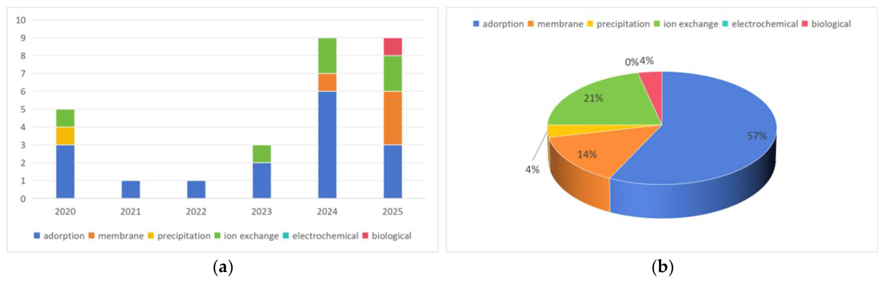
Figure 2.
Technical Roadmap for Medical Radioactive Wastewater Treatment.
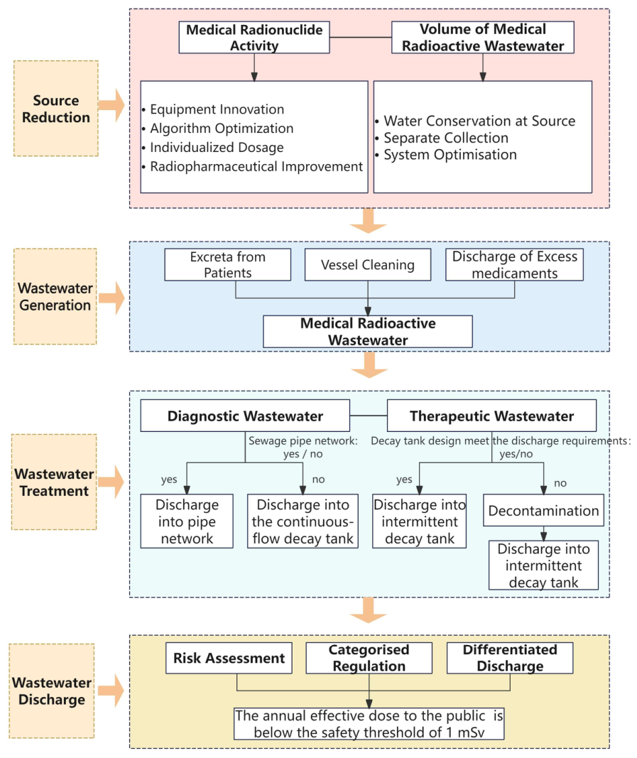

Table 1.
Main physical properties and medical applications of common nuclear medicine radionuclides.
Table 1.
Main physical properties and medical applications of common nuclear medicine radionuclides.
| Radionuclide | Half-life | Main radiation type | Clinical application | Category |
|---|---|---|---|---|
| ⁹⁹mTc | 6.02 h | γ-ray | SPECT imaging of multiple organs (most widely used) | Diagnostic |
| ¹⁸F | 109.8 min | β⁺ (positron) | PET/CT imaging for tumours, the brain and the myocardium | Diagnostic |
| ⁶⁸Ga | 67.7 min | β⁺ (positron) | PET/CT imaging for prostate cancer and neuroendocrine tumours | Diagnostic |
| ¹²³I | 13.2 h | γ-ray | Thyroid function and nodule diagnosis | Diagnostic |
| ¹³¹I | 8.02 days | β⁻ + γ | Hyperthyroidism, differentiated thyroid cancer (therapy + imaging) | Theranostic |
| ¹⁷⁷Lu | 6.64 d | β⁻ + γ | Targeted therapy for neuroendocrine tumours, prostate cancer | Therapeutic |
| ⁹⁰Y | 64.1 h | β⁻ | Liver cancer microspheres, tumour-targeted therapy | Therapeutic |
| ²²³Ra | 11.4 days | α-ray | Treatment of bone metastases in prostate cancer | Therapeutic |
| ²²⁵Ac | 10.0 days | α-ray | Targeted alpha therapy for advanced solid tumours | Therapeutic |
| ⁸⁹Sr | 50.5 d | β⁻ | Palliative analgesia for bone metastases | Therapeutic |
| ¹⁵³Sm | 46.3 h | β⁻ + γ | Pain relief and lesion inhibition in bone metastases | Therapeutic |
| ³²P | 14.3 days | β⁻ | Haematological diseases, cutaneous haemangioma | Therapeutic |
| ¹²⁵I | 59.4 days | γ-ray | In vitro assay, brachytherapy seed implantation | Adjuvant/ Therapeutic |
Table 2.
Proportion of administered activity discharged to sewers.
| Nuclide and form | Disease or condition treated | Proportion of activity discharged to sewers (%) |
|---|---|---|
| 198 Au colloid | Malignant disease | 0 |
| 131 I | Hyperthyroidism | 54 |
| 131 I | Thyroid carcinoma | 84-90 |
| 131 I MIBG1 | Pheochromocytoma | 89 |
| 32 P phosphate | Polycythaemia, etc. | 42 |
| 89 Sr chloride | Bone metastases | 92 |
1 meta-iodobenzylguanidine (MIBG).
Table 3.
Approaches to the treatment of medical radioactive waste in different countries and institutions.
Table 3.
Approaches to the treatment of medical radioactive waste in different countries and institutions.
| Radioactive Contaminants | Require collection or Not | Treatment Method | |
|---|---|---|---|
| WHO standard | Radioactive wastewater | Required | Stored in a secure area and discharged after decay to background levels |
| ICRP Publication 25 | Liquids spilled during laboratory operations and normal cleaning solutions for glassware | Not required | Treated via the drainage and sewage systems |
| Blood and excreta from patients undergoing radionuclide therapy | Not required | Discharged via bathroom drains | |
| ICRP Publication 105 | Excreta from patients undergoing radionuclide therapy | Not recommended | Discharged into the sewage system |
| IAEA | Excreta from patients undergoing diagnostic imaging | Not required | Discharged via the toilet drain |
| Excreta from patients undergoing therapy | Not required | Discharged after dilution in a continuous wastewater treatment system | |
| China | Radioactive liquid and solid waste | Required | a) Radioactive liquid waste containing nuclides with a half-life of less than 24 hours may be released directly after a storage period exceeding 30 days; b) Radioactive liquid waste containing nuclides with a half-life of more than 24 hours may be released after a storage period exceeding 10 times the longest half-life (including a storage period exceeding 180 days for waste containing iodine-131) |
| Italy | Radioactive waste liquids discharged by patients | Generally required | Stored in a dedicated septic tank for approximately 10 hours |
| UK | Radioactive liquid and solid waste | Subject to the limits specified by the wastewater treatment operator | If the discharge limits are not exceeded, the waste may be discharged into the sewage system; however, radioactive barium sulphate must not be discharged |
| Spain | Excreta from patients treated in the nuclear medicine department | Not required | Discharged via the toilet drain |
| France | Urine from patients treated with short-lived radioactive elements | Must | Collected in a shielded room and connected to a septic tank to delay and allow the decay |
| Urine from patients treated with radioactive elements having a long half-life | - | Handled by a designated facility | |
| USA | Excreta from patients undergoing medical diagnosis or treatment with radioactive substances | Not required | Discharged directly into the municipal sewerage system |
| Oman | Radioactive wastewater | Recommended | In areas lacking a centralised sewage system, decay tanks are still recommended as a safe disposal method. |
Table 4.
Discharge standards for medical radioactive wastewater in different countries.
| Country | Regulations and Standards | Control Method | Standard Values |
|---|---|---|---|
| China | GB 18466-2005 | Concentration | Total α activity at the total discharge outlet for radioactive waste liquid ≤ 1 Bq/L, total β activity ≤ 10 Bq/L, and the activity concentration of 131I ≤ 10 Bq/L |
| HJ 1188—2021 | Concentration | Total α ≤ 1 Bq/L and total β ≤ 10 Bq/L at the total discharge outlet for radioactive waste liquid; the activity concentration of 131I shall not exceed 10 Bq/L | |
| GB 18871-2002 | Concentration and total | Firstly, discharges must not exceed the discharge limits approved by the regulatory authority, including both total discharge limits and concentration limits; Secondly, for waste liquid meeting the criteria for low-level radioactive waste, the discharge requirements are as follows: 1) The total activity discharged per month shall be ≤ 10 ALImin; 2) The activity of each discharge shall be ≤ 1 ALImin, and the discharge point shall be flushed with a volume of water not less than three times the discharge volume after each discharge. |
|
| USA | 10 CFR Part 20 - Standards for Protection Against Radiation | Monthly average concentration and annual total | The monthly average concentration must not exceed the specified limit; the total annual amount of radioactive material must not exceed 185 GBq for 3H and 37 GBq for 14C, and the total amount of other radioactive materials must not exceed 37 GBq |
| France | ASN Position Papers & Technical Instructions | Concentration | The total radioactivity level in wastewater discharged by healthcare facilities into the municipal sewer system must be below 10 Bq/L; the discharge limit for 131I may be relaxed to 100 Bq/L |
| UK | The Environmental Permitting (England and Wales) Regulations 2016 | Monthly average concentration and annual total | Must not exceed the prescribed concentration and total quantity limits |
| India | AERB/NRF/SG/RW-10 | Annual total, daily total and monthly average concentration | The total annual discharge shall not exceed 37 GBq; for 131I, the daily discharge shall not exceed 3.7 MBq and the monthly average concentration shall not exceed 22.2 MBq/m³ |
| Oman | Ministerial Decree No.249/97 | Daily total and monthly average concentration |
131I daily emission limit: 3.7 MBq, Monthly average concentration limit: 22.2 MBq/m³ |
| Slovenia | 96/29/Euratom of 13 May | Annual total and quarterly total | Annual total 200 GBq, quarterly total 80 GBq |
| Brazil | Resolução CNEN 167/14 | Monthly average concentration and annual total | For 131I in liquid waste, the total monthly activity released must not exceed 1.9×10⁴ Bq/m³, and the total annual amount of 131I discharged into the sanitary sewer system must not exceed 1×10⁷ Bq |
Disclaimer/Publisher’s Note: The statements, opinions and data contained in all publications are solely those of the individual author(s) and contributor(s) and not of MDPI and/or the editor(s). MDPI and/or the editor(s) disclaim responsibility for any injury to people or property resulting from any ideas, methods, instructions or products referred to in the content. |
© 2026 by the authors. Licensee MDPI, Basel, Switzerland. This article is an open access article distributed under the terms and conditions of the Creative Commons Attribution (CC BY) license (http://creativecommons.org/licenses/by/4.0/).
Copyright: This open access article is published under a Creative Commons CC BY 4.0 license, which permit the free download, distribution, and reuse, provided that the author and preprint are cited in any reuse.


