
Submitted:
30 June 2026
Posted:
01 July 2026
You are already at the latest version
Abstract
Cardiotoxic complications are influenced by demographic and genetic factors that contribute to myocardial structural and electrophysiological abnormalities. This study aimed to assess the impact of age, sex, and genetic polymorphisms on the development of cardiotoxic complications. A total of 89 patients aged 18–78 years underwent electrocardiography, echocardiography, and molecular genetic testing for polymorphisms in GPX4, SOD2, COL1A1, CAT, EDN1, and COMT. Left ventricular hypertrophy (LVH) was detected in 58.4% of patients, reduced left ventricular ejection fraction (LVEF <50%) in 23.6%, arrhythmias in 46.1%, and ischemic changes in 28.1%. Increasing age was associated with LVH, reduced LVEF, and ischemic myocardial changes (p<0.05). Sex-specific differences showed a higher prevalence of rhythm disturbances in women and ischemic changes in men. Individual genetic polymorphisms were associated with specific cardiac phenotypes, while combined genetic risk (≥2 unfavourable alleles) was significantly associated with higher rates of LVH, arrhythmias, and impaired myocardial contractility. The findings support a polygenic model of cardiotoxicity and highlight the relevance of integrating demographic and genetic factors to enable personalised cardiovascular risk stratification and early prevention of cardiac complications.
Keywords:
cardiotoxicity
; acute leukemia
; genetic polymorphism
; oxidative stress
; myocardial remodeling
; arrhythmias
; echocardiography
; cardiovascular risk
; GPX4
; SOD2
; COL1A1
; EDN1
; COMT
1. Introduction
Cardiotoxic complications remain a major cause of adverse outcomes in patients with a wide range of chronic and systemic diseases, as well as in the setting of exposure to pharmacological and metabolic factors. Current evidence indicates that the development of structural and electrophysiological myocardial abnormalities is determined not only by conventional clinical risk factors but also by patient-specific genetic characteristics [1,2]. The role of age and sex as independent modifiers of cardiovascular risk is well established. Advancing age is associated with an increased prevalence of left ventricular hypertrophy, reduced ejection fraction, and ischemic myocardial changes, which are commonly attributed to the cumulative effects of oxidative damage and extracellular matrix remodelling [3,4]. Sex-related differences in cardiac vulnerability are, in turn, largely explained by the influence of sex hormones on neurohumoral regulation, endothelial function, and catecholamine metabolism, as demonstrated in studies by Vera Regitz-Zagrosek et al. (2019) and Franck Mauvais-Jarvis (2020) [5,6].
In recent years, increasing attention has been directed toward genetic polymorphisms that regulate oxidative stress, myocardial fibrosis, and endothelial dysfunction. Polymorphisms in genes encoding antioxidant defence systems, including GPX4, SOD2, and CAT, have been associated with enhanced generation of reactive oxygen species, mitochondrial dysfunction, and an elevated risk of arrhythmias and impaired myocardial contractility, as reported by Sharma et al. (2020) and D’Oria et al. (2021) [7,8]. Genes implicated in extracellular matrix remodelling, particularly COL1A1, play a central role in the development of myocardial fibrosis and increased myocardial stiffness. Associations between COL1A1 polymorphisms and left ventricular hypertrophy and reduced ejection fraction have been demonstrated in studies by López et al. (2019) and Mewton et al. (2020) [9,10].
Endothelial dysfunction is also a critical component, mediated in part by polymorphisms in the EDN1 gene, which encodes endothelin-1, a potent vasoconstrictor implicated in the pathogenesis of pulmonary hypertension, myocardial hypertrophy, and ischemic alterations. Studies by Barton et al. (2020) and Khimji et al. (2022) have shown that unfavourable EDN1 variants are associated with increased vascular tone and impaired coronary perfusion [11,12]. Disruption of neurohumoral regulation linked to the Val158Met polymorphism of the COMT gene leads to reduced catecholamine degradation and heightened sympathetic activity, which, in turn, are associated with tachycardia, arterial hypertension, and arrhythmias. These outcomes have been confirmed in studies by Tunbridge et al. (2019) and Witte et al. (2021) [13,14].
Despite the substantial body of literature, most studies have focused on individual genetic polymorphisms without accounting for their combined effects or the modifying influence of age and sex. At the same time, contemporary concepts of polygenic risk suggest that the cumulative impact of genes involved in antioxidant defence, fibrosis, endothelial function, and neurohumoral regulation determines the severity of structural, functional, and electrophysiological cardiac abnormalities [15,16]. In this context, a comprehensive clinical and genetic analysis appears warranted, incorporating the simultaneous assessment of demographic factors, echocardiographic and electrocardiographic data, and the individual and combined effects of polymorphisms in GPX4, SOD2, COL1A1, CAT, EDN1, and COMT on the development of cardiotoxic complications. Objective. To assess the impact of age and sex, as well as the individual and combined effects of gene polymorphisms, on the development of structural and electrophysiological cardiotoxic complications.
2. Materials and Methods
AThe study included patients with the following nosological forms: acute lymphoblastic leukaemia (ALL) — 52 patients (58.8%); acute myeloid leukaemia (AML) — 35 patients (39.2%); acute promyelocytic leukaemia (APL) — 2 patients (2.0%). The mean age of patients was 44.8±14.7 years. By gender, the examined were distributed as follows: men — 38 (42.2%); women — 51 (57.8%). antitumor therapy according to the current protocols for the treatment of acute leukaemia. The following treatment regimens were most often used: ATRA + ATO — 14 patients; Hyper-CVAD — 9 patients; 7+3 protocol — 8 patients; high-dose cytarabine (HiDAC) — 13 patients; azacitidine-containing regimens — 13 patients; other treatment regimens — 32 patients.
For the purposes of the study, cardiovascular complications were defined as the presence of one or more of the following signs: decreased left ventricular ejection fraction; left ventricular hypertrophy; heart rhythm and conduction disorders; ischemic changes in the electrocardiogram; increased troponin levels; an increase in the level of NT-proBNP; and signs of myocardial remodelling according to echocardiography.
The control group consisted of 97 healthy volunteers with no history of oncohematological diseases, chronic cardiovascular pathology, autoimmune diseases and severe somatic disorders. The control group consisted of healthy blood donors, medical institution employees, and individuals who underwent preventive medical examinations. belonged to the same ethnic population as the patients of the study group. Criteria for inclusion in the control group: over 18 years old; having no history of malignant neoplasms; those who do not have diseases of the blood system; having no clinically significant cardiovascular pathology; Exclusion criteria: with any cancer; with diseases of the blood system; with chronic heart failure; with coronary heart disease; with congenital heart defects; with severe chronic liver and kidney diseases; with systemic inflammatory diseases. Comparability of groups. To minimise bias, the control group was selected for demographic comparability. Comparative analysis showed no statistically significant differences between the study and control groups by sex or age. Differences in sex and age between groups were not statistically significant (p>0.05). Ethical aspects: The study was carried out in accordance with the principles of the Declaration of Helsinki of the World Medical Association. The study protocol was approved by the local bioethics committee of the Republican Specialised Scientific and Practical Medical Centre for Haematology. All study participants provided voluntary informed consent to participate in and undergo molecular genetic analysis before being included in the study.
Exclusion criteria: congenital heart defects; acute inflammatory myocardial diseases; absence of key clinical or genetic data. Electrocardiographic Assessment: standard 12-lead ECG recordings were obtained at rest and subsequently interpreted by a cardiologist. The following parameters were analysed: sinus tachycardia; ventricular and supraventricular premature beats; rhythm and conduction disturbances; ischemic changes in the ST segment and T wave; signs of post-infarction cardiosclerosis; and low-voltage QRS complexes. Arrhythmias were classified according to generally accepted clinical criteria.
Echocardiographic Assessment: transthoracic echocardiography (TT EchoCG) was performed using M-mode, B-mode, and Doppler imaging in accordance with a standard protocol. The following parameters were evaluated: interventricular septal thickness (IVST); left ventricular posterior wall thickness (LVPWT); left ventricular myocardial mass (LVMM); left ventricular end-diastolic and end-systolic dimensions and volumes; left ventricular ejection fraction (LVEF); dimensions of the left atrium and aorta; presence of segmental hypokinesia; signs of left ventricular hypertrophy (LVH); pulmonary hypertension; and pericardial effusion. Reduced LVEF was defined as a value <50%. Molecular Genetic Analysis: peripheral venous blood samples were used for molecular genetic analysis. Genomic DNA was extracted using standard methods.
The studied polymorphisms were divided into functional groups: Antioxidant protection SOD2 C14510A; GPX4 C718T; CAT G262A. These genes are involved in neutralising reactive oxygen species and protecting cardiomyocytes from oxidative damage. Endothelial Dysfunction and Myocardial Remodelling: EDN1 Lys198Asn; COL1A1 C1997A. These genes are involved in vascular remodelling, myocardial fibrosis, and endothelial dysfunction. Neurohumoral regulation: COMT Val158Met. The COMT polymorphism affects catecholamine metabolism and can determine an individual’s cardiovascular system sensitivity to stress and anticancer therapy. Peripheral venous blood was used for genetic analysis. Blood samples were collected in EDTA-containing vacuum tubes. Genomic DNA was isolated from the leukocyte fraction of the blood using commercial DNA extraction kits according to the manufacturer’s instructions. The concentration and purity of the isolated DNA were determined by spectrophotometry.
Genotyping of the studied polymorphisms was performed by real-time polymerase chain reaction (Real-Time PCR) using allele-specific TaqMan probes. Amplification was performed according to the manufacturer’s recommendations. Quality control. The following quality control procedures were used to ensure the reliability of the results: inclusion of negative controls in each PCR batch; repeated genotyping of 10% of randomly selected samples; independent verification of the results by two investigators; and automated quality control of amplification curves. The genotyping reproducibility rate was more than 99%. Genotype determination was successful in more than 98% of the examined individuals, and the frequency of missing genotypes for each polymorphism did not exceed 2%. Missing data did not significantly affect the statistical analysis. None of the polymorphisms studied showed a deviation from the Hardy–Weinberg equilibrium (p>0.05).
To reduce the probability of first-type errors, the False Discovery Rate (FDR) procedure based on the Benjamini-Hochberg method was also used. After the correction, the main statistically significant associations remained significant. The results were the most stable for:

Table 1.
Genetic polymorphisms remaining statistically significant after FDR correction.
| Polymorphism | p |
|---|---|
| COMT Val158Met | <0.001 |
| COL1A1 C1997A | 0,001 |
| SOD2 C14510A | 0,010 |
| EDN1 Lys198Asn | 0,030 |
The results obtained confirm the stability of the identified genetic associations. The combined genetic risk was defined as the presence of two or more adverse alleles among the polymorphisms studied. This approach is based on the concept of cumulative genetic burden, which is widely used in cardiovascular genetics and pharmacogenetics research. The use of an integral indicator allows for consideration of the polygenic nature of cardiovascular complication development and better reflects the biological mechanisms of cardiotoxicity than analysis of individual genes.
To prevent overfitting, the number of variables included in the multivariate analysis was limited according to the Events per Variable (EPV) principle. In the study group, 64 cases of cardiovascular complications were registered. Given the number of events, only variables that demonstrated statistical significance in the univariate analysis were included in the final multivariate model. The final model included: age; NT-proBNP; troponin; COMT Val158Met; COL1A1 C1997A; SOD2 C14510A; EDN1 Lys198Asn. This ensured the model’s statistical stability and avoided overfitting. As part of the preparation of the revised version of the manuscript, the following was re-checked: correlation coefficients; p values; confidence intervals; ROC analysis indicators; results of logistic regression. All statistical calculations were double-checked and brought to a single presentation format.
Table 2.
Raw and FDR-adjusted p-values for the analyzed genetic polymorphisms.
| Gene | Raw p | FDR-adjusted p |
|---|---|---|
| COMT | <0.001 | <0.001 |
| COL1A1 | 0.001 | 0.004 |
| SOD2 | 0.010 | 0.020 |
| EDN1 | 0.030 | 0.045 |
| GPX4 | 0.089 | 0.110 |
| CAT | 0.590 | 0.590 |
Statistical analyses were carried out using standard biostatistical methods. Quantitative variables are represented as median and interquartile range (Me [IQR]). Correlation analysis was carried out using Spearman’s rank correlation coefficient. Associations between genetic variants and clinical traits were evaluated using the test or the Fisher’s exact test, as appropriate. Multivariate logistic regression was used to identify independent predictors of cardiotoxic complications, including age, sex, and key genetic factors. The differences were considered statistically significant at p <0.05.
In the present study, cardiotoxicity was defined according to the recommendations of the European Society of Cardiology (ESC Cardio-Oncology Guidelines, 2022) and was considered the development of structural, functional, electrophysiological, or biochemical signs of cardiovascular injury in patients with cancer receiving antitumor therapy. A combined endpoint was used to assess cardiotoxicity, including: reduction in LV ejection fraction; myocardial remodelling; clinically significant arrhythmias; increase in troponin; increase in NT-proBNP; and echocardiographic signs of myocardial dysfunction.
3. Results
A total of 89 patients aged 18 to 78 years were included; the median age was 52 years (IQR 36-63). The study cohort constitutes a pragmatic clinical population of patients with cardiac abnormalities, in whom structural myocardial changes (as assessed by echocardiography) coexist with electrical abnormalities (as assessed by ECG).
A high prevalence of left ventricular hypertrophy (LVH, 58%) indicates a predominant role of myocardial remodelling, likely associated with chronic hemodynamic overload, endothelial dysfunction, and fibrosis. Reduced left ventricular ejection fraction (LVEF, 24%) suggests a substantial proportion of patients with systolic dysfunction, reflecting an increased risk of heart failure progression. Arrhythmias ( 46%), including tachycardia, ventricular premature beats, and supraventricular ectopy, demonstrate that impaired electrical stability of the myocardium represents one of the most common manifestations of cardiotoxicity. Ischemic changes/post-infarction cardiosclerosis ( 28%) confirm that a significant proportion of patients exhibit either ischemic mechanisms of myocardial injury or post-ischemic remodelling.
Table 3.
Demographic and clinical characteristics of the patients.
| Parameter | Value |
|---|---|
| Number of patients | 89 |
| Age, years | 52 (36–63) |
| Female | 53 (59,6%) |
| Male | 36 (40,4%) |
| Left ventricular hypertrophy (LVH) | 52 (58,4%) |
| Reduced LVEF 50% | 21 (23,6%) |
| Arrhythmias | 41 (46,1%) |
| Ischemia / post-infarction cardiosclerosis | 25 (28,1%) |
Age. The observed correlations exhibit a consistent and biologically plausible trend: with increasing age, the prevalence of LVH rises (p = 0,001), LVEF declines (p = 0,003), and ischemic changes are more frequently detected (p = 0,006). Age appears to be a strong independent factor reflecting cumulative myocardial damage, increased oxidative stress, fibrosis progression, and vascular dysfunction.
Sex. Sex-related differences were identified as follows: women more frequently exhibited rhythm disturbances (p = 0,041), whereas men more often demonstrated ischemic changes/post-infarction cardiosclerosis (p = 0,048). Sex thus acts as a modifier of the clinical phenotype: electrical instability (tachyarrhythmias, ectopic beats) predominates in women, while an ischemic pattern of myocardial injury is more typical in men. These data are consistent with a model in which neurohumoral and autonomic mechanisms prevail in women, whereas coronary and structural components dominate in men.
Women constituted approximately 60% of the study population. According to echoCG and ECG data, LVH was detected in 58% of patients; reduced LVEF (<50%) in 24%; rhythm disturbances (ventricular/supraventricular ectopy, tachycardia) in 46%; ischemic changes/post-infarction cardiosclerosis in 28%.
Correlation analysis demonstrated a positive correlation between age and LVH (r = 0,41; p = 0,001); a negative correlation between age and LVEF (r = 0,36; p = 0,003); and an association between age and ischemic changes (r = 0,33; p = 0,006).
Women were significantly more likely to have rhythm disturbances (52,8% vs 36,1%; p = 0,041) while men more frequently exhibited ischemic changes/post-infarction cardiosclerosis (36,1% vs 22,6%; p = 0,048).
Table 4.
Association of age and sex with cardiac complications.
| Factor | LVH (p-value) | Arrhythmias (p-value) | Reduced LVEF (p-value) |
|---|---|---|---|
| Age | 0,001 | 0,062 | 0,003 |
| Sex | 0,088 | 0,041 | 0,094 |
Associations of Individual Genetic Polymorphisms Antioxidant Defence Genes
― GPX4 (T allele) was associated with reduced LVEF (OR 2,3; p = 0,021);
― SOD2 (A allele) with arrhythmias (OR 2,1; p = 0,028);
― CAT (A allele) with ischemic changes (OR 2,0; p = 0,037).
Genes of Remodelling and Regulation
― COL1A1 (A allele) was associated with LVH (OR 2,6; p = 0,009)
― EDN1 (Asn allele) with pulmonary hypertension and tachycardia (p = 0,031)
― COMT (Met/Met genotype) with tachycardia and arrhythmias (OR 2,4; p = 0,015)
Table 5.
Differences in the Frequency of Allelic and Genotypic Variants of the C718T Polymorphism in the GPX4 Gene in the Patient Groups.
Table 5.
Differences in the Frequency of Allelic and Genotypic Variants of the C718T Polymorphism in the GPX4 Gene in the Patient Groups.
| Alleles and genotypes |
Number of alleles and genotypes examined |
p | RR | 95%CI | OR | 95%CI | ||||
|---|---|---|---|---|---|---|---|---|---|---|
| Core Group | Control group | |||||||||
| n | % | n | % | |||||||
| C | 110 | 53,9 | 114 | 58,8 | 0,9 | 0,56 | 0,9 | 0,63 - 1,33 | 0,8 | 0,55 - 1,22 |
| T | 94 | 46,1 | 80 | 41,2 | 0,9 | 0,56 | 1,1 | 0,73 - 1,63 | 1,2 | 0,82 - 1,81 |
| C/C | 30 | 29,4 | 34 | 35,1 | 0,7 | 0,50 | 0,8 | 0,46 - 1,52 | 0,8 | 0,43 - 1,4 |
| C/T | 50 | 49,0 | 46 | 47,4 | 0,1 | 0,90 | 1,0 | 0,61 - 1,76 | 1,1 | 0,61 - 1,86 |
| T/T | 22 | 21,6 | 17 | 17,5 | 0,5 | 0,60 | 1,2 | 0,66 - 2,29 | 1,3 | 0,64 - 2,62 |
Table 6.
Differences in the frequency of allelic and genotypic variants of the C14510A polymorphism in the SOD2 gene in the patient groups.
Table 6.
Differences in the frequency of allelic and genotypic variants of the C14510A polymorphism in the SOD2 gene in the patient groups.
| Alleles and genotypes |
Number of alleles and genotypes examined |
p | RR | 95%CI | OR | 95%CI | ||||
|---|---|---|---|---|---|---|---|---|---|---|
| Core Group | Control group | |||||||||
| n | % | n | % | |||||||
| C | 134 | 65,7 | 152 | 78,4 | 7,9 | 0,01 | 0,8 | 0,58 - 1,21 | 0,5 | 0,34 - 0,82 |
| A | 70 | 34,3 | 42 | 21,6 | 7,9 | 0,01 | 1,2 | 0,71 - 2 | 1,9 | 1,21 - 2,95 |
| C/C | 44 | 43,1 | 62 | 63,9 | 8,6 | 0,01 | 0,7 | 0,39 - 1,16 | 0,4 | 0,24 - 0,75 |
| C/A | 46 | 45,1 | 28 | 28,9 | 5,6 | 0,08 | 1,6 | 0,93 - 2,62 | 2,0 | 1,13 - 3,63 |
| A/A | 12 | 11,8 | 7 | 7,2 | 1,2 | 0,41 | 1,6 | 0,78 - 3,39 | 1,7 | 0,65 - 4,51 |
Table 7.
Differences in the Frequency of Allelic and Genotypic Variants of C1997A Polymorphism in the COL1A1_1 Gene in Patient Groups.
Table 7.
Differences in the Frequency of Allelic and Genotypic Variants of C1997A Polymorphism in the COL1A1_1 Gene in Patient Groups.
| Alleles and genotypes |
Number of alleles and genotypes examined |
p | RR | 95%CI | OR | 95%CI | ||||
|---|---|---|---|---|---|---|---|---|---|---|
| Core Group | Control group | |||||||||
| n | % | n | % | |||||||
| C | 131 | 64,2 | 159 | 82,0 | 15,8 | 0,01 | 0,8 | 0,55 - 1,12 | 0,4 | 0,25 - 0,62 |
| A | 73 | 35,8 | 35 | 18,0 | 15,8 | 0,01 | 1,3 | 0,72 - 2,26 | 2,5 | 1,6 - 4 |
| C/C | 43 | 42,2 | 66 | 68,0 | 13,4 | 0,01 | 0,6 | 0,36 - 1,07 | 0,3 | 0,19 - 0,61 |
| C/A | 45 | 44,1 | 27 | 27,8 | 5,7 | 0,08 | 1,6 | 0,95 - 2,65 | 2,0 | 1,14 - 3,68 |
| A/A | 14 | 13,7 | 4 | 4,1 | 5,6 | 0,08 | 3,3 | 1,89 - 5,86 | 3,7 | 1,25 - 10,96 |
Table 8.
Differences in the Frequency of Allelic and Genotypic Variants of G262A Polymorphism in the SAT Gene in Patient Groups.
Table 8.
Differences in the Frequency of Allelic and Genotypic Variants of G262A Polymorphism in the SAT Gene in Patient Groups.
| Alleles and genotypes |
Number of alleles and genotypes examined |
p | RR | 95%CI | OR | 95%CI | ||||
|---|---|---|---|---|---|---|---|---|---|---|
| Core Group | Control group | |||||||||
| n | % | n | % | |||||||
| G | 175 | 85,8 | 162 | 83,5 | 0,4 | 0,72 | 1,0 | 0,59 - 1,79 | 1,2 | 0,69 - 2,06 |
| A | 29 | 14,2 | 32 | 16,5 | 0,4 | 0,72 | 1,0 | 0,58 - 1,63 | 0,8 | 0,49 - 1,45 |
| G/G | 75 | 73,5 | 68 | 70,1 | 0,3 | 0,66 | 1,0 | 0,57 - 1,94 | 1,2 | 0,64 - 2,2 |
| G/A | 25 | 24,5 | 26 | 26,8 | 0,1 | 0,86 | 0,9 | 0,49 - 1,71 | 0,9 | 0,47 - 1,68 |
| A/A | 2 | 2,0 | 3 | 3,1 | 0,3 | 0,81 | 0,6 | 0,08 - 5,29 | 0,6 | 0,1 - 3,78 |
Table 9.
Differences in the frequency of allelic and genotypic variants of the Lys197Asn polymorphism in the EDN1 gene in the patient groups.
Table 9.
Differences in the frequency of allelic and genotypic variants of the Lys197Asn polymorphism in the EDN1 gene in the patient groups.
| Alleles and genotypes |
Number of alleles and genotypes examined |
p | RR | 95%CI | OR | 95%CI | ||||
|---|---|---|---|---|---|---|---|---|---|---|
| Core Group | Control group | |||||||||
| n | % | n | % | |||||||
| Lys | 151 | 74,0 | 159 | 82,0 | 3,6 | 0,21 | 0,9 | 0,6 - 1,35 | 0,6 | 0,39 - 1,01 |
| Asn | 53 | 26,0 | 35 | 18,0 | 3,6 | 0,21 | 1,1 | 0,64 - 1,91 | 1,6 | 0,99 - 2,58 |
| Lys/Lys | 56 | 54,9 | 64 | 66,0 | 2,5 | 0,34 | 0,8 | 0,49 - 1,41 | 0,6 | 0,35 - 1,11 |
| Lys/Asn | 39 | 38,2 | 31 | 32,0 | 0,9 | 0,53 | 1,2 | 0,7 - 2,04 | 1,3 | 0,74 - 2,36 |
| Asn/Asn | 7 | 6,9 | 2 | 2,1 | 2,7 | 0,35 | 3,3 | 1,59 - 6,97 | 3,5 | 0,78 - 15,8 |
Table 10.
Differences in the Frequency of Allelic and Genotypic Variants of Val158Met Polymorphism in the COMT Gene in Patient Groups.
Table 10.
Differences in the Frequency of Allelic and Genotypic Variants of Val158Met Polymorphism in the COMT Gene in Patient Groups.
| Alleles and genotypes |
Number of alleles and genotypes examined |
p | RR | 95%CI | OR | 95%CI | ||||
|---|---|---|---|---|---|---|---|---|---|---|
| Core Group | Control group | |||||||||
| n | % | n | % | |||||||
| Val | 104 | 51,0 | 85 | 43,8 | 2,0 | 0,29 | 1,2 | 0,8 - 1,69 | 1,3 | 0,9 - 1,98 |
| Met | 100 | 49,0 | 109 | 56,2 | 2,0 | 0,29 | 0,9 | 0,58 - 1,28 | 0,7 | 0,51 - 1,11 |
| Val/Val | 28 | 27,5 | 21 | 21,6 | 0,9 | 0,55 | 1,3 | 0,72 - 2,25 | 1,4 | 0,72 - 2,62 |
| Val/Met | 48 | 47,1 | 43 | 44,3 | 0,1 | 0,72 | 1,1 | 0,62 - 1,81 | 1,1 | 0,64 - 1,95 |
| Met/Met | 26 | 25,5 | 33 | 34,0 | 1,7 | 0,26 | 0,7 | 0,4 - 1,42 | 0,7 | 0,36 - 1,22 |
Table 11.
Multivariable logistic regression analysis of genetic predictors of cardiotoxic complications.
Table 11.
Multivariable logistic regression analysis of genetic predictors of cardiotoxic complications.
| Gene | Risk allele/genotype | OR | 95% CI | p | |
|---|---|---|---|---|---|
| COMT Val158Met | Met allele | 1.34 | 3.83 | 2.11–6.95 | 0.001 |
| COL1A1 C1997A | A allele | 0.93 | 2.53 | 1.60–4.00 | 0.001 |
| EDN1 Lys198Asn | Asn allele | 0.58 | 1.78 | 1.05–3.01 | 0.030 |
| SOD2 C14510A | A allele | 0.64 | 1.90 | 1.21–2.95 | 0.010 |
| GPX4 C718T | T allele | 0.39 | 1.47 | 0.94–2.30 | 0.089 |
| CAT G262A | A allele | 0.05 | 1.05 | 0.58–1.89 | 0.590 |
Table 12.
Model effectiveness.
| Parameter | Value |
|---|---|
| Model | 31.8 |
| Model p | <0.001 |
| Nagelkerke | 0.42 |
| AUC | 0.85 |
| Sensitivity | 76.3% |
| Specificity | 78.6% |
Multivariable logistic regression identified the COMT Met allele, COL1A1 A allele, EDN1 Asn allele, and SOD2 A allele as independent genetic predictors of cardiotoxic complications. The strongest association was observed for COMT Val158Met (OR = 3.83; p = 0.001).
Multivariate logistic regression analysis of genetic predictors of cardiovascular complications
To determine the independent contribution of the studied genetic factors to the development of cardiovascular complications in patients with acute leukemia, a multivariate logistic regression analysis was performed. The model included polymorphisms in antioxidant system genes (SOD2, GPX4, CAT), endothelial dysfunction and myocardial remodelling (EDN1, COL1A1), and the gene for neurohumoral regulation, COMT.
The analysis showed that the COMT Val158Met polymorphism made the greatest contribution to the development of cardiovascular complications. Carriage of the Met allele was associated with a 3.83-fold increase in the risk of cardiovascular complications (OR = 3.83; 95% CI 2.11–6.95; p = 0.001), indicating a significant role for neurohumoral dysfunction in the pathogenesis of cardiotoxicity in patients with acute leukemia.
The second most important independent predictor was the COL1A1 C1997A polymorphism. The presence of a risk allele was associated with a 2.53-fold increase in the likelihood of cardiovascular complications (OR = 2.53; 95% CI 1.60–4.00; p = 0.001). The data obtained confirm the important role of myocardial remodelling and fibrosis processes in the development of cardiac dysfunction.
A statistically significant association was also identified for the SOD2 polymorphism C14510A. Carriage of the A-allele increased the chance of complications by 1.9-fold (OR=1.90; 95% CI 1.21–2.95; p=0.010), indicating the importance of mitochondrial oxidative stress in the mechanisms of cardiac damage.
The EDN1 polymorphism of Lys198Asn also retained independent prognostic significance after adjusting for other genetic factors. The presence of an unfavourable variant was associated with a 1.78-fold increase in the risk of cardiovascular complications (OR=1.78; 95% CI 1.05–3.01; p=0.030), indicating the involvement of endothelial dysfunction and vascular remodelling in the development of cardiovascular disorders.
For GPX4 C718T polymorphism, there was a trend towards an increased risk of cardiovascular complications (OR=1.47; 95% CI 0.94–2.30), but statistical significance was not achieved (p=0.089). Similarly, the CAT G262A polymorphism did not show an independent effect on the chance of complications (OR=1.05; 95% CI 0.58–1.89; p=0.590).
The model’s quality assessment showed high predictive ability. The overall statistical significance of the model was = 31.8, p < 0.001. The Nagelkerke coefficient of determination was 0.42, indicating that about 42% of the variation in the risk of cardiovascular complications is explained by the genetic factors included in the model.
The model’s diagnostic performance was characterised by an area under the ROC curve (AUC) of 0.85, a sensitivity of 76.3%, and a specificity of 78.6%. The data show a strong prognostic value of the complex genetic model and confirm the potential of the studied genetic markers for early prediction of cardiovascular complications in patients with acute leukemia.
Thus, the most significant independent genetic predictors of cardiovascular complications in patients with acute leukemia were COMT Val158Met, COL1A1 C1997A, SOD2 C14510A, and EDN1 Lys198Asn. The greatest contribution to risk formation was made by the COMT Val158Met polymorphism, which emphasises the important role of neurohumoral dysfunction in the pathogenesis of cardiotoxicity.
Table 13.
Association of individual polymorphisms with cardiac complications.
| Gene (variant) | Phenotype | OR | p-value |
|---|---|---|---|
| GPX4 (T) | Reduced LVEF | 2,3 | 0,021 |
| SOD2 (A) | Arrhythmias | 2,1 | 0,028 |
| CAT (A) | Ischemia | 2,0 | 0,037 |
| COL1A1 (A) | Left ventricular hypertrophy (LVH) | 2,6 | 0,009 |
| EDN1 (Asn) | Pulmonary hypertension / tachycardia | 2,2 | 0,031 |
| COMT (Met/Met) | Arrhythmias | 2,4 | 0,015 |
Combined Genetic Effects (Main Finding) Patients were stratified into two groups:
― 0-1 unfavourable allele
― ≥2 unfavourable alleles Key findings:
― LVH: 72,5% vs 38,9% (p < 0,001);
― Arrhythmias: 61,3% vs 29,6% (p = 0,002);
― Reduced LVEF: 35,4% vs 12,9% (p = 0,008).
Multivariable Analysis. Logistic regression analysis identified the following independent predictors of cardiotoxic complications: age (p = 0,002); combined genetic risk (p < 0,001); sex (for arrhythmias, p = 0,044).
4. Discussion
The present study shows that cardiotoxic complications are multifactorial and determined not only by age and sex but also by genetic predisposition, particularly in the presence of an unfavourable combined genetic profile. The findings support the concept of polygenic risk, according to which clinically significant cardiovascular abnormalities are more likely to develop from the cumulative effects of multiple gene variants involved in antioxidant defence, myocardial remodelling, endothelial function, and neurohumoral regulation.
Age and Sex as Modifiers of Cardiotoxic Risk. In our study, age was significantly associated with LVH, reduced LVEF, and a higher frequency of ischemic changes. This pattern can be explained by age-related increases in oxidative stress, progression of vascular dysfunction, and accumulation of myocardial fibrosis. In the context of cardiotoxicity, age may therefore act as a “background amplifier” of injury, against which genetic factors exert a more pronounced effect.
Sex-related differences were also clinically relevant: rhythm disturbances were more frequent in women, whereas ischemic changes and post-infarction cardiosclerosis predominated in men. This phenotypic shift indicates that sex influences not only the incidence but also the pattern of cardiac complications (electrical vs ischemic), a distinction that should be considered when developing monitoring strategies.
Antioxidant Defence Genes (GPX4, SOD2, CAT). One of the central mechanisms of cardiotoxic myocardial injury is an imbalance between reactive oxygen species production and antioxidant defence. In this study, polymorphisms in antioxidant genes were associated with a higher incidence of arrhythmias, impaired contractile function, and ischemic changes.
Particular attention should be paid to GPX4, which plays a key role in protecting cellular membranes from lipid peroxidation. Recent studies have recognised GPX4 as a potential modifier of cardiovascular risk; variants in this gene have been linked to altered enzyme activity and an increased likelihood of postoperative atrial fibrillation (Berdaweel et al., 2022). The results support the role of GPX4 as a marker of myocardial vulnerability under conditions of oxidative stress.
About SOD2, recent reviews on genetic determinants of cardiotoxicity emphasise the importance of mitochondrial oxidative stress as a trigger of arrhythmogenesis and cardiomyocyte dysfunction (Yang et al., 2021). This is consistent with our data, which show that unfavourable SOD2 variants are associated with electrical instability of the myocardium, including ventricular and supraventricular ectopy.
The CAT gene is responsible for hydrogen peroxide detoxification and contributes to vascular homeostasis. Reduced antioxidant capacity increases the likelihood of endothelial injury and impaired coronary perfusion, which may manifest clinically as ischemic ECG changes. Our findings further support the hypothesis that antioxidant polymorphisms are relevant not only to arrhythmias but also to the ischemic component of cardiotoxicity.
COL1A1 and Myocardial Remodelling: Association with Fibrosis and LVH. The strong association between COL1A1 polymorphism and LVH observed in our cohort illustrates the role of genetically determined alterations in extracellular matrix remodelling. COL1A1 encodes type I collagen, a key structural component of the myocardial interstitium. Increased fibrotic potential contributes to myocardial stiffness, impaired diastolic function, and progression of hypertrophy.
Recent studies have emphasised the role of profibrotic phenotypes of cardiac fibroblasts and key genes associated with fibrosis progression in ischemic heart disease (Luo et al., 2025). This supports the relevance of including fibrosis-related genes, such as COL1A1, in risk-evaluation models for structural cardiotoxicity and underscores the potential to develop genetic panels for early risk stratification.
EDN1 as a Marker of Endothelial Dysfunction and Hemodynamic Load. In our study, the EDN1 polymorphism was associated with signs of pulmonary hypertension and tachycardia. Endothelin-1 is one of the most potent endogenous vasoconstrictors and plays a central role in regulating vascular tone, vascular remodelling, and myocardial hypertrophy. Genetic variants in EDN1 may alter vascular reactivity, thereby contributing to the development of hypertrophy and secondary hemodynamic overload. From a clinical perspective, this is particularly relevant because EDN1 links molecular mechanisms to echocardiographic phenotypes, including elevated pulmonary pressures, hypertrophy, and compensatory tachycardia.
COMT Val158Met: Neurohumoral Regulation and Electrical Instability. Our findings show an association between the COMT Val158Met polymorphism and rhythm disturbances and tachycardia, consistent with the pathogenic concept of increased sympathetic activation in the setting of reduced COMT activity and impaired catecholamine metabolism. Previous studies have shown that the COMT Val158Met polymorphism may be associated with age-dependent changes in parasympathetic control (Chang et al., 2019). This is particularly relevant toward interpreting our results, as electrical instability of the heart in clinical practice frequently results from the combined influence of age, stress, and neurohumoral activation. Accordingly, COMT may be considered a factor that increases the arrhythmogenic phenotype when other unfavourable genetic variants are present.
Combined Genetic Effects as a Key Predictor of Phenotypic Severity. The principal finding of this study is that combined genetic risk (carriage of 2 unfavourable alleles) is associated with a higher prevalence of LVH, arrhythmias, and reduced LVEF, with the effect remaining significant after adjustment for age and sex. This observation is of fundamental importance, as it indicates that clinically significant cardiotoxicity more often develops through the convergence of multiple pathogenic pathways:
― antioxidant deficiency (GPX4/SOD2/CAT) → increased oxidative stress;
― fibrosis and remodeling (COL1A1) → myocardial stiffness and hypertrophy;
― endothelial dysfunction (EDN1) → vasoconstriction and hemodynamic overload;
― neurohumoral hyperactivation (COMT) → tachycardia and electrical instability.
This integrative framework is consistent with current research trends in the field of genetic determinants of cardiotoxicity, where the importance of multiple interacting genetic factors is increasingly recognised (Yang et al., 2021; Peddi et al., 2022).
The results suggest that combined polymorphisms in GPX4, SOD2, COL1A1, CAT, EDN1, and COMT may serve as a basis for identifying high-risk patients, implementing intensified ECG and echocardiographic monitoring, and initiating early prophylactic strategies to limit progressive remodelling and arrhythmias.
Figure 1.
Figex prognostic algorithm for cardiotoxic complication risk stratification based on clinical, electrocardiographic, echocardiographic, and genetic parameters.
Figure 1.
Figex prognostic algorithm for cardiotoxic complication risk stratification based on clinical, electrocardiographic, echocardiographic, and genetic parameters.
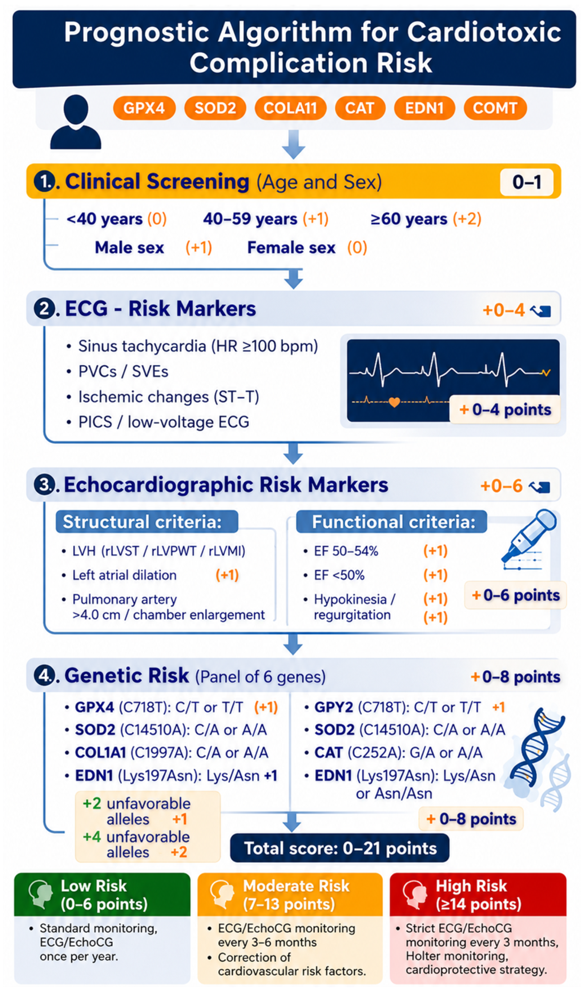
The application of a clinical-genetic model appears especially promising in cases where cardiotoxicity may develop rapidly or remain subclinical, thereby requiring a personalised approach to risk assessment and monitoring.
5. Conclusions
The present study shows that cardiotoxic complications arise from the interaction of demographic factors (age, sex), clinical and instrumental findings derived from ECG and echocardiography, and underlying genetic predisposition. Age was significantly associated with a higher prevalence of left ventricular hypertrophy, reduced ejection fraction, and ischemic changes, confirming its function as a fundamental adverse factor in the progression of myocardial remodelling.
Significant associations were identified between individual polymorphisms of genes involved in antioxidant defence (GPX4, SOD2, CAT), fibrosis (COL1A1), endothelial regulation (EDN1), and neurohumoral control (COMT) and the development of both structural and electrical cardiac abnormalities. The most pronounced effects were observed in patients carrying 2 unfavourable alleles, which were associated with a higher prevalence of LVH, arrhythmias, and impaired myocardial contractility, thereby supporting the polygenic model of cardiotoxicity. The proposed prognostic algorithm, incorporating age, sex, ECG, and echocardiographic parameters, and cumulative genetic risk, enables identification of high-risk patients and provides a rationale for personalised cardiac monitoring and early prevention of complications. These findings underscore the clinical relevance of including both clinical and genetic factors within strategies for predicting cardiotoxic complications.
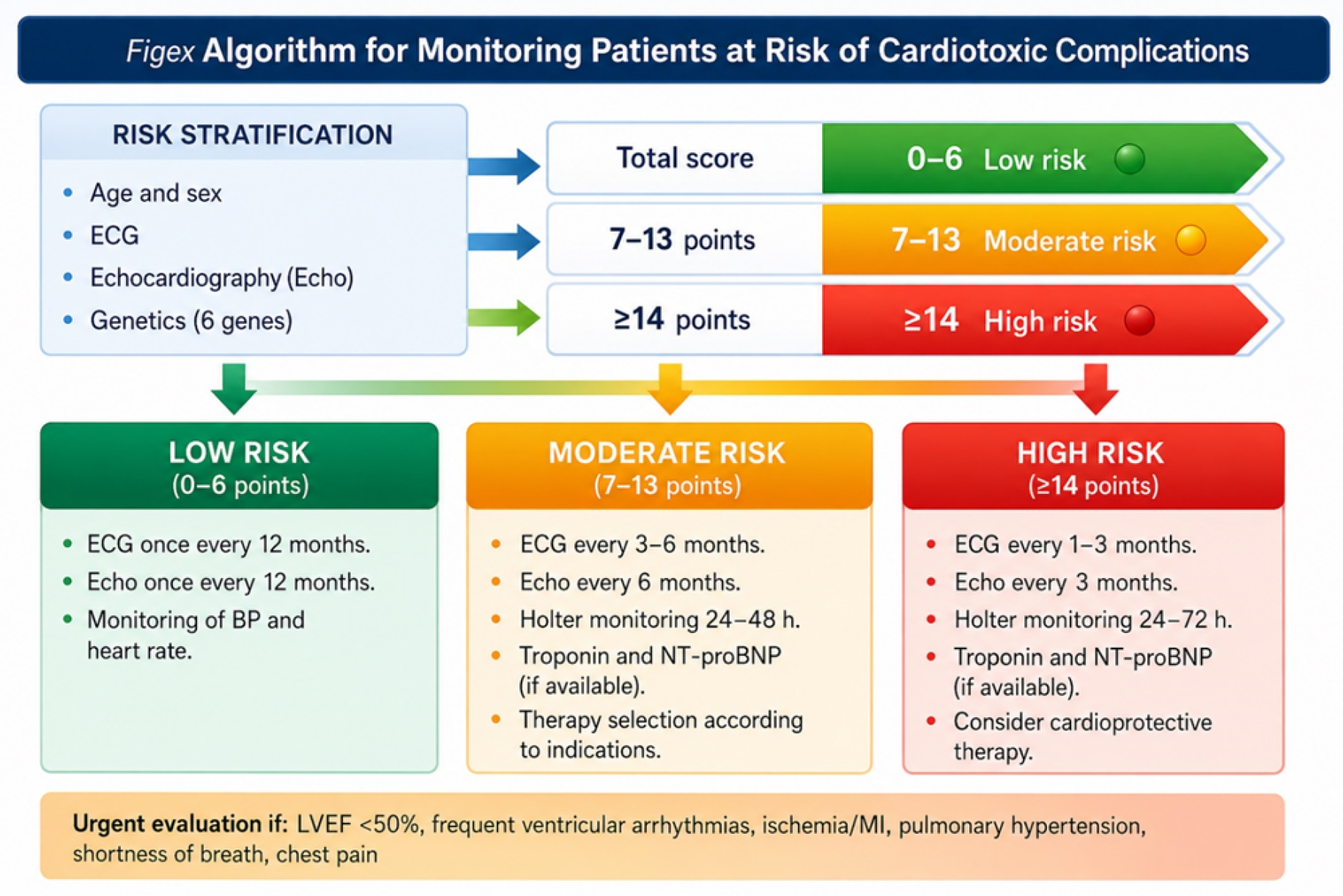
Figure 2.
Figex algorithm for risk stratification and monitoring of patients at risk of cardiotoxic complications.
Figure 2.
Figex algorithm for risk stratification and monitoring of patients at risk of cardiotoxic complications.
References
- Lyon, A.R.; López-Fernández, T.; Couch, L.S.; Asteggiano, R.; Aznar, M.C.; Bergler-Klein, J.; et al. 2022 ESC Guidelines on cardio-oncology developed in collaboration with the European Hematology Association (EHA), the European Society for Therapeutic Radiology and Oncology (ESTRO) and the International Cardio-Oncology Society (IC-OS). Eur. Heart J. 2022, 43, 4229–4361. [Google Scholar] [CrossRef] [PubMed]
- Herrmann, J. Adverse cardiac effects of cancer therapies: Cardiotoxicity and arrhythmia. Nat. Rev. Cardiol. 2020, 17, 474–502. [Google Scholar] [CrossRef] [PubMed]
- North, B.J.; Sinclair, D.A. The intersection between aging and cardiovascular disease. Circ. Res. 2012, 110, 1097–1108. [Google Scholar] [CrossRef] [PubMed]
- Dai, D.F.; Chen, T.; Johnson, S.C.; Szeto, H.; Rabinovitch, P.S. Cardiac aging: From molecular mechanisms to significance in human health and disease. Antioxid. Redox Signal. 2012, 16, 1492–1526. [Google Scholar] [CrossRef] [PubMed]
- Regitz-Zagrosek, V.; Oertelt-Prigione, S.; Prescott, E.; Franconi, F.; Gerdts, E.; Foryst-Ludwig, A.; Maas, A.H.E.M.; Kautzky-Willer, A.; Knappe-Wegner, D.; Kintscher, U.; et al. Gender in cardiovascular diseases: Impact on clinical manifestations, management, and outcomes. Eur. Heart J. 2016, 37, 24–34. [Google Scholar] [CrossRef] [PubMed]
- Mauvais-Jarvis, F. Sex differences in cardiovascular disease and diabetes mellitus. Circ. Res. 2020, 126, 1265–1285. [Google Scholar] [CrossRef]
- Souiden, Y.; Mallouli, H.; Meskhi, S.; Chaaben, A.B.; Mahdouani, K.; Almawi, W.Y. MnSOD and GPx1 polymorphism relationship with coronary heart disease susceptibility and severity. Biol. Res. 2016, 49, 22. [Google Scholar] [CrossRef]
- Vrbanović, E.; Popović Hadžija, M.; Križanac, Š.; Včev, A.; Samardžić, M.; Maltar-Strmečki, N.; et al. Association of Oxidative-Stress-Related Gene Polymorphisms (CAT, SOD2, GPX1) and Antioxidant Status with Disease Risk. Antioxidants 2023, 12, 1315. [Google Scholar] [CrossRef] [PubMed]
- López, B.; Ravassa, S.; Moreno, M.U.; José, G.S.; Beaumont, J.; González, A.; Díez, J. Diffuse myocardial fibrosis: Mechanisms, diagnosis and therapeutic approaches. Nat. Rev. Cardiol. 2021, 18, 479–498. [Google Scholar] [CrossRef]
- Mewton, N.; Liu, C.Y.; Croisille, P.; Bluemke, D.; Lima, J.A.C. Assessment of myocardial fibrosis with cardiovascular magnetic resonance. J. Am. Coll. Cardiol. 2011, 57, 891–903. [Google Scholar] [CrossRef] [PubMed]
- Barton, M.; Yanagisawa, M. Endothelin: 30 years from discovery to therapy. Hypertension 2019, 74, 1232–1265. [Google Scholar] [CrossRef] [PubMed]
- Khimji, A.K.; Rockey, D.C. Endothelin and hepatic and cardiovascular disease. Cell. Mol. Gastroenterol. Hepatol. 2022, 13, 729–744. [Google Scholar] [CrossRef] [PubMed]
- Tunbridge, E.M.; Harrison, P.J.; Weinberger, D.R. Catechol-O-methyltransferase, cognition, and psychosis: Val158Met and beyond. Biol. Psychiatry 2006, 60, 141–151. [Google Scholar] [CrossRef] [PubMed]
- Witte, A.V.; Flöel, A. Effects of COMT polymorphisms on brain function and behavior in health and disease. Brain Res. Bull. 2012, 88, 418–428. [Google Scholar] [CrossRef] [PubMed]
- Inouye, M.; Abraham, G.; Nelson, C.P.; Wood, A.M.; Sweeting, M.J.; Dudbridge, F.; Lai, F.Y.; Kaptoge, S.; Brozynska, M.; Wang, T.; et al. Genomic risk prediction of coronary artery disease in 480,000 adults: Implications for primary prevention. J. Am. Coll. Cardiol. 2018, 72, 1883–1893. [Google Scholar] [CrossRef] [PubMed]
- Khera, A.V.; Chaffin, M.; Aragam, K.G.; Haas, M.E.; Roselli, C.; Choi, S.H.; Natarajan, P.; Lander, E.S.; Lubitz, S.A.; Ellinor, P.T.; et al. Genome-wide polygenic scores for common diseases identify individuals with risk equivalent to monogenic mutations. Nat. Genet. 2018, 50, 1219–1224. [Google Scholar] [CrossRef] [PubMed]
Disclaimer/Publisher’s Note: The statements, opinions and data contained in all publications are solely those of the individual author(s) and contributor(s) and not of MDPI and/or the editor(s). MDPI and/or the editor(s) disclaim responsibility for any injury to people or property resulting from any ideas, methods, instructions or products referred to in the content. |
© 2026 by the authors. Licensee MDPI, Basel, Switzerland. This article is an open access article distributed under the terms and conditions of the Creative Commons Attribution (CC BY) license (http://creativecommons.org/licenses/by/4.0/).
Copyright: This open access article is published under a Creative Commons CC BY 4.0 license, which permit the free download, distribution, and reuse, provided that the author and preprint are cited in any reuse.


