

Submitted:
25 June 2026
Posted:
01 July 2026
You are already at the latest version
Abstract
Background: Chronic Multi-symptom Illness (CMI) is an idiopathic condition that can cause severe disability in U.S. Veterans. Objectives: 1. The collection of data to assist in the development of a hypothesis that describes the pathophysiology of CMI and related conditions including Gulf War Illness and Chronic Fatigue Syndrome. 2. Development of a better understanding of the consequences of this syndrome on the study subjects impaired level of functioning. Design: This is an observational VA Pilot study of a cohort of 40 US Veterans with Chronic Multi-symptom illness studied between 2019 and 2025. Methods: The study population was drawn from U.S. veterans who receive medical care from the W.G. Hefner VA Health Care System in Salisbury, N.C. U.S. veterans were initially referred to the Rehabilitation Medicine Service (RMS) for various diagnoses associated with chronic or persistent pain. From this pool of RMS patients, 36 males and 4 females who were diagnosed with Chronic Multi-symptom Illness and who met the study inclusion criteria were selected to participate in the study. Data collected from this study group included: demographic, medical history, and physical symptom information, psychological and cognitive evaluation and testing data, physical exam results, and serum lab test data. Lab tests were performed twice on the study subjects. Lab tests included serum active vitamin D precursor, 25(OH)D3, levels, calcium levels, and human IgM and IgG antibody levels to dUTPase enzymes from three latent herpetic viruses, Varicella Zoster virus (VZV), Human Herpes 6 virus (HH6V), and Epstein Barr virus (EBV). Serum active vitamin D precursor levels were examined because active vitamin D has a role in immune system function including the suppression of latent viruses and the modulation (attenuation) of the immune system’s inflammatory response to microbials including latent viruses. The herpesvirus dUTPases from EBV, HH6V, and VZV are proteins/enzymes in viral DNA replication but not structural components of the mature virion. In this pilot study, antibodies against these proteins were used as biomarkers for abortive lytic replication, a process that can stimulate an inflammatory immune process. This abortive lytic inflammatory process is hypothesized to be distinct from the inflammatory response associated with full lytic viral reactivation, which results in the production of complete virions. Results: Demographic Data – 75% of study subject men and 50 % of women served in combat. Comparison of our pilot study population to the general VA population revealed differences: US Marines made up 25% of our study population versus 9.9% in the general VA population, and African American/Black study subjects made up 30% or our study population verses 15% in the VA general population. Our study subjects had high service connected, disability/ compensation ratings: 55% or 22/40 had a rating of greater than or equal to 80%, and 22.5% or 9/40 had a rating of greater than or equal to 100%. Neurological findings in study subjects included hyperalgesia to pin prick over the skin, which was present in 95% of study subjects, tinnitus (ringing in the ears) in 82% of study subjects, decreased balance on heel to toe walking which was normal in only 28.9% of study subjects with their eyes open and in only 2.7% with eyes closed. This test could not be performed in 18.4% with eyes open and in 40.5% with their eyes closed. Two study subjects developed severe quadriparesis and idiopathic neurocognitive disorders later in the study after their initial interview, physical exam, cognitive testing and blood tests were performed. Serum Lab Tests: Vitamin D, 25(OH)D3 Levels: in Trial 1 – Deficient range 11/40 (27.5%), Insufficient range 17/40 (42.5%) in Trial 2 – Deficient range 8/36 (22.2%), Insufficient range 11/36 (30.6%) Note: Only two study subjects were deficient for both seasonally adjusted 25(OH)D3 levels checked in Trials 1&2 Human IgG antibody levels to Herpesvirus dUTPase proteins: Trial 1 serum test results for human IgG serum antibody levels to herpes virus dUTPase proteins were negative for all three viruses tested in 10/40 (25%) of study subjects and positive for all three viruses examined in 7/40 (17.5%) of study subjects. Trial 2 serum test results for human IgG antibody levels to herpes virus dUTPase enzymes were negative for all three viruses tested in only 1/36 (2.8%) study subjects. Human IgG dUTPase antibodies were positive for all three viruses in 17/36 (47%) study subjects. In Trial 1 – Human VZV IgG was Pos. (elevated) for VZV dUTPase in 11/40 (12.5%) of study subjects, In Trial 2 – Human VZV IgG was Pos. (elevated) for VZV dUTPases in 23/36 (64%) of study subjects.
Keywords:
chronic multi-symptom illness
; low 25(OH)D3 storage
; human herpesvirus dUTPase antibodies
; abortive lytic virus replication
; Amyotrophic Lateral Sclerosis
; neurodegenerat neurocognitive disorder of unknown etiology
1. Introduction
Since 1990-91, many U.S. military service members returning from the wars in Iraq and Afghanistan have suffered chronic, often disabling, multi-symptom illnesses for which the pathophysiology or etiology of their symptoms is unknown.[1,2,3,4] Based on a wide range of epidemiological studies, chronic multi-symptom illness associated with the first Gulf War affected at least one quarter (range 25% to 32%) of the U.S. service members who served in the Gulf War region during the 1990 to 1991 time frame.[5] Currently the Department of Veterans Affairs define chronic multi-symptom illnesses (CMI) as illnesses with clusters of often disabling symptoms (including chronic pain, fatigue, headaches, indigestion, insomnia, dizziness, respiratory disorders, and memory problems) for which an underlying cause or causes have not been determined.[6]
Multi-symptom illnesses such as Gulf War Illness (GWI) and Myalgic Encephalomyelitis (ME) /chronic fatigue syndrome (CFS) have been a vexing problem for many U.S. veterans as well as others suffering from these illnesses. These symptom-based illnesses often include chronic severe pain. Medical management of these multi-symptom illnesses has likewise been a frustrating experience for many medical providers. Similar illnesses have been documented in U.S. Service members since the U.S. Civil War. [7]
This pilot study was initiated based in part on a latent viral immune inflammatory response model for chronic multi symptom illnesses and on the findings of frequent early chronic multi-symptom illness among returning U.S. Marines from Iraq and Afghanistan between 2010 and 2011. [8,9] Young U.S. Marines often returned with signs of hyperalgesia to pin prick in the skin and signs of decreased balance on the Tandem Gait Test. These two abnormal signs were associated with the symptom of tinnitus and with deficient and insufficient levels of stored active vitamin D precursor, 25(OH)D3. This current study was also initiated based on the previous discovery of human antibody titers to latent herpetic virus deoxyuridine triphosphate nucleotidohydrolase (dUTPase), enzymes detected in the serum of U.S. veterans who were diagnosed with chronic multi-symptom illnesses such as chronic fatigue syndrome and Gulf War Illness. [10,11,12,13,14,15,16] Specific human IgM and IgG herpetic virus dUTPase antibodies indicate the presence of antigenic herpetic virus dUTPase enzymes in the body. These enzymes are pathological in that they stimulate an inflammatory immune response in the tissues where they are expressed such as within the cells of the nervous system. The dUTPase proteins are expressed during herpetic virus lytic and abortive lytic replication.
Distinct or unique dUTPase enzymes are present in different life forms including some viruses, bacteria, fungi, plants, animals, and humans. Herpetic virus dUTPase enzymes occur in a monomeric form. When latent herpetic viruses are present in humans, their dUTPase enzymes stimulate the immune system. The resulting inflammatory response includes the generation of specific human antibodies directed against them.[15] Purposes of the dUTPase enzyme in different organisms, including latent herpetic viruses, are to produce dUMP for synthesizing DNA and to remove dUTP from the deoxynucleotide pool which in turn can help to reduce the possibility of dUTP being erroneously incorporated into an organism’s DNA. [17] Human, anti-herpesvirus dUTPase IgM and IgG antibodies in the serum have been used in this study as biomarkers to indicate the past and current, presence of inflammatory herpetic virus dUTPase enzymes in the nervous system and other tissues of the body. [18]
Human herpesvirus dUTPase IgG and IgM antibody levels, measured during this pilot study from each of the three subfamilies of herpetic viruses (alpha, beta, and gamma herpesvirinae), were reported in a previous article.[18] The herpes virus dUTPase enzymes are recognized as antigenic to the human immune system, and are inflammatory.[12,14,16] In this study, human herpetic virus IgG and IgM, dUTPase antibody levels were measured to alpha Varicella Zoster (VZV; ORF8), beta Human Virus 6 (HHV6; U45), and gamma Epstein Barr (EBV; BLLF3) viruses.
Rather than the complete synthesis of latent virus virions during lytic replication, the study participants in this paper are proposed to have a new latent virus inflammatory response disease. It is characterized by the synthesis of antigenic inflammatory molecules by latent virus DNA during abortive lytic replication.[19,20] With this newly recognized form of viral illness, there is generally no fever, no elevated white blood cell count, or other typical signs of an acute viral infection. Rather, the inflammatory response is characterized by the presence of specific IgG and IgM antibodies and an accompanying inflammatory (cytokine) response to a molecule produced by a specific latent virus during viral DNA replication.
In this study, the measurement and presence of human IgG and IgM antibodies to specific herpesvirus dUTPase enzymes are used as biomarkers and as documentation for this proposed newly recognized mechanism of viral illness.[18] Symptoms and physical exam findings are used to characterize the effect and the location of the latent virus inflammatory response. Because certain herpetic latent viruses, especially varicella zoster and Herpes Simplex 1 and 2 are present in the nervous system, inflammation affecting nervous system cells/tissues are proposed to initially cause hyperexcitability in the affected cells followed by damage and even cell death. For example, tinnitus and decreased balance are symptoms that suggest the middle ear as a focus of an inflammatory response. Similarly, neuropathic pain and hyperalgesia to pin prick in the skin point to the cells of the sensory ganglia and above as the point of the inflammation.
Herpesviruses are large DNA viruses. Of the three examined in this pilot study, EBV has the smallest coding capacity, and it encodes for at least 80 proteins; some of which are only expressed during latency or lytic (abortive lytic) replication. HHV-6 and VZV encode for more proteins, but like EBV some of these proteins are only expressed during latency while others are only expressed during lytic (abortive lytic) replication.
Latent (Latency) [21,22] and lytic (abortive lytic) [23,24,25,26,27,28,29,30,31,32,33,34,35,36,37,38,39,40,41,42] replication are states in which herpes viruses exist. In latency only a few viral genes are expressed which allows the virus to avoid the host immune system and to persist for the lifetime of the individual. In latency the virus does NOT replicate its DNA and thus no new virus is formed.
If something triggers the latent virus it will reactivate, which means it will enter the lytic (abortive lytic) state. In the lytic state a different set of genes are expressed in a cascade manner (alpha, beta and gamma genes). If all become expressed, this process results in new virus being formed. This is termed a PRODUCTIVE INFECTION. If for some reason, this process of lytic replication is not completed, no new virus is formed, and the state is referred to as abortive lytic. However, some virus proteins such as the dUTPase are expressed during abortive lytic replication. Abortive lytic replication is really what happens when you treat a patient with acyclovir. Acyclovir inhibits virus DNA elongation thus preventing new viruses for forming but it doesn’t prevent the expression of alpha or beta genes and thus those dUTPase proteins are made. It is possible that herpes virus abortive lytic replication can be occurring simultaneously in the same nervous tissue as a variable and perhaps small amount of lytic replication. This might result in a significant amount of inflammation but without a significant amount of cell lysis.
A small observational pilot study of U.S. Veterans with Chronic Multi-symptom Illness (CMI) was conducted at the Bill Hefner VA Medical Center between 2019 and 2025 in Salisbury North Carolina. This study involved the collection of a significant amount of demographic, medical, psychological, and symptom history. A physical exam, cognitive and psychological testing/evaluations were also performed at the beginning of this study. Two sets of blood samples were drawn and tested over the course of this study. Each set of blood tests included measurement of serum calcium and vitamin D precursor, 25(OH)D3, levels, multiple inflammatory cytokine levels, and three specific herpes virus dUTPase enzyme IgM and IgG human antibody levels. Cytokine and herpes virus human dUTPase antibody level blood test results are presented in greater detail in a separate report. [18]
This report focuses on demographic information, medical history including concurrent diagnoses, physical exam findings, and a brief review of the psychological and cognitive testing in the study subjects from this VA pilot study.
The clinical report below also includes an analysis of the 25(OH)D3 , active vitamin D precursor lab results and a brief review of our study subject’s herpes virus dUTPase, human antibody test results[18]. This report will also explore how vitamin D status and latent herpetic viruses may interact in the pathophysiology of chronic multi-symptom and system illnesses. This study’s findings support the hypothesis that some idiopathic chronic multi-symptom and system illnesses have an underlying pathophysiology related to the body’s immune system’s inability to suppress abortive lytic replication of latent herpesviruses. The immune system’s inability to suppress herpes virus abortive lytic replication is hypothesized to result in an inflammatory response in cells where the latent herpesvirus DNA resides.
2. Methods
This study was approved by the Salisbury VA Medical Center Investigational Review Board (IRB) and Research Committee, IRB Study # 18-020, Project # 1575420-4.
2.1. Patient Selection
The study population was drawn from U.S. veterans who receive medical care from the W.G. Hefner VA Health Care System in Salisbury, N.C. U.S. veterans were initially referred to the Rehabilitation Medicine Service (RMS) for various diagnoses most often associated with chronic or persistent pain. Once seen and evaluated in the RMS clinic by department physiatrists, certain patients who met the study criteria were then referred for consideration for this study. From this RMS input of patients, 36 U.S. male and 4 female veterans with chronic multi-symptom illnesses were selected to participate in this pilot study.
Inclusion Criteria: U.S. Veterans (male and female) treated by the W.G. Hefner VA Health Care System who have some of the idiopathic symptoms of chronic multi-symptom illnesses described in the Introduction section above were eligible for this study. Specific inclusion criteria for symptomatic veterans in this pilot study included the presence of at least one sign on physical exam involving the nervous system (i.e., hyperalgesia to pin prick, hyperhidrosis, decreased balance, increased reflexes etc.) and at least two symptoms (physical, psychological or cognitive symptoms such as significant neuropathic or musculoskeletal pain, fatigue, dyspnea, anxiety, depression, etc.).
Exclusion criteria: U.S. Veterans who are unable to give voluntary informed consent for either cognitive or psychological reasons were excluded from the study. Veterans who are unable or unwilling to undergo the required blood testing were excluded.
2.2. Demographic, Medical, Psychological, and Symptom History
Demographic, medical, and symptom histories were obtained using a questionnaire and the VA electronic medical record. These histories were then reviewed by the examining physician. The medical history in this report contains five parts: concurrent medical diagnoses, history of viral infections, trauma history, medications, and symptoms. Psychological information was obtained through interview and testing by two neuropsychologists. Wherever possible, information collected was verified through the VA patient electronic medical record. A complete list of symptoms is included in Appendix 1.
2.3. Physical Exam
The physical exam was performed once by a single VA medical provider upon the initial encounter with the 40 VA pilot study subjects and later, multiple times in two study subjects whose physical condition changed dramatically during the study. The complete findings of the initial physical exam are listed in detail in Appendix 2. The physical exam is an important component of the evaluation of U.S. veterans with chronic multi-symptom illness. Unlike symptoms, components of the physical exam are meant to be findings which can be more objectively and consistently measured over time and between different examiners and across different patient populations. When objective findings on physical exam fall outside of an accepted norm they can be referred to as abnormal signs.
The purpose of the physical exam was to determine a profile of abnormal signs in our group of US veterans with chronic multi-symptom illness. There was a specific focus on any signs related to the somatic and autonomic nervous systems and on nervous system reflexes including the gag reflex, glossopharyngeal nerve - cranial nerve (IX) and balance, vestibulocochlear nerve – cranial nerve (VIII) and deep tendon reflexes in the extremities.
Pain is a very common symptom in patients with chronic multi-symptom illnesses. Pain is a symptom which can be very subjective depending on the patient’s interpretation of the pain sensations. Alternately, the patient’s reaction to light pin prick or touch is more objective. Hyperalgesia is an abnormal sign of increased sensitivity to pin prick over skin. This increased sensation is often bilateral and exists over the skin of the posterior neck or back and corresponding upper or lower extremities. It can also occur over the skin of the face and may occur over the skin of just one side of the body. Because it most often occurs over the skin of the neck or back and corresponding limbs, it may represent an inflammatory reaction affecting the sensory ganglia of the spine. It is also possible that it originates further up in the sensory pathways of the central nervous system. Similarly, allodynia which is a painful reaction to light touch over skin covering some parts of the body but not others. Allodynia can be dramatic and can represent an objectively abnormal sign but is less common.
A complaint of dizziness or decreased balance can be quite disturbing but is a subjective complaint. A staggering gait on heel to toe walking along a straight line (the tandem gait test) can be a dramatic objective sign of decreased balance especially if visual feedback is taken away and the patient must rely on their vestibular system of the middle ear to accomplish this task.
To look for objective signs on physical exam in this study population, our study subjects underwent examination of motor and sensory function in the somatic nervous systems and evaluation for other signs in the autonomic nervous system such as hyperhidrosis and hyperemia of the skin. The skin and oral cavity mucosa were also examined for rashes or intraoral lesions that represent signs of inflammation. In our U.S. veteran population, abnormal signs involving the nervous system are often associated with a variety of symptoms which are perceived and interpreted by the brains of our U.S. veterans with chronic multi-symptom illnesses. Evaluation of parts of the peripheral nervous system was a major focus for this W.G. Hefner VA pilot study, “Medical Evaluation of U.S. Veterans with Chronic Multi-symptom Illness”.
2.4. Description of Psychological and Cognitive Evaluation and Testing
Measures of cognitive and psychological function were obtained using the following assessment tools:
- Montreal Cognitive Assessment (MOCA)
- Conners Continuous Performance Test, 3rd Edition (CPT-3)
- DSM-5 Self Rated Cross Cutting Symptoms Level 1, Adult (DSM-5 Level 1)
- DSM-5 Self Rated Somatic Symptom Level 2, Adult (DSM-5 Somatic)
- DSM-5 Self Rated Anxiety Scale Level 2, Adult (DSM-5 Anxiety)
- Neurobehavioral Symptom Inventory and Mild Brain Injury Atypical Symptom Scale (NSI+MBIAS)
- The Pittsburg Sleep Quality Index (PSQI)
2.5. Lab Data
The lab data presented in this report includes the results obtained from two blood draws from the study subjects. The mean time between blood draws was 110 weeks, ranging from 32-151 weeks. The long interval between the two blood collection trials was due to the study being placed on hold between March 2020 and June 2021 due to the COVID pandemic. Of the original 40 study subjects, a second set of blood samples was obtained from 36 study subjects (90%). All blood samples were drawn at the Salisbury VA Medical Center clinical lab. Samples for serum vitamin D precursor, 25(OH)D3, levels and calcium levels were processed at the Salisbury VA Medical Center. Serum samples for herpes virus dUTPase, IgM and IgG antibody levels and cytokine level determination were sent to the Institute for Behavioral Medicine Research, Ohio State University Wexner Center. The results of the lab tests run at Ohio State University are reported in detail and analyzed in a separate article.[18] An overview of the IgG test results are presented in this report and reviewed in the discussion section for their clinical significance.
2.6. Serum 25(OH)D3 and Calcium Serum Levels
The measurement of the active vitamin D precursor, 25(OH)D3, was included in our study because low vitamin D precursor storage states have been found in Veterans with chronic multi-symptom illnesses.[9] Active vitamin D may play a significant role in the suppression of latent viruses and in the mitigation of the inflammation associated with viral antigens.[9], [43,44,45] The treatment of our study subjects who were found to have vitamin D deficiency or insufficiency was not part of our study. The study subjects with low vitamin D status including deficiency and insufficiency, on either set of blood work, were referred to their assigned VA treating physicians who then put them on vitamin D supplementation or adjusted their current vitamin D dose. The dose of supplemental vitamin D3 used to treat our study subjects was not monitored or recorded since treatment of chronic multi-symptom illness was not part of this study.
The guidelines used in this study for assessing 25(OH)D3 levels have been recommended by the Endocrine Society[46] and used by two outside commercial labs, Lab Corp of America, and Quest Diagnostic Nichols Institute. The guidelines recommended by the Endocrine Society and used by our pilot study are as follows: vitamin D deficiency (<20 ng/mL), vitamin D insufficiency (20 ng/mL to <30 ng/mL), and normal or sufficient for bone health (30 ng/mL to 100 ng/mL). At Salisbury VA Medical Center, the normal range for calcium levels was 8.5 to 10.5 mg/dl at the time of the blood collections.
When comparing the serum 25(OH)D3 levels in the study subjects at various times of the year, their test results were converted to seasonally adjusted, annual mean, serum 25(OH)D3 levels using the “25 Hydroxyvitamin D Calculator for Seasonal Adjustment” developed at the Kidney Research Institute, University of Washington.[47,48,49] When comparing serum 25(OH)D3 levels with other blood tests, the non-adjusted serum 25(OH)D3 levels were used. The measurement of serum 25(OH)D3 levels is currently our clinical test for assessing vitamin D status. Because it is a measurement of the last precursor prior to conversion to the active form of vitamin D, 1,25(OH)2D3, it is not a measurement of active vitamin D in the body, or its demand by the body. Rather it is a measurement of vitamin D precursor storage in the body.[50]
Serum calcium levels were measured along with serum 25(OH)D3 levels. This was done because serum active vitamin D, 1,25(OH)2D3, facilitates the absorption of calcium from the intestine which is one of its main functions. High calcium levels can be a sign of too much exogenous oral vitamin D3 and can result in elevated calcium levels (vitamin D toxicity).[51]
2.7. Herpetic Virus dUTPase Antibody Serum Levels
Each set of blood tests (Trials 1 and 2) separated in time included one human IgM and IgG herpesvirus dUTPase antibody level from each of the three subfamilies of herpesvirus: alpha-Varicella Zoster (VZV; ORF8), beta-Human Virus 6 (HHV6; U45), and gamma-Epstein Barr (EBV; BLLF3) viruses. Detailed testing methodology and results have been reported in a separate article.[18] The dUTPase blood samples were drawn, allowed to clot over 30 minutes, spun down to convert them to serum samples, frozen at -80 degrees Celsius, and then sent in batches to the Institute for Behavioral Medicine Research, Ohio State University, Columbus, OH for further processing and analysis.
In our VA pilot study, elevated and changing serum human dUTPase enzyme IgG and IgM antibody levels were used as biomarkers for evidence of herpetic virus antigenic/ inflammatory dUTPase protein synthesis by herpetic virus DNA.[18] The herpetic virus dUTPase enzymes were not directly measured. Rather, unbound human antibodies to these herpetic enzymes are measured from study subjects’ serum using a dUTPase protein-specific ELISA assay.[52] The specificity for assaying serum dUTPase antibody levels was achieved by using purified recombinant VZV, HHV6 and EBV dUTPase proteins.
Analyses of the data from both sets of blood tests were performed the same way during the VA pilot study. Means (SD) were calculated across all the participants for each of the serum calcium and serum 25(OH)D3 blood draws (denoted as trial 1 and 2). Where appropriate, median (IQI) values were also computed when data values were skewed. Paired t-tests for differences between the two test sessions, and Pearson correlations were calculated to look for relationships among the variables. A significance level (p equal to or less than 0.05) was used for all statistical tests. Statistical analyses were performed using SPSS Statistics Version 23 (IBM Corporation, Armonk, NY, USA). Statistical analysis of dUTPase and cytokine levels are presented in detail in a separate article. [18]
3. Results
3.1. Demographic and Other Participant Data

Table 1.
VA Pilot Study Participant Characteristics and Demographics Compared to those of the Entire U.S. VA Population.
Table 1.
VA Pilot Study Participant Characteristics and Demographics Compared to those of the Entire U.S. VA Population.
| Salisbury VA study participants | Entire US Veteran Population |
| Sex: Male n = 36 Female n = 4 Ratio M/F 9 to 1 Study Participant Enrollment: 3/25/2019 – 3/13/2020 |
Male n = 16,998,424 [53] Female n = 1,601,292 Ratio M/F 10.6 to 1 |
| Study Group’s Median Age 49 Years: Age Range in 2019/20: 31-76 years 70’s (4), 60’s (10), 50’s (5), 40’s (6), and 30’s (15) |
VA Population Median Age 65 Years: [54] Age Ranges in 2020 [55]: 30-79 years: 70’s (4,502481), 60’s (3,606,055), 50’s (3,417,648), 40’s (2,273,736), and 30’s (2,025,275) |
| Combat Experience: male 27 of 36 - 75% female 2 of 4 - 50% |
Overall, about 29 percent of US veterans have experienced combat. Of those who have served after 9/11/2001, about 49 percent have experienced combat.[56] |
| Racial make-up of the VA Study Group: White/Non-Hispanic 27/40 - 67.5% Black / African American 12/40 - 30% Hispanic/Latino 1/40 - 2.5% |
Racial make-up US Veteran Population: [57] White/Non-Hispanic-72.9% Black / African American - 15.5 % Hispanic/Latino - 5.4% Other - 6.2% |
| Study subjects by Branch of Military: Army + Army NG (24-60%), Marine Corps (10- 25%), Air Force (4-10%), Navy (1-2.5%), Coast Guard (1-2.5%) |
Percentage of U.S. veterans by Branch: [58,59] Army 50.2%, Marine Corps 9.9%, Navy 24.4%, Air Force 19.6%, Coast Guard 1.4% [13] |
Table 2.
Percent Service-Connected (SC) % - Compensation/Disability in Study Group Subjects.
| none | 7 |
| 0% | 1 |
| 10% | 3 |
| 40% | 2 |
| 50% | 1 |
| 60% | 1 |
| 70% | 3 |
| 80% | 6 |
| 90% | 7 |
| 100% | 9 |
| Total percent with SC | 33/40 (82.5%) |
| Percent with SC > or = 80 % | 22/40 (55%) |
| Percent with SC > or = 100% | 9/40 (22.5%) |
Note: 0% means service connected for a condition but with no compensable disability.
3.2. Occupation Type or Status
Skilled trade 10, Admin/clerical/sales 6, Transportation 4, Professional 4, Self-employed 1, general laborer 1, Disabled 5, Retired 5, None listed 4.
3.3. Social and Other Habits
Self-Reported Tobacco Use: Never 18 (45%), Previous 7 (17.5%). Current - cigarettes 12 (30%), oral 3 (7.5%), oral plus cigarettes 2 (5%)
Self-Reported Alcohol Consumption: No alcohol 27 (67.5%), 3 (1-2 drinks/day – 7.5%), 6 (4-7 drinks/day – 15%), 2 (12-15 drinks per day – 5%), 2 (26-28 drinks per day – 5%)
Self-Reported Illicit Drug Consumption: None 38 (95%), 1 (1 drug – 2.5%), 1 (6 drugs – 2.5%)
Self-Reported Sunscreen Use: Often 12 (30%), Seldom 12 (30%), Never 16 (40%)
3.3. Medical History Summary – Self-Reported and from the VA Electronic Health Record-EHR (See Appendix 1 for Complete Data Set.)
The medical history is presented in this manuscript in five parts: non mental health medical diagnoses, history of viral infections, trauma history, medications, and symptoms. After chronic multi-symptom illness, the most frequent concurrent medical diagnosis made in our study population was a history of trauma: to extremities excluding hands and feet – 75%, to head and neck – 45%, to hands and feet – 37.5%. Following trauma, the most frequent diagnoses included: history of chicken pox (varicella) and/or shingles (Zoster) - 70%, arthritis - 57.5%, hypertension – 52.5%, mild traumatic brain injury (TBI) - 50%, gastro esophageal reflux disease (GERD) - 45%, and obstructive sleep apnea (OSA) - 25%.
The most frequently prescribed medications to study subjects were for pain and included: oral and topical anti-inflammatory meds – oral 45%, topical 22.5%, antiseizure meds for nerve pain – 27.5%, muscle relaxers – 17.5%, and narcotics – 10%. Following prescribed pain medications by frequency were mental health disorder meds – 40%, hypertension and gastroesophageal reflux (GERD) meds – 37.5% each, sleep meds – 15%, and anti-viral and migraine headache meds – 12.5% each. The most frequent over the counter nutritional supplements prescribed/recommended included vitamin D3 – 72.5%, omega 3 fatty acids 42.5%, and other nutritional supplements including multi-vitamins – 42.5%.
The most frequently reported symptom was pain. Median Chronic pain level was 5 on a scale of 1- 10 and chronic pain occurred in 95% of study subjects. Chronic nerve pain was reported by 77.5% of study subjects and chronic joint pain was reported by 90% of study patients. More severe acute exacerbations of pain (median pain level was 7 on a scale of 1-10) was reported by 95% of study patients with 77.5% of study subjects reporting nerve pain exacerbations and 92.5% of study subjects reporting exacerbations of joint pain. Two study subjects – 5% reported no significant pain symptoms.
Pain symptoms were usually the reason for referral of these study subjects to the VA Rehab Medicine Service (RMS) and ultimately part of the reason for referral to the RMS Chronic Multi-Symptom Illness VA pilot study. Tinnitus was the next most frequent symptom in the study group – 82%, followed by chronic fatigue – 80%, sleep disturbance – 75%, and GI symptoms – 72.5%. This distribution of symptoms is consistent with a profile of chronic multi-symptom illnesses symptoms except for tinnitus which is usually not elicited from study subjects unless asked for specifically.
3.4. Physical Exam Summary (See Appendix 2 for Complete Data Set.)
The majority (60%) of our study population had a BMI of 30 or more and were obese. Just less than one-third of our study population had a dark complexion. These two findings can contribute to low 25(OH)D3 vitamin D precursor (storage) levels in the serum. In our study group, 45% had some form of skin rash and one-third of these were moderate and 7.5% had an intra-oral lesion. These signs may indicate an inflammatory process going on in the skin and oral mucosa.
3.5. Vascular and Autonomic Nervous Systems
On physical exam, 27.5% (11) of study subjects exhibited stage 1 (Systolic BP 130 – 140 mmHg) or stage 2 hypertension (systolic BP > 140 mmHg) and 30% (12) exhibited diastolic hypertension only (BP > 80 mmHg) . Study Participant blood pressure (BP) ranges (definitions) are based on blood pressure stages per the American Heart Association [60] Ten percent (4) of study patients had both systolic and diastolic hypertension. Of the above 57.5% (23) of US veterans who were documented to have hypertension on physical exam at the beginning of the study, only 37.5% (15) were on treatment for hypertension at the time their blood pressures were taken. This group of treated veterans included all with systolic hypertension except one veteran who had not been previously been diagnosed with hypertension.
In our study group going from supine to standing, 7.5% of study subjects demonstrated orthostatic systolic hypotension (drop of 20 mmHg points or more) and an additional 7.5% exhibited slightly smaller drops of (15,17, and 19 points). Furthermore, 7.5% experienced diastolic orthostatic hypotension with a drop of 10 mmHg points or more.
In our study group, 20.5% of study subjects were noted to have hyperhidrosis in either the palms or soles on physical exam. Two study subjects, (5.1%), exhibited hyperemia in the skin of either the head and neck area or the extremities.
3.6. Motor System
Deep tendon reflexes in 5% of the study subjects were increased in the upper extremities and in 15.4% of the lower extremities (> 3/4). Gag reflexes were increased in 20% of study subjects. In the study group, 10.3% were unable to walk on tip toes and heels. Seven (17.4%) of study subjects including a double lower extremity amputee, were unable to perform a deep knee bend.
In those who could perform the Tandem Gait Test (walking heel to toe along a straight line 10-15 feet forward and backward) was normal in 28.9% of study subjects with their eyes open, and in only 2.7% with their eyes closed. In those who could perform this test, it was impaired in 52.6% with their eyes open, and in 56.8% with their eyes closed. This test could not be performed in 18.4% with their eyes open and in 40.5% with their eyes closed.
Motor system diagnoses made after the initial physical evaluation (exam):
Quadriparesis 2/40 (5%) of study subjects, diagnosed in Sept. of 2022 and in Oct. of 2024
3.7. Sensory System
Sensation to light touch was intact or normal in the skin of the upper extremities in 97.5% of study subjects and intact or normal in the skin of the right lower extremity in 87.2% and in the left lower extremity in 84.6%.
Exaggerated painful sensation to pin prick (hyperalgesia) was -elevated in the skin of 95% of study subject’s upper extremities and in the skin of 82.1% of the study subject’s lower extremities. The skin over the cervical spine was hyperalgesic to pin prick in 77.5% of study subjects and the skin over the lumbar spine was hyperalgesic in 85% of study subjects.
3.8. Mental Health Data Summary
Cognitive and psychological testing/evaluation:
Diagnoses Source # of study subjects with diagnoses
Post Traumatic Stress Disorder Self-reported 21 (52.5%)
Chart (Electronic 14 (35.0%)
Health Record -EHR)
Service Connected 16 (40.0%)
Depression Self-reported 11 (27.5%)
Chart (EHR) 14 (35.0%)
Service Connected 1 (2.5%)
Anxiety Self-reported 8 (20.0%)
Chart (EHR) 14 (35.0%)
Service Connected 3 (7.5%)
Sleep Disturbance Self-Reported 30 (75.0%)
Sleep Apnea 10 (25.0%)
Initial Insomnia 22 (55.0%)
Middle Insomnia 26 (65.0%)
Chart (EHR) 14 (35.0%)
3.9. Cognitive Function Test Results
About half of the study subjects demonstrated at least a moderate likelihood of having an attention deficit disorder on the CPT-3 test.
3.10. Psychological Outcome Test Results
Within the study group, there was depression (74%), anger (65%), anxiety (60%), memory complaints (57%) and substance use issues (57%) in most of our study subjects on psychological tests. In both the Cognitive Function and Psychological Outcome Test results there was no correlation between serum 25(OH)D3 serum levels and test results.
Three to five years after initial psychological and cognitive testing, two study subjects developed significant cognitive decline with the diagnosis of “Major or Mild Neurocognitive Disorder Due to Unknown Etiology”.
3.11. Lab Results for Serum 25(OH)D3 Vitamin D and Calcium Levels:
Table 3.
Vitamin D Data from 40 study Subjects .

| 1 |
28.0 | 17.5 | 31 | 18 | -13 | 9.1 | 8.5 | -0.6 | |
| 2 | 24.6 | 18.0 | 28 | 16 | -12 | 9.4 | 9.2 | -0.2 | |
| 3 | 6.8 | 8.0 | 9.0 | ||||||
| 4 | 44.9 | 61.3 | 47 | 60 | +13 | 8.9 | 9.2 | +0.3 | |
| 5 | 22.8 | 24 | 9.1 | ||||||
| 6 | 26.8 | 10.7 | 29 | 11 | -18 | 9.1 | 9.2 | +0.1 | |
| 7 | 8.8 | 21.8 | 11 | 20 | +9 | 8.9 | 9.3 | +0.4 | |
| 8 | 27.9 | 77.1 | 30 | 80 | +50 | 9.4 | 9.4 | 0.0 | |
| 9 | 16.6 | 19.7 | 16 | 23 | +7 | 9.4 | 9.5 | +0.1 | |
| 10 | 17.6 | 19.4 | 17 | 20 | +3 | 9.3 | 9.0 | -0.3 | |
| 11 | 24.2 | 26.0 | 24 | 23 | -1 | 9.3 | 9.1 | -0.2 | |
| 12 | 30.9 | 62.5 | 28 | 60 | +32 | 9.2 | 9.2 | 0.0 | |
| 13 | 34.8 | 51.4 | 31 | 51 | +20 | 9.2 | 9.2 | 0.0 | |
| 14 | 26.3 | 26.5 | 23 | 26 | +3 | 9.1 | 9.3 | +0.2 | |
| 15 | 21.7 | 15.6 | 18 | 16 | -2 | 9.5 | 9.6 | +0.1 | |
| 16 | 23.6 | 17.8 | 20 | 21 | +1 | 9.0 | 8.8 | -0.2 | |
| 17 | 22.7 | 21.0 | 19 | 19 | 0 | 8.7 | 9.1 | +0.4 | |
| 18 | 23.8 | 16.7 | 20 | 13 | +3 | 8.5 | 9.1 | +0.6 | |
| 19 | 13.9 | 19.4 | 10 | 21 | +1 | 8.3 | 8.8 | +0.5 | |
| 20 | 20.6 | 63.7 | 17 | 66 | +49 | 9.6 | 9.1 | +0.5 | |
| 21 | 23.3 | 38.4 | 20 | 35 | +15 | 8.8 | 8.5 | -0.3 | |
| 22 | 15.9 | 83 | 12 | 81 | +69 | 8.8 | 10 | +1.2 | |
| 23 | 44.7 | 44.8 | 42 | 47 | +5 | 9.7 | 9.9 | +0.2 | |
| 24 | 21.0 | 28.6 | 19 | 32 | +13 | 9.6 | 9.6 | 0.0 | |
| 25 | 21.7 | 23.6 | 20 | 26 | +6 | 9.5 | 9.3 | -0.2 | |
| 26 | 26.3 | 27 | 9.7 | ||||||
| 27 | 27.9 | 84.0 | 31 | 86 | +55 | 9.3 | 8.8 | -0.5 | |
| 28 | 20.9 | 56.1 | 21 | 59 | +38 | 9.9 | 9.5 | -0.4 | |
| 29 | 29.5 | 32.6 | 30 | 29 | -1 | 9.3 | 9.9 | +0.6 | |
| 30 | 19.2 | 35.2 | 19 | 32 | +3 | 9.2 | 8.7 | -0.5 | |
| 31 | 49.7 | 75.2 | 52 | 78 | +26 | 9.1 | 9.8 | +0.7 | |
| 32 | 27.5 | 29 | 8.9 | ||||||
| 33 | 36.1 | 20.1 | 38 | 18 | -20 | 9.2 | 9.1 | -0.1 | |
| 34 | 26.0 | 12.1 | 28 | 16 | -12 | 9.5 | 10 | +0.5 | |
| 35 | 23.0 | 23.0 | 25 | 27 | +2 | 9.3 | 9.6 | +0.3 | |
| 36 | 16.5 | 29.7 | 20 | 33 | +13 | 9.0 | 9.7 | +0.7 | |
| 37 | 23.6 | 27.9 | 27 | 26 | -1 | 9.7 | 8.9 | -0.8 | |
| 38 | 54.8 | 38.4 | 64 | 35 | -29 | 9.5 | 9.2 | -0.3 | |
| 39 | 33.4 | 34.4 | 37 | 38 | +1 | 10.6 | 10.9 | +0.3 | |
| 40 | 42.6 | 57.7 | 46 | 58 | +12 | 9.3 | 9.3 | 0.0 |
In the first blood draw of 40 study subjects, 11 adjusted 25(OH)D3 levels were in the deficient range (less than 20 ng/ml), 17 were in the insufficient range (greater than 20 to less than 30 ng/ml), and 12 were in the normal range (greater than 30 ng/ml). Range of actual serum 25(OH)D3 levels in the first trial was 6.78 to 54.82 ng/ml. Mean serum 25(OH)D3 level in this first trial was 26.5 ng/ml. Median serum 25(OH)D3 level was approximately 24 ng/ml (average of participants 20 and 21).
In the second blood draw of 36 study subjects, 8 adjusted serum 25(OH)D3 levels were in the deficient range, 11 were in the insufficient range, and 17 were in the normal range. Range of actual serum 25(OH)D3 levels was 10.7 to 84.0 ng/ml. Mean serum 25(OH)D3 level in this second trial was 36.7 ng/ml. Median serum 25(OH)D3 level was approximately 28.25 (average of participants 18 and 19). Only two study subjects were in the deficiency range on both blood draws. Overall, 19 (47.5%) of our study subjects had deficient serum 25(OH)D3 levels on one of their blood draws. In addition, a large percentage of our study subjects were either deficient or insufficient in 25(OH)D3 levels on the first (28/40 – 70%) and second (19/36 – 52.8%) blood draws.
Box Plot of Seasonally Adjusted 25(OH)D3 Levels in Trial 1 verses Trial 2
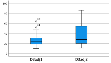
The box plots above compare seasonally adjusted 25(OH)D3, active vitamin D3 precursor levels in trial 1 (left side) and trial 2 (right side).
3.12. Summary of Lab Results in VA CMI Pilot Study Subjects for Human Herpes Virus dUTPase Antibody Levels [18]
- Trial 1 serum test results for human IgG serum antibody levels to herpes virus dUTPase proteins were negative for all three viruses in 10 out of 40 (25%) study serum tests and positive for all three viruses in 7 of 40 (17.5%) study serum tests.
Trial 1 human antibody levels to the Varicella Zoster dUTPase enzyme were positive in 11 out of 40 study subjects.
- 2.
- Trial 2 serum test results for human IgG antibody levels to herpes virus dUTPase enzymes were negative for all three viruses in only 1 out of 36 (2.8%) study subjects. Human IgG dUTPase antibodies were positive for all three viruses in 17 out of 36 (47%) study subjects.
Trial 2 human antibody levels were positive for the Varicella Zoster dUTPase enzyme in 23 out of 36 (64%) study subjects.
Positive-Test result was defined in both trials, using the same ELISA measurement technique. A serum sample that led to a human antibody herpes virus dUTPase signal twice over the background OD (Optical Density) of the control serum (95% Confidence Level) was considered elevated or positive.
3.13. Trial 1 and 2 Human dUTPase Antibody Serum Test Results in 2 Study Subjects Who Were Later Diagnosed with Severe Neurodegenerative Disorders within 3-5 years after starting this study.
Initial pilot study data including Trial 1 lab serum tests were collected from the study subjects prior to the study being placed on hold during the peak of the COVID epidemic. (i.e. between March 2020 and June 2021)
Significant neurodegenerative conditions were diagnosed in two study subjects after this VA pilot study reopened and after their Trial 2 blood tests had been collected. One study subject was diagnosed with Amyotrophic Lateral Sclerosis in September 2022 and was followed at the Atrium Health Wake Forest Baptist ALS Clinic until his passing in hospice care on 20 June 2025. Besides ALS and quadriparesis, he had two other significant diagnoses. The first was “Major or Mild Neurocognitive Disorder Due to Unknown Etiology”. The second was terminal metastatic appendiceal cancer (diagnosed in December 2019 after his appendix ruptured and 8 months after entering our study). Serum IgG and IgM human antibody levels against Varicella Zoster dUTPase enzymes were both positive (elevated) in both his serum blood draws (Trials 1&2).
The second study subject developed a progressive, neurodegenerative disorder which included quadriparesis with some sparing of the left upper extremity and a rapidly progressing decline in cognitive function, “Major or Mild Neurocognitive Disorder Due to Unknown Etiology” during 2024-2025. He has been evaluated at the Duke University Health Neuromuscular Clinic in August 2025. He currently requires further neurological evaluation to determine a more precise diagnosis for his neurodegenerative disorder. IgG human antibody levels to Varicella Zoster dUTPase enzyme were elevated/positive in both blood draws (Trials 1&2). IgM human antibody level to the Varicella Zoster dUTPase enzyme was elevated/positive in the first blood draw only (Trial 1).
The data for Varicella Zoster in these two study subjects is presented because this is an observational VA study of veterans with chronic multi-system Illness (CMI) and these diagnoses were very unexpected observations in these two VA pilot study subjects with a CMI initial diagnosis. (See Discussion Section.)
4. Discussion
Based on earlier work,[8]--[16] this VA Pilot study was designed to provide a multi-perspective snapshot of U.S. veterans with chronic multi-symptom illnesses. This snapshot included the recording of demographic information, a pertinent medical history, a focused physical examination, a mental health assessment including cognitive function, and the measurement of certain laboratory blood tests.
4.1. Demographic Information
Pertinent demographic information of our study group in comparison to the overall veteran population is listed in Table 1. Our study subject’s mean age was lower than the mean age of the general VA population. Combat experience was higher in our study population than in the general VA population even when compared to the higher rate of combat experienced in veterans since 9/11/2001. The African American/Black racial makeup in our study population was nearly twice that of the general U.S. veteran population.
In our study group, 33/40 (82.5%) of our pilot study subjects (Table 2) were service connected for a compensable injury or condition. Slightly over half (55%) were rated at 80% or higher. In the overall U.S. veteran population, about 27% have a service-connected injury or condition and of those 27%, 47% have a service-connected condition and a rating of 60% or higher.[56,58,59] In our study group, of the 82.5% who have a service-connected condition, 65% have a rating of 60% or higher and 22.5% have a rating of 100% or higher. Because of the funneling referral effect of sending VA Medical Center patients to the Rehabilitation Medicine Service with severe pain and chronic, severely disabling, multi-symptom illnesses, this study’s subjects may be more severely affected by CMI than the average veteran population with CMI.
Of our study’s 29 combat veterans, 4 served during the Vietnam War, 11 served during the First Gulf War, and 14 served during Operation Enduring Freedom (OEF) or Operation Iraqi Freedom (OIF). Of our study’s 40 veterans, 24.2% were service connected (SC) for only physical conditions, 6.1% were SC for only mental health/ psychological conditions, and 69.7% were SC for both physical and mental health/psychological conditions. 5 of 7 veterans in our study with no SC conditions had not served in combat. Our VA Pilot Study subjects with CMI had similar symptoms and signs but served in multiple different conflicts, in multiple different locations, and at multiple different periods. This finding is consistent with an in-depth study done shortly after the first Gulf War which concluded that the disabling symptoms affecting veterans from the first Gulf War (whatever the cause) were like those affecting veterans from other conflicts dating back to al least the U.S. Civil War.[7]
4.2. Medical History - Current and Past Diagnoses, and Symptoms
The most frequent past medical diagnoses were episodes of trauma which is not surprising given the nature of serving in the military and in combat. The history of chicken pox (varicella) and/or shingles (Zoster) 70% is also not surprising but is significant in that the presence of latent VZV in the body provides a basis for the possible presence of VZV abortive lytic replication as one source of inflammation causing neuropathic pain in 77.5% of U.S. service members with CMI. Tinnitus was the next most frequent symptom in the study group – 82%, followed by chronic fatigue – 80%, sleep disturbance – 75%, and GI symptoms – 72.5%. This distribution of symptoms is consistent with a profile of chronic multi-symptom illness except for tinnitus which is usually not elicited from patients unless asked about specifically.
4.3. Physical Exam and Signs
The reason for our physical exam focus on the nervous system is the hypothesis that many if not most of the signs and symptoms in our U.S. veteran CMI study group emanate from the nervous system. It is hypothesized that CMI signs and symptoms are primarily related to an immune system inflammatory response to latent herpesvirus abortive lytic replication rather than predominately lytic replication occurring in nervous system cells. Typical lytic replication results in a virus infection including cell lysis, fever, elevated white count etc. Abortive lytic replication is proposed to be caused by dUTPase molecules produced during DNA replication without complete virion synthesis. Human IgG and IgM antibodies to herpes virus dUTPase enzymes, synthesized by latent herpes virus DNA in many parts of the nervous system, are used as biomarkers in this study for this abortive lytic replication. The inflammation caused by dUTPase and possibly other molecules produced during abortive lytic replication is proposed to be the cause of symptoms and signs on physical exam in our study subjects with CMI.
The DNA of herpetic viruses, especially varicella zoster and simplex viruses, has been found using PCR throughout the central and peripheral nervous systems affecting the brain, cranial nerves in the brain stem, and peripheral nervous system including the autonomic and somatic nervous systems.[61,62] The reactivation of latent herpetic viruses, especially varicella zoster, has been reported as associated with a failure of the immune system to suppress these viruses. The pain caused by reactivation of varicella zoster has been attributed to inflammation of the sensory ganglia and nerves.[63] The neuropathic pain of varicella zoster can be associated with hyperalgesia to pin prick and allodynia. This pain is not always associated with skin lesions or rash and when this occurs the condition is referred to as “zoster sine herpete”.[64] Finally, motor nerves can be affected during an outbreak of herpes zoster or simplex causing weakness or paralysis of certain muscles such as those of the brachial plexus or face.[61,62]
On the physical exam, hyperalgesia to pin prick over the skin of the upper extremities was the most frequent abnormal sign and was found in 95% of the study subjects. Hyperalgesia to pin prick may be a manifestation of acute zoster, post herpetic neuralgia, and zoster sine herpete as well as for other herpetic viruses such as Herpes simplex 1&2.[65,66] Hyperalgesia to pin prick is a very frequent abnormal sensation in the skin of our study subjects and is commonly associated with one of the three herpes virus dUTPase enzymes (VZV) whose human antibody levels were measured in this study. The elevated VZV herpetic virus dUTPase enzyme antibody levels were found to be positive in at least one of the two blood specimens drawn from each study subject who had two blood draws (36/40). Hyperalgesia to pin prick may also be connected to the symptom of neuropathic pain which was reported to be present in 77.5% of study subjects.
The next frequent abnormal sign on physical exam was impaired balance on heel to toe walking along a straight line (tandem gait). A high prevalence of the latent (alpha) herpes simplex one virus DNA has been reported in the trigeminal geniculate and in the vestibular ganglia – vestibulocochlear nerve, cranial nerve VIII. [67] The sign of decreased balance in at least 71% of our study subjects and the symptom of tinnitus reported to be present in 82% of study subjects may be related to the latent HS1 virus whose dUTPase enzyme was not measured in this study.
In our study group, no study subjects on initial physical exam had what appeared to be a rash consistent with shingles, although many different, atypical, appearing rashes have been documented to be associated with herpetic virus DNA. [68] Herpetic Virus dUTPase enzymes have not yet been measured in the serum only their antibodies. However, HHV-6 antigens and Epstein Barr virus dUTPase enzymes have been recently measured in the postmortem brain tissue of those with chronic fatigue syndrome. [69]
4.4. Cognitive and Psychological Testing/Evaluation
Initial testing of cognitive function did not reveal significant impairment other than moderate attention deficit disorder in about one half of our study subjects. Psychological testing revealed the presence of moderate to large amounts of emotions including depression, anger, anxiety, somatoform, and sleep disturbance symptoms.
4.5. Serum 25(OH)D3 and Calcium Levels
Serum 25(OH)D3 levels and the storage of this active vitamin D precursor go up or down in response to the rate of 25(OH)D3 utilized by the body to make active vitamin D, the amount of 25(OH)D3 being synthesized by the body from sunlight, the amount introduced into the body from D3 supplementation and the amount of 25(OH)D3 degraded or gotten rid of by the body. [50] The level of storage of the precursor to active vitamin D can be viewed as analogous to the charge on a battery. When the demand for active vitamin D increases significantly and the level of vitamin D precursor storage is adequate or even high, the precursor level will drop unless it is adequately replaced. Conversely, when demand for active vitamin D is low, low storage of the precursor (battery charge) is adequate. However, when the demand for active vitamin D increases, a higher level of vitamin D precursor (battery charge) is needed to produce sufficient active vitamin D during certain stressors such as pregnancy, trauma, or infection.[50]
There were continuous numbers of insufficient and deficient levels of active vitamin D precursor among our study subjects despite the knowledge of low vitamin D status early in the study and treatment by their physicians. In the 36 study subjects that had two blood draws, 25 study subjects’ serum 25(OH)D3 levels went up (average 18 points), 10 study subjects levels went down (average 10 points) and one remained the same. This reflects an overall improvement in vitamin D status between the first and second blood draws that was statistically significant on the paired sample test – Table 4. The definitions of sufficiency, insufficiency, and deficiency were established with respect to bone health and not immune system health. Levels of required stored vitamin D to make active vitamin D may change significantly depending on changing circumstances the body finds itself in.
Using the Pearson Correlation, non-seasonally adjusted vitamin D precursor, 25(OH)D3 levels in Table 5 did not correlate with serum calcium levels in each trial. In a previous larger study describing the effect of 25(OH)D3 on serum calcium, the Pearson correlation between the two measurements was 0.46 among men and 0.40 among women, suggesting a moderate day-to-day variation in calcium intake.[70] This correlation level may be expected because active vitamin D controls calcium absorption not its precursor and 25(OH)D3 levels in the serum do not correlate with active vitamin D levels in the serum except at very low 25(OH)D3 levels. [50] The fact that the effect of 25(OH)D3 on serum calcium levels in our pilot study was not seen, could also be due to the small sample size in our study.
4.6. Serum Human Herpesvirus dUTPase Antibody Levels
There is growing evidence that abortive lytic herpes virus reactivation may be a commonly unrecognized pathological process resulting in a human immune system inflammatory response in tissues where latent herpes virus DNA resides. Latent Varicella Zoster and Herpes Simplex Viruses are known to reside in nervous system tissue and are known to cause severe pain. They are also known to cause encephalitis and lower motor neuron impairment. [61,62]
An independent study of the postmortem brain tissue in patients with chronic fatigue syndrome supports the concept of multiple herpetic viruses being active at the same time in the nervous system.[69] Interestingly, in this postmortem study of brain and spinal cord tissue, active latent EBV and HH6V herpesviruses, co-existed in the same patient with chronic fatigue syndrome but not in the same tissue locations in the brain and nervous system.
4.7. Trial 1 and 2 Human dUTPase Antibody Serum Test Results in 2 Study Subjects Who Were Later Diagnosed with Severe Neurodegenerative Disorders within 3-5 years after starting this study.
Latent herpes zoster virus IgG and IgM antibody responses to dUTPase enzymes in these two study subjects are presented separately because the finding of severe cognitive and motor impairments were unexpected observations in this VA pilot study of veterans with a CMI initial diagnosis. (See Discussion Section, Physical Exam and Signs)
Quadriparesis and significant cognitive decline were observed in two study subjects after this VA pilot study reopened and after their Trial 2 blood tests had been collected. One study subject was diagnosed with Amyotrophic Lateral Sclerosis in September 2022 and was followed at the Atrium Health Wake Forest Baptist ALS Clinic until his passing on 20 June 2025. Besides ALS and quadriparesis, he had two other significant diagnoses including: “Major or Mild Neurocognitive Disorder Due to Unknown Etiology” and metastatic appendiceal cancer (diagnosed in December 2019 after his appendix ruptured and 8 months after entering our study). This study subjects Serum IgG and IgM human antibody levels against Varicella Zoster dUTPase enzymes were both positive (elevated) in both his serum blood draws (Trials 1&2).
The second study subject developed a neurodegenerative disorder which included quadriparesis with some sparing of the left upper extremity and rapidly progressing cognitive decline, “Major or Mild Neurocognitive Disorder Due to Unknown Etiology”, during 2024-2025. He has been evaluated at the Duke University Health Neuromuscular Clinic in August 2025. This study subject is currently undergoing further evaluation to determine a more precise diagnosis for their neurodegenerative disorder. IgG human antibody levels to Varicella Zoster dUTPase enzyme were elevated/positive in both blood draws (Trials 1&2). IgM human antibody level to the Varicella Zoster dUTPase enzyme was elevated/positive in the first blood draw only (Trial 1).
It is well documented that Varicella Zoster can damage many parts of the central and peripheral nervous system including upper and lower motor neurons. [61,62] The finding of one study subject with ALS and one other study subject with a neurodegenerative disorder clinically characterized by quadriparesis and a neurocognitive disorder of undetermined etiology would seem at first to be very different from and unrelated to chronic multi-symptom illness. Recent research has expanded our understanding of ALS and some other neurodegenerative diseases with respect to the parts of the nervous system affected (i.e. ALS is not just a motor neuron disease).
ALS patients can have symptoms and signs beyond just somatic motor dysfunction. [71,72,73,74,75,76,77,78] These recent articles document an array of symptoms which are common in patients with ALS and in patients with chronic multi-symptom illnesses. A recent article also reviews similarities in immune inflammatory responses found in traditional neuroinflammatory and neurodegenerative disorders such as Alzheimer’s disease, Parkinson’s disease, and Amyotrophic Lateral Sclerosis with those found in chronic multi-symptom illnesses such as Gulf War Illness and myalgic encephalomyelitis.[79]
Finally, one previous case report describes the presence of an immunological response in the spinal fluid (CSF) to herpes simplex viruses in a 47-year-old patient diagnosed with ALS, type Vulplan-Bernardt.[80] A subsequent 12 year follow up after antiviral treatment of herpes simplex treatment reported negative antibodies (below the level of measurement) to herpes simplex virus in the CSF and reported the patient doing well clinically. [81]
5. Study Limitations
1. This was an observational pilot study with a small number of participants. The goal was to collect pilot study data over many aspects of the 40-study subject’s physical and mental health and social background.
2. There was no control group of veterans at the W.G. Hefner VA Medical Genter without chronic multi-symptom illness used in this study.
3. Serum inflammatory, antigenic, herpesvirus dUTPase protein levels were not directly measured but human IgG and IgM antibodies to these herpetic virus dUTPase proteins were measured.
4. The measurement of the active vitamin D precursor, 25(OH)D3, is a measurement of vitamin D precursor storage in the body and to some extent the body’s ability to produce active vitamin D, 1,25(OH)2D3 (the battery storage analogy). It does not reflect the body’s actual intracellular active vitamin D concentration, synthesis, utilization, and demand.[5]
6. Conclusions:
1. Chronic Multi-Symptom and System Illnesses in the US Veteran population are common, idiopathic, often quite disabling, and are associated with significant physical and psychological suffering despite being generally non-lethal.
2. In this study, the study subjects often had deficient or insufficient active vitamin D precursor (storage) levels. This deficiency and insufficiency of active vitamin D precursor storage may limit the synthesis of active vitamin D during periods of increased demand.[50] Insufficiency of active vitamin D may impair two of the functions of the immune system, namely the suppression of latent viruses, and the modulation/attenuation of the inflammatory response to latent virus from abortive lytic replication. [43,44,45]
3. Measurable unique dUTPase IgG and IgM dUTPase antibody levels to one of the three sub-families of human herpetic viruses (VZV, EBV, and HH6V) were present in most of our study subjects in at least one blood draw specimen.[18] The presence of changing IgG and IgM antibodies to the herpetic virus dUTPase proteins is indirect evidence for the presence of an inflammatory immune response to herpes virus dUTPase proteins in our study subjects during this study.
4. A high percentage of humans are infected with latent herpes virus DNA. Because virus latency is so common, it may be considered a normal state or phenomenon. However, the presence of herpes virus DNA in the body can result in the production of molecules like the dUTPase proteins during abortive lytic replication. Herpes virus dUTPase proteins are pathological in humans because they induce an inflammatory response in the cells or tissues where they are present. This is an additional pathological expression of the latent viruses different in nature from the regeneration of whole virions which destroy cells by lysing them (lytic replication) and which produce their own inflammatory reaction.
5. Varicella zoster dUTPase IgM and IgG antibodies were present in the serum of two study subjects who developed severe neurodegenerative disorders including significant cognitive decline and quadriparesis. These conditions occurred after their initial diagnosis of chronic multi-symptom illness. Alpha herpesviruses (varicella zoster and herpes simplex) are known to cause injury to the nervous system including motor neurons. The capability of latent VZV virus to infect and inflame motor neurons suggests that abortive lytic replication may be contributing to their idiopathic neurodegenerative disorders.
One of these study subjects (diagnosed and treated for ALS) was a Gulf War veteran and both ALS and Chronic Multi-symptom Illness occur more frequently in Gulf War Veterans and are presumptive diagnoses for service connection by the VA.
6. Remaining questions include: when and where (in what tissues/cells) are these antigenic proteins present and how much inflammation are they causing? The locations where latent herpetic viruses reside have been determined for many herpetic viruses using PCR. The physical exam with a focus on the nervous system gives us a clue to where some of the latent herpes virus inflammation might be occurring, especially in the case of herpes zoster.
The simultaneous presence of 3 different herpesvirus dUTPase protein antibodies in our study subjects suggests that more than one dUTPase protein may be causing inflammation in the same or in different tissues/cells at the same time. This makes determining which one or more of the dUTPase proteins are causing significant inflammation/tissue damage and in which tissues/cells they are active a challenge. The presence of human antibodies to the antigenic dUTPase proteins (enzymes) suggests a new mechanism of chronic viral disease (abortive lytic replication) independent of classical viral lytic replication and lysis of cells.
7. Tasks Going Forward
- Could some idiopathic neurodegenerative disorders like ALS be just another rare presentation of an inflammatory process in various neurons caused by abortive lytic replication of latent varicella zoster or simplex herpes viruses following the spread of latent herpes zoster/simplex DNA into various nervous system cells/tissues?
There may be a test to exclude this possibility. The same technique that was used to document Epstein Barr, inflammatory, herpes virus, dUTPase proteins in the postmortem brain and spinal cord tissues of individuals who were diagnosed with chronic fatigue syndrome at the time of their deaths could be used to look for and document the presence of one or more inflammatory herpes virus dUTPase proteins in the brains and spinal cords of individuals who have died of ALS or other forms of neurodegenerative disease.[69]
This was achieved by using a recombinant herpetic Epstein Barr virus (EBV) dUTPase protein (synthesized in the Behavioral Medicine Research Lab, at Ohio State University).[11] The recombinant herpes virus EBV dUTPase protein was next used to produce a polyclonal antibody in a rabbit. This antibody was then used in a standard immunohistochemical technique to identify the EBV antigenic protein. This rabbit dUTPase protein antibody was deployed in the postmortem brain tissues of patients who had chronic fatigue syndrome (CFS) and in patients who did not have CFS. The brain tissues of patients with CFS had positive responses to the detection of viral dUTPase protein using rabbit derived EBV dUTPase antibodies.
The postmortem control tissues with no history of CFS showed no evidence of Epstein Barr or Human Herpes 6 herpes virus dUTPase enzymes. The focus of this postmortem brain tissue study was to identify Human Herpes Virus 6, (HHV-6) proteins and virus miRNA in ME/CSF patients and not in controls. The study also, however, looked for and detected EBV dUTPase inflammatory proteins in the tissues of CFS patients but not in the tissues from the controls. [6]
- 2.
- More direct tests are needed to measure inflammatory herpetic virus dUTPase proteins (and possibly other biomarker molecules of early latent viral abortive lytic replication) in the serum, spinal fluid, and tissues/cells of live human bodies. Specifically assays for measuring both free herpetic virus dUTPase proteins, and the combined dUTPase enzyme and antibody complex in plasma or spinal fluid may also be needed.
Herpetic dUTPase proteins have not yet been detected in the serum using synthesized dUTPase antibodies to capture the unbound herpetic virus dUTPase proteins. The concentration of free or unbound herpetic virus dUTPase proteins in spun down serum samples may be below the limits of detection using the current assay. Alternatively, it may be possible to use the bottom-up LC-MS (liquid chromatography-tandem mass spectrometry) method to detect free dUTPase protein and dUTPase antibody/protein complexes in samples that are not spun down.
- 3.
- Tissue examination in live humans using imaging techniques may be needed to directly identify these selected biomarkers, their quantity, and their location to connect symptoms and signs on physical exam with a pathological inflammatory process in the body. To accomplish this, an imaging test perhaps using tagged antibodies introduced into the body would be necessary. Complicating an imaging process is the possible/probable presence of multiple DNA based latent virus dUTPases causing inflammation at the same time and location or in different locations (cells/tissues) of the nervous system or other tissues of the body.
- 4.
- Finally, questions arise as to what is promoting latent virus abortive lytic replication and its accompanying inflammation and is it increasing in frequency? Is it caused by a new form of impaired immune system? CMI patients often have low or low normal serum vitamin D precursor storage levels. Active vitamin D may have a role in the immune system’s suppression of latent herpes viruses and their dUTPase proteins, and in the modulation of the body’s inflammatory response to these herpes virus dUTPase proteins.[43,44,45]
To determine the body’s intracellular synthesis of active vitamin D, any fluctuating increase in demand for active vitamin D, and the body’s overall concentration of active vitamin D, a new method of determining these parameters will be needed. [50] One way to acquire this information may be to measure active vitamin D, 1,25(OH)2D3, and its end metabolites along with its precursor, 25(OH)D3 and its end metabolites, in the serum and in the urine and then back calculate the level of and synthesis of active vitamin D in the body. To achieve this as with all other clinical tests, lab standards from normal subjects will also have to be determined for the end metabolite values.
Author Contributions
Norman Chiu: assisted in understanding the vitamin D data, reviewed and edited the manuscript and contributed to Future Tasks section of the manuscript. Brandon Cox: performed lab experiments and reviewed data at OSU. Paula Goolkasian designed experiments and study, performed statistical analyses, wrote and edited manuscript. S Lad performed psychological and cognitive testing and analyzed data. James Plunkett: screened patients for study, treated study subjects during the study, provided clinical input for study design and data analysis. Irene Mena Palomo: performed experiments at OSU and analyzed data. Robert Shura designed study, performed psychological and cognitive testing, analyzed data, wrote and edited manuscript: Maria Eugenia Ariza: designed experiments and study, analyzed data, wrote and edited manuscript: Marshall Williams designed experiments and study, analyzed data, wrote and edited manuscript. Sean R. Maloney: designed experiments and study, analyzed data, interviewed and examined patients, wrote and edited manuscript.
Funding
This work-study was supported by the National Institutes of Health (NIH/NIAID) grant R01 AI084898 to M.E.A. and M.V.W., The Salisbury Foundation for Research and Education—Grant # 18-020, Project # 1575420-4 and The National Association of Veterans' Research and Education Foundation, NAVREF.
Data Availability Statement
The data that support the findings of this study are available on request from the corresponding author. The data are not publicly available due to privacy or ethical restrictions.
Acknowledgments
This study is supported by the U.S. Department of Veterans Affairs, Research Department, W.G. (Bill) Hefner VA Medical Center, Salisbury, NC. The welfare of human subjects was protected and the W.G. (Bill) Hefner VA Medical Center Institutional Review Board and Research Committee approved all research involving human subjects. (Study: IRB Protocol 18-020— “Medical Evaluation of U.S. Veterans with Chronic Multi-symptom illnesses”. Sean R. Maloney is a part-time physician employee of the U.S. Government. This study was prepared as part of his official duties. Title 17, USC, 105 provides that “Copyright protection under this title is not available for any work of the U.S. Government”. Title 17, USC, 101 defines a U.S. Government work as “a work prepared by an employee of the U.S. Government as part of that person's official duties”. We would also like to acknowledge the assistance of Marie Carmen Espiritu, MD for her assistance in screening patients and treating some of the study subject’s low vitamin D status. We would also like to thank Dr. Jarred Younger, University of Alabama Birmingham USA for providing GWI sera samples, Drs. Silvia de Sanjose, National Cancer Institute, Bethesda, MD, USA; ISGlobal, Barcelona, Spain and Ian W. Lipkin, Columbia University for providing control sera/plasma samples respectively, and Britney Martinez, Dylan Brown and Geetika Kaur for technical assistance.
Ethics Statement
Participants recruited via the Salisbury VA Health Care System and who were enrolled in the VA pilot study, “Medical Evaluation of U.S. Veterans with Chronic Multi-symptom Illness” signed an informed consent approved by the Salisbury VA Medical Center's Research & Development Committee (R&DC) and the W.G. (Bill) Hefner VA Medical Center Institutional Review Board (IRB Protocol 18-020). The welfare of human subjects was protected. Sean R. Maloney is a parttime physician employee of the U.S. Government. This study was prepared as part of his official duties. Title 17, USC, 105 provides that “Copyright protection under this title is not available for any work of the U.S. Government”. Title 17, USC, 101 defines a U.S. Government work as “a work prepared by an employee of the U.S. Government as part of that person's official duties”.
Conflicts of Interest
The authors declare no conflicts of interest.
References
- Public Health: Gulf War Veterans’ Medically Unexplained Illnesses. Department of Veterans Affairs. 16 May 2016. Available online: https://www.publichealth.va.gov/exposure/gulfwar/medically-unexplained-illness.asp (accessed on 30 April 2018).
- Cory-Slechta, D.; Wedge, R. (Eds.) Gulf War and Health. In Update of Health Effects of Serving in the Gulf War, 2016; The National Academies Press: Washington, DC; Volume 10.
- McAndrew, L.M.; Helmer, D.A.; Phillips, L.A.; Chandler, H.K.; Ray, K.; Quigley, K.S. Iraq and Afghanistan Veterans report symptoms consistent with chronic multisymptom illness one year after deployment. J. Rehab Res. Dev. 2016, 53(1), 59–70. [Google Scholar] [CrossRef]
- Smith, T.C.; Powell, T.M.; Jacobson, I.G.; Smith, B.; Hooper, T.M.; Boyko, E.J.; Gacksletter, G.D. Chronic Multisymptom Illness: A Comparison of Iraq and Afghanistan Deployers With Veterans of the 1991 Gulf War. Am. J. Epidemiol. 2014, 180(12), 1176–1187. [Google Scholar] [CrossRef] [PubMed]
- Public Health: Gulf War Veterans’ Medically Unexplained Illnesses. Department of Veterans Affairs. 16 May 2016. Available online: https://www.publichealth.va.gov/exposure/gulfwar/medically-unexplained-illness.asp (accessed on 30 April 2018).
- Research Advisory Committee on Gulf War Veterans’ Illnesses. Gulf War Illness and the Health of Gulf War Veterans: Scientific Findings and Recommendations. Executive Summary: pg. 4, U.S Government Printing Office, Washington, D.C. November 2008. Available online: https://www.va.gov./RAC-GWV1/docs/Committee_Documents/GW1andHealthofGWVeterans_RAC-GWVIReport_2008.pdf (accessed on 11 June 2018).
- Hyams, K.C.; Wignail, F.S.; Roswell, R. War syndromes and their evaluations: From the U.S. Civil War to the Persian Gulf. Ann. Intern. Med. 1996, 125, 398–405. [Google Scholar] [CrossRef] [PubMed]
- Maloney, S.R.; Jensen, S.; Gil-Rivas, V.; Goolkasian, P. Latent viral immune inflammatory response model for chronic multisymptom illness. Med. Hypotheses 2013, 80, 220–229. Available online: http://www.ncbi.nlm.nih.gov/pubmed/23265363. [CrossRef] [PubMed]
- Maloney, S.R.; Goolkasian, P. Low Vitamin D States Observed in U.S. Marines and Navy Sailors with Early Multi-Symptom Illness. Biomolecules 2020, 10; 1032, pgs. 1–12. [Google Scholar]
- Ariza, M.E.; Rivailler, P.; Glaser, R.; Chen, M.; Williams, M.V. Epstein-Barr Virus Encoded dUTPase Containing Exosomes Modulate Innate and Adaptive Immune Responses in Human Dendritic Cells and Peripheral Blood Mononuclear Cells. PLoS ONE 2013, 8, e69827. [Google Scholar] [PubMed]
- Aubrecht, T.G.; Weil, Z.M.; Ariza, M.E.; Williams, M.; Reader, B.; Glaser, R.; Sheridan, J.; Nelson, R.J. Epstein-Barr virus (EBV)-Encoded dUTPase and Chronic Restraint Induce Impaired Learning and Memory and Sickness Responses. Physiol. Behav. 2014, 137, 18–24. [Google Scholar] [PubMed]
- Williams, M.V.; Cox, B.; Lafuse, W.P.; Ariza, M.E. Epstein-Barr Virus dUTPase Induces Neuroinflammatory Mediators: Implications for Myalgic Encephalomyelitis/Chronic Fatigue Syndrome. Clin. Ther. 2019, Volume 41; Number 5, 1–16. [Google Scholar]
- Ariza, M.E.; Glaser, R.; Williams, M.V. Human herpesviruses-encoded dUTPases: A family of proteins that modulate dendritic cell function and innate immunity. Front. Microbiol. Virol. 2014, 5, 505. [Google Scholar]
- Williams, M.V.; Brandon, C.; Ariza, M.E. Herpesviruses dUTPases: A New Family of Pathogen-Associated Molecular Pattern (PAMP) Proteins with Implications for Human Disease. Pathogens 2017, 6, 2. [Google Scholar]
- Ariza, E.A. Myalgic Encephalomyelitis/Chronic Fatigue Syndrome: The Human Herpesviruses Are Back! Biomolecules 2021, 11, 185. [Google Scholar] [CrossRef] [PubMed]
- Ariza, M.E.; Cox, B.; Martinez, B.; Mena-Polomo, I.; Zarate, G.J.; Williams, M.V. Viral dUTPases: Modulators of Innate Immunity. Biomolecules 2022, 12, 227. [Google Scholar] [CrossRef] [PubMed]
- Vertessy, B.G.; Toth, J. Keeping Uracil Out of DNA: Physiological Role, Structure, and Catalytic Mechanism of dUTPases. Acc. Chem. Res. 2009, 42(1), 97–106. [Google Scholar] [CrossRef] [PubMed]
- Cox, B.; Goolkasian, P.; Palomo, I.M.; Williams, M.V.; Maloney, S.R.; Ariza, M.E. Reactivation of Latent Herpesviruses and a Faulty Antiviral Response may Contribute to Chronic Multi-Symptom and Multi-System Illnesses in U.S. Military Veterans. J. Med. Virol. 2025, 97(5), e70400. [Google Scholar] [CrossRef] [PubMed]
- SoRelle, E.D.; Haynes, L.E.; Willard, K.A.; Chang, B.; Ch’ng, J.; Christofk, H.; Luftig, M.A. Epstein-Barr virus reactivation induces divergent abortive, reprogrammed, and host shutoff states by lytic progression. PLoS Pathog. 2024, 20(10), e1012341. [Google Scholar] [CrossRef] [PubMed]
- Weidner-Glunde, M.; Kruminis-Kaszkiel, E.; Savanagouder, M. Herpesviral Latency - Common Themes. Pathogens 2020, 9(2), 125. [Google Scholar] [CrossRef] [PubMed]
- Cohen, J.I. Herpesvirus latency. J. Clin. Invest 2020, V130;7, 3361–3369. [Google Scholar] [CrossRef]
- Stoeger, T.; Adler, H. “Novel” Triggers of Herpesvirus Reactivation and Their Potential Health Relevance. Front. Microbiol. 2019, 9, 3207. [Google Scholar] [CrossRef] [PubMed]
- Laichalk, L.L.; Thorley-Lawson, D.A. Terminal differentiation into plasma cells initiates the replicative cycle of Epstein-Barr virus in vivo. J. Virol. 2005, 79, 1296–1307. [Google Scholar] [CrossRef] [PubMed]
- Al Tabaa, Y.; Tuaillon, E.; Bollore, K.; Foulongne, V.; Petitjean, G.; Seigneurin, J.M.; Duperray, C.; Desgranges, C.; Vendrell, J.P. Functional Epstein-Barr virus reservoir in plasma cells derived from infected blood memory B cells. Blood 2009, 113, 604–611. [Google Scholar] [PubMed]
- Al Tabaa, Y.; Tuaillon, E.; Jeziorski, E.; Ouedraogo, D.E.; Bollore, K.; Rubbo, P.A.; Foulongne, V.; Rodiere, M.; Vendrell, J.P. B-cell polyclonal activation and Epstein-Barr viral abortive lytic cycle are two key features in acute infectious mononucleosis. J. Clin. Virol. 2011, 52, 33–37. [Google Scholar] [PubMed]
- Altmann, M.; Hammerschmidt, W. Epstein-Barr virus provides a new paradigm: A requirement for the immediate inhibition of apoptosis. PLoS Biol. 2005, 3, e404. [Google Scholar] [CrossRef] [PubMed]
- Wen, W.; Iwakiri, D.; Yamamoto, K.; Maruo, S.; Kanda, T.; Takada, K. Epstein-Barr virus BZLF1 gene, a switch from latency to lytic replication, is expressed as an immediate-early gene after primary infection of B lymphocytes. J. Virol. 2007, 81, 1037–1042. [Google Scholar] [PubMed]
- Shannon-Lowe, C.; Adland, E.; Bell, A.I.; Delecluse, H.J.; Rickman, A.B.; Rowe, M. Features distinguishing Epstein-Barr virus infections of epithelial cells and B cells: Viral genome expression, genome maintenance, and genome amplification. J. Virol. 2009, 83, 7749–7760. [Google Scholar] [CrossRef] [PubMed]
- Kalla, M.; Hammerschmidt, W. Human B cells on their route to latent infection-early but transient expression of lytic genes of Epstein-Barr virus. Eur. J. Cell Biol. 2012, 91, 65–69. [Google Scholar] [PubMed]
- Perez, L. Characterizing cytomegalovirus infection one cell at a time. Nat. Microbiol. 2021, 6, 1477–1478. [Google Scholar] [CrossRef] [PubMed]
- Hein, M.Y.; Weissman, J.S. Functional single-cell genomics of human cytomegalovirus infection. Nat. Biotechnol. 2022, 40, 391–401. [Google Scholar] [PubMed]
- Prusty, B.K.; Gulve, N.; Chowdhury, S.R.; Schuster, M.; Strempel, S.; Descamps, V.; Rudel, T. HHV-6 encoded small non-coding RNAs define an immediate and early stage in viral replication. Genom. Med. 2018, 3, 25. [Google Scholar] [CrossRef]
- Scholz, B.A.; Harth-Hertle, M.L.; Malterer, G.; Haas, J.; Ellwart, J.; Schulz, T.F.; Kempkes, B. Abortive lytic reactivation of KSHV in CBF1/CSL deficient human cell lines. PLoS Pathog. 2013, 9(5), e1003336. [Google Scholar] [PubMed]
- Campbell, D.M.; Rappocciolo, G.; Jenkins, F.J.; Rinaldo, C.R. Dendritic cells: key players in human herpesvirus 8 infection and pathogenesis. Front Microbiol. 2014, 452. [Google Scholar] [CrossRef] [PubMed]
- Aneja, K.K.; Yuan, Y. Reactivation and lytic replication of Kaposi’s sarcoma-associated herpesvirus: an update. Front Microbiol. 2017, 8, 613. [Google Scholar] [PubMed]
- Ma, J.Z.; Russell, T.A.; Spelman, T.; CarboneF, R.; Tscharke, D.C. Lytic replication is frequent in HSV-1 latent infection and correlates with the engagement of a cell-intrinsic transcriptional response. PLoS Pathog. 2014, 10(8), e1004361. [Google Scholar] [PubMed]
- Drayman, N.; Patel, P.; Vistain, L.; Tay, S. HSV-1 single cell analysis reveals the activtion of anti-viral and developmental programs in distinct sub-populations. eLIFE 2019, 8, e46339. [Google Scholar] [PubMed]
- Kobiler, O.; Afriat, A. The fate of incoming HSV-1 genomes entering the nucleus. Curr. Issues Mol. Biol. 2021, 41(1), 221–266. [Google Scholar] [CrossRef] [PubMed]
- Fredrikson, J.P.; Domanico, L.F.; Pratt, S.L.; Loveday, E.K.; Taylor, M.P.; Chang, C.B. Single-cell herpes simplex virus type 1 infection of neyrons usingdrop-based microfluidics reveals heterogeneous replication kinetics. Sci. Adv. 2024, 10, eadk9185. [Google Scholar] [PubMed]
- Laemmie, L.; Goldstein, R.S.; Kinchington, P.R. Modeling Varicella zoster virus persistence and reactivation-closer to resolving a perplexing persistent state. Front Microbiol. 2019, 10, 1634. [Google Scholar]
- Warner, B.E.; Yee, M.B.; Zhang, M.; Hornung, R.S.; Kaufer, B.B.; Visalli, R.J.; Kramer, P.R.; Goins, W.F.; Kinchington, P.R. Varicella-zoster virus early infection but not complete replication is required for the induction of chronic hypersensitivity in rat models of postherpetic neuralgia. PLoS Pathog. 2021, 17(7), e1009689. [Google Scholar] [CrossRef] [PubMed]
- Wang, J.; Bruckner, N.; Weissmann, S.; Gunther, T.; Zhu, S.; Vogt, C.; Sun, G.; Guo, R.; Bruno, R.; Ritter, B.; Steinbruck, L.; Kaufer, B.B.; Depledge, D.P.; Grundhoff, A.; Viejo-Borbolla, A. Repression of varicella zoster gene express during quiescent infection in the absence of detectable histone deposition. PLoS Pathog. 2025, 21(2), e1012367. [Google Scholar] [CrossRef] [PubMed]
- Hewsin, M. Vitamin D and the intracrinology of innate immunity. Mol. Cell Endocrinol. 2010, 321, 103–111. [Google Scholar] [CrossRef]
- White, J.H. Emerging Roles of Vitamin D-Induced Antimicrobial Peptides in Antiviral Innate Immunity. Nutrients Assessed. 2023, 2022. 14, 284. [Google Scholar] [CrossRef] [PubMed]
- Galdo-Torres, D.; Andreu, S.; Caballero, O.; Hernandez-Ruiz, I.; Ripa, I.; Bello-Morales, R.; Lopez-Guerrero, J.A. Immune Modulatory Effects of Vitamin D on Herpesvirus Infections. Int. J. Mol. Sci. 2025, 26, 1767. [Google Scholar] [CrossRef] [PubMed]
- Holick, M.F.; Binkley, N.C.; Bischo-Ferrari, H.A.; Gordon, C.M.; Hanley, D.A.; Heaney, R.P.; Murad, M.H.; Weaver, C.M. Evaluation, Treatment, and Prevention of Vitamin D Deficiency: An Endocrine Society Clinical Practice Guideline. J. Clin. Endocrinol. Metab. 2011, 96, 1911–1930. [Google Scholar] [CrossRef] [PubMed]
- Kidney Research Institute. 25 Hydroxyvitamin D Calculator for Seasonal Adjustment. University of Washington. Available online: https://kri.washington.edu/calculator (accessed on 31 January 2019).
- Sachs, M.C.; Shoben, A.; Levin, G.P.; Robinson-Cohen, C.; Hoofnable, A.N.; Swords-Jenny, J.N.; Ix, J.H.; Budo, M.; Lutsey, P.L.; Siscovick, D.S.; et al. Estimating mean annual 25-hydroxyvitamin D concentrations from single measurements: The Multi-Ethnic Study of Atherosclerosis. Am. J. Clin. Nutr. 2013, 97, 1243–1251. [Google Scholar] [CrossRef] [PubMed]
- Budo, M.; Lutsey, P.L.; Siscovick, D.S.; et al. Estimating mean annual 25-hydroxyvitamin D concentrations from single measurements: The Multi-Ethnic Study of Atherosclerosis. Am. J. Clin. Nutr. 2013, 97, 1243–1251. [Google Scholar] [CrossRef]
- Maloney, S.R. A Mathematical Model for Determining the Body’s Fluctuating Need for and Synthesis of Active Vitamin D. Biomedicines 2023, 11, 324. [Google Scholar] [CrossRef] [PubMed]
- Marcinowska-Suchowierska, E.; Kupisz-Urbańska, M.; Łukaszkiewicz, J.; Płudowski, P.; Jones, G. Vitamin D Toxicity-A Clinical Perspective. Front Endocrinol. 2018, 9, 550. [Google Scholar] [CrossRef] [PubMed] [PubMed Central]
- Halpin, P.; Williams, M.V.; Kilmas, N.G.; Fletcher, M.A.; Barnes, Z.; Ariza, M.E. Myalgic encephalomyelitis/chronic fatigue syndrome and gulf war illness patients exhibit increased humoral responses to the herpesvirus-encoded dUTPase: Implications in disease pathophysiology. J. Med. Virol. 2017, 26, jmv24810 epub. [Google Scholar]
- National Center for Veteran’s Analysis and Statistics; United States Department of Veterans Affairs. Profile of Veterans: 2016 Data from the American Community Survey. February 2018; pp. pgs. 1–45. Available online: https://www.va.gov/vetdata/docs/SpecialReports/Profile_of_Veterans_2016.pdf.
- Census Bureau Releases New Report on Veterans, Press Release Number CB20-TPS.30, June 02, 2020, US Census Bureau, US Government, Census Bureau Releases New Report on Veterans assessed Dec. 20, 2023
- VetPop2020 National Data, 1L, National Center for Veterans Analysis and Statistics, US Department of Veterans Affairs, VetPop2020 National Data, 1L | Department of Veterans Affairs Open Data Portal (va.gov) assessed Dec. 20. 2023. [PubMed]
- Deployment, combat, and their consequences. The American Veteran Experience and the Post-9/11 Generation, Sept. 10, 2019. Pew Research Center, 2023. Available online: https://www.pewresearch.org/social-trends/2019/09/10/deployment-.
- Minority Veteran Statistics, Population Page, Racial and Ethnic Minority Veterans, Office of Health Equity, U.S. Dept. of Veterans Affairs April 2016. Available online: https://www.va.gov/HEALTHEQUITY/Race_Ethnicity.asp (accessed on 27 February 2021).
- National Center for Veteran’s Analysis and Statistics; United States Department of Veterans Affairs. Profile of Veterans: 2016 Data from the American Community Survey Profile of Veterans: 2009 (va.gov) Assessed 2018, pgs. 1–45.
- Military Service Experience, VA.gov Home, VA Dept. of Veterans Affairs. 15 February 2024. Available online: https://www.va.gov/vetdata/docs/surveysandstudies/military.pdf.
- WebMD. Hypertension-High Blood Pressure Guide: diastolic and systolic. Available online: https://www.webmd.com/hypertension-high-blood-pressure/guide/diastolic-.
- Kennedy, P.G.E. The Spectrum of Neurological Manifestations of Varicella-Zoster Virus Reactivation. Viruses 2023, 15(8), 1663. [Google Scholar] [CrossRef]
- Gilden, D.; Nagel, M.; Cohrs, R.; Mahalingam, R.; Baird, N. Varicella Zoster Virus in the Nervous System. (version 1) F1000Research 2015. 4(F1000 Faculty Rev). Last updated. 15 FEB 2016; p. 1356. [Google Scholar]
- Nair, P.A.; Patel, B.C. Herpes Zoster 2023. Stat Pearls, NCBI Bookshelf; National Library of Medicine.
- Zhou, J.; Li, J.; Ma, L.; Cao, S. Zoster sine herpete: a review. Korean J. Pain 2020, 3, 208–215. [Google Scholar] [CrossRef]
- Haanpa, M.; Laippala, P.; Nurmikko, T. Allodynia and Pinprick Hypesthesia in Acute Herpes Zoster, and the Development of Postherpetic Neuralgia. J. Pain Symptom Manag. 2000, Vol 20(1), 50–58. [Google Scholar] [CrossRef]
- Jain, E.; Eldon, E.; Witkin, L.; Cron, J. Herpes Simplex Virus-1 Complicated by Postherpetic Neuralgia: A Case Report. Pain Med. Case Rep. 2024, Vol 8(No. 4), 133–136. [Google Scholar] [CrossRef]
- Theil, D.; Arbusow, V.; Derfuss, T.; Strupp, M.; Pfeiffer, M.; Masco, A.; et al. Prevalence of HSV-1 latency in human Trigeminal geniculate, and vestibular ganglia and its implications for cranial nerve syndromes. Brain Pathol. 2001, 11(4), 408–13. [Google Scholar] [PubMed]
- Daruish, M.; Cazzato, G.; Markiewics, D.; Taibjee, S.; Fortarezza, F.; Calonje, E. A Review of Atypical Cutaneous histological Manifestations of Herpes Zoster. Viruses 2024, 16, 1035. [Google Scholar] [CrossRef] [PubMed]
- Kasimir, F.; Toomey, D.; Liu, Z.; Kaiping, A.C.; Ariza, M.E.; Prusty, B.K. Tissue specific signature of HHV-6 infection in ME/CFS. Front. Mol. Biosci. 2022. [Google Scholar] [CrossRef] [PubMed]
- Bischoff-Ferrari, H.A.; Kiel, D.P.; Dawson-Hughes, B.; Orav, J.E.; Li, R.; Spiegelman, D.; Dietrich, T.; Willett, W.C. Dietary Calcium and Serum 25-Hydroxyvitamin D Status in Relation to BMD Among U.S. Adults. J. Bone Min. Res. 2008, 24(5), 935–942. [Google Scholar] [CrossRef] [PubMed]
- Bombaci, A.; Lupica, A.; Pozzi, F.E.; Remoli, G.; Manera, U.; Di Stefano, V. Sensory neuropathy in amyotrophic lateral sclerosis: a systemic review. J. Neurol. 2023, 270-5677-5691. [Google Scholar] [CrossRef]
- Rubio, M.A.; Herrando-Grabulosa, M.; Navarro, X. Sensory Involvement in Amyotrophic Lateral Sclerosis, 2022. Int. J. Mol. Sci. 23, 15521. [CrossRef] [PubMed]
- Riancho, J.; Paz-Fajordo, L.; de Munain, Al. Clinical and preclinical evidence of somatosensory involvement in amyotrophic lateral sclerosis. Br. J. Pharmacol. 2021, 178, 1257–1268. [Google Scholar] [CrossRef] [PubMed]
- Dubbioso, R.; Provitera, V.; Pacella, D.; Santoro, L.; Manganelli, F.; Nolano, M. Autonomic dysfunction is associated with disease progression and survival in amyotrophic lateral sclerosis: a prospective longitudinal cohort study. J. Neurol. 2023, 270-4968-4977. [Google Scholar] [CrossRef] [PubMed]
- Oprisan, A.L.; PopescuBO. Dysautonomia in Amyotrophic Lateral Sclerosis. Int. J. Mol. Sci. 2023, 24, 14927. [Google Scholar] [CrossRef] [PubMed]
- Chio, A.; Mora, G.; Lauria, G. Pain in amyotrophic lateral sclerosis. Lancet Neurol. 2017, 16, 144–57. [Google Scholar] [CrossRef] [PubMed]
- Alencar, M.A.; Soares, B.L.; de Andrade Rangel, M.F.; Abdo, J.S.; de Almeida, R.A.P.; de Araujo, C.M.; de Souza, L.C.; de Cassia Gomes, G. Fatigue in amyotrophic lateral sclerosis and correlated factors. Arq. Neuropsiquiatr. 2022, 80(10), 1045–1051. [Google Scholar] [CrossRef] [PubMed]
- Bjelica, B.; Bartels, M.-B.; Hesebeck-Brinckmann, J.; Petri, S. Non-motor symptoms in patients with amyotrophic lateral sclerosis: current state and future directions. J. Neurol. 2024, 271, 3953–3977. [Google Scholar] [CrossRef]
- Cohen, J.; Mathew, A.; Dourvetakis, K.D.; Sanchez-Guerrero, E.; Pangeni, R.P.; Gurusamy, N.; Aenlle, K.K.; Ravindran, G.; Twahir, A.; Isler, D.; Sosa-Garcia, S.R.; Llizo, A.; Bested, A.C.; Theoharis, T.C.; Klimas, N.G.; Kempuraj, D. Recent Research Trends in Neurodegenerative Disorders. Cells 2024, 13, 511. [Google Scholar] [CrossRef] [PubMed]
- Ferri-de-Barros, J.E.; Moreira, M. Amyotrophic lateral sclerosis and herpes virus. Report of an unusual case: a cause or casual association? Arq. Neuropsiquiatr. 1998, 56(2), 307–11. [Google Scholar] [PubMed]
- Ferri-de-Barros, J.E.; Moreira, M. Amyotrophic lateral sclerosis and herpes virus: A casual or causal association? Twelve years after. Arq. Neuropsiquiatr. 2010, 68(6), 978–979. [Google Scholar] [CrossRef] [PubMed]
- Bischoff-Ferrari, H.A.; Kiel, D.P.; Dawson-Hughes, B.; Orav, J.E.; Li, R.; Spiegelman, D.; Dietrich, T.; Willett, W.C. Dietary Calcium and Serum 25-Hydroxyvitamin D Status in Relation to BMD Among U.S. Adults. J. Bone Min. Res. 2008, 24(5), 935–942. [Google Scholar] [CrossRef] [PubMed]
Table 4.
Paired Sample Test (paired differences) for Adj. Vit. D and Ca Levels.

| 25(OH)D3 Adjusted |
26.45 (11.67)
n = 40 |
36.67 (21.84) n = 36 |
-2.70
p =.011 |
| Calcium mg/dL | 9.25 (.401) n = 40 |
9.31 (.476) n = 36 | -.81 p=.425 |
Table 5.
Relationship between Vitamin D3 and Calcium in Trials 1 and 2.
| ____________________________________________________________________ | ||||
| Pearson Correlation | Sig. (2-tailed) | 95% Confidence Intervals (2-tailed) | ||
| Lower | Upper | |||
| VitD3T1 - CaT1 | .237 | .142 | -.081 | .510 |
| VitD3T2 – CaT2 | .127 | .462 | -.211 | .437 |
| Note: The subscript T1 represents the data collected on Trial 1 and T2 is the data from Trial 2. | ||||
Disclaimer/Publisher’s Note: The statements, opinions and data contained in all publications are solely those of the individual author(s) and contributor(s) and not of MDPI and/or the editor(s). MDPI and/or the editor(s) disclaim responsibility for any injury to people or property resulting from any ideas, methods, instructions or products referred to in the content. |
© 2026 by the authors. Licensee MDPI, Basel, Switzerland. This article is an open access article distributed under the terms and conditions of the Creative Commons Attribution (CC BY) license (http://creativecommons.org/licenses/by/4.0/).
Copyright: This open access article is published under a Creative Commons CC BY 4.0 license, which permit the free download, distribution, and reuse, provided that the author and preprint are cited in any reuse.


