

Submitted:
14 June 2026
Posted:
24 June 2026
You are already at the latest version
Abstract
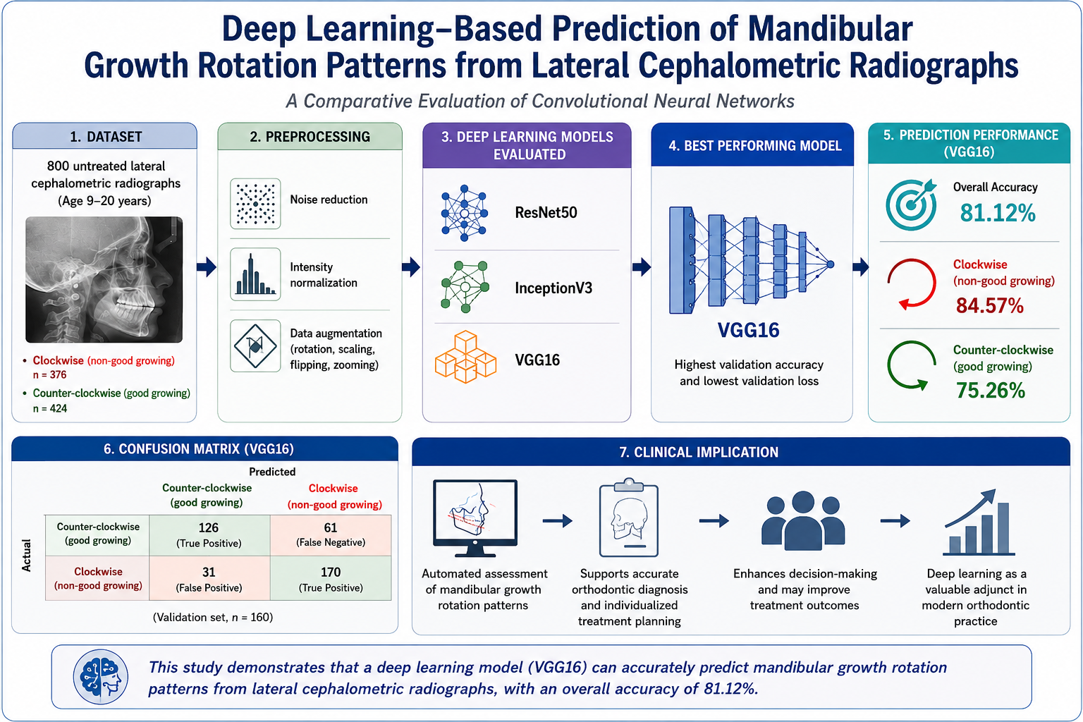
Background/Objectives: Prediction of mandibular growth rotation is important for orthodontic diagnosis and treatment planning, but conventional cephalometric methods are subjective and clinician-dependent. Deep learning may improve diagnostic consistency and efficiency. This study aimed to develop and evaluate a model for predicting mandibular growth rotation patterns from lateral cephalometric radiographs. Methods: A total of 800 untreated lateral cephalometric radiographs from individuals aged 9–20 years were retrospectively collected and classified as clockwise (n = 376) or counter-clockwise (n = 424) growth rotation patterns. The dataset was divided into training (80%) and validation (20%) sets. Three pre-trained convolutional neural network architectures (ResNet50, InceptionV3, and VGG16) were trained and compared. Image preprocessing included noise reduction, intensity normalization, and data augmentation. Model performance was evaluated using accuracy, precision, recall, F1-score, validation loss, and confusion matrix analysis. Results: VGG16 demonstrated the best performance, achieving the highest validation accuracy and lowest validation loss among the tested models. The model correctly predicted clockwise growth rotation with an accuracy of 84.57% and counter-clockwise growth rotation with an accuracy of 75.26%. Overall classification accuracy was 81.12%. Precision values were 0.85 and 0.73, recall values were 0.67 and 0.88, and F1-scores were 0.75 and 0.80 for counter-clockwise and clockwise growth patterns, respectively. Conclusions: The proposed VGG16-based model demonstrated promising accuracy in predicting mandibular growth rotation patterns from lateral cephalometric radiographs. Deep learning may serve as a valuable adjunct to orthodontic diagnosis and treatment planning, with further improvements possible through larger datasets and enhanced computational resources.
Keywords:
artificial intelligence
; deep learning
; convolutional neural network
; mandibular growth rotation
; cephalometric radiography
; orthodontics
; growth prediction
; predictive modelling
1. Introduction
The prediction of craniofacial growth remains one of the most challenging aspects of orthodontic diagnosis and treatment planning. Among the various components of facial growth, mandibular growth rotation plays a critical role in determining skeletal relationships, facial aesthetics, occlusal development, and treatment outcomes in growing patients. The mandible grows downward and forward through a combination of posterior growth and anterior displacement, accompanied by rotational changes that substantially influence the sagittal and vertical dimensions of the face. Unfavourable clockwise rotation is frequently associated with increased lower facial height, skeletal open bite tendencies, and reduced treatment stability, whereas counter-clockwise rotation is generally associated with more favourable growth characteristics. Consequently, accurate prediction of mandibular growth rotation is of considerable importance for individualized treatment planning and long-term outcome prediction. [1,2]
Several methods have been proposed to evaluate and predict mandibular growth rotation. Björk's landmark implant studies provided valuable insights into mandibular growth and remodeling and described three principal approaches for predicting mandibular shape changes: longitudinal, metric, and structural methods [3,4]. Among these, the structural method gained widespread acceptance because it relied on characteristic anatomical features observable on routine lateral cephalometric radiographs. Implant studies further demonstrated that mandibular growth occurs predominantly at the condylar region, while appositional and resorptive remodeling along the lower border contributes significantly to rotational changes of the mandible [1,4]. Building upon these findings, Skieller et al. identified four morphological characteristics—mandibular inclination, intermolar angle, lower border morphology, and symphysis inclination—as important indicators of future mandibular growth rotation, reporting an accuracy of approximately 88% [5].
Despite their clinical usefulness, traditional methods for predicting mandibular growth rotation remain dependent on subjective interpretation and clinician experience. Variations in landmark identification, observer bias, and the inherent complexity of craniofacial growth can affect the consistency and reliability of growth prediction. The increasing availability of digital radiographic records and computational technologies has created opportunities to overcome these limitations through automated image analysis.
Artificial intelligence (AI) has emerged as a transformative technology in healthcare and medical imaging. Since its inception in the mid-twentieth century, AI has evolved into a broad field encompassing machine learning (ML) and deep learning (DL), both of which are capable of identifying complex patterns within large datasets and generating predictive models with minimal human intervention [6]. In orthodontics, AI applications have expanded rapidly and have demonstrated promising results in cephalometric landmark detection, diagnosis, treatment planning, growth assessment, and clinical decision support systems [7]. In particular, convolutional neural networks (CNNs) have shown exceptional performance in image classification tasks because of their ability to automatically extract and learn relevant features directly from radiographic images.
Although AI has been successfully applied to several orthodontic diagnostic tasks, limited evidence exists regarding its application in the prediction of mandibular growth rotation patterns. Given the clinical significance of growth rotation and the limitations associated with conventional prediction methods, there is a need to investigate whether deep learning models can reliably classify mandibular growth patterns using routinely acquired cephalometric radiographs.
Therefore, the aim of the present study was to develop and evaluate a novel deep learning framework for predicting mandibular growth rotation patterns from lateral cephalometric radiographs. The performance of three pre-trained convolutional neural network architectures was compared to identify the most effective model for automated classification of clockwise and counter-clockwise mandibular growth rotation patterns. It was hypothesized that deep learning-based analysis of cephalometric radiographs could provide a reliable and objective approach for supporting orthodontic diagnosis and treatment planning
2. Materials and Methods
2.1. Study Sample and Dataset Preparation
A retrospective dataset comprising 800 lateral cephalometric radiographs of untreated patients aged 9-20 years was obtained from the Department of Oral Medicine and Radiology. The selected age range represented the active growth period during which mandibular growth rotation patterns can be effectively assessed and are of considerable importance in orthodontic diagnosis and treatment planning.
Only high-resolution digital cephalograms with adequate contrast, sharpness, and complete visualization of craniofacial structures were included. Radiographs exhibiting orthodontic appliances, jewellery, metallic artefacts, incorrect head positioning, motion artefacts, or poor image quality were excluded to ensure consistency and reliability of image interpretation.
The radiographs were manually categorized according to mandibular growth rotation patterns using established morphological indicators described by Björk and Skieller, including mandibular plane inclination, gonial angle morphology, antegonial notch depth, and lower border contour of the mandible. These classifications served as the ground-truth labels for model development and validation. The dataset comprised 424 radiographs representing counter-clockwise growth rotation (good-growing) patterns and 376 radiographs representing clockwise growth rotation (non-good-growing) patterns.
The complete dataset was randomly divided into training (80%) and validation (20%) subsets while maintaining class distribution across both groups. The distribution of radiographs in each subset is presented in Table 1.
The overall study workflow, including dataset preparation, classification, and model development, is illustrated in Figure 1.
2.2. Deep Learning Model Development
Model development and training were performed using Python programming language (Python Software Foundation, Wilmington, DE, USA) and the Keras deep learning framework (Keras Team, Google LLC, Mountain View, CA, USA) on a Lenovo IdeaPad 3 workstation (Lenovo Group Ltd., Beijing, China) equipped with an AMD Radeon Graphics Processing Unit (Advanced Micro Devices, Inc., Santa Clara, CA, USA). Three widely used pre-trained convolutional neural network (CNN) architectures employing transfer learning were evaluated and compared:
- ResNet50 (Microsoft Research, Redmond, WA, USA)
- InceptionV3 (Google Inc., Mountain View, CA, USA)
- VGG16 (Visual Geometry Group, University of Oxford, Oxford, UK)
Transfer learning was adopted to utilize feature representations learned from large-scale image datasets, thereby improving model performance while reducing training time and computational requirements.
2.3. Image Preprocessing and Feature Learning
Lateral cephalometric radiographs served as the input images for the proposed deep learning framework. Prior to model training, all radiographs underwent a standardized preprocessing pipeline to enhance image quality and ensure suitability for machine learning analysis. Preprocessing procedures included noise reduction to improve image clarity, intensity normalization to standardize pixel values across the dataset, and data augmentation to increase dataset variability and improve model robustness.
Following preprocessing, relevant image features associated with mandibular morphology and growth rotation patterns were extracted automatically by the CNN architectures. Unlike conventional cephalometric analyses that rely on manual landmark identification, the deep learning models learned hierarchical image representations directly from the radiographs. These learned features were subsequently utilized for classification of mandibular growth rotation patterns into good-growing (counter-clockwise) and non-good-growing (clockwise) categories.
The overall model training and classification workflow is illustrated in Figure 2.
2.4. Model Training
The selected CNN architectures were trained using the preprocessed and annotated dataset. During training, the models learned to differentiate between clockwise and counter-clockwise growth rotation patterns through iterative optimization of internal network parameters. Supervised learning was employed using labelled datasets, and network weights were updated using backpropagation algorithms to minimize the classification loss function and improve predictive performance.
Fine-tuning of the pre-trained networks was performed to adapt the learned feature representations specifically to the task of mandibular growth rotation classification. This process enabled the models to identify complex morphological patterns present within cephalometric radiographs and improve classification accuracy.
2.5. Model Evaluation and Performance Assessment
Following training, the performance of the deep learning models was evaluated using the independent validation dataset. Model performance was assessed using a comprehensive set of classification metrics. A confusion matrix was generated to quantify true positive, true negative, false positive, and false negative predictions, thereby providing a detailed assessment of classification outcomes.
Additional performance measures included accuracy, precision, recall, and F1-score, which collectively evaluated overall model effectiveness as well as the balance between sensitivity and specificity. To further assess learning behaviour, training and validation accuracy and loss curves were analysed to monitor convergence, detect potential overfitting or underfitting, and evaluate model generalizability to previously unseen data.
In the final classification stage, the trained model processed cephalometric radiographs and predicted whether the mandibular growth pattern corresponded to a good-growing (counter-clockwise) or non-good-growing (clockwise) category. The evaluation process ultimately facilitated the development of an automated deep learning framework capable of assisting clinicians in the assessment and prediction of mandibular growth rotation patterns from lateral cephalometric radiographs.
3. Results
A total of 800 lateral cephalometric radiographs were included in the study and categorized into two mandibular growth rotation groups. The dataset comprised 376 radiographs with clockwise growth rotation patterns and 424 radiographs with counter-clockwise growth rotation patterns. The complete dataset was randomly divided into training and validation subsets in an 80:20 ratio. Accordingly, 640 radiographs were used for model training and 160 radiographs were used for validation.
The performance of the three pre-trained convolutional neural network models, ResNet50, InceptionV3, and VGG16, was compared using validation accuracy and validation loss. Among the three models, VGG16 demonstrated the best overall performance, with the highest validation accuracy and the lowest validation loss. Therefore, VGG16 was selected as the final model for automated classification of mandibular growth rotation patterns from lateral cephalometric radiographs (Figure 3).
The confusion matrix for the VGG16 model demonstrated that the model was able to classify both clockwise and counter-clockwise mandibular growth rotation patterns with reasonable accuracy (Figure 4). The model correctly identified counter-clockwise growth rotation patterns, representing good-growing mandibles, with an accuracy of 75.26%, while 24.73% of these cases were misclassified as clockwise growth rotation patterns. The model correctly identified clockwise growth rotation patterns, representing non-good-growing mandibles, with an accuracy of 84.57%, while 15.42% of these cases were misclassified as counter-clockwise growth rotation patterns.
The overall accuracy of the VGG16 model was calculated using the following formula:
Accuracy = (True Positives + True Negatives) / Total Sample
Based on the confusion matrix values, the model achieved an overall accuracy of 81.12%, indicating that the VGG16 model correctly classified most radiographs into their respective mandibular growth rotation categories.
The training and validation accuracy and loss curves of the VGG16 model are shown in Figure 5. These curves were used to assess the learning behaviour of the model during training. The model demonstrated progressive improvement in accuracy with a corresponding reduction in loss, indicating effective learning and acceptable model convergence.
The validation classification report further supported the performance of the VGG16 model. For counter-clockwise growth rotation patterns, the precision, recall, and F1-score were 0.85, 0.67, and 0.75, respectively. For clockwise growth rotation patterns, the precision, recall, and F1-score were 0.73, 0.88, and 0.80, respectively. These findings indicate that the model showed better sensitivity in detecting clockwise growth rotation patterns, while demonstrating higher precision for counter-clockwise growth rotation patterns.
Overall, the VGG16 model outperformed ResNet50 and InceptionV3 and was selected as the most suitable deep learning architecture for the proposed automated mandibular growth rotation classification framework.
4. Discussion
Accurate prediction of mandibular growth rotation remains one of the most challenging yet clinically significant aspects of orthodontic diagnosis and treatment planning. Mandibular rotation has a profound influence on facial aesthetics, occlusal relationships, vertical facial proportions, and long-term treatment stability. Since growth modification procedures and treatment mechanics are largely dependent on anticipated growth patterns, the ability to predict mandibular growth rotation accurately is of considerable value to the orthodontist [8,9].
Historically, Björk's implant studies provided the foundation for understanding mandibular growth and remodeling and led to the development of structural indicators for predicting future growth direction [3,4]. Subsequent work by Skieller et al. demonstrated that specific morphological characteristics of the mandible could be used to estimate future growth rotation with relatively high accuracy [5]. Despite their clinical relevance, these approaches rely heavily on subjective interpretation of cephalometric structures and are influenced by observer experience, image quality, and variability in landmark identification. Furthermore, previous investigations have reported inconsistent results regarding the predictive reliability of these structural indicators when applied to broader patient populations [2,12,13]. These limitations highlight the need for more objective and reproducible approaches to growth prediction.
Recent advances in artificial intelligence have transformed image-based diagnostics across medicine and dentistry. Deep learning algorithms, particularly convolutional neural networks, have demonstrated an exceptional ability to identify complex image patterns that may not be readily apparent to human observers [6,10]. In orthodontics, AI has been successfully applied to cephalometric landmark identification, skeletal classification, growth assessment, and treatment planning, demonstrating performance levels comparable to or exceeding traditional approaches [7,15,16,17,18,19,20,21,22,23,24,25,26,27,28]. However, the application of deep learning for automated prediction of mandibular growth rotation has remained largely unexplored.
The present study developed and evaluated a deep learning framework for classification of mandibular growth rotation patterns using lateral cephalometric radiographs. Among the three pre-trained CNN architectures investigated, VGG16 demonstrated superior performance, achieving the highest validation accuracy and the lowest validation loss. The final model achieved an overall classification accuracy of 81.12%, with correct identification rates of 84.57% for clockwise growth rotation patterns and 75.26% for counter-clockwise growth rotation patterns. These findings suggest that deep learning algorithms are capable of extracting clinically relevant morphological information directly from cephalometric radiographs and can effectively differentiate between distinct mandibular growth rotation patterns.
The higher predictive performance observed for clockwise growth patterns may be attributed to the presence of more pronounced morphological characteristics typically associated with vertical growth, such as increased mandibular plane angle, steeper gonial angle, and deeper antegonial notch morphology. In contrast, the counter-clockwise group included both horizontal and average growth patterns, which may have introduced greater morphological variability and contributed to the comparatively lower classification accuracy observed for this category. This difference is reflected in the classification metrics, where the model demonstrated higher recall for clockwise growth patterns and higher precision for counter-clockwise growth patterns.
The findings of the present investigation are consistent with the growing body of evidence supporting the use of AI in orthodontic diagnosis. Lee et al. reported accuracy levels approaching 90% for automated cephalometric landmark identification [15], while Kök et al. and several subsequent investigators demonstrated the successful application of machine learning and deep learning techniques for cervical vertebral maturation assessment and growth prediction [16,19,20,21,22,23,24,25,26,27,28]. Similarly, Qu et al. reported diagnostic accuracies ranging from 75.24% to 82.52% for AI-assisted assessment of mandibular asymmetry [29], whereas Myers et al. demonstrated prediction accuracies of approximately 70–80% for long-term craniofacial growth modelling using machine learning approaches [31]. The overall accuracy of 81.12% achieved in the present study is therefore comparable with previously reported AI-based orthodontic diagnostic systems and supports the feasibility of automated mandibular growth rotation assessment.
A notable strength of the present study is the relatively large dataset of 800 cephalometric radiographs used for model development and validation. In addition, the comparison of multiple state-of-the-art CNN architectures enabled identification of the most suitable network for this specific classification task. The use of transfer learning further improved model efficiency and allowed meaningful feature extraction despite the moderate dataset size.
Nevertheless, several limitations should be acknowledged. The dataset was obtained from a single institution, which may limit the generalizability of the findings to different populations and imaging systems. The classification labels were based on morphological assessment of growth patterns rather than longitudinal growth observations, and therefore represent predicted growth tendencies rather than actual future growth outcomes. Furthermore, although the model demonstrated promising performance, misclassification was still observed in both growth categories, indicating that mandibular growth prediction remains a complex biological phenomenon influenced by multiple genetic and environmental factors. Future studies should incorporate larger multicentre datasets, external validation cohorts, and longitudinal growth records to further improve model robustness and clinical applicability.
Overall, the findings of this study demonstrate that deep learning-based analysis of lateral cephalometric radiographs can provide an objective and efficient method for classification of mandibular growth rotation patterns. Such systems have the potential to function as clinical decision-support tools, assisting orthodontists in diagnosis, growth assessment, and treatment planning while reducing reliance on subjective interpretation.
5. Conclusions
A novel deep learning framework was developed and evaluated for automated classification of mandibular growth rotation patterns using lateral cephalometric radiographs. Among the three convolutional neural network architectures investigated, VGG16 demonstrated the best performance, achieving an overall classification accuracy of 81.12%, with prediction accuracies of 84.57% and 75.26% for clockwise and counter-clockwise growth rotation patterns, respectively.
The findings indicate that deep learning algorithms are capable of identifying clinically relevant morphological features associated with mandibular growth rotation directly from cephalometric images and can provide objective classification of growth patterns. This approach has the potential to enhance diagnostic consistency, reduce observer-dependent variability, and support orthodontic treatment planning.
The findings of the present study support the feasibility of integrating deep learning-based image analysis into orthodontic growth assessment. With further validation and refinement, such systems may serve as valuable clinical decision-support tools for predicting mandibular growth rotation and enhancing treatment planning.
Author Contributions
Dharma R. Mallikarjunaiah: conceptualization, methodology, software, validation, supervision; Jyolsna Sreemony: formal analysis, investigation, resources, data curation; Narayan Gandedkar: writing - original draft preparation, writing- review and editing; Akshai Shetty, Amarnath B. Chikkamuniswamy and Prashanth C. Shivashankar: visualization, project administration. All authors have read and agreed to the published version of the manuscript.
Funding
This research received no external funding.
Institutional Review Board Statement
The study was conducted in accordance with the Declaration of Helsinki and approved by the Institutional Review Board (Research Sustenance & Institutional Review Board Committee) of D.A.P.M.R.V. Dental College, Bengaluru (Protocol No. 475/Vol-2/2023; approved on 15 May 2023).
Informed Consent Statement
Patient consent was waived due to the retrospective nature of the study. All data were collected from existing records, and patient anonymity and confidentiality were strictly maintained throughout the study.
Data Availability Statement
The data presented in this study are not publicly available due to privacy and ethical restrictions.
Conflicts of Interest
The authors declare no conflict of interest.
References
- Björk, A. Facial growth in man, studied with the aid of metallic implants. Acta Odontol. Scand. 1955, 13(1), 9–34. [Google Scholar] [CrossRef] [PubMed]
- Leslie, L.R.; Southard, T.E.; Southard, K.A.; Casko, J.S.; Jakobsen, J.R.; Tolley, E.A.; Hillis, S.L.; Carolan, C.; Logue, M. Prediction of mandibular growth rotation: assessment of the Skieller, Björk, and Linde-Hansen method. Am. J. Orthod. Dentofac. Orthop. 1998, 114(6), 659–67. [Google Scholar] [CrossRef] [PubMed]
- Björk, A. Prediction of mandibular growth rotation. Am. J. Orthod. 1969, 55, 585–99. [Google Scholar] [CrossRef] [PubMed]
- Björk, A. Variations in the growth pattern of the human mandible: longitudinal radiographic study by the implant method. J. Dent. Res. 1963, 42, 400–11. [Google Scholar] [CrossRef]
- Skieller, V.; Björk, A.; Linde-Hansen, T. Prediction of mandibular growth rotation evaluated from a longitudinal implant sample. Am. J. Orthod. 1984, 86(5), 359–70. [Google Scholar] [CrossRef] [PubMed]
- Khanagar, S.B.; Al-Ehaideb, A.; Maganur, P.C.; Vishwanathaiah, S.; Patil, S.; Baeshen, H.A.; et al. Developments, application, and performance of artificial intelligence in dentistry - a systematic review. J. Dent. Sci. 2021, 16, 508–22. [Google Scholar] [PubMed]
- Prasad, J.; Mallikarjunaiah, D.R.; Shetty, A.; Gandedkar, N.; Chikkamuniswamy, A.B.; Shivashankar, P.C. Machine Learning Predictive Model as Clinical Decision Support System in Orthodontic Treatment Planning. Dent. J. 2022, 11(1), 1. [Google Scholar] [CrossRef] [PubMed] [PubMed Central]
- Buschang, P. H.; Jacob, H. B. Mandibular rotation revisited: What makes it so important? Am. J. Orthod. Dentofac. Orthop. 2019, 155(6), 795–807. [Google Scholar]
- Björk, A.; Skieller, V. Normal and abnormal growth of the mandible. A synthesis of longitudinal cephalometric implant studies over a period of 25 years. Eur. J. Orthod. 1983, 5(1), 1–46. [Google Scholar] [CrossRef] [PubMed]
- Schwendicke, F.; Samek, W.; Krois, J. Artificial Intelligence in Dentistry: Chances and Challenges. J. Dent. Res. 2020, 99(7), 769–774. [Google Scholar] [CrossRef] [PubMed] [PubMed Central]
- Tajmir, S.H.; Lee, H.; Shailam, R.; Gale, H.I.; Nguyen, J.C.; Westra, S.J.; Lim, R.; Yune, S.; Gee, M.S.; Do, S. Artificial intelligence-assisted interpretation of bone age radiographs improves accuracy and decreases variability. Skelet. Radiol. 2019, 48(2), 275–283. [Google Scholar]
- Davidovitch, M.; Eleftheriadi, I.; Kostaki, A.; Shpack, N. The use of Bjork's indications of growth for evaluation of extremes of skeletal morphology. Eur. J. Orthod. 2016, 38(6), 555–562. [Google Scholar] [CrossRef] [PubMed]
- von Bremen, J.; Pancherz, H. Association between Björk's structural signs of mandibular growth rotation and skeletofacial morphology. Angle Orthod. 2005, 75(4), 506–9. [Google Scholar] [CrossRef] [PubMed]
- Gomes, Rahul; Kamrowski, C.; Langlois, Jordan; Rozario, Papia; Dircks, Ian; Grottodden, Keegan; Martinez, Matthew; Tee, Wei; Sargeant, Kyle; LaFleur, Corbin; Haley, Mitchell. A Comprehensive Review of Machine Learning Used to Combat COVID-19. Diagnostics 2022, 12, 1853. [Google Scholar] [CrossRef] [PubMed]
- Lee, H.; et al. Effectiveness of Human–Artificial Intelligence Collaboration in Cephalometric Landmark Detection. Diagnostics 2022, 12(11), 2877. [Google Scholar] [CrossRef] [PubMed]
- Kök, H.; Acilar, A.M.; İzgi, M.S. Usage and comparison of artificial intelligence algorithms for determination of growth and development by cervical vertebrae stages in orthodontics. Prog. Orthod. 2019, 20(1), 41. [Google Scholar] [CrossRef] [PubMed]
- Yu, H. J.; Cho, S. R.; Kim, M. J.; Kim, W. H.; Kim, J. W.; Choi, J. Automated skeletal classification with lateral cephalometry based on artificial intelligence. J. Dent. Res. 2020, 99(3), 249–256. [Google Scholar] [CrossRef] [PubMed]
- Jung, S. K.; Kim, T. W. New approach for the diagnosis of extractions with neural network machine learning. Am. J. Orthod. Dentofac. Orthop. 2016, 149(1), 127–133. [Google Scholar] [CrossRef] [PubMed]
- Makaremi, M.; Lacaule, C.; Mohammad-Djafari, A. Deep learning and artificial intelligence for the determination of the cervical vertebra maturation degree from lateral radiography. Entropy 2019, 21, 1222. [Google Scholar] [CrossRef]
- Amasya, H.; Yildirim, D.; Aydogan, T.; Kemaloglu, N.; Orhan, K. Cervical vertebral maturation assessment on lateral cephalometric radiographs using artificial intelligence: comparison of machine learning classifier models. Dentomaxillofac Radiol. 2020, 49, 20190441. [Google Scholar] [CrossRef] [PubMed]
- Li, Haizhen; Chen, Yanlong; Wang, Qing; Gong, Xu; Lei, Yi; Tian, Jialiang; Gao, Xuemei. Convolutional neural network-based automatic cervical vertebral maturation classification method. Dentomaxillofacial Radiol. 2022, Volume 51(Issue 6). [Google Scholar]
- Mathew, R.; Palatinus, S.; Padala, S.; Alshehri, A.; Awadh, W.; Bhandi, S.; Thomas, J.; Patil, S. Neural networks for classification of cervical vertebrae maturation: a systematic review. Angle Orthod. 2022, 92(6), 796–804. [Google Scholar] [CrossRef] [PubMed]
- Radwan, M.T.; Sin, Ç.; Akkaya, N.; Vahdettin, L. Artificial intelligence-based algorithm for cervical vertebrae maturation stage assessment. Orthod. Craniofac Res. 2023, 26(3), 349 355. [Google Scholar] [PubMed]
- Atici, S. F.; Elnagar, M. H.; Ansari, R.; Allareddy, V.; Suhaym, O.; Cetin, A. E. A novel continuous classification system for the cervical vertebrae maturation (CVM) stages using convolutional neural networks. Comput. Biol. Med. 2021, 134, 104453. [Google Scholar]
- Zhou, J.; Zhou, H.; Pu, L.; Gao, Y.; Tang, Z.; Yang, Y.; You, M.; Yang, Z.; Lai, W.; Long, H. Development of an Artificial Intelligence System for the Automatic Evaluation of Cervical Vertebral Maturation Status. Diagnostics 2021, 11(12), 2200. [Google Scholar] [CrossRef] [PubMed]
- Nogueira-Reis, F.; Cascante-Sequeira, D.; Farias-Gomes, A.; de Macedo, M. M. G.; Watanabe, R. N.; Santiago, A. G.; Tabchoury, C. P. M.; Freitas, D. Q. Determination of the pubertal growth spurt by artificial intelligence analysis of cervical vertebrae maturation in lateral cephalometric radiographs. Oral Surg. Oral Med. Oral Pathol. Oral Radiol. 2024, 138(2), 306–315. [Google Scholar] [CrossRef] [PubMed]
- Rana, S. S.; Nath, B.; Chaudhari, P. K.; Vichare, S. Cervical Vertebral Maturation Assessment using various Machine Learning techniques on Lateral cephalogram: A systematic literature review. J. Oral Biol. Craniofacial Res. 2023, 13(5), 642–651. [Google Scholar] [CrossRef]
- Khazaei, M.; Mollabashi, V.; Khotanlou, H.; Farhadian, M. Automatic determination of pubertal growth spurts based on the cervical vertebral maturation staging using deep convolutional neural networks. J. World Fed. Orthod. 2023, 12(2), 56–63. [Google Scholar] [CrossRef] [PubMed]
- Qu, W.; Qiu, Z.; Lam, K. C.; Sakaran, K. G.; Chen, H.; Lin, Y. Artificial intelligence-assisted identification and assessment of mandibular asymmetry on panoramic radiography. Oral Surg. Oral Med. Oral Pathol. Oral Radiol. 2023, 135(1), 78–86. [Google Scholar]
- Wood, T.; Anigbo, J. O.; Eckert, G.; Stewart, K. T.; Dundar, M. M.; Turkkahraman, H. Prediction of the Post-Pubertal Mandibular Length and Y Axis of Growth by Using Various Machine Learning Techniques: A Retrospective Longitudinal Study. Diagnostics 2023, 13(6), 1035. [Google Scholar] [CrossRef] [PubMed]
- Myers, M.; Brown, M. D.; Badirli, S.; Eckert, G. J.; Johnson, D. H.-M.; Turkkahraman, H. Long-Term Predictive Modelling of the Craniofacial Complex Using Machine Learning on 2D Cephalometric Radiographs. Diagnostics 2023, 13(5), 897. [Google Scholar]
- Enlow, E.H.; Harris, D.B. A study of the postnatal growth of the human mandible. Am. J. Orthod. 1964, 50, 25–50. [Google Scholar] [CrossRef]
- McNamara, J.A., Jr.; Graber, L.W. Mandibular growth in the Rhesus monkey. Am. J. Phys. Anthropol. 1975, 42, 15–24. [Google Scholar] [CrossRef] [PubMed]
- Brodie, A.G. Facial patterns: a theme on variation. Angle Orthod. 1946, 16, 75–87. [Google Scholar]
- Schudy, F.F. The rotation of the mandible resulting from growth: its implications in orthodontic treatment. Angle Orthod. 1965, 35, 36–50. [Google Scholar] [CrossRef] [PubMed]
- Björk, A.; Skieller, V. Facial development and tooth eruption: an implant study at the age of puberty. Am. J. Orthod. 1972, 62, 339–83. [Google Scholar] [PubMed]
- Baumrind, S.; Korn, E.L.; West, E.E. Prediction of mandibular rotation: an empirical test of clincian performance. Am. J. Orthod. 1984, 86, 371–85. [Google Scholar] [CrossRef] [PubMed]
- Park, W. J.; Park, J.-B. History and application of artificial neural networks in dentistry. Eur. J. Dent. 2018, 12(4), 594–601. [Google Scholar] [CrossRef] [PubMed]
- Björk, A. The use of metallic implants in the study of facial growth in children: Method and application. Am. J. Phys. Anthropol. 1968, 29(2), 243–254. [Google Scholar] [CrossRef] [PubMed]
- Björk, A.; Skieller, V. Facial development and tooth eruption: An implant study at the age of puberty. Am. J. Orthod. 1972, 62(4), 339–383. [Google Scholar] [PubMed]
- Alipour Shoari, S.; Sadrolashrafi, S. V.; Sohrabi, A.; Afrouzian, R.; Ebrahimi, P.; Kouhsoltani, M.; Kouh Soltani, M. Estimating mandibular growth stage based on cervical vertebral maturation in lateral cephalometric radiographs using artificial intelligence. Prog. Orthod. 2024, 25(1), Article 27. [Google Scholar] [CrossRef]
Figure 1.
Study design. Workflow illustrating dataset selection, classification of mandibular growth rotation patterns, training of three CNN architectures (ResNet50, InceptionV3, and VGG16), and model development using the Python–Keras framework on an AMD Radeon GPU platform.
Figure 1.
Study design. Workflow illustrating dataset selection, classification of mandibular growth rotation patterns, training of three CNN architectures (ResNet50, InceptionV3, and VGG16), and model development using the Python–Keras framework on an AMD Radeon GPU platform.
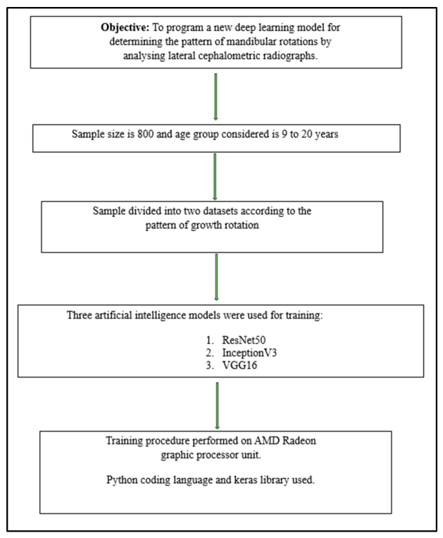
Figure 2.
Illustration of the model training and classification process. Workflow of the deep learning framework for mandibular growth rotation classification. Lateral cephalometric radiographs were used as input images and underwent preprocessing, including noise reduction, intensity normalization, and data augmentation. Relevant image features were subsequently extracted and analysed using three pre-trained convolutional neural network architectures (ResNet50, InceptionV3, and VGG16). Following model training and fine-tuning, performance was evaluated using accuracy and confusion matrix analysis. The final output classified mandibular growth rotation patterns as good-growing (counter-clockwise) or non-good-growing (clockwise).
Figure 2.
Illustration of the model training and classification process. Workflow of the deep learning framework for mandibular growth rotation classification. Lateral cephalometric radiographs were used as input images and underwent preprocessing, including noise reduction, intensity normalization, and data augmentation. Relevant image features were subsequently extracted and analysed using three pre-trained convolutional neural network architectures (ResNet50, InceptionV3, and VGG16). Following model training and fine-tuning, performance was evaluated using accuracy and confusion matrix analysis. The final output classified mandibular growth rotation patterns as good-growing (counter-clockwise) or non-good-growing (clockwise).
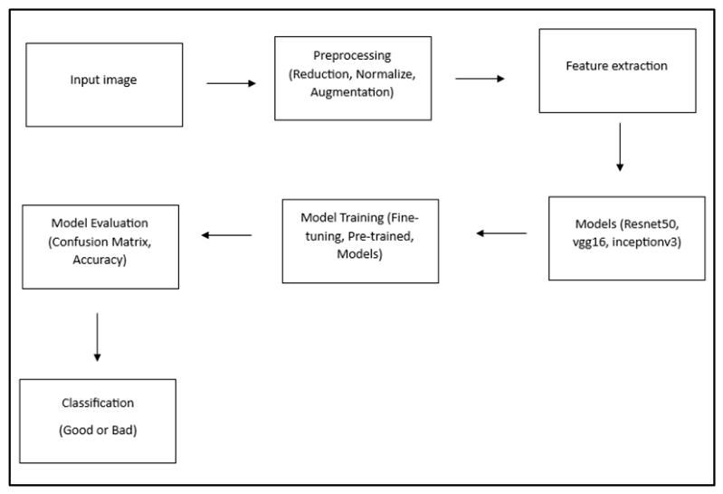
Figure 3.
Validation performance comparison of the three convolutional neural network models. Validation accuracy (top) and validation loss (bottom) curves for VGG16, InceptionV3, and ResNet50 across the training epochs. Among the evaluated architectures, VGG16 demonstrated the highest and most stable validation accuracy, accompanied by the lowest validation loss, indicating superior classification performance and generalizability for mandibular growth rotation pattern prediction.
Figure 3.
Validation performance comparison of the three convolutional neural network models. Validation accuracy (top) and validation loss (bottom) curves for VGG16, InceptionV3, and ResNet50 across the training epochs. Among the evaluated architectures, VGG16 demonstrated the highest and most stable validation accuracy, accompanied by the lowest validation loss, indicating superior classification performance and generalizability for mandibular growth rotation pattern prediction.
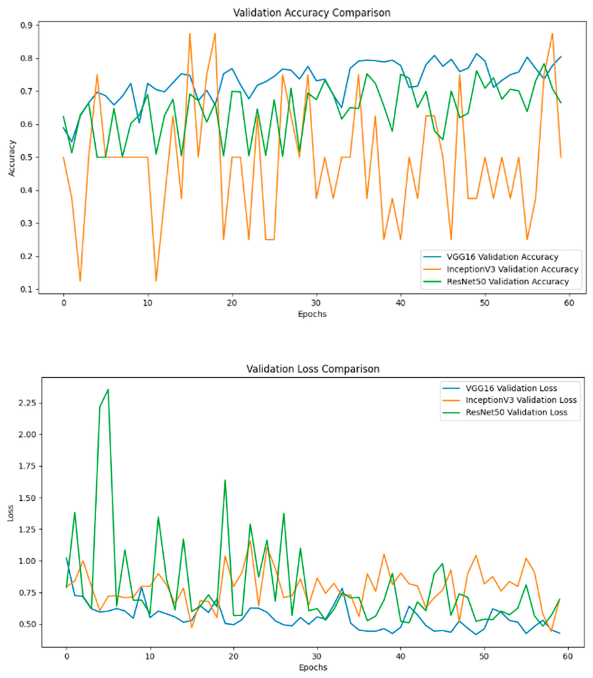
Figure 4.
Confusion matrix of the VGG16 model for classification of mandibular growth rotation patterns. Confusion matrix of the VGG16 model for classification of mandibular growth rotation patterns. The matrix presents the distribution of true and predicted classifications for clockwise (non-good-growing) and counter-clockwise (good-growing) mandibular growth rotation patterns. The VGG16 model correctly classified 318 clockwise and 331 counter-clockwise cases, while 58 clockwise cases were misclassified as counter-clockwise and 93 counter-clockwise cases were misclassified as clockwise. The results demonstrate the model's ability to effectively differentiate between the two mandibular growth rotation patterns.
Figure 4.
Confusion matrix of the VGG16 model for classification of mandibular growth rotation patterns. Confusion matrix of the VGG16 model for classification of mandibular growth rotation patterns. The matrix presents the distribution of true and predicted classifications for clockwise (non-good-growing) and counter-clockwise (good-growing) mandibular growth rotation patterns. The VGG16 model correctly classified 318 clockwise and 331 counter-clockwise cases, while 58 clockwise cases were misclassified as counter-clockwise and 93 counter-clockwise cases were misclassified as clockwise. The results demonstrate the model's ability to effectively differentiate between the two mandibular growth rotation patterns.
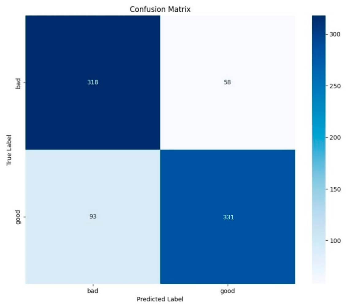
Figure 5.
Training and validation accuracy and loss curves of the VGG16 model. Training and validation accuracy (top) and loss (bottom) curves obtained during model training over 60 epochs. Both training and validation accuracy showed a progressive increase with increasing epochs, while the corresponding loss values decreased, indicating effective feature learning and model convergence. The close agreement between the training and validation curves suggests satisfactory generalization performance with minimal evidence of overfitting.
Figure 5.
Training and validation accuracy and loss curves of the VGG16 model. Training and validation accuracy (top) and loss (bottom) curves obtained during model training over 60 epochs. Both training and validation accuracy showed a progressive increase with increasing epochs, while the corresponding loss values decreased, indicating effective feature learning and model convergence. The close agreement between the training and validation curves suggests satisfactory generalization performance with minimal evidence of overfitting.
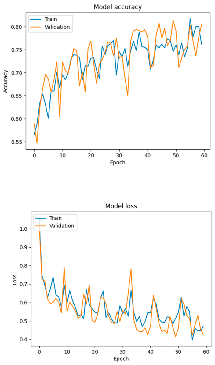

Table 1.
Distribution of the dataset used for model training and validation.
| Dataset | Clockwise rotation pattern | Counter-clockwise rotation pattern |
|---|---|---|
| Training set | 300 | 340 |
| Validation set | 76 | 84 |
| Total | 376 | 424 |
Disclaimer/Publisher’s Note: The statements, opinions and data contained in all publications are solely those of the individual author(s) and contributor(s) and not of MDPI and/or the editor(s). MDPI and/or the editor(s) disclaim responsibility for any injury to people or property resulting from any ideas, methods, instructions or products referred to in the content. |
© 2026 by the authors. Licensee MDPI, Basel, Switzerland. This article is an open access article distributed under the terms and conditions of the Creative Commons Attribution (CC BY) license (http://creativecommons.org/licenses/by/4.0/).
Copyright: This open access article is published under a Creative Commons CC BY 4.0 license, which permit the free download, distribution, and reuse, provided that the author and preprint are cited in any reuse.


