
Submitted:
29 July 2026
Posted:
31 July 2026
You are already at the latest version
Abstract
Coxiella burnetii, the bacterium responsible for Q fever, is an obligate intracellular pathogen. Q fever is a global zoonotic disease, except in New Zealand, and has significant economic and public health consequences. In Africa, although studies on human and animal seroprevalence are well documented, knowledge regarding the evolution and phylogenetic structure of circulating C. burnetii strains remains incomplete. This review analyses the genetic diversity, phylogenetic structure, and zoonotic potential of C. burnetii strains circulating in Africa and assesses the zoonotic potential of the various identified genotypes. This study involved reviewing scientific data published in Africa between January 2006 and January 2026 on molecular characterisation, as well as on the diagnostic methods used: MST (Multispacer Sequence Typing) and MLVA (Multiple-Locus Variable Number Tandem Repeat Analysis). Our analysis show marked genetic heterogeneity, characterised by the predominance of variants specific to the African continent. The MST group QT19 is more prevalent in West Africa, whereas East and North Africa exhibit distinct profiles. An assessment of pathogenicity reveals that African C. burnetii strains harbour important virulence determinants, such as the QpH1 plasmid, the adaA gene, and specific Type IVB secretion system effectors, causing acute forms of Q fever, case of atypical pneumonia, and chronic endocarditis in humans. Based on these findings, this study highlights the need to implement a comprehensive One Health surveillance strategy that combines systematic genetic characterisation with rigorous clinical monitoring to stay ahead of the threat of emerging zoonotic diseases in Africa. This structured literature review offers recommendations aimed at strengthening molecular surveillance, developing African C. burnetii genomics networks, and integrating One Health approaches to improve understanding of the epidemiology of Q fever and its zoonotic potential on the African continent.
Keywords:
Coxiella burnetii
; genetic diversity
; phylogenetics
; zoonotic potential
; Africa
1. Introduction
Coxiella burnetii, the causative agent of Q fever, or “query fever, is an intracellular bacterium belonging to the class Gammaproteobacteria. The Q fever is a worldwide zoonosis, characterized by an inefficient protective response to C. burnetii infection leading to endocarditis. A long-term treatment consisting of the association of doxycycline with hydroxychloroquine improves the prognosis despite the persisting risk of relapses [1]. C. burnetii is characterised by high environmental resistance and a broad host range, including domestic ruminants, wildlife, ticks, and humans. Cattle, sheep, and goats are the primary animal reservoirs; during parturition or abortion, infected females can shed large quantities of the bacterium into the environment. Transmission to humans occurs primarily through inhalation of contaminated aerosols from livestock farms or infected biological materials, while transmission through the ingestion of raw milk remains rare or secondary. Molecular approaches, notably Multispacer Sequence Typing (MST), Multiple-Locus Variable Number Tandem Repeat Analysis (MLVA), and, more recently, whole-genome sequencing (WGS), have improved our understanding of the genetic diversity and phylogenetic structure of C. burnetii. These methods have shown that the species does not constitute a homogeneous population but comprises several lineages that differ in geographic distribution, hosts, virulence, and zoonotic potential [2]. In Africa, the circulation of C. burnetii has been documented in numerous hosts, specifically cattle, sheep, goats, camelids, rodents, ticks, and humans. However, data on genotypes and phylogeny remain limited due to insufficient molecular surveillance, limited access to diagnostic infrastructure, and the priority given to other zoonoses. A few studies conducted in Ethiopia have revealed the existence of novel genotypes, such as a new MST profile proposed as ST52, and specific MLVA profiles in ticks of the genus Amblyomma, suggesting that African genetic diversity remains underexplored [3].
2. Methods
2.1. Scientific Research and Selection Criteria
This review was conducted and reported in line with the Preferred Reporting Items for Systematic Reviews and Meta-Analyses (PRISMA 2020) recommendations, adapted to a qualitative synthesis of molecular data on C. burnetii in Africa. The objective was to identify and evaluate scientific articles reporting the genetic diversity, phylogenetic structure, genotypes and zoonotic potential of C. burnetii strains circulating in Africa between January 2006 and January 2026. A comprehensive literature search was carried out in two bibliographic platforms (Harzing’s Publish or Perish and ResearchRabbit), interrogating multiple databases and search engines, including PubMed/Medline, Web of Science, Scopus, Cochrane Library, Semantic Scholar and CrossRef, as well as Google Scholar (via Harzing’s Publish or Perish) (Table 1). The search terms combined medical subject headings and free-text keywords using Boolean operators (AND/OR): “Coxiella burnetii” OR “Q fever” OR “Coxiellosis” AND “genetic diversity” OR “phylogenetic” OR “genotyping” OR “MST” OR “MLVA” OR “molecular typing” AND “Africa” OR “West Africa” OR “North Africa” OR “East Africa” OR “Southern Africa”. The target population comprised domestic ruminants (cattle, sheep, goats), ticks, rodents, camelids, dogs and humans sampled in African countries. For greater precision, this review included studies on C. burnetii strains from African countries, regardless of where the molecular analyses were conducted. Thus, studies analysing African in laboratories located outside Africa were also included, provided that the biological samples originated from Africa and that the study presented original data on molecular genotyping or phylogeny (Table 1).
2.2. Eligibility Criteria
Studies were eligible for inclusion if they met the following criteria: Population: Domestic ruminants (cattle, sheep, goats), ticks, rodents, camelids, dogs or humans sampled in Africa. Intervention/exposure: Detection and molecular genotyping of C. burnetii using PCR/qPCR targeting IS1111 or other genes, MST, MLVA, VNTR, icd gene typing or whole-genome sequencing (WGS). Comparator: Not applicable (descriptive or comparative molecular/genotyping studies without a specific intervention comparator). Outcome: Identification of C. burnetii genotypes, phylogenetic groups or genetic profiles, and/or assessment of zoonotic potential based on molecular data. Study design: Original research articles reporting primary molecular data, or theses containing genotyping data, published in English or French between January 2006 and January 2026. Studies were excluded if they reported serological data only without molecular characterisation; were narrative reviews, editorials, conference abstracts or opinion papers without primary data; those whose samples were collected outside Africa; or did not report genotyping or phylogenetic results.
2.3. Study Selection and Data Management
All records retrieved were imported into Zotero for reference management and de-duplication, based on author name, article title, year of publication, journal, volume and DOI. Study selection proceeded in two stages: Stage 1 – Title and abstract screening: Records were screened against the eligibility criteria. At this stage, 89 records remained after duplicate removal and were assessed by title and abstract. Stage 2 – Full-text assessment: Full texts of potentially relevant articles were retrieved and examined in detail. Reasons for exclusion (e.g., serological data only, absence of molecular typing, no primary data, non-African setting) were documented. The full process of study selection and data management is described in Figure 1. The methodological quality of each included study was assessed using an adapted version of the STROBE checklist for observational studies, supplemented by criteria specific to molecular epidemiology (clarity of sampling strategy, explicit description of PCR targets, validation of genotyping methods, and availability of sequence data in public repositories). Each item was scored as “yes”, “partial” or “no”, and overall study quality was categorised as high (≥75% of criteria met), moderate (50–74%) or low (<50%). Given the limited number and heterogeneity of the available studies, this quality assessment was used to inform the qualitative interpretation of molecular data rather than to weight studies in a quantitative meta-analysis.
3. Results
3.1. Genomic Groups, Genetic Diversity, and Genotyping Tools for C. burnetii Infection
C. burnetii exhibits limited genetic diversity but comprises several distinct genetic groups, designated as groups I through VI. These groups are defined by genomic restriction profiles and confirmed by various genotyping approaches, namely MLVA, MST, and SNP analyses, reflecting clonal lineages associated with differences in geographic distribution, host range, and virulence [4,5]. Groups I, II, and III are phylogenetically closely related and exhibit high genomic conservation, with a limited number of single nucleotide polymorphisms (SNPs) within each group. Group I includes the reference strain Nine Mile Phase I (RSA493), which is widely used as an experimental model. Group II includes European genotypes, among which are isolates associated with Q fever in the Netherlands, while Group III is dominated by MST20-type isolates, which are more commonly associated with cattle. Group V, on the other hand, exhibits a higher level of polymorphism compared to the Nine Mile reference strain and includes North American isolates, such as the Q212 strain. Group IV appears to be one of the most distantly related groups in phylogenetic trees based on SNPs or MST profiles; it notably includes MST8-type isolates often associated with small ruminants. Group VI is represented by the Dugway strains, isolated from rodents in the United States, and is considered to have low or no virulence [6]. The MST typing method for C. burnetii was developed by Glazunova et al. and optimised by Hornstra et al. [7,8]. The main advantage of MST typing lies in its inter-laboratory reliability, which is inherent to the sequencing approach. However, compared with other methods, this technique is more expensive and time-consuming to perform, while also requiring DNA extracts of high quality and concentration. Molecular approaches (Table 2), specifically MST and MLVA, have been used to study the genetic diversity of C. burnetii. However, whole-genome sequencing (WGS) is now considered the gold standard for molecular characterisation, offering substantial advantages over conventional methods. Indeed, MST relies on the analysis of a limited number of intergenic regions, while MLVA targets selected VNTR loci. Although these methods remain useful for genotyping and routine epidemiological surveillance, they do not allow exploration of the entire genome or the direct identification of variations involving plasmids or the accessory genome. In contrast, WGS allows simultaneous analysis of chromosome and plasmid sequences (QpH1, QpRS, QpDV, QpDG), thereby facilitating the identification of structural variations, pseudogenes, and rearrangements that are missed by targeted methods. WGS also provides access to the accessory genome, including genes involved in host adaptation, cellular interactions, and secretion systems, thereby offering an integrated view of strain diversity [9]. Although MST and MLVA remain useful in settings where sequencing resources are limited, WGS should be prioritised for the detailed characterisation of new strains, comparison of local isolates with international reference genomes, and exploration of the genomic determinants of virulence [8,10].
3.2. Distribution of C. burnetii Infection in Africa
The data from this structured literature review show a high concentration of studies in North Africa, which accounts for 40% of the included studies (5/13), followed by East Africa (3/13; 23.1%), Southern Africa (3/13; 23.1%), and West Africa (2/13; 15.4%). This distribution reflects disparities in surveillance and diagnostic capabilities rather than the actual distribution of C. burnetii infection across the continent [3,11,12]. This uneven distribution reflects diagnostic and research capabilities rather than the actual prevalence of the infection on the African continent. Several studies indicate that C. burnetii is likely endemic in African livestock systems, particularly in pastoral settings where contact between animals, humans, and the environment facilitates transmission, and most cases remain undetected and unreported. As shown in Table 3, the literature search conducted using various search engines yielded 96 articles and 3 theses related to the genetic diversity of C. burnetii strains. Eighty-six articles were excluded based on the inclusion criteria. Thirteen articles met the inclusion criteria and were therefore selected for this systematic review. The data from the 13 articles meeting the inclusion criteria were synthesised. Indeed, Table 3 presents the geographic distribution in Africa, diagnostic and molecular typing methods, the genetic diversity of C. burnetii, and the variability of the species in question. Of the 13 articles, 2 were from West Africa using MST genotyping, 5 from North Africa using MST and MLVA genotyping, 3 from East Africa using MST, MLVA, and VNTR genotyping, and 3 from Southern Africa using icd gene typing and MLVA. Table 3, also summarises research conducted on the genetic diversity of C. burnetii strains in Africa from 2014 to 2026, involving various hosts, namely ticks, cattle, goats, sheep, rodents, dogs, and humans, using different molecular diagnostic methods (PCR, qPCR, MST, MLVA, and icd gene typing). These studies reveal significant genetic heterogeneity (Table 3 and Figure 2), with the identification of several new genotypes depending on the region. Inter-species transmission (animals–ticks–humans) is common, indicating a high zoonotic potential. However, certain lineages show similarities across geographic regions or with human strains, suggesting continent-wide spread and underscoring the need for improved genetic surveillance. In West Africa, significant genetic variability has been observed, with several new genotypes identified in Guinea as well as in Senegal. In North Africa, the dominant genotype in circulation is MST20, which has been observed in multiple hosts, indicating multi-host transmission and the emergence of new genetic profiles. The data show that in East Africa, genetic diversity is high, with new African genotypes suggesting that vectors play a role in maintaining the pathogen. As for Southern Africa, there is genetic variability among the different hosts and a similarity to strains from Europe and the Middle East. This geographic heterogeneity can be explained by significant differences in scientific and technological capacity among African regions. North African countries generally have more developed molecular biology infrastructures, access to sequencing platforms, well-structured academic networks, and international collaborations that facilitate the generation of molecular data. In contrast, in other regions of Africa, specifically West Africa, molecular characterisation activities remain limited due to a shortage of specialised laboratories, restricted access to sequencing technologies, the high cost of genomic analyses, and inadequate surveillance of zoonoses. Consequently, the underrepresentation of certain African regions in the literature reflects inequalities in diagnostic and research capacity more than a true absence of C. burnetii circulation [13,14,15].
3.3. Comparison of Molecular Genotypes of C. burnetii Identified in Different Regions of the World
To better contextualise the genotypes identified in Africa, a systematic comparison was conducted with molecular genotype data available from other regions of the world. Indeed, the Table 4, summarises, for each region, the main hosts studied, the molecular methods used, and the MST/MLVA genotypes reported in the literature. The African genotype, particularly MST20, which is associated with cattle, is found in Europe and the Middle East, suggesting historical transmission linked to livestock movement between these regions. At the same time, the identification of new genotypes, particularly in West Africa (MST86–MST89) and East Africa (MST52), highlights local diversity that has not yet been sufficiently explored. Unlike Europe or the Americas, where high-resolution methods such as WGS have enabled better characterisation of genomic groups and their virulence profiles, Africa remains under-documented. These results underscore the need to strengthen standardised molecular studies on the continent in order to better place African strains within the global epidemiology of Q fever.
3.4. Assessment of Zoonotic Potential
Analysis of the pathogenicity of African isolates highlights a major zoonotic risk. Clinical studies in humans combined with molecular typing have demonstrated the direct involvement of the ST19 genotype, endemic in West Africa, in the emergence of rural outbreaks characterised by severe acute pneumonia and persistent fevers [16]. African strains of C. burnetii exhibit significant genetic diversity, characterised by different MST genotypes as well as several plasmid types, notably QpH1 and QpDG, which contribute to the phylogenetic structure of circulating lineages. However, the virulence potential of C. burnetii cannot be inferred solely from the presence of the com1 gene, as this gene is highly conserved in virtually all strains. Although some studies have identified associations between plasmids, the adaA gene, and certain clinical profiles, virulence is now considered a multifactorial trait resulting from the interaction between multiple genetic determinants, genomic polymorphisms, and gene expression regulation [17,18,19]. The clinical impact of these characteristics is most evident in North Africa [8]. Clinical studies indicate that ST19 infections frequently cause severe acute pneumonia and prolonged fever, unlike other genotypes, which often present with milder symptoms. The differential virulence of C. burnetii genotypes is increasingly recognised, with Group I strains (of which ST19 is a representative) considered more virulent in animal and human models [16].
4. Discussion
The genetic diversity of C. burnetii strains circulating in Africa highlights a complex epidemiological landscape at the human-animal-environment interface. The coexistence of multiple, divergent genotypes underscores the urgent need for enhanced molecular surveillance to track transmission dynamic and anticipate zoonotic outbreaks [20]. However, to fully understand and mitigate these risks, it is imperative to move beyond mere genotype reporting and deeply analyze the specific phylogenetic clades, their associated virulence determinants, and their direct zoonotic implications. Our review demonstrates a pronounced geographic and clinical dichotomy among African C. burnetii lineages. In West Africa, genomic Group I clades prominently represented by the MST19 genotype are highly prevalent. Historically, Group I strains are linked to acute, severe clinical manifestations in humans, notably atypical pneumonia [16]. The heightened virulence of these clades is strongly correlated with specific genomic determinants, including the presence of the adaA gene and the QpH1 plasmid. These molecular factors equip the bacterium to aggressively modulate the host’s innate immune response and evade macrophage apoptosis, thereby driving acute respiratory infections [21]. Consequently, the emergence and circulation of Group I strains represent an immediate, acute threat to human respiratory health in these regions. In contrast, genomic Group III clades, primarily represented by the MST20 profile, dominate the epidemiological landscape in North and East Africa. These clades exhibit a strong tropism for cattle and frequently harbour the QpRS plasmid or specific QpH1 variants. Unlike the explosive acute outbreaks associated with Group I, Group III strains are primarily drivers of endemic infections in livestock, leading to insidious, chronic reproductive disorders and prolonged bacterial shedding in milk. The zoonotic implications here are distinct but equally severe: human exposure to Group III clades, predominantly through the consumption of unpasteurized dairy products, is strongly associated with the development of chronic Q fever, including silent but fatal infective endocarditis [20]. Furthermore, the identification of novel African genotypes, including MST52 in Ethiopia and the recently described MST86-MST89 lineages in Guinea, suggests that C. brunetii is undergoing region-specific evolutionary diversification. These findings highlight the limited representation of African strains in current genomic databases and emphasize the need for expanded genomic surveillance. Comprehensive whole-genome sequencing (WGS) is required to characterize the complete repertoire of Dot/Icm type IVB secretion system (T4BSS) effectors, which are essential for host- cell manipulation, intracellular replication, and adaptation to diverse mammalian hosts [22]. While advocating for advanced genomic surveillance to track these virulence determinants is scientifically sound, its implementation remains severely hindered by the current state of diagnostic facilities in most African countries. Significant infrastructural barriers including chronic underfunding, cold chain instability for reagents, a lack of bioinformatics expertise, and limited laboratory capacity render WGS impractical outside of a few specialized reference centers? Therefore, a more pragmatic, high-impact intermediate strategy is urgently needed. Public health efforts should focus on strengthening decentralized molecular diagnostic capacities using robust, cost-effective tools such as targeted qPCR or MLVA, supported by regional collaborations and international genomics networks. The interpretation of the data remains limited due to the heterogeneity of the diagnostic and typing methods used, as well as the limited geographic coverage. The disparity in PCR diagnostic methods targeting the IS1111 repetitive element, the varying MLVA panels, and the non-uniform MST protocols may introduce significant biases when comparing results across different regions. The limited representativeness of the data, with research concentrated in certain, better-documented areas, restricts the generalizability of the results to the entire African continent [7,11].
5. Conclusion
This structured literature review demonstrates that C. burnetii circulating in Africa exhibits considerable genetic diversity, including several unique African genotypes with important zoonotic implications. Molecular genotyping, particularly whole-genome sequencing (WGS), is essential for improving epidemiological surveillance, understanding transmission dynamics, and detecting emerging lineages. Strengthening genomic surveillance, harmonizing genotyping methods, and integrating these approaches within a One Health framework will enhance the prevention, diagnosis, and control of Q fever while supporting evidence-based public health interventions and the future development of improved diagnostic tools and vaccines adapted to African strains.
Author Contributions
Mama Agnès TEA, Albert Sourou SALAKO, Abdoulaye Oury BARRY and Eric GHIGO conducted the literature review, analysed the bibliographic data, and drafted and revised the manuscript.
Funding
Mama Agnès TEA, Albert Sourou SALAKO, Abdoulaye Oury BARRY and Eric GHIGO are employed of the Ministry of Higher Education and Scientific Research (MESRS).
Institutional Review Board Statement
Not applicable.
Informed Consent Statement
Not applicable.
Data Availability Statement
No new data were created or analysed in this study. Data sharing is not applicable to this article.
Acknowledgments
Not applicable.
Conflicts of Interest
The authors declare that they have no conflicts of interest.
References
- Raoult, D.; Marrie, T. J.; Mege, J. L. Natural history and pathophysiology of Q fever. Lancet Infect. Dis. 2005, 5, 219–226. [Google Scholar] [CrossRef] [PubMed]
- Arricau-Bouvery, N.; Rodolakis, A. Is Q fever an emerging or re-emerging zoonosis? Vet. Res. 2005, 36, 327–349. [Google Scholar] [CrossRef] [PubMed]
- Rahal, M.; Salhi, O.; Ouchetati, I.; Khelifi Touhami, N. A.; Ouchene, N. Global epidemiology and molecular typing of Coxiella burnetii: A systematic review of Q fever in humans and animals. Comp. Immunol. Microbiol. Infect. Dis. 2025, 123, 102401. [Google Scholar] [CrossRef] [PubMed]
- Metters, G.; Norville, I. H.; Titball, R. W.; Hemsley, C. M. From cell culture to cynomolgus macaque: infection models show lineage-specific virulence potential of Coxiella burnetii. J. Med. Microbiol. 2019, 68, 1419–1430. [Google Scholar] [CrossRef] [PubMed]
- D’Amato, F.; Eldin, C.; Raoult, D. The contribution of genomics to the study of Q fever. Future Microbiol. 2016, 11, 253–272. [Google Scholar] [CrossRef] [PubMed]
- Brangsch, H.; Berens, C.; Fuchs, S.; Fitzgerald, S.; McNeilly, T.N.; Lührmann, A.; Mertens-Scholz, K. Genome characteristics and type IV effector protein repertoire of Coxiella burnetii depend rather on Genomic Groups than on host species. BMC Microbiol. 2026, 26, 393. [Google Scholar] [CrossRef] [PubMed]
- Glazunova, O.; Roux, V.; Freylikman, O.; Sekeyova, Z.; Fournous, G.; Tyczka, J.; Raoult, D. Coxiella burnetii Genotyping. Emerg. Infect. Dis. 2005, 11, 1211–1217. [Google Scholar] [CrossRef] [PubMed]
- Hornstra, H.M.; Priestley, R.A.; Georgia, S.M.; Kachur, S.; Birdsell, D.N.; Hilsabeck, R.; Pearson, T. Rapid typing of Coxiella burnetii. PLoS ONE 2011, 6, e26201. [Google Scholar] [CrossRef] [PubMed]
- Yuan, J.; Zhang, Y.; Song, L.; Luo, Z.-Q. Modulation of host cell pathways by Coxiella burnetii Dot/Icm effectors. mLife 2025, 4, 577–596. [Google Scholar] [CrossRef] [PubMed]
- Mbiri, P.; Muleya, W.; Moyo, E.; Samkange, A.; Matomola, O.C.; Charamba, V.; Chitanga, S. Molecular Detection and Prevalence of Coxiella burnetii in Ticks from Namibia: A Regional and Genus-Specific Analysis. Pathogens 2025, 14, 1262. [Google Scholar] [CrossRef] [PubMed]
- Eldin, C.; Mélenotte, C.; Mediannikov, O.; Ghigo, E.; Million, M.; Edouard, S.; Raoult, D. From Q Fever to Coxiella burnetii Infection: a Paradigm Change. Clin. Microbiol. Rev. 2017, 30, 115–190. [Google Scholar] [CrossRef] [PubMed]
- Kampschreur, L. M.; et al. Chronic Q fever in the Netherlands 5 years after the start of the Q fever epidemic: results from the Dutch chronic Q fever database. J. Clin. Microbiol. 2014, 52, 1637–1643. [Google Scholar] [CrossRef] [PubMed]
- Ochola, R. The Case for Genomic Surveillance in Africa. Trop. Med. Infect. Dis. 2025, 10, 129. [Google Scholar] [CrossRef] [PubMed]
- Nkengasong, J. N.; Yao, K.; Onyebujoh, P. Laboratory medicine in low-income and middle-income countries: progress and challenges. The Lancet 2018, 391, 1873–1875. [Google Scholar] [CrossRef] [PubMed]
- Petti, C. A.; Polage, C. R.; Quinn, T. C.; Ronald, A. R.; Sande, M. A. Laboratory Medicine in Africa: A Barrier to Effective Health Care. Clin. Infect. Dis. 2006, 42, 377–382. [Google Scholar] [CrossRef] [PubMed]
- Long, C. M.; Beare, P. A.; Cockrell, D. C.; Larson, C. L.; Heinzen, R. A. Comparative virulence of diverse Coxiella burnetii strains. Virulence 2019, 10(1), 133–150. [Google Scholar] [CrossRef] [PubMed]
- Hemsley, C. M.; Essex-Lopresti, A.; Norville, I. H.; Titball, R. W. Correlating Genotyping Data of Coxiella burnetii with Genomic Groups. Pathogens 2021, 10, 604. [Google Scholar] [CrossRef] [PubMed]
- Massung, R. F.; Cutler, S. J.; Frangoulidis, D. Molecular Typing of Coxiella burnetii (Q Fever). in Coxiella burnetii: Recent Advances and New Perspectives in Research of the Q Fever Bacterium; Toman, R., Heinzen, R. A., Samuel, J. E., Mege, J.-L., Eds.; Springer Netherlands: Dordrecht, 2012; pp. 381–396. [Google Scholar] [CrossRef] [PubMed]
- Mezouar, S.; Desnues, B.; Bechah, Y.; Bardin, N.; Devaux, C.; Mege, J.L. Pathogenicity and Virulence of Coxiella burnetii: Focus on Q fever. Virulence 2025, 16, 2495842. [Google Scholar] [CrossRef] [PubMed]
- Khademi, P.; Tukmechi, A.; Sgroi, G.; Ownagh, A.; Enferadi, A.; Khalili, M.; Mardani, K. Molecular and genotyping techniques in diagnosis of Coxiella burnetii: An overview. Infect. Genet. Evol. 2024, 123, 105655. [Google Scholar] [CrossRef] [PubMed]
- Luo, S.; Lu, S.; Fan, H.; Chen, Z.; Sun, Z.; Hu, Y.; Song, L. The Coxiella burnetii QpH1 Plasmid Is a Virulence Factor for Colonizing Bone Marrow-Derived Murine Macrophages. J. Bacteriol. 2021, 203, 10.1128/jb.00588-20. [Google Scholar] [CrossRef] [PubMed]
- Sulyok, K. M.; Hornok, S.; Abichu, G.; Erdélyi, K.; Gyuranecz, M. Identification of novel Coxiella burnetii genotypes from Ethiopian ticks. PLoS ONE 2014, 9, e113213. [Google Scholar] [CrossRef] [PubMed]
- Camara, K.; Hammoud, A.; Diallo, H.M.; Diarra, A.Z.; Camara, A.O.D.; Kaba, L.; Mediannikov, O. Genetic diversity of Coxiella burnetii in ticks from the Republic of Guinea. Ticks Tick. Borne Dis. 2026, 17, 102597. [Google Scholar] [CrossRef] [PubMed]
- Mangombi-Pambou, J.; Granjon, L.; Labarrere, C.; Kane, M.; Niang, Y.; Fournier, P. E.; Mediannikov, O. New Genotype of Coxiella burnetii Causing Epizootic Q Fever Outbreak in Rodents, northern Senegal. Emerg. Infect. Dis. 2023, 29(5), 1078. [Google Scholar] [CrossRef] [PubMed]
- Ghaoui, H.; Bitam, I.; Zaidi, S.; Achour, N.; Zenia, S.; Idres, T.; Fournier, P.E. Molecular detection and MST genotyping of Coxiella burnetii in ruminants and stray dogs and cats in Northern Algeria. Comp. Immunol. Microbiol. Infect. Dis. 2024, 106, 102126. [Google Scholar] [CrossRef] [PubMed]
- Rahal, M.; Bitam, I. Génotypage de Coxiella burnetii, chlamydia spp et leptospira spp détectées dans des tissus placentaires chez des vaches laitières ayant avorté dans le nord de l’Algérie; Ecole Nationale Supérieure Vétérinaire, 2019. [Google Scholar]
- Menadi, S.E.; Chisu, V.; Santucciu, C.; Di Domenico, M.; Curini, V.; Masala, G. Serological, Molecular Prevalence and Genotyping of Coxiella burnetii in Dairy Cattle Herds in Northeastern Algeria. Vet. Sci. 2022, 9, 40. [Google Scholar] [CrossRef] [PubMed]
- Delaloye, J.; Pillonel, T.; Smaoui, M.; Znazen, A.; Abid, L.; Greub, G. Culture-independent genome sequencing of Coxiella burnetii from a native heart valve of a Tunisian patient with severe infective endocarditis. New Microbes New Infect. 2018, 21, 31–35. [Google Scholar] [CrossRef] [PubMed]
- Selim, A.; Abdelrahman, A.; Thiéry, R.; Sidi-Boumedine, K. Molecular typing of Coxiella burnetii from sheep in Egypt. Comp. Immunol. Microbiol. Infect. Dis. 2019, 67, 101353. [Google Scholar] [CrossRef] [PubMed]
- Frangoulidis, D.; Kahlhofer, C.; Said, A.S.; Osman, A.Y.; Chitimia-Dobler, L.; Shuaib, Y.A. High Prevalence and New Genotype of Coxiella burnetii in Ticks Infesting Camels in Somalia. Pathogens 2021, 10, 741. [Google Scholar] [CrossRef] [PubMed]
- Kumsa, B.; Socolovschi, C.; Almeras, L.; Raoult, D.; Parola, P. Occurrence and Genotyping of Coxiella burnetii in Ixodid Ticks in Oromia, Ethiopia. Am. J. Trop. Med. Hyg. 2015, 93, 1074–1081. [Google Scholar] [CrossRef] [PubMed]
- Chitanga, S.; Simulundu, E.; Simuunza, M.C.; Changula, K.; Qiu, Y.; Kajihara, M.; Hang’ombe, B.M. First molecular detection and genetic characterization of Coxiella burnetii in Zambian dogs and rodents. Parasites Vectors 2018, 11, 40. [Google Scholar] [CrossRef] [PubMed]
- Mangena, M.; Gcebe, N.; Pierneef, R.; Thompson, P. N.; Adesiyun, A. A. Q Fever: Seroprevalence, Risk Factors in Slaughter Livestock and Genotypes of Coxiella burnetii in South Africa. Pathogens 2021, 10, 258. [Google Scholar] [CrossRef] [PubMed]
- Hemsley, C.M.; O’Neill, P.A.; Essex-Lopresti, A.; Norville, I.H.; Atkins, T.P.; Titball, R.W. Extensive genome analysis of Coxiella burnetii reveals limited evolution within genomic groups. BMC Genom. 2019, 20, 441. [Google Scholar] [CrossRef] [PubMed]
- Angelakis, E.; Raoult, D. Q fever. Vet. Microbiol. 2010, 140, 297–309. [Google Scholar] [CrossRef] [PubMed]
- Mobarez, M. A.; Khalili, M.; Mostafavi, E.; Esmaeili, S. Molecular detection of Coxiella burnetii infection in aborted samples of domestic ruminants in Iran. PLoS ONE 2021, 16, e0250116. [Google Scholar] [CrossRef] [PubMed]
Figure 1.
Flow chart of the study selection process. Our search identified 96 records, after which publications were selected as described in the flow chart.
Figure 1.
Flow chart of the study selection process. Our search identified 96 records, after which publications were selected as described in the flow chart.
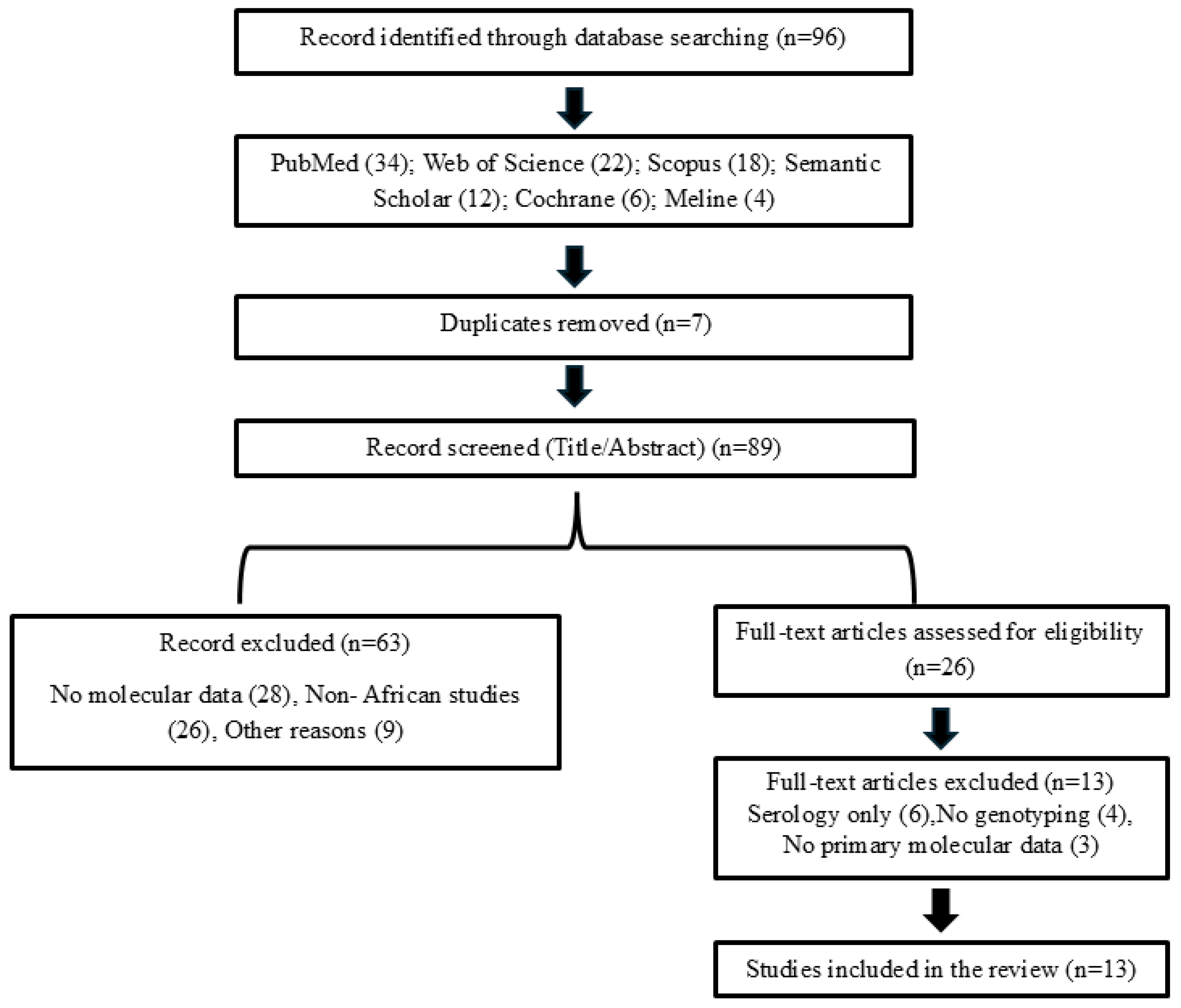
Figure 2.
Geographic distribution of Coxiella burnetii genotypes identified across Africa. Based on Table 2, the Figure 2 shows the geographic distribution of the genotypes identified across Africa, was included to help visualize regional genetic diversity.
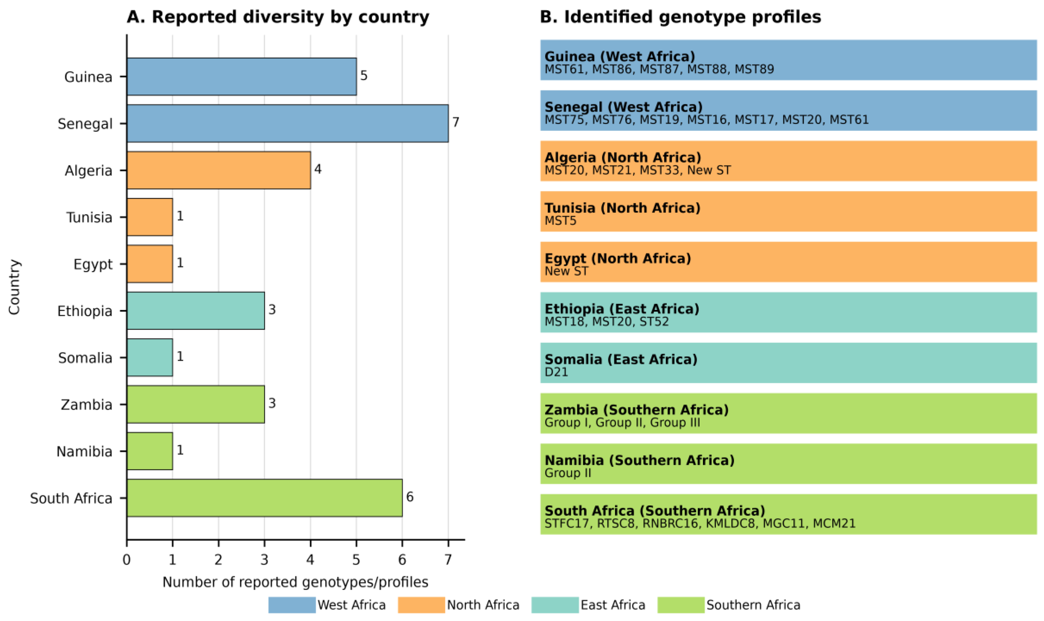

Table 1.
Summary of the literature review strategy, eligibility criteria, and study selection process.
Table 1.
Summary of the literature review strategy, eligibility criteria, and study selection process.
| Elements | Description |
| Research Period | African scientific publications released between 2006 and 2026 were included. |
| Research Objective | To identify studies on the molecular characterization, genetic diversity, phylogenetic structure, and zoonotic potential of Coxiella burnetii in Africa. |
| Document Sources | Data were collected from the scientific literature available in bibliographic databases (Harzing’s Publish or Perish and ResearchRabbit) using the search engines Cochrane, PubMed, Web of Science, Scopus, and Semantic Scholar, as well as Medline and CrossRef. |
| Search Criteria | The terms used in the search bar were “Q fever,” “Coxiellosis,” “Coxiella burnetii,” “genetic diversity,” “phylogenetic structure,” “Coxiella burnetii strain,” and “Africa.” |
| Inclusion Criteria | The following were included: (i) studies conducted in Africa; (ii) studies on Coxiella burnetii in animals, humans, and ticks; (iii) studies that used molecular or genotyping methods such as PCR, qPCR, MST, MLVA, or WGS; (iv) articles providing actionable data on genotyping, strains, or diagnostic approaches. |
| Exclusion Criteria | (i) articles not related to the African context; (ii) studies not directly focused on Coxiella burnetii; (iii) publications lacking molecular data; (iv) duplicates; (v) abstracts for which full text was not available when essential data could not be extracted. |
| Number of studies identified | A total of 99 studies, comprising all publications identified by the literature search using the defined keywords |
| Number of excluded studies | A total of 86 studies were excluded. These studies were deemed irrelevant after reviewing the title, abstract, or full text. |
| Number of included studies | A total of 13 studies were selected for the final analysis, as presented in Table 2 of the manuscript |
| Data extracted | For each included study, the following information was extracted: country, year, host type, samples analysed, molecular methods used, genotypes identified, and main results |
| Purpose of the selection process | This approach made it possible to compile a homogeneous set of relevant studies to analyse the genetic diversity and zoonotic potential of African strains of Coxiella burnetii. |
Table 2.
Comparison of molecular methods for the characterisation of Coxiella burnetii in Africa.
| Methods | Principle | Resolution | Advantages | Limitations | Cost | Technical requirements | Applicability in Africa |
| MST (Multi-Spacer Sequence Typing) | Sequencing of 10 intergenic regions (spacers) | Average (discrimination among major lineages) | Standardized, reproducible, global database | Limited Response to Local Outbreaks | Moderate | Standard Sanger sequencing | Good (routinely accessible) |
| MLVA (Multiple Loci VNTR Analysis) | Analysis of tandem repeat numbers at 17 loci | High (characteristics of similar strains) |
Highly specific for epidemiological tracing | Risk of homoplasy; difficult to compare results across laboratories | Moderate | PCR and (capillary) electrophoresis | Good (basic equipment available) |
| WGS (Whole-Genome Sequencing) | Comprehensive sequencing of the entire genome (chromosomes and plasmids) | Maximum identification of single mutations, plasmids, and accessory genes | Comprehensive View Virulence, resistance, evolution, genome, accessory |
High cost; requires advanced bioinformatics expertise | Advanced | High-Throughput Sequencing, Server Infrastructure, and Bioinformatics | Under development (requires reference platforms) |
Table 3.
Geographic distribution of studies conducted in Africa, diagnostic methods, and genetic diversity of C. burnetii.
Table 3.
Geographic distribution of studies conducted in Africa, diagnostic methods, and genetic diversity of C. burnetii.
| Region | Country | Year | Hosts | Background | Diagnostic Methods | Identified Genotypes | References |
| West Africa | Guinea | 2026 | Ticks | livestock-human interface | MST | MST61*, MST86*, MST87* MST88*, MST89* |
[23] |
| Senegal | 2023 | Ticks / Humans | Rural areas | MST | MST75, MST76, MST19, MST16, MST17, MST20, MST61 |
[24] | |
| North Africa | Algeria | 2024 | Cattle/dogs/cats | Livestock and Stray Animals | MST | MST20, MST21, MST33 | [25] |
| Algeria | 2019 | Cattle | Livestock Farming | MST | MST20 | [26] | |
| Algeria | 2022 | Dairy Cattle | Livestock Farming | MST | MST12, MST32* | [27] | |
| Tunisia | 2018 | Human | Urban environment | MST | MST5 | [28] | |
| Egypt | 2019 | Ewes | Spontaneous abortions | MST, MLVA | MST*, MLVA* MLVA* | [29] | |
| East Africa | Ethiopia | 2014 | Ticks | Ixodidae | MST | MST18, MST20, MST52* | [22] |
| Somalia | 2021 | Ticks | Ticks infesting camels | MLVA | D21* | [30] | |
| Ethiopia | 2015 | Ixodid ticks | Collected from domestic animals | MST | MST18, MST20 | [31] | |
| Southern Africa | Zambia | 2018 | Dogs/rodents/cattle | Livestock and Stray Animals | Typage icd | Groupe I-III | [32] |
| Namibia | 2025 | Ticks | Livestock Farming | Typage icd | Groupe II | [10] | |
| South Africa | 2021 | Cattle/Sheep | Livestock Farming | MLVA | STFC17*, RTSC8*, RNBRC16*, KMLDC8*, MGC11*, MCM21* |
[33] |
Legend: MST = Multispacer Sequence Typing; MLVA = Multiple-Locus Variable-Number Tandem Repeat Analysis; icd = isocitrate dehydrogenase; *= Genotype identified for the first time in Africa, according to the cited studies.
Table 4.
Main hosts, molecular methods, and MST/MLVA genotypes reported by region in the literature.
Table 4.
Main hosts, molecular methods, and MST/MLVA genotypes reported by region in the literature.
| Region | Hosts studied | Molecular methods | Types of genotypes / MST-MLVA profiles identified | Comparison with Africa | References |
| Africa | Cattle, sheep, goats, camels, humans, rodents, ticks | PCR, qPCR, MST, MLVA | MST20, predominant in several hosts; presence of distinct MST profiles associated with local epidemiological contexts. New genotypes, including MST86, MST87, MST88 and MST89, have been identified in Guinea. MST52 has also been proposed as a new sequence type. | Profiles close to those described in Europe, particularly MST20, suggesting historical exchanges linked to livestock movements. | [11,23,24,25] |
| Europe | Cattle, sheep, goats, camels, humans, ticks | MST, MLVA, SNP typing, WGS | MST8 (group IV, goats), MST13, MST18, MST20 (group III, cattle), MST32, MST33 (Dutch outbreak, group II-b), MST62–MST65 (sub-lineage II-d). | Europe has more comprehensive data; MST20 is shared with North Africa, suggesting intercontinental circulation. | [7,8,34] |
| America | Cattle, sheep, goats, camels, humans, ticks | PCR, qPCR, MST, MLVA, SNP typing, WGS | MST16, MST17 (French Guiana, associated with severe forms), MST20, MST21 (Nova Scotia, group V, North America); distinct genomic profiles with characterized polymorphisms. | Profiles such as MST17 are associated with increased virulence, highlighting the clinical relevance of phylogenetic characterization. MST20 is also shared with North Africa. | [4,8,17,35] |
| Middle East / Asia | Cattle, sheep, goats, humans, ticks | PCR, qPCR, MST, MLVA | MST20 is dominant in many countries; five distinct MST genotypes have been identified in Iran; MST62–MST65, corresponding to sub-lineage II-d, are predominant in Iran; variable MLVA profiles have also been reported. | As in Africa, MST20 is found in cattle, indicating the global circulation of this lineage; other profiles appear to be more localized. | [17,36] |
Legend: PCR = Polymerase Chain Reaction; qPCR = Real-Time Polymerase Chain Reaction; MST = Multispacer Sequence Typing; MLVA = Multiple-Locus Variable-Number Tandem Repeat Analysis; WGS= Whole Genome Sequencing.
Disclaimer/Publisher’s Note: The statements, opinions and data contained in all publications are solely those of the individual author(s) and contributor(s) and not of MDPI and/or the editor(s). MDPI and/or the editor(s) disclaim responsibility for any injury to people or property resulting from any ideas, methods, instructions or products referred to in the content. |
© 2026 by the authors. Licensee MDPI, Basel, Switzerland. This article is an open access article distributed under the terms and conditions of the Creative Commons Attribution (CC BY) license (http://creativecommons.org/licenses/by/4.0/).
Copyright: This open access article is published under a Creative Commons CC BY 4.0 license, which permit the free download, distribution, and reuse, provided that the author and preprint are cited in any reuse.