

Submitted:
12 June 2026
Posted:
15 June 2026
You are already at the latest version
Abstract
Background/Objectives: Silver nanoparticles (AgNPs) hold great promise for antimicrobial, antiviral, and anticancer applications; however, concerns about their toxicity remain a major barrier to their medical use. This study systematically evaluates the acute oral toxicity of five well-characterized AgNP formulations from the Argovit™ family, which previously showed remarkable biomedical properties. Methods: We determine the median lethal dose (LD50), assess clinical signs, and identify the histopathological alterations in mice following oral administration for each formulation. The LD50 was calculated for AgNPs (metallic silver plus stabilizing agent) and for the complete formulation (including water). Results: All formulations showed exceptionally high LD50 values (86,020–150,535 mg/kg) considering the complete formulation, surpassing those reported for other AgNPs in the literature (typically ≤5,000 mg/kg). Clinical signs and toxicological assessment reveal differences in the response of the AgNPs formulations, but all exhibit extremely low toxicity. Histopathological changes were minimal compared to those caused by other AgNP formulations, even though we used doses several times higher. The estimated human LD50 values for the assessed Argovit AgNPs formulations are 17-30 times higher than those reported for other AgNP formulations in the literature. Conclusions: The remarkably low toxicity found in this work contradicts the prevailing assumption that only biogenically synthesized AgNP are biocompatible, highlighting these synthetic PVP-AgNP formulations as a highly safe alternative. The study suggests that the low LD50 may be due to the peculiar structure of Argovit AgNP formulations, which, according to recent data, consists of Ag cores incorporated into PVP nano- and microgels. A high percentage of rapid elimination of Argovit AgNPs from organisms (>98%) via feces also contributes to low LD50. To our knowledge, the present work is the first to study the LD50 of AgNPs-nanogel systems.
Keywords:
silver nanoparticles
; remarkably low LD50
; acute toxicity test
; OECD Guideline 420
; impact on applications in nanomedicine
1. Introduction
The growing challenge of treating complex diseases, such as drug-resistant infections and cancer, has intensified the search for new, safer therapeutic strategies. Among the most promising nanomaterials, silver nanoparticles (AgNPs) have gained significant attention due to their broad-spectrum biological activities, including antimicrobial, antiviral, anti-fungal, anti-inflammatory, and anticancer effects [1,2,3,4,5]. As a result, AgNPs are currently among the most widely studied nanomaterials in the biomedical sciences.
However, ongoing concerns regarding their safety have limited the efficient clinical translation of AgNP application. The prevailing paradigm asserts that chemically or physically synthesized AgNPs are inherently toxic, while green-synthesized AgNP formulations, although considered less harmful, exhibit inconsistent chemical composition. Indeed, various studies have reported cytotoxic and genotoxic effects of AgNPs across multiple models, closely related to their physicochemical properties (size, shape, surface coating, dose, and exposure time) [6,7,8,9,10]. Surprisingly, despite extensive studies of AgNPs, only a few works have accurately determined the median lethal dose (LD50) of AgNPs—an essential toxicological benchmark—and even fewer have included complete formulation data such as contents of metallic silver, stabilizer, and solvent. This gap hinders meaningful comparisons between AgNP formulations and weakens the toxicological foundation necessary for clinical development.
To address this issue, we evaluated five well-characterized formulations of Argovit™ AgNPs synthesized by a high-energy electron beam method and stabilized with polyvinylpyrrolidone (PVP) with different molecular masses. All AgNP formulations contain a specific Ag/PVP ratio, with well-characterized concentrations of metallic silver, the stabilizer, and the solvent. In earlier studies, these formulations exhibited high physicochemical stability and scalability and have demonstrated selective anticancer activity, genoprotective effects, and minimal environmental toxicity [11,12,13,14]. Despite all studied formulations exhibiting from good to remarkable results on each evaluated system, differences in PVP used for each formulation show a specific response, e.g., AgNP formulation synthesized with PVP K-30 exhibits the best anticancer effect in colon; AgNP formulation with K-17 results in the best antihyperglycemic effect [15] while using PVP 12.6 KDa provides the best abiotic elicitor to improve the yields of plant-derived bioactive compounds of pharmaceutical interest. Differences in the nature of coating agents play a key role in optimizing AgNP formulations for specific applications.
Therefore, the purpose of this article was to measure the LD50 of these five AgNP formulations in mice using the standardized scientific test method, OECD Guideline 420 (Table 1), and to compare the obtained LD50 values with those published for other AgNP formulations.
Table 1 presents GHS Hazard Categorization for Acute Toxicity by Oral Route according to OECD 420 [16]. The categorization includes the LD50 corresponding to five acute toxicity levels: 1) lethally toxic, 2) toxic, 3) harmful, 4) low hazard, 5) without significant danger, and a not classified level defined as harmless.
2. Materials and Methods
2.1. Silver Nanoparticles
The Scientific and Production Center Vector-Vita Ltd. (Novosibirsk, Russia) kindly donated five formulations of ArgovitTM AgNPs. Each of the five formulations contains approximately 1.2% wt. metallic silver, 18.8% wt. of stabilizing agent (polyvinylpyrrolidone (PVP)), and 80% wt. of distilled water. Each formulation has an overall concentration of 200 mg/mL of AgNPs (considering metallic Ag and PVP) suspended in water. All formulations have been characterized by DLS, UV-Vis, HR-TEM, TGA, and DSC. The summary of the physicochemical properties of each AgNPs formulation is shown in Table 2, adapted from [17].
Characteristics of the PVP stabilizer and electron adsorbed doses used in the preparation of every AgNP formulation are presented in Table 3.
Ag1 and Ag2 samples showed the influence of PVP MW variation at 15 kGy, while Ag3 and Ag4 at 45 kGy. Ag2 and Ag3 revised the absorbed dose variation, with 15 kGy for Ag2 and 45 kGy for Ag3 at constant MW. The influence of the PVP manufacturers (Boai NKY Pharmaceuticals Ltd. and LLC AK Sintvita) was revised on Ag2 and Ag5, respectively, maintaining the same MW and electron-absorbed dose.
2.2. Acute Toxicity Test (OECD Guideline 420)
The GHS approach (Table 1) uses the observation of clear signs of toxicity or death in at least three of the five mice as the test endpoint. The lethal dose for all AgNP formulations was studied on BALB/c mice following the instructions of the OECD Guideline 420 for Acute Oral Toxicity Assay-Fixed Dose Procedure [18]. The experimental protocol (No. 001/2018) was approved by the Ethical Committee of the Autonomous University of Baja California, México. First, an observational study was conducted with each formulation to select the appropriate starting dose. A female mouse was administered a single dose of 4,300 mg/kg of each AgNP formulation and observed for 14 days. Changes in clinical signs, including skin and fur appearance, eyes, membranes, nausea, vomiting, diarrhea, tremors, convulsions, salivation, lethargy, sleep, coma, general behavior, respiratory pattern, and death, were carefully recorded. Since no signs of toxicity were observed during the observational study, the limit test with five healthy nulliparous females was conducted at a starting dose of 4,300 mg/kg of AgNPs. Twenty-five female mice (body weight ~18.6 g) were randomly divided into five groups and kept in polycarbonate cages at 22±2 °C, 60% humidity, and a 12/12-h light-dark cycle. These groups were designated by the AgNP formulation administered: AgNP1, AgNP2, AgNP3, AgNP4, and AgNP5. Mice were given repeated doses of 4,300 mg/kg every two hours until three of the five mice in each group died. During the experiment, the changes in clinical signs were carefully registered. Animals with very low mobility and a decreased respiratory rate, without coma, for up to 30 minutes were classified as agonic. LD50 doses were calculated in mg/kg for AgNPs (metallic silver with stabilizer) and the complete AgNPs formulation (metallic silver with stabilizer and water).
2.3. Histopathology
In addition to the main method used in this work (Acute toxicity test), histopathological studies were also conducted. Since histological studies are time-consuming, the number of animals per group was determined based on LD50 results, survival rate, and clinical signs. The intestine, liver, kidney, lungs, and heart were collected and placed in 10% formaldehyde for histopathological evaluation as recommended by the OECD Guide 420 [18]. Tissues were stained with hematoxylin and eosin (H&E) and examined microscopically (ZEISS Primo Star 1, Jena, Germany) for evidence of cellular inflammation or damage.
2.4. Statistical Analysis
Kaplan-Meier survival curves were constructed to assess survival probability as a function of cumulative dose for each AgNP formulation, using dose (mg/kg of metallic silver) as a proxy for time in classical survival analysis. The Kaplan–Meier estimator is a nonparametric statistic that estimates the survival function S(t), which represents the probability that an individual survives beyond a given dose. It is calculated as:
where dᵢ is the number of events (deaths) at dose level tᵢ, and nᵢ is the number of subjects at risk before dose tᵢ.
In this context, each increment in the administered dose was treated as a time-like event, and death was considered the event of interest. The survival function was updated at each dose level where at least one death occurred. Right censoring was applied to animals that survived the most significant cumulative dose without dying. The curves were generated using GraphPad Prism version 8.0, and the survival functions for each AgNP group were compared descriptively.
3. Results and Discussion
Table 4 presents the LD50 calculated for two cases: for AgNP (metallic Ag and PVP) and the complete formulation (metallic Ag, PVP, and H2O). The LD50 (metallic Ag and PVP) range for the five AgNP formulations is 17,204-30,107 mg/kg (Table 4). The administration of AgNP1 exhibits the lowest LD50 value of 17,204 mg/kg, whereas the AgNP4 formulation led to the highest dose (30,107 mg/kg) (Table 4). The LD50 values follow the order: AgNP4 > AgNP5 > AgNP3 = AgNP2 > AgNP1.
3.1. The Kaplan–Meier Survival Curves
Figure 1 shows the Kaplan–Meier survival curves for mice exposed to five different Argovit™ AgNPs formulations. As the number of administered doses increases, distinct differences in survival appear among the groups. AgNP1 produced the fastest decline, reaching the death of three out of five mice at a cumulative dose of 17,204 mg/kg of AgNPs and total mortality by the fifth dose. AgNP2 gradually decreased, with complete mortality after the sixth dose. In contrast, AgNP3, AgNP4, and AgNP5 showed delayed lethality. AgNP4 maintained 80% survival through the seventh dose at 30,100 mg/kg, followed closely by AgNP5, which maintained 100% survival through the fifth dose and 80% through the sixth. These survival trends align with the calculated LD50 values and confirm the lower acute oral toxicity of AgNP4 and AgNP5.
3.2. Frequency and Severity of Clinical Signs
Table 5 illustrates the frequency and severity of clinical signs presented by the AgNP formulations. The most frequently observed symptoms (observed in 15-25 mice across all groups) were: hypothermia (the most severe symptom), lethargy and closed or half-closed eyes (moderate symptoms), and hunched back/ruffled fur (mild symptoms). The least frequently observed (in only 1-2 mice across all groups) were the following moderate symptoms: mustard-colored urine, eye discharge, blepharitis, and hiccup. The remaining symptoms were observed at an average frequency (ranging from 3 to 10 mice across all groups).
The percentages of mild, moderate, and severe clinical signs for each formulation are presented in Table 6. AgNP3, AgNP4, and AgNP5 caused milder clinical signs. AgNP3 was mainly associated with general fatigue and vomiting reflexes, whereas AgNP4 and AgNP5 showed limited systemic symptoms, suggesting lower potential for toxicity. The clinical sign patterns reinforce the survival ranking and LD50 data, positioning AgNP3, AgNP4, and AgNP5 as the least toxic formulations, and highlighting the utility of clinical signs as early indicators of nanoparticle toxicity. The samples can be arranged in the following order: AgNP3 < AgNP5 < AgNP4 < AgNP1 < AgNP2, with AgNP2 showing the highest percentage of severe signs and AgNP3 the lowest.
Figure 2 displays the percentage of mice with severe symptoms relative to the initial number of mice. Severe dehydration has been reported to cause cardiovascular effects that affect thermoregulation, which could explain the hypothermia. Another complication of dehydration is the neurological effects, such as confusion, delirium, and seizures [19], which could explain agony. Thus, high doses of AgNPs can lead to dehydration of the body, which in turn can trigger other severe symptoms, including death. On the other hand, frequent administration of AgNPs could lead to overhydration, resulting in water intoxication and most of the symptoms described in Table 4 and Table 5 [20]. In the case of dyspnoea, it could have been due to pulmonary aspiration, since these mice also exhibited a vomiting reflex. Figure SI-1 presents the same data as Figure 2 for all observed symptoms.
3.3. Histopathological Results
The histopathological changes observed in five organs after administration of silver nanoparticles (AgNPs) are presented in Table 7, which illustrates the percentage of mice with these changes. No specific changes were identified in the kidney after targeted searching. Fewer changes were observed in the heart and lungs. A greater number of changes were observed in the liver and spleen. The percentages of mice with significantly different changes are marked in blue in Table 6. Analysis of these values indicates that the highest percentage of histological change was observed in AgNP3, followed by AgNP4 and AgNP5, with the lowest changes in AgNP1 and AgNP2. Interestingly, two of five mice in the AgNP3 group developed pericardial calcification, and the AgNP3 formulation was the only one to show lymphoid infiltrate in the heart. Hence, the samples can be arranged in the following histological abnormalities order: AgNP1~AgNP2>AgNP5~AgNP4>AgNP3
The photographs in Figure 3 illustrate the representative changes observed in the five organs. In the liver, megahepatocytes and vacuolization were observed (Figure 3a and b, respectively). The pigmented areas in the red pulp, apoptosis in the white pulp, congestion, and extramedullary hematopoiesis (Figure 3c and d) were observed in the spleen. Congestion and hemorrhage were registered in the kidney (Figure 3e and f). Figure 3g and h demonstrate hemorrhage and alveolar macrophages with pigment, respectively, in the lung. In the heart, pigment deposits on the valvular surface and pigment residues in the lumen of the cardiac chambers and pericardial calcification (Figure 3i and j) were registered.
3.4. Performance Order of AgNP Formulations Regarding LD50, Survival Rate, Clinical Signs, and Histopathological Changes
Our results showed that each formulation exhibits distinct performance across LD50, survival rate, clinical signs, and histopathological changes. Table 8 summarizes the identified patterns in AgNP formulations with respect to LD50, survival rate, clinical signs, and histopathological changes.
Data from Table 8 on LD50, survival rate, and clinical signs indicate that AgNP4 and AgNP5 are the least toxic and most promising for completing their preclinical portfolio first. On the other hand, although AgNP1 and AgNP2 formulations exhibited the highest toxic effects among the five formulations assessed in this work, their LD50 values are sufficiently high to warrant consideration for future biomedical applications.
It is interesting to examine the cause of the observation described above. Regarding LD50, survival rate, and clinical signs, the AgNP4 and AgNP5 groups exhibit lower toxicity, while the AgNP1 and AgNP2 groups show more serious damage in the experimental group (Table 8). This pattern is consistent with the recorded histopathological damage. The histological changes in the AgNP1 and AgNP2 groups are minimal compared with those in the other groups because they died more rapidly at a lower dose than those in the AgNP4 and AgNP5 groups (Figure 1). Considering the similarity in physicochemical properties among the five AgNP formulations assessed in this work (Table 3), the main difference that can explain the patterns described in Table 8 is the size of the coating polymer. The longer the polymer, the higher the stability of the AgNP formulation and the lower the toxicity in whole animals.
3.5. Comparison with LD50’s of Other AgNP Formulations
It should be emphasized that toxicologists use the LD50 value as the first step in assessing a substance’s toxicity [21]. LD50 is the basis for planning further assessments, including chronic and sub-chronic toxicity, pharmacodynamic and pharmacokinetic assays, and other studies. The OECD guidelines help classify new substances in the Global Harmonized System (GHS). This system includes five LD50 categories, with substances in Category 1 considered the most toxic and those in Category 5 considered harmless [22] (Table 1). The values of LD50 of the five AgNPs formulations studied (metallic Ag with PVP) are in the range of 17,204-30,107 mg/kg (Table 4). According to the Hazard Characterization presented in Table 1, the values for the AgNPs studied here exceed 3.4-6.0 times the LD50 category 5 (>5,000 mg/kg) (Table 4). In other words, the LD50 of the studied AgNP formulations exceeds the LD50 of the “Harmless” category by 340-600%. What category do the complete AgNP formulations correspond to? The LD50 values for the five AgNP formulations studied, calculated for the complete formulations, range from 86,020 to 150,535 mg/kg (Table 4). These values exceed the LD50 for the “Harmless” category by 17.2-30.1 times, or 1,720-3,010%. It was of great interest to compare the LD50 values of the AgNP formulations studied here with those of other formulations of silver nanoparticles.
Considering that the determination of the LD50 is a key factor in assessing the toxicity of a substance [21], it was surprising to find that we could identify only twelve articles reporting this parameter for AgNP formulations. Table 9 summarizes the experimental details and results obtained from these publications. The concentration of AgNPs was mentioned only in two of these works [17,23], while the other 10 did not specify the concentration of the AgNPs or the content of metallic silver within the AgNP formulation [24,25,26,27,28,29,30,31,32,33]. The AgNP size and stabilizer type were also unspecified in several studies. In 9 previous publications, precise LD50 values were not provided; LD50 was reported as >2,000 or >5,000 mg/kg. This is further confirmation that the LD50 of AgNP formulations has been poorly studied. The estimated Human equivalent dose for the complete formulation studied in this work and in previously published works was also included in Table 9.
So, we could not find any other LD50 results for AgNP formulations beyond those presented in Table 9. Comparing research results under identical conditions is ideal, but when unavailable, data should be compared with the closest available studies by highlighting similarities and differences. We could not compare our results with other AgNP formulations stabilized with PVP, as LD50 values for such formulations have not been reported in the literature. Table 9 shows that we could compare our LD50 results only with those obtained for AgNPs synthesized by green synthesis, as they are the only ones reported in the literature.
Thus, despite the current paradigm that AgNPs are toxic and dangerous for medical use, we found that the most basic toxicological study (LD50) was conducted on only a small number of AgNP formulations. The comparison of the LD50 values for the five Argovit™ AgNP formulations studied in this work, and those reported for other AgNP formulations, is presented in Figure 4. When the concentrations of metallic silver and stabilizing agents of the formulations are indicated in the works, it is possible to calculate the LD50 for comparative purposes: 1) active component (metallic Ag), 2) complete AgNPs (metallic Ag with stabilizer), and 3) complete formulation (metallic Ag + stabilizer + water). Regrettably, the previous studies presented in Figure 4 do not specify which of the three methods was used to report the LD50. Figure 4a shows the comparison for AgNPs, considering the complete formulation (metallic Ag + stabilizer + water).
For five of the twelve AgNP formulations, it was found that the LD50 was more than 2,000 [27,29,31,32,33] and five works found that were greater than 5,000 [23,25,26,28,30] (Figure 4a). In any case, the difference between the LD50 of Argovit™ AgNPs tested in this research and those previously published is notable (Figure 4a). As confirmed via email, some researchers calculate the LD50 using the complete AgNP formulation [23,33]. In this case, the LD50 difference between AgNP4 and the values reported in those works ranged from 30.11-188.17 times (3,011-18,817%) higher. Two other surprising points are presented below. Firstly, four of our five AgNPs formulations have similar LD50 as drinking water (LD50 >90,000 mg/kg) [34] (Figure 4a). Secondly, the LD50 of our five AgNPs formulations is 1.64-3.63 times higher than that of sucrose (LD50 of 32,500 mg/kg) [34] (Figure 4a). Hence, Argovit™ AgNP formulations exhibit lower acute oral toxicity than sucrose, a naturally occurring disaccharide composed of glucose and fructose, the main constituent of white sugar, and one of the least toxic compounds. The LD50 of AgNP4 (calculated for Ag and stabilizer) is 6.02–37.6 times (602-3,760%) higher than those published in previous works (Figure 4b). Figure 4b also shows that the LD50 values for some pharmacological drugs (horizontal-colored lines) are close to those reported for previously published AgNP formulations.
LD50 values for Ag2-Ag5 formulations range from 107,525 to 150,535 mg/kg. As mentioned above, the LD50 of tap water is >90,000 mg/kg [34]. This indicates that the LD50 values reported for Ag2-Ag5 are the same as those for tap water, which is among the least toxic substances [31]. It suggests that, in the case of Ag2-Ag5, it is impossible to distinguish which compound exactly (AgNPs or water) killed the mice. Is it possible that water killed the mice? Is this a reasonable suggestion? Due to the characteristics of AgNPs assessed in this work, it is possible that water intoxication by overhydration could produce death in mice. Also, overhydration may be responsible for the symptoms observed in mice described in Table 4 and Table 5.
Analysis of LD50 and physicochemical properties of five AgNP formulations showed that LD50 rises (Table 3) with an increase in PVP molecular mass (Table 3). The high polymer content stabilizes the formulations, forming a nanogel that prolongs AgNPs’ lifespan and appears to act as an extended-release system [35,36]. However, further work will be necessary to examine more deeply the relationship between physicochemical parameters and LD50. The most important revelation of the present work is that all five Argovit™ AgNP formulations showed significantly higher LD50 values (up to 37.6-fold, or 3,760% higher) than the value corresponding to a “Harmless” level.
Although calculating human LD50 values from mouse LD50 data is not always accurate, this approach remains one of the most effective methods to date for a wide range of chemical compounds [37]. The scaling factor to transform LD50 from a mouse to a human is 0.081 [38]. The LD50 for AgNP4 in humans corresponds to 12,193 mg/Kg or 853.88 g of concentrated AgNP/day/person (average weight of 70 Kg). It is difficult to imagine that a human could withstand such a dose, because concentrated AgNP formulations have a very bitter taste with a strong metallic aftertaste. Figure 5 and Figure 6 illustrate the conversion of the administered volume of the AgNP formulation to a human equivalent.
From the LD50 values reported for rodent species (Table 9), we calculated the corresponding human dose using the previously published scaling factors by Nair and colleagues (2008) [38]. The result of this exercise is shown in Figure 6. LD50 values calculated for humans for five Argovit AgNP formulations range from 6,967.62 to 12,193.34 mg/kg, while the human LD50 values reported in twelve earlier publications do not exceed 405 mg/kg. Hence, based on data from literature and present research, we conclude that the human LD50 of Argovit AgNP formulations is 17-30 times higher than that of other AgNP formulations.
Notably, nine of the twelve AgNP formulations reported by other scientific groups were obtained via green synthesis. Of the remaining two, one was obtained using mild green conditions, and the other does not indicate the method (Table 9). Many studies report that AgNPs synthesized by green synthesis (using microbiological or plant derivatives) are less toxic than AgNPs synthesized by chemical methods [39,40]. Recent advancements in nanobiotechnology have demonstrated the potential of biosynthesized AgNPs as eco-friendly, cost-effective, and biocompatible agents, particularly for cancer treatment [41]. On the other hand, green synthesis has some disadvantages, such as variability in the chemical composition of synthesized AgNPs, primarily due to environmental factors like weather conditions, time constraints, and overall plant production. Additionally, the availability of raw materials in some regions, the need for extensive extraction techniques [42], and the difficulty in monitoring and controlling the biomolecules involved in the synthesis [41].
Five Argovit™ AgNP formulations studied in this work were synthesized by the physicochemical method, which included 1) the reduction of silver ions with high-energy electrons (physical stage) and 2) the stabilization of the formed metallic silver nuclei with organic molecules of polyvinylpyrrolidone (chemical stage). Hence, the results obtained here for Argovit™ AgNPs challenge the paradigm that AgNPs produced via green synthesis are less toxic than those synthesized by chemical or physical methods. Argovit™ AgNP formulations synthesized via physicochemical methods exhibit extremely low toxicity, with in vivo acute oral toxicity at least 6.02–37.6 times lower than that of biogenically synthesized AgNPs.
As shown in Table 6 and Table 7, the histopathological analysis reveals no severe changes. The liver and spleen present the most significant changes (Table 7). We found several publications reporting the acute oral toxicity of AgNPs in Albino rats with total doses of 4,000 and 4,200 mg/kg [43,44], and in Wistar rats with doses of 3,000 and 3,950 mg/kg [45,46]. The AgNP LD50 in our study ranged from 17,204 to 30,107 mg/kg. Hence, applying 6-10 times higher AgNPs dose we did not observe kidney tissue damage (Table 7), while other AgNP formulations (with 6-10 times lower doses) showed swollen epithelium with cytoplasmic vacuolization in renal tubules, thickening of the basement membrane, and destruction of some mitochondrial cristae, podocyte elongation and thinning of their primary and secondary processes, and thickening of the basement membrane of the capillary tufts [44].
Using very high doses in the present work (17,204–30,107 mg/kg of AgNPs), pigmented deposits are observed on valves, pericardial calcification, and scant lymphoid infiltrate, while other AgNP formulations at much lower doses—4,000 mg/kg in albino rats [43] and 3,950 mg/kg in Wistar rats [46] caused the appearance of extravasated red blood cells and congested blood vessels, inflammatory cellular infiltration and vacuolation of cardiac muscle fibers, interruption with wide spaces, deposition of collagen fibers between cardiac muscle fibers [43], as well as fragmentation of sarcoplasm and degenerative changes in myocardial fibers [46]. We used very high doses (17,204–30,107 mg/kg) to observe only hemorrhage and pigment in alveolar macrophages in the lung (Table 7). In contrast, with other AgNP formulations administered at 4,000 mg/kg [43] and 3,950 mg/kg [46], additional findings were reported, including collapse of some alveoli, emphysematous dilatation, thickening of interalveolar septa, deposition of collagen fibers, congested blood vessels, and inflammatory infiltrates [43]. An influx of polymorphonuclear cells, lymphocytes, and macrophages into alveoli and perivascular areas was also described [46].
The liver is the largest solid organ in the body. It removes toxins from the blood and maintains blood sugar levels, among other fundamental physiological functions. Obviously, at such high doses (17,204–30,107 mg/kg), it is hard to expect AgNPs not to affect hepatic tissue. We observed congestion, vacuolization, megahepatocytes, Kupffer cell pigmentation, and, to a lesser extent, cytoplasmic inclusions (Table 5). Notably, Kupffer cell pigmentation may reflect the internalization of AgNPs. On the other hand, AgNPs formulations reported in other publications, administered at 4,200 mg/kg in albino rats [44], more pronounced liver damage was observed, including sinusoidal narrowing, exaggerated hepatocytes with hypertrophic nucleoli, and accumulation of lipid globules in both the nucleoplasm and cytoplasm. Furthermore, electron microscopy revealed that Kupffer cells exhibited a hypertrophied morphology with altered mitochondrial cristae, large lysosomes, and endosomes filled with silver nanoparticles [44].
3.6. The Reasons for the Low Toxicity of ArgovitTM AgNP Formulations
It is worth mentioning that the Argovit™ AgNP formulation (AgNP2) has previously demonstrated low cyto- and genotoxicity in mouse erythrocytes and human lymphocytes, and low hemolysis in human erythrocytes [11,47,48]. Why do ArgovitTM AgNPs studied in the present work exhibit lower toxicity than AgNP formulations studied by other authors? The extremely low toxicity could be related to the high stability conferred by the Ag/PVP [17] which also promotes the formation of nanogels [50,51]. The results of recent studies using HRTEM, DLS, UV-visible spectroscopy, XPS, luminescence, FTIR, viscosity, and electrophoresis indicate that the structure of AgNPs in the Argovit™ formulation family is a novel model comprising PVP nanohydrogels incorporating Ag cores and/or PVP-capped Ag cores [49,50]. It is known that nanogels, through their unique physicochemical properties, such as small size, high surface area, and tunable surface charge, can effectively encapsulate and deliver therapeutic agents to target sites while minimizing off-target effects and toxicity [35,36,51]. Further research is needed to better understand how the unique Argovit™ AgNP structure contributes to their low toxicity.
The evidence for the AgNPs-nanogel structure is provided in [50,51], with increased viscosity and FTIR data (the appearance of new peaks attributed to vibrations of C-N and C-O bands) being the most direct indicators of nanogel formation [50]. We were unable to find any studies in the literature on the LD50 of AgNPs-nanogel formulations. To our knowledge, the present work is the first to study the LD50 of AgNPs-nanogel systems. Currently, the development of AgNPs-nanogels is an emerging field of nanoscience. Only a few studies have focused on the synthesis and characterization of these systems. In [50], data on synthesis, characterization methods, toxicity, biological activity results, certification, and commercialization of AgNPs-nanogels reported in the literature are compared with those of ArgovitTM AgNPs. In terms of these parameters, Argovit AgNPs-nanogels have been studied incomparably more extensively than other AgNPs-nanogel formulations [49]. The extraordinary properties of ArgovitTM AgNPs attract the attention of researchers. In earlier studies, AgNP1-AgNP5 formulations studied here exhibited high physicochemical stability and scalability and have demonstrated selective anticancer activity, genoprotective effects, and minimal environmental toxicity [11,12,13,14]. Despite all studied formulations exhibiting from good to remarkable results on each evaluated system, differences in PVP used for each formulation show a specific response, e.g., AgNP formulation synthesized with PVP K-30 exhibits the best anticancer effect in colon; AgNP formulation with K-17 results in the best antihyperglycemic effect [15], while using PVP 12.6 KDa provides the best abiotic elicitor to improve the yields of plant-derived bioactive compounds of pharmaceutical interest. Differences in coating agents play a key role in optimizing AgNP formulations for specific applications.
Furthermore, the remarkable stability of Argovit™ AgNP formulations is also reflected in their clearance efficiency. Biodistribution and bioaccumulation of AgNP5 have been measured by the radioactive tracer method (RTM) after the intragastric administration of a single 1 mg dose of AgNPs, following the activity of the radioactive isotope 110mAg in whole blood, liver, spleen, pancreas, gonads, kidneys, lungs, heart, brain, gastrointestinal organs, musculoskeletal frame with hair and skin, and feces. The results shown that more than 98% of the administered silver was eliminated in feces after 24h with minimal percentages in target organs [52]. Similar results were obtained after subchronic administration of this AgNP formulation [53,54].
In addition to very low toxicity, Argovit™ used in this research has demonstrated high potential for several medical uses, e.g., treating foot injuries in diabetic patients [55,56], returning the bacteria’s susceptibility to antibiotics [47], antiparasitic activity [57], as a preventive agent for SARS-CoV-2 infection [58], antiviral activity [59], as an antifungal agent [60]. Regarding cancer treatments, Argovit™ AgNPs have demonstrated that they can induce non-immunogenic tumour cell death [61] decrease the tumor growth rate as well as increase the life expectancy in mice with melanoma [62]. Moreover, [13] found that healthy cells were 16 times more resistant than cancer cells, indicating the selective cytotoxic response of AgNPs.
One of the most significant challenges for medicine is to achieve an effective treatment against cancer without adverse effects, considering that cancer treatments are among the most toxic drugs. Argovit™ formulations are between 107 and 940 times less harmful than the antitumor drug cyclophosphamide, which has an oral LD50 of 160 mg/kg [63] (Figure 4b). A similar pattern is observed when comparing the LD50 of Argovit™ with those of other drugs shown in Figure 4b. For example, Argovit™ AgNP formulations have LD50 values 5.23-9.91 times higher than tetracycline, a widely used broad-spectrum antibiotic; 10.46-19.07 times higher than albendazole, an antiparasitic drug; and 60.44-106.52 times higher than amphotericin B, an antifungal drug.
The low toxicity and broad biological activity of Argovit™ AgNP formulations set them apart from many other AgNP formulations, making them viable candidates for further development across diverse fields, including nanomedicine, environmental science, and food safety. Further in vivo studies and clinical trials will be required to investigate their potential and examine new areas where these nanoparticles could affect public health and industrial applications.
5. Conclusions
This study assesses the acute oral toxicity associated with five well-characterized formulations of Argovit™ AgNPs. The results reveal LD50 values ranging from 86,020 to 150,535 mg/kg for the complete AgNP formulations, placing them above the “harmless” threshold under the OECD classification. The LD50 of Argovit AgNP formulations was found to be 17-30 times higher than the human LD50 reported for other AgNP formulations in the literature.
Survival analysis, clinical observations, and histopathological evaluations collectively confirm the low acute toxicity of these formulations, even at doses substantially higher than those used in previous studies. Notably, no significant tissue damage was observed in critical organs, such as the kidneys and heart, despite high exposure levels. The previous results revealed that more than 98% of the administered silver was eliminated in feces after 24h, with minimal percentages in target organs, which also contributes to the low LD50 of these AgNPs formulations. The study suggests that the low LD50 may be due to the higher stability and the peculiar structure of Argovit AgNP formulations, which, according to recent data, consists of AgNPs incorporated into PVP nano- and microgels. To our knowledge, the present work is the first to study the LD50 of AgNPs-nanogel systems.
These findings challenge the prevailing notion that AgNPs synthesized by physicochemical methods are inherently more toxic than those synthesized by green methods. Argovit™ AgNP formulations are promising candidates characterized by their low toxicity, high physicochemical stability, and robust potential for scalable biomedical applications.
Supplementary Materials
The following supporting information can be downloaded at the website of this paper posted on Preprints.org.
Author Contributions
Author Contributions: Conceptualization, Y.T.-M, J.C.G-R, and N.B.; methodology, L.M.V.-S., P.A.M.-M, and E.R.M.-S.; Software, R.L.V.-G, J.C.G-R, L.M.V.-S., and A.G.-A.; Validation, R.L.V.-G, L.J.V.-G., A.G.-A., L.E.A.-A., and M.E.A.-G.; formal analysis, R.L.V.-G, L.J.V.-G., A.G.-A., L.E.A.-A., M.E.A.-G, and A.P.; Investigation, L.M.V.-S., P.A.M.-M, and E.R.M.-S; resources, N.B., Y.T.-M., J.C.G.-R., and L.M.V.-S.; data curation, L.M.V.-S., P.A.M.-M, and E.R.M.-S.; writing—original draft preparation, L.M.V.-S., N.B., R.L.V.-G, L.J.V.-G., A.G.-A., L.E.A.-A., M.E.A.-G, J.C.G.-R., and A.P., writing—review and editing, Y.T.-M, J.C.G-R, N.B. M.E.A.-G, and A.P.; visualization, L.M.V.-S., P.A.M.-M, A.G.-A., and E.R.M.-S.; supervision, Y.T.-M, J.C.G.-R, N.B. and L.M.V.-S.; project administration, L.M.V.-S.; funding acquisition, L.M.V.-S. All authors have read and agreed to the published version of the manuscript.
Funding
This research was funded by the Secretariat of Science, Humanities, Technology, and Innovation (SECIHTI, Mexico) through the CBF-2025-G-82 project.
Institutional Review Board Statement
The study was conducted in accordance with the Declaration of Helsinki, and approved by the Ethical Committee of the Autonomous University of Baja California (protocol code 001/2018, Approval Date: August 13, 2018).
Informed Consent Statement
Not applicable.
Data Availability Statement
In this work, all original data are presented within the article.
Conflicts of Interest
The authors declare no conflict of interest.
Abbreviations
The following abbreviations are used in this manuscript:
| AgNPs | Silver nanoparticles |
| LD50 | Median lethal dose |
| PVP | Polyvinylpyrrolidone |
| v/v | The volume-volume percentage |
| nm | Nanometer |
| AgNP1-AgNP5 | Five specific AgNP formulation |
| GHS | Global Harmonized System |
| MW | Molecular weight |
References
- Jain, N.; Jain, P.; Rajput, D.; Patil, U. K. Green synthesized plant-based silver nanoparticles: therapeutic prospective for anticancer and antiviral activity. Micro Nano Syst. Lett. 2021, 9(1), 5. [Google Scholar] [CrossRef]
- Bhatia, D.; Mittal, A.; Malik, D. K. Antimicrobial potential and in vitro cytotoxicity study of polyvinyl pyrollidone-stabilised silver nanoparticles synthesised from Lysinibacillus boronitolerans. IET Nanobiotechnology 2021, 15(4), 427–440. [Google Scholar] [CrossRef] [PubMed]
- Bruna, T.; Maldonado-Bravo, F.; Jara, P.; Caro, N. Silver Nanoparticles and Their Antibacterial Applications. Int. J. Mol. Sci. 2021, 22(13), 7202. [Google Scholar] [CrossRef]
- Bold, B.-E.; Urnukhsaikhan, E.; Mishig-Ochir, T. Biosynthesis of silver nanoparticles with antibacterial, antioxidant, anti-inflammatory properties and their burn wound healing efficacy. Front. Chem. 2022, 10, 972534. [Google Scholar] [CrossRef]
- Dias, M.; Zhang, R.; Lammers, T.; Pallares, R. M. Clinical translation and landscape of silver nanoparticles. Drug Deliv. Transl. Res. 15(3), 789–797, 2025. [CrossRef] [PubMed]
- Ferdous, Z.; Nemmar, A. Health Impact of Silver Nanoparticles: A Review of the Biodistribution and Toxicity Following Various Routes of Exposure. Int. J. Mol. Sci. 2020, 21(7), 2375. [Google Scholar] [CrossRef]
- Iskuzhina, L.; et al. Advances in the Toxicity Assessment of Silver Nanoparticles Derived from a Sphagnum fallax Extract for Monolayers and Spheroids. Biomolecules 2024, 14(6), 611. [Google Scholar] [CrossRef]
- Noga, M.; Milan, J.; Frydrych, A.; Jurowski, K. Toxicological Aspects, Safety Assessment, and Green Toxicology of Silver Nanoparticles (AgNPs)—Critical Review: State of the Art. Int. J. Mol. Sci. 2023, 24(6), 5133. [Google Scholar] [CrossRef]
- Bélteky, P.; et al. Are Smaller Nanoparticles Always Better? Understanding the Biological Effect of Size-Dependent Silver Nanoparticle Aggregation Under Biorelevant Conditions. Int. J. Nanomed. 2021, 16(0), 3021–3040. [Google Scholar] [CrossRef] [PubMed]
- Heidari, R.; Taghizadeh, S.-M.; Karami-Darehnaranji, M.; Mirzaei, E.; Berenjian, A.; Ebrahiminezhad, A. Bioavailability and biocompatibility of FeOOH nanostructures as iron supplements: the matter of particle’s shape. Appl. Phys. A 2023, 129(10), 697. [Google Scholar] [CrossRef]
- Castañeda-Yslas, I. Y.; et al. AgNPs ArgovitTM Modulates Cyclophosphamide-Induced Genotoxicity on Peripheral Blood Erythrocytes In Vivo. Nanomaterials 2021, 11(8), 2096. [Google Scholar] [CrossRef]
- Castañeda-Yslas, Y.; et al. Protective Effect of Silver Nanoparticles Against Cytosine Arabinoside Genotoxicity: An In Vivo Micronucleus Assay. Int. J. Environ. Res. Public Heal. 2024, 21(12), 1689. [Google Scholar] [CrossRef]
- E. V. Plotnikov et al., A Comparative Study of Cancer Cells Susceptibility to Silver Nanoparticles Produced by Electron Beam. Pharmaceutics 2023, 15(3), 962. [CrossRef] [PubMed]
- Scherer, M. D.; et al. Cytotoxic and genotoxic effects of silver nanoparticles on meristematic cells of Allium cepa roots: A close analysis of particle size dependence. Sci. Total Environ. 2019, 660, 459–467. [Google Scholar] [CrossRef] [PubMed]
- Pineda, V. V.; de la Paz, A. A.; Bogdanchikova, N.; Pestrykov, A.; Fernández, J. J. A.; Campos, M. R. S. Properties of AgNPs stabilized with polyvinylpyrrolidone relevant to antidiabetic agents. Nanoscale 18(2), 918–931, 2025. [CrossRef] [PubMed]
- OECD. “OECD Guidelines for the Testing of Chemicals, Section 4,” 2021. Available online: https://www.oecd.org/en/publications/oecd-guidelines-for-the-testing-of-chemicals-section-4_20745788.html.
- Cruz-Ramírez, O. U.; et al. Antitumor Activity against Human Colorectal Adenocarcinoma of Silver Nanoparticles: Influence of [Ag]/[PVP] Ratio. Pharmaceutics 2021, 13(7), 1000. [Google Scholar] [CrossRef]
- OECD/OCDE. “OECD GUIDELINE FOR TESTING OF CHEMICALS Acute Oral Toxicity—Fixed Dose Procedure,” Dec. 17, 2002. Available online: https://www.oecd.org/en/publications/test-no-420-acute-oral-toxicity-fixed-dose-procedure_9789264070943-en/support-materials.html.
- Taylor, K.; Tripathi, A. K. Adult Dehydration—StatPearls—NCBI Bookshelf. Available online: https://www.ncbi.nlm.nih.gov/books/NBK555956/ (accessed on 23 June 2025).
- Hew-Butler, T.; Smith-Hale, V.; Pollard-McGrandy, A.; VanSumeren, M. Of Mice and Men—The Physiology, Psychology, and Pathology of Overhydration. Nutrients 2019, 11(7), 1539. [Google Scholar] [CrossRef]
- Pillai, S. K.; Kobayashi, K.; Michael, M.; Mathai, T.; Sivakumar, B.; Sadasivan6, P. John William Trevan’s concept of Median Lethal Dose (LD50/LC50)—more misused than used. J. Pre-Clin. Clin. Res. 2021. [Google Scholar] [CrossRef]
- GHS. Globally harmonized system of classification and labelling of chemicals. 2011. Available online: https://unece.org/DAM/trans/danger/publi/ghs/ghs_rev04/English/ST-SG-AC10-30-Rev4e.pdf.
- Olobayotan, I. W.; Akin-Osanaiye, B. C.; Onuh, O. A. Antibacterial Activity and Acute Toxicity Testing of Biosynthesized Silver Nanoparticles against Methicilin-Resistant Staphylococcus aureus and Pseudomonas aeruginosa. bioRxiv 2021, 2021.06.02.446742. [Google Scholar] [CrossRef]
- Aboelmaati, M. G.; et al. Biogenic and biocompatible silver nanoparticles for an apoptotic anti-ovarian activity and as polydopamine-functionalized antibiotic carrier for an augmented antibiofilm activity. Colloids Surf. B Biointerfaces 2021, 206, 111935. [Google Scholar] [CrossRef] [PubMed]
- Abuh, O. O.; Surakat, O. A.; Azeez, L. A.; Rufia, A. M.; Fasasi, K. A.; Adeleke, M. A. Green-synthesized silver and iron nanoparticles from Alstonia boonei and Terminalia catappa as antimalarial agents in mice models. J. Parasit. Dis. 49(4), 951–964, 2025. [CrossRef] [PubMed]
- Banerjee, P.; Nath, D. A Phytochemical Approach to Synthesize Silver Nanoparticles for Non-Toxic Biomedical Application and Study on their Antibacterial Efficacy. Symbiosis 2015. [Google Scholar] [CrossRef] [PubMed]
- Dasgupta, N.; Ranjan, S.; Ramalingam, C.; Gandhi, M. Silver nanoparticles engineered by thermal co-reduction approach induces liver damage in Wistar rats: acute and sub-chronic toxicity analysis. 3 Biotech. 2019, 9(4), 125. [Google Scholar] [CrossRef]
- Hamad, S. M.; Shnawa, B. H.; Jalil, P. J.; Ahmed, M. H. Assessment of the Therapeutic Efficacy of Silver Nanoparticles against Secondary Cystic Echinococcosis in BALB/c Mice. Surfaces 2022, 5(1), 91–112. [Google Scholar] [CrossRef]
- Kim, S.; et al. Single and repeated oral dose toxicity studies of silver nanoparticles in rats. Toxicol. Lett. 2011, 205, S285. [Google Scholar] [CrossRef]
- Maneeattanapinyo, P.; Banlunara, W.; Thammacharoen, C.; Ekgasit, S.; Kaewamatawong, T. An Evaluation of Acute Toxicity of Colloidal Silver Nanoparticles. J. Vet. Med. Sci. 2011, 73(11), 1417. [Google Scholar] [CrossRef] [PubMed]
- Mosaad, Y. O.; et al. BAAE-AgNPs Improve Symptoms of Diabetes in STZ-induced Diabetic Rats. Curr. Pharm. Biotechnol. 2023, 24(14), 1812–1826. [Google Scholar] [CrossRef] [PubMed]
- Odeyemi, S. W.; Mare, J. D. L.; Edkins, A. L.; Afolayan, A. J. In vitro and in vivo toxicity assessment of biologically synthesized silver nanoparticles from Elaeodendron croceum. J. Complement. Integr. Med. 2019, 16(3), 20180184. [Google Scholar] [CrossRef]
- Padilla-Camberos, E.; Juárez-Navarro, K. J.; Sanchez-Hernandez, I. M.; Torres-Gonzalez, O. R.; Flores-Fernandez, J. M. Toxicological Evaluation of Silver Nanoparticles Synthesized with Peel Extract of Stenocereus queretaroensis. Materials 2022, 15(16), 5700. [Google Scholar] [CrossRef]
- Strey, K. Water as a Standard Substance of a Logarithmic Poison Scale. J. Biosci. Med. 2024, 12(01), 86–92. [Google Scholar] [CrossRef]
- Modi, D. M.; Modi, A. D. A perspective on next generation nanomedicine: Nanogels for therapeutic delivery in biomedical research. Brain Disord. 2023, 11, 100089. [Google Scholar] [CrossRef]
- Sarkar, B.; Roy, S. D.; Shil, D. Nanogels as next-generation drug delivery systems: Design, advances, and biomedical applications. J. Biol. Methods 0(0), e99010090, 2025. [CrossRef]
- Dearden, J. C.; Hewitt, M. Prediction of Human Lethal Doses and Concentrations of MEIC Chemicals from Rodent LD50 Values: An Attempt to Make Some Reparation. Altern. Lab. Anim. 2021, 49(1–2), 10–21. [Google Scholar] [CrossRef] [PubMed]
- Nair, A.; Morsy, M. A.; Jacob, S. Dose translation between laboratory animals and human in preclinical and clinical phases of drug development. Drug Dev. Res. 2018, 79(8), 373–382. [Google Scholar] [CrossRef] [PubMed]
- Amooaghaie, R.; Saeri, M. R.; Azizi, M. Synthesis, characterization and biocompatibility of silver nanoparticles synthesized from Nigella sativa leaf extract in comparison with chemical silver nanoparticles. Ecotoxicol. Environ. Saf. 2015, 120, 400–408. [Google Scholar] [CrossRef] [PubMed]
- Mirkatuli, H. A.; Baghbani, M.; Yahyaei, B. Comparison of the possible histopathological changes of the rat neonatal cerebellum induced by toxic and nontoxic doses of biological silver nanoparticles with chemical silver nanoparticles. Brain Behav. 2021, 11(8), e2319. [Google Scholar] [CrossRef] [PubMed]
- Ratan, Z. A.; et al. Green Chemistry Synthesis of Silver Nanoparticles and Their Potential Anticancer Effects. Cancers 2020, 12(4), 855. [Google Scholar] [CrossRef]
- Nie, P.; Zhao, Y.; Xu, H. Synthesis, applications, toxicity and toxicity mechanisms of silver nanoparticles: A review. Ecotoxicol. Environ. Saf. 2023, 253, 114636. [Google Scholar] [CrossRef] [PubMed]
- Elnasr, S. E. A.; Ragab, A. M. H.; Ragab, M.; zaher, shaimaa. Effect of Different Doses of Silver Nanoparticles on Lung and Cardiac Muscle of Adult Female Albino Rat (A Histological and Immunohistochemical Study). Egypt. J. Histol. 2024. [Google Scholar] [CrossRef] [PubMed]
- Sarhan, O. M. M.; Hussein, R. M. Effects of intraperitoneally injected silver nanoparticles on histological structures and blood parameters in the albino rat. Int. J. Nanomed. 2014, 9(0), 1505–1517. [Google Scholar] [CrossRef] [PubMed]
- Sulaiman, F. A.; et al. Biochemical and morphological alterations caused by silver nanoparticles in Wistar rats. J. Acute Med. 2015, 5(4), 96–102. [Google Scholar] [CrossRef]
- Yousef, M. I.; Abuzreda, A. A.; Kamel, M. A.-N. Cardiotoxicity and lung toxicity in male rats induced by long-term exposure to iron oxide and silver nanoparticles. Exp. Ther. Med. 2019, 18(6), 4329–4339. [Google Scholar] [CrossRef]
- Nefedova, E.; Shkil, N.; Vazquez-Gomez, R. L.; Garibo, D.; Pestryakov, A.; Bogdanchikova, N. AgNPs Targeting the Drug Resistance Problem of Staphylococcus aureus: Susceptibility to Antibiotics and Efflux Effect. Pharmaceutics 2022, 14(4), 763. [Google Scholar] [CrossRef]
- Ruiz-Ruiz, B.; et al. Cytokinesis-Block Micronucleus Assay Using Human Lymphocytes as a Sensitive Tool for Cytotoxicity/Genotoxicity Evaluation of AgNPs. ACS Omega 2020, 5(21), 12005–12015. [Google Scholar] [CrossRef] [PubMed]
- Bogdanchikova, N.; et al. Ag nanoparticles via PVP synthesis: Evidence of nanohydrogel formation, characterization, and review of nanomedicine applications. Nano-Struct. Nano-Objects 45, 101601, 2026. [CrossRef]
- Serrato-Barragan, J. A.; et al. Shedding light on the structure of silver nanoparticles with promising properties for nano-oncology. J. Nanoparticle Res. 27(9), 238, 2025. [CrossRef]
- Mastella, P.; Todaro, B.; Luin, S. Nanogels: Recent Advances in Synthesis and Biomedical Applications. Nanomaterials 2024, 14(15), 1300. [Google Scholar] [CrossRef]
- Gmoshinski, I. V.; et al. Nanomaterials and nanotechnologies: methods of analysis and control. Russ. Chem. Rev. 2013, 82(1), 48–76. [Google Scholar] [CrossRef]
- Antsiferova, A. A.; Kopaeva, M. Yu.; Kochkin, V. N.; Kashkarov, P. K. Kinetics of Silver Accumulation in Tissues of Laboratory Mice after Long-Term Oral Administration of Silver Nanoparticles. Nanomaterials 2021, 11(12), 3204. [Google Scholar] [CrossRef]
- Buzulukov, Yu. P.; Arianova, E. A.; Demin, V. F.; Safenkova, I. V.; Gmoshinski, I. V.; Tutelyan, V. A. Bioaccumulation of silver and gold nanoparticles in organs and tissues of rats studied by neutron activation analysis. Biol. Bull. 2014, 41(3), 255–263. [Google Scholar] [CrossRef]
- Hernández, C. A. A.; Juarez-Moreno, K.; Castañeda-Juarez, M. E.; HoracioAlmanza-Reyes; Pestryakov, A.; Bogdanchikova, N. Silver Nanoparticles for the Rapid Healing of Diabetic Foot Ulcers. Int. J. Med. Nano Res. 2017. [Google Scholar] [CrossRef]
- Villarreal-Gómez, L. J.; et al. Antimicrobial Effect of Electrospun Nanofibers Loaded with Silver Nanoparticles: Influence of Ag Incorporation Method. J. Nanomater. 2021, 2021(1), 1–15. [Google Scholar] [CrossRef]
- Valenzuela-Salas, L. M.; et al. New Protein-Coated Silver Nanoparticles: Characterization, Antitumor and Amoebicidal Activity, Antiproliferative Selectivity, Genotoxicity, and Biocompatibility Evaluation. Pharmaceutics 2021, 13(1), 65. [Google Scholar] [CrossRef]
- Almanza-Reyes, H.; et al. Evaluation of silver nanoparticles for the prevention of SARS-CoV-2 infection in health workers: In vitro and in vivo. PLoS ONE 2021, 16(8), e0256401. [Google Scholar] [CrossRef]
- Borrego, B.; et al. Potential application of silver nanoparticles to control the infectivity of Rift Valley fever virus in vitro and in vivo. Nanomed. Nanotechnol. Biol. Med. 2016, 12(5), 1185–1192. [Google Scholar] [CrossRef] [PubMed]
- Guerra, J. D.; et al. Selective antifungal activity of silver nanoparticles: A comparative study between Candida tropicalis and Saccharomyces boulardii. Colloid Interface Sci. Commun. 2020, 37, 100280. [Google Scholar] [CrossRef]
- Garcia, M. R. G.; et al. Silver nanoparticles induce a non-immunogenic tumor cell death. J. Immunotoxicol. 2023, 20(1), 2175078. [Google Scholar] [CrossRef] [PubMed]
- Valenzuela-Salas, L. M.; et al. Antiproliferative and Antitumour Effect of Nongenotoxic Silver Nanoparticles on Melanoma Models. Oxidative Med. Cell. Longev. 2019, 2019, 4528241. [Google Scholar] [CrossRef]
- Hou, F. X.; Yang, H. F.; Yu, T.; Chen, W. The immunosuppressive effects of 10mg/kg cyclophosphamide in Wistar rats. Environ. Toxicol. Pharmacol. 2007, 24(1), 30–36. [Google Scholar] [CrossRef] [PubMed]
Figure 1.
Kaplan-Meier survival curves for the five AgNP formulations.
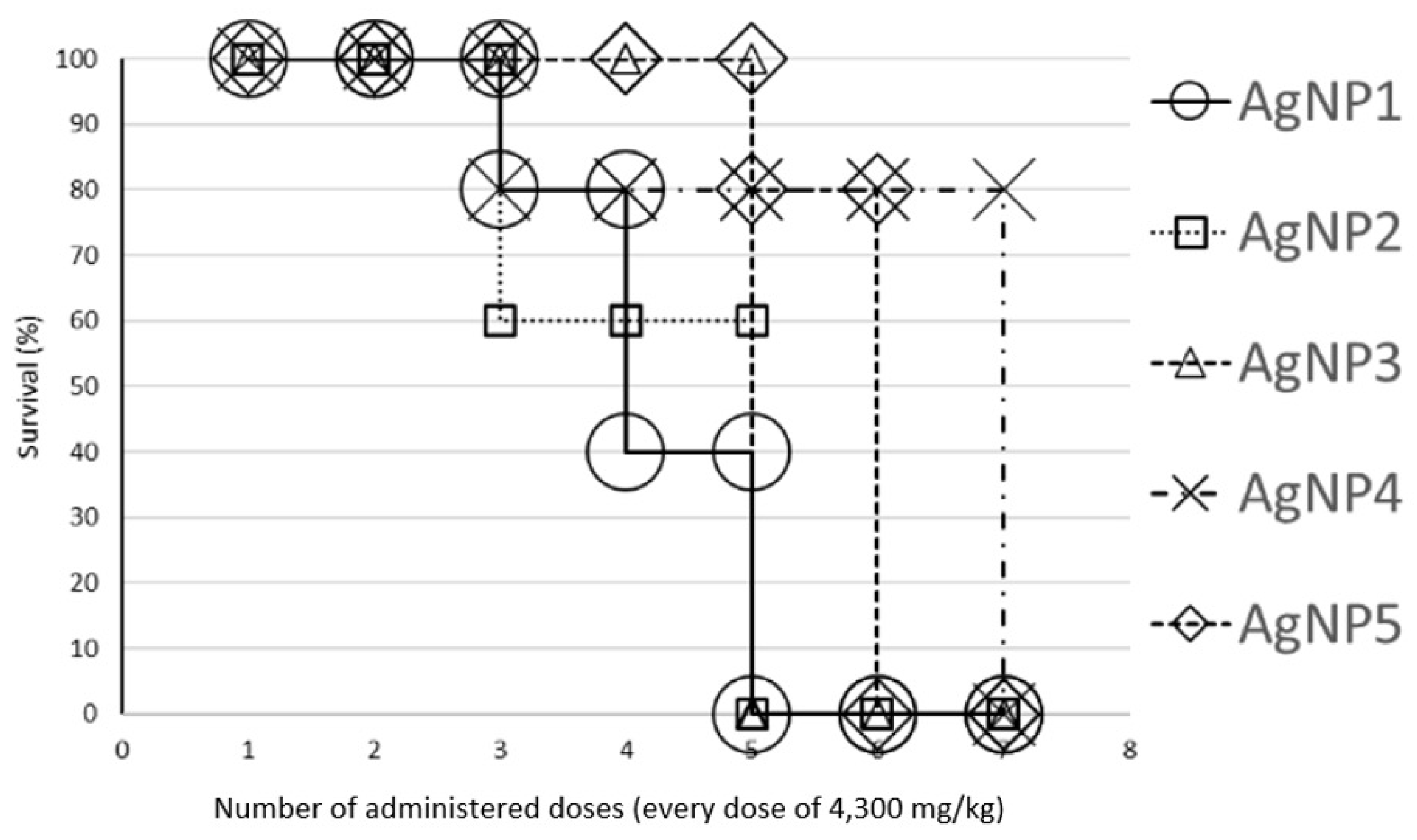
Figure 2.
The percentage of severe symptoms relative to the initial number of mice is shown using colored columns. The x-axis represents the cumulative number of AgNP doses (each 4300 mg/kg) administered at two-hour intervals throughout the experiment. Each subfigure corresponds to a specific AgNP formulation (AgNP1-AgNP5). Vertical dotted lines separate data by symptom for each AgNP formulation.
Figure 2.
The percentage of severe symptoms relative to the initial number of mice is shown using colored columns. The x-axis represents the cumulative number of AgNP doses (each 4300 mg/kg) administered at two-hour intervals throughout the experiment. Each subfigure corresponds to a specific AgNP formulation (AgNP1-AgNP5). Vertical dotted lines separate data by symptom for each AgNP formulation.
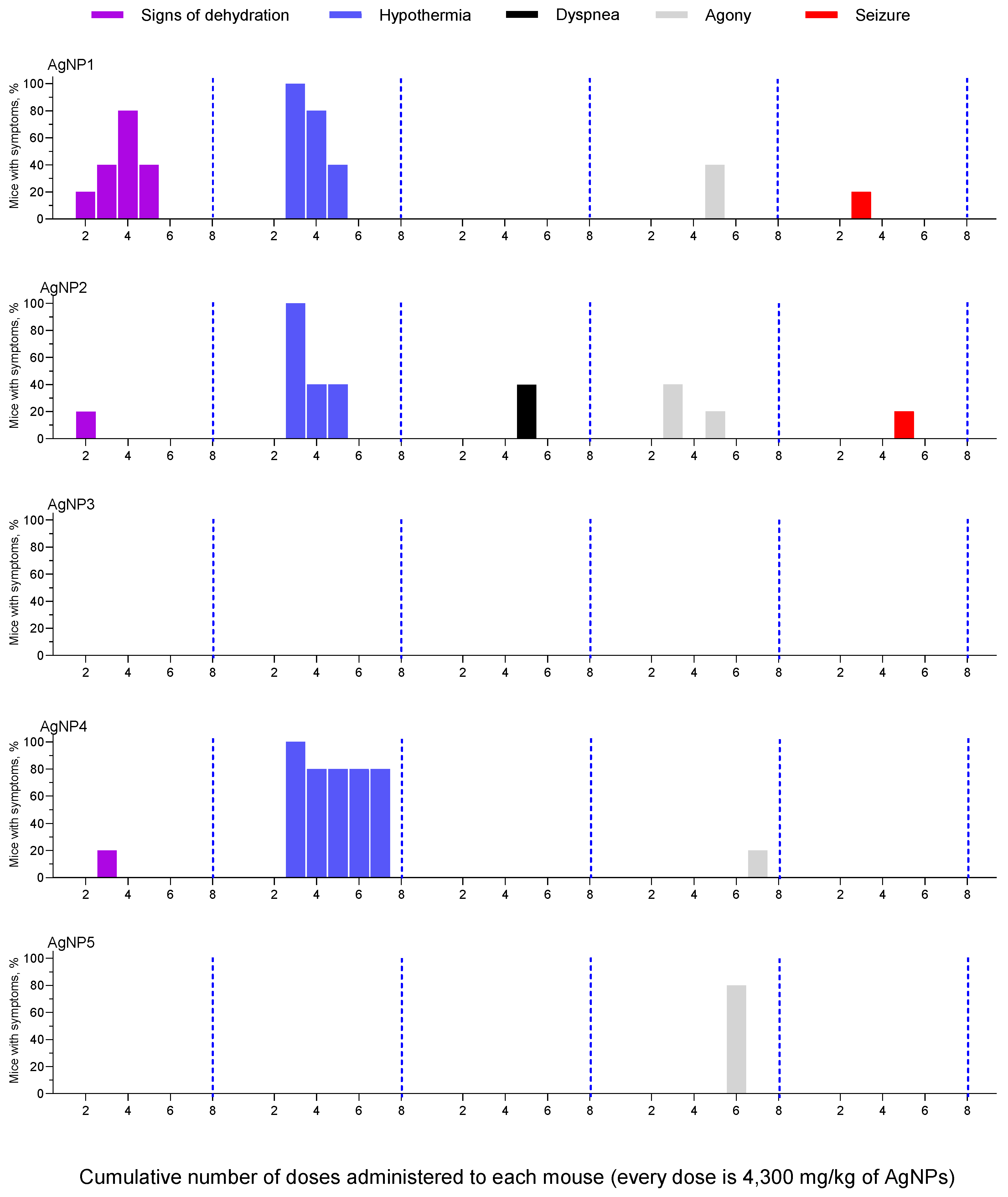
Figure 3.
Photographs of the liver (a, b), spleen (c, d), kidney (e, f), lung (g, h), and heart (i, j), showing the histopathological changes described in the main text. a) megahepatocyte (circle) and vacuolization (black arrows); b) Kupffer cells with pigment (arrowheads); c) areas of pigment in the red pulp (asterisks) and apoptosis in white pulp (circle); d) congestion (arrowheads) and extramedullary hematopoiesis (circles); e and f) congestion (arrows); g) hemorrhage (arrowhead and circles); h) alveolar macrophages with pigment (arrows); i) pigment deposit on the valvular surface (arrowhead) and pigment residues in the lumen of the cardiac chambers (asterisks); j) pericardial calcification (arrows).
Figure 3.
Photographs of the liver (a, b), spleen (c, d), kidney (e, f), lung (g, h), and heart (i, j), showing the histopathological changes described in the main text. a) megahepatocyte (circle) and vacuolization (black arrows); b) Kupffer cells with pigment (arrowheads); c) areas of pigment in the red pulp (asterisks) and apoptosis in white pulp (circle); d) congestion (arrowheads) and extramedullary hematopoiesis (circles); e and f) congestion (arrows); g) hemorrhage (arrowhead and circles); h) alveolar macrophages with pigment (arrows); i) pigment deposit on the valvular surface (arrowhead) and pigment residues in the lumen of the cardiac chambers (asterisks); j) pericardial calcification (arrows).
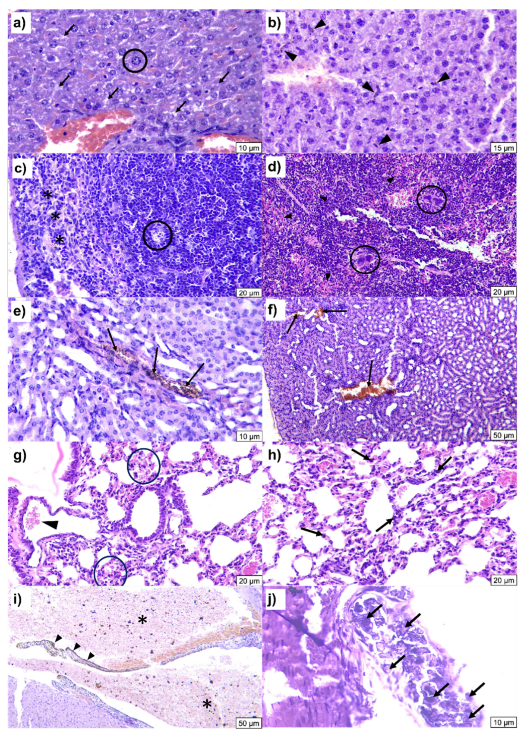
Figure 4.
Comparison of the LD50 of the five Argovit™ AgNP formulations studied in this work with those reported from other scientific groups: a) LD50 calculated for total AgNP formulation (metallic Ag and stabilizer, and water) and b) LD50 calculated for AgNPs (metallic Ag together with stabilizer). LD50 values for some substances and the GHS category 5 have also been included as horizontal, colored lines for comparative purposes.
Figure 4.
Comparison of the LD50 of the five Argovit™ AgNP formulations studied in this work with those reported from other scientific groups: a) LD50 calculated for total AgNP formulation (metallic Ag and stabilizer, and water) and b) LD50 calculated for AgNPs (metallic Ag together with stabilizer). LD50 values for some substances and the GHS category 5 have also been included as horizontal, colored lines for comparative purposes.
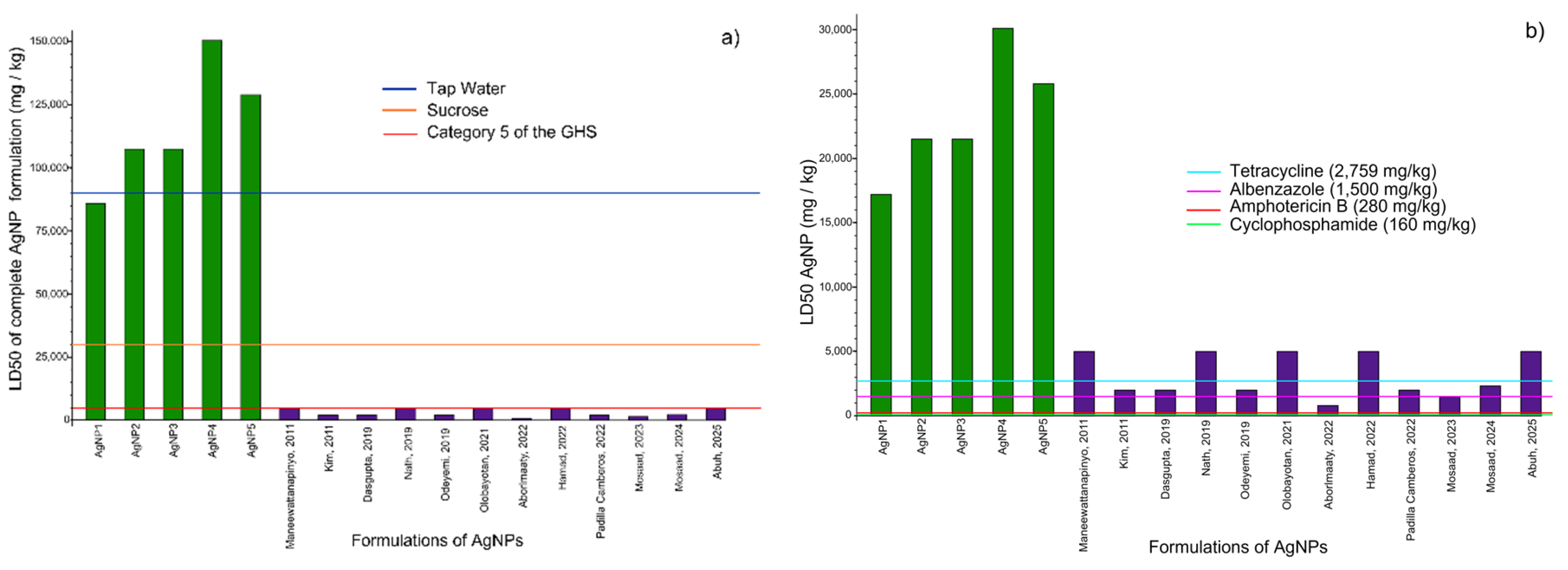
Figure 5.
Equivalency of AgNP volume administered to a human, calculated considering the concentration of Argovit formulation (200 mg/mL).
Figure 5.
Equivalency of AgNP volume administered to a human, calculated considering the concentration of Argovit formulation (200 mg/mL).
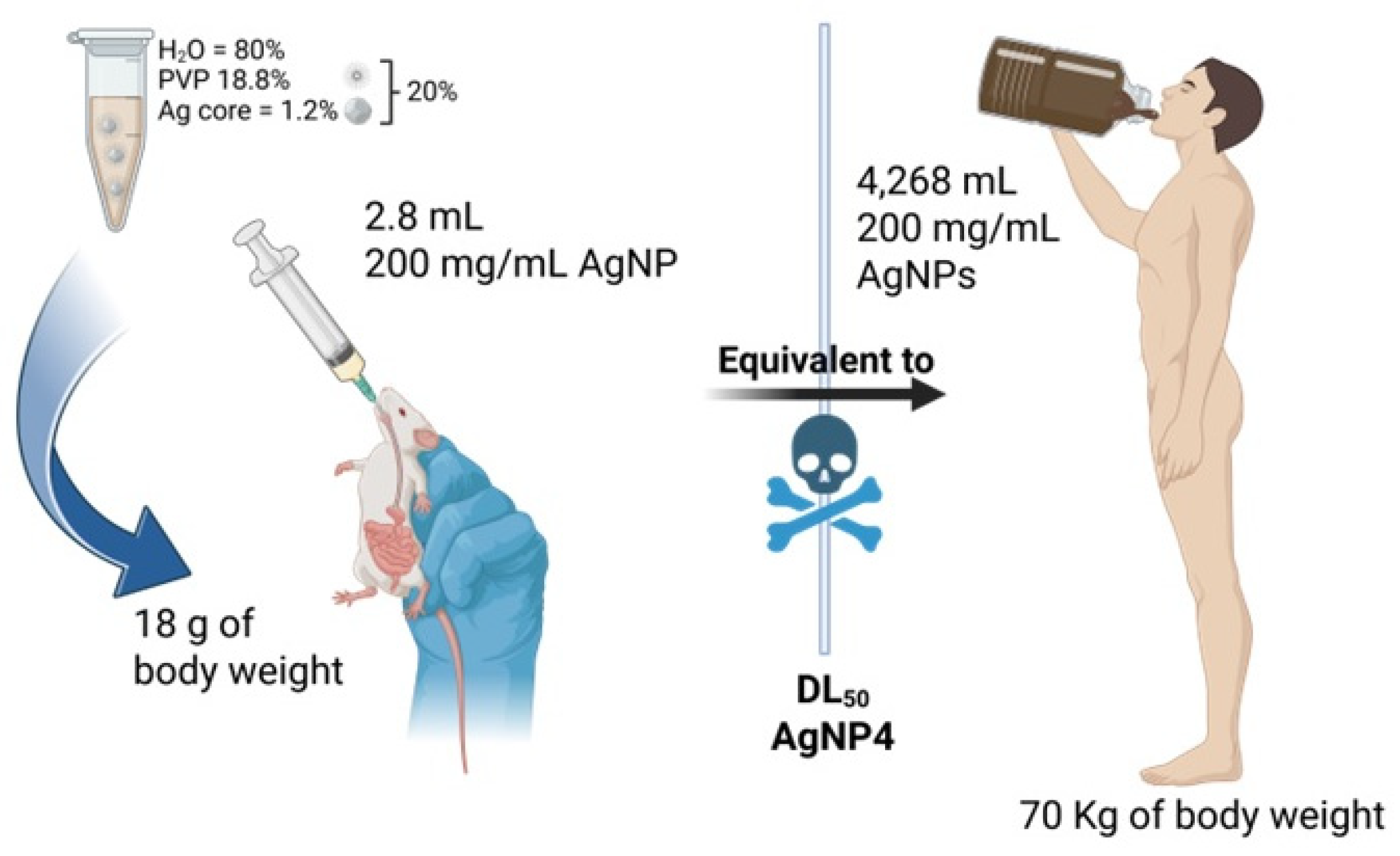
Figure 6.
LD50 for humans calculated from mouse LD50 using the scaling factors previously published by Nair, et. al. (2008) [38].
Figure 6.
LD50 for humans calculated from mouse LD50 using the scaling factors previously published by Nair, et. al. (2008) [38].
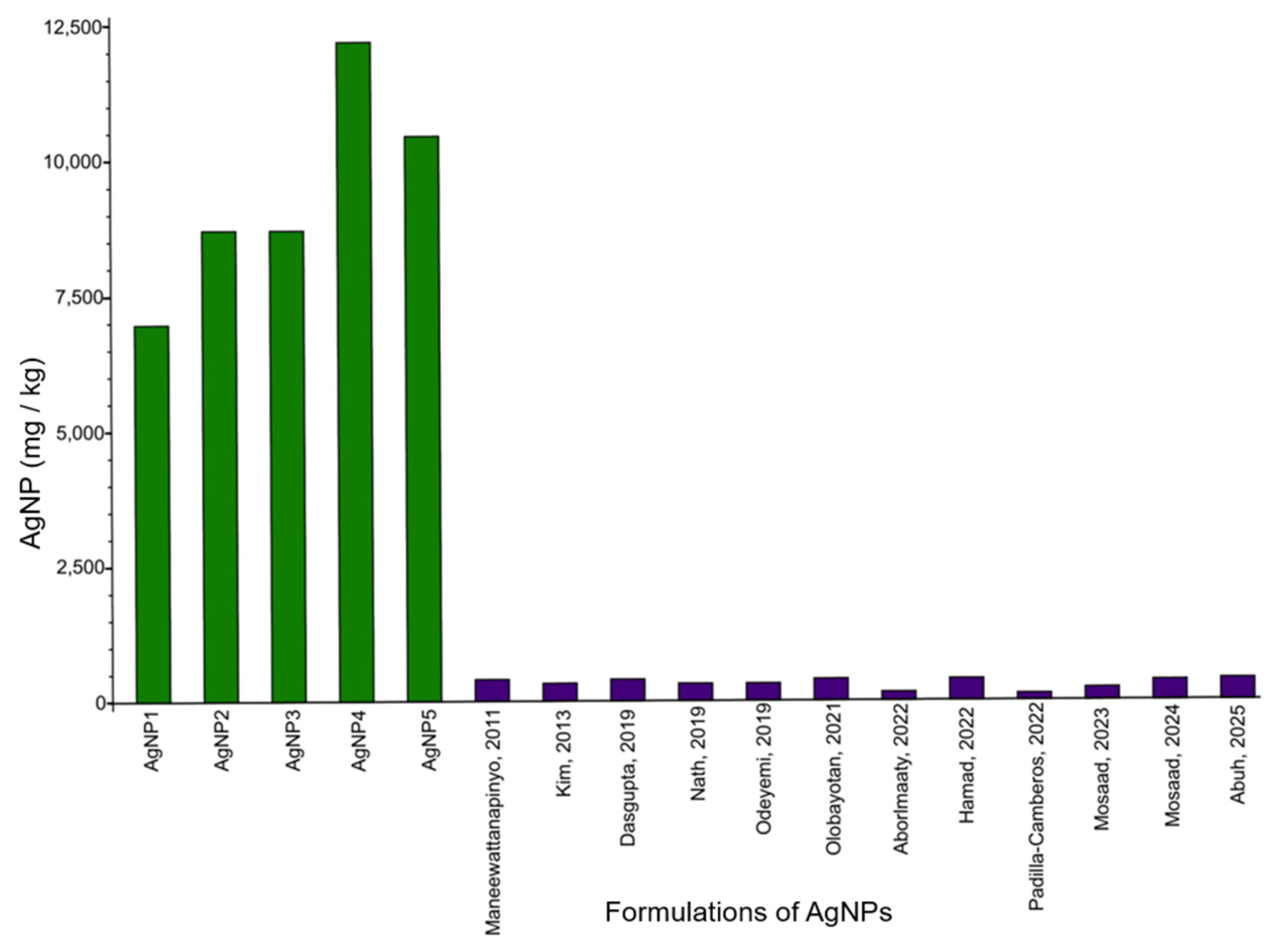

Table 1.
GHS Hazard Categorization for Acute Toxicity by Oral Route according to OECD 420 [16].
Table 1.
GHS Hazard Categorization for Acute Toxicity by Oral Route according to OECD 420 [16].
| Acute toxicity level | Toxicity definition | Dose (mg/kg) |
| 1 | Lethally toxic | ≤ 5 |
| 2 | Toxic | >5–<50 |
| 3 | Harmful | >50–<300 |
| 4 | Low hazard | >300–<2,000 |
| 5 | Without significant danger | >2,000–<5,000 |
| Not classified | Harmless | >5,000 |
Table 2.
Physicochemical properties of five Argovit lots. The data were adapted from Cruz-Ramirez et al., 2021 [17].
Table 2.
Physicochemical properties of five Argovit lots. The data were adapted from Cruz-Ramirez et al., 2021 [17].
| Properties | AgNP1 | AgNP2 | AgNP3 | AgNP4 | AgNP5 |
| Average diameter of metallic cores ∅Ag (nm) | 16.4±8.0 | 25.4±13.2 | 19.0±9.3 | 16.4±8.1 | 30.6±23.2 |
| TEM size distribution (nm) | 5–40 | 5–60 | 5–40 | 5–40 | 5–80 |
| PVP | K-15* | K-17* | K-17* | K-30* | 12.6 KDa |
| Hydrodynamic diameter ∅hydro (nm) | 448.7 | 90.4 | 43.8 | 483.2 | 121.1 |
| Polydisoersity index (PDI) | 0.813 | 0.270 | 0.433 | 0.555 | 0.280 |
| Zeta potential ζ (mV) | −0.872 | −4.56 | +5.13 | −0.464 | −1.46 |
| Surface Plasmon Resonance (λ) | 415 | 402, 444 | 402 | 406-549 | 429 |
| TGA analysis of metallic silver (% ω/ω) | 1.14±0.02 | 1.32±0.05 | 1.26±0.03 | 1.19±0.01 | 1.31±0.01 |
| PVP (% ω/ω) | 19.62±0.30 | 24.49±0.70 | 24.43±0.20 | 20.92±0.42 | 21.67±0.50 |
| H2O (% ω/ω) | 79.24±0.45 | 74.25±0.60 | 75.74±0.25 | 77.89±0.80 | 77.02±0.40 |
| Morphology | Spherical | Spherical | Mostly spherical** | Mostly spherical** | Spherical |
* Molecular masses of the AgNP formulations are presented in Table 3. ** A small portion of the metallic Ag particles (less than 3% and 1%) had triangular and rod shapes, respectively, in TEM micrographs.
Table 3.
Characteristics of the PVP stabilizer and electron adsorbed doses used in the preparation of every AgNP formulation.
Table 3.
Characteristics of the PVP stabilizer and electron adsorbed doses used in the preparation of every AgNP formulation.
| AgNP sample | PVP type and its molecular mass kDa | Electron absorbed dose, kGy | PVP manufacturer |
| AgNP1 | K-15: 8–12 kDa | 15 | Boai NKY Pharmaceuticals Ltd., China |
| AgNP2 | K-17, 10–16 kDa | 15 | Boai NKY Pharmaceuticals Ltd., China |
| AgNP3 | K-17: 10–16 kDa | 45 | Boai NKY Pharmaceuticals Ltd., China |
| AgNP4 | K-30: 45–58 kDa | 45 | Boai NKY Pharmaceuticals Ltd., China |
| AgNP5 | 12.6 kDa | 15 | LLC AK Sintvita, Tula region, Russia |
Table 4.
LD50 calculated for AgNP (metallic Ag and PVP) and the complete formulation (metallic Ag, PVP, and water).
Table 4.
LD50 calculated for AgNP (metallic Ag and PVP) and the complete formulation (metallic Ag, PVP, and water).
| AgNP formulation | LD50 (mg/kg) calculated for | |
| AgNPs (Ag+PVP) |
Complete formulation (Ag+PVP+H2O) | |
| AgNP1 | 17,204 | 86,020 |
| AgNP2 | 21,505 | 107,525 |
| AgNP3 | 21,505 | 107,525 |
| AgNP4 | 30,107 | 150,535 |
| AgNP5 | 25,806 | 129,030 |
Table 5.
Frequency and severity of clinical signs presented by the AgNP formulations.
| Frequency | Description | Severity of clinical signs | ||
| Mild | Moderate | Severe | ||
| Frequently observed | The sign was observed in 15-25 mice across all groups. | Hunched back/ruffled fur* | Lethargy* | Hypothermia 1, 2, 4 |
| Closed or half-closed eyes 1, 3, 4, 5 | ||||
| Observed with average frequency | The sign was observed in 3-10 mice across all groups. | Pasty stools 2, 3, 4, 5 | Abdominal distension 1, 2 | Dyspnoea 2 |
| Hyperaemic ears 4, 5 | Vomiting reflex 2 | Agony 1, 2, 4, 5 | ||
| Erect tail 3 | Signs of dehydration 1, 2, 4 | |||
| Startled response 1, 2, 5 | Seizure 1, 2 | |||
| Rarely observed | The sign was observed in only 1-2 mice across all groups. | Mustard-coloured urine 5 | ||
| Eye discharge 4 | ||||
| Blepharitis 4,5 | ||||
| Hiccup 4 | ||||
* Signs observed in all mice. 1,2,3,4 or 5 Signs observed in AgNP1, AgNP2, AgNP3, AgNP4, or AgNP5, respectively.
Table 6.
Number and percentage of clinical signs presented by mice after administration of AgNP formulations.
Table 6.
Number and percentage of clinical signs presented by mice after administration of AgNP formulations.
| AgNP formulation | Number of clinical signs presented by mice after administration of AgNP formulation | ||
| Mild | Moderate | Severe | |
| AgNP1 | 1(11.2%) | 4(44.4%) | 4(44.4%) |
| AgNP2 | 2(18.2%) | 4(36.4%) | 5(45.4%) |
| AgNP3 | 2(40%) | 3(60%) | 0(0.0%) |
| AgNP4 | 3(27.3%) | 5(45.4%) | 3(27.3%) |
| AgNP5 | 3(33.3%) | 5(55.6%) | 1(11.1%) |
Table 7.
Percentage of mice with histopathological changes in five organs after AgNP administration.
Table 7.
Percentage of mice with histopathological changes in five organs after AgNP administration.
| Organ | Observations | Percentage (%) of mice with histological changes histopathological changes were observed |
||||
| AgNP1 | AgNP2 | AgNP3 | AgNP4 | AgNP5 | ||
| Liver | Congestion | 100 | 100 | 100 | 100 | 100 |
| Vacuolization | 100 | 100 | 100 | 100 | 100 | |
| Mega hepatocyte | 100 | 100 | 100 | 80 | 100 | |
| Kupffer Cells Pigmented | 100 | 33 | 40 | 60 | 80 | |
| Cytoplasmic inclusion | 0 | 0 | 60 | 20 | 0 | |
| Spleen | Congestion | 100 | 100 | 100 | 100 | 100 |
| White pulp apoptosis | 100 | 100 | 80 | 100 | 100 | |
| Extramedullary hematopoiesis | 100 | 100 | 100 | 100 | 100 | |
| Kidney | Tubular regeneration | 0 | 0 | 0 | 0 | 0 |
| Medullary calcification | 0 | 0 | 0 | 0 | 0 | |
| Interstitial lymphocytic infiltration | 0 | 0 | 0 | 0 | 0 | |
| Lung | Haemorrhage | 100 | 100 | 100 | 100 | 100 |
| Interstitial lymphocytic infiltration | 0 | 0 | 0 | 0 | 0 | |
| Pigment in alveolar macrophages | 0 | 100 | 100 | 100 | 100 | |
| Lymphoid hyperplasia | 0 | 0 | 0 | 0 | 0 | |
| Heart | Thrombus | 0 | 0 | 0 | 0 | 0 |
| Pericardial calcification | 0 | 0 | 40 | 20 | 20 | |
| Myocardial calcification | 0 | 0 | 0 | 0 | 0 | |
| Endocardial calcification | 0 | 0 | 0 | 0 | 0 | |
| Lymphoid infiltrate | 0 | 0 | 20 | 0 | 0 | |
| Neutrophil infiltration | 0 | 0 | 0 | 0 | 0 | |
Table 8.
Performance order of AgNP formulations regarding LD50, survival rate, clinical signs, and histopathological changes.
Table 8.
Performance order of AgNP formulations regarding LD50, survival rate, clinical signs, and histopathological changes.
| Parameter | Order | Best | Worst |
| LD50 | AgNP4>AgNP5>AgNP3=AgNP2>AgNP1 | AgNP4 | AgNP1 |
| Survival rate | AgNP4>AgNP5>AgNP3>AgNP2>AgNP1 | AgNP4 | AgNP1 |
| Clinical signs | AgNP3<AgNP5<AgNP4<AgNP1<AgNP2 | AgNP3 | AgNP2 |
| Histopathology | AgNP1~AgNP2<AgNP5~AgNP4<AgNP3 | AgNP1 | AgNP3 |
Table 9.
The comparison of LD50 in rodents and the corresponding calculated dose in humans for the AgNP formulations studied here and the twelve described in the literature.
Table 9.
The comparison of LD50 in rodents and the corresponding calculated dose in humans for the AgNP formulations studied here and the twelve described in the literature.
| Size of Ag nuclei (nm) | Type of AgNP synthesis | Stabilization agent | Animal model | Dose (mg/kg) | LD50 of complete formulation in mice (mg/kg) | Human equivalent dose of the complete formulation (mg/kg) | Dose of AgNP (g) for a person weighing 70 kg | Reference |
|---|---|---|---|---|---|---|---|---|
| 16.4 ± 8.0 (AgNP1) | Reduction of Ag+ with a high-energy electron beam | PVP | Mice | Repeated doses of 4,300 | 86,020 | 6,967.62 | 487.73 | Present work |
| 25.4 ± 13.2 (AgNP2) | 107,525 | 8,709.53 | 609.91 | |||||
| 19.0 ± 9.3 (AgNP3) | 107,525 | 8,709.53 | 609.91 | |||||
| 16.4 ± 8.1 (AgNP4) | 150,535 | 12,193.34 | 853.88 | |||||
| 30.6 ± 23.2 (AgNP5) | 129,030 | 10,451.43 | 731.89 | |||||
| 10-20 | Green | Starch | Mice | Single dose of 5,000 | >5000 | >405 | >28.36 | [30] |
| 19 | Not specified | Not specified | Rats | Not specified | >2,061 | >333.8 | >23.38 | [29] |
| 3-10 | Green | Saraca asoca | Mice | 5 | >5,000 | >405 | >28.36 | [26] |
| 60 | Mild green | Trisodium citrate | Rats | 500, 1,000, 2,000 | >2,000 | >324 |
22.69 | [27] |
| Not specified | Green | Not specified | Rats | 500, 1000, 2,000 | >2,000 | >324 | 22.69 | [32] |
| Not specified | Green | Kolanut (Cola nitida) pod extract | Mice | 10, 100, 1,000, 1,500, 3,000, and 5,000 | >5,000 | >405 | >28.36 | [23] |
| Not specified | Green | Aqueous S. queretaroensis peel extract | Mice | 2,000 | >2,000 | >162 | >11.34 | [33] |
| 50-80 | Green | Leaf extracts of Z. spina-christi | Mice | 50, 100, 200, and | >5,000 | >405 | >28.36 | [28] |
| 12.87 | Green | Thymus vulgaris | Rats | 100, 200, 400, 600, 800, and 1,000 | 800 | 129.6 | 9.08 | [24] |
| 49.33 ± 7.59 | Green | Balanites aegyptiaca | Rats | Not specified | 1,470 | 238.1 | 16.68 | [31] |
| 38.20 ± 2.5 | 2,330 | 377.5 | 26.43 | |||||
| Not specified | Green |
T. catappa A. boonei |
Mice | 1) 10, 100, and 1000 mg/kg 2) 1600, 2900, and 5000 mg/kg |
>5,000 | >405 | >28.36 | [25] |
Disclaimer/Publisher’s Note: The statements, opinions and data contained in all publications are solely those of the individual author(s) and contributor(s) and not of MDPI and/or the editor(s). MDPI and/or the editor(s) disclaim responsibility for any injury to people or property resulting from any ideas, methods, instructions or products referred to in the content. |
© 2026 by the authors. Licensee MDPI, Basel, Switzerland. This article is an open access article distributed under the terms and conditions of the Creative Commons Attribution (CC BY) license.
Copyright: This open access article is published under a Creative Commons CC BY 4.0 license, which permit the free download, distribution, and reuse, provided that the author and preprint are cited in any reuse.


