

Submitted:
10 June 2026
Posted:
11 June 2026
You are already at the latest version
Abstract
The cavernous sinus remains one of the most anatomically complex and surgically challenging regions of the skull base because of its intimate relationships with the internal carotid artery and multiple cranial nerves.
Detailed microsurgical anatomical knowledge is essential for safe skull base and parasellar surgery. A stepwise microsurgical cadaveric dissection of the cavernous sinus was performed on six adult formalinfixed specimens using a standard pterional extradural approach under magnification with a Leica operating microscope. Progressive extradural and interdural dissections were carried out to expose the lateral wall of the cavernous sinus, cranial nerves III, IV, V1, V2, and VI, the intracavernous internal carotid artery, and the principal surgical triangles of the cavernous sinus. The dissection demonstrated the multilayered interdural architecture of the lateral wall and allowed identification of the interdural cleavage plane extending from the
superior orbital fissure to Meckel’s cave. Cranial nerves III, IV, V1, and V2 were identified within the lateral wall, whereas the abducens nerve coursed medially within the venous compartment adjacent to the cavernous internal carotid artery. Parkinson’s triangle and the anteromedial and infratrochlear corridors provided useful operative windows to the intracavernous compartment. Stepwise cadaveric dissection remains an effective educational tool for understanding the microsurgical anatomy of the cavernous sinus and improving operative orientation during skull base procedures.
Keywords:
cadaveric dissection
; cavernous sinus
; microsurgical anatomy
; skull base surgery
; skull base triangles
1. Introduction
The cavernous sinus represents one of the most intricate anatomical regions of the cranial base because of the density of neurovascular structures traversing its compartments. The sinus contains the cavernous segment of the internal carotid artery (ICA), cranial nerves III, IV, V1, V2, and VI, as well as a complex venous plexus communicating with the superior and inferior petrosal sinuses, ophthalmic veins, and basilar venous plexus [1,2,3].
Historically, surgical access to the cavernous sinus was considered hazardous because of the risk of cranial nerve injury and catastrophic vascular complications. The pioneering anatomical and surgical works of Parkinson, Dolenc, Kawase, and Rhoton established the anatomical foundations that enabled modern skull base approaches to this region [4,5,6,7,8]. The development of extradural and interdural approaches considerably improved operative exposure while minimizing neurovascular morbidity.
Despite significant advances in endoscopic and minimally invasive skull base surgery, microsurgical anatomical understanding of the cavernous sinus remains fundamental. Precise identification of dural layers, interdural cleavage planes, cranial nerve trajectories, and surgical triangles is essential for procedures involving cavernous sinus tumors, aneurysms, petroclival lesions, pituitary adenomas with parasellar extension, and trigeminal schwannomas [9,10,11].
The purpose of this study was to provide a detailed stepwise cadaveric dissection of the cavernous sinus
using a microsurgical extradural approach, emphasizing surgically relevant anatomical landmarks and operative corridors.
2. Materials and Methods
Specimens
Six adult cadaveric heads fixed in formalin were studied at the Laboratory of Anatomy of the Faculty of Medicine, Pharmacy and Dental Medicine, Sidi Mohammed Ben Abdellah University, Fez, Morocco.
Specimens demonstrated no evidence of prior cranial trauma, surgery, or skull base pathology.
No arterial or venous silicone injection was performed. Although this limited visualization of venous channels and small vascular branches, the specimens allowed adequate demonstration of dural layers, cranial nerves, and major intracavernous arterial structures.
Institutional authorization for anatomical dissection was obtained in accordance with laboratory regulations and ethical standards governing cadaveric studies.
Microsurgical Equipment
All dissections were performed under microscopic magnification using a Leica operating microscope (Leica Microsystems, Wetzlar, Germany). Standard microsurgical instruments, including microdissectors, bipolar forceps, microscissors, and suction devices, were utilized.
Surgical Technique
For didactic purposes, a superior craniectomy involving removal of the calvaria was initially performed in all specimens. The brain was subsequently sectioned at the level of the mesencephalon and removed to obtain an unobstructed superior view of the skull base and cavernous sinus region.
Step 1: Exposure of the Lateral Wall of the Cavernous Sinus
Bulleted The meningo-orbital band was identified and sectioned to facilitate extradural peeling of the temporal dura propria. Progressive elevation of the dura exposed the anterior clinoid process, superior orbital fissure, foramen rotundum, and Meckel’s cave. The lateral wall of the cavernous sinus was identified as the dural plane extending between the superior orbital fissure anteriorly and Meckel’s cave posteriorly (Figure 1).
Step 2: Interdural Peeling
All An interdural cleavage plane was developed between the periosteal and meningeal dural layers. The dissection was initiated at the level of the maxillary nerve (V2) near the foramen rotundum and progressively extended anteriorly and posteriorly (Figure 2).
Step 3: Identification of Cranial Nerves
This The oculomotor nerve (III), trochlear nerve (IV), ophthalmic division of the trigeminal nerve (V1), and maxillary division (V2) were identified within the lateral wall of the cavernous sinus. The abducens nerve (VI) was visualized within the venous compartment medial to the lateral wall and inferolateral to the intracavernous ICA (Figure 3)
The oculomotor nerve (CN III) occupies the superior position, followed by the trochlear nerve (IV). The ophthalmic division (V1) is located inferiorly, while the maxillary division (V2) courses along the inferior aspect of the lateral wall before exiting via the foramen rotundum. The abducens nerve (VI) is seen within the cavernous sinus proper, adjacent to the intracavernous internal carotid artery (ICA), Ant clin : anterior clinoid process.
Step 4: Exposure of Intracavernous Vascular Structures
The cavernous segment of the ICA was exposed with identification of its posterior vertical segment, posterior bend, horizontal segment, anterior bend, and anterior vertical segment. The meningohypophyseal trunk and inferolateral trunk were identified when present (Figure 4).
The artery is seen after passing above the fibrocartilaginous region of the foramen lacerum beneath the petrolingual ligament. The posterior ascending segment, posterior genu, horizontal segment, anterior genu, and anterior ascending segment are identified in relation to surrounding dural and bony landmarks. The close relationship between the intracavernous internal carotid artery and the abducens nerve is demonstrated, particularly at the posterior cavernous compartment. The clinoid segment is shown within the narrow osseous corridor formed by the anterior clinoid process, optic strut, and carotid sulcus.
Step 5: Surgical Triangles
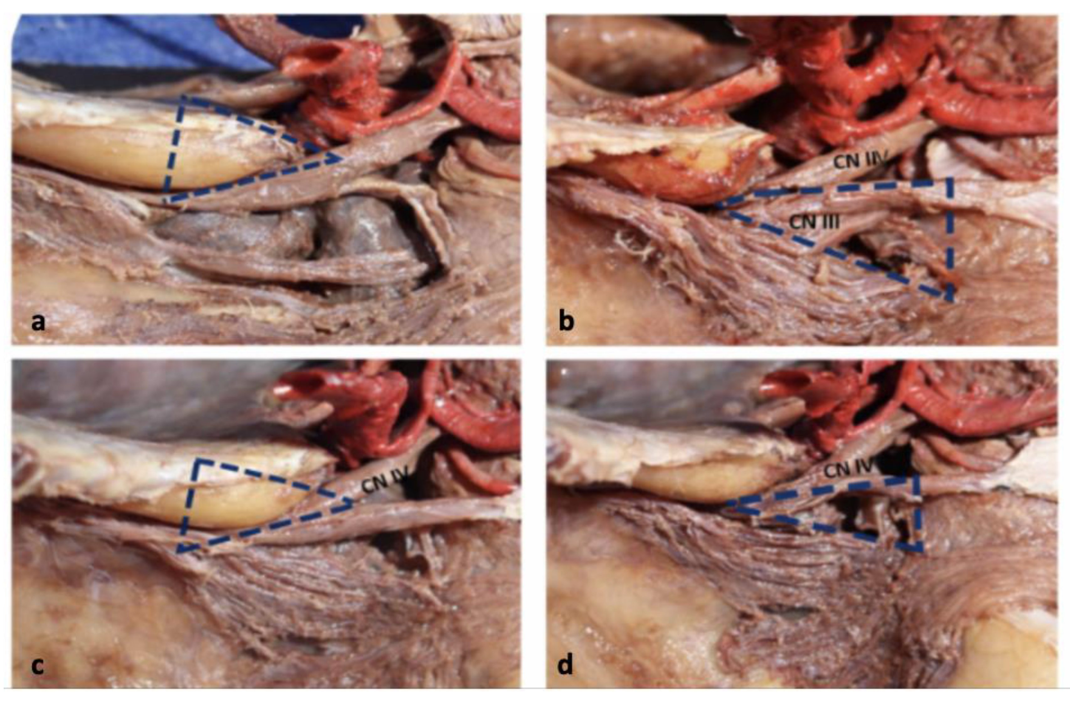
The anatomical limits of Parkinson’s triangle, the infratrochlear triangle, and anteromedial operative corridors were identified and analyzed according to their surgical accessibility and neurovascular relationships (Figure 5).
3. Results
3.1. General Anatomical Findings
The cavernous sinus consistently demonstrated a multilayered interdural architecture composed of periosteal and meningeal dural layers. The venous compartment occupied the space medial to the lateral wall and surrounded the cavernous segment of the ICA.
3.2. Lateral Wall Anatomy
The lateral wall was composed of two distinct dural layers. Cranial nerves III, IV, V1, and V2 were located between these layers within the lateral wall. The oculomotor nerve occupied the superior aspect of the wall, followed inferiorly by the trochlear nerve, ophthalmic nerve, and maxillary nerve. The interdural cleavage plane was most readily identifiable at the level of V2 near the foramen rotundum. This cleavage plane extended posteriorly toward Meckel’s cave and anteriorly toward the superior orbital fissure (Figure 1 and Figure 2).
3.3.. Cranial Nerve Relationships
The oculomotor nerve entered the roof of the cavernous sinus lateral to the posterior clinoid process before coursing anteriorly toward the superior orbital fissure.
The trochlear nerve followed a thinner and more superior trajectory along the lateral wall. The ophthalmic division (V1) coursed within the intermediate portion of the lateral wall toward the superior orbital fissure, whereas V2 exited through the foramen rotundum.
The abducens nerve followed a distinct intracavernous trajectory medial to the lateral wall and lateral to the ICA. Its intimate relationship with the cavernous carotid artery was consistently observed (Figure 3).
3.4. Intracavernous Internal Carotid Artery
The cavernous ICA demonstrated the classical S-shaped configuration in all specimens. The posterior vertical segment ascended from the petroclival region before turning anteriorly into the horizontal segment. The anterior bend then continued superiorly toward the clinoid segment.
The meningohypophyseal trunk was identified in five specimens, arising from the posterior genu of the ICA. The inferolateral trunk was observed in four specimens. Small branches corresponding to McConnell arteries supplying the pituitary capsule were identified in several dissections (Figure 4).
3.5. Surgical Triangles
Parkinson’s triangle was consistently identified between cranial nerves IV and V1. This corridor provided direct visualization of the intracavernous ICA and abducens nerve.
The infratrochlear triangle offered access to the posterior cavernous sinus compartment and posterior bend of the ICA. The anteromedial corridor allowed visualization of the anterior cavernous sinus compartment and proximal superior orbital fissure (Figure 5).
4. Discussion
The cavernous sinus remains one of the most challenging regions in skull base surgery because of its complex neurovascular anatomy and multilayered dural architecture. Comprehensive microsurgical anatomical understanding is therefore essential for minimizing morbidity during approaches to parasellar and petroclival lesions.
The present study demonstrates the importance of interdural dissection for safe exposure of the cavernous sinus. The cleavage plane identified between the periosteal and meningeal layers corresponds to the surgical plane originally emphasized by Dolenc during extradural transcavernous approaches [5]. This interdural corridor enables exposure of cranial nerves embedded within the lateral wall while limiting venous bleeding and minimizing manipulation of the intracavernous compartment.
Consistent with prior anatomical studies, cranial nerves III, IV, V1, and V2 were located within the lateral wall, whereas the abducens nerve followed a medial intracavernous trajectory adjacent to the ICA [6,7,8]. The vulnerability of the abducens nerve during cavernous sinus surgery is directly related to this anatomical relationship.
The interdural cleavage plane was most consistently identified at the level of V2 near the foramen rotundum. This observation is surgically relevant because V2 represents a reliable landmark during extradural skull base approaches.
The cavernous ICA demonstrated the characteristic carotid siphon configuration described in classical anatomical literature [7]. Identification of the meningohypophyseal trunk and inferolateral trunk remains essential during surgery because these branches may contribute to cranial nerve vascularization and tumor blood supply.
The surgical triangles of the cavernous sinus remain important operative corridors for accessing distinct intracavernous compartments. Parkinson first described the infratrochlear triangle as a route to carotidcavernous fistulas and aneurysms [4]. Subsequent anatomical refinements by Dolenc and Rhoton expanded the operative applications of these corridors [5,7].
Although endoscopic endonasal surgery has significantly expanded access to the parasellar region, microsurgical transcranial approaches remain indispensable for selected lesions with lateral extension, vascular encasement, or complex cranial nerve relationships [10,11,12]. Therefore, cadaveric microsurgical training continues to play a crucial role in skull base surgical education.
This study has several limitations. First, vascular injection was not performed, limiting visualization of venous channels and small arterial branches. Second, the sample size was relatively limited. Third, formalin fixation may alter tissue elasticity and venous sinus morphology compared with living anatomy. Finally, no morphometric analysis was conducted.
Despite these limitations, the study provides a practical stepwise anatomical framework for understanding microsurgical approaches to the cavernous sinus.
5. Conclusions
The cavernous sinus possesses a complex multilayered anatomy requiring precise understanding of interdural planes, cranial nerve trajectories, and vascular relationships. Extradural and interdural microsurgical dissections provide safe operative corridors to this region while minimizing neurovascular injury.
Cadaveric dissection remains an essential educational method for skull base surgical training. The present study highlights the importance of the lateral wall anatomy, inte dural cleavage plane, and cavernous sinus triangles as key anatomical landmarks during cavernous sinus surgery
Author Contributions
All authors have reviewed the final version to be published and agreed to be accountable for all aspects of the work. Concept and design: Hammoud Marouane, Abdesslam Bouassria, Soufiane Mellas, Mustapha Elkouache, Khalid Chakour. Acquisition, analysis, or interpretation of data: Hammoud Marouane, Abdesslam Bouassria, Soufane Mellas, Mustapha Elkouache, Khalid Chakour. Drafting of the manuscript: Hammoud Marouane, Abdesslam Bouassria, Soufiane Mellas, Mustapha Elkouache, Khalid Chakour. Critical review of the manuscript for important intellectual content: Hammoud Marouane, Adeslam. Bouassria, Soufiane Mellas, Mustapha Elkouache, Khalid Chakour. Supervision: Hammoud Marouane, Abdesslam Bouassria, Soufiane Mellas, Mustapha Elkouache, Khalid Chakour
Funding
This research received no external funding
Institutional Review Board Statement
Not applicable
Informed Consent Statement
Not applicable.
Conflicts of Interest
The authors declare no conflicts of interest.
Abbreviations
The following abbreviations are used in this manuscript:
| ACP | Anterior clinoid process |
| CN | Cranial nerve |
| CS | Cavernous sinus |
| ICA | Internal carotid artery |
| V1 | ophthalmic division of the trigeminal nerve |
| V2 | Maxillary division of the trigeminal nerve |
| V3 | Mandibular division of the trigeminal nerve |
| VI | Abducens nerve |
| III | Oculomotor nerve |
| IV | Trochlear nerve |
References
- D. Parkinson: A surgical approach to the cavernous portion of the carotid artery. Anatomical studies and case report. J. Neurosurg. vol. 23, 474:483. [CrossRef]
- F. S. Harris and A. L. Rhoton: Anatomy of the cavernous sinus. A microsurgical study. J. Neurosurg. vol. 45(no. 2), 169–180. [CrossRef] [PubMed]
- Yasuda, A.M.; Campero, A.; Martins, C.; et al. The medial wall of the cavernous sinus: microsurgical anatomy. Neurosurgery (Lippincott Williams & Wilkins) 2004, 55, 179–190. [Google Scholar] [CrossRef]
- Yasuda, A.; Campero, A.; Martins, C.; et al. Microsurgical anatomy and approaches to the cavernous sinus. Neurosurgery 2008, 62, 1240–1263. [Google Scholar] [CrossRef] [PubMed]
- Umansky, F.; Nathan, H. The lateral wall of the cavernous sinus. With special reference to the nerves related to it. J. Neurosurg. 1982, vol. 56(no. 2), 228–234. [Google Scholar] [CrossRef] [PubMed]
- Sindou, M.; Jacquesson, T. Surgical Anatomy of the Cavernous Sinus. Brain Anatomy and Neurosurgical Approaches: A Practical, Illustrated, Easy-to-Use Guide 2023, 495–514. [Google Scholar] [CrossRef]
- Inoue, T.; Rhoton, A. L.; Theele, D.; Barry, M. E. ‘Surgical approaches to the cavernous sinus: a microsurgical study’. Neurosurgery 1990, vol. 26(no. 6), 903. [Google Scholar] [CrossRef]
- MCCONNELL, E.M. The arterial blood supply of the human hypophysis cerebri. Anat. Rec. 1953, vol. 115(no. 2 115), 175-203 175-203. [Google Scholar] [CrossRef] [PubMed]
- D. Parkinson: Carotid cavernous fistula: direct repair with preservation of the carotid artery. Technical note. J. Neurosurg. 1973, vol. 38(no. 1), 99–106. [CrossRef] [PubMed]
- Isolan, G. R.; Krayenbühl, N.; de Oliveira, E.; et al. Al-Mefty, ‘Microsurgical Anatomy of the Cavernous Sinus: Measurements of the Triangles in and around It’. Skull Base vol. 17, 357–2007. [CrossRef] [PubMed]
- Dolenc, V. V. Extradural approach to intracavernous ICA aneurysms. Acta Neurochir. Suppl. 72, 99–106. [CrossRef] [PubMed]
- Watanabe, A.; Nagaseki, Y.; Ohkubo, S.; et al. Anatomical variations of the ten triangles around the cavernous sinus. Clin. Anat. 2003, 16, 9–14. [Google Scholar] [CrossRef] [PubMed]
Figure 1.
External configuration of the cavernous sinus. (a) Superior view of the endocranial cavity after superior craniectomy performed for didactic exposure, demonstrating the left cavernous sinus (blue arrow). (b) Lateral wall of the left cavernous sinus showing its principal anatomical landmarks and dural relationships. The superior orbital fissure is identified anteriorly, the anterior clinoid process superiorly, and Meckel’s cave posteriorly.
Figure 1.
External configuration of the cavernous sinus. (a) Superior view of the endocranial cavity after superior craniectomy performed for didactic exposure, demonstrating the left cavernous sinus (blue arrow). (b) Lateral wall of the left cavernous sinus showing its principal anatomical landmarks and dural relationships. The superior orbital fissure is identified anteriorly, the anterior clinoid process superiorly, and Meckel’s cave posteriorly.
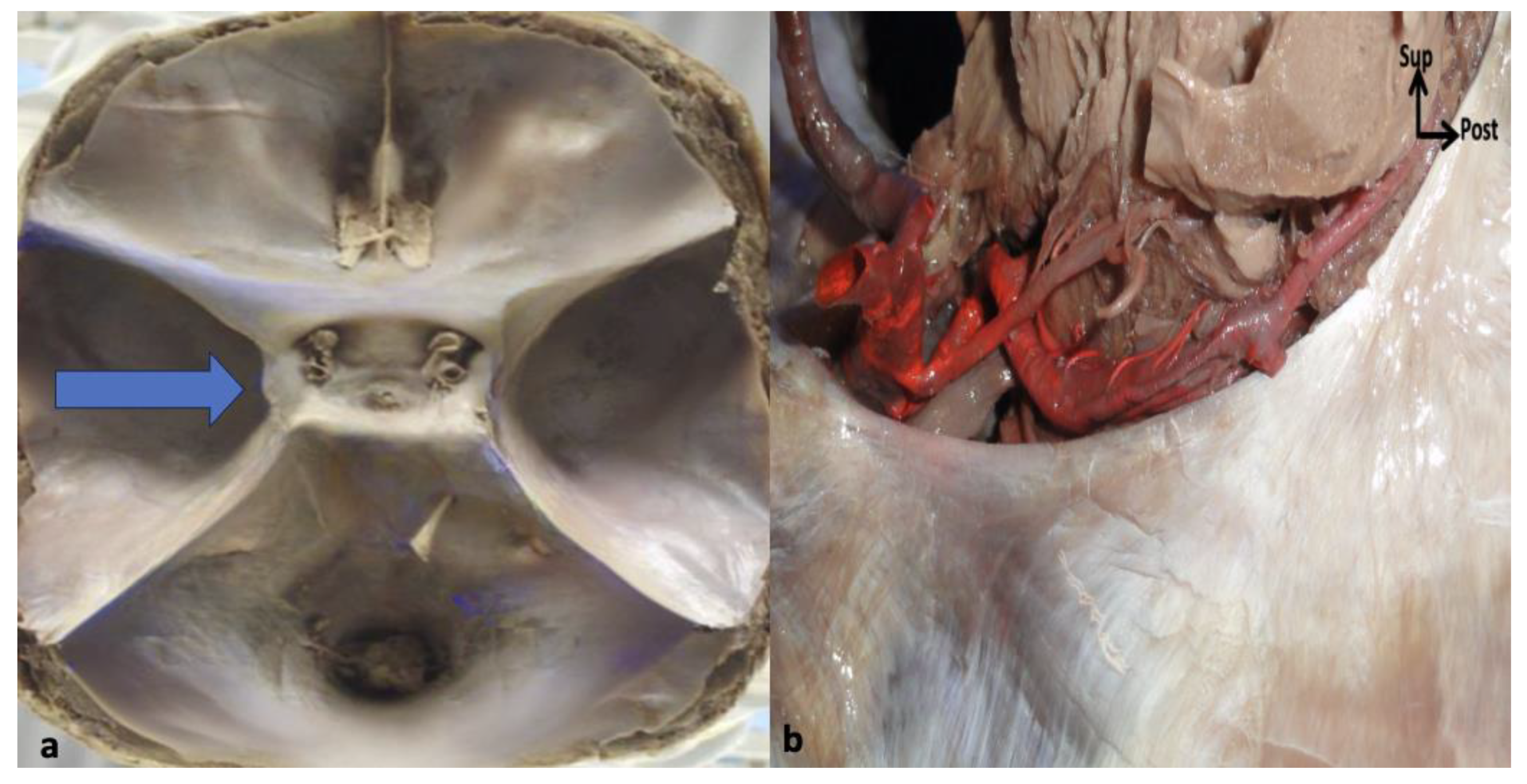
Figure 2.
Dural organization of the lateral wall of the cavernous sinus. (a) Coronal section through the cavernous sinus demonstrating the multilayered dural organization. The outer dural envelope consists of a superficial meningeal layer (orange), while the periosteal layer (green) contributes to the endosteal dural lining. The periosteal layer further splits into two laminae, forming the inner dural layers of the lateral wall and roof of the cavernous sinus. (b) Microsurgical dissection demonstrating the interdural cleavage plane between the superficial meningeal layer and the deep endosteal dural layer. The dissection is initiated at the level of the maxillary nerve (V2) and extended anteriorly toward the superior orbital fissure and posteriorly toward Meckel’s cave. Progressive interdural peeling allows sequential exposure of cranial nerves embedded within the lateral wall while preserving the integrity of the venous compartment of the cavernous sinus.
Figure 2.
Dural organization of the lateral wall of the cavernous sinus. (a) Coronal section through the cavernous sinus demonstrating the multilayered dural organization. The outer dural envelope consists of a superficial meningeal layer (orange), while the periosteal layer (green) contributes to the endosteal dural lining. The periosteal layer further splits into two laminae, forming the inner dural layers of the lateral wall and roof of the cavernous sinus. (b) Microsurgical dissection demonstrating the interdural cleavage plane between the superficial meningeal layer and the deep endosteal dural layer. The dissection is initiated at the level of the maxillary nerve (V2) and extended anteriorly toward the superior orbital fissure and posteriorly toward Meckel’s cave. Progressive interdural peeling allows sequential exposure of cranial nerves embedded within the lateral wall while preserving the integrity of the venous compartment of the cavernous sinus.
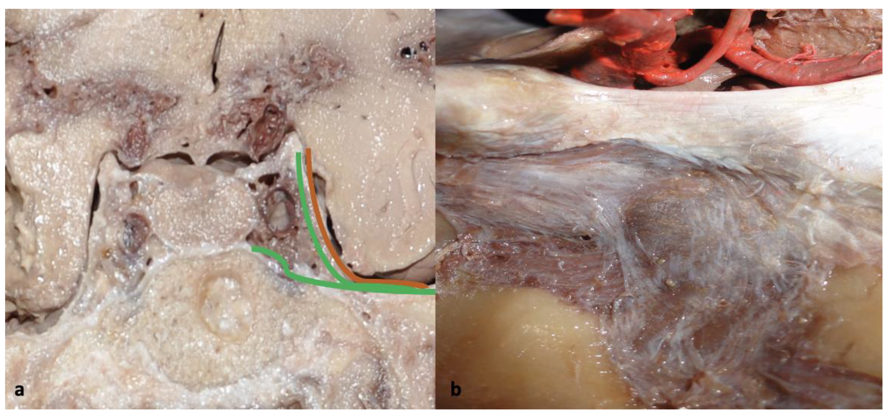
Figure 3.
Sequential exposure of cranial nerves within the lateral cavernous wall after interdral peeling.
Figure 3.
Sequential exposure of cranial nerves within the lateral cavernous wall after interdral peeling.
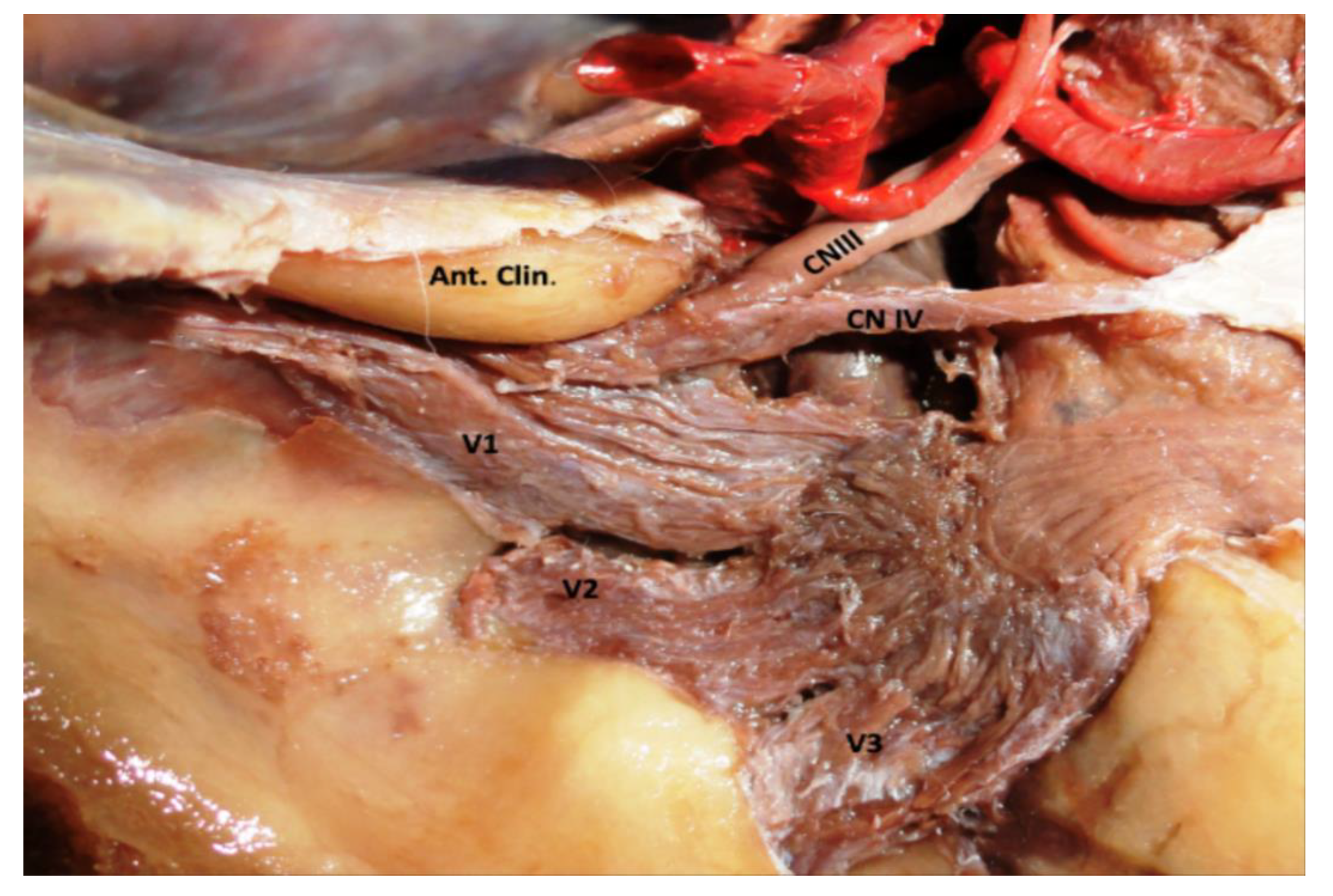
Figure 4.
Exposure of the intracavernous internal carotid artery.
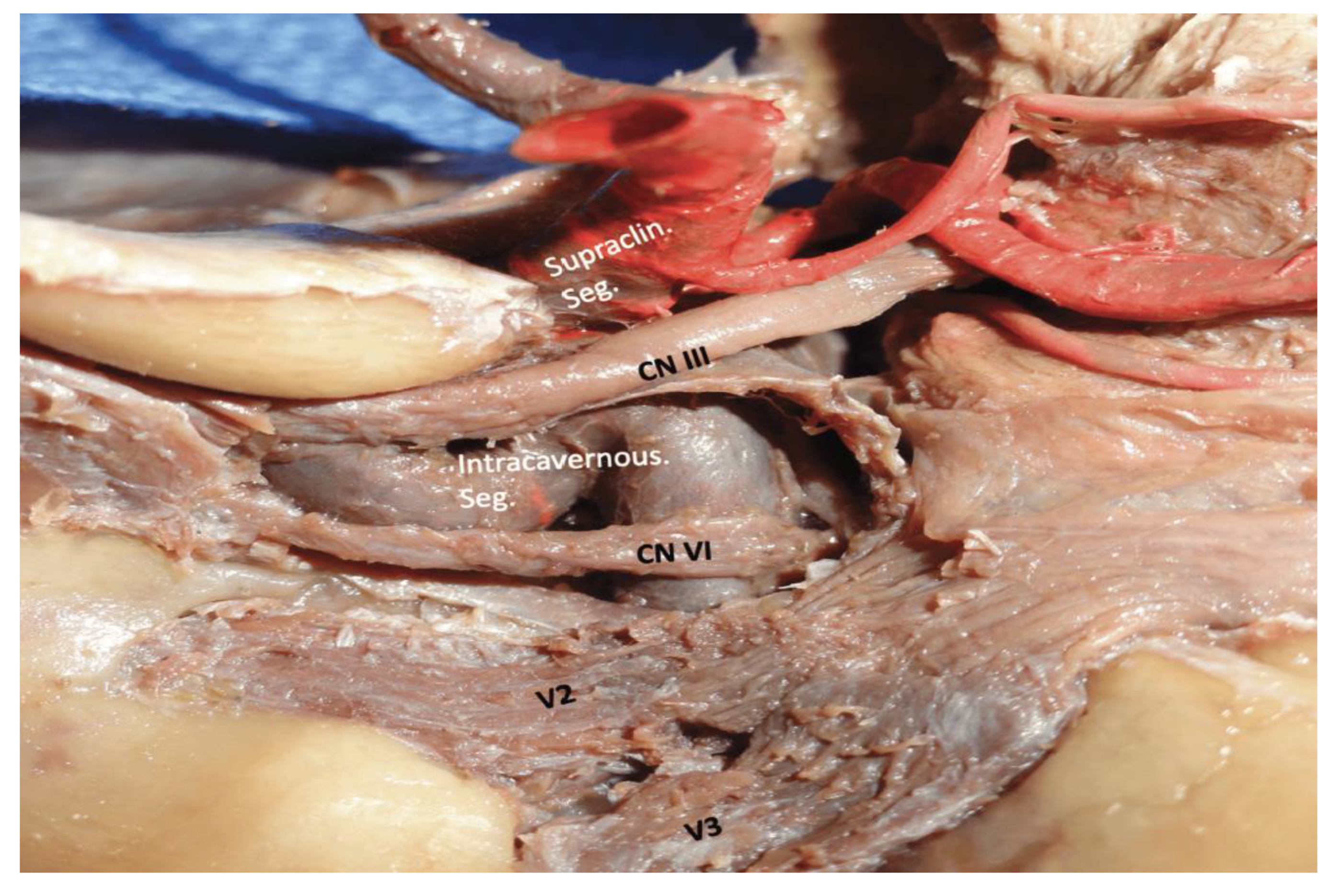
Figure 5.
The principal triangle of the cavernous sinus. (a) Clinoidal triangle: Located between the optic nerve and oculomotor nerve; provides access to the clinoid segment of the ICA following anterior clinoidectomy. (b) Oculomotor triangle: Defined by dural folds at the entry zone of cranial nerve III; serves as an orientation landmark in roof dissection. (c) Supratrochlear triangle: Situated between the oculomotor and trochlear nerves; offers limited but useful superior exposure. (d) Infratrochlear (Parkinson’s) triangle: Located between the trochlear nerve and V1; provides the most direct access to the posterior genu and horizontal segment of the cavernous ICA.
Figure 5.
The principal triangle of the cavernous sinus. (a) Clinoidal triangle: Located between the optic nerve and oculomotor nerve; provides access to the clinoid segment of the ICA following anterior clinoidectomy. (b) Oculomotor triangle: Defined by dural folds at the entry zone of cranial nerve III; serves as an orientation landmark in roof dissection. (c) Supratrochlear triangle: Situated between the oculomotor and trochlear nerves; offers limited but useful superior exposure. (d) Infratrochlear (Parkinson’s) triangle: Located between the trochlear nerve and V1; provides the most direct access to the posterior genu and horizontal segment of the cavernous ICA.
Disclaimer/Publisher’s Note: The statements, opinions and data contained in all publications are solely those of the individual author(s) and contributor(s) and not of MDPI and/or the editor(s). MDPI and/or the editor(s) disclaim responsibility for any injury to people or property resulting from any ideas, methods, instructions or products referred to in the content. |
© 2026 by the authors. Licensee MDPI, Basel, Switzerland. This article is an open access article distributed under the terms and conditions of the Creative Commons Attribution (CC BY) license (http://creativecommons.org/licenses/by/4.0/).
Copyright: This open access article is published under a Creative Commons CC BY 4.0 license, which permit the free download, distribution, and reuse, provided that the author and preprint are cited in any reuse.