

Submitted:
08 June 2026
Posted:
09 June 2026
You are already at the latest version
Abstract
Acute myeloid leukemia (AML) develops within a bone marrow environment that influences leukemic stem-cell behavior, residual disease, and response to therapy. This review examines evidence that the marrow microenvironment is not only a site of leukemic growth, but can actively shape AML initiation, maintenance, and treatment resistance. Clinical observations such as donor cell leukemia after allogeneic transplantation, together with experimental models in which stromal or osteolineage abnormalities induce myeloid disease, suggest that altered niches may contribute to leukemogenesis in selected settings. In established AML, vascular and endosteal compartments provide adhesive, chemokine, inflammatory, and metabolic signals that promote leukemic-cell retention, quiescence, survival, and chemotherapy tolerance. AML cells also remodel the surrounding marrow, suppressing normal hematopoiesis and generating stromal, endothelial, osteoblastic, adipocytic, and immune-cell programs that favor leukemic persistence. These interactions are especially relevant to drug resistance, including resistance to venetoclax-based therapy, where cytokine-mediated changes in apoptotic dependence, fatty-acid metabolism, mitochondrial adaptation, and stromal support may all contribute. Several therapeutic approaches have attempted to disrupt niche-mediated protection, including targeting CXCL12/CXCR4 signaling, adhesion pathways, inflammatory circuits, Hedgehog signaling, and metabolic dependencies. Although early-phase studies have shown activity in some AML subsets, randomized evidence remains limited and results have been inconsistent. We discuss how a better understanding of microenvironmental biology may help define when niche-directed therapy is most likely to complement conventional and molecularly targeted AML treatment.
Keywords:
bone marrow microenvironment
; acute myeloid leukemia
; leukemia stem cell
1. Introduction
Darwin observed that “a grain in the balance will determine which individual shall live or die, which species will increase or become extinct” (On the Origin of Species, 1861, chapter XIV). Evolution proceeds through small, random variations, of which only a few prove advantageous: the surviving species is the one that adapts most favorably to its environment.
In the 1950s and 1960s, Till and McCulloch proposed a hierarchical, pyramidal organization of hematopoiesis, with a self-renewing, asymmetrically dividing hematopoietic stem cell (HSC) at its apex (1). Asymmetric mitosis allows the cell both to differentiate toward a specific function and to self-regenerate at the top of the pyramid, maintaining the stem pool in quiescence and ensuring tissue inexhaustibility. The same logic applies to neoplastic tissues: relapse after morphological remission implies persistence of a quiescent, chemoresistant cancer stem cell with indefinite self-renewal, which drives carcinogenesis (2).
Stemness is not a cell-surface identity but a function, defined by the capacity to regenerate the tissue of origin in serial xenotransplantation into immunodeficient hosts (3). A stem cell — normal or leukemic — is conventionally defined by its capacity to sustain tissue in at least three serial transplants; hence the terms leukemic stem cell (LSC) or leukemia-initiating cell (L-IC). The observation that LSCs share the CD34⁺/CD38⁻ immunophenotype of HSCs led to the hypothesis that acute myeloid leukemia (AML) arises from oncogenic transformation of a normal HSC (4).
Immunophenotypic studies of LSCs began to disclose a role for the bone marrow microenvironment (BMM) in AML pathogenesis. In NOD/SCID/IL2Rγc-deficient models, lacking B, T, and NK cells (5), even committed progenitors expressing CD38 or CD45RA can regenerate AML in serial transplants, fulfilling LSC criteria (6). In a sufficiently permissive microenvironment, stemness is therefore not restricted to primitive cells.
These data reframe malignancy: aggressiveness depends not only on genetic and epigenetic traits but on the tumor microenvironment that shapes proliferative advantage. A neoplasm is an ecosystem in which malignant cells interact with non-neoplastic components that modulate tumor fitness and response to therapy (7), honoring Darwin’s intuition at a microscopic scale.
2. Clues From Clinical Experience
Allogeneic hematopoietic stem-cell transplantation (HSCT) is a curative treatment for eligible patients with high-risk AML. Sustained remission depends on the graft-versus-leukemia (GVL) effect exerted by donor immunity against residual leukemia stem cells (LSCs) (8). Relapse — the principal obstacle to cure — still occurs in 30–40% of transplanted patients (9). Most relapses are of host origin; donor cell leukemia (DCL), in which leukemia arises de novo from engrafted donor hematopoiesis, is rare but mechanistically informative.
Registry data estimate DCL at 0.08% (European Society for Blood and Marrow Transplantation, EBMT) and 0.16% (Japan Society for Hematopoietic Cell Transplantation, JSHCT) across indications (10,11). Among AML recipients, the reported incidence approaches 2%, with individual series describing rates up to 4–6%, and with a clear preponderance in adults (12). DCL develops after a median of 28 months post-HSCT, although the range is wide (1 month to 24 years). The first described case (1971) involved a 16-year-old female with acute lymphoblastic leukemia (ALL) who, after transplantation from her brother, developed donor-derived ALL in XY hematopoietic cells (12).
Diagnostic sensitivity has improved with chimerism analysis by short-tandem-repeat (STR) and variable-number-tandem-repeat (VNTR) profiling. The pathogenesis is multifactorial, reflecting donor, recipient, and procedure-related contributions in a multiple-hit model.
Prior to 2010, cytogenetic analysis of DCL identified chromosome 7 abnormalities in 23% of cases versus ≤5% of de novo AML (13) — a distribution reminiscent of therapy-related myeloid neoplasms and consistent with persistent mutagenic effects of recipient-directed chemotherapy and conditioning on the marrow stroma. Next-generation sequencing has refined this picture. Among the transcription factors recurrently mutated in DCL — CEBPA, GATA2, RUNX1, and JAK2 — three are typically of germline origin and one is predominantly somatic (13). Donor clonal hematopoiesis, particularly DNMT3A-mutated clonal hematopoiesis of indeterminate potential (CHIP), has been documented in recipients who later developed DCL (14,15); in related donors aged ≥50 years, CHIP prevalence reaches 16% (16). Additional recurrently mutated genes cluster into splicing regulators (DDX41), epigenetic modifiers (ASXL1, DNMT3A, EZH2, IDH1/2), and DNA-repair genes (CHEK1, XPD, XRCC3) (13).
That donors do not typically develop AML, except in extremely rare cases, suggests that the recipient microenvironment plays a significant role in supporting leukemogenesis. Preclinical data link alterations in bone marrow stromal cells and other niche components to the development and maintenance of leukemia and to chemotherapy resistance (17–21). Aldoss et al. reported two sequential myeloid DCLs in a single recipient after two HSCTs from different donors — a matched-related transplant followed three years later by a first DCL, then, after a new induction, a second transplant from a young unrelated donor with subsequent development of a second DCL from the second donor. The authors concluded that the recipient likely had a leukemogenic microenvironment that predisposed donor cells to transform (22,23).
DCL after cord-blood transplant (CBT) occurs earlier (median 14.5 vs 36 months) and is more frequently associated with chromosome 7 abnormalities. Fusion genes such as TEL-AML1 or AML1-ETO may be present in cord cells and predispose to expansion of a pre-leukemic clone; these are class 2 genetic alterations that impair differentiation but do not alone induce overt AML, and they often occur years before disease onset. Because cord blood cells are less likely to have acquired predisposing genetic mutations, DCLs after CBT provide a compelling argument for a role of the microenvironment in initiating leukemic transformation.
3. Possible Direct Oncogenic Role of the Bone Marrow Microenvironment
Mesenchymal stromal cells (MSCs) sustain marrow homeostasis by regulating HSC self-renewal, differentiation, and immune tolerance. Primary MSCs from patients with myelodysplastic syndromes (MDS) and AML differ structurally, genetically, and functionally from healthy controls; dysregulation of KIT-ligand, angiopoietin-1, and Jagged1 signaling is associated with impaired osteogenic commitment and reduced growth capacity (24–27). TGF-β signaling has been implicated as an important contributor to the aberrant MSC phenotype in both MDS and AML (28). While the role of the BMM in disease maintenance and chemoresistance is increasingly recognized, its contribution to disease initiation is only now emerging.
Raaijmakers et al. deleted Dicer1 — a ribonuclease III endonuclease indispensable for microRNA biogenesis — in murine MSCs at the osteolineage differentiation stage (19). Osteolineage-specific loss disrupted osteoblastic differentiation without affecting the osteoclastic component, induced severe cytopenias with marked myelodysplastic features, and accelerated progression to acute leukemia. Transplantation of hematopoietic cells from Dicer1-mutant mice into wild-type recipients restored normal hematopoiesis, and the emergent leukemic clone retained wild-type Dicer1 — demonstrating that the initiating event lay within the altered marrow microenvironment, not the hematopoietic compartment. Dicer1 inactivation in osteoprogenitors was also linked to downregulation of the Shwachman-Bodian-Diamond (Sbds) gene, which is involved in ribosomal biogenesis and whose germline loss produces Shwachman-Bodian-Diamond syndrome — characterized by skeletal alterations, inefficient hematopoiesis with bone marrow failure, and increased propensity to progress to MDS and AML. Reduced expression of Dicer1, Drosha, and Sbds has since been confirmed in MSCs from MDS and AML patients, together with loss of miR-155, miR-181a, and miR-222 — microRNAs that regulate hematopoiesis (29–31).
Activating β-catenin mutations in murine osteoblasts reproduce an analogous niche-initiated leukemia (32). β-catenin activation drives osteoblastic Jagged1 expression cooperatively with FoxO1, engaging Notch signaling in adjacent hematopoietic progenitors and producing AML with recurrent karyotypic alterations and autonomous progression (33). Notch activation in this setting is associated with chemoresistance.
Epigenetic dysregulation also contributes to the altered functional state of the BMM. In primary stromal cells from MDS patients, aberrant cytosine hypermethylation silences FRZB, a WNT antagonist, thereby activating the WNT/β-catenin pathway in surrounding HSCs (34). WNT activity affects HSC homeostasis in a dose-dependent manner, and increased WNT/β-catenin signaling has been linked to leukemic transformation (35,36). Whether this stromal methylation defect precedes emergence of the myelodysplastic clone remains unresolved.
Cytotoxic therapy imprints a durable pro-inflammatory state on the MSC compartment. Alkylating agents induce cellular senescence and a senescence-associated secretory phenotype (SASP) with elevated pro-inflammatory cytokines and reactive oxygen species (37). Multi-omic profiling of MSCs from patients with therapy-related myeloid neoplasms (t-MN) confirms induction of CDKN1A and β-galactosidase, FOS repression, elevated cytokine output, and impaired proliferation — a senescent-secretory signature that is stable regardless of the latency between cytotoxic exposure and t-MN diagnosis (38). Analogous changes are seen in MSCs from patients with Fanconi anemia (FA), an inherited disease characterized by congenital abnormalities, bone marrow failure, and increased susceptibility to myeloid malignancies: baseline downregulation of FLT3-ligand, TGF-β1, and CXCL12, with cytoplasmic mislocalization of Dicer1 even before etoposide exposure. In healthy-donor MSCs, etoposide redistributes Dicer1 to the cytoplasm and induces ER-stress genes (GADD34, ATF4, NUPR1, HSPB), phenocopying the FA baseline (39). Taken together, these data indicate that microenvironmental alterations may contribute to the pathogenesis of myeloid neoplasms, including AML, and suggest that they may also serve as therapeutic targets.
4. the Medullary Vascular Niche
Evidence indicates that self-renewing HSCs reside in the bone-marrow perivascular niche rather than exclusively in long-term dormant reserves (Figure 1). This specialized, protective compartment is composed of MSCs (40,41) and endothelial cells of sinusoidal microvessels (42,43). From this location, HSCs monitor blood-borne factors and activate the recruitment of additional HSCs from quiescent endosteal niches to stimulate hematopoiesis and osteogenesis, counteracting hematological stress (44). During embryogenesis, primitive hematopoiesis arises from hemogenic endothelium of the aorta-gonad-mesonephros region and yolk sac via endothelial-to-hematopoietic transition (45–47); the close spatial relationship between endothelial and hematopoietic progenitors persists into adulthood, with the vascular niche traditionally considered comparatively oxygen-rich relative to the endosteum (48) — a view that direct intravital oxygen measurements have since challenged and partially inverted (see §5).
Sinusoids are fenestrated, low-pressure channels (49,50) that maintain niche homeostasis through secretion of IL-1, IL-6, IL-8, GM-CSF, TNF, G-CSF, chemokines, and matrix metalloproteinases (51). The endosteal niche contains transition-zone vessels (52), roughly 15% of HSCs (53), and approximately 10% of marrow volume. The central vascular niche occupies the remaining ~90% of marrow volume and is estimated to harbor ~85% of HSCs (43). The endosteal compartment is relatively drug-resistant and supports post-chemotherapy regeneration (54), whereas the vascular compartment is sensitive to irradiation and myeloablation (55). Two-thirds of perivascular HSCs associate with sinusoids and LepR⁺ stromal cells; the remaining third associate with arterioles and NG2⁺/Nestinhigh pericytes (56–58).
Specific alterations in the BM vascular niche can act synergistically with genetic or epigenetic lesions in hematopoietic cells to facilitate mutant-cell survival and expansion, contribute to progression and protection from chemotherapy, and promote relapse. Using intravital two-photon microscopy, Passaro et al. showed that vascular leakiness and increased hypoxia in AML are dependent on endothelial nitric oxide (NO); induction chemotherapy that fails to restore normal vasculature may be associated with poor prognosis, while inhibition of NO production promotes normal hematopoiesis and enhances treatment response in preclinical models (59). CXCL12-abundant reticular (CAR) cells are heterogeneously distributed in the perivascular endothelium: arteriolar CARs (NG2⁺/LepR⁻) enforce HSC quiescence, whereas sinusoidal CARs (NG2⁻/LepR⁺) support retention and expansion (57,60,61). The consistent association of HSCs with CXCL12-secreting reticular cells in both vascular and endosteal locations blurs the historical distinction between the two niches (59,62).
Increased microvascular density has been documented in ALL (63), chronic myeloid leukemia (CML) (64–66), AML (67), and multiple myeloma (MM) (68) and carries prognostic weight (69). Tumor neovasculature is abnormally permeable and tortuous (70,71). Endothelial cells (ECs) and leukemic cells engage in bidirectional signaling: ECs provide trophic and paracrine support, while blasts release proangiogenic factors (FGF-2, VEGF-A, VEGF-C, angiopoietins) and cytokines such as GM-CSF (72,73). AML cells can become quiescent and resist chemotherapy by integrating into the vascular endothelium, forming vascular tissue-associated AML (V-AML) (74–76). Combining anti-angiogenic therapy with consolidation chemotherapy has been proposed as a practical approach for patients with acute leukemia (77).
Adrenomedullin (ADM) is a 52-amino-acid peptide mainly synthesized by endothelial cells, vascular smooth muscle cells, and pericytes (78–80). It is expressed in many human tissues and exerts predominantly hypotensive, immunomodulatory, and vasoactive effects, including those observed during septic shock (81–83). ADM signals through calcitonin-receptor-like receptor (CALCRL) in complex with receptor activity-modifying proteins (RAMPs); among these, the CALCRL–RAMP2 heterodimer mediates canonical ADM signaling. ADM is also expressed by leukemia stem cells and defines an inflammatory signature in AML (84). Moreover, ADM is overexpressed in solid tumors under hypoxic conditions via HIF-1 regulation (85), exhibiting pro-angiogenic activity and stimulating tumor progression and chemoresistance (86). Our group showed that AML cell lines constitutively express ADM and its receptors to sustain proliferation and block differentiation, suppressing Cullin-family gene expression and activating PI3K/AKT and MAPK/ERK (87). Larrue et al. subsequently demonstrated that the ADM–CALCRL axis preserves relapse-initiating cells (RICs) that persist after chemotherapy; high CALCRL or ADM expression in RICs correlates with adverse outcome, and CALCRL knockdown reduces LSC frequency and restores cytarabine sensitivity in patient-derived xenografts (88).
Events that promote blast–endothelium adhesion are of particular interest given the capacity of AML cells to integrate into the vascular wall. ADM downregulates CD38 on blasts — thereby weakening the CD38/hyaluronic-acid axis — and simultaneously upregulates CD31, enhancing trans-endothelial migration and circulatory dissemination (87,89). In murine models, Cogle et al. documented progressive localization of AML blasts around hepatic portal vessels and their incorporation into endothelial syncytia (V-AML) that acquire quiescence and chemoresistance (76). Analogous observations in chronic myeloproliferative neoplasms, where marrow endothelial cells carry disease-defining mutations, suggest similar endothelial participation in other hematologic malignancies.
5. the Osteoblastic Niche
In AML, LSCs preferentially home to the osteoblastic compartment — the endosteal surface lined by osteoblasts — rather than to the vascular niche, as confirmed by homing and serial engraftment experiments (21,90) (Figure 1). Elevated endosteal ATP supports LSC retention through P2X7 signaling (91). LSCs in this compartment exhibit high rates of symmetric division, strong colony-forming potential, elevated glycolytic flux, and superior leukemogenic capacity in vivo (90). Expression of active Rac1 in murine AML reduces disease latency by enhancing niche homing, quiescence, and apoptosis protection (92).
Oxygen tension in the leukemic endosteum falls to ~0.1% (90,93), substantially below that of the vascular niche; in healthy mice the relationship is inverted, with deeper peri-sinusoidal regions of the vascular niche slightly more hypoxic (~1.3%) than the endosteum (94). Hypoxia can promote HSC quiescence (95) and, via hypoxia-associated factor (HAF), HIF-1α, and HIF-2α, upregulates OCT-3/4, NANOG, and SOX2 — transcription factors whose ectopic expression can reprogram somatic cells into induced pluripotency (96–99). In cancer stem cells, MYC, NANOG, and SOX2 stabilize HIF-2α, which represses p53 and preserves self-renewal (100); loss of p53 in turn impairs MDM2-mediated HIF-1α degradation (101). Although a clear consensus has not been reached, increased expression of OCT4 and NANOG has been reported in AML relative to normal marrow (102).
In endosteal LSCs, HIF-1 drives metabolic reprogramming by upregulating PDK2 and blocking pyruvate entry into the tricarboxylic acid (TCA) cycle (103). Concurrent AMPK activation induces GLUT1 and the pentose-phosphate pathway, raising reduced glutathione and buffering against reactive oxygen species (ROS)-mediated oxidative stress (90,104). In T-cell acute lymphoblastic leukemia (T-ALL), NOTCH mutations engage AMPK with comparable cytoprotective effect (105), supporting AMPK as a therapeutic target for sensitization. Conversely, oxidative phosphorylation (OXPHOS) is required to maintain quiescent human LSCs (106), leaving the quantitative relationship between the endosteum, hypoxia, glucose availability, and LSC fate open.
Blasts engage osteoblasts through fibronectin–hyaluronic-acid/CD44, ICAM1/LFA, WNT/Frizzled, Notch/Jagged, CXCL12/CXCR4, VCAM1/VLA4, and N-cadherin/N-cadherin pairings, sustaining a stemness program in vitro (107–109). Osteoblasts shield AML cells from chemotherapy- and chemokine-induced apoptosis. CXCR4-high AML cells escape CXCL12-mediated apoptosis in co-culture, independently of direct contact or MSC involvement (110); this protection is lost after histone-deacetylase inhibition through downregulation of tissue-nonspecific alkaline phosphatase (111,112). Osteoblasts protect blasts from cytarabine in both two- and three-dimensional systems (113), and AML-derived osteoblasts shield cells from daunorubicin more effectively than healthy-donor osteoblasts (114).
The interaction is reciprocal. Osteoblast numbers are reduced in AML patients and murine models (115–117). Co-culture with AML blasts arrests stromal differentiation at a pre-osteoblastic stage that overexpresses pro-leukemogenic cytokines (IL-6, CCL2, CXCL8) (118). Elevated marrow CCL3 suppresses mature osteoblasts, as previously shown in MM (117,119). Genetic depletion of osteoblasts accelerates leukemic engraftment, while pharmacologic preservation of osteoblasts restores marrow function, reduces tumor burden, and prolongs survival (116).
Mechanistically, blast-derived kynurenine binds serotonin receptor 1B (HTR1B) on osteoblasts, inducing serum amyloid A, which upregulates IDO1 — the rate-limiting enzyme in kynurenine synthesis — and closes a self-sustaining kynurenine–HTR1B–SAA–IDO1 loop that generates a leukemia-permissive niche (120). Secondary transplantation of long-term HSC-phenotype cells from osteoblast-ablated donors paradoxically prolongs survival, because separation from the endosteum disengages LSCs from quiescence, drives proliferation and differentiation, and depletes the LSC pool (121).
Taken together, this evidence indicates that cells expressing the characteristic LSC functional program reside in the endosteal niche through direct and indirect interactions with microenvironmental cells that recruit and anchor them there. Osteoblasts, in turn, are instructed by AML blasts either to preserve LSC properties or to actively generate functional LSCs — cells held in a state of metabolic and proliferative quiescence, resistant to oxidative stress, and shielded from chemotherapy. The osteoblastic niche therefore remains a main suspect for the persistence of measurable residual disease (MRD) during therapy.
6. Chemoresistance Mechanisms Induced by the Bone Marrow Microenvironment
Chemoresistance in AML depends on a set of non-cell-autonomous mechanisms that the preceding sections have begun to outline: MSC- and osteoblast-mediated protection, adhesion- and chemokine-driven survival signaling, immune cell-mediated drug tolerance, and metabolic reprogramming sustained by the niche (Figure 2). This section integrates those mechanisms.
Contact with MSCs from healthy donors or AML patients confers a side-population (SP) phenotype on AML blasts through α4-integrin engagement and stroma-dependent activation of ABC transporters; SP cells are quiescent and chemoresistant in vitro and in patient-derived xenografts (122). Co-culture with AML-derived MSCs activates Notch signaling more strongly than MSCs from healthy donors, amplifying proliferation and drug tolerance (123). Furthermore, co-culture of AML blasts with MSCs engages a self-sustaining loop in which AML cells, via the apoptosis repressor with caspase recruitment domain (ARC) protein, activate NF-κB and drive IL-1β secretion. IL-1β induces COX-2 expression in MSCs, which release prostaglandin E2 (PGE2); PGE2 feeds back onto AML cells and activates a β-catenin/ARC axis, increasing their oncogenic and chemoresistance potential (124). ARC had previously been reported as an independent adverse prognostic factor in AML (125). These findings are consistent with the notion that a pro-inflammatory microenvironment may be associated with greater aggressiveness, chemoresistance, and unfavorable clinical outcomes.
In the HS-5 co-culture model, stromal contact shifts AML metabolism toward OXPHOS and ATP synthesis, inhibiting AMPK and activating mTORC1 (126). Genetic AMPK knockout accelerates leukemia growth and chemoresistance, supporting the OXPHOS–AMPK axis as a potential therapeutic target.
The CXCL12/CXCR4 axis is a major mediator of niche-mediated resistance to AML therapies, including FLT3 and IDH inhibitors (127). CXCR4 is expressed on HSCs and is overexpressed on AML cells, where it correlates with poor prognosis; chemotherapy and the hypoxic niche further upregulate it (128,129). High CXCR4 allows leukemic clones to outcompete normal HSCs for niche occupancy and drive their exhaustion (130). Blast–stroma adhesion provides a protective environment from chemotherapy-induced cell death by promoting leukemic cell growth and activating anti-apoptotic signals (131). In particular, the CXCL12/CXCR4 axis promotes leukemic cell survival by activating AKT and MAPK pathways and by overexpressing the anti-apoptotic protein BCL-xL — a consequence of CXCR4-driven downregulation of miR-let-7a, which otherwise targets BCL-xL for degradation (132). Stromal GM-CSF, G-CSF, and other cytokines engage JAK/STAT to switch anti-apoptotic dependence from BCL-2 to BCL-xL, producing refractoriness to venetoclax that is reversed ex vivo and in murine models by ruxolitinib (133). CXCL12 also recruits macrophages and polarizes them toward an immunosuppressive M2 phenotype (134–136). Celik et al. reported that AML marrow is enriched for MSCs secreting CXCL8, which supports leukemic survival via PI3K/AKT (137).
Bone-marrow macrophages are major effectors of chemoresistance. CD169⁺ macrophages are required for cytarabine and doxorubicin resistance in murine AML (138); M-CSF-induced CD163⁺CD206⁺ M2 macrophages protect AML cell lines from daunorubicin (139). M2-like tumor-associated macrophages (TAMs) regulate their own polarization (140) and support leukemic cell survival by rewiring their metabolic profile to rely more on amino acids, fatty acids, and oxidative metabolism than healthy macrophages (141). TAMs also sustain the survival of drug-resistant LSCs, which drive disease relapse (142).
LSC drug tolerance depends on metabolic plasticity shaped by the niche (143). One important mechanism of relapse to venetoclax/azacitidine involves increased fatty-acid oxidation (FAO) in LSCs, which can be overcome experimentally by etomoxir (144,145). FAO provides NADH, FADH₂, and acetyl-CoA for mitochondrial respiration and the TCA cycle (146). Bone-marrow adipocytes release CXCL12 to retain LSCs in their proximity (131) and deliver free fatty acids in response to LSC-derived TNF-α, IL-1α, IL-1β, and CSF2 (147). LSCs upregulate the fatty-acid transporter CD36 and the lipid chaperone FABP4, and nuclear activation of PPARγ drives CD36, FABP4, and BCL2 transcription, sustaining drug resistance (148,149). Shafat et al. confirmed that AML blasts activate adipocyte lipolysis and that FABP4 knockdown suppresses leukemic proliferation in co-culture and prolongs survival in Hoxa9/Meis1-driven AML (150). As noted in §5, LSCs maintain low ROS levels; BCL-2 inhibition selectively eliminates quiescent LSCs by disrupting OXPHOS (106), and FAO inhibition triggers the integrated stress response and apoptosis (151–153).
Metabolic cooperation extends to organelle transfer. Following OXPHOS inhibition, AML cells form nanotubes through which functional mitochondria are transferred from stroma to leukemic cells, improving their metabolic activity and promoting drug resistance. The process is driven by ROS-mediated activation of the PI3K/AKT pathway, which opens a transmembrane gap-junction channel enabling mitochondrial trafficking; the resulting increase in mitochondrial mass and ATP production provides a survival advantage (154). AML chemoresistance therefore reflects, at least in part, metabolic cooperation between the leukemic clone and its BMM, rather than being explained solely by cell-intrinsic mechanisms.
7. Remodeling of the Niche Induced by Cross-Talk with Leukemic Cells
Research has mainly described how the BMM affects leukemic cells, with only limited focus on the reciprocal relationship. Increasing evidence indicates a “partnership” between the two components, in which leukemic cells also modify the marrow niches and thereby improve the overall “fitness” of the neoplasm (Figure 1). One of the most informative mechanisms of communication between leukemic cells and their surroundings is the production of exosomes, which are principal mediators of intercellular signaling.
Exosomes carry diverse molecular cargo — RNA (especially microRNA), proteins, lipids, and DNA — and have potential as disease biomarkers or as targets for novel therapies. AML blasts use exosomes to suppress normal hematopoietic progenitors both indirectly, by altering the supporting marrow niche into a microenvironment favorable to leukemic cells (including modification of CXCL12-dependent signaling, discussed in §4 and §6), and directly, by delivering inhibitory microRNA signals to hematopoietic progenitors (155). Exosomes isolated from leukemic blast cultures or from the plasma of AML mouse models are rich in miR-150 and miR-155, which impair the clonogenic ability of normal HSCs by inhibiting c-Myc transcription (156).
Exosomes derived from AML blasts also downregulate genes that support normal hematopoiesis in marrow stromal cells — including IGF1, CXCL12, KITL, and IL-7 — as well as osteogenic genes such as IGF1, OCN, and COL1A1. At the same time, they upregulate DKK1 in the same cells, inhibiting the Wnt/β-catenin pathway, which is essential to physiological hematopoiesis and osteogenesis. Forced DKK1 expression in osteoblasts leads to osteopenia and destruction of the HSC niche, resulting in hematopoietic defects (115). A further study demonstrated that exosomes from an AML cell line induce IL-8 production by bone marrow MSCs, which in turn promotes chemoresistance to etoposide by increasing expression of the 70-kDa heat-shock protein (HSP70) and lysosomal-associated membrane protein 3 (CD63) (157). Overall, these studies highlight the capacity of AML cells to create a pro-leukemic niche that hinders normal hematopoiesis and activates chemotherapy-resistance mechanisms.
8. Therapeutic Implications
These mechanisms provide a rationale for therapeutic targeting of the BMM (Figure 2). The CXCL12/CXCR4 axis is the most extensively studied target. Plerixafor, a CXCR4 partial agonist, has been evaluated across phase I–II trials in newly diagnosed and relapsed/refractory (R/R) AML with intensive, non-intensive, salvage, and conditioning regimens (158–166). In responders it at least doubles blast mobilization into peripheral blood (159,160). Trial NCT00906945 was terminated for futility at interim analysis, and historical comparisons have shown no consistent survival advantage (165). No randomized data are available.
Motixafortide (BL-8040), LY2510924, and the IgG4 antibody ulocuplumab have produced encouraging activity in R/R AML (167,168). Ulocuplumab — exploiting IgG pharmacokinetics and potential antibody-dependent cellular cytotoxicity — administered one week before mitoxantrone–etoposide–cytarabine (MEC) achieved fivefold blast mobilization, four complete remissions (CR) or CR with incomplete count recovery (CRi) before chemotherapy, and an overall response rate (ORR) of 51% (169). Nonetheless, randomized trials of motixafortide plus high-dose cytarabine (HiDAC) consolidation and of ulocuplumab plus low-dose cytarabine (LDAC) in unfit patients failed to improve response or overall survival (OS) (170,171). Timing and intensity of chemotherapy are therefore likely determinants of synergy.
Tipifarnib, a farnesyltransferase inhibitor extensively studied in AML (172–176), illustrates the same principle. Re-analysis of NCT00027872 identified high baseline CXCL12 expression as predictive of response, a finding replicated in R/R peripheral T-cell lymphoma (177–179); tipifarnib reduces CXCL12 transcription in pancreatic and bone-marrow stromal cells in vitro and ex vivo (178,180). Yet phase III trials in newly diagnosed elderly unfit patients and as post-induction maintenance showed no response or survival benefit (181–183). Early-phase combinations with chemotherapy produced prolonged CR duration in matched historical comparisons but have not been validated in randomized studies (184–187).
Dociparstat (CX-01), a 2-O,3-O-desulfated heparin without anticoagulant activity, antagonizes CXCL12, PF4, HMGB1, and P-selectin (188,189). Disruption of the CXCL12 signal is associated not with mobilization of AML blasts but with inhibition of chemotaxis (188,190). In combination with 7+3 induction it delivered CR/CRi rates approaching 90% versus 50% for chemotherapy alone, with longer event-free survival (EFS); the randomized phase III NCT04571645 was terminated for slow accrual (188,191,192). Its pleiotropic effects on PF4 and P-selectin may additionally mitigate chemotherapy-induced cytopenias and blast adhesion (189,193).
Uproleselan (GMI-1271), a synthetic glycomimetic E-selectin antagonist, targets cell adhesion to the endothelium and the pro-survival signaling it activates (194,195). Early-phase trials reported ORRs of 48%, 80%, and 39% with salvage, induction, and bridge-to-transplant regimens in cohorts enriched for adverse cytogenetics and secondary AML (196,197). The phase III trial combining uproleselan with MEC or FLAI did not meet its OS primary endpoint (hazard ratio, HR, 0.89; 95% CI 0.69–1.15) but suggested a significant benefit in refractory disease (31.2 vs 10.1 months) (198), consistent with higher E-selectin ligand expression on refractory blasts (196). CR/CRi rates with motixafortide have similarly been higher in refractory than relapsed disease (159,168), suggesting that chemorefractory patients may preferentially benefit from niche-targeted therapy.
β-catenin dysregulation can be disrupted by sequestering the coactivator CBP with the peptidomimetic CWP232291 (199), by inhibiting the MUC1-C/β-catenin interaction with GO-203 (200–202), or by GSK-3 inhibition. As single agents in R/R AML, all three failed to produce meaningful response despite acceptable safety; only GO-203 combined with decitabine achieved a 43% CR/CRi rate (203–205). The kynurenine–HTR1B–SAA–IDO1 axis has been addressed through IDO1 inhibition: epacadostat development was halted, while indoximod with standard induction delivered 84% CR/CRi and MRD negativity in half of a small, predominantly adverse-risk cohort (206–208). Bioinformatic analyses link IDO1 overexpression to altered B-cell subsets and elevated IL-10, both reversible in vitro by IDO1 inhibition (209).
Pro-inflammatory cytokines — IL-1β, IL-6, IL-8 — released by blasts, MSCs, and pre-osteoblasts sustain the altered niche and drive resistance to daunorubicin and cytarabine, with IL-6 as a recurrent mediator (210,211). In the phase I TOCILAM trial, tocilizumab added to standard induction produced an acceptable response, but one-year survival was below 50%, consistent with the adverse/intermediate European LeukemiaNet (ELN) 2017 risk profile (212).
The Hedgehog pathway regulates HSC quiescence and LSC maintenance (213–215). AML cells exploit both autocrine PTCH1 signaling and paracrine Hh stimulation from osteoblasts, endothelial cells, and stroma. Smoothened inhibitors have shown limited activity; glasdegib as a single agent is active only in R/R AML (216–218). In newly diagnosed patients, randomized trials demonstrated CR/CRi and OS benefit for glasdegib plus LDAC but only a CR/CRi benefit when combined with hypomethylating agents (219–223). The randomized phase III BRIGHT AML 1019 did not confirm an OS advantage (HR 1.05; 95% CI 0.782–1.408; P = 0.749) despite earlier evidence of cytarabine sensitization through UGT1A downregulation (221,224,225); placebo was favored in ELN 2017 intermediate-risk and GLI2-mutated subgroups.
Overall, with the exception of glasdegib plus LDAC, no niche-directed strategy has entered standard practice. Single-agent activity is limited, so combination design — especially with venetoclax-based regimens — and timing require rigorous definition in new trials. Subgroup selection on the basis of niche biology (secondary, therapy-related, refractory, and high-risk disease) is likely to be an important determinant of clinical signal.
9. Conclusions
The BMM has a major role in AML biology and pathogenesis, sustaining leukemic cell survival, proliferation, and drug resistance through a network of cellular and molecular interactions. The vascular and osteoblastic niches, together with adipocytes and tumor-associated macrophages, are remodeled by AML blasts into a permissive compartment that favors disease progression. Metabolic reprogramming — including enhanced fatty-acid oxidation and mitochondrial transfer from stromal cells — contributes to chemoresistance, while exosome-mediated communication allows AML cells to further reshape the niche, suppress normal hematopoiesis, and reinforce leukemic fitness.
The tumor microenvironment, although composed of non-neoplastic cells, must therefore be considered in any comprehensive model of AML biology. In selected contexts — stromal-initiated murine models, donor cell leukemia, and DCL after cord-blood transplantation — it may also contribute to leukemogenesis.
For therapy, the implication is not to replace clone-directed treatment but to combine it with strategies that disrupt protective niche interactions, quiescence, and metabolic plasticity. Preliminary clinical signals with CXCR4, E-selectin, IDO1, and Hedgehog-directed approaches are encouraging in some settings, particularly refractory, secondary, or therapy-related AML, but randomized confirmation, rational combinations, and better patient selection remain necessary.
Author Contributions
Conception and design: MG; Collection and assembly of the literature: RDM, VR, RDL; Manuscript writing: MG, FDM, GC, AS; Manuscript revision: ALL THE AUTHORS; Final approval of manuscript: ALL THE AUTHORS; Accountable for all aspects of the work: MG, FDM.
Acknowledgments
The authors would also like to acknowledge the “Associazione Italiana Leucemie – AIL” (sections of Treviso) for the help and support throughout the years.
Conflicts of Interest
none.
References
- TILL, J.E.; MCCULLOCH, E.A.; SIMINOVITCH, L. A STOCHASTIC MODEL OF STEM CELL PROLIFERATION, BASED ON THE GROWTH OF SPLEEN COLONY-FORMING CELLS. Proc Natl Acad Sci U S A 1964, 51(1), 29–36. [Google Scholar] [CrossRef] [PubMed] [PubMed Central]
- Reya, T.; Morrison, S.J.; Clarke, M.F.; Weissman, I.L. Stem cells, cancer, and cancer stem cells. Nature 2001, 414(6859), 105–11. [Google Scholar] [CrossRef] [PubMed]
- Lapidot, T.; Sirard, C.; Vormoor, J.; Murdoch, B.; Hoang, T.; Caceres-Cortes, J.; et al. A cell initiating human acute myeloid leukaemia after transplantation into SCID mice. Nature 1994, 367(6464), 645–8. [Google Scholar] [CrossRef] [PubMed]
- Tan, B.T.; Park, C.Y.; Ailles, L.E.; Weissman, I.L. The cancer stem cell hypothesis: a work in progress. Lab Investig. J. Tech Methods Pathol. 2006, 86(12), 1203–7. [Google Scholar] [CrossRef] [PubMed]
- Shultz, L.D.; Ishikawa, F.; Greiner, D.L. Humanized mice in translational biomedical research. Nat. Rev. Immunol. 2007, 7(2), 118–30. [Google Scholar] [CrossRef] [PubMed]
- Sarry, J.E.; Murphy, K.; Perry, R.; Sanchez, P.V.; Secreto, A.; Keefer, C.; et al. Human acute myelogenous leukemia stem cells are rare and heterogeneous when assayed in NOD/SCID/IL2Rγc-deficient mice. J. Clin. Investig. 2011, 121(1), 384–95. [Google Scholar] [CrossRef] [PubMed Central]
- Junttila, M.R.; de Sauvage, F.J. Influence of tumour micro-environment heterogeneity on therapeutic response. Nature 2013, 501(7467), 346–54. [Google Scholar] [CrossRef] [PubMed]
- Barrett, A.J. Understanding and harnessing the graft-versus-leukaemia effect. Br. J. Haematol. 2008, 142(6), 877–88. [Google Scholar] [CrossRef] [PubMed]
- Barrett, A.J.; Battiwalla, M. Relapse after allogeneic stem cell transplantation. Expert Rev. Hematol. 2010, 3(4), 429–41. [Google Scholar] [CrossRef] [PubMed] [PubMed Central]
- Kato, M.; Yamashita, T.; Suzuki, R.; Matsumoto, K.; Nishimori, H.; Takahashi, S.; et al. Donor cell-derived hematological malignancy: a survey by the Japan Society for Hematopoietic Cell Transplantation. Leukemia 2016, 30(8), 1742–5. [Google Scholar] [CrossRef] [PubMed]
- Engel, N.; Rovo, A.; Badoglio, M.; Labopin, M.; Basak, G.W.; Beguin, Y.; et al. European experience and risk factor analysis of donor cell-derived leukaemias/MDS following haematopoietic cell transplantation. Leukemia 2019, 33(2), 508–17. [Google Scholar] [CrossRef] [PubMed]
- Yanada, M.; Konuma, T.; Yamasaki, S.; Kondo, T.; Fukuda, T.; Shingai, N.; et al. Relapse of acute myeloid leukemia after allogeneic hematopoietic cell transplantation: clinical features and outcomes. Bone Marrow Transplant. 2021, 56(5), 1126–33. [Google Scholar] [CrossRef] [PubMed]
- Williams, L.; Doucette, K.; Karp, J.E.; Lai, C. Genetics of donor cell leukemia in acute myelogenous leukemia and myelodysplastic syndrome. Bone Marrow Transpl. 2021, 56(7), 1535–49. [Google Scholar] [CrossRef] [PubMed]
- Yasuda, T.; Ueno, T.; Fukumura, K.; Yamato, A.; Ando, M.; Yamaguchi, H.; et al. Leukemic evolution of donor-derived cells harboring IDH2 and DNMT3A mutations after allogeneic stem cell transplantation. Leukemia 2014, 28(2), 426–8. [Google Scholar] [CrossRef] [PubMed]
- Gondek, L.P.; Zheng, G.; Ghiaur, G.; DeZern, A.E.; Matsui, W.; Yegnasubramanian, S.; et al. Donor cell leukemia arising from clonal hematopoiesis after bone marrow transplantation. Leukemia 2016, 30(9), 1916–20. [Google Scholar] [CrossRef] [PubMed] [PubMed Central]
- Frick, M.; Chan, W.; Arends, C.M.; Hablesreiter, R.; Halik, A.; Heuser, M.; et al. Role of Donor Clonal Hematopoiesis in Allogeneic Hematopoietic Stem-Cell Transplantation. J. Clin. Oncol. Off. J. Am. Soc. Clin. Oncol. 2019, 37(5), 375–85. [Google Scholar] [CrossRef] [PubMed]
- Duarte, D.; Hawkins, E.D.; Lo Celso, C. The interplay of leukemia cells and the bone marrow microenvironment. Blood 2018, 131(14), 1507–11. [Google Scholar] [CrossRef] [PubMed]
- Witkowski, M.T.; Kousteni, S.; Aifantis, I. Mapping and targeting of the leukemic microenvironment. J Exp Med 2020, (2), 217. [Google Scholar] [CrossRef] [PubMed] [PubMed Central]
- Raaijmakers, M.H.G.P.; Mukherjee, S.; Guo, S.; Zhang, S.; Kobayashi, T.; Schoonmaker, J.A.; et al. Bone progenitor dysfunction induces myelodysplasia and secondary leukaemia. Nature 2010, 464(7290), 852–7. [Google Scholar] [CrossRef] [PubMed] [PubMed Central]
- Colmone, A.; Amorim, M.; Pontier, A.L.; Wang, S.; Jablonski, E.; Sipkins, D.A. Leukemic cells create bone marrow niches that disrupt the behavior of normal hematopoietic progenitor cells. Science 2008, 322(5909), 1861–5. [Google Scholar] [CrossRef] [PubMed]
- Ishikawa, F.; Yoshida, S.; Saito, Y.; Hijikata, A.; Kitamura, H.; Tanaka, S.; et al. Chemotherapy-resistant human AML stem cells home to and engraft within the bone-marrow endosteal region. Nat. Biotechnol. 2007, 25(11), 1315–21. [Google Scholar] [CrossRef] [PubMed]
- Aldoss, I.; Clark, M.; Marcucci, G.; Forman, S.J. Donor derived leukemia in allogeneic transplantation. Leuk. Lymphoma 2021, 62(12), 2823–30. [Google Scholar] [CrossRef] [PubMed]
- Aldoss, I.; Song, J.Y.; Curtin, P.T.; Forman, S.J. Multiple donor-derived leukemias in a recipient of allogeneic hematopoietic cell transplantation for myeloid malignancy. Blood Adv. 2020, 4(19), 4798–801. [Google Scholar] [CrossRef] [PubMed] [PubMed Central]
- Pinho, S.; Frenette, P.S. Haematopoietic stem cell activity and interactions with the niche. Nat. Rev. Mol. Cell Biol. 2019, 20(5), 303–20. [Google Scholar] [CrossRef] [PubMed Central]
- Goulard, M.; Dosquet, C.; Bonnet, D. Role of the microenvironment in myeloid malignancies. Cell Mol. Life Sci. CMLS 2018, 75(8), 1377–91. [Google Scholar] [CrossRef] [PubMed] [PubMed Central]
- Geyh, S.; Oz, S.; Cadeddu, R.P.; Fröbel, J.; Brückner, B.; Kündgen, A.; et al. Insufficient stromal support in MDS results from molecular and functional deficits of mesenchymal stromal cells. Leukemia 2013, 27(9), 1841–51. [Google Scholar] [CrossRef] [PubMed]
- Ferrer, R.A.; Wobus, M.; List, C.; Wehner, R.; Schönefeldt, C.; Brocard, B.; et al. Mesenchymal stromal cells from patients with myelodyplastic syndrome display distinct functional alterations that are modulated by lenalidomide. Haematologica 2013, 98(11), 1677–85. [Google Scholar] [CrossRef] [PubMed] [PubMed Central]
- Geyh, S.; Rodríguez-Paredes, M.; Jäger, P.; Koch, A.; Bormann, F.; Gutekunst, J.; et al. Transforming growth factor β1-mediated functional inhibition of mesenchymal stromal cells in myelodysplastic syndromes and acute myeloid leukemia. Haematologica 2018, 103(9), 1462–71. [Google Scholar] [CrossRef] [PubMed] [PubMed Central]
- Santamaría, C.; Muntión, S.; Rosón, B.; Blanco, B.; López-Villar, O.; Carrancio, S.; et al. Impaired expression of DICER, DROSHA, SBDS and some microRNAs in mesenchymal stromal cells from myelodysplastic syndrome patients. Haematologica 2012, 97(8), 1218–24. [Google Scholar] [CrossRef] [PubMed] [PubMed Central]
- Ozdogan, H.; Gur Dedeoglu, B.; Oztemur Islakoglu, Y.; Aydos, A.; Kose, S.; Atalay, A.; et al. DICER1 gene and miRNA dysregulation in mesenchymal stem cells of patients with myelodysplastic syndrome and acute myeloblastic leukemia. Leuk. Res. 2017, 63, 62–71. [Google Scholar] [CrossRef] [PubMed]
- Zhao, Y.; Wu, D.; Fei, C.; Guo, J.; Gu, S.; Zhu, Y.; et al. Down-regulation of Dicer1 promotes cellular senescence and decreases the differentiation and stem cell-supporting capacities of mesenchymal stromal cells in patients with myelodysplastic syndrome. Haematologica 2015, 100(2), 194–204. [Google Scholar] [CrossRef] [PubMed] [PubMed Central]
- Kode, A.; Manavalan, J.S.; Mosialou, I.; Bhagat, G.; Rathinam, C.V.; Luo, N.; et al. Leukaemogenesis induced by an activating β-catenin mutation in osteoblasts. Nature 2014, 506(7487), 240–4. [Google Scholar] [CrossRef] [PubMed] [PubMed Central]
- Kode, A.; Mosialou, I.; Manavalan, S.J.; Rathinam, C.V.; Friedman, R.A.; Teruya-Feldstein, J.; et al. FoxO1-dependent induction of acute myeloid leukemia by osteoblasts in mice. Leukemia 2016, 30(1), 1–13. [Google Scholar] [CrossRef] [PubMed] [PubMed Central]
- Bhagat, T.D.; Chen, S.; Bartenstein, M.; Barlowe, A.T.; Von Ahrens, D.; Choudhary, G.S.; et al. Epigenetically Aberrant Stroma in MDS Propagates Disease via Wnt/β-Catenin Activation. Cancer Res 2017, 77(18), 4846–57. [Google Scholar] [CrossRef] [PubMed Central]
- Hu, Y.; Chen, Y.; Douglas, L.; Li, S. beta-Catenin is essential for survival of leukemic stem cells insensitive to kinase inhibition in mice with BCR-ABL-induced chronic myeloid leukemia. Leukemia 2009, 23(1), 109–16. [Google Scholar] [CrossRef] [PubMed]
- Luis, T.C.; Naber, B.A.E.; Roozen, P.P.C.; Brugman, M.H.; de Haas, E.F.E.; Ghazvini, M.; et al. Canonical wnt signaling regulates hematopoiesis in a dosage-dependent fashion. Cell Stem Cell 2011, 9(4), 345–56. [Google Scholar] [CrossRef] [PubMed]
- Stoddart, A.; Wang, J.; Fernald, A.A.; Davis, E.M.; Johnson, C.R.; Hu, C.; et al. Cytotoxic Therapy-Induced Effects on Both Hematopoietic and Marrow Stromal Cells Promotes Therapy-Related Myeloid Neoplasms. Blood Cancer Discov. 2020, 1(1), 32–47. [Google Scholar] [CrossRef] [PubMed Central]
- Kutyna, M.M.; Kok, C.H.; Lim, Y.; Tran, E.N.H.; Campbell, D.; Paton, S.; et al. A senescence stress secretome is a hallmark of therapy-related myeloid neoplasm stromal tissue occurring soon after cytotoxic exposure. Leukemia 2022, 36(11), 2678–89. [Google Scholar] [CrossRef]
- Özdemir, C.; Muratoğlu, B.; Özel, B.N.; Alpdündar-Bulut, E.; Tonyalı, G.; Ünal, Ş.; et al. Multiparametric analysis of etoposide exposed mesenchymal stem cells and Fanconi anemia cells: implications in development of secondary myeloid malignancy. Clin. Exp. Med. 2023, 23(8), 4511–24. [Google Scholar] [CrossRef] [PubMed]
- Wang, Y.X.; Deng, Z.H.; Li, Y.Y.; Bai, K.; Ma, J.; Liu, Y.; et al. Function of hematopoiesis and bone marrow niche in inflammation and non-hematopoietic diseases. Life Med 2025, 4(3), lnaf015. [Google Scholar] [CrossRef] [PubMed] [PubMed Central]
- Szade, K.; Gulati, G.S.; Chan, C.K.F.; Kao, K.S.; Miyanishi, M.; Marjon, K.D.; et al. Where Hematopoietic Stem Cells Live: The Bone Marrow Niche. Antioxid. Redox Signal 2018, 29(2), 191–204. [Google Scholar] [CrossRef] [PubMed]
- Morrison, S.J.; Scadden, D.T. The bone marrow niche for haematopoietic stem cells. Nature 2014, 505(7483), 327–34. [Google Scholar] [CrossRef]
- Acar, M.; Kocherlakota, K.S.; Murphy, M.M.; Peyer, J.G.; Oguro, H.; Inra, C.N.; et al. Deep imaging of bone marrow shows non-dividing stem cells are mainly perisinusoidal. Nature 2015, 526(7571), 126–30. [Google Scholar] [CrossRef]
- Wilson, A.; Laurenti, E.; Oser, G.; van der Wath, R.C.; Blanco-Bose, W.; Jaworski, M.; et al. Hematopoietic Stem Cells Reversibly Switch from Dormancy to Self-Renewal during Homeostasis and Repair. Cell 2008, 135(6), 1118–29. [Google Scholar] [CrossRef] [PubMed]
- Bertrand, J.Y.; Giroux, S.; Golub, R.; Klaine, M.; Jalil, A.; Boucontet, L.; et al. Characterization of purified intraembryonic hematopoietic stem cells as a tool to define their site of origin. Proc. Natl. Acad. Sci. 2005, 102(1), 134–9. [Google Scholar] [CrossRef]
- Lancrin, C.; Sroczynska, P.; Stephenson, C.; Allen, T.; Kouskoff, V.; Lacaud, G. The haemangioblast generates haematopoietic cells through a haemogenic endothelium stage. Nature 2009, 457(7231), 892–5. [Google Scholar] [CrossRef] [PubMed]
- Kissa, K.; Herbomel, P. Blood stem cells emerge from aortic endothelium by a novel type of cell transition. Nature 2010, 464(7285), 112–5. [Google Scholar] [CrossRef]
- Kunisaki, Y.; Frenette, P.S. Influences of vascular niches on hematopoietic stem cell fate. Int. J. Hematol. 2014, 99(6), 699–705. [Google Scholar] [CrossRef]
- Orr, F.W.; Wang, H.H.; Lafrenie, R.M.; Scherbarth, S.; Nance, D.M. Interactions between cancer cells and the endothelium in metastasis. J. Pathol. 2000, 190(3), 310–29. [Google Scholar] [CrossRef]
- Nombela-Arrieta, C.; Manz, M.G. Quantification and three-dimensional microanatomical organization of the bone marrow. Blood Adv. 2017, 1(6), 407–16. [Google Scholar] [CrossRef]
- Kobayashi, H.; Butler, J.M.; O’Donnell, R.; Kobayashi, M.; Ding, B.S.; Bonner, B.; et al. Angiocrine factors from Akt-activated endothelial cells balance self-renewal and differentiation of haematopoietic stem cells. Nat. Cell Biol. 2010, 12(11), 1046–56. [Google Scholar] [CrossRef]
- Bixel, M.G.; Kusumbe, A.P.; Ramasamy, S.K.; Sivaraj, K.K.; Butz, S.; Vestweber, D.; et al. Flow Dynamics and HSPC Homing in Bone Marrow Microvessels. Cell Rep. 2017, 18(7), 1804–16. [Google Scholar] [CrossRef]
- Beerman, I.; Luis, T.C.; Singbrant, S.; Lo Celso, C.; Méndez-Ferrer, S. The evolving view of the hematopoietic stem cell niche. Exp Hematol 2017, 50, 22–6 28189651. [Google Scholar] [CrossRef]
- Zhao, M.; Tao, F.; Venkatraman, A.; Li, Z.; Smith, S.E.; Unruh, J.; et al. N-Cadherin-Expressing Bone and Marrow Stromal Progenitor Cells Maintain Reserve Hematopoietic Stem Cells. Cell Rep. 2019, 26(3), 652–669.e6. [Google Scholar] [CrossRef]
- Hooper, A.T.; Butler, J.M.; Nolan, D.J.; Kranz, A.; Iida, K.; Kobayashi, M.; et al. Engraftment and Reconstitution of Hematopoiesis Is Dependent on VEGFR2-Mediated Regeneration of Sinusoidal Endothelial Cells. Cell Stem Cell 2009, 4(3), 263–74. [Google Scholar] [CrossRef] [PubMed]
- Asada, N.; Takeishi, S.; Frenette, P.S. Complexity of bone marrow hematopoietic stem cell niche. Int. J. Hematol. 2017, 106(1), 45–54. [Google Scholar] [CrossRef] [PubMed] [PubMed Central]
- Kunisaki, Y.; Bruns, I.; Scheiermann, C.; Ahmed, J.; Pinho, S.; Zhang, D.; et al. Arteriolar niches maintain haematopoietic stem cell quiescence. Nature 2013, 502(7473), 637–43. [Google Scholar] [CrossRef]
- Silberstein, L.E.; Lin, C.P. A New Image of the Hematopoietic Stem Cell Vascular Niche. Cell Stem Cell 2013, 13(5), 514–6. [Google Scholar] [CrossRef] [PubMed]
- Passaro, D.; Di Tullio, A.; Abarrategi, A.; Rouault-Pierre, K.; Foster, K.; Ariza-McNaughton, L.; et al. Increased Vascular Permeability in the Bone Marrow Microenvironment Contributes to Disease Progression and Drug Response in Acute Myeloid Leukemia. Cancer Cell 2017, 32(3), 324–341.e6. [Google Scholar] [CrossRef]
- Mendelson, A.; Frenette, P.S. Hematopoietic stem cell niche maintenance during homeostasis and regeneration. Nat. Med. 2014, 20(8), 833–46. [Google Scholar] [CrossRef]
- Sugiyama, T.; Kohara, H.; Noda, M.; Nagasawa, T. Maintenance of the Hematopoietic Stem Cell Pool by CXCL12-CXCR4 Chemokine Signaling in Bone Marrow Stromal Cell Niches. Immunity 2006, 25(6), 977–88. [Google Scholar] [CrossRef]
- Cheng, H.; Sun, G.; Cheng, T. Hematopoiesis and microenvironment in hematological malignancies. Cell Regen. 2018, 7(1), 22–6. [Google Scholar] [CrossRef] [PubMed]
- Dander, E.; Palmi, C.; D’Amico, G.; Cazzaniga, G. The Bone Marrow Niche in B-Cell Acute Lymphoblastic Leukemia: The Role of Microenvironment from Pre-Leukemia to Overt Leukemia. In Int J Mol Sci; PubMed; PubMed Central, 23 Apr 2021; 9. [Google Scholar] [CrossRef] [PubMed] [PubMed Central]
- Aguayo, A.; Kantarjian, H.; Manshouri, T.; Gidel, C.; Estey, E.; Thomas, D.; et al. Angiogenesis in acute and chronic leukemias and myelodysplastic syndromes. Blood 2000, 96(6), 2240–5. [Google Scholar] [CrossRef]
- Lundberg, L.G.; Lerner, R.; Sundelin, P.; Rogers, R.; Folkman, J.; Palmblad, J. Bone Marrow in Polycythemia Vera, Chronic Myelocytic Leukemia, and Myelofibrosis Has an Increased Vascularity. Am. J. Pathol. 2000, 157(1), 15–9. [Google Scholar] [CrossRef]
- Korkolopoulou, P.; Viniou, N.; Kavantzas, N.; Patsouris, E.; Thymara, I.; Pavlopoulos, P.M.; et al. Clinicopathologic correlations of bone marrow angiogenesis in chronic myeloid leukemia: a morphometric study. Leukemia 2003, 17(1), 89–97. [Google Scholar] [CrossRef] [PubMed]
- Hussong, J.W.; Rodgers, G.M.; Shami, P.J. Evidence of increased angiogenesis in patients with acute myeloid leukemia. Blood 2000, 95(1), 309–13. [Google Scholar] [CrossRef] [PubMed]
- Giuliani, N.; Storti, P.; Bolzoni, M.; Palma, B.D.; Bonomini, S. Angiogenesis and Multiple Myeloma. Cancer Microenviron. 2011, 4(3), 325–37. [Google Scholar] [CrossRef]
- Nico, B.; Benagiano, V.; Mangieri, D.; Maruotti, N.; Vacca, A.; Ribatti, D. Evaluation of microvascular density in tumors: pro and contra. Histol. Histopathol. 2008, 23(5), 601–7. [Google Scholar] [CrossRef] [PubMed]
- Shahrabi, S.; Rezaeeyan, H.; Ahmadzadeh, A.; Shahjahani, M.; Saki, N. Bone Marrow Blood Vessels: Normal and Neoplastic Niche. Oncol. Rev. 2016, 10(2), 306. [Google Scholar] [CrossRef] [PubMed] [PubMed Central]
- Katayama, Y.; Uchino, J.; Chihara, Y.; Tamiya, N.; Kaneko, Y.; Yamada, T.; et al. Tumor Neovascularization and Developments in Therapeutics. Cancers 2019, 11(3). [Google Scholar] [CrossRef]
- Hassanshahi, M.; Hassanshahi, A.; Khabbazi, S.; Su, Y.W.; Xian, C.J. Bone marrow sinusoidal endothelium: damage and potential regeneration following cancer radiotherapy or chemotherapy. Angiogenesis 2017, 20(4), 427–42. [Google Scholar] [CrossRef]
- Lugano, R.; Ramachandran, M.; Dimberg, A. Tumor angiogenesis: causes, consequences, challenges and opportunities. Cell Mol. Life Sci. 2020, 77(9), 1745–70. [Google Scholar] [CrossRef] [PubMed]
- Cogle, C.R.; Bosse, R.C.; Brewer, T.; Migdady, Y.; Shirzad, R.; Kampen, K.R.; et al. Acute myeloid leukemia in the vascular niche. Cancer Lett. 2016, 380(2), 552–60. [Google Scholar] [CrossRef]
- Cogle, C.R.; Madlambayan, G.J.; Goldman, D.C.; Al Masri, A.; Leon, R.P.; Dunlap, J.; et al. Acute Myeloid Leukemia Cells Generate Leukemic Endothelial Cells with Leukemogenic Potential: Blood Vessels As Sanctuaries for Leukemia Relapse. Blood 2011, 118(21), 241–241. [Google Scholar] [CrossRef]
- Cogle, C.R.; Goldman, D.C.; Madlambayan, G.J.; Leon, R.P.; Al Masri, A.; Clark, H.A.; et al. Functional integration of acute myeloid leukemia into the vascular niche. Leukemia 2014, 28(10), 1978–87. [Google Scholar] [CrossRef]
- Ma, J.; Waxman, D.J. Combination of antiangiogenesis with chemotherapy for more effective cancer treatment. Mol. Cancer Ther. 2008, 7(12), 3670–84. [Google Scholar] [CrossRef] [PubMed]
- Berenguer-Daizé, C.; Boudouresque, F.; Bastide, C.; Tounsi, A.; Benyahia, Z.; Acunzo, J.; et al. Adrenomedullin Blockade Suppresses Growth of Human Hormone–Independent Prostate Tumor Xenograft in Mice. Clin. Cancer Res. 2013, 19(22), 6138–50. [Google Scholar] [CrossRef]
- Sugo, S.; Minamino, N.; Shoji, H.; Kangawa, K.; Kitamura, K.; Eto, T.; et al. Production and Secretion of Adrenomedullin from Vascular Smooth-Muscle Cells: Augmented Production by Tumor Necrosis Factor-α. Biochem Biophys. Res. Commun. 1994, 203(1), 719–26. [Google Scholar] [CrossRef]
- Karpinich, Natalie O.; Hoopes, Samantha L.; Kechele, Daniel O.; Lenhart, Patricia M.; Caron, Kathleen M. Adrenomedullin Function in Vascular Endothelial Cells: Insights from Genetic Mouse Models. Curr. Hypertens. Rev. 2011, 7(4), 228–39. [Google Scholar] [CrossRef]
- Nikitenko, L.L.; Smith, D.M.; Hague, S.; Wilson, C.R.; Bicknell, R.; Rees, M.C.P. Adrenomedullin and the microvasculature. Trends Pharmacol. Sci. 2002, 23(3), 101–3. [Google Scholar] [CrossRef] [PubMed]
- Rullé, S.; Kioon, M.D.A.; Asensio, C.; Mussard, J.; Ea, H.K.; Boissier, M.C.; et al. Adrenomedullin, a neuropeptide with immunoregulatory properties induces semi-mature tolerogenic dendritic cells. Immunology 2012, 136(2), 252–64. [Google Scholar] [CrossRef]
- Kitamura, K.; Kangawa, K.; Kawamoto, M.; Ichiki, Y.; Nakamura, S.; Matsuo, H.; et al. Adrenomedullin: A Novel Hypotensive Peptide Isolated from Human Pheochromocytoma. Biochem Biophys. Res. Commun. 1993, 192(2), 553–60. [Google Scholar] [CrossRef]
- Simonetti, G.; Angeli, D.; Petracci, E.; Fonzi, E.; Vedovato, S.; Sperotto, A.; et al. Adrenomedullin Expression Characterizes Leukemia Stem Cells and Associates With an Inflammatory Signature in Acute Myeloid Leukemia. Front Oncol [Internet]. 2021, pp. 11–2021. Available online: https://www.frontiersin.org/journals/oncology/articles/10.3389/fonc.2021.684396.
- Nguyen, S.V.; Claycomb, W.C. Hypoxia Regulates the Expression of the Adrenomedullin and HIF-1 Genes in Cultured HL-1 Cardiomyocytes. Biochem Biophys. Res. Commun. 1999, 265(2), 382–6. [Google Scholar] [CrossRef]
- Nakamura, Misa; Han, Bo; Nunobiki, Osamu; Kakudo, Kennichi. Adrenomedullin: A Tumor Progression Factor via Angiogenic Control. Curr. Cancer Drug Targets 2006, 6(7), 635–43. [Google Scholar] [CrossRef]
- Di Liddo, R.; Bridi, D.; Gottardi, M.; De Angeli, S.; Grandi, C.; Tasso, A.; et al. Adrenomedullin in the growth modulation and differentiation of acute myeloid leukemia cells. Int. J. Oncol. 2016, 48(4), 1659–69. [Google Scholar] [CrossRef] [PubMed]
- Larrue, C.; Guiraud, N.; Mouchel, P.L.; Dubois, M.; Farge, T.; Gotanègre, M.; et al. Adrenomedullin-CALCRL axis controls relapse-initiating drug tolerant acute myeloid leukemia cells. Nat. Commun. 2021, 12(1), 422. [Google Scholar] [CrossRef]
- Gallay, N.; Anani, L.; Lopez, A.; Colombat, P.; Binet, C.; Domenech, J.; et al. The Role of Platelet/Endothelial Cell Adhesion Molecule–1 (CD31) and CD38 Antigens in Marrow Microenvironmental Retention of Acute Myelogenous Leukemia Cells. Cancer Res. 2007, 67(18), 8624–32. [Google Scholar] [CrossRef]
- Hao, X.; Gu, H.; Chen, C.; Huang, D.; Zhao, Y.; Xie, L.; et al. Metabolic Imaging Reveals a Unique Preference of Symmetric Cell Division and Homing of Leukemia-Initiating Cells in an Endosteal Niche. Cell Metab. 2019, 29(4), 950–965.e6. [Google Scholar] [CrossRef] [PubMed]
- He, X.; Wan, J.; Yang, X.; Zhang, X.; Huang, D.; Li, X.; et al. Bone marrow niche ATP levels determine leukemia-initiating cell activity via P2X7 in leukemic models. J. Clin. Investig. 2021, 131(4), 140242. [Google Scholar] [CrossRef] [PubMed Central]
- Chen, S.; Li, H.; Li, S.; Yu, J.; Wang, M.; Xing, H.; et al. Rac1 GTPase Promotes Interaction of Hematopoietic Stem/Progenitor Cell with Niche and Participates in Leukemia Initiation and Maintenance in Mouse. Stem Cells Dayt Ohio 2016, 34(7), 1730–41. [Google Scholar] [CrossRef] [PubMed]
- Rieger, C.T.; Fiegl, M. Microenvironmental oxygen partial pressure in acute myeloid leukemia: Is there really a role for hypoxia? Exp. Hematol. 2016, 44(7), 578–82. [Google Scholar] [CrossRef] [PubMed]
- Spencer, J.A.; Ferraro, F.; Roussakis, E.; Klein, A.; Wu, J.; Runnels, J.M.; et al. Direct measurement of local oxygen concentration in the bone marrow of live animals. Nature 2014, 508(7495), 269–73. [Google Scholar] [CrossRef]
- Guitart, A.V.; Hammoud, M.; Dello Sbarba, P.; Ivanovic, Z.; Praloran, V. Slow-cycling/quiescence balance of hematopoietic stem cells is related to physiological gradient of oxygen. Exp. Hematol. 2010, 38(10), 847–51. [Google Scholar] [CrossRef] [PubMed]
- Koh, M.Y.; Lemos, R., Jr.; Liu, X.; Powis, G. The Hypoxia-Associated Factor Switches Cells from HIF-1α- to HIF-2α-Dependent Signaling Promoting Stem Cell Characteristics, Aggressive Tumor Growth and Invasion. Cancer Res. 2011, 71(11), 4015–27. [Google Scholar] [CrossRef] [PubMed]
- Das, B.; Pal, B.; Bhuyan, R.; Li, H.; Sarma, A.; Gayan, S.; et al. MYC Regulates the HIF2α Stemness Pathway via Nanog and Sox2 to Maintain Self-Renewal in Cancer Stem Cells versus Non-Stem Cancer Cells. Cancer Res. 2019, 79(16), 4015–25. [Google Scholar] [CrossRef] [PubMed Central]
- Huang, J.; Zhang, Y.; Bersenev, A.; O’Brien, W.T.; Tong, W.; Emerson, S.G.; et al. Pivotal role for glycogen synthase kinase-3 in hematopoietic stem cell homeostasis in mice. J. Clin. Investig. 2009, 119(12), 3519–29. [Google Scholar] [CrossRef] [PubMed Central]
- Zhao, Q.; Ren, H.; Feng, S.; Chi, Y.; He, Y.; Yang, D.; et al. Aberrant expression and significance of OCT-4A transcription factor in leukemia cells. Blood Cells Mol. Dis. 2015, 54(1), 90–6. [Google Scholar] [CrossRef]
- Yi, J.; Zhou, L.Y.; Yi, Y.Y.; Zhu, X.; Su, X.Y.; Zhao, Q.; et al. Low Expression of Pseudogene POU5F1B Affects Diagnosis and Prognosis in Acute Myeloid Leukemia (AML). Med. Sci. Monit. Int. Med. J. Exp. Clin. Res. 2019, 25, 4952–9. [Google Scholar] [CrossRef] [PubMed] [PubMed Central]
- Aanei, C.M.; Devêvre, E.; Șerban, A.; Tavernier-Tardy, E.; Guyotat, D.; Campos Catafal, L. High-Dimensional Mass Cytometry Analysis of Embryonic Antigens and Their Signaling Pathways in Myeloid Cells from Bone Marrow Aspirates in AML Patients at Diagnosis. Cancers 2023, (19), 15. [Google Scholar] [CrossRef] [PubMed] [PubMed Central]
- Dello Sbarba, P.; Cheloni, G. Tissue ‘Hypoxia’ and the Maintenance of Leukemia Stem Cells. Adv. Exp. Med. Biol. 2019, 1143, 129–45. [Google Scholar] [CrossRef] [PubMed]
- Wang, Y.; Liu, Y.; Malek, S.N.; Zheng, P.; Liu, Y. Targeting HIF1α eliminates cancer stem cells in hematological malignancies. Cell Stem Cell 2011, 8(4), 399–411. [Google Scholar] [CrossRef] [PubMed Central]
- Saito, Y.; Chapple, R.H.; Lin, A.; Kitano, A.; Nakada, D. AMPK Protects Leukemia-Initiating Cells in Myeloid Leukemias from Metabolic Stress in the Bone Marrow. Cell Stem Cell 2015, 17(5), 585–96. [Google Scholar] [CrossRef] [PubMed] [PubMed Central]
- Kishton, R.J.; Barnes, C.E.; Nichols, A.G.; Cohen, S.; Gerriets, V.A.; Siska, P.J.; et al. AMPK Is Essential to Balance Glycolysis and Mitochondrial Metabolism to Control T-ALL Cell Stress and Survival. Cell Metab. 2016, 23(4), 649–62. [Google Scholar] [CrossRef] [PubMed]
- Lagadinou, E.D.; Sach, A.; Callahan, K.; Rossi, R.M.; Neering, S.J.; Minhajuddin, M.; et al. BCL-2 Inhibition Targets Oxidative Phosphorylation and Selectively Eradicates Quiescent Human Leukemia Stem Cells. Cell Stem Cell 2013, 12(3), 329–41. [Google Scholar] [CrossRef]
- TOHDA, S. NOTCH Signaling Roles in Acute Myeloid Leukemia Cell Growth and Interaction with other Stemness-related Signals. Anticancer Res. 2014, 34(11), 6259. [Google Scholar]
- Tavor, S.; Petit, I. Can inhibition of the SDF-1/CXCR4 axis eradicate acute leukemia? Semin Cancer Biol. 2010, 20(3), 178–85. [Google Scholar] [CrossRef] [PubMed]
- Morath, I.; Hartmann, T.N.; Orian-Rousseau, V. CD44: More than a mere stem cell marker. In Int J Biochem Cell Biol; PubMed, Dec 2016; Volume 81, Pt A, pp. 166–73. [Google Scholar] [CrossRef] [PubMed]
- Kremer, K.N.; Dudakovic, A.; McGee-Lawrence, M.E.; Philips, R.L.; Hess, A.D.; Smith, B.D.; et al. Osteoblasts protect AML cells from SDF-1-induced apoptosis. J. Cell Biochem. 2014, 115(6), 1128–37. [Google Scholar] [CrossRef] [PubMed] [PubMed Central]
- Sterner, R.M.; Kremer, K.N.; Dudakovic, A.; Westendorf, J.J.; van Wijnen, A.J.; Hedin, K.E. Tissue-Nonspecific Alkaline Phosphatase Is Required for MC3T3 Osteoblast-Mediated Protection of Acute Myeloid Leukemia Cells from Apoptosis. J Immunol Baltim Md 1950 2018, 201(3), 1086–96. [Google Scholar] [CrossRef] [PubMed] [PubMed Central]
- Kremer, K.N.; Dudakovic, A.; Hess, A.D.; Smith, B.D.; Karp, J.E.; Kaufmann, S.H.; et al. Histone Deacetylase Inhibitors Target the Leukemic Microenvironment by Enhancing a Nherf1-Protein Phosphatase 1α-TAZ Signaling Pathway in Osteoblasts. J. Biol. Chem. 2015, 290(49), 29478–92. [Google Scholar] [CrossRef] [PubMed Central]
- Shen, Z.H.; Zeng, D.F.; Kong, P.Y.; Ma, Y.Y.; Zhang, X. AMD3100 and G-CSF disrupt the cross-talk between leukemia cells and the endosteal niche and enhance their sensitivity to chemotherapeutic drugs in biomimetic polystyrene scaffolds. Blood Cells Mol Dis. 2016, 59, 16–24. [Google Scholar] [CrossRef] [PubMed]
- Dong-Feng, Z.; Ting, L.; Yong, Z.; Cheng, C.; Xi, Z.; Pei-Yan, K. The TPO/c-MPL pathway in the bone marrow may protect leukemia cells from chemotherapy in AML Patients. Pathol. Oncol. Res. POR 2014, 20(2), 309–17. [Google Scholar] [CrossRef] [PubMed]
- Kumar, B.; Garcia, M.; Weng, L.; Jung, X.; Murakami, J.L.; Hu, X.; et al. Acute myeloid leukemia transforms the bone marrow niche into a leukemia-permissive microenvironment through exosome secretion. Leukemia 2018, 32(3), 575–87. [Google Scholar] [CrossRef] [PubMed] [PubMed Central]
- Krevvata, M.; Silva, B.C.; Manavalan, J.S.; Galan-Diez, M.; Kode, A.; Matthews, B.G.; et al. Inhibition of leukemia cell engraftment and disease progression in mice by osteoblasts. Blood 2014, 124(18), 2834–46. [Google Scholar] [CrossRef] [PubMed] [PubMed Central]
- Frisch, B.J.; Ashton, J.M.; Xing, L.; Becker, M.W.; Jordan, C.T.; Calvi, L.M. Functional inhibition of osteoblastic cells in an in vivo mouse model of myeloid leukemia. In Blood; PubMed Central, 12 Jan 2012; Volume 119, 2, pp. 540–50. [Google Scholar] [CrossRef] [PubMed] [PubMed Central]
- Tomasoni, C.; Arsuffi, C.; Donsante, S.; Corsi, A.; Riminucci, M.; Biondi, A.; et al. AML alters bone marrow stromal cell osteogenic commitment via Notch signaling. Front Immunol. 2023, 14, 1320497. [Google Scholar] [CrossRef] [PubMed] [PubMed Central]
- Vallet, S.; Pozzi, S.; Patel, K.; Vaghela, N.; Fulciniti, M.T.; Veiby, P.; et al. A novel role for CCL3 (MIP-1α) in myeloma-induced bone disease via osteocalcin downregulation and inhibition of osteoblast function. Leukemia 2011, 25(7), 1174–81. [Google Scholar] [CrossRef] [PubMed Central]
- Galán-Díez, M.; Borot, F.; Ali, A.M.; Zhao, J.; Gil-Iturbe, E.; Shan, X.; et al. Subversion of Serotonin Receptor Signaling in Osteoblasts by Kynurenine Drives Acute Myeloid Leukemia. Cancer Discov. 2022, 12(4), 1106–27. [Google Scholar] [CrossRef] [PubMed Central]
- Bowers, M.; Zhang, B.; Ho, Y.; Agarwal, P.; Chen, C.C.; Bhatia, R. Osteoblast ablation reduces normal long-term hematopoietic stem cell self-renewal but accelerates leukemia development. Blood 2015, 125(17), 2678–88. [Google Scholar] [CrossRef] [PubMed Central]
- Boutin, L.; Arnautou, P.; Trignol, A.; Ségot, A.; Farge, T.; Desterke, C.; et al. Mesenchymal stromal cells confer chemoresistance to myeloid leukemia blasts through Side Population functionality and ABC transporter activation. Haematologica 2020, 105(4), 987–9998. [Google Scholar] [CrossRef] [PubMed] [PubMed Central]
- Takam Kamga, P.; Dal Collo, G.; Cassaro, A.; Bazzoni, R.; Delfino, P.; Adamo, A.; et al. Small Molecule Inhibitors of Microenvironmental Wnt/β-Catenin Signaling Enhance the Chemosensitivity of Acute Myeloid Leukemia. Cancers 2020, 12(9). [Google Scholar] [CrossRef] [PubMed] [PubMed Central]
- Carter, B.Z.; Mak, P.Y.; Wang, X.; Tao, W.; Ruvolo, V.; Mak, D.; et al. An ARC-Regulated IL1β/Cox-2/PGE2/β-Catenin/ARC Circuit Controls Leukemia-Microenvironment Interactions and Confers Drug Resistance in AML. Cancer Res. 2019, 79(6), 1165–77. [Google Scholar] [CrossRef] [PubMed] [PubMed Central]
- Carter, B.Z.; Qiu, Y.H.; Zhang, N.; Coombes, K.R.; Mak, D.H.; Thomas, D.A.; et al. Expression of ARC (apoptosis repressor with caspase recruitment domain), an antiapoptotic protein, is strongly prognostic in AML. Blood 2011, 117(3), 780–7. [Google Scholar] [CrossRef] [PubMed Central]
- You, R.; Hou, D.; Wang, B.; Liu, J.; Wang, X.; Xiao, Q.; et al. Bone marrow microenvironment drives AML cell OXPHOS addiction and AMPK inhibition to resist chemotherapy. In J Leukoc Biol.; PubMed Central, Aug 2022; Volume 112, 2, pp. 299–311. [Google Scholar] [CrossRef] [PubMed] [PubMed Central]
- Cho, B.S.; Kim, H.J.; Konopleva, M. Targeting the CXCL12/CXCR4 axis in acute myeloid leukemia: from bench to bedside. Korean J. Intern Med. 2017, 32(2), 248–57. [Google Scholar] [CrossRef] [PubMed Central]
- Sison, E.A.R.; McIntyre, E.; Magoon, D.; Brown, P. Dynamic chemotherapy-induced upregulation of CXCR4 expression: a mechanism of therapeutic resistance in pediatric AML. Mol Cancer Res MCR 2013, 11(9), 1004–16. [Google Scholar] [CrossRef] [PubMed] [PubMed Central]
- Fiegl, M.; Samudio, I.; Clise-Dwyer, K.; Burks, J.K.; Mnjoyan, Z.; Andreeff, M. CXCR4 expression and biologic activity in acute myeloid leukemia are dependent on oxygen partial pressure. Blood 2009, 113(7), 1504–12. [Google Scholar] [CrossRef] [PubMed] [PubMed Central]
- Ciciarello, M.; Corradi, G.; Forte, D.; Cavo, M.; Curti, A. Emerging Bone Marrow Microenvironment-Driven Mechanisms of Drug Resistance in Acute Myeloid Leukemia: Tangle or Chance? Cancers 2021, (21), 13. [Google Scholar] [CrossRef] [PubMed] [PubMed Central]
- Kim, D.; Kim, J.; Yoon, J.H.; Ghim, J.; Yea, K.; Song, P.; et al. CXCL12 secreted from adipose tissue recruits macrophages and induces insulin resistance in mice. Diabetologia 2014, 57(7), 1456–65. [Google Scholar] [CrossRef] [PubMed]
- Chen, Y.; Jacamo, R.; Konopleva, M.; Garzon, R.; Croce, C.; Andreeff, M. CXCR4 downregulation of let-7a drives chemoresistance in acute myeloid leukemia. J. Clin. Invest. 2013, 123(6), 2395–407. [Google Scholar] [CrossRef] [PubMed] [PubMed Central]
- Karjalainen, R.; Pemovska, T.; Popa, M.; Liu, M.; Javarappa, K.K.; Majumder, M.M.; et al. JAK1/2 and BCL2 inhibitors synergize to counteract bone marrow stromal cell-induced protection of AML. Blood 2017, 130(6), 789–802. [Google Scholar] [CrossRef] [PubMed]
- Rigo, A.; Gottardi, M.; Zamò, A.; Mauri, P.; Bonifacio, M.; Krampera, M.; et al. Macrophages may promote cancer growth via a GM-CSF/HB-EGF paracrine loop that is enhanced by CXCL12. Mol. Cancer 2010, 9(1), 273. [Google Scholar] [CrossRef]
- Sánchez-Martín, L.; Estecha, A.; Samaniego, R.; Sánchez-Ramón, S.; Vega, M.Á.; Sánchez-Mateos, P. The chemokine CXCL12 regulates monocyte-macrophage differentiation and RUNX3 expression. Blood 2011, 117(1), 88–97. [Google Scholar] [CrossRef]
- Babazadeh, S.; Nassiri, S.M.; Siavashi, V.; Sahlabadi, M.; Hajinasrollah, M.; Zamani-Ahmadmahmudi, M. Macrophage polarization by MSC-derived CXCL12 determines tumor growth. Cell Mol. Biol. Lett. 2021, 26(1), 30. [Google Scholar] [CrossRef]
- Çelik, H.; Lindblad, K.E.; Popescu, B.; Gui, G.; Goswami, M.; Valdez, J.; et al. Highly multiplexed proteomic assessment of human bone marrow in acute myeloid leukemia. Blood Adv. 2020, 4(2), 367–79. [Google Scholar] [CrossRef] [PubMed]
- Keech, T.; McGirr, C.; Winkler, I.G.; Levesque, J.P. Macrophage Involvement in the Response of Acute Myeloid Leukaemia to Chemotherapy. Blood 2017, 130, 5069. [Google Scholar] [CrossRef]
- Miari, K.E.; Guzman, M.L.; Wheadon, H.; Williams, M.T.S. Macrophages in Acute Myeloid Leukaemia: Significant Players in Therapy Resistance and Patient Outcomes. Front Cell Dev Biol [Internet]. 2021, pp. 9–2021. Available online: https://www.frontiersin.org/journals/cell-and-developmental-biology/articles/10.3389/fcell.2021.692800.
- Vitale, I.; Manic, G.; Coussens, L.M.; Kroemer, G.; Galluzzi, L. Macrophages and Metabolism in the Tumor Microenvironment. Cell Metab. 2019, 30(1), 36–50. [Google Scholar] [CrossRef] [PubMed]
- Xiao, L.; Wang, Q.; Peng, H. Tumor-associated macrophages: new insights on their metabolic regulation and their influence in cancer immunotherapy. Front Immunol [Internet]. 2023, pp. 14–2023. Available online: https://www.frontiersin.org/journals/immunology/articles/10.3389/fimmu.2023.1157291.
- Spertini, C.; Bénéchet, A.P.; Birch, F.; Bellotti, A.; Román-Trufero, M.; Arber, C.; et al. Macrophage migration inhibitory factor blockade reprograms macrophages and disrupts prosurvival signaling in acute myeloid leukemia. Cell Death Discov. 2024, 10(1), 157. [Google Scholar] [CrossRef]
- Mesbahi, Y.; Trahair, T.N.; Lock, R.B.; Connerty, P. Exploring the Metabolic Landscape of AML: From Haematopoietic Stem Cells to Myeloblasts and Leukaemic Stem Cells. Front Oncol [Internet]. 2022, pp. 12–2022. Available online: https://www.frontiersin.org/journals/oncology/articles/10.3389/fonc.2022.807266.
- Bolandi, S.M.; Pakjoo, M.; Beigi, P.; Kiani, M.; Allahgholipour, A.; Goudarzi, N.; et al. A Role for the Bone Marrow Microenvironment in Drug Resistance of Acute Myeloid Leukemia. Cells 2021, 10(11). [Google Scholar] [CrossRef]
- Stevens, B.M.; Jones, C.L.; Pollyea, D.A.; Culp-Hill, R.; D’Alessandro, A.; Winters, A.; et al. Fatty acid metabolism underlies venetoclax resistance in acute myeloid leukemia stem cells. Nat. Cancer 2020, 1(12), 1176–87. [Google Scholar] [CrossRef]
- Carracedo, A.; Cantley, L.C.; Pandolfi, P.P. Cancer metabolism: fatty acid oxidation in the limelight. Nat. Rev. Cancer 2013, 13(4), 227–32. [Google Scholar] [CrossRef]
- Ye, H.; Adane, B.; Khan, N.; Sullivan, T.; Minhajuddin, M.; Gasparetto, M.; et al. Leukemic Stem Cells Evade Chemotherapy by Metabolic Adaptation to an Adipose Tissue Niche. Cell Stem Cell 2016, 19(1), 23–37. [Google Scholar] [CrossRef]
- Itoh, T.; Fairall, L.; Amin, K.; Inaba, Y.; Szanto, A.; Balint, B.L.; et al. Structural basis for the activation of PPARγ by oxidized fatty acids. Nat. Struct. Mol. Biol. 2008, 15(9), 924–31. [Google Scholar] [CrossRef]
- Tabe, Y.; Yamamoto, S.; Saitoh, K.; Sekihara, K.; Monma, N.; Ikeo, K.; et al. Bone Marrow Adipocytes Facilitate Fatty Acid Oxidation Activating AMPK and a Transcriptional Network Supporting Survival of Acute Monocytic Leukemia Cells. Cancer Res. 2017, 77(6), 1453–64. [Google Scholar] [CrossRef] [PubMed]
- Shafat, M.S.; Oellerich, T.; Mohr, S.; Robinson, S.D.; Edwards, D.R.; Marlein, C.R.; et al. Leukemic blasts program bone marrow adipocytes to generate a protumoral microenvironment. Blood 2017, 129(10), 1320–32. [Google Scholar] [CrossRef]
- Jaswal, J.S.; Keung, W.; Wang, W.; Ussher, J.R.; Lopaschuk, G.D. Targeting fatty acid and carbohydrate oxidation — A novel therapeutic intervention in the ischemic and failing heart. Mitochondria Cardioprot. 2011, 1813(7), 1333–50. [Google Scholar] [CrossRef]
- Tabe, Y.; Konopleva, M. Resistance to energy metabolism - targeted therapy of AML cells residual in the bone marrow microenvironment. Cancer Drug Resist Alhambra Calif. 2023, 6(1), 138–50. [Google Scholar] [CrossRef] [PubMed] [PubMed Central]
- Tabe, Y.; Saitoh, K.; Yang, H.; Sekihara, K.; Yamatani, K.; Ruvolo, V.; et al. Inhibition of FAO in AML co-cultured with BM adipocytes: mechanisms of survival and chemosensitization to cytarabine. Sci. Rep. 2018, 8(1), 16837. [Google Scholar] [CrossRef]
- Moschoi, R.; Imbert, V.; Nebout, M.; Chiche, J.; Mary, D.; Prebet, T.; et al. Protective mitochondrial transfer from bone marrow stromal cells to acute myeloid leukemic cells during chemotherapy. Blood 2016, 128(2), 253–64. [Google Scholar] [CrossRef]
- Boyiadzis, M.; Whiteside, T.L. Exosomes in acute myeloid leukemia inhibit hematopoiesis. Curr Opin Hematol [Internet]. 2018, 25. Available online: https://journals.lww.com/co-hematology/fulltext/2018/07000/exosomes_in_acute_myeloid_leukemia_inhibit.7.aspx.
- Hornick, N.I.; Doron, B.; Abdelhamed, S.; Huan, J.; Harrington, C.A.; Shen, R.; et al. AML suppresses hematopoiesis by releasing exosomes that contain microRNAs targeting c-MYB. Sci. Signal 2016, 9(444), ra88–ra88. [Google Scholar] [CrossRef]
- Chen, T.; Zhang, G.; Kong, L.; Xu, S.; Wang, Y.; Dong, M. Leukemia-derived exosomes induced IL-8 production in bone marrow stromal cells to protect the leukemia cells against chemotherapy. Life Sci. 2019, 221, 187–95. [Google Scholar] [CrossRef]
- Michelis, F.V.; Hedley, D.W.; Malhotra, S.; Chow, S.; Loach, D.; Gupta, V.; et al. Mobilization of Leukemic Cells Using Plerixafor as Part of a Myeloablative Preparative Regimen for Patients with Acute Myelogenous Leukemia Undergoing Allografting: Assessment of Safety and Tolerability. Biol. Blood Marrow Transpl. J. Am. Soc. Blood Marrow Transpl. 2019, 25(6), 1158–63. [Google Scholar] [CrossRef] [PubMed]
- Martínez-Cuadrón, D.; Boluda, B.; Martínez, P.; Bergua, J.; Rodríguez-Veiga, R.; Esteve, J.; et al. A phase I-II study of plerixafor in combination with fludarabine, idarubicin, cytarabine, and G-CSF (PLERIFLAG regimen) for the treatment of patients with the first early-relapsed or refractory acute myeloid leukemia. Ann. Hematol. 2018, 97(5), 763–72. [Google Scholar] [CrossRef] [PubMed]
- Roboz, G.J.; Ritchie, E.K.; Dault, Y.; Lam, L.; Marshall, D.C.; Cruz, N.M.; et al. Phase I trial of plerixafor combined with decitabine in newly diagnosed older patients with acute myeloid leukemia. Haematologica 2018, 103(8), 1308–16. [Google Scholar] [CrossRef] [PubMed] [PubMed Central]
- Konopleva, M.; Benton, C.B.; Thall, P.F.; Zeng, Z.; Shpall, E.; Ciurea, S.; et al. Leukemia cell mobilization with G-CSF plus plerixafor during busulfan-fludarabine conditioning for allogeneic stem cell transplantation. Bone Marrow Transpl. 2015, 50(7), 939–46. [Google Scholar] [CrossRef] [PubMed] [PubMed Central]
- Heiblig, M.; Elhamri, M.; Thomas, X.; Plesa, A.; Raffoux, E.; Hayette, S. A phase 1 study of chemosensitization with plerixafor plus G-CSF in adults with relapsed acute myeloid leukemia. Leuk Res. 2018, 72, 7–11. [Google Scholar] [CrossRef] [PubMed]
- Cooper, T.M.; Sison, E.A.R.; Baker, S.D.; Li, L.; Ahmed, A.; Trippett, T.; et al. A phase 1 study of the CXCR4 antagonist plerixafor in combination with high-dose cytarabine and etoposide in children with relapsed or refractory acute leukemias or myelodysplastic syndrome: A Pediatric Oncology Experimental Therapeutics Investigators’ Consortium study (POE 10-03). Pediatr. Blood Cancer 2017, 64(8). [Google Scholar] [CrossRef] [PubMed] [PubMed Central]
- Borthakur, G.; Zeng, Z.; Cortes, J.E.; Chen, H.C.; Huang, X.; Konopleva, M.; et al. Phase 1 study of combinatorial sorafenib, G-CSF, and plerixafor treatment in relapsed/refractory, FLT3-ITD-mutated acute myelogenous leukemia patients. Am J Hematol. 2020, 95(11), 1296–303. [Google Scholar] [CrossRef] [PubMed]
- Uy, G.L.; Rettig, M.P.; Stone, R.M.; Konopleva, M.Y.; Andreeff, M.; McFarland, K.; et al. A phase 1/2 study of chemosensitization with plerixafor plus G-CSF in relapsed or refractory acute myeloid leukemia. Blood Cancer J. 2017, 7(3), e542–e542. [Google Scholar] [CrossRef]
- Uy, G.L.; Avigan, D.; Cortes, J.E.; Becker, P.S.; Chen, R.W.; Liesveld, J.L.; et al. Safety and Tolerability of Plerixafor in Combination with Cytarabine and Daunorubicin in Patients with Newly Diagnosed Acute Myeloid Leukemia- Preliminary Results From a Phase I Study. Blood 2011, 118(21), 82–82. [Google Scholar] [CrossRef]
- Boddu, P.; Borthakur, G.; Naqvi, K.; Wierda, W.G.; Bose, P.; Jabbour, E.; et al. Initial report of a phase I study of LY2510924 with idarubicin and cytarabine (IA) in relapsed/refractory (R/R) AML. J. Clin. Oncol. 2018, 36((15_) suppl, 7026–7026. [Google Scholar] [CrossRef]
- Borthakur, G.; Ofran, Y.; Tallman, M.S.; Foran, J.; Uy, G.L.; DiPersio, J.F.; et al. BL-8040 CXCR4 antagonist is safe and demonstrates antileukemic activity in combination with cytarabine for the treatment of relapsed/refractory acute myelogenous leukemia: An open-label safety and efficacy phase 2a study. Cancer 2021, 127(8), 1246–59. [Google Scholar] [CrossRef]
- Zhang, Y.; Saavedra, E.; Tang, R.; Gu, Y.; Lappin, P.; Trajkovic, D.; et al. Targeting primary acute myeloid leukemia with a new CXCR4 antagonist IgG1 antibody (PF-06747143). Sci. Rep. 2017, 7(1), 7305. [Google Scholar] [CrossRef]
- Jaramillo Segura, S.; Wass, M.; Schaffrath, J.; Rieger, K.; Nogai, A.; Haenel, M.; et al. Double-Blind, Placebo Controlled, Randomized, Multicenter, Phase II Study to Assess the Efficacy of the High Affinity CXCR4 Inhibitor BL-8040 As Addition to Consolidation Therapy in AML By the SAL and OSHO Leukemia Study Groups. Blood 2022, 140 (Supplement 1), 3338–40. [Google Scholar] [CrossRef]
- Squibb, Bristol-Myers. Bristol-Myers Squibb. A Phase 1/2, Open-label Randomized Study of Ulocuplumab (BMS-936564) in Combination With Low Dose Cytarabine in Subjects With Newly Diagnosed Acute Myeloid Leukemia [Clinical trial registration] [Internet]. clinicaltrials.gov; 2021 Aug [cited 2025 Sep 9]. Clinical trial registration no.: NCT02305563. Available from: https://clinicaltrials.gov/study/NCT02305563.
- Kirschbaum, M.H.; Synold, T.; Stein, A.S.; Tuscano, J.; Zain, J.M.; Popplewell, L.; et al. A phase 1 trial dose-escalation study of tipifarnib on a week-on, week-off schedule in relapsed, refractory or high-risk myeloid leukemia. Leukemia 2011, 25(10), 1543–7. [Google Scholar] [CrossRef] [PubMed]
- Karp, J.E.; Smith, B.D.; Gojo, I.; Lancet, J.E.; Greer, J.; Klein, M.; et al. Phase II Trial of Tipifarnib as Maintenance Therapy in First Complete Remission in Adults with Acute Myelogenous Leukemia and Poor-Risk Features. Clin. Cancer Res. 2008, 14(10), 3077–82. [Google Scholar] [CrossRef] [PubMed]
- Muus, P.; Langemeijer, S.; van Bijnen, S.; Blijlevens, N.; de Witte, T. A phase I clinical trial to study the safety of treatment with tipifarnib combined with bortezomib in patients with advanced stages of myelodysplastic syndrome and oligoblastic acute myeloid leukemia. Leuk. Res. 2021, 105, 106573. [Google Scholar] [CrossRef]
- Luger, S.M.; Wang, V.X.; Rowe, J.M.; Litzow, M.R.; Paietta, E.; Ketterling, R.P.; et al. Tipifarnib as maintenance therapy did not improve disease-free survival in patients with acute myelogenous leukemia at high risk of relapse: Results of the phase III randomized E2902 trial. Leuk. Res. 2021, 111, 106736. [Google Scholar] [CrossRef]
- Zimmerman, T.M.; Harlin, H.; Odenike, O.M.; Berk, S.; Sprague, E.; Karrison, T.; et al. Dose-Ranging Pharmacodynamic Study of Tipifarnib (R115777) in Patients With Relapsed and Refractory Hematologic Malignancies. J. Clin. Oncol. 2004, 22(23), 4816–22. [Google Scholar] [CrossRef] [PubMed]
- Gualberto, A.; Scholz, C.; Janes, M.R.; Kessler, L.; Raza, A. The CXCL12/CXCR4 Pathway As a Potential Target of Tipifarnib in Acute Myeloid Leukemia and Myelodysplastic Syndromes. Blood 2017, 130, 3957. [Google Scholar] [CrossRef]
- Gualberto, A.; Scholz, C.; Mishra, V.; Janes, M.R.; Kessler, L.; Cutsem, E.V.; et al. Abstract CT191: Mechanism of action of the farnesyltransferase inhibitor, tipifarnib, and its clinical applications. Cancer Res. 2019, 79((13_) Supplement. [Google Scholar] [CrossRef]
- Cummins, K.D.; Frey, N.V.; Nelson, A.M.; Schmidt, A.H.; Luger, S.M.; Isaacs, R.; et al. Treating Relapsed / Refractory (RR) AML with Biodegradable Anti-CD123 CAR Modified T Cells. Blood 2017, 130, 1359–1359. [Google Scholar]
- Malik, S.; Westcott, J.M.; Brekken, R.A.; Burrows, F.J. CXCL12 in Pancreatic Cancer: Its Function and Potential as a Therapeutic Drug Target. Cancers 2022, 14(1). [Google Scholar] [CrossRef]
- Harousseau, J.L.; Martinelli, G.; Jedrzejczak, W.W.; Brandwein, J.M.; Bordessoule, D.; Masszi, T.; et al. A randomized phase 3 study of tipifarnib compared with best supportive care, including hydroxyurea, in the treatment of newly diagnosed acute myeloid leukemia in patients 70 years or older. Blood 2009, 114(6), 1166–73. [Google Scholar] [CrossRef]
- Erba, H.P.; Othus, M.; Walter, R.B.; Kirschbaum, M.H.; Tallman, M.S.; Larson, R.A.; et al. Four different regimens of farnesyltransferase inhibitor tipifarnib in older, untreated acute myeloid leukemia patients: North American Intergroup Phase II study SWOG S0432. Leuk. Res. 2014, 38(3), 329–33. [Google Scholar] [CrossRef] [PubMed]
- Lancet, J.E.; Gojo, I.; Gotlib, J.; Feldman, E.J.; Greer, J.; Liesveld, J.L.; et al. A phase 2 study of the farnesyltransferase inhibitor tipifarnib in poor-risk and elderly patients with previously untreated acute myelogenous leukemia. Blood 2006, 109(4), 1387–94. [Google Scholar] [CrossRef] [PubMed]
- Jabbour, E.; Kantarjian, H.; Ravandi, F.; Garcia-Manero, G.; Estrov, Z.; Verstovsek, S.; et al. A phase 1-2 study of a farnesyltransferase inhibitor, tipifarnib, combined with idarubicin and cytarabine for patients with newly diagnosed acute myeloid leukemia and high-risk myelodysplastic syndrome. Cancer 2011, 117(6), 1236–44. [Google Scholar] [CrossRef] [PubMed]
- Karp, J.E.; Flatten, K.; Feldman, E.J.; Greer, J.M.; Loegering, D.A.; Ricklis, R.M.; et al. Active oral regimen for elderly adults with newly diagnosed acute myelogenous leukemia: a preclinical and phase 1 trial of the farnesyltransferase inhibitor tipifarnib (R115777, Zarnestra) combined with etoposide. Blood 2009, 113(20), 4841–52. [Google Scholar] [CrossRef]
- Karp, J.E.; Vener, T.I.; Raponi, M.; Ritchie, E.K.; Smith, B.D.; Gore, S.D.; et al. Multi-institutional phase 2 clinical and pharmacogenomic trial of tipifarnib plus etoposide for elderly adults with newly diagnosed acute myelogenous leukemia. Blood 2012, 119(1), 55–63. [Google Scholar] [CrossRef]
- Brandwein, J.M.; Leber, B.F.; Howson-Jan, K.; Schimmer, A.D.; Schuh, A.C.; Gupta, V.; et al. A phase I study of tipifarnib combined with conventional induction and consolidation therapy for previously untreated patients with acute myeloid leukemia aged 60 years and over. Leukemia 2009, 23(4), 631–4. [Google Scholar] [CrossRef]
- Kovacsovics, T.J.; Mims, A.; Salama, M.E.; Pantin, J.; Rao, N.; Kosak, K.M.; et al. Combination of the low anticoagulant heparin CX-01 with chemotherapy for the treatment of acute myeloid leukemia. Blood Adv. 2018, 2(4), 381–9. [Google Scholar] [CrossRef]
- Rao, N.V.; Argyle, B.; Xu, X.; Reynolds, P.R.; Walenga, J.M.; Prechel, M.; et al. Low anticoagulant heparin targets multiple sites of inflammation, suppresses heparin-induced thrombocytopenia, and inhibits interaction of RAGE with its ligands. Am. J. Physiol.-Cell Physiol. 2010, 299(1), C97–110. [Google Scholar] [CrossRef]
- Huselton, E.; Rettig, M.P.; Campbell, K.; Cashen, A.F.; DiPersio, J.F.; Gao, F.; et al. Combination of dociparstat sodium (DSTAT), a CXCL12/CXCR4 inhibitor, with azacitidine for the treatment of hypomethylating agent refractory AML and MDS. Leuk. Res. 2021, 110, 106713. [Google Scholar] [CrossRef]
- Chimerix. A Randomized, Double-blind, Placebo-controlled Study to Evaluate the Efficacy and Safety of Dociparstat Sodium in Combination With Standard Chemotherapy for the Treatment of Newly Diagnosed Acute Myeloid Leukemia [Clinical trial registration] [Internet]. Clinical trial registration no.: NCT04571645; clinicaltrials.gov. 2024 Mar [cited 2025 Sep 9. Available online: https://clinicaltrials.gov/study/NCT04571645.
- Kovacsovics, T.; Levy, M.Y.; Cook, R.J.; Kolitz, J.E.; Westervelt, P.; Donnellan, W.B.; et al. A randomized phase II trial of CX-01 with standard therapy in elderly patients with acute myeloid leukemia (AML). J. Clin. Oncol. 2019, 37((15_) suppl, 7001–7001. [Google Scholar] [CrossRef]
- Bruns, I.; Lucas, D.; Pinho, S.; Ahmed, J.; Lambert, M.P.; Kunisaki, Y.; et al. Megakaryocytes regulate hematopoietic stem cell quiescence through CXCL4 secretion. Nat. Med. 2014, 20(11), 1315–20. [Google Scholar] [CrossRef]
- Erbani, J.; Tay, J.; Barbier, V.; Levesque, J.P.; Winkler, I.G. Erbani J, Tay J, Barbier V, Levesque JP, Winkler IG. Acute Myeloid Leukemia Chemo-Resistance Is Mediated by E-selectin Receptor CD162 in Bone Marrow Niches. Front Cell Dev Biol [Internet]. 2020;Volume 8-2020. Available from: https://www.frontiersin.org/journals/cell-and-developmental-biology/articles/10.3389/fcell.2020.00668. 2020. [Google Scholar]
- Barbier, V.; Erbani, J.; Fiveash, C.; Davies, J.M.; Tay, J.; Tallack, M.R.; et al. Endothelial E-selectin inhibition improves acute myeloid leukaemia therapy by disrupting vascular niche-mediated chemoresistance. Nat. Commun. 2020, 11(1), 2042. [Google Scholar] [CrossRef]
- DeAngelo, D.J.; Jonas, B.A.; Liesveld, J.L.; Bixby, D.L.; Advani, A.S.; Marlton, P.; et al. Phase 1/2 study of uproleselan added to chemotherapy in patients with relapsed or refractory acute myeloid leukemia. Blood 2022, 139(8), 1135–46. [Google Scholar] [CrossRef]
- Almanza Huante, E.; Bataller, A.; Hammond, D.E.; Chien, K.S.; DiNardo, C.D.; Short, N.J.; et al. Uproleselan Added to Cladribine Plus Low Dose Cytarabine (LDAC) in Patients with Treated Secondary Acute Myeloid Leukemia (TS-AML). Blood 2023, 142 (Supplement 1), 2922–2922. [Google Scholar] [CrossRef]
- DeAngelo, D.J.; Schuh, A.C.; Jonas, B.A.; Becker, P.S.; Advani, A.S.; Uy, G.L.; et al. Efficacy and Safety of Uproleselan Combined with Chemotherapy Vs. Chemotherapy Alone in Relapsed/Refractory Acute Myeloid Leukemia: Findings from an International Phase 3 Trial. Blood 2024, 144 (Supplement 1), 733–733. [Google Scholar] [CrossRef]
- Benoit, Y.D.; Mitchell, R.R.; Risueño, R.M.; Orlando, L.; Tanasijevic, B.; Boyd, A.L.; et al. Sam68 Allows Selective Targeting of Human Cancer Stem Cells. Cell Chem. Biol. 2017, 24(7), 833–844.e9. [Google Scholar] [CrossRef] [PubMed]
- Rajabi, H.; Ahmad, R.; Jin, C.; Kosugi, M.; Alam, M.; Joshi, M.D.; et al. MUC1-C Oncoprotein Induces TCF7L2 Transcription Factor Activation and Promotes Cyclin D1 Expression in Human Breast Cancer Cells*. J. Biol. Chem. 2012, 287(13), 10703–13. [Google Scholar] [CrossRef]
- Huang, L.; Chen, D.; Liu, D.; Yin, L.; Kharbanda, S.; Kufe, D. MUC1 Oncoprotein Blocks Glycogen Synthase Kinase 3β–Mediated Phosphorylation and Degradation of β-Catenin. Cancer Res. 2005, 65(22), 10413–22. [Google Scholar] [CrossRef]
- Kufe, D.W. MUC1-C oncoprotein as a target in breast cancer: activation of signaling pathways and therapeutic approaches. Oncogene 2013, 32(9), 1073–81. [Google Scholar] [CrossRef]
- Rizzieri, D.A.; Cooley, S.; Odenike, O.; Moonan, L.; Chow, K.H.; Jackson, K.; et al. An open-label phase 2 study of glycogen synthase kinase-3 inhibitor LY2090314 in patients with acute leukemia. Leuk. Lymphoma 2016, 57(8), 1800–6. [Google Scholar] [CrossRef]
- Lee, J.H.; Faderl, S.; Pagel, J.M.; Jung, C.W.; Yoon, S.S.; Pardanani, A.D.; et al. Phase 1 study of CWP232291 in patients with relapsed or refractory acute myeloid leukemia and myelodysplastic syndrome. Blood Adv. 2020, 4(9), 2032–43. [Google Scholar] [CrossRef]
- Liegel, J.; Rosenblatt, J.; Stone, R.M.; McMasters, M.; Levine, J.D.; Nahas, M.; et al. Phase I/Ib Trial of the MUC1 Inhibitor GO-203-2C Alone and in Combination with Decitabine for Acute Myeloid Leukemia. Blood 2017, 130, 2659. [Google Scholar] [CrossRef]
- Insel Gruppe, A.G.; University Hospital Bern. Epacadostat With Cladribine and Cytarabine (ECC) in Relapsed / Refractory AML Patients Fit for Intensive Chemotherapy; a Phase I Study. [Clinical trial registration] [Internet]. clinicaltrials.gov; 2018 Sep [cited 2025 Sep 9]. Clinical trial registration no.: NCT03491579. Available from: https://clinicaltrials.gov/study/NCT03491579.
- Insel Gruppe, A.G.; University Hospital Bern. Epacadostat With Idarubicin and Cytarabine (EIC) for First-line Treatment of AML Patients Fit for Intensive Chemotherapy; a Phase I Study [Clinical trial registration] [Internet]. clinicaltrials.gov; 2018 Sep [cited 2025 Sep 9]. Clinical trial registration no.: NCT03444649. Available from: https://clinicaltrials.gov/study/NCT03444649.
- Emadi, A.; Duong, V.H.; Pantin, J.; Imran, M.; Koka, R.; Singh, Z.; et al. Indoximod Combined with Standard Induction Chemotherapy Is Well Tolerated and Induces a High Rate of Complete Remission with MRD-Negativity in Patients with Newly Diagnosed AML: Results from a Phase 1 Trial. Blood 2018, 132 (Supplement 1), 332–332. [Google Scholar] [CrossRef]
- Yao, Y.; ying, Liu Y; feng, Li J; shuo, Chen Y; Shi, L.; Shen, Y.; et al. Indoleamine 2,3-dioxygenase 1 alters the proportions of B cell subpopulations in the microenvironment of acute myeloid leukemia. Mol. BioMed 2025, 6(1), 23. [Google Scholar] [CrossRef]
- Reikvam, H.; Brenner, A.K.; Hagen, K.M.; Liseth, K.; Skrede, S.; Hatfield, K.J.; et al. The cytokine-mediated crosstalk between primary human acute myeloid cells and mesenchymal stem cells alters the local cytokine network and the global gene expression profile of the mesenchymal cells. Stem Cell Res. 2015, 15(3), 530–41. [Google Scholar] [CrossRef]
- Zhang, Y.; Guo, H.; Zhang, Z.; Lu, W.; Zhu, J.; Shi, J. IL-6 promotes chemoresistance via upregulating CD36 mediated fatty acids uptake in acute myeloid leukemia. Exp. Cell Res. 2022, 415(1), 113112. [Google Scholar] [CrossRef]
- Peterlin, P.; Garnier, A.; Le Bourgeois, A.; Guillaume, T.; Le Bris, Y.; Theisen, O.; et al. Tocilizumab in combination with a standard induction chemotherapy in acute myeloid leukaemia patients (TOCILAM study): a single-centre, single-arm, phase 1 trial. eClinicalMedicine 2023, 64. [Google Scholar] [CrossRef] [PubMed]
- Sadarangani, A.; Pineda, G.; Lennon, K.M.; Chun, H.J.; Shih, A.; Schairer, A.E.; et al. GLI2 inhibition abrogates human leukemia stem cell dormancy. J. Transl. Med. 2015, 13(1), 98. [Google Scholar] [CrossRef]
- Fukushima, N.; Minami, Y.; Kakiuchi, S.; Kuwatsuka, Y.; Hayakawa, F.; Jamieson, C.; et al. Small-molecule Hedgehog inhibitor attenuates the leukemia-initiation potential of acute myeloid leukemia cells. Cancer Sci. 2016, 107(10), 1422–9. [Google Scholar] [CrossRef]
- Trowbridge, J.J.; Scott, M.P.; Bhatia, M. Hedgehog modulates cell cycle regulators in stem cells to control hematopoietic regeneration. Proc. Natl. Acad. Sci. 2006, 103(38), 14134–9. [Google Scholar] [CrossRef]
- Roche, Hoffmann-La. A PHASE IB/II STUDY TO EVALUATE THE SAFETY AND EFFICACY OF VISMODEGIB IN RELAPSED/REFRACTORY ACUTE MYELOGENOUS LEUKEMIA (AML) AND RELAPSED/REFRACTORY HIGH-RISK MYELODYSPLASTIC SYNDROME (MDS) [Clinical trial registration] [Internet] Clinical trial registration no.: NCT01880437.
- Pharmaceuticals, Novartis. A Phase II Multi-center, Open Label, Randomized Study to Assess Safety and Efficacy of Two Different Schedules of Oral LDE225 in Adult Patients With Relapsed/Refractory or Untreated Elderly Patients With Acute Leukemia [Clinical trial registration] [Internet]. clinicaltrials.gov; 2016 Jul [cited 2025 Sep 9]. Clinical trial registration no.: NCT01826214. Available from: https://clinicaltrials.gov/study/NCT01826214.
- Martinelli, G.; Oehler, V.G.; Papayannidis, C.; Courtney, R.; Shaik, M.N.; Zhang, X.; et al. Treatment with PF-04449913, an oral smoothened antagonist, in patients with myeloid malignancies: a phase 1 safety and pharmacokinetics study. Lancet Haematol. 2015, 2(8), e339–46. [Google Scholar] [CrossRef]
- Heuser, M.; Smith, B.D.; Fiedler, W.; Sekeres, M.A.; Montesinos, P.; Leber, B.; et al. Clinical benefit of glasdegib plus low-dose cytarabine in patients with de novo and secondary acute myeloid leukemia: long-term analysis of a phase II randomized trial. Ann. Hematol. 2021, 100(5), 1181–94. [Google Scholar] [CrossRef]
- Savona, M.R.; Pollyea, D.A.; Stock, W.; Oehler, V.G.; Schroeder, M.A.; Lancet, J.; et al. Phase Ib Study of Glasdegib, a Hedgehog Pathway Inhibitor, in Combination with Standard Chemotherapy in Patients with AML or High-Risk MDS. Clin. Cancer Res. Off. J. Am. Assoc. Cancer Res. 2018, 24(10), 2294–303. [Google Scholar] [CrossRef] [PubMed]
- Sekeres, M.A.; Montesinos, P.; Novak, J.; Wang, J.; Jeyakumar, D.; Tomlinson, B.; et al. Glasdegib plus intensive or non-intensive chemotherapy for untreated acute myeloid leukemia: results from the randomized, phase 3 BRIGHT AML 1019 trial. Leukemia 2023, 37(10), 2017–26. [Google Scholar] [CrossRef]
- Sekeres, M.A.; Schuster, M.; Joris, M.; Krauter, J.; Maertens, J.; Breems, D.; et al. A phase 1b study of glasdegib + azacitidine in patients with untreated acute myeloid leukemia and higher-risk myelodysplastic syndromes. Ann. Hematol. 2022, 101(8), 1689–701. [Google Scholar] [CrossRef]
- Tibes, R.; Kosiorek, H.E.; Dueck, A.C.; Palmer, J.; Sproat, L.; Bogenberger, J.; et al. Phase 1/1b study of azacitidine and hedgehog pathway inhibitor sonidegib in patients with myeloid neoplasms. Cancer 2023, 129(15), 2321–30. [Google Scholar] [CrossRef]
- Assouline, S.; Gasiorek, J.; Bergeron, J.; Lambert, C.; Culjkovic-Kraljacic, B.; Cocolakis, E.; et al. Molecular targeting of the UDP-glucuronosyltransferase enzymes in high-eukaryotic translation initiation factor 4E refractory/relapsed acute myeloid leukemia patients: a randomized phase II trial of vismodegib, ribavirin with or without decitabine. Haematologica 2023, 108(11), 2946–58. [Google Scholar] [CrossRef]
- Cortes, J.E.; Douglas Smith, B.; Wang, E.S.; Merchant, A.; Oehler, V.G.; Arellano, M.; et al. Glasdegib in combination with cytarabine and daunorubicin in patients with AML or high-risk MDS: Phase 2 study results. Am. J. Hematol. 2018, 93(11), 1301–10. [Google Scholar] [CrossRef]
Figure 1.
Bone marrow niches that sustain AML and their reciprocal remodeling. The endosteal/osteoblastic niche (left) retains LSCs through P2X7/ATP signaling, hypoxia–HIF-1α–AMPK metabolic reprogramming, and adhesion via CXCL12/CXCR4, VCAM-1/VLA-4, CD44 and N-cadherin, generating a low-ROS, drug-tolerant state; AML blasts in turn arrest osteoblast differentiation at a pre-osteoblastic stage with elevated IL-6, CCL2, and CXCL8. The vascular niche (right) supports CXCL12-mediated retention near sinusoidal LepR⁺ stromal cells and arteriolar NG2⁺ pericytes, with V-AML integration into the endothelial wall conferring quiescence and chemoresistance; endothelial NO drives vascular leakiness and hypoxia, while the ADM–CALCRL–RAMP2 axis preserves relapse-initiating cells.
Figure 1.
Bone marrow niches that sustain AML and their reciprocal remodeling. The endosteal/osteoblastic niche (left) retains LSCs through P2X7/ATP signaling, hypoxia–HIF-1α–AMPK metabolic reprogramming, and adhesion via CXCL12/CXCR4, VCAM-1/VLA-4, CD44 and N-cadherin, generating a low-ROS, drug-tolerant state; AML blasts in turn arrest osteoblast differentiation at a pre-osteoblastic stage with elevated IL-6, CCL2, and CXCL8. The vascular niche (right) supports CXCL12-mediated retention near sinusoidal LepR⁺ stromal cells and arteriolar NG2⁺ pericytes, with V-AML integration into the endothelial wall conferring quiescence and chemoresistance; endothelial NO drives vascular leakiness and hypoxia, while the ADM–CALCRL–RAMP2 axis preserves relapse-initiating cells.
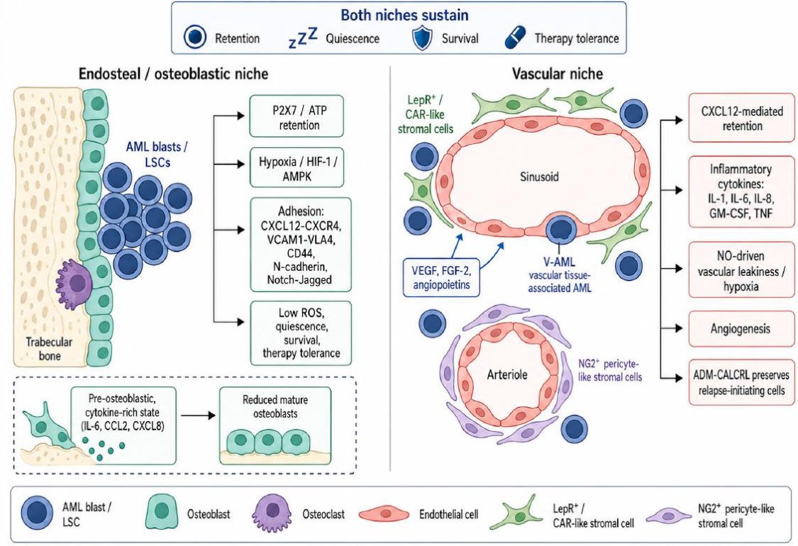
Figure 2.
Microenvironment-driven chemoresistance and therapeutic opportunities in AML. The LSC integrates four protective inputs from the marrow microenvironment: adhesion- and chemokine-mediated survival signaling, lipid transfer from adipocytes feeding fatty-acid oxidation and BCL-2-dependent quiescence, mitochondrial transfer through tunneling nanotubes from MSCs, and stromal pro-inflammatory cytokines that engage JAK–STAT to switch anti-apoptotic dependence from BCL-2 to BCL-xL. Exosome-mediated remodeling of normal hematopoiesis (miR-150, miR-155) and stromal MSC reprogramming (IL-8) reinforce a leukemia-permissive niche. The right column lists the principal therapeutic strategies under clinical evaluation: CXCR4 antagonists (plerixafor, motixafortide, ulocuplumab), the E-selectin antagonist uproleselan, JAK and IL-6 inhibitors (ruxolitinib, tocilizumab), metabolic targeting of FAO/OXPHOS, IDO1 inhibition, and the Hedgehog inhibitor glasdegib.
Figure 2.
Microenvironment-driven chemoresistance and therapeutic opportunities in AML. The LSC integrates four protective inputs from the marrow microenvironment: adhesion- and chemokine-mediated survival signaling, lipid transfer from adipocytes feeding fatty-acid oxidation and BCL-2-dependent quiescence, mitochondrial transfer through tunneling nanotubes from MSCs, and stromal pro-inflammatory cytokines that engage JAK–STAT to switch anti-apoptotic dependence from BCL-2 to BCL-xL. Exosome-mediated remodeling of normal hematopoiesis (miR-150, miR-155) and stromal MSC reprogramming (IL-8) reinforce a leukemia-permissive niche. The right column lists the principal therapeutic strategies under clinical evaluation: CXCR4 antagonists (plerixafor, motixafortide, ulocuplumab), the E-selectin antagonist uproleselan, JAK and IL-6 inhibitors (ruxolitinib, tocilizumab), metabolic targeting of FAO/OXPHOS, IDO1 inhibition, and the Hedgehog inhibitor glasdegib.
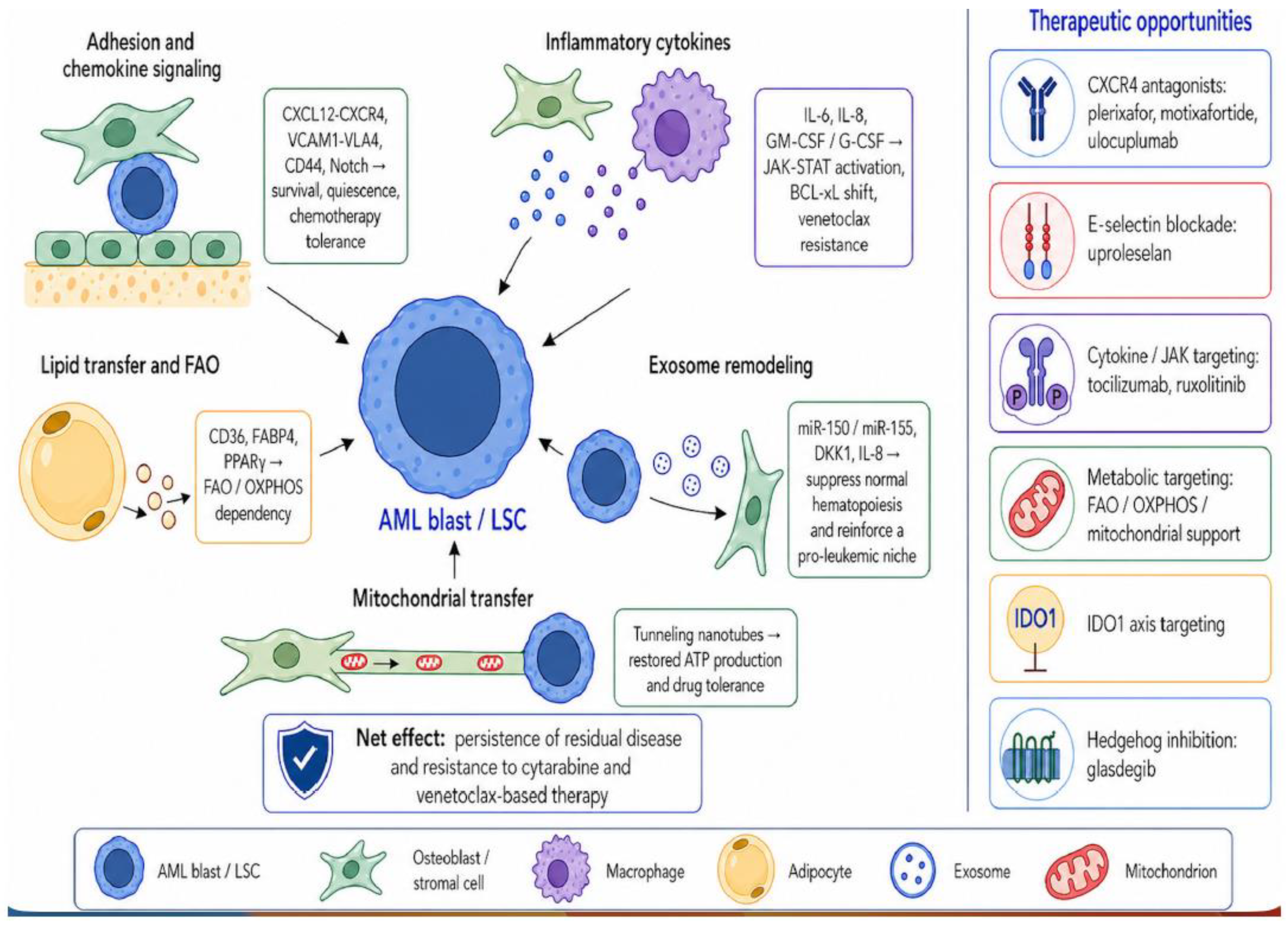
Disclaimer/Publisher’s Note: The statements, opinions and data contained in all publications are solely those of the individual author(s) and contributor(s) and not of MDPI and/or the editor(s). MDPI and/or the editor(s) disclaim responsibility for any injury to people or property resulting from any ideas, methods, instructions or products referred to in the content. |
© 2026 by the authors. Licensee MDPI, Basel, Switzerland. This article is an open access article distributed under the terms and conditions of the Creative Commons Attribution (CC BY) license (http://creativecommons.org/licenses/by/4.0/).
Copyright: This open access article is published under a Creative Commons CC BY 4.0 license, which permit the free download, distribution, and reuse, provided that the author and preprint are cited in any reuse.


