

Submitted:
29 May 2026
Posted:
01 June 2026
You are already at the latest version
Abstract
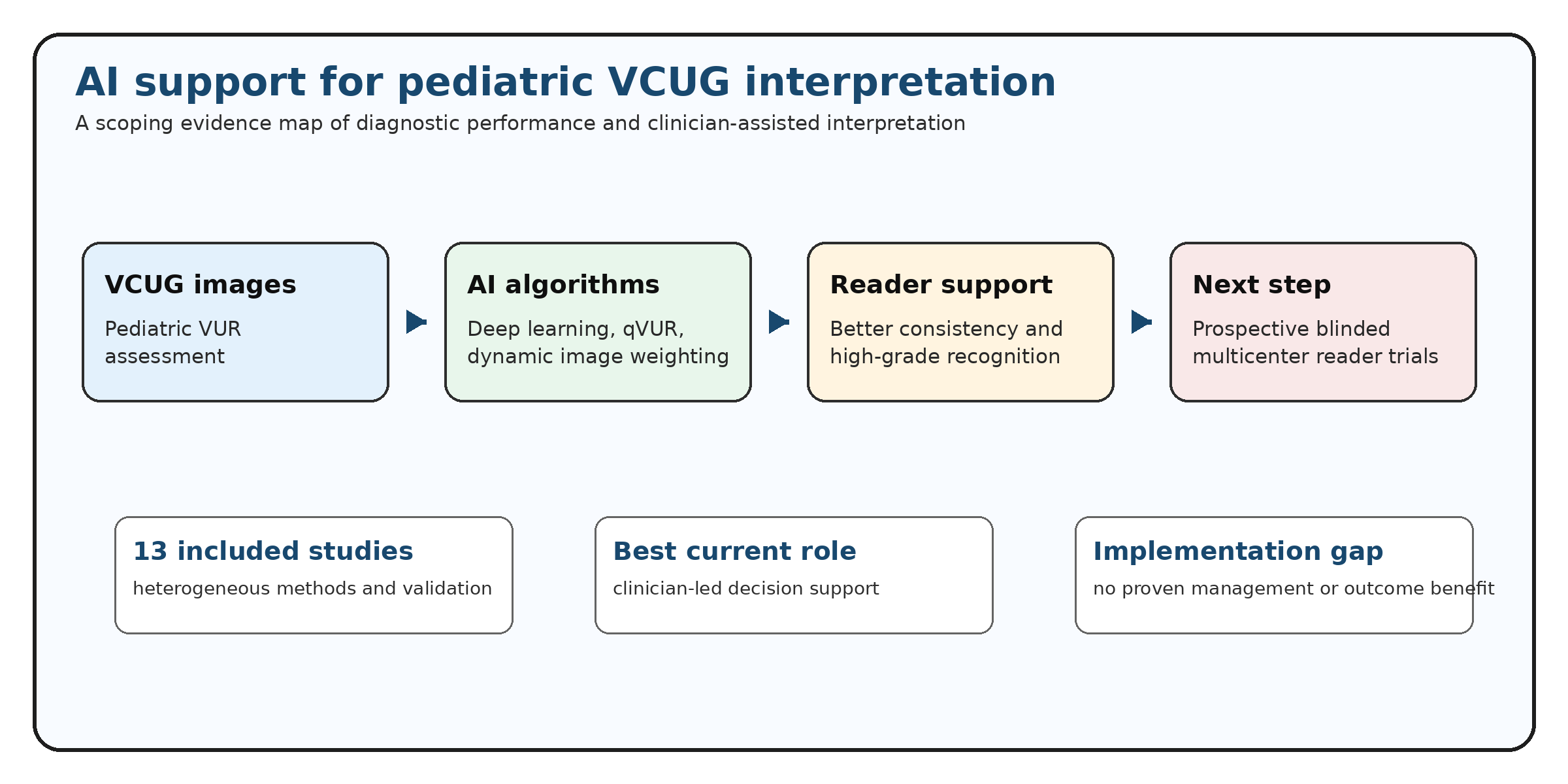
Background/Objectives: Vesicoureteral reflux (VUR) is an important pediatric urologic condition in which accurate grading on voiding cystourethrography (VCUG) may influence counseling, surveillance, antimicrobial prophylaxis, and surgical decision-making. Visual grading is partly subjective, particularly around the grade III-IV boundary. This review mapped the available evidence on AI-based pediatric VUR assessment on VCUG, with emphasis on diagnostic performance, clinician comparison, AI-assisted interpretation, and implementation gaps. Methods: A computer-assisted scoping evidence map was conducted using an exported screening dataset containing 500 records and an extraction dataset containing 13 included studies. Studies were classified as direct VCUG-based VUR grading, low/high-grade classification, AI-assisted clinician interpretation, or indirect clinical prediction. Because the exported files did not document independent duplicate screening, full-text verification, or database-specific deduplication, the synthesis was designed as an evidence map rather than a formal diagnostic meta-analysis. Results: Thirteen studies published between 2019 and 2025 were included. Reported AI performance was generally promising but heterogeneous. The strongest externally validated Deep-VCUG study reported external AUC values of 0.944 for unilateral reflux and 0.924 for bilateral reflux. A qVUR model achieved AUC 0.84 and improved grading reliability 3.6-fold. A multicenter VCUG-DAM study showed marked improvement in clinician AUC with AI support. Direct AI-versus-clinician evidence remained limited. Conclusions: AI may support pediatric VCUG interpretation by improving grading consistency and high-grade VUR recognition. However, current evidence does not establish autonomous superiority over radiologists. AI should currently be considered a decision-support tool rather than a replacement for expert clinician interpretation.
Keywords:
vesicoureteral reflux
; voiding cystourethrography
; artificial intelligence
; deep learning
; pediatric urology
; scoping review
1. Introduction
Vesicoureteral reflux (VUR) is characterized by retrograde urine flow from the bladder into the ureter and renal collecting system. Accurate diagnosis and grading are clinically relevant because reflux severity may influence surveillance, parental counseling, antimicrobial prophylaxis, endoscopic injection, ureteral reimplantation, and long-term renal risk assessment [1,2].
Voiding cystourethrography (VCUG) remains the conventional imaging method for diagnosing and grading VUR. The International Reflux Grading System classifies reflux from grade I to grade V according to reflux extension, ureteral dilatation, tortuosity, renal pelvic dilatation, and calyceal distortion [1]. Although widely used, this grading system depends on visual interpretation and may show clinically relevant variability between readers.
Reader variability is especially important around the grade III-IV boundary. For pediatric urologists, this distinction may affect counseling, follow-up intensity, prophylaxis discussions, and the threshold for intervention. Therefore, a tool that improves consistency at this threshold could be clinically useful even if it does not replace radiologist interpretation.
AI, machine learning, and deep learning have recently been applied to pediatric VCUG interpretation. These approaches include quantitative feature-based models, random forest classifiers, support vector machines, convolutional neural networks, ensemble learning systems, dual-stream architectures, and dynamic multi-image weighting systems. Reported performance is often promising, but the literature is heterogeneous in target task, imaging input, architecture, validation strategy, comparator design, and outcome reporting.
The key clinical question is therefore not simply whether an AI model can produce a high AUC in a selected dataset. A more clinically relevant question is whether AI can improve meaningful VCUG interpretation compared with clinicians, and whether the evidence is strong enough to support clinical implementation. This review aimed to map the available evidence, separate direct grading studies from indirect prediction studies, evaluate evidence quality, and clarify the current role of AI-assisted pediatric VUR interpretation.
2. Materials and Methods
2.1. Design and Reporting Framework
A scoping evidence synthesis design was selected because the literature is heterogeneous in population, imaging input, AI architecture, validation strategy, comparator design, target outcome, and performance reporting. The manuscript was organized according to PRISMA Extension for Scoping Reviews (PRISMA-ScR) principles [3]. Because the supplied dataset did not include formal database exports, independent duplicate screening records, or a full-text verification field, this article is best interpreted as a computer-assisted scoping evidence map and not as a completed conventional systematic review with meta-analysis.
2.2. Information Sources and Search Strategy
Two exported files generated with Elicit were used as the primary evidence source for the synthesis: a screening export containing 500 records and an extraction export containing detailed data for 13 included studies. The exact export date was not available in the supplied manuscript file. Elicit was used as an organizational support tool; the clinical interpretation, evidence categorization, and final conclusions were author verified. For transparency, the search strategy below is provided as a reproducible example for future formal database verification:
Example PubMed/MEDLINE search strategy: (“vesicoureteral reflux” OR “vesico-ureteral reflux” OR VUR) AND (“voiding cystourethrography” OR “voiding cystourethrogram” OR VCUG) AND (“artificial intelligence” OR “machine learning” OR “deep learning” OR “convolutional neural network” OR CNN OR “computer-aided diagnosis”) AND (child OR children OR pediatric OR paediatric OR infant).
For a future formal systematic or scoping review, database-specific exports from PubMed/MEDLINE, Embase, Scopus, Web of Science, and Google Scholar should be added, deduplicated, independently screened, and incorporated into an expanded PRISMA-ScR flow diagram.
2.3. Eligibility Criteria
- Included pediatric patients or pediatric imaging data.
- Evaluated AI, machine learning, deep learning, or computer-aided diagnostic approaches.
- Addressed VUR detection, VUR grading, low/high-grade classification, AI-assisted interpretation, or clinically related VUR prediction.
- Used VCUG or accepted VUR-related imaging or clinical data.
- Reported diagnostic performance, reliability, agreement, or clinician-comparison metrics.
- Used human pediatric datasets.
Studies were excluded if they were unrelated to pediatric VUR, lacked AI methodology, used non-human data only, were editorials, isolated case reports, or technical reports without clinically interpretable outcomes.
2.4. Study Selection and Data Charting
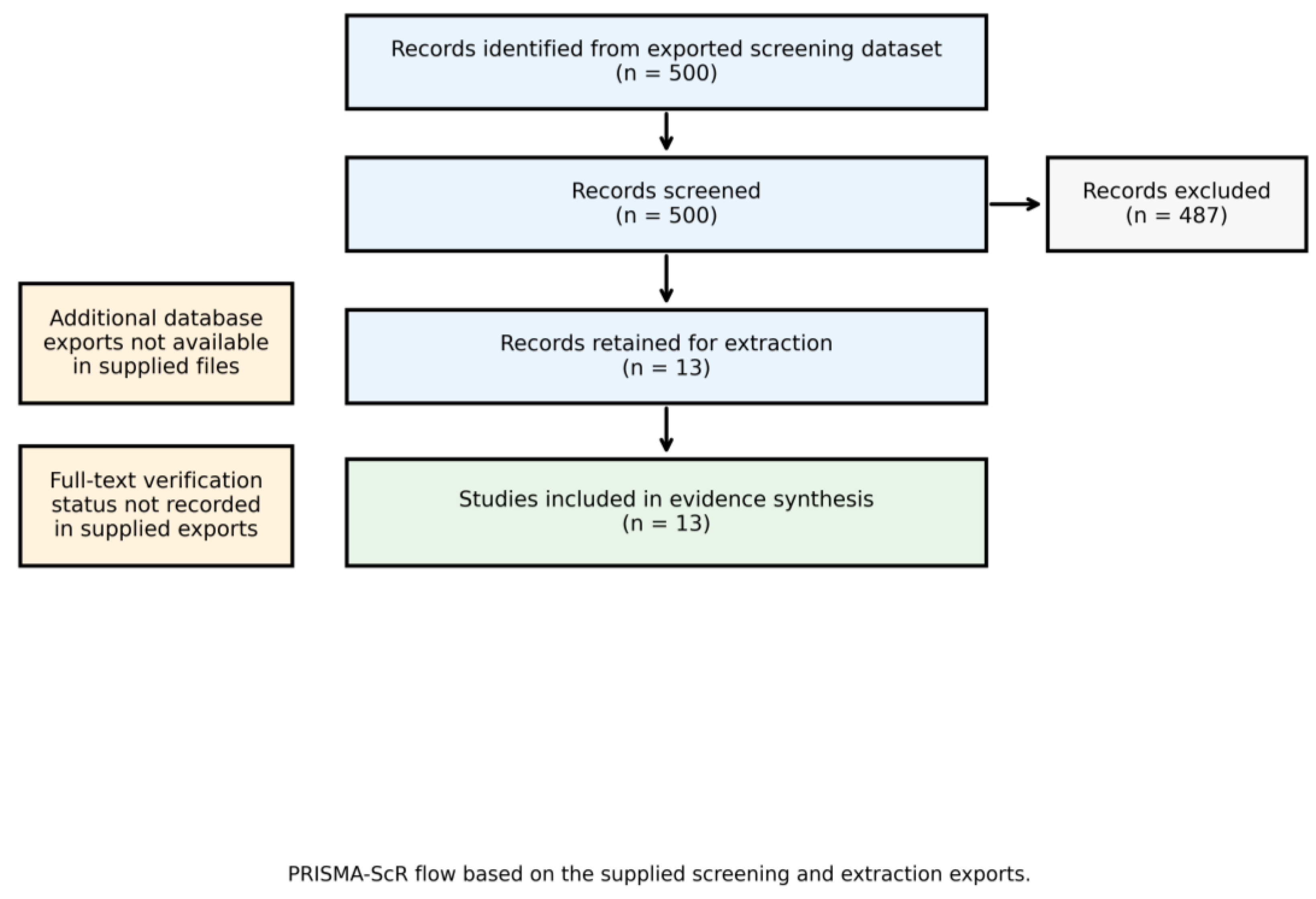
Study selection and data extraction were based on the supplied screening and extraction exports. The screening export classified 13 records as included and 487 as excluded. Extracted variables included title, authors, year, DOI, journal or conference venue, AI performance metrics, radiologist or clinician performance metrics, AI algorithm details, direct comparison results, study population, VUR grading system, and dataset or validation characteristics.
The supplied exports did not document independent dual-reviewer verification. This limitation is disclosed explicitly. Before conversion into a full systematic review, eligibility decisions and extracted data should be independently verified by at least two reviewers, with disagreements resolved by consensus or a third reviewer.
2.5. Evidence Classification and Confidence Mapping
To reduce methodological heterogeneity, studies were classified into four groups: direct VCUG grading studies, supportive low/high-grade classification studies, AI-assisted interpretation studies, and indirect clinical prediction studies. Direct grading and AI-assisted interpretation studies were prioritized in the narrative synthesis. Confidence was described qualitatively based on study type, sample size, validation strategy, comparator design, and reporting detail available in the extraction export.
2.6. Synthesis Approach
Because of heterogeneity across AI architectures, unit of analysis, target outcomes, validation designs, and performance metrics, a pooled diagnostic meta-analysis was not performed. Findings were summarized narratively and tabulated by clinical evidence role.
3. Results
3.1. Study Selection
The screening export contained 500 records. Thirteen studies were included and 487 records were excluded as not directly relevant or below the screening threshold. No additional database records were available in the supplied files.
Figure 1.
PRISMA-ScR study selection flow diagram. The supplied screening export identified 500 records; 13 studies were included and 487 records were excluded. The diagram should be expanded if formal database exports are added.
Figure 1.
PRISMA-ScR study selection flow diagram. The supplied screening export identified 500 records; 13 studies were included and 487 records were excluded. The diagram should be expanded if formal database exports are added.
3.2. Characteristics of Included Studies
The 13 included studies were published between 2019 and 2025. Dataset size ranged from small public or single-center image collections to multicenter datasets containing thousands of VCUG images. Some studies analyzed images, others renal units, and others patients. AI approaches included ensemble learning, hybrid convolutional neural networks, dual-stream CNN architectures, random forest models, support vector machines, multilayer perceptrons, gradient boosting, optimal classification trees, and dynamic multi-image weighting systems.

Table 1.
Included studies and evidence role based on the supplied extraction export.
| Study | Year | Venue | DOI | Evidence category | Confidence |
|---|---|---|---|---|---|
| Li et al. [4] | 2024 | eClinicalMedicine | 10.1016/j.eclinm.2024.102466 | Direct VCUG grading / AI-assisted interpretation | High/moderate |
| Khondker et al. [5] | 2022 | Journal of Urology | 10.1097/JU.0000000000002987 | Quantitative grading / reliability | High/moderate |
| Wu et al. [6] | 2025 | Research | 10.34133/research.0771 | AI-assisted interpretation | High/moderate |
| Khondker et al. [7] | 2022 | Journal of Pediatric Urology | 10.1016/j.jpurol.2021.10.009 | Quantitative grading | Moderate |
| Eroglu et al. [8] | 2021 | Computer Methods and Programs in Biomedicine | 10.1016/j.cmpb.2021.106369 | Direct VCUG grading | Moderate |
| Chen et al. [9] | 2025 | Journal of Imaging Informatics in Medicine | 10.1007/s10278-025-01438-1 | Direct VCUG grading | Moderate |
| Wang et al. [10] | 2024 | Journal of Pediatric Urology | 10.1016/j.jpurol.2023.11.003 | Indirect prediction | Low/moderate |
| Ergun et al. [11] | 2024 | Journal of Surgery and Medicine | 10.28982/josam.8020 | Direct VCUG grading | Low/moderate |
| Kabir et al. [12] | 2024 | Journal of Pediatric Urology | 10.1016/j.jpurol.2023.10.030 | Low/high-grade classification | Low/moderate |
| Alqaraleh et al. [13] | 2025 | Data and Metadata | 10.56294/dm2025756 | Classification study | Low/moderate |
| Estrada et al. [14] | 2019 | Journal of Urology | 10.1097/JU.0000000000000186 | Indirect clinical prediction | Moderate |
| Kose et al. [15] | 2020 | BioMed Research International | 10.1155/2020/1895076 | Indirect clinical prediction | Low/moderate |
| Wahed et al. [16] | 2024 | NETAPPS Conference | 10.1109/NETAPPS63333.2024.10823509 | Conference / limited report | Low |
3.3. AI Diagnostic Performance
AI algorithms generally demonstrated promising performance, but reported results varied according to study design, validation rigor, unit of analysis, and target task. The strongest externally validated evidence came from Li et al., whose Deep-VCUG model reported external AUC values of 0.944 for unilateral reflux and 0.924 for bilateral reflux [4]. Khondker et al. reported AUC 0.84 and demonstrated a 3.6-fold improvement in grading reliability compared with traditional assessment [5]. Wu et al. showed strong performance for side-specific reflux recognition and substantial improvement in clinician performance with AI support [6].
Smaller studies reported very high or near-perfect metrics, including training accuracy approaching 100% and AUC values near 1.0 [11,13,16]. These findings should be interpreted cautiously because of small sample size, limited external validation, and possible overfitting.
Table 2.
Clinically relevant AI performance findings.
| Study | Main finding | Clinical interpretation |
|---|---|---|
| Li 2024 [4] | External AUC 0.944 for unilateral VUR and 0.924 for bilateral VUR | Strong externally validated signal |
| Khondker 2022 [5] | AUC 0.84; reliability improved 3.6-fold | AI may improve grading consistency |
| Wu 2025 [6] | Clinician AUC markedly improved with AI assistance | Strong signal for clinician support |
| Eroglu 2021 [8] | Accuracy 96.9% | Promising, but validation details should be interpreted cautiously |
| Chen 2025 [9] | Patient-level accuracy 0.84; AUC 0.82 | Clinically plausible side-specific performance |
| Small/public datasets [11,13,16] | Near-perfect or limited-report metrics | Exploratory only; overfitting and reporting-bias risk |
3.4. Radiologist and Clinician Comparator Evidence
Radiologist-specific comparator evidence was limited. Most studies used radiologist labels as reference standards but did not independently quantify radiologist performance. Khondker et al. reported inter-rater reliability of 0.44 and median agreement of 0.71, supporting the concern that conventional VUR grading is variable [5]. Wu et al. reported baseline clinician AUC values before AI assistance and demonstrated substantial improvement after AI support [6].
Table 3.
AI-assisted clinician interpretation studies.
| Study | Baseline clinician performance | AI-assisted performance | Interpretation |
|---|---|---|---|
| Li 2024 [4] | Clinician comparison performed | Improved junior and senior clinician performance | AI may support readers across experience levels |
| Khondker 2022 [5] | Similar accuracy; lower reliability | Reliability improved 3.6-fold | AI may standardize grading |
| Wu 2025 [6] | Left VUR AUC 0.6288; right VUR AUC 0.7305 | Left VUR AUC 0.9641; right VUR AUC 0.9506 | Strongest signal for AI-assisted interpretation |
3.5. Direct Versus Indirect Evidence
Direct VCUG grading studies are the most clinically relevant for pediatric VUR interpretation [4,8,9,11]. Supportive low/high-grade or quantitative VUR studies are relevant for severity stratification but may not fully reproduce standard grade I-V reporting [5,7,12,13]. AI-assisted interpretation studies are the most relevant for real-world workflow because they evaluate whether AI improves clinician performance [4,6]. Indirect prediction studies, including models predicting dilating VUR, recurrent urinary tract infection risk, or likely benefit from VCUG, should not be interpreted as proof that AI can grade reflux severity on VCUG [10,14,15].
3.6. Clinical Management Implications
No included study definitively demonstrated that AI changes management decisions such as antibiotic prophylaxis, endoscopic injection, ureteral reimplantation, follow-up intensity, or renal outcome surveillance. Current evidence primarily demonstrates improvement in image classification, grading consistency, and clinician diagnostic performance. Therefore, AI should currently be interpreted as a decision-support tool rather than a management-changing intervention.
4. Evidence Quality Map
Table 4.
Evidence quality and implementation concerns.
| Domain | Main concern | Clinical impact |
|---|---|---|
| Search reproducibility | Screening and extraction exports were used; conventional database exports were not available | Limits claims of comprehensive systematic search |
| Full-text assessment | Full-text verification status was not available in the supplied export information | Requires manual verification before a formal systematic review |
| Study heterogeneity | Different target tasks, datasets, units of analysis, and outcomes | Prevents reliable meta-analysis |
| Comparator evidence | Limited direct AI-versus-radiologist or AI-versus-clinician studies | Restricts superiority claims |
| Validation quality | External validation was limited to selected studies | Real-world performance remains uncertain |
| Small datasets | Near-perfect metrics were reported in some small cohorts | Overfitting risk |
| Clinical management evidence | No outcome-based management studies | Limits implementation claims |
| Reporting quality | Incomplete sensitivity, calibration, confidence interval, and decision-curve reporting | Reduces comparability and implementation readiness |
5. Discussion
5.1. Principal Findings
This scoping evidence synthesis shows that AI algorithms have promising performance for pediatric VUR assessment on VCUG, but current evidence does not support autonomous replacement of radiologist interpretation. The most clinically relevant signal is not AI superiority over radiologists, but the possibility that AI may improve grading consistency and reduce interobserver variability.
This distinction is clinically important around the grade III-IV boundary, where grading differences can alter counseling, surveillance intensity, and discussion of intervention. AI may be most useful when it provides standardized support for image interpretation and reduces uncertainty in high-grade VUR recognition.
5.2. Interpretation in the Context of Current Evidence
The strongest evidence comes from multicenter or externally validated studies [4,6]. In contrast, near-perfect results from small datasets should be interpreted cautiously because of overfitting risk, limited case diversity, and insufficient external validation [11,13,16].
An important contribution of this review is the separation of direct VCUG grading studies from indirect clinical prediction models. Models predicting dilating VUR, recurrent urinary tract infection, or the probability of benefiting from VCUG may be clinically useful, but they should not be interpreted as proof that AI can accurately grade reflux severity on VCUG [10,14,15].
5.3. Clinical Implementation
The most realistic near-term role of AI in pediatric VUR imaging is decision support. AI may help standardize grading, support less experienced readers, improve communication between radiology and pediatric urology teams, and reduce uncertainty in high-grade VUR recognition. Final interpretation and management decisions should remain clinician-led.
For implementation, future systems should be tested in reader studies that compare AI-alone, clinician-alone, and AI-assisted clinician interpretation on the same locked external dataset. Studies should report not only AUC and accuracy, but also sensitivity for high-grade VUR, calibration, decision-curve analysis, confidence intervals, reporting time, error patterns, and performance across age, sex, unilateral/bilateral disease, and imaging quality subgroups.
5.4. Reporting Standards for Future AI studies
Future research should follow established AI and diagnostic accuracy reporting frameworks, including CLAIM, TRIPOD+AI, and STARD-AI, to improve reproducibility, transparency, and clinical applicability [17,18,19,20]. Predefined model locking, external validation, data governance, and clear human-AI interaction protocols are especially important before clinical deployment.
5.5. Limitations
This review has several limitations. First, although the supplied screening and extraction exports provide a reproducible basis for the current synthesis, formal database exports from PubMed/MEDLINE, Embase, Scopus, Web of Science, and Google Scholar were not available. Second, full-text verification status was not available in the supplied export information and should be completed manually before a formal systematic review. Third, the included studies were heterogeneous in AI architecture, target task, validation strategy, and comparator design. Fourth, only a small number of studies directly compared AI with clinician performance. Fifth, no study definitively showed that AI changes management decisions or improves patient outcomes.
5.6. Future Directions
Future studies should use prospective multicenter reader-based designs. The ideal study should include AI-alone, radiologist-alone, pediatric urologist-alone, and AI-assisted reader arms evaluated on the same locked external dataset. Future studies should also evaluate grade III-IV discordance, high-grade VUR sensitivity, calibration, decision-curve analysis, reporting time, PACS integration, management impact, and patient outcomes.
6. Conclusions
AI algorithms show promising diagnostic performance for pediatric VUR assessment on VCUG, particularly in externally validated and multicenter studies. However, current evidence does not prove autonomous superiority over radiologists. AI should currently be considered a decision-support tool rather than a replacement for expert clinician interpretation. Its most clinically relevant role may be improving grading consistency, supporting high-grade VUR recognition, and reducing uncertainty around the grade III-IV boundary. Prospective multicenter blinded reader studies are required before AI-assisted VCUG grading can be recommended for routine clinical implementation.
Supplementary Materials
The following supporting information can be downloaded at the website of this paper posted on Preprints.org: Supplementary File S1: PRISMA-ScR checklist; Supplementary File S2: screening export; Supplementary File S3: extraction export. If the CSV exports are not uploaded as supplementary files, the Data Availability Statement should be revised accordingly.
Author Contributions
Conceptualization, Y.A.B.; methodology, Y.A.B.; evidence synthesis, Y.A.B.; writing-original draft preparation, Y.A.B.; writing-review and editing, Y.A.B. The author has read and agreed to the published version of the manuscript.
Funding
This research received no external funding.
Institutional Review Board Statement
Not applicable. This article is an evidence synthesis of previously published studies and does not include new patient-level data.
Informed Consent Statement
Not applicable. This article does not include new patient-level data or identifiable patient information.
Data Availability Statement
The data summarized in this review were derived from the supplied screening and extraction exports and previously published studies. The exports should be submitted as supplementary files where journal policy and licensing permit; otherwise, they are available from the corresponding author upon reasonable request.
Acknowledgments
The author thanks all researchers whose work contributed to the evidence base summarized in this review.
Use of AI Tools
Elicit was used to support literature screening and extraction organization. Generative AI tools were used for language editing, formatting, and manuscript organization. The author verified the scientific interpretation, conclusions, and final submitted content.
Conflicts of Interest
The author declares no conflicts of interest.
References
- Lebowitz, R.L.; Olbing, H.; Parkkulainen, K.V.; Smellie, J.M.; Tamminen-Mobius, T.E. International system of radiographic grading of vesicoureteric reflux. Pediatr. Radiol. 1985, 15, 105–109. [CrossRef]
- Peters, C.A.; Skoog, S.J.; Arant, B.S.; Copp, H.L.; Elder, J.S.; Hudson, R.G.; Khoury, A.E.; Lorenzo, A.J.; Pohl, H.G.; Shapiro, E.; Snodgrass, W.T.; Diaz, M. Summary of the AUA guideline on management of primary vesicoureteral reflux in children. J. Urol. 2010, 184, 1134–1144. [CrossRef]
- Tricco, A.C.; Lillie, E.; Zarin, W.; O’Brien, K.K.; Colquhoun, H.; Levac, D.; Moher, D.; Peters, M.D.J.; Horsley, T.; Weeks, L.; et al. PRISMA Extension for Scoping Reviews (PRISMA-ScR): checklist and explanation. Ann. Intern. Med. 2018, 169, 467–473. [CrossRef]
- Li, Z.; Tan, Z.; Wang, Z.; Tang, W.; Ren, X.; Fu, J.; Wang, G.; Chu, H.; Chen, J.; Duan, Y.; et al. Development and multi-institutional validation of a deep learning model for grading of vesicoureteral reflux on voiding cystourethrogram: a retrospective multicenter study. EClinicalMedicine 2024, 69, 102466. [CrossRef]
- Khondker, A.; Kwong, J.C.C.; Yadav, P.; Chan, J.Y.H.; Singh, A.; Skreta, M.; et al. Multi-institutional validation of improved vesicoureteral reflux assessment with simple and machine learning approaches. J. Urol. 2022, 208, 1314–1322. [CrossRef]
- Wu, M.; Li, Z.; Liu, Y.; et al. Dynamic multi-image weighting for automated detection and diagnosis of abnormal urinary tract on voiding cystourethrography with a deep learning system: a retrospective, large-scale, multicenter study. Research 2025, 8, 0771. [CrossRef]
- Khondker, A.; Kwong, J.C.C.; Rickard, M.; Skreta, M.; Keefe, D.T.; Lorenzo, A.J.; Erdman, L. A machine learning-based approach for quantitative grading of vesicoureteral reflux from voiding cystourethrograms: methods and proof of concept. J. Pediatr. Urol. 2022, 18, e1–e78-78.e7. [CrossRef]
- Eroglu, Y.; Yildirim, K.; Cinar, A.; Yildirim, M. Diagnosis and grading of vesicoureteral reflux on voiding cystourethrography images in children using a deep hybrid model. Comput. Methods Programs Biomed. 2021, 210, 106369. [CrossRef]
- Chen, G.; Su, L.; Wang, S.; Liu, X.; Wu, W.; Zhang, F.; Zhao, Y.; Zhu, L.; Zhang, H.; Wang, X.; Yu, G. Automated grading of vesicoureteral reflux (VUR) using a dual-stream CNN model with deep supervision. J. Imaging Inform. Med. 2025, 38, 3517–3525. [CrossRef]
- Wang, H.-H.S.; Li, M.; Cahill, D.; Panagides, J.; Logvinenko, T.; Chow, J.; Nelson, C. A machine learning algorithm predicting risk of dilating VUR among infants with hydronephrosis using UTD classification. J. Pediatr. Urol. 2024, 20, 271–278. [CrossRef]
- Ergun, O.; Serel, T.A.; Ozturk, S.A.; Serel, H.B.; Soyupek, S.; Hoscan, B. Deep-learning-based diagnosis and grading of vesicoureteral reflux: a novel approach for improved clinical decision-making. J. Surg. Med. 2024, 8, 12–16. [CrossRef]
- Kabir, S.; Pippi Salle, J.L.; Chowdhury, M.E.H.; Abbas, T.O. Quantification of vesicoureteral reflux using machine learning. J. Pediatr. Urol. 2024, 20, 257–264. [CrossRef]
- Alqaraleh, M.; Alzboon, M.S.; Al-Batah, M.S.; Al Aesa, L.Y.; Abu-Arqoub, M.H.; Marie, R.R.; Alsmadi, F.H. Machine learning-based quantification of vesicoureteral reflux with enhancing accuracy and efficiency. Data Metadata 2025, 4, 756. [CrossRef]
- Estrada, C.R.; Nelson, C.P.; Wang, H.H.; Bertsimas, D.; Dunn, J.; Li, M.; Zhuo, D.; et al. Targeted workup after initial febrile urinary tract infection: using a novel machine learning model to identify children most likely to benefit from voiding cystourethrogram. J. Urol. 2019, 202, 144–152. [CrossRef]
- Kose, T.; Ozgur, S.; Cosgun, E.; Keskinoglu, A.; Keskinoglu, P. Effect of missing data imputation on deep learning prediction performance for vesicoureteral reflux and recurrent urinary tract infection clinical study. BioMed Res. Int. 2020, 2020, 1895076. [CrossRef]
- Wahed, M.A.; Alzboon, M.; Alqaraleh, M.; et al. Enhancing diagnostic precision in pediatric urology: machine learning models for automated grading of vesicoureteral reflux. In Proceedings of the 2024 7th International Conference on Internet Applications, Protocols, and Services (NETAPPS), 2024. [CrossRef]
- Mongan, J.; Moy, L.; Kahn, C.E., Jr. Checklist for Artificial Intelligence in Medical Imaging (CLAIM): a guide for authors and reviewers. Radiol. Artif. Intell. 2020, 2, e200029. [CrossRef]
- Tejani, A.S.; Klontzas, M.E.; Gatti, A.A.; et al. Checklist for Artificial Intelligence in Medical Imaging: CLAIM 2024 update. Radiol. Artif. Intell. 2024. [CrossRef]
- Collins, G.S.; Moons, K.G.M.; Dhiman, P.; Riley, R.D.; Beam, A.L.; Van Calster, B.; et al. TRIPOD+AI statement: updated guidance for reporting clinical prediction models that use regression or machine learning methods. BMJ 2024, 385, e078378. [CrossRef]
- Sounderajah, V.; Guni, A.; Liu, X.; et al. The STARD-AI reporting guideline for diagnostic accuracy studies using artificial intelligence. Nat. Med. 2025. [CrossRef]
Disclaimer/Publisher’s Note: The statements, opinions and data contained in all publications are solely those of the individual author(s) and contributor(s) and not of MDPI and/or the editor(s). MDPI and/or the editor(s) disclaim responsibility for any injury to people or property resulting from any ideas, methods, instructions or products referred to in the content. |
© 2026 by the authors. Licensee MDPI, Basel, Switzerland. This article is an open access article distributed under the terms and conditions of the Creative Commons Attribution (CC BY) license (http://creativecommons.org/licenses/by/4.0/).
Copyright: This open access article is published under a Creative Commons CC BY 4.0 license, which permit the free download, distribution, and reuse, provided that the author and preprint are cited in any reuse.