

Submitted:
23 May 2026
Posted:
25 May 2026
You are already at the latest version
Abstract
Background: Multiparameter flow cytometry is widely used in the diagnosis of acute leukemia, allowing rapid identification of leukemic cells based on their immunophenotype. The EuroFlow Acute Leukemia Orientation Tube (ALOT) was designed as a standardized screening tool to support early diagnostic orientation and guide further, more targeted testing. In this study, we assessed the diagnostic performance of the ALOT panel in pediatric patients with suspected acute leukemia. Methods: A total of 254 pediatric patients (0–18 years) with suspected acute leukemia were analyzed. Bone marrow samples were assessed using multiparameter flow cytometry with the EuroFlow ALOT panel, comprising eight markers (MPO, cyCD79a, CD34, CD19, CD3, cyCD3, CD7, and CD45). Final diagnoses were established using extended immunophenotypic panels and additional diagnostic methods when required. Diagnostic performance was assessed by calculating sensitivity, specificity, precision, accuracy, and negative predictive value. Results: Among 254 patients, 234 were diagnosed with hematologic disorders, while 20 had normal bone marrow findings. The ALOT panel correctly identified all pathological samples and did not misclassify any normal sample, resulting in 100% sensitivity, specificity, positive predictive value, negative predictive value, and accuracy for discrimination between abnormal and normal samples. In terms of exact diagnostic orientation, ALOT correctly classified 244 of 254 cases (96.1%) using a single-tube approach. The remaining 10 cases (3.9%), including rare entities such as Burkitt leukemia, chronic myeloid leukemia, and transient myeloproliferative syndrome, required extended immunophenotypic evaluation. Importantly, these cases were not false-negative results, as all were correctly identified as abnormal. Conclusions: The EuroFlow ALOT panel is a reliable screening tool for rapid diagnostic orientation in pediatric acute leukemia. Its implementation facilitates targeted selection of extended immunophenotypic panels, improving the efficiency and cost-effectiveness of diagnostic workflows.
Keywords:
acute leukemia
; flow cytometry
; immunophenotyping
; pediatric hematology
; screening
; acute leukemia orientation tube (ALOT)
; EuroFlow
; diagnostic algorithm
1. Introduction
Acute leukemias (AL), including acute lymphoblastic leukemia (ALL) and acute myeloid leukemia (AML), are the most common malignancies in the pediatric population, representing approximately 30% of all pediatric cancers [1]. Due to their aggressive clinical course, fast and accurate diagnosis is essential to enable the initiation of appropriate treatment [2]. Multiparameter flow cytometry is a key diagnostic tool in the diagnosis of ALL, allowing rapid immunophenotypic characterization of leukemic cells using specific surface and intracellular markers [3,4]. This method demonstrates high sensitivity and specificity, enabling accurate diagnosis and further classification of leukemias [4,5]. Compared to other diagnostic methods, flow cytometry offers wide availability and shorter turnaround time, which is crucial in rapidly progressing hematologic malignancies [6]. Molecular methods serve as an important complementary tool in pediatric leukemia diagnostics, allowing identification of specific genetic mutations and chromosomal abnormalities. Although these findings have significant prognostic value and guide therapeutic decisions, molecular analyses are often time-consuming. Therefore, screening approaches such as the Acute Leukemia Orientation Tube (ALOT) play a key role at the initial stage of diagnosis [7,8,9]. The ALOT panel, developed by the EuroFlow Consortium, is a standardized flow cytometric screening tool designed for the initial assessment of bone marrow or peripheral blood involvement in patients with suspected acute leukemia. It consists of a single-tube antibody combination targeting key markers expressed on leukemic cells of different lineages. The use of ALOT allows rapid narrowing of the diagnostic possibilities to the most common leukemia subtypes, thereby facilitating the selection of appropriate extended immunophenotypic panels and reducing both diagnostic time and cost. In this study, we evaluated the diagnostic performance of the ALOT panel in pediatric patients and assessed its usefulness in routine clinical practice.
2. Materials and Methods
2.1. Study Population
The study included 254 pediatric patients aged 0–18 years who were hospitalized in the Department of Pediatric Hematology and Oncology in Zabrze (Table 1) with suspected acute leukemia based on clinical symptoms (e.g., lymphadenopathy, weakness, fever) and preliminary laboratory findings (complete blood count and manual peripheral blood smear). All patients underwent bone marrow aspiration biopsy, and samples were analyzed using cytomorphological and flow cytometric methods, as well as additional diagnostic tests when necessary.
2.2. ALOT and Flow Cytometry
Immunophenotypic characterization of leukemic cells was performed using multiparameter flow cytometry at the Department of Immunology, Medical University of Silesia in Katowice. For initial diagnostic evaluation, all patients were analyzed using the single-tube ALOT panel. The antibody combination included the following markers: MPO-FITC, cyCD79a-PE, CD34-PerCP-Cy5.5, CD19-PE-Cy7, CD3-APC-H7, cyCD3-Pacific Blue (BD Horizon V450), CD7-APC, and CD45-BD Horizon V500-C. Samples were processed and analyzed according to standardized procedures validated by the EuroFlow Consortium [6,9].
2.3. Rationale for Marker Selection in the ALOT Panel
The ALOT panel was designed to enable rapid discrimination between the major acute leukemia lineages by combining markers specific for myeloid, B-lymphoid, and T-lymphoid differentiation, as well as markers of cellular immaturity. Myeloperoxidase (MPO) is a key marker of myeloid lineage and allows identification of acute myeloid leukemia. CD19 and cytoplasmic CD79a (cyCD79a) are specific for B-cell lineage and are essential for the diagnosis of B-cell precursor ALL. CD3 and cytoplasmic CD3 (cyCD3) are highly specific markers of T-cell lineage, enabling identification of T-ALL. CD7, although not entirely lineage-specific, is frequently expressed in T-cell leukemias and some myeloid leukemias, supporting lineage assignment. CD34 is a marker of hematopoietic progenitor cells and reflects cellular immaturity, which is characteristic of acute leukemias. CD45, a pan-leukocyte marker, is crucial for gating strategies and allows distinction between leukemic blasts and normal hematopoietic cells based on CD45 expression and side scatter properties.
2.4. Results Analysis
Based on leukemia-specific immunophenotyping, each patient was assigned to one of the following diagnostic categories:
1. B-cell precursor acute lymphoblastic leukemia (BCP-ALL)
2. T-cell acute lymphoblastic leukemia (T-ALL)
3. Acute myeloid leukemia (AML)
4. Burkitt leukemia/lymphoma (B-AL)
5. Chronic myeloid leukemia (CML)
6. Myelodysplastic syndrome with excess blasts (MDS-EB)
7. Transient myeloproliferative syndrome (TMS)
8. Normal bone marrow findings
Subsequently, all diagnoses were confirmed using multiparameter flow cytometry with extended immunophenotypic panels dedicated to specific leukemia subtypes. To evaluate the diagnostic performance of the ALOT screening test in acute leukemia, the following parameters were calculated: sensitivity, specificity, precision, accuracy, and negative predictive value. Sensitivity reflects the ability of the test to correctly identify patients with the disease (true positives), whereas specificity reflects the ability to correctly identify individuals without the disease (true negatives). Precision (positive predictive value) represents the proportion of true positive results among all positive results. Accuracy indicates the proportion of correctly classified cases among all evaluated cases. Negative predictive value represents the probability that patients with a negative test result truly do not have leukemia. The following formulas were applied:
1. Sensitivity = TP / (TP + FN)
2. Specificity = TN / (TN + FP)
3. Precision = TP / (TP + FP)
4. Accuracy = (TP + TN) / (TP + TN + FP + FN)
5. Negative predictive value (NPV) = TN / (TN + FN)
Where:
- TP (true positive) – number of correctly identified leukemia cases
- FN (false negative) – number of cases not identified as leukemia by ALOT but confirmed by further testing
- TN (true negative) – number of correctly identified non-leukemia cases
- FP (false positive) – number of cases incorrectly identified as leukemia
Because ALOT is designed as a screening and orientation tool rather than a fully comprehensive diagnostic assay, its performance was evaluated at two levels: (1) discrimination between abnormal and normal samples, and (2) exact diagnostic classification using a single-tube approach. Cases requiring additional immunophenotypic characterization were classified as indeterminate rather than false-negative results.
3. Results
3.1. Study Cohort
Among the 254 patients analyzed using the ALOT screening panel, a definitive diagnosis was established in 244 cases. In the remaining 10 cases, additional immunophenotypic panels were required; however, none of these patients were ultimately classified as healthy. Normal bone marrow findings were identified in 20 cases. Within the study cohort, 186 cases of acute lymphoblastic leukemia (ALL) were identified, including 162 cases of B-cell precursor ALL (BCP-ALL) and 24 cases of T-cell ALL (T-ALL). In addition, 33 cases of acute myeloid leukemia (AML) and 5 cases of myelodysplastic syndrome with excess blasts (MDS-EB) were diagnosed. Less frequent diagnoses included Burkitt leukemia (BL, n=5), chronic myeloid leukemia (CML, n=2), and transient myeloproliferative syndrome (TMS, n=3).
3.2. Diagnostic Performance of the ALOT Panel
The ALOT panel demonstrated excellent diagnostic performance as a screening and orientation tool. All cases of BCP-ALL, T-ALL, AML, and MDS were correctly identified at the initial stage, resulting in 100% sensitivity and specificity for these entities within the scope of diagnostic orientation (Table 2). In contrast, the ALOT panel did not provide sufficient immunophenotypic resolution for definitive classification of such hematologic disorders as Burkitt leukemia, chronic myeloid leukemia, and transient myeloproliferative syndrome, for which additional immunophenotypic staining was required in all cases. Importantly, these cases were not misclassified as normal, but rather recognized as abnormal and directed for further diagnostic work-up.
Overall, the ALOT panel enabled correct initial diagnostic orientation in the majority of cases and significantly reduced the need for extended diagnostic panels in common leukemia subtypes.
3.3. Global Diagnostic Parameters
When evaluated as a screening tool for discrimination between abnormal and normal samples, the ALOT panel achieved 100% sensitivity and 100% specificity, as no pathological sample was classified as normal and no normal sample was classified as pathological. Positive predictive value, negative predictive value, and overall accuracy were also 100%. In terms of exact diagnostic classification using a single-tube approach, ALOT correctly classified 244 of 254 cases (96.1%). The remaining 10 cases (3.9%) required extension of the immunophenotypic panel for final diagnosis. Importantly, none of these cases were misclassified as normal, and therefore they were considered indeterminate rather than false-negative results.
4. Discussion
Multiparameter flow cytometry is a key diagnostic tool in the evaluation of acute leukemias in both pediatric and adult patients [2,4]. It enables rapid and precise immunophenotypic characterization of leukemic cells, which is essential for accurate classification and prognostic stratification [10]. In addition to its diagnostic role, flow cytometry allows for monitoring of treatment response, including minimal residual disease (MRD), which is critical for long-term disease control [3,11,12]. Owing to its ability to simultaneously assess multiple cellular markers, it is particularly useful in distinguishing between different hematologic malignancies at an early stage of the diagnostic process [2]. An ideal screening test should enable early detection of disease, thereby facilitating prompt initiation of appropriate therapy. Such a test should be characterized by high sensitivity and specificity, as well as high positive and negative predictive values. In addition, it should be widely available, cost-effective, easy to perform, and reproducible across different laboratories. These criteria provide a useful framework for evaluating the clinical utility of screening tools in hematologic diagnostics. The rapid turnaround time and broad availability of flow cytometry support its use as an initial screening modality in suspected acute leukemia. The single-tube ALOT panel, developed by the EuroFlow Consortium and comprising a limited set of lineage-specific markers, appears to meet several criteria of an ideal screening test. The findings of the present study support its high diagnostic performance in the initial evaluation of pediatric acute leukemia. In particular, ALOT proved highly effective in the initial orientation of common leukemia subtypes, such as ALL and AML, enabling timely diagnostic orientation and facilitating early clinical decision-making. Previous EuroFlow-based studies reported high diagnostic accuracy of standardized screening panels, typically exceeding 90% for major leukemia subtypes [10,11]. In this context, the performance observed in our cohort is consistent with these findings. According to van Dongen et al. [9], the ALOT panel, as part of the EuroFlow standardization approach, plays a central role in rapid orientation of leukemia diagnostics and narrowing the differential diagnosis to specific subtypes [10,11]. In the present cohort, the ALOT panel achieved 100% sensitivity within the predefined diagnostic categories for BCP-ALL, T-ALL, AML, and MDS. These findings are in line with EuroFlow standardization studies, which also reported high sensitivity for these disease categories [11]. The absence of false-positive results in non-leukemic cases further underscores the high specificity of the method and supports its role as a reliable screening tool, consistent with data reported in the literature [10,11,13]. The ALOT panel includes markers selected to detect the most common leukemia subtypes; therefore, its diagnostic performance is inherently limited in rare entities. In our study, cases of Burkitt leukemia, CML, and transient myeloproliferative syndrome required additional immunophenotypic characterization beyond the initial ALOT panel. Importantly, however, the preliminary information obtained from ALOT enabled a targeted selection of extended diagnostic panels, thereby streamlining the diagnostic workflow and reducing unnecessary testing. An additional diagnostic challenge in flow cytometric evaluation of acute leukemias is represented by mixed phenotype acute leukemia (MPAL), which was not observed in the present cohort. MPAL is characterized by the co-expression of markers of more than one lineage, often requiring strict application of WHO classification criteria and extended immunophenotypic panels for accurate diagnosis. In such cases, limited screening panels, including ALOT, may not provide sufficient resolution for definitive classification. Nevertheless, the use of an initial orientation tube may still facilitate early recognition of aberrant immunophenotypes and guide the selection of appropriate extended diagnostic panels [7,9]. A major strength of the ALOT approach lies in its standardization. The panel is based on EuroFlow-validated protocols for sample preparation and data acquisition, which have been implemented in many leading centers worldwide. This ensures a high level of reproducibility and comparability of results across laboratories [10,11]. Furthermore, advanced analytical tools developed within the EuroFlow framework, such as principal component analysis (PCA) and database-guided automated gating (e.g., Compass), enhance data interpretation, reduce operator-dependent variability, and support rapid and objective classification of acute leukemias [10,11]. Taken together, the results of this study support the role of the ALOT panel as a highly effective screening tool for the rapid identification of common pediatric acute leukemias. Its implementation in routine clinical practice facilitates early initiation of therapy and improves the efficiency and cost-effectiveness of diagnostic workflows. In cases of rare or atypical hematologic malignancies, such as Burkitt leukemia or CML, ALOT should be complemented by extended flow cytometry panels and molecular analyses to achieve full diagnostic characterization and prognostic assessment [8,10,11]. Importantly, the interpretation of diagnostic performance metrics for ALOT requires consideration of its intended role as a screening and orientation tool. In this context, cases requiring additional immunophenotypic characterization should not be considered false-negative results, as the panel correctly identified them as abnormal and directed further diagnostic work-up. Therefore, classical binary performance metrics should be interpreted separately from the rate of exact one-tube classification. The use of ALOT as a first-line screening tool supports a stepwise, algorithm-based diagnostic approach. A proposed diagnostic algorithm incorporating ALOT as the initial step is presented in Figure 1.
Author Contributions
Conceptualization, J.B. and T.S.; methodology, J.B. and Ł.S.; formal analysis, J.B.; investigation, J.B., Ł.S., Ł.S. and B.P.; data curation, J.B.; writing—original draft preparation, J.B.; writing—review and editing, J.B., Ł.S., Ł.S., B.P. and T.S.; visualization, J.B.; supervision, T.S. All authors have read and agreed to the published version of the manuscript.
Funding
This research was funded by the Young Scientist Grant of the Medical University of Silesia, grant number PCN-2-090/N/0/N.
Institutional Review Board Statement
The study was conducted in accordance with the Declaration of Helsinki and approved by the Ethics Committee of the Medical University of Silesia, Katowice, Poland (protocol code KNW/0022/KB1/153/1/16/17; approval date: 3 October 2017).
Informed Consent Statement
Informed consent was obtained from all subjects involved in the study.
Data Availability Statement
The data presented in this study are available from the corresponding author upon reasonable request. The data are not publicly available due to privacy and ethical restrictions.
Conflicts of Interest
The authors declare no conflicts of interest.:
Abbreviations
The following abbreviations are used in this manuscript:
| ALOT | Acute Leukemia Orientation Tube |
| ALL | Acute Lymphoblastic Leukemia |
| BCP-ALL | B-cell Precursor Acute Lymphoblastic Leukemia |
| T-ALL | T-cell Acute Lymphoblastic Leukemia |
| AML | Acute Myeloid Leukemia |
| MDS | Myelodysplastic Syndrome |
| MDS-EB | Myelodysplastic Syndrome with Excess Blasts |
| BL | Burkitt Leukemia / Burkitt Lymphoma |
| B-AL | Burkitt Acute Leukemia |
| CML | Chronic Myeloid Leukemia |
| TMS | Transient Myeloproliferative Syndrome |
| MPAL | Mixed Phenotype Acute Leukemia |
| MRD | Minimal Residual Disease |
| PCA | Principal Component Analysis |
| TP | True Positive |
| TN | True Negative |
| FP | False Positive |
| FN | False Negative |
| NPV | Negative Predictive Value |
| PPV | Positive Predictive Value |
| CD | Cluster of Differentiation |
| WHO | World Health Organization |
References
- Pui, C.H.; Robison, L.L.; Look, A.T. Acute lymphoblastic leukaemia. Lancet 2008, 371, 1030–1043. [Google Scholar] [CrossRef] [PubMed]
- Orfao, A.; Ortuño, F.; de Santiago, M.; Lopez, A.; San Miguel, J. Immunophenotyping of acute leukemias and myelodysplastic syndromes. Cytom. A 2004, 58, 62–71. [Google Scholar] [CrossRef] [PubMed]
- Weir, E.G.; Borowitz, M.J. Flow cytometry in the diagnosis of acute leukemia. Semin Hematol. 2001, 38, 124–138. [Google Scholar] [CrossRef] [PubMed]
- Béné, M.C.; Nebe, T.; Bettelheim, P.; Buldini, B.; Bumbea, H.; Kern, W.; Lacombe, F.; Lemez, P.; Marinov, I.; Matutes, E.; Maynadié, M.; Oelschlagel, U.; Orfao, A.; Schabath, R.; Solenthaler, M.; Tschurtschenthaler, G.; Vladareanu, A.M.; Zini, G.; Faure, G.C.; Porwit, A. Immunophenotyping of acute leukemia and lymphoproliferative disorders: a consensus proposal of the European LeukemiaNet Work Package 10. Leukemia 2011, 25, 567–574. [Google Scholar] [CrossRef] [PubMed]
- Wood, B.L. Flow cytometry in the diagnosis and monitoring of acute leukemia. J. Hematop. 2015, 8, 191–199. [Google Scholar] [CrossRef]
- Kalina, T.; Flores-Montero, J.; van der Velden, V.H.; Martin-Ayuso, M.; Böttcher, S.; Ritgen, M.; Almeida, J.; Lhermitte, L.; Asnafi, V.; Mendonça, A.; de Tute, R.; Cullen, M.; Sedek, L.; Vidriales, M.B.; Pérez, J.J.; te Marvelde, J.G.; Mejstrikova, E.; Hrusak, O.; Szczepański, T.; van Dongen, J.J.; Orfao, A. EuroFlow standardization of flow cytometer instrument settings and immunophenotyping protocols. Leukemia 2012, 26, 1986–2010. [Google Scholar] [CrossRef] [PubMed]
- Swerdlow, S.H.; Campo, E.; Pileri, S.A.; Harris, N.L.; Stein, H.; Siebert, R.; Advani, R.; Ghielmini, M.; Salles, G.A.; Zelenetz, A.D.; Jaffe, E.S. The 2016 revision of the World Health Organization classification of lymphoid neoplasms. Blood 2016, 127, 2375–2390. [Google Scholar] [CrossRef] [PubMed]
- King, R.L.; Naghashpour, M.; Watt, C.D.; Morrissette, J.J.; Bagg, A. A comparative analysis of molecular genetic and conventional cytogenetic detection of diagnostically important translocations in more than 400 cases of acute leukemia. Am. J. Clin. Pathol. 2011, 135, 921–928. [Google Scholar] [CrossRef] [PubMed]
- van Dongen, J.J.M.; Lhermitte, L.; Böttcher, S.; Almeida, J.; van der Velden, V.H.J.; Flores-Montero, J.; Rawstron, A.; Asnafi, V.; Lécrevisse, Q.; Lucio, P.; Mejstrikova, E.; Szczepański, T.; Kalina, T.; de Tute, R.; Brüggemann, M.; Sedek, L.; Cullen, M.; Langerak, A.W.; Mendonça, A.; Macintyre, E.; Martin-Ayuso, M.; Hrusak, O.; Vidriales, M.B.; Orfao, A. EuroFlow antibody panels for standardized n-dimensional flow cytometric immunophenotyping of normal, reactive and malignant leukocytes. Leukemia 2012, 26, 1908–1975. [Google Scholar] [CrossRef] [PubMed]
- Lhermitte, L.; Mejstrikova, E.; van der Sluijs-Gelling, A.J.; Grigore, G.E.; Sedek, L.; Bras, A.E.; Gaipa, G.; Sobral da Costa, E.; Novakova, M.; Sonneveld, E.; Buracchi, C.; de Sá Bacelar, T.; te Marvelde, J.G.; Trinquand, A.; Asnafi, V.; Szczepański, T.; Matarraz, S.; Lopez, A.; Vidriales, B.; Bulsa, J.; Hrusak, O.; Kalina, T.; Lecrevisse, Q.; Martin-Ayuso, M.; Brüggemann, M.; Verde, J.; Fernandez, P.; Burgos, L.; Paiva, B.; Pedreira, C.E.; van Dongen, J.J.M.; Orfao, A.; van der Velden, V.H.J. Automated database-guided expert-supervised orientation for immunophenotypic diagnosis and classification of acute leukemia. Leukemia 2018, 32, 874–881. [Google Scholar] [CrossRef] [PubMed]
- Lhermitte, L.; Barreau, S.; Morf, D.; Fernandez, P.; Grigore, G.; Barrena, S.; de Bie, M.; Flores-Montero, J.; Brüggemann, M.; Mejstrikova, E.; Nierkens, S.; Burgos, L.; Caetano, J.; Gaipa, G.; Buracchi, C.; da Costa, E.S.; Sedek, L.; Szczepański, T.; Aanei, C.M.; van der Sluijs-Gelling, A.; Delgado, A.H.; Fluxa, R.; Lecrevisse, Q.; Pedreira, C.E.; van Dongen, J.J.M.; Orfao, A.; van der Velden, V.H.J. Automated identification of leukocyte subsets improves standardization of database-guided expert-supervised diagnostic orientation in acute leukemia: a EuroFlow study. Mod. Pathol. 2021, 34, 59–69. [Google Scholar] [CrossRef] [PubMed]
- Campana, D. Minimal residual disease in acute lymphoblastic leukemia. Hematol. Am. Soc. Hematol. Educ. Program 2010, 2010, 7–12. [Google Scholar] [CrossRef] [PubMed]
- Cheng, F.M.; Lo, S.C.; Lin, C.C.; Lo, W.J.; Chien, S.Y.; Sun, T.H.; Hsu, K.C. Deep learning assists in acute leukemia detection and cell classification via flow cytometry using the acute leukemia orientation tube. Sci. Rep. 2024, 14, 8350. [Google Scholar] [CrossRef] [PubMed]
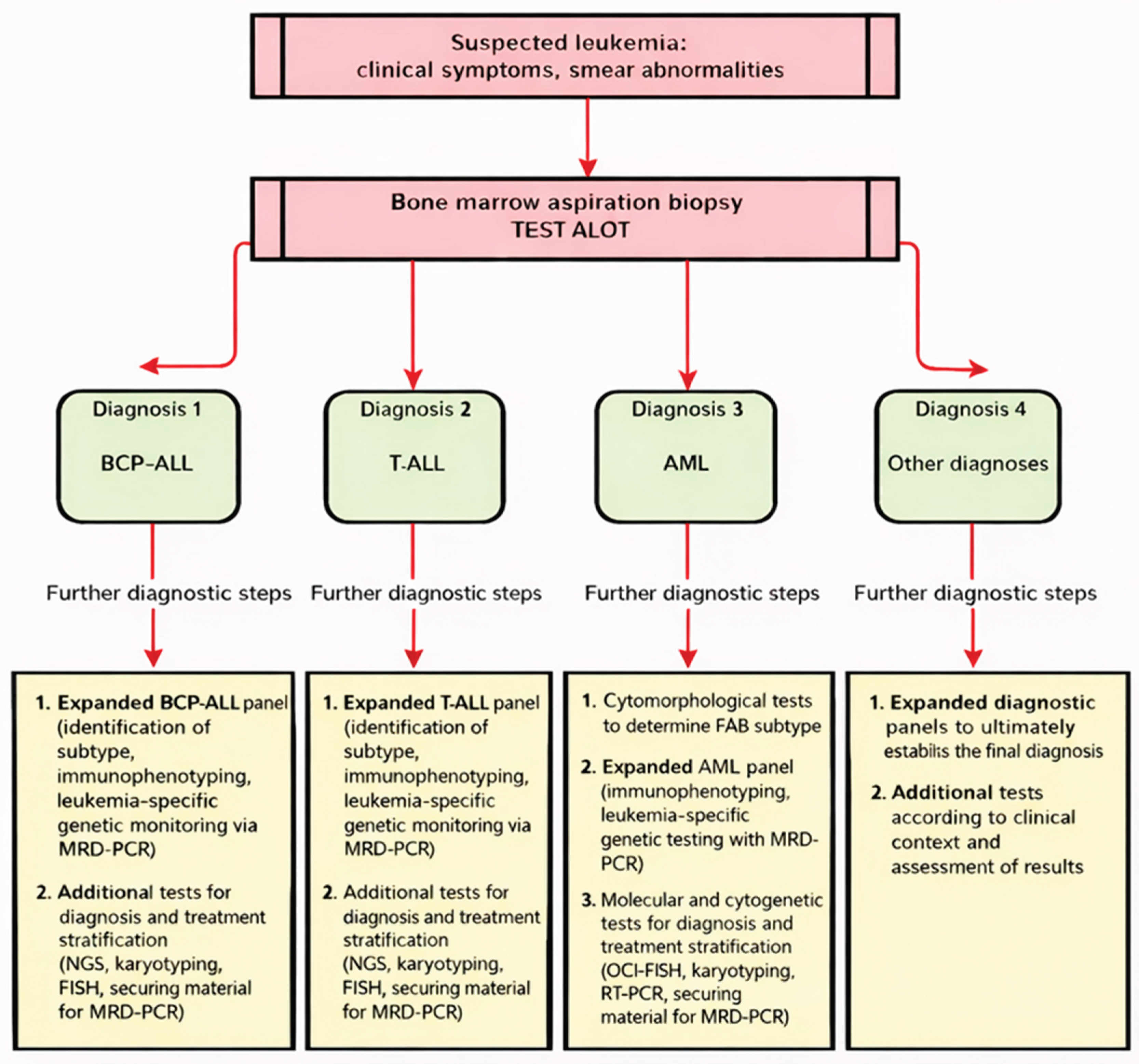

Table 1.
Characteristics of the study population.
| Leukemia type (WHO classification) | Number of cases | Females | Males | Age range (years) |
| BCP-ALL | 162 | 82 | 80 | 0–18 |
| T-ALL | 24 | 6 | 18 | 1–18 |
| AML | 33 | 13 | 20 | 0–18 |
| MDS | 5 | 1 | 4 | 7–10 |
| BL | 5 | 1 | 4 | 8–13 |
| CML | 2 | 1 | 1 | 16–19 |
| TMS | 3 | 0 | 3 | 0–1/12 |
| Normal bone marrow |
20 | 6 | 14 | 0-18 |
Table 2.
Performance of the ALOT panel in the orientation of hematologic disorders.
| Diagnosis | n | Correctly oriented | Additional testing | Orientation rate (%) |
| BCP-ALL | 162 | 162 | 0 | 100% |
| T-ALL | 24 | 24 | 0 | 100% |
| AML | 33 | 33 | 0 | 100% |
| MDS | 5 | 5 | 0 | 100% |
| BL | 5 | 0 | 5 | 0% |
| CML | 2 | 0 | 2 | 0% |
| TMS | 3 | 0 | 3 | 0% |
| Normal bone marrow |
20 | 20 | 0 | 100% |
| All cases | 254 | 244 | 10 | 96.1% |
Disclaimer/Publisher’s Note: The statements, opinions and data contained in all publications are solely those of the individual author(s) and contributor(s) and not of MDPI and/or the editor(s). MDPI and/or the editor(s) disclaim responsibility for any injury to people or property resulting from any ideas, methods, instructions or products referred to in the content. |
© 2026 by the authors. Licensee MDPI, Basel, Switzerland. This article is an open access article distributed under the terms and conditions of the Creative Commons Attribution (CC BY) license (http://creativecommons.org/licenses/by/4.0/).
Copyright: This open access article is published under a Creative Commons CC BY 4.0 license, which permit the free download, distribution, and reuse, provided that the author and preprint are cited in any reuse.