

Submitted:
18 May 2026
Posted:
19 May 2026
You are already at the latest version
Abstract
Objective: Thyroid cartilage calcification and glucose metabolism may vary with age and gender. This study aimed to investigate the role of [18F]-sodium fluoride ([18F]NaF) and 2-[18F]fluoro-2-deoxy-D-glucose ([18F]FDG) PET/CT for the evaluation of physiological molecular calcification and glucose metabolism of thyroid cartilage with age. Methods: This retrospective study analyzed [18F]NaF and [18F]FDG PET/CT images from the CAMONA study (NCT01724749). Regions of interest were placed around the thyroid cartilage on CT images using OsiriX software. The mean standardized uptake value (SUVmean) was measured for [18F]NaF and [18F]FDG PET/CT images. Pearson correlation coefficients were calculated to evaluate the effects of aging on the uptake of [18F]NaF and [18F]FDG in the thyroid cartilage. Results: A total of 127 healthy subjects (65 females and 62 males) with a mean age of 48.46±14.13 (range 21–75) years for [18F]NaF PET/CT and a total of 114 healthy subjects (52 females and 63 males) with a mean age of 49.05±14.29 (range 21–75) years for [18F]FDG PET/CT were included. A significant positive correlation was observed between age and SUVmean of [18F]NaF in the thyroid cartilage (r=0.18, p=0.04). This result indicates that molecular calcification of this cartilage increases with aging. However, the correlation between age and SUVmean of [18F]FDG in the thyroid cartilage was not statistically significant (r=-0.09 p=0.31). Conclusion: This study presents a novel methodology for the determination of molecular calcification and glucose metabolism of laryngeal cartilage using [18F]NaF and [18F]FDG PET/CT. Molecular calcification of thyroid cartilage was found to increase with age, whereas glucose metabolism did not show a statistically significant correlation with age.
Keywords:
calcification
; glucose metabolism
; positron emission tomography
; thyroid cartilage
; [18F]NaF
; [18F]FDG
1. Introduction
The thyroid cartilage (TC) is the largest among the nine cartilages in the larynx and is composed of two plates that meet in the center. It also has superior and inferior horns, which are key landmarks for airway and laryngeal procedures (Glikson et al., 2017). The progressive process of calcification in the thyroid cartilage, beginning in adolescence and increasing with age, is fully developed in individuals over 70 years (Schultz, 2015; Wenaas et al., 2016).
Positron emission tomography/computed tomography (PET/CT) with [18F]-sodium fluoride ([18F]NaF) can measure both blood flow and osteoblast activity in bone and soft tissue (Park et al., 2021). [18F]NaF is well-known for its bone-seeking properties, with high skeletal uptake and quick blood clearance from the circulation following intravenous infusion (Raynor et al., 2016; Bin Saeedan et al., 2016). Extraosseous tissues have also shown [18F]NaF uptake in previous studies (Seraj et al., 2019; Al-Zaghal et al., 2018c). Such accumulation of [18F]NaF has been documented in several settings as a sign of soft tissue calcification (Al-Zaghal et al., 2018b; Asmar et al., 2017; Shao et al., 2016). Physiological calcification tends to build up in tissues such as the choroid plexus, pineal gland, and habenula over a person’s lifetime (Al-Zaghal et al., 2018a; Blomberg et al., 2017; Murad et al., 2022). Among the respiratory tract organs, a case report described tracheobronchial tree calcifications as detected on [18F]-NaF PET/CT in a 75-year-old woman (Al-Zaghal et al., 2018c). However, no attempts have been made to study the calcification of thyroid cartilage using [18F]NaF PET/CT. Explaining the link between [18F]NaF uptake and age in thyroid cartilage will require understanding its underlying physiology.
2-[18F]fluoro-2-deoxy-D-glucose ([18F]FDG) is a radioactive tracer that is becoming increasingly useful in the diagnosis and monitoring of inflammatory diseases, as well as in determining the presence, stage, and biologic behavior of tumors (Kung et al., 2019; Hustinx et al., 1999). However, the uptake of [18F]FDG is an indicator of glucose metabolism not only in inflammatory and cancerous tissues but also in healthy tissues such as the lung, myocardium, liver, spleen, bone marrow, and cartilage (Raynor et al., 2016). Several studies have reported that [18F]FDG uptake imaged by PET/CT in healthy tissues before or during treatment is linked to different factors, including possible treatment-related adverse events (Malaih et al., 2022; Kostakoglu et al., 2004).
The process of aging and the presence of degenerative diseases have a significant impact on cartilage within the human body (Sokoloff, 1983). This distinction pertains to the differentiation between the degenerative processes associated with the natural aging of adult cartilage and the alterations that transpire during skeletal growth. The phenomenon of age-related degradation in cartilage, such as those found in the costal, nasal septal, laryngeal, and tracheobronchial regions, has been documented in diverse mammalian species (Sokoloff, 1983). The pathogenesis of degenerative joint disease proposes that articular cartilage experiences senescent alterations akin to those observed in extra-articular cartilage. The process of localized calcification and endochondral ossification in costal cartilage with advancing age has been documented. Fibrillation, which is distinguished by the presence of minute irregularities on the surface of cartilage, serves as an initial indication of cartilage deterioration in the context of aging and degenerative processes (Sokoloff, 1983).
While significant research has been performed on the use of various anatomical locations for determining an individual’s age, such as in rib cartilage, which becomes more calcified with age, there is relatively little literature on the use of the thyroid cartilage for age estimation (Park et al., 2022; Deng et al., 2015; Shehu et al., 2022). The thyroid cartilage gradually calcifies as individuals age (Jadav et al., 2022). Previous studies on age estimation from laryngeal cartilage calcification have reported contradictory results, with some finding a significant correlation between age and laryngeal cartilage calcification and others finding it to be an unreliable method (Ross et al., 2001). Further research on different population groups is needed to better understand laryngeal cartilage calcification in relation to age.
This study aims to achieve two objectives: 1) to develop a method for measuring the molecular calcification and glucose metabolism of thyroid cartilage and 2) to examine the correlation between aging and the molecular calcification and glucose metabolism of these cartilage.
2. Materials and Methods
2.1. Subjects
This retrospective cross-sectional study examined a total of 139 healthy subjects from the study known as “Cardiovascular Molecular Calcification Assessed by [18F]NaF PET/CT (CAMONA) (NCT01724749). The CAMONA study recruited subjects from 2012 to 2016. The study was approved by the Danish National Committee on Health Research Ethics. All subjects gave written consent, and the Declaration of Helsinki was met. To enroll healthy patients in the CAMONA study, the exclusion criteria were the presence of malignancy, immunodeficiency syndromes, autoimmune disease, alcohol abuse, any symptoms suggesting cardiovascular disease, illicit drug use, pregnancy, or any prescription medication. Due to technical issues (incomplete data sets and insufficient image quality), 12 subjects from the [18F]NaF group and 25 subjects from the [18F]FDG were excluded for further investigations.
2.2. PET/CT Imaging
All participants underwent whole-body [18F]NaF and [18F]FDG PET/CT acquisition on two different days within two weeks. In brief, [18F]-NaF PET/CT imaging was performed 90 minutes after the injection of 2.2 MBq/kg of [18F]NaF with 2.5 min/bed. After fasting overnight for at least 8 hours and confirming serum glucose concentration < 8 mmol/L, [18F]FDG PET/CT imaging was performed 180 minutes after the injection of 4 MBq/kg of [18F]FDG with 3.5 min/bed. The following scanners with compatible full-body spatial resolutions were used for image acquisition: GE Discovery RX, STE, and 690/710 imaging systems (General Electrical Healthcare, Milwaukee, WI, USA). PET was adjusted for attenuation, scatter, scanner dead time, and random coincidences. For attenuation correction and anatomical alignment, low-dose CT imaging (140 kVp, 30–110mA, noise index 25, 0.8 seconds per rotation, slice thickness 3.75 mm) was performed.
2.3. Image Processing
A trained physician, who was blinded to the subjects’ demographics and clinical history, manually placed regions of interest (ROI) around the thyroid cartilage in each axial slice using a DICOM viewer (OsiriX MD Soft- ware; Pixmeo SARL, Bernex, Switzerland) (Figure 1). The mean standardized uptake value (SUVmean) was calculated from the ROI in each axial slice. Each subject’s SUVmean was given as the average value of all voxels measured within all ROIs placed on thyroid cartilage combined.
2.4. Data Analysis
Pearson correlation coefficient was calculated to test for association between SUVmean and age for both [18F]NaF and [18F]FDG PET. A p-value less than 0.05 was considered statistically significant. For visualization, scatterplots and exploratory univariable linear regressions were used.
3. Results
A total of 127 subjects (65 females and 62 males) with a mean age of 48.46 ± 14.13 years for [18F]-NaF PET/CT and a total of 114 healthy subjects (52 females and 62 males) with a mean age of 49.05 ± 14.29 (range 21–75) years for [18F]FDG PET/CT were identified. Physiological molecular calcification and glucose metabolism of thyroid cartilage were measured with [18F]NaF and [18F]FDG PET/CT, respectively. The study showed that age was positively correlated with SUVmean of [18F]NaF in the thyroid cartilage in a mixed-gender group (r = 0.18, p = 0.04) and that the correlation was not significant with [18F]FDG uptake in a mixed-gender group (r = -0.09, p = 0.31).
When the subjects were divided based on gender, a strong positive correlation was observed in females (r = 0.28, p = 0.023) for the [18F]NaF uptake. Although a positive trend was observed in the male group, the correlation was not significant (r = 0.13, p = 0.32) (Table 1) (Figure 2). For the [18F]FDG uptake, a strong negative correlation was observed in males (r = -0.31, p = 0.01), but the correlation was not significant in females (r = 0.07, p = 0.63) (Table 2) (Figure 3).
4. Discussion
Aging involves functional processes and anatomical changes that do not represent disease in elderly asymptomatic patients, including cartilage calcification. Aging is associated with a wide range of metabolic changes, including alterations in glucose metabolism and metabolic rate. The current study presents a novel methodology for the non-invasive quantification of molecular calcification and glucose metabolism of laryngeal cartilage using [18F]NaF and [18F]FDG PET/CT. To our knowledge, this is the first study to assess the effects of aging on the uptake of [18F]NaF and [18F]FDG in the thyroid cartilage. Several studies have discussed changes in calcification and glucose metabolism in thyroid cartilage based on CT and PET (Naimo et al., 2013; Ouellet et al., 2011; Glikson et al., 2017; Schultz, 2015); however, none of them have used PET/CT imaging.
In the current study, we found a significant positive correlation between age and SUVmean of [18F]NaF uptake in the thyroid cartilage in a mixed-gender group, indicating an increase in molecular calcification of this cartilage with aging. Moreover, [18F]NaF uptake in our study demonstrated a high positive correlation in females. Although the male group showed an upward tendency, the correlation was non-significant. However, [18F]FDG metabolism of this cartilage did not show a statistically significant correlation with age.
The study’s findings are significant as they suggest that [18F]NaF PET/CT imaging can be used to measure molecular calcification in the thyroid cartilage. Calcification of the thyroid cartilage is a process that occurs as a part of normal aging, where the cartilage undergoes mineralization and hardening (Mupparapu and Vuppalapati, 2005). Our study demonstrated a strong positive correlation between age and [18F]NaF uptake in females. Although the male group demonstrated a positive trend, the correlation was non-significant, and the underlying reasons are not clear. Available findings in the literature suggest that there may be differences in the calcification of the thyroid cartilage in females and males. Studies have investigated these potential differences with varying results. One study has found that there is a lower prevalence and degree of calcification in females compared to males (Mupparapu and Vuppalapati, 2005), while another has found no significant difference between the two genders (Schultz, 2015). One possible explanation for these discrepancies may be differences in the age and health status of the study participants (Schultz, 2015). For example, some studies may have included older individuals with more advanced stages of calcification, which could skew the results towards one gender. Similarly, there could be hormonal factors (for example, effects of sex hormones) impacting the degree of cartilage calcification among females and males (Claassen et al., 2011). The reaction of cartilage to estrogen administration has been studied (Zondek, 1936). Although researchers have identified sex hormone receptors for 17β-estradiol, progesterone, and testosterone in human chondrocytes, as well as in immortalized chondrocyte cell lines, there is still limited understanding of how sex hormones affect cartilage metabolism (Claassen et al., 2011). Understanding the exact mechanism by which hormonal factors may affect the calcification of thyroid cartilage will require further investigation.
Shehu et al. examined the normal molecular calcification of the costal cartilage in relation to age and sex using [18F]NaF-PET/CT in the CAMONA subjects, similar to our study (Shehu et al., 2022). Both sexes had an age-related increase in calcification using [18F]NaF-PET/CT measures, but only men had a substantial age-related increase in calcification when they performed CT measures (Shehu et al., 2022). This highlights that although the calcification of cartilage is age-related, there is a possibility that the calcification pattern of cartilage in females and males may vary depending on the type/location of cartilage studied. For example, costal cartilage, which is probably more prone to damage and calcification in males, may have resulted in this finding.
Overall, while there is some evidence to suggest that there may be differences in the calcification of the thyroid cartilage between females and males, the exact nature of these differences remains unclear. Further research is needed to better understand the underlying mechanisms and potential implications of these gender differences.
It is worth noting that the [18F]FDG metabolism of the thyroid cartilage did not exhibit a statistically significant correlation with age in the mixed-gender group. After separating the subjects into groups based on gender, a significant negative correlation for [18F]FDG uptake was seen in the male group but not in the female group.
Our study has some limitations. Firstly, the sample size was relatively small. Additionally, while the study showed promising results, there are limitations to the methodology and delineation of the ROIs. For example, adjacent tissues near the cartilage, like thyroid tissue, could also have radiotracer uptake, making it difficult to completely exclude overlapping radiotracer uptake in the cartilage of interest. Given the study’s limitations, future studies with larger sample sizes and more diverse populations may be needed to confirm these findings.
5. Conclusions
Our findings indicate that increasing age is associated with an increase in [18F]NaF uptake in the thyroid cartilage in mixed-gender and female groups, as well as with a significant decrease of [18F]FDG uptake in the thyroid cartilage in the male group. These data provide evidence for the potential role of [18F]NaF and [18F]FDG in assessing molecular calcification and glucose metabolism in thyroid cartilage. The methodology presented in this study could be useful to further assess the effects of aging on other cartilaginous structures throughout the body. The ability to determine age-related trends in molecular calcification and glucose metabolism in thyroid cartilage may offer valuable diagnostic insights and potentially inform preventive interventions. The gender-specific variations observed underscore the need for appropriate approaches in clinical assessments, recognizing the distinct physiological patterns in males and females. As we move forward, exploring the wider applicability of this methodology in diverse cartilaginous structures across the body holds the key to unlocking a comprehensive understanding of age-related changes and gender-specific distinctions in cartilage physiology.
Author Contributions
DAT, AA, and TJW conceptualized and designed the study, and DAT and MER defined the intellectual content and study methodology. NM, SBS, MI, MA, BBS, and SKN were involved with data acquisition and analysis, and NM, SBS, JG, OHG, and GJ prepared the manuscript. Finally, NM, SBS, MI, MA, JG, OHG, GJ, BBS, SKN, TJW, MER, AA, and DAT reviewed, edited, and finalized the manuscript. AA, DAT, MER provided supervision. All the authors have made significant contributions to this work and have approved the manuscript.
Funding
No funds were received for this study.
Institutional Review Board Statement
The Danish National Committee on Health Research Ethics approved the “Cardiovascular Molecular Calcification Assessed by [18F]NaF PET/CT (CAMONA)” study (NCT01724749).
Informed Consent Statement
All participant(s) gave written, informed consent, and the Declaration of Helsinki was met. Informed consent for publication was provided by the participant(s).
Data Availability Statement
Data can be made available by requesting the corresponding author.
Conflicts of Interest
The author(s) declared no potential conflicts of interest with respect to the research, authorship, and/or publication of this article.
References
- Al-Zaghal, A.; Mehdizadeh Seraj, S.; Werner, T.J.; et al. Assessment of Physiological Intracranial Calcification in Healthy Adults Using (18)F-NaF PET/CT. J. Nucl. Med. 2018, Epub ahead of print 20180712. [Google Scholar] [CrossRef]
- Al-Zaghal, A.; Werner, T.J.; Hoilund-Carlsen, P.F.; et al. The Detection of Uterine Leiomyoma (Fibroid) Calcifications on 18F-NaF PET/CT. Clin. Nucl. Med. 2018, 43, e287–e288. [Google Scholar] [CrossRef] [PubMed]
- Al-Zaghal, A.; Werner, T.J.; Hoilund-Carlsen, P.F.; et al. Identification of Tracheobronchial Tree Calcifications Using Molecular Imaging Probes: 18F-NaF PET/CT. Clin. Nucl. Med. 2018, 43, e278–e279. [Google Scholar] [CrossRef]
- Asmar, A.; Simonsen, L.; Svolgaard, B.; et al. Unexpected Diffuse 18F-NaF Uptake in the Lung Parenchyma in a Patient With Severe Hypercalcemia Due to Myelomatosis. Clin. Nucl. Med. 2017, 42, 68–69. [Google Scholar] [CrossRef]
- Bin Saeedan, M.; Aljohani, I.M.; Khushaim, A.O.; et al. Thyroid computed tomography imaging: pictorial review of variable pathologies. Insights Imaging 2016, 7, 601–617. [Google Scholar] [CrossRef]
- Blomberg, B.A.; de Jong, P.A.; Thomassen, A.; et al. Thoracic aorta calcification but not inflammation is associated with increased cardiovascular disease risk: results of the CAMONA study. Eur. J. Nucl. Med. Mol. Imaging 2017, 44, 249–258. [Google Scholar] [CrossRef] [PubMed]
- Claassen, H.; Schicht, M.; Paulsen, F. Impact of sex hormones, insulin, growth factors and peptides on cartilage health and disease. Prog. Histochem Cytochem 2011, 45, 239–293. [Google Scholar] [CrossRef] [PubMed]
- Deng, H.; Zheng, W.; Jankovic, J. Genetics and molecular biology of brain calcification. Ageing Res. Rev. 2015, 22, 20–38. [Google Scholar] [CrossRef]
- Glikson, E.; Sagiv, D.; Eyal, A.; et al. The anatomical evolution of the thyroid cartilage from childhood to adulthood: A computed tomography evaluation. Laryngoscope 2017, 127, E354–E358. [Google Scholar] [CrossRef]
- Hustinx, R.; Smith, R.J.; Benard, F.; et al. Dual time point fluorine-18 fluorodeoxyglucose positron emission tomography: a potential method to differentiate malignancy from inflammation and normal tissue in the head and neck. Eur. J. Nucl. Med. 1999, 26, 1345–1348. [Google Scholar] [CrossRef]
- Jadav, D.; Shedge, R.; Kanchan, T.; et al. Age related changes in thyroid and cricoid cartilages: An autopsy based radiological analysis. J. Forensic Leg. Med. 2022, 85, 102299. [Google Scholar] [CrossRef]
- Kostakoglu, L.; Hardoff, R.; Mirtcheva, R.; et al. PET-CT fusion imaging in differentiating physiologic from pathologic FDG uptake. Radiographics 2004, 24, 1411–1431. [Google Scholar] [CrossRef] [PubMed]
- Kung, B.T.; Seraj, S.M.; Zadeh, M.Z.; et al. An update on the role of (18)F-FDG-PET/CT in major infectious and inflammatory diseases. Am. J. Nucl. Med. Mol. Imaging 2019, 9, 255–273. [Google Scholar]
- Malaih, A.A.; Dunn, J.T.; Nygard, L.; et al. Test-retest repeatability and interobserver variation of healthy tissue metabolism using 18F-FDG PET/CT of the thorax among lung cancer patients. Nucl. Med. Commun. 2022, 43, 549–559. [Google Scholar] [CrossRef]
- Mupparapu, M.; Vuppalapati, A. Ossification of laryngeal cartilages on lateral cephalometric radiographs. Angle Orthod. 2005, 75, 196–201. [Google Scholar] [CrossRef]
- Murad, V.; Kim, E.E.; Paeng, J.-C.; et al. Atlas and Anatomy of PET/CT. Atlas and Anatomy of PET/MRI, PET/CT and SPECT/CT; Springer, 2022; pp. 53–180. [Google Scholar]
- Naimo, P.; O’Donnell, C.; Bassed, R.; et al. The use of computed tomography in determining developmental changes, anomalies, and trauma of the thyroid cartilage. Forensic Sci. Med. Pathol. 2013, 9, 377–385. [Google Scholar] [CrossRef]
- Ouellet, V.; Routhier-Labadie, A.; Bellemare, W.; et al. Outdoor temperature, age, sex, body mass index, and diabetic status determine the prevalence, mass, and glucose-uptake activity of 18F-FDG-detected BAT in humans. J. Clin. Endocrinol. Metab. 2011, 96, 192–199. [Google Scholar] [CrossRef]
- Park, P.S.U.; Raynor, W.; Werner, T.; et al. Role of NaF-PET/CT in detecting age-associated changes in the trabecular bone metabolism. Soc. Nucl. Med. 2022. [Google Scholar]
- Park, P.S.U.; Raynor, W.Y.; Sun, Y.; et al. (18)F-Sodium Fluoride PET as a Diagnostic Modality for Metabolic, Autoimmune, and Osteogenic Bone Disorders: Cellular Mechanisms and Clinical Applications. Int. J. Mol. Sci. 2021, 22(12). [Google Scholar]
- Raynor, W.; Houshmand, S.; Gholami, S.; et al. Evolving Role of Molecular Imaging with (18)F-Sodium Fluoride PET as a Biomarker for Calcium Metabolism. Curr. Osteoporos. Rep. 2016, 14, 115–125. [Google Scholar] [CrossRef] [PubMed]
- Ross, J.S.; Stagliano, N.E.; Donovan, M.J.; et al. Atherosclerosis and cancer: common molecular pathways of disease development and progression. Ann. N Y Acad. Sci. 2001, 947, 271-292; discussion 292-273. [Google Scholar] [CrossRef]
- Schultz, N.T. Trends and Patterns in the Ossification of Thyroid Cartilage. 2015. [Google Scholar]
- Seraj, S.M.; Al-Zaghal, A.; Ostergaard, B.; et al. Identification of Heterotopic Ossification Using 18F-NaF PET/CT. Clin. Nucl. Med. 2019, 44, 319–320. [Google Scholar] [CrossRef] [PubMed]
- Shao, F.; Zou, Y.; Cai, L.; et al. Unexpected Detection of Urinary Bladder Cancer on Dual Phase 18F-NaF PET/CT in a Patient With Back Pain. Clin. Nucl. Med. 2016, 41, 902–904. [Google Scholar] [CrossRef]
- Shehu, V.; Glennan, P.; Park, P.S.U.; et al. Quantitative Analysis of Costal Cartilage Calcification in Aging Adults with [18F] NaF-PET/CT. Soc. Nucl. Med. 2022. [Google Scholar]
- Sokoloff, L. Aging and degenerative diseases affecting cartilage. In Cartilage; Elsevier, 1983; pp. 109–141. [Google Scholar]
- Wenaas, A.E.; Tran, B.; Ongkasuwan, J. The progression of thyroid cartilage calcification as it relates to the utilization of laryngeal ultrasound. Laryngoscope 2016, 126, 913–917. [Google Scholar] [CrossRef]
- Zondek, B. Impairment of anterior pituitary functions by follicular hormone. 1936. [Google Scholar] [CrossRef]
Figure 1.
Regions of interest placed around the thyroid cartilage in axial slices during segmentation.
Figure 1.
Regions of interest placed around the thyroid cartilage in axial slices during segmentation.
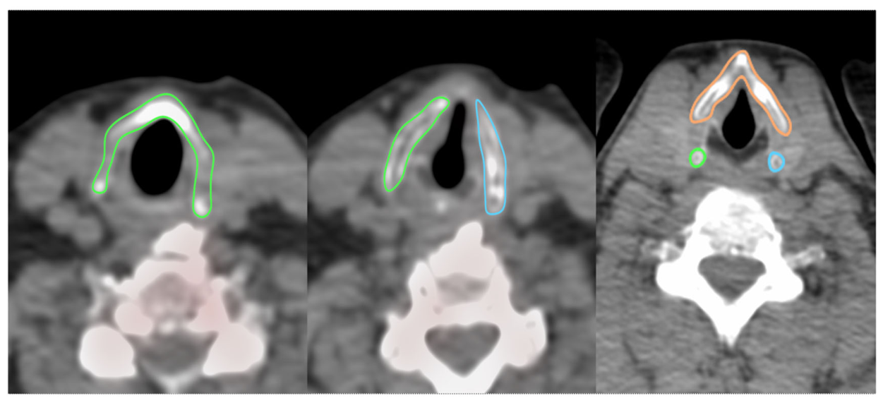
Figure 2.
Scatterplots illustrating the relationships between [18F]NaF uptake and age in different gender groups: (A) mixed-gender, (B) female, (C) male.
Figure 2.
Scatterplots illustrating the relationships between [18F]NaF uptake and age in different gender groups: (A) mixed-gender, (B) female, (C) male.
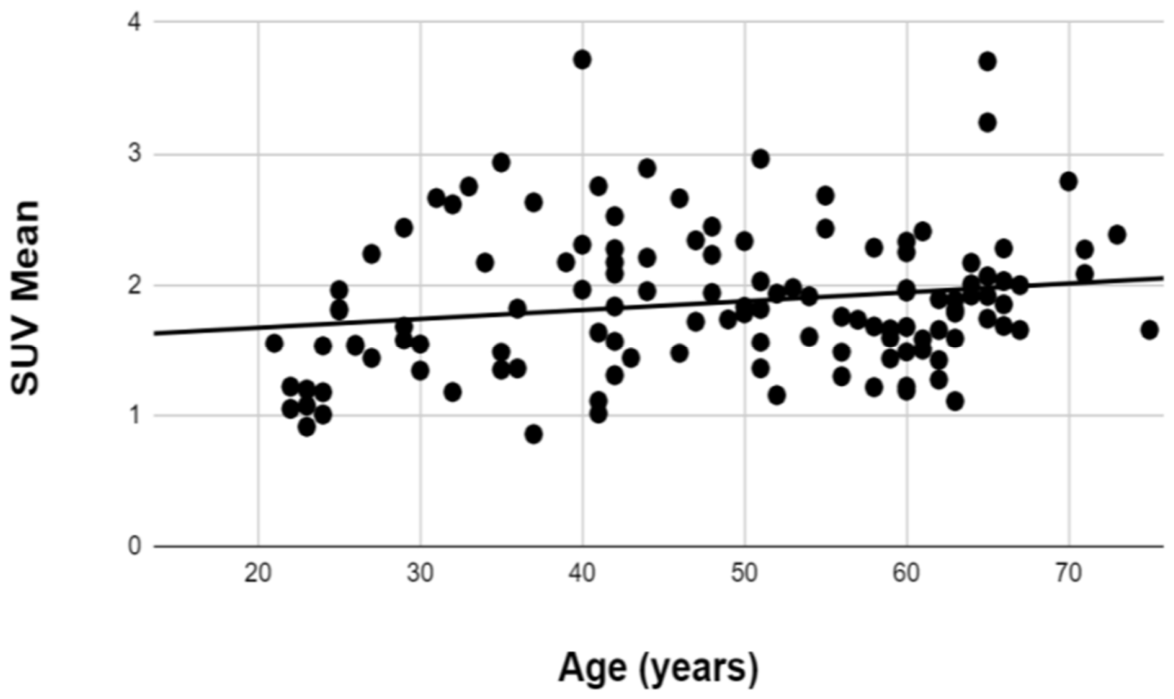
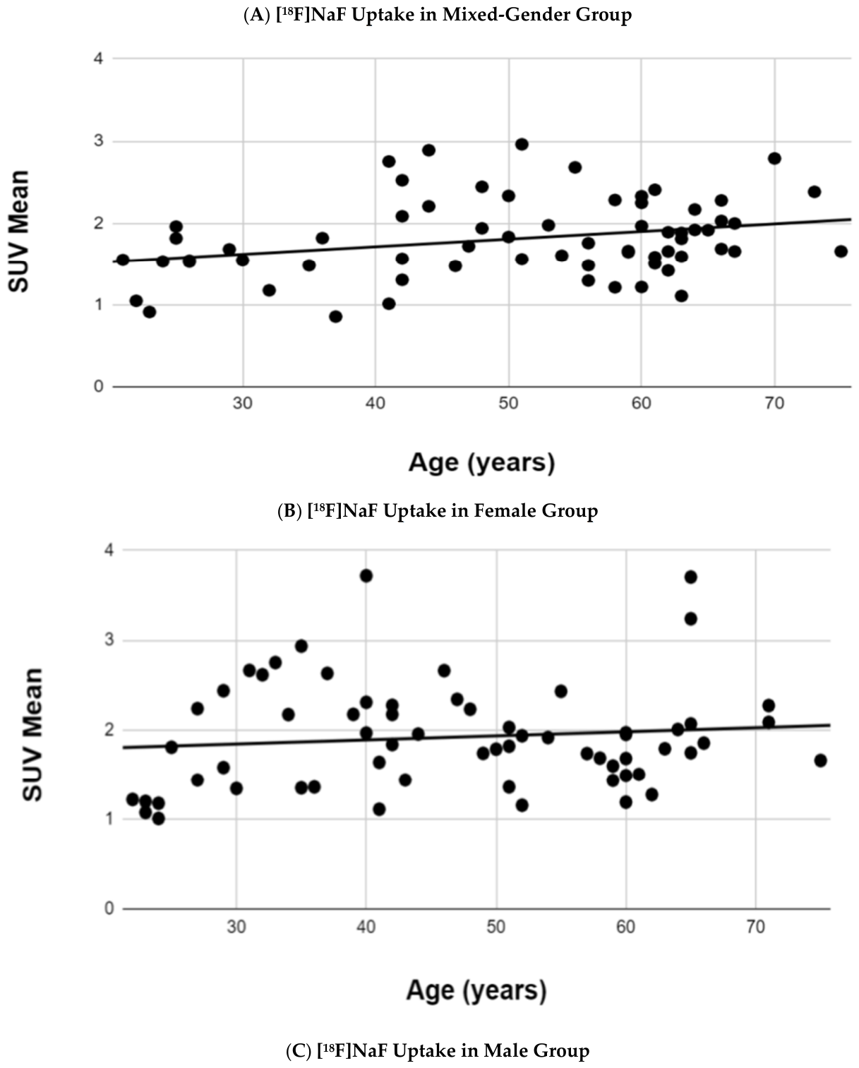
Figure 3.
Scatterplots illustrating the relationships between [18F]FDG uptake and age in different gender groups: (A) mixed-gender, (B) female, and (C) male.
Figure 3.
Scatterplots illustrating the relationships between [18F]FDG uptake and age in different gender groups: (A) mixed-gender, (B) female, and (C) male.
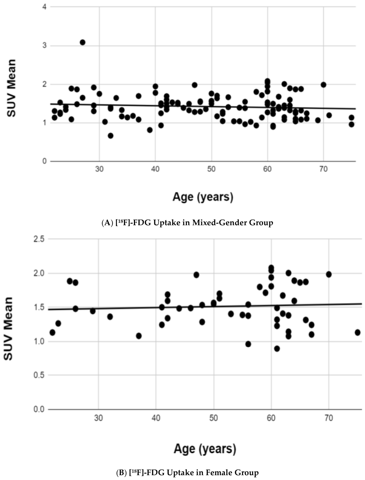
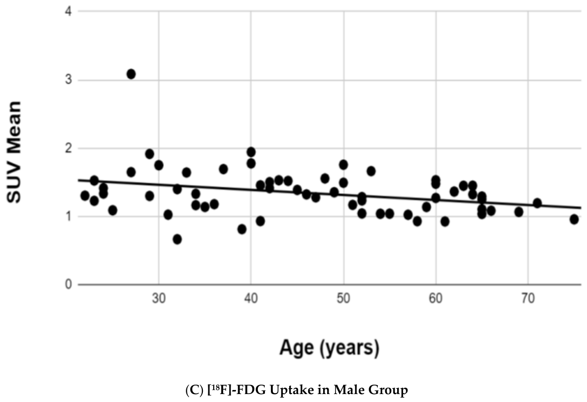

Table 1.
Pearson correlation between [18F]NaF uptake in thyroid cartilage and age for different groups.
Table 1.
Pearson correlation between [18F]NaF uptake in thyroid cartilage and age for different groups.
| Group | Pearson Correlation Coefficient (r) | P-Value |
|---|---|---|
| Mixed-Gender | 0.18 | 0.04 |
| Female | 0.28 | 0.02 |
| Male | 0.13 | 0.32 |
Table 2.
Pearson correlation between [18F]FDG uptake in thyroid cartilage and age for different groups.
Table 2.
Pearson correlation between [18F]FDG uptake in thyroid cartilage and age for different groups.
| Group | Pearson Correlation Coefficient (r) | P-Value |
|---|---|---|
| Mixed-Gender | -0.09 | 0.31 |
| Female | 0.06 | 0.63 |
| Male | -0.31 | 0.01 |
Disclaimer/Publisher’s Note: The statements, opinions and data contained in all publications are solely those of the individual author(s) and contributor(s) and not of MDPI and/or the editor(s). MDPI and/or the editor(s) disclaim responsibility for any injury to people or property resulting from any ideas, methods, instructions or products referred to in the content. |
© 2026 by the authors. Licensee MDPI, Basel, Switzerland. This article is an open access article distributed under the terms and conditions of the Creative Commons Attribution (CC BY) license (http://creativecommons.org/licenses/by/4.0/).
Copyright: This open access article is published under a Creative Commons CC BY 4.0 license, which permit the free download, distribution, and reuse, provided that the author and preprint are cited in any reuse.