

Submitted:
15 May 2026
Posted:
18 May 2026
You are already at the latest version
Abstract
The study considers the hypothesis (supported by a compatible theory) that the vibratory test (SVIN) corresponds to unilateral weakness (UW) in caloric tests and that the head-shaking test (HST) is directly proportional to directional preponderance (DP). A group of 76 patients with vestibular deficits at various stages of compensation, confirmed by bithermal tests using the Fitzgerald-Hallpike technique and evaluated with Jongkees' formulas, was studied and compared with SVIN and HST results. The statistical study shows a marked correlation between SVIN and UW and between HST and DP. At the same time, the cross-correlations between SVIN and DP, and between HST and UW, are significantly weaker, confirming the hypothesis. It is concluded that, in cases where caloric testing is not considered necessary, these two bedside functional tests can give a precise idea of the results of caloric testing and that this interpretation can highlight the great clinical potential of SVIN and HST performed in combination.
Keywords:
acute unilateral vestibulopathy
; unilateral weakness
; directional preponderance
; SVIN
; head shaking test
1. Introduction
The Skull Vibration Induced Nystagmus (SVIN) and the Head Shaking Test (HST) are among the tests routinely performed to diagnose vertigo.
Alongside these, caloric tests continue to be performed to complete the diagnosis: clinicians often perform only two tests (right warm and left warm) instead of four to shorten the duration and, at the same time, cause less discomfort to the patient [1]. In fact, caloric tests are generally well tolerated, but there are cases in which they can cause vagal disturbances, even persisting for several hours, especially in patients with migraine and/or motion sickness.
By doing the caloric tests we can obtain, through Jongkees' formulas, the unilateral values weakness (UW) and directional preponderance (DP), which tell us, respectively, how much one lateral semicircular canal functions compared to the other and whether there is clinical or subclinical nystagmus, and towards which side it is directed.
This data is important to get a clear idea of:
Deficient side and importance of the labyrinthine lesion;
Degree of imbalance between the two hemisystems.
Unfortunately, this information cannot be directly obtained from bedside observation; Therefore, instrumental assessment is necessary to establish an accurate functional diagnosis.
However, being in possession of two simple tests of great diagnostic value such as the SVIN and the HST (“Bedside functional tests” BFT), we can perhaps get an idea of UW and DP without necessarily having to perform instrumental tests.
The aim of this work is to explore this possibility, both theoretically and practically, not so much to avoid caloric testing, but to better understand the meaning of BFT.
The Head Shaking Test [2].
It consists of rotating the patient's head 20 times in the horizontal plane at approximately 1 cycle (right-left) per second. At the conclusion, post-HST nystagmus (HSny) is observed, preferably with inhibited fixation. The test can also be performed in the vertical plane.
The response is usually a nystagmus directed toward the dominant side, and in particular cases, as Menière's disease also toward the deficient side. We will not consider the rather rare possibility of a perverted nystagmus in this paper (nystagmus in the vertical plane from horizontal stimulation) because in this case, the interpretation is not a controversial pathology of the central nervous system.
The literature interprets HSny as the result of excitatory stimulation of the two hemisystems whose algebraic sum is performed at the Velocity Storage level and then downloaded at the end of the stimulation [3]: it will not be possible to evoke an HSny in case of inefficiency of the Velocity Storage.
The test is harmless and generally does not cause patient discomfort, but it should be avoided in cases of neck stiffness or pathology.
The test appears to be closely related to the progress of compensation: as compensation proceeds after a deficit, the intensity of HSny progressively decreases over time until it disappears [4]. Consequently, it may be a direct expression of directional preponderance, which in turn is directly related to the progress of compensation [5].
There remain doubts about the interpretation of the test:
How is it possible that there is an HSny directed towards the deficit side?
How is it possible that we can come across pathological HSny in patients who are normal on instrumental tests?
Why does the response to the HST vary over time? The HSny may disappear in subsequent checks, or it may even reverse direction (for example, check at time t=0, HSny directed to the right, check at time t=1, HSny directed to the left).
The Skull Vibrational Test (Dumas Test) [6]
The vibration test should never be missing from a vestibular examination, both for the simplicity of its execution and for the valuable information it provides.
A device that vibrates at a frequency of 100 hertz is placed behind the right and left ears, with at least 10 seconds of stimulation. You can also try the cranial vertex for another 10 seconds.
If, for example, the right vestibular hemisystem does not function, or functions poorly, during the vibration and without latency, a nystagmus directed to the left (therefore towards the healthy side) is observed, whether the vibrator is applied to the right or left mastoid [7].
Vertical nystagmus may also appear, especially when vibrating at the vertex, as a possible sign of a dehiscence of the superior semicircular canal [8].
This test has a sensitivity of nearly 90% [7,9,10]: if the caloric tests are normal and the vibration test is pathological, the caloric tests were probably performed incorrectly.
The SVIN is not affected by compensation: if performed on a patient who had a deficit a year earlier, it is still altered if the deficit persists [1]1. It compensates for natural head movements (which are around 1 – 2 hertz) and there is no need to develop compensation for frequencies that are not found in everyday life.
The SVIN does not bother the patient; some slightly more sensitive individuals are impressed by the sensation associated with vibration, but it is exceptional to find a patient who is unable to undergo the examination.
The fact that the test remains altered for as long as the deficit remains and is present even in the case of complete compensation seems to link the test directly to Unilateral Weakness. Does not require the presence of Velocity Storage [12].
The HST (2 Hz and its harmonics), SVINT (100 Hz), and CT (<0.01 Hz) are used in the multifrequency analysis of the vestibule, which is especially useful for Peripheral Vestibular Loss (PVL) cases. These likely interact with sensory cells that differ in topography or frequency sensitivity. Since the inner ear primarily operates at high frequencies, the HIT and SVINT are the most effective and physiologically relevant tests when compared to the caloric test [13]
Hypotheses for the interpretation of the two tests:
1. HST (Figure 1)
When an HST is performed and the head rotates to the right, the cupula of the right semicircular canal moves towards the utricle, while that of the left semicircular canal moves away from the utricle.
When the patient turns his head to the left, the cupula of the left semicircular canal moves towards the utricle, while that of the right semicircular canal moves away from the utricle.
When caloric tests are performed with hot water, the cupula moves in a utriculipetal direction and excites; when cold tests are performed, the cupula moves in a utriculifugal direction and inhibits.
When patients turn their head to the right during an HST test, they are in the same situation as if they were experiencing a warm right and a cold left simultaneously. Conversely, when they turn their head to the left, it is as if they experience a warm left and a cold right; the effect on cupula displacement remains the same. Velocity Storage converts the stimulus from phasic to tonic because it performs the algebraic sum, and at the end of a complete cycle (first to the right and then to the left), we obtain:
(RW+LC)-(LW+RC)
Where the minus sign takes into account the direction of the nystagmus. This expression is the numerator of the formula for directional preponderance.
So, the HST result is directly proportional to the Directional Preponderance.
In fact, the HST indicates the level of compensation: if the HST is normal, it does not mean that the patient is normal; rather, it indicates that there is no uncompensated deficit.
2. SVIN (Figure 2)
During vibration, both cupulas move towards the utricle because, by favouring the path with the lowest impedance, the vibration travels along the shunt through the cranial vault, and the direction of the wave relative to the ear is always from the outside to the inside. Furthermore, since this is a very high-frequency stimulation, during the utriculipetal compression phase in the lateral canal, we obtain an excitatory current, and during the utriculifugal rarefaction phase, there is a resetting of the discharge.
Conversely, if the direction of the cupula's movement was utriculifugal, then in the case of right areflexia, one would expect a right-directed SVIN, and vice versa. However, this does not occur; rather, the opposite is observed.
The sum of the stimulations (which does not require Velocity Storage because they are very close together), therefore, represents excitation on both the right and the left.
If the two channels are well-matched or symmetrical, the two sides are equivalent. However, if one side functions less effectively, nystagmus is induced that matches the difference between the two sides.
The result is similar to a warm stimulation on the right and, at the same time, a warm one on the left.
Considering two cycles: (RW+LW)+(RW+LW) but the sum of the hot ones must equal the sum of the cold ones (“Temperature Effect ” [14]).
So: (RW+LW)=(RC+LC)
so, it is possible to replace:
(RW+LW)+(RW+LW)
(RW+LW)+(RC+LC)
(RW+RC)-(LW+LC)
(The sign takes into account left-directed nystagmus)
This is the numerator of the fraction representing the Unilateral Weakness.
Therefore, the intensity of vibratory nystagmus is directly proportional to the Unilateral Weakness.
It therefore seems that performing SVIN and HST enables detection of UW and DP, respectively, even without caloric testing.
The advantages are clear:
- caloric tests are more exposed to the risk of technical error than HST and SVIN;
- the discomfort caused to the patient is significantly less;
- The condition of the tympanic membrane does not influence the response.
By evaluating the tests in combination, all possible clinical situations can be deduced, as shown in Table 1.
There may be apparent normality in bilateral areflexia, but there is more than one “bedside” system to diagnose bilateral areflexia: for example, the optotype test [15] or the Head Impulse Test [16].
Interpretation of the two coupled tests
Let's consider an example of an acute right lateral semicircular canal defect. The patient will display acute symptoms of objective external vertigo, combined with vagal symptoms due to the dominance of the left vestibular system over the right. Objectively, a deficient nystagmus may be observed (sometimes after a brief irritative phase), followed by a rapid phase directed to the left.
Case 1. The right labyrinth remains in deficit.
Over the following days, the system begins to compensate, and as time goes by, this compensation becomes increasingly effective. The Unilateral Weakness remains unchanged because the function of the deficient ear remains constant and low, but Directional Preponderance will tend to decrease progressively due to the effect of compensation. Since the SVIN is related to the UW and the HST is related to the DP, it is expected that the former will remain pathological (present with constant intensity), while the latter will diminish over time until it disappears, demonstrating more effective compensation.
Case 2. The right labyrinth gradually resumes functioning.
As the situation normalises, the UW decreases until it returns to normal if the channel fully regains its function. The DP also tends to attenuate and disappear, not due to compensation but because the balance between the two hemisystems is restored. Both the SVIN and the HST will therefore become less and less evident until they disappear, a sign, this time, not of effective compensation but of a true functional recovery of the system.
Case 3. The right labyrinth resumes functioning completely and rapidly.
The two labyrinths function well, but a portion of "compensatory nystagmus" remains, elaborated by the central nervous system to assist the side with the deficit, which must be progressively reabsorbed. Until that moment, we can expect a DP directed towards the side that was previously in deficit and a normal UW. Consequently, we will have a normal SVIN and an HSny present and directed towards the side that was previously in deficit. It is to be expected that in the following days the compensation, no longer necessary, will be eliminated and the HST will also normalize. Until that moment, however, we will be faced with the phenomenon, strange only in appearance, of an HSny that inverts its direction compared to the acute phase (previously the rapid phase of the nystagmus was directed towards the healthy side, now it is directed towards the opposite side, the one that was deficient). This is the well-known phenomenon of Recovery Nystagmus (Bechterew) [17], which can therefore be clearly highlighted by SVIN and HST, but would go unnoticed if caloric tests were limited to hot caloric stimuli.
Case 4.
The right labyrinth resumes functioning quickly but incompletely.
This is the typical case of Meniere's disease, but it can also occur in other clinical contexts.
In this case, the situation is the same as previously described (case 3), except that partial recovery means we still have a right UW, but it is associated with a left DP. In fact, excess compensation still exists, but the functionality of the deficient side has not reached that of the more efficient side. The result is once again only apparently paradoxical: the SVIN will be directed to the left (toward the efficient side), and the HSny will be directed to the right due to the presence of excess compensation.
2. Materials and Methods
To verify the theoretical hypotheses developed to date, 76 patients with acute or stabilised deficits in one vestibular hemi-system were included in clinical practice. Informed consent was obtained from all subjects involved in the study.
Patients underwent a thorough history, audiometry, and vestibular examination, including a search for spontaneous and evoked nystagmus, by ocular and cephalic manoeuvres (lateral, rebound, position, and positioning nystagmus), and Skull Vibration Nystagmus Test. SVINT was applied with a battery-powered device designed for physiotherapy, placed perpendicular to the skin just behind the ear. Fundamental frequency, measured with the aid of a phonometer, was 100 Hz (60-120 Hz). Vibratory stimulation was applied for about 10 sec, first on one side, on the other and on the vertex. The test was considered positive when persistent horizontal or horizontal torsion nystagmus was evoked at least in two sides for the duration of the stimulus and if the phenomenon was repeatable.
a head-shaking test, and a clinical head-impulse test. Patients with spontaneous nystagmus underwent the Standing protocol [18], and if a central nervous system disorder was suspected, they underwent contrast-enhanced brain MRI. After initial symptomatic therapy lasting a few days, a vestibular rehabilitation program was proposed to be performed at home, with periodic checkups.
Caloric testing was performed with hot and cold water. The maximum speed of the slow-phase of nystagmus evoked by each ear was analysed for Unilateral Weakness and Directional Preponderance as determined by Jongkees formulae. Abnormal Unilateral Weakness or Directional Preponderance was defined when the difference between the two ears was 30% or more.
When possible, eye movements (saccadic and pursuit) were studied.
Inclusion criteria:
- – Patients underwent both caloric testing and SVIN and HST.
- – Subjects agreed to be recruited into the study.
Exclusion criteria:
- – Otological pathologies that do not allow caloric testing
- – Central nervous system disorders
- – Patients' refusal to undergo one of the battery tests
- – Patients' refusal to be included in the study.
Seventy-six patients, aged between 17 and 87 years (mean 57.8, standard deviation 16.4), were enrolled in the study.
Once the UW and DP values were obtained (evaluated as pathological/normal, according to the laboratory regulations), they were compared with the SVIN and HST tests, again evaluating them as normal test (absent nystagmus) / pathological test (present nystagmus).
In this way, UW were compared with SVIN and DP with HST to potentially reject the null hypothesis that they originated from the same population.
For comparison, UW with DP and HST with SVIN were also evaluated.
Statistical methods: we have used R Core Team (A Language and Environment for Statistical Computing. R Foundation for Statistical Computing, Vienna, Austria. https://www.R-project.org/. RStudio 2025.05.0 Build 496 © 2009-2025 Posit Software, PBC packages (" vcd ", " epiR ", " forestplot "- 2024)
Seventy-six patients undergoing caloric testing were included, with dichotomous classification into:
- – Unilateral weakness (UW): UW+ (pathological) vs UW− (normal)
- – Directional preponderance (DP): DP+ (pathological) vs DP− (normal)
- – Vibratory test (SVIN): SVIN+ (pathological) vs SVIN− (normal)
- – Head shaking test (HST): HST+ (pathological) vs HST− (normal).
The primary objective was to evaluate the association between UW and SVIN and between DP and HST, hypothesized as measures of the same physical phenomenon and compare them with the “cross-matched” UW–HST and DP–SVIN associations.
Since all variables are dichotomous (pathological / normal), the chi-square test of independence (χ²) for 2×2 tables was used.
- – The coefficient φ (phi) was calculated as a measure of the strength of the association (equivalent to a correlation for dichotomous variables);
- – Odds Ratio (OR) was calculated to quantify the magnitude of the effect.
No assumption of normality is required, since we are not analyzing continuous variables but only categorical outcomes.
3. Results
As for the strong expected associations, we get:
1. Association between UW and SVIN: OR = 96.3; coefficient ϕ ≈ 0.80
Chi-square (χ², 1 df) X 2 ≈ 48.6(p≪0.001)
Interpretation: Pathological UW is strongly associated with pathological SVIN. The association is very strong (φ ≈ 0.80), and the extremely high OR indicates that the presence of UW+ massively increases the likelihood of SVIN+.
2. Association between DP and HST: OR = 52.3; coefficient ϕ ≈ 0.68
Chi-square (χ², 1 df) χ 2 ≈ 35 (p≪0.001)
Interpretation: Pathological DP is strongly associated with pathological HST. Here too, the association is strong (φ ≈ 0.68), and the OR is very high, consistent with the idea that HST expresses the same directional preponderance seen in caloric testing.
Cross-associations (weak expectations):
3. “Cross-matched” association between UW and HST: OR = 4.5; coefficient ϕ ≈ 0.29
Chi-square (χ², 1 df) χ 2 ≈ 6.4 (p ≈ 0.01)
Interpretation: UW and HST are significantly associated, but the strength of the association is modest compared to “physiologically matched” pairs (UW–SVIN and DP–HST).
4. “Cross-relationship” between DP and SVIN: OR = 8.9; coefficient ϕ ≈ 0.35
Chi-square (χ², 1 df) χ 2 ≈ 9.3 (p ≈ 0.003)
Interpretation: DP and SVIN are significantly associated, but with moderate strength, lower than the UW–SVIN and DP–HST associations.
The analysis of associations between caloric tests and dynamic tests showed a strong concordance between UW and SVIN (φ = 0.80; p < 0.001; OR ≈ 96) and between DP and HST (φ = 0.68; p < 0.001; OR ≈ 52). However, the cross-lagged associations were significantly weaker: UW–HST (φ = 0.29; p ≈ 0.01; OR ≈ 4.5) and DP–SVIN (φ = 0.35; p ≈ 0.003; OR ≈ 8.9).
4. Discussion
The reported case studies appear to be consistent with the predictions from the theoretical section. In particular, a clear correspondence is evident between UW and SVIN, and between DP and HST. In reality, there is also a correspondence between UW and HST and between DP and SVIN, but to a much lesser extent. The presence of a correlation in these cases is attributable to the peculiar, labyrinthine situation in which a UW and a DP can coexist. The “Bedside Functional tests”, are able to predict the presence of a canal deficit and also the degree of compensation. No quantitative assessment is reported, and this may seem reductive given the Jongkees formulas' percentage-based responses, but this is solely due to the approach taken to bedside functional testing: if we had recorded and measured the resulting nystagmus, we could have obtained a precise quantitative picture of the situation. For example, in a patient with a stable deficit undergoing compensation, we would have had a constant SVIN across the various observations and a progressively lower HSny until exhaustion. The angular velocity of the slow phase of the SVIN is directly proportional to the extent of the deficit (UW), the angular velocity of the slow phase of the HSny is directly proportional to the compensation (DP). It's a fact that caloric tests are being performed less frequently, despite providing valuable information for the evaluation of patients with vertigo, due to the time required and the potential discomfort associated with motion sickness. Integrated assessment of SVIN and HST could "replace" caloric tests if the examiner deems them unnecessary. It should be added that caloric tests can be affected by stimulation errors, regardless of the examiner's skill and experience, due to asymmetrical calibre or torsion of the external auditory canals. Often, apparent dysreflexia is due to imprecise irrigation for anatomical reasons. In these cases, a discrepancy between caloric tests and bedside functional tests could perhaps be attributed more to intrinsic defects in the caloric tests than to an invalid response to the BFT. In any case, what perhaps seems most appropriate to take into consideration is the real semiological value of the latter. It appears that the SVIN accurately assesses a possible deficit in labyrinth function, whereas the HST evaluates the central compensation mechanism more than the canal function. The association of the two tests could therefore provide precise information on the symmetry of lateral semicircular canal function and on their position relative to compensation.The problem of non-instrumental diagnosis of bilateral vestibular areflexias remains, but at least their suspicion should not be difficult, even with only a bedside approach.The rapid impulse test has a low sensitivity in partial unilateral deficits 18,19,20, but in areflexias, even unilateral, the sensitivity rises to 100% 21. Sensitivity will be even higher in bilateral forms.
Weak points of this work may be:
– Trials exploring different ranges of stimulation (low frequencies for caloric testing, high frequencies for HST, very high frequencies for SVIN) have been compared, but research on a real patient population does not appear to have been affected by this heterogeneity.
– The study is not prospective but retrospective, which prevented a complete design. For example, it would have been interesting to evaluate the behaviour of video-HIT in this context.
– It is not a quantitative study regarding test responses, but is limited to the categorical absent/present response.
These points could serve as a starting point for further improvement.
5. Conclusions
Bedside vestibular testing, which has improved substantially in recent years, is likely capable of providing information equivalent to that obtained with caloric testing.
In the presented case series, the correlations between UW and SVIN, and between DP and HST, were very satisfactory.
An interpretation of bedside functional tests is proposed not only as an alternative to caloric tests where these are contraindicated or otherwise inadvisable, but also for the purpose of a more precise interpretation of these tests.
Author Contributions
Conceptualization, investigation, writing—original draft preparation MG; review and editing, visualization, supervision, GN. All authors have read and agreed to the published version of the manuscript.
Funding
“This research received no external funding
Institutional Review Board Statement
Ethical approval was not required for the studies involving humans because patients underwent routine vestibular testing. The studies were conducted in accordance with the local legislation and institutional requirements. Written informed consent for participation was not required from the participants or the participants’ legal guardians/next of kin in accordance with the national legislation and institutional requirements.
Informed Consent Statement
Informed consent was obtained from all subjects involved in the study
Conflicts of Interest
The authors declare that the research was conducted in the absence of any commercial or financial relationships that could be construed as a potential conflict of interest.
Abbreviations
The following abbreviations are used in this manuscript:
| UW | Unilateral Weakness |
| SVINT | Skull Vibrational nystagmus Test |
| SVIN | Skull Vibrational nystagmus |
| HST | Head Shaking Test |
| HSny | post-HST nystagmus |
| DP | Directional Preponderance |
| BFT | Bedside functional tests |
| PVL | Peripheral Vestibular Loss |
References
- Alhabib, S.F.; Saliba, I. Reliability of Monothermal Caloric Test as Screening Test of Vestibular System. J. Clin. Med. 2022, 11, 6977. [Google Scholar] [CrossRef] [PubMed] [PubMed Central]
- Jacobson, G.P.; Newman, C.W.; Safadi, I. Sensitivity and specificity of the head-shaking test for detecting vestibular system abnormalities. Ann. Otol. Rhinol. Laryngol. 1990, 99, 539–542. [Google Scholar] [CrossRef] [PubMed]
- Hain, T.C.; Fetter, M.; Zee, D.S. Head-shaking nystagmus in patients with unilateral peripheral vestibular lesions. Am. J. Otolaryngol. 1987, 8, 36–47. [Google Scholar] [CrossRef] [PubMed]
- Striteska, M.; Valis, M.; Chrobok, V.; Profant, O.; Califano, L.; Syba, J.; Trnkova, K.; Kremlacek, J.; Chovanec, M. Head-shaking-induced nystagmus reflects dynamic vestibular compensation: A 2-year follow-up study. Front Neurol. 2022, 13, 949696. [Google Scholar] [CrossRef] [PubMed] [PubMed Central]
- Striteska, M.; Valis, M.; Chrobok, V.; Profant, O.; Califano, L.; Syba, J.; Trnkova, K.; Kremlacek, J.; Chovanec, M. Head-shaking-induced nystagmus reflects dynamic vestibular compensation: A 2-year follow-up study. Front Neurol. 2022, 13, 949696. [Google Scholar] [CrossRef] [PubMed] [PubMed Central]
- Dumas, G.; Perrin, P.; Schmerber, S. Nystagmus induced by high frequency vibrations of the skull in total unilateral peripheral vestibular lesions. Acta Otolaryngol. 2008, 128, 255–262. [Google Scholar] [CrossRef] [PubMed]
- Michel, J.; Dumas, G.; Lavieille, J.P.; Charachon, R. Diagnostic value of vibration-induced nystagmus obtained by combined vibratory stimulation applied to the neck muscles and skull of 300 vertiginous patients. Rev. Laryngol. Otol. Rhinol. (Bord) 2001, 122, 89–94. [Google Scholar] [PubMed]
- Dumas, G.; Tan, H.; Dumas, L.; Perrin, P.; Lion, A.; Schmerber, S. Skull vibration induced nystagmus in patients with superior semicircular canal dehiscence. Eur. Ann. Otorhinolaryngol. Head. Neck Dis. 2019, 136, 263–272. [Google Scholar] [CrossRef] [PubMed]
- Nuti, D.; Mandalà, M. Sensitivity and specificity of mastoid vibration test in detection of effects of vestibular neuritis. Acta Otorhinolaryngol. Ital. 2005, 25, 271–276. [Google Scholar] [PubMed] [PubMed Central]
- Koo, J.W.; Kim, J.S.; Hong, S.K. Vibration-induced nystagmus after acute peripheral vestibular loss: comparative study with other vestibule-ocular reflex tests in the yaw plane. Otol. Neurotol. 2011, 32, 466–471. [Google Scholar] [CrossRef] [PubMed]
- Baloh, R.W.; Kerber, K. Clinical Neurophysiology of the vestibular system, 4th Ed. ed; Oxford Universuty Press, 2011. [Google Scholar]
- Curthoys, I.S.; Zee, D.S.; Dumas, G.; Pastras, C.J.; Dlugaiczyk, J. Skull vibration induced nystagmus, velocity storage and self-stability. Front Neurol. 2025, 16, 1533842. [Google Scholar] [CrossRef] [PubMed] [PubMed Central]
- Stockwell CW: ENG Workbook. ProEd, Austin Texas, 1983.
- Lambert, S.; Sigrist, A.; Delaspre, O.; Pelizzone, M.; Guyot, J.P. Measurement of dynamic visual acuity in patients with vestibular areflexia. Acta Otolaryngol. 2010, 130, 820–823. [Google Scholar] [CrossRef] [PubMed]
- van Dooren, T.S.; Lucieer, F.M.P.; Duijn, S.; Janssen, A.M.L.; Guinand, N.; Pérez Fornos, A.; Van Rompaey, V.; Kingma, H.; Ramat, S.; van de Berg, R. The Functional Head Impulse Test to Assess Oscillopsia in Bilateral Vestibulopathy. Front Neurol. 2019, 10, 365. [Google Scholar] [CrossRef] [PubMed] [PubMed Central]
- Kim, Y.; Jin, S.; Kim, J.S.; Koo, J.W. Bechterew's Phenomenon in Bilateral Sequential Vestibular Neuritis: A Report of Two Cases. Front Neurol. 2022, 13, 844676. [Google Scholar] [CrossRef] [PubMed] [PubMed Central]
- Vanni, S.; Pecci, R.; Casati, C.; et al. STANDING, a four-step bedside algorithm for differential diagnosis of acute vertigo in the emergency department. Acta Otorhinolaryngol. Ital. 2014, 34, 419–426. [Google Scholar] [PubMed]
- Kim, S.H.; Lee, S.U.; Cho, B.H.; Cho, K.H.; Yu, S.; Kim, B.J.; Kim, J.S. Analyses of Head-Impulse Tests in Patients With Posterior Circulation Stroke and Vestibular Neuritis. Neurology 2023, 100, e2374–e2385. [Google Scholar] [CrossRef] [PubMed] [PubMed Central]
- Walther, L.E.; Löhler, J.; Agrawal, Y.; Motschall, E.; Schubach, F.; Meerpohl, J.J.; Schmucker, C. Evaluating the Diagnostic Accuracy of the Head-Impulse Test: A Scoping Review. JAMA Otolaryngol. Head. Neck Surg. 2019, 145, 550–560. [Google Scholar] [CrossRef] [PubMed]
- Lopez-Escamez, J.A. Video Head-Impulse Testing vs Clinical Diagnosis of Vestibular Disorders. JAMA Otolaryngol. Head. Neck Surg. 2019, 145, 561–562. [Google Scholar] [CrossRef] [PubMed]
- Halmagyi, G.M.; Curthoys, I.S. A clinical sign of canal paresis. Arch. Neurol. 1988, 45, 737–739. [Google Scholar] [CrossRef] [PubMed]
Figure 1.
Head Shaking Test (HST) dynamics. During rightward rotation, the right semicircular canal cupula moves in an ampullopetal (excitatory) direction, while the left cupula moves ampullofugal (inhibitory). Conversely, leftward rotation triggers left ampullopetal and right ampullofugal displacement. Each head turn mimics the combined physiological response of a warm caloric test on the side of rotation and a cold caloric test on the contralateral side. Image prompt-based AI-generated image (Google Gemini, 2026) created according to the author's specifications.
Figure 1.
Head Shaking Test (HST) dynamics. During rightward rotation, the right semicircular canal cupula moves in an ampullopetal (excitatory) direction, while the left cupula moves ampullofugal (inhibitory). Conversely, leftward rotation triggers left ampullopetal and right ampullofugal displacement. Each head turn mimics the combined physiological response of a warm caloric test on the side of rotation and a cold caloric test on the contralateral side. Image prompt-based AI-generated image (Google Gemini, 2026) created according to the author's specifications.
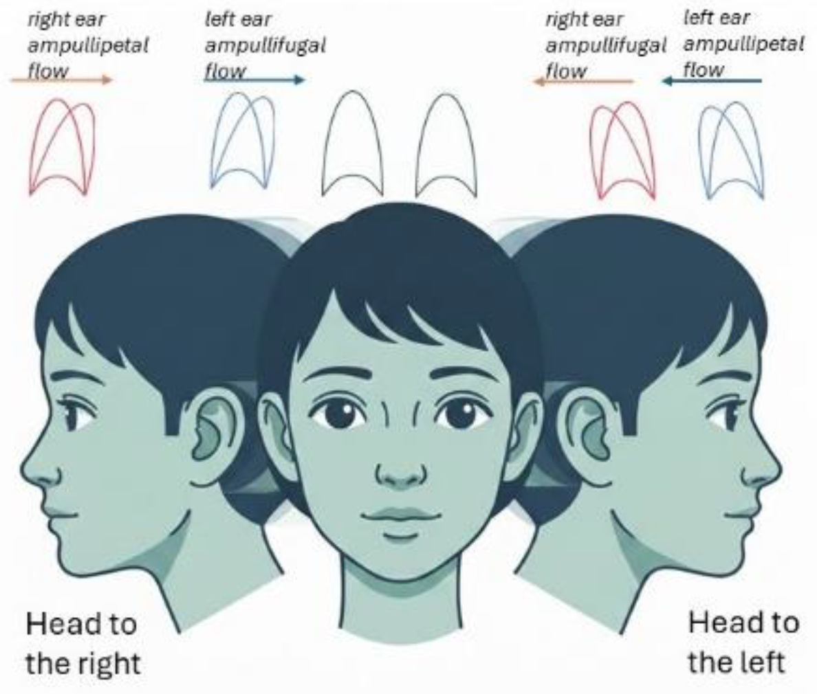
Figure 2.
Vibratory Test Dynamics. During mastoid or vertex vibration, both lateral semicircular canal cupulae are displaced in an ampullopetal direction. In healthy or symmetrical vestibular systems, the bilateral inputs balance each other out. However, in the presence of a unilateral deficit, a vibration-induced nystagmus (VIN) emerges, representing the functional asymmetry between the two sides. The stimulus mimics a simultaneous bilateral warm caloric stimulation; the resulting nystagmus reflects the difference in responsiveness between the intact and the impaired side.
Figure 2.
Vibratory Test Dynamics. During mastoid or vertex vibration, both lateral semicircular canal cupulae are displaced in an ampullopetal direction. In healthy or symmetrical vestibular systems, the bilateral inputs balance each other out. However, in the presence of a unilateral deficit, a vibration-induced nystagmus (VIN) emerges, representing the functional asymmetry between the two sides. The stimulus mimics a simultaneous bilateral warm caloric stimulation; the resulting nystagmus reflects the difference in responsiveness between the intact and the impaired side.
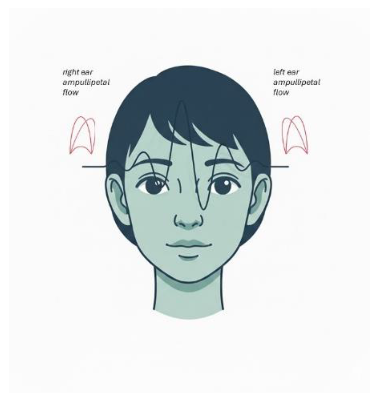
Figure 3.
UW/SVIN association, DP/HST (strong associations) and UW/HST and DP/SVIN (weak associations). Pathological UW is strongly associated with pathological SVIN, pathological DP is strongly associated with pathological HST, UW/HST and DP/SVIN are significantly associated, but the strength of the association is modest compared to the “physiologically corresponding” pairs (UW–SVIN and DP–HST).
Figure 3.
UW/SVIN association, DP/HST (strong associations) and UW/HST and DP/SVIN (weak associations). Pathological UW is strongly associated with pathological SVIN, pathological DP is strongly associated with pathological HST, UW/HST and DP/SVIN are significantly associated, but the strength of the association is modest compared to the “physiologically corresponding” pairs (UW–SVIN and DP–HST).
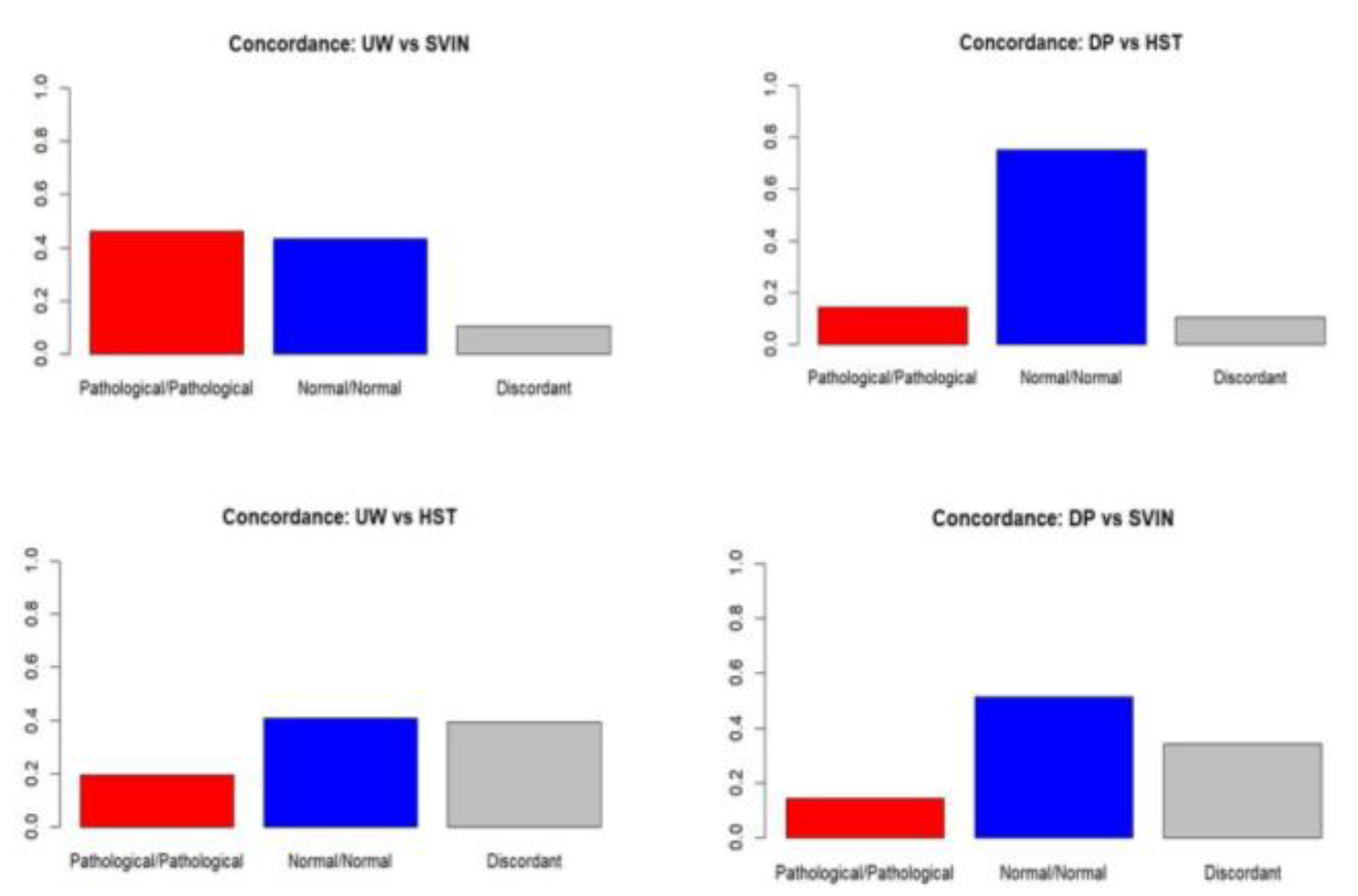
Figure 4.
Analysis of the associations between caloric tests and dynamic tests showed a strong correlation between UW and SVIN (φ = 0.80; p < 0.001) and between DP and HST (φ = 0.68; p < 0.001). Cross-associations, on the other hand, were significantly weaker: UW–HST (φ = 0.29; p ≈ 0.01) and DP–SVIN (φ = 0.35; p ≈ 0.003).
Figure 4.
Analysis of the associations between caloric tests and dynamic tests showed a strong correlation between UW and SVIN (φ = 0.80; p < 0.001) and between DP and HST (φ = 0.68; p < 0.001). Cross-associations, on the other hand, were significantly weaker: UW–HST (φ = 0.29; p ≈ 0.01) and DP–SVIN (φ = 0.35; p ≈ 0.003).
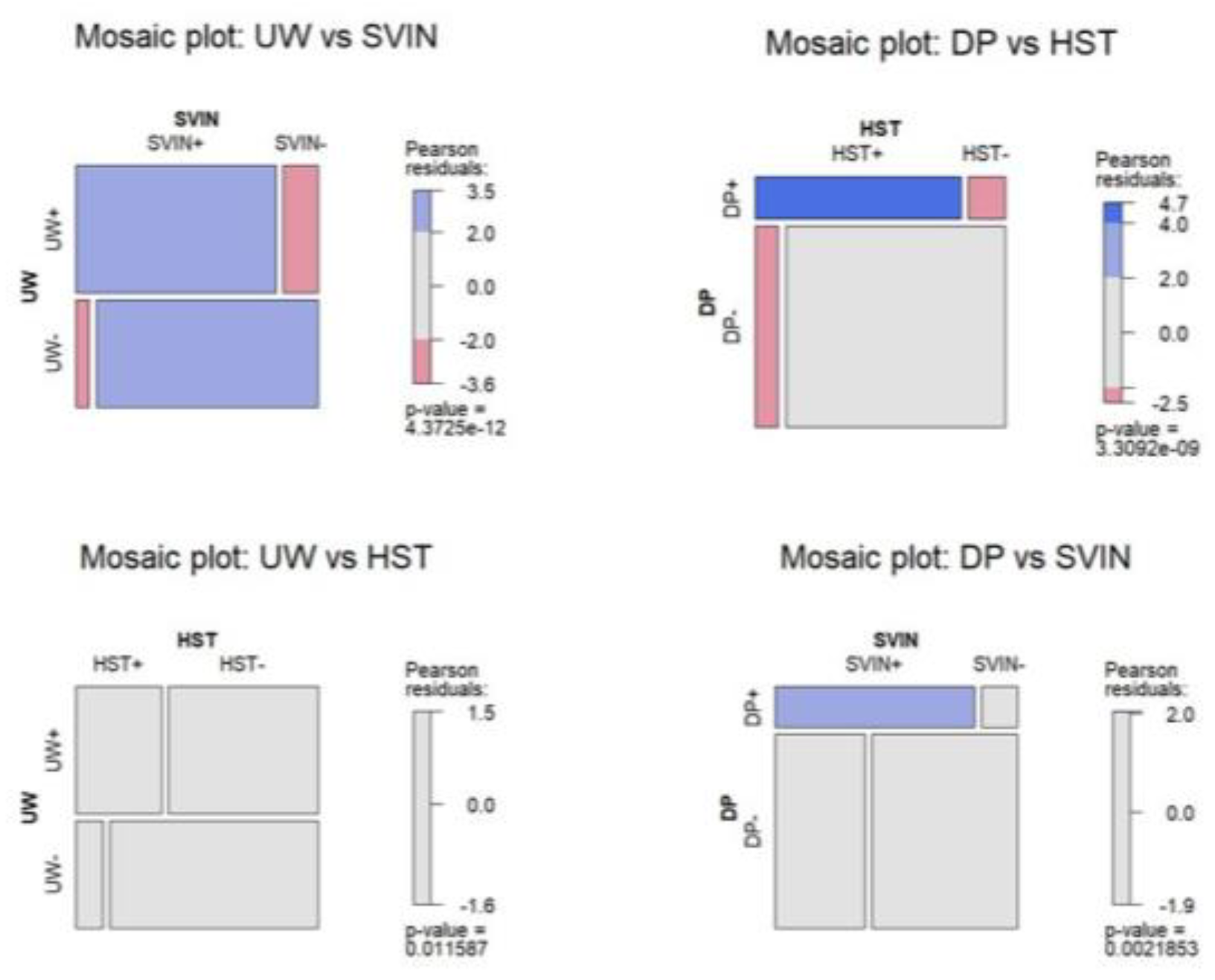
Figure 5.
Analysis of the associations between caloric tests and dynamic tests showed a strong correlation between UW and SVIN (OR ≈ 96) and between DP and HST (OR ≈ 52). Cross-associations, on the other hand, were significantly weaker: UW–HST (OR ≈ 4.5) and DP–SVIN (OR ≈ 8.9).
Figure 5.
Analysis of the associations between caloric tests and dynamic tests showed a strong correlation between UW and SVIN (OR ≈ 96) and between DP and HST (OR ≈ 52). Cross-associations, on the other hand, were significantly weaker: UW–HST (OR ≈ 4.5) and DP–SVIN (OR ≈ 8.9).
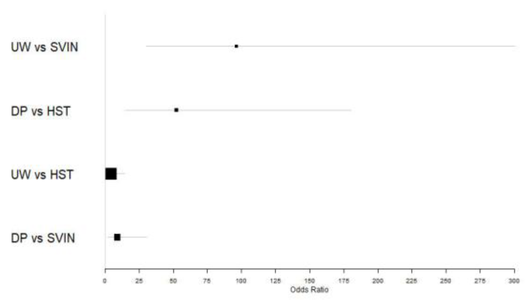

Table 1.
Bedside functional tests and their correlation with UW and DP. Their combination allows us to determine the patient's functional status. SVIN: Skull Vibratory Induced Nystagmus; HST: Head Shaking Nystagmus; UW: Unilateral Weakness; DP: Directional Preponderance.
Table 1.
Bedside functional tests and their correlation with UW and DP. Their combination allows us to determine the patient's functional status. SVIN: Skull Vibratory Induced Nystagmus; HST: Head Shaking Nystagmus; UW: Unilateral Weakness; DP: Directional Preponderance.
| SVIN | HST | Caloric | Issue |
| Present | Absent | UW+ DP+ | Uncompensated deficit |
| Present | Absent | UW+ DP Absent | Compensated deficit |
| Absent | Present | UW Absent DP + | Recovery nystagmus |
| Absent | Absent | UW Absent DP Absent | Absent |
Table 2.
Comparison of caloric test results (UW) and SVIN. The correlation is very strong.
| SVIN + | SVIN - | TOTAL | |
| UW + | 35 | 6 | 41 |
| UW - | 2 | 33 | 35 |
| T | 37 | 39 | 76 |
Table 3.
Comparison of caloric test results (DP) and HST. The correlation is very strong.
| HST + | HST - | TOTAL | |
| DP + | 11 | 2 | 13 |
| DP - | 6 | 57 | 63 |
| T | 17 | 59 | 76 |
Table 4.
Comparison of caloric test results (UW) and HST. The correlation is weak.
| HST + | HST - | TOTAL | |
| UW + | 15 | 26 | 41 |
| UW - | 4 | 31 | 35 |
| T | 19 | 57 | 76 |
Table 5.
Comparison of caloric test results (DP) and SVIN. The correlation is weak.
| SVIN + | SVIN - | TOTAL | |
| DP + | 11 | 2 | 13 |
| DP - | 24 | 39 | 63 |
| T | 35 | 41 | 76 |
Disclaimer/Publisher’s Note: The statements, opinions and data contained in all publications are solely those of the individual author(s) and contributor(s) and not of MDPI and/or the editor(s). MDPI and/or the editor(s) disclaim responsibility for any injury to people or property resulting from any ideas, methods, instructions or products referred to in the content. |
© 2026 by the authors. Licensee MDPI, Basel, Switzerland. This article is an open access article distributed under the terms and conditions of the Creative Commons Attribution (CC BY) license (http://creativecommons.org/licenses/by/4.0/).
Copyright: This open access article is published under a Creative Commons CC BY 4.0 license, which permit the free download, distribution, and reuse, provided that the author and preprint are cited in any reuse.