

Submitted:
01 July 2026
Posted:
03 July 2026
You are already at the latest version
Abstract
Lipedema affects an estimated 11 to 12% of women and is characterized by bilateral, symmetric, painful enlargement of the limb subcutaneous fat that resists dietary weight loss. Its primary substrate is increasingly understood to be adipo-vascular and connective: genome-wide association studies implicate adipose, vascular, and extracellular-matrix loci rather than immune genes, and the affected fat is metabolically healthier than its mass predicts. Inflammation is itself well documented in lipedema tissue, but its type 2, reparative character suggests that it amplifies this substrate rather than initiating the disease. Against this substrate, however, a focused neuroimmune cluster remains unexplained by purely structural models: a diagnostically accurate quantitative sensory testing pattern, elevated tissue histamine, frequent comorbid fibromyalgia, and disease that is more severe in limbs carrying an additional local vascular trigger. We hypothesize that altered tissue mechanics and adipo-vascular biology help trigger a localized immune cascade, so that these observations may define a clinically identifiable subgroup exhibiting a type 2 immune and mast cell-activation endotype, nested within the broader adipo-vascular disorder. The core framework is deliberately limited to this testable sequence: adipo-vascular susceptibility, per-cell mast cell/type 2 activation, histaminergic peripheral sensitization, the quantitative sensory testing signature, and pain generation in a clinically identifiable endotype. We argue that the endotype is best characterized by per-cell mast cell secretory activity rather than abundance, which is not consistently increased in lipedema tissue. It is defined a priori, before treatment exposure, by a composite of baseline features (affected-fat erythema or warmth, atopic or mast cell-activation features, and the characteristic quantitative sensory testing pattern), with response to mast cell-directed therapy reserved as its prospective test rather than a defining criterion. We present the framework as a graded synthesis, distinguishing evidence shown directly in lipedema from that inferred from related fields and from untested hypotheses. Broader adipogenic, gut-immune, HLA-associated, cancer-related, ADHD-related, and systemic immunomodulatory concepts are separated into a dedicated exploratory section; they are secondary hypotheses and are not required for the central mast cell-histamine-QST-pain claim. The framework identifies mast cell activation as a potential therapeutic target in this subgroup for future investigation, pending prospective validation.
Keywords:
lipedema
; endotype
; mast cells
; type 2 innate immunity
; histamine
; quantitative sensory testing
; gluteofemoral adipose tissue
; adipo-vascular
1. Introduction
Lipedema is a chronic disorder of the limb subcutaneous adipose tissue affecting an estimated 11 to 12% of women [1,2]. Its clinical hallmarks have been recognized since the original description by Allen and Hines [3]: bilateral symmetric fat deposition that spares the feet, pressure-evoked disproportionate pain, easy bruising, and resistance to weight-loss interventions (Figure 1). Yet lipedema still lacks an objective biomarker, a dedicated diagnostic code, and a disease-modifying therapy, and its diagnosis is typically delayed by more than two decades.
What is known about its causes. Converging lines of evidence place the initial substrate of lipedema in adipose, vascular, and connective-tissue biology. A genome-wide association study of a lipedema phenotype in the UK Biobank, and its clinically ascertained replication and meta-analysis, implicate adipo-vascular and extracellular-matrix (ECM) loci (VEGFA, GRB14-COBLL1, RSPO3) with pathway enrichment in vascular and actin/EGFR signalling and no top signal in immune, inflammasome, or mast cell genes [4,5]. Inflammation is itself well documented in lipedema, from crown-like macrophage structures around dying adipocytes [6] and a distinct M2 macrophage infiltrate that drives adipocyte differentiation [7] to an IL-6 risk polymorphism [8]; its type 2, reparative character and its coupling to adipocyte stress, however, position it as an amplifier of this substrate rather than the initiating lesion, a distinction this framework is designed to test. Gluteofemoral fat distribution is itself the most heritable adiposity trait and is genetically associated with cardiometabolic protection [5], and the affected depot in lipedema is metabolically healthier than its mass predicts [9]. Microvascular barrier dysfunction with increased endothelial permeability is documented early and in normal-weight tissue [10,11]; connective-tissue laxity with frequent joint hypermobility is a recognized associated feature [11,12]. On this reading, lipedema is, at its germline root, a systemic adipo-vascular and connective programme of which the limb fat is the most visible sign. We adopt this substrate as the foundation for everything that follows.
What this substrate does not explain. A focused cluster of observations is not readily accounted for by structural adipo-vascular changes alone: a quantitative sensory testing (QST) pattern abnormal in only 2 of 13 parameters with near-perfect diagnostic classification [13]; tissue histamine reported elevated 2.2-fold above controls in a preliminary metabolomic study [14]; fibromyalgia in 35 to 40% of patients [15,16]; estrogen-dependent onset and exacerbation; and disease that is reproducibly more severe in a limb carrying an additional local vascular trigger (Amato et al., asymmetric-lipedema case series, under review). These are immunological and neuroimmune phenomena, and they cluster around a specific testable sequence: mast cell activation, histaminergic peripheral sensitization, the QST signature, and pain in a clinically identifiable subgroup. Broader systemic correlations, including immunoglobulin, HLA/gluten, cancer-related, and neuropsychiatric associations, are not used here to define the core framework and are considered only as exploratory extensions in Section 9.
The hypothesis and scope boundary. We propose that this subgroup constitutes a type 2 immune and mast cell-activation endotype of lipedema. We name the operational construct the gfWAT-IIT2 framework, after its two anchors: the gluteofemoral white adipose tissue (gfWAT) as the paradigmatic affected depot, and innate type 2 immunity (IIT2) as the resulting immunological state of the endotype. The framework's core claim is narrow and deliberately ordered by evidence strength: (i) the disease substrate is adipo-vascular and connective (Section 3); (ii) a clinically recognizable subgroup superimposes a type 2 / mast cell-activation state on that substrate, defined by per-cell mast cell secretory activity rather than by mast cell abundance (Section 4); and (iii) within this subgroup, mast cell-derived mediators generate the characteristic sensory signature and pain phenotype (Section 4 to 5). This core stands or falls on the mast cell-histamine-QST-pain prediction, not on broader systemic correlations. Candidate adipogenic feedback, HLA/gluten and gut-immune pathways, MCAS susceptibility, tertiary lymphoid structures, cancer-related associations, ADHD-related mechanisms, and other systemic immunomodulatory concepts are presented only as secondary, exploratory, non-essential extensions in Section 9. We make explicit throughout which elements are demonstrated directly in lipedema, inferred from related fields, or hypothetical (Section 7), compare the core framework with existing models (Section 6), and derive falsifiable predictions separated into core and exploratory tiers (Section 8). The therapeutic corollary, that in this endotype the rational target is mast cell activation and trigger removal rather than the adipocyte, is offered as a research-stage hypothesis (Section 10), not a clinical recommendation.
2. Methods of Evidence Synthesis
This is a Hypothesis and Theory article. Its purpose is to integrate dispersed, independently reported observations into a single, internally consistent, and falsifiable mechanistic model, and to specify the experiments that would confirm or refute it. It is not a systematic review, and we do not present it as one; a population-intervention-comparator-outcome (PICO) structured systematic review is the appropriate instrument for estimating an effect size from comparable studies, whereas the task here is mechanistic synthesis across heterogeneous fields (adipose immunology, mast cell biology, histaminergic neuroscience, vascular medicine, genetics). We have nonetheless made the search and the evidence weighting explicit, to address the legitimate concern that a narrative synthesis can otherwise blur the line between fact and inference.
Search strategy. Relevant publications were identified through structured PubMed/MEDLINE searches (inception to May 2026) combining the terms lipedema with each of: mast cell, type 2 immunity, innate lymphoid cell, NLRP3, histamine, quantitative sensory testing, gluteofemoral adipose tissue, estrogen receptor, GWAS, fibromyalgia, transcriptome. Seminal papers were expanded by forward and backward citation tracking. Mechanistic evidence from adjacent fields was included only where a lipedema-relevant bridge could be stated explicitly.
Evidence grading. Every node and connection in the framework is assigned to one of three transparency levels, used consistently in the text, in the framework figure (Figure 2), and in the Evidence Summary Table (Section 7):
- Direct: demonstrated in human lipedema tissue or patients.
- Indirect: demonstrated in related conditions, model systems, or general immunology/pain literature, and extrapolated to lipedema.
- Hypothetical: a plausible link not yet tested in any system, formalized as a prediction.
Throughout, we use deliberately calibrated language: is consistent with and may explain for indirect or correlational support, reserving demonstrates and establishes for findings with direct lipedema evidence. Associations are not interpreted as causation unless a controlled comparison (e.g., the within-patient asymmetric-limb comparison, Section 5.2) licenses it.
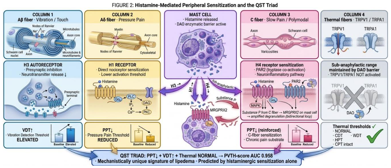
Figure 2.
The gfWAT-IIT2 endotype cascade, coded by evidence level. Three trigger classes (hormonal-transition, microbial/metabolic, local vascular) converge on per-cell mast cell activation in susceptible gluteofemoral white adipose tissue, against an adipo-vascular/connective germline substrate (top). Mast cell degranulation releases histamine (H1/H3/H4), tryptase (PAR2), and other mediators; core downstream nodes include type 2 polarization (ILC2/eosinophil-derived IL-4/13, M2 macrophages), the sensory triad (PPT↓, VDT↑, thermal normal), perivenular fibrosis, and pain-relevant clinical features (fibromyalgia, sarcopenic-valgus). Exploratory systemic or neuropsychiatric nodes shown in the figure are coded as dotted and are treated only as non-load-bearing extensions in Section 9. Line coding indicates evidence level: solid = direct evidence in lipedema; dashed = indirect or inferred from related fields; dotted = hypothetical or untested. The germline substrate and the QST pattern are solid; M2 polarization and tryptase-fibrosis are mixed; the ERα-mast cell trigger, ILC2/eosinophil source, DAO source, and ADHD/HNMT links are dashed or dotted.
Figure 2.
The gfWAT-IIT2 endotype cascade, coded by evidence level. Three trigger classes (hormonal-transition, microbial/metabolic, local vascular) converge on per-cell mast cell activation in susceptible gluteofemoral white adipose tissue, against an adipo-vascular/connective germline substrate (top). Mast cell degranulation releases histamine (H1/H3/H4), tryptase (PAR2), and other mediators; core downstream nodes include type 2 polarization (ILC2/eosinophil-derived IL-4/13, M2 macrophages), the sensory triad (PPT↓, VDT↑, thermal normal), perivenular fibrosis, and pain-relevant clinical features (fibromyalgia, sarcopenic-valgus). Exploratory systemic or neuropsychiatric nodes shown in the figure are coded as dotted and are treated only as non-load-bearing extensions in Section 9. Line coding indicates evidence level: solid = direct evidence in lipedema; dashed = indirect or inferred from related fields; dotted = hypothetical or untested. The germline substrate and the QST pattern are solid; M2 polarization and tryptase-fibrosis are mixed; the ERα-mast cell trigger, ILC2/eosinophil source, DAO source, and ADHD/HNMT links are dashed or dotted.
Re-analysis of public transcriptomic data. Several statements below rest on our own re-analysis of a publicly available bulk RNA-seq dataset of lipedema versus control adipose tissue (the multi-omics cohort of Straub et al. [17]). Because those data are openly deposited, the analysis is fully reproducible, and we summarize here what it showed so that readers can verify or extend it. The pipeline is reproducible from the raw data (transcripts quantified with salmon and summarized to gene symbols with tximport, then modelled in DESeq2: a baseline design of ~site + group yielded 1,501 differentially expressed genes, whereas re-specifying it as ~site + leukocyte-fraction + group, the leukocyte fraction estimated by deconvolution, collapsed the count to four). The collapse occurs because estimated blood/leukocyte content correlated with disease status at r ≈ −0.7 and so dominated as a confounder. The apparent cohort-level immune signal, reduced mast cell, M2-macrophage, and canonical NLRP3-inflammasome transcripts, was therefore non-identifiable from cell composition rather than a true biological difference, and no transcriptional lipolytic brake was evident (β-adrenergic receptors and lipases were, if anything, mildly up-regulated). We cite these findings throughout as unpublished data (A.C.M. Amato, unpublished data, 2026), used only to bound what bulk transcriptomics can and cannot establish (Section 9.1 and Section 11.2), not as positive evidence for the endotype.
3. The Adipo-Vascular and Connective Substrate: Upstream Initiating Factors
Before any immune layer, the framework requires an account of why gfWAT accumulates abnormally and becomes vulnerable. We summarize the upstream factors that most plausibly initiate the disease, all of which are independent of the type 2 endotype and would remain valid even if the endotype claim were refuted.
3.1. Genetic and Depot Architecture
The replicated germline architecture of a lipedema phenotype is adipo-vascular and ECM, not immune. A genome-wide association study of a lipedema phenotype replicated VEGFA (endothelial permeability) and GRB14-COBLL1 (insulin/adipocyte signalling) [4], while additional adipo-vascular and ECM loci that shape the gluteofemoral depot, including RSPO3 (WNT/adipogenesis), derive from gynoid-fat and waist-to-hip-ratio genetics [5]. Gluteofemoral fat distribution is the most heritable adiposity trait (SNP-heritability h2 ≈ 0.52 in women), with a genetic architecture nearly independent of visceral and abdominal subcutaneous fat [5]. The depot is also distinguished by an ERβ-enriched estrogen-receptor profile relative to abdominal subcutaneous fat [18,19], and early reviews described familial clustering with a heritable component [20]. Two intracrine mechanisms increase local estrogenic loading. First, AKR1C1/AKR1C2 variants alter local progesterone and androgen metabolism, favouring a locally estrogenic, pro-adipogenic milieu [21,22]. Second, an aromatase (CYP19A1) amplification loop documented in lipedema gfWAT [10] can sustain a locally hyper-estrogenic microenvironment even after systemic estrogen declines, a candidate explanation for why some patients progress, or even present, after menopause.
3.2. The Receptor Shift and a Pro-Adipogenic Estrogen Response
Lipedema adipose-derived stem cells (ASCs) show an ERβ-dominant receptor profile with an altered ERα and GPER response to estrogen [23]. On estrogen exposure, lipedema ASCs fail to mount the proliferative and aromatase (CYP19A1) responses seen in healthy ASCs, yet on adipogenic differentiation they paradoxically up-regulate the adipogenic isoform PPARγ2 and recruit ERα [23], a cell-autonomous, pro-adipogenic (fat-storage) bias that is independent of caloric intake and does not require an inflammatory explanation. Consistent with this, our own unpublished re-analysis of public lipedema adipose RNA-seq finds no transcriptional lipolytic brake (β-adrenergic receptors and lipases are if anything mildly up-regulated), locating diet-resistance in baseline depot biology rather than in a lipedema-specific transcriptional suppression (A.C.M. Amato, unpublished data, 2026). This baseline depot biology accounts for a well-recognized clinical hallmark: the gluteofemoral disproportion persists after weight loss, which lowers fat across depots without correcting the lower-limb predominance [1,9]. The core framework does not require explaining this baseline resistance immunologically. A separate exploratory question, treated in Section 9.1, is whether the active inflammatory and hypoxic milieu can add a reversible functional impairment of limb-fat mobilization distinct from the structural baseline resistance.
3.3. Microvascular and Connective Fragility
Endothelial barrier dysfunction precedes overt morphology: the lipedema stromal-vascular secretome lowers VE-cadherin and raises permeability in healthy endothelium [10]. Connective-tissue laxity and joint hypermobility co-occur in 44 to 60% of patients, frequently from childhood and years before lipedema onset [11]. These features supply the structural, pre-immune trigger surface on which any downstream immune amplification acts.
This substrate is the framework's upstream. The type 2 endotype proposed below is, on present evidence, best read as a downstream amplifier that is present in some patients, not as the germline cause of the disease. We return to this explicitly in Section 4.6 and Section 11.
4. The Type 2 Immune/Mast Cell-Activation Endotype
4.1. Operational Definition: Activity, Not Abundance; A Subgroup, Not All Patients
We define the endotype by function and clinical recognizability, not by cell counts. This distinction is essential and is what reconciles the framework with the tissue data. The construct has two parts: a stable, germline adipo-vascular susceptibility trait (Section 3) and, superimposed on it, a reversible type 2 / mast cell activity state (developed as a two-axis continuum in Section 9.6); what we term the endotype is the subgroup that, on this trait, reaches a high-amplitude activity state. Reading the immune component as a fluctuating state rather than a fixed subtype is what reconciles it with the immunologically quiet cohort average (most patients are sampled at low activity) and what makes the central prediction a within-patient, reversible one (Section 8). The largest histological series found mast cell numerical density (CD117+ cells per high-power field) not significantly different between lipedema and controls [24], and bulk adipose transcriptomes do not show a uniformly mast cell- or M2-enriched tissue (see Section 4.6). The framework therefore does not claim that lipedema tissue is, on average, mast cell-rich. It claims that, in an identifiable subgroup, mast cells are hyper-secretory per cell, consistent with elevated tissue histamine (2.2-fold; ref. 14) against unchanged cell numbers, and that this functional state, not cellular expansion, is the pathological substrate of the endotype.
Operationally, we propose that the endotype is recognizable at the bedside by: (i) cutaneous vasomotor signs (erythema, flushing, warmth of the affected fat); (ii) an atopic or mast cell-activation diathesis (rhinitis, urticaria, dermographism, features compatible with mast cell activation syndrome, MCAS); and (iii) the characteristic quantitative sensory testing pattern (reduced pressure pain threshold with elevated vibration detection threshold) together with, where measurable, elevated local or urinary histamine-pathway metabolites. This is the falsifiable heart of the proposal: the endotype is the subgroup in whom these pre-treatment features co-segregate, and the central hypothesis is that they predict response to mast cell-directed therapy (Prediction P3), a test we deliberately keep separate from the defining features to avoid circularity. To make the construct operational and testable, we propose a set of candidate, treatment-naïve baseline variables for prospectively identifying the active state, summarized in Box 1.
Box 1 | Candidate baseline variables for prospective identification of the gfWAT-IIT2 active state (research use only; treatment-naïve).
The active state should be identified before treatment exposure through a multidomain baseline assessment. Candidate variables include cutaneous vasomotor signs of the affected depot (erythema, flushing, or warmth); an atopic or mast cell-activation diathesis (urticaria, dermographism, rhinitis, or other features compatible with mast cell activation syndrome); the quantitative sensory testing pattern of reduced pressure pain threshold with elevated vibration detection threshold and preserved thermal thresholds, where available; and, where measurable, local or urinary histamine-pathway metabolites. Asymmetric disease with a unilateral local vascular trigger, and comorbid fibromyalgia, may be supportive.
These are candidate variables, not diagnostic criteria: they should not yet be assigned fixed weights or thresholds, and they require prospective evaluation to determine whether they co-segregate and predict pharmacological response. Response to mast cell-directed therapy is deliberately excluded, since it is the framework's prospective test of the state (Prediction P3), not a defining feature.
A caveat applies to the evidence behind this definition. The QST triad, the mast cell density and per-cell activity data, and the bulk transcriptomic signatures are all drawn from unstratified lipedema cohorts and have not yet been shown to co-segregate specifically within the subgroup these variables define. Applying cohort-level findings to the proposed subgroup is therefore itself a hypothesis: if the QST triad proves equally common in patients without vasomotor or atopic features, or if mast cell stabilization alters it irrespective of those features, the subgroup loses its specificity and the framework reverts to a continuous severity axis rather than a discrete state. This is precisely what predictions P1 to P4 are designed to test.
4.2. Triggers and Mast Cell Activation
Three classes of stimulus can drive mast cell activation in susceptible gfWAT, each via a distinct entry point (Figure 2):
- Hormonal-transition. The best-characterized candidate route is estrogenic: estradiol triggers rapid non-genomic degranulation in mast cell lines via ERα [25]. Whether gfWAT mast cells express estrogen receptors at functionally relevant density is not established, and tissue studies at other estrogen-responsive sites are inconsistent: estrogen can activate mast cells in ovarian endometriomas [26], yet estrogen receptors are not consistently detected on the mast cells themselves. This mechanism is therefore, at this stage, an indirect extrapolation formalized as a prerequisite test (Prediction P2). Clinically, the trigger maps to hormonal-transition windows rather than to an absolute sex-steroid level: onset clusters at puberty and around the menopausal transition, and exacerbation is reported with hormonal contraceptives by 58.8% of patients [27]. Because several of these windows (pregnancy, combined and progestin-containing contraception) are not purely estrogenic, the framework frames this mast cell-activation trigger as the hormonal transition itself, distinct from the local intracrine estrogen loading that shapes the adipogenic substrate (Section 3.1); whether transitions and stable exposure differ in their capacity to activate gfWAT mast cells is itself testable.
- Microbial/metabolic. Intestinal-derived lipopolysaccharide (LPS) primes the NLRP3 inflammasome (TLR4 → NF-κB → canonical NLRP3). A second, lipedema-specific link from the gut to the inflammasome runs through the MIF-CD74 axis: macrophage migration inhibitory factor and its receptor CD74 are required for NLRP3 activation and are elevated, at the mRNA and cellular level, directly in lipedema subcutaneous adipose independently of body mass index [28], while a gut MIF-CD74-NF-κB-NLRP3 pathway is documented at the intestinal barrier [29], positioning MIF-CD74 as a candidate upstream node feeding the inflammasome. β-hydroxybutyrate (BHB), produced on a ketogenic diet, inhibits NLRP3 assembly [30], a candidate mechanism for the diet's reported efficacy independent of caloric restriction [31]. We note that NLRP3 activity has not been directly measured in lipedema tissue; our unpublished re-analysis finds the canonical inflammasome transcripts reduced at the cohort level, a finding that is non-identifiable from cell composition in bulk data (A.C.M. Amato, unpublished data, 2026) and is therefore neither confirmation nor refutation (Section 4.6).
- Local vascular. Venous stasis and local hypoxia can prime NLRP3, plausibly through mitochondrial reactive oxygen species, with the non-canonical pathway (caspase-4/5/11) engaged where cytosolic microbial products are present; this trigger class is supported by the asymmetric-lipedema observation (Section 5.2).
Beyond these discrete triggers, the substrate itself supplies a mechanical route to the same node: the altered extracellular matrix and connective laxity (Section 3) raise interstitial tension that engages the mechanosensitive channel PIEZO1 on mast cells, driving degranulation, a response sensitized by the type 2 alarmin interleukin-33 and clinically echoed by dermographism and a mechanically evoked Darier-type sign [32]. This route is, however, cell-specific rather than a single vector: PIEZO1 is pro-inflammatory in mast cells but restrains type 2 innate lymphoid cells [33], so mechanical load is expected to modulate, not uniformly drive, the type 2 milieu, consistent with the balance-based reading the framework applies elsewhere (Section 10.4). Two further inputs are noted only briefly, as candidate modulators rather than core triggers: a systemic mast cell activation syndrome [34] is a trait-level constitutional amplifier (not itself a local trigger) that should lower the degranulation threshold and define a candidate high-responder subgroup (Prediction P16), and physical exertion is hormetic, protective in moderation but a candidate behavioural precipitant only in chronic overtraining via exercise-induced intestinal permeability feeding the microbial node [35,36,37]. These entry points act as a convergence hub rather than a closed list: a broad range of clinically reported precipitants, from hormonal transitions and gut-derived endotoxin to venous stasis, mechanical load, and psychological stress, each feed one or more of the same nodes, so that it is the convergence on per-cell mast cell activation, not any single trigger, that the framework asserts.
4.3. ILC2s, Eosinophils, and M2 Polarization Within the Endotype
In lean (non-obese, homeostatic) adipose tissue, eosinophils sustain M2 macrophage polarization via interleukin-4/13 without adaptive T cells [38]. In lipedema gfWAT, a dominant M2 transcriptomic signature with in-vitro functional causality has been reported [7], and CD163+ M2 enrichment specific to lipedema (versus lipohypertrophy and lymphedema controls) by an orthogonal histological (immunohistochemistry and qPCR) cohort [39], even though, as noted, total bulk immune content is not uniformly elevated. The cellular source of interleukin-4/13 in lipedema gfWAT (ILC2, eosinophil, or mast cell) has not been characterized and is a critical open question (Prediction P1). We flag explicitly that the M2 signal is an area where orthogonal methods disagree (enrichment by CyTOF/immunohistochemistry; reduction in some bulk datasets), which we attribute to composition confounding and resolve only by single-cell methods (Section 4.6).
4.4. Histamine and the Sensory Signature: A Parsimonious, Not Exclusive, Explanation
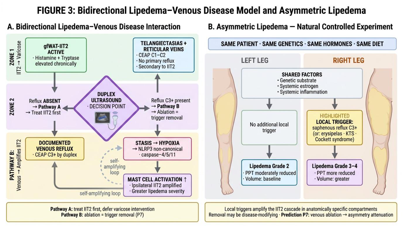
The framework offers what we believe is the first explicit mechanistic account of the lipedema QST triad, abnormality in exactly 2 of 13 parameters (reduced pressure pain threshold, PPT↓; elevated vibration detection threshold, VDT↑) with normal thermal thresholds, the only published pattern achieving near-perfect diagnostic classification [13] (Figure 3):
- PPT↓, histamine sensitizes Aδ fibres via H1 and C fibres via H4 receptors; mast cell tryptase co-activates C fibres via PAR2.
- VDT↑, two non-exclusive routes are plausible: histamine acting on presynaptic H3 receptors is proposed to suppress Aβ (vibration-sensing) transmission, but the large myelinated Aβ fibres are also the most vulnerable to ischemia and compression, so part of the VDT elevation may be structural rather than mediator-driven. On either reading the robustly mediator-dependent component of the signature is carried by PPT; this fibre-specific assignment is a framework-derived inference and is not yet directly demonstrated.
- Thermal sparing, predicted if tissue histamine stays within a sub-anaphylactic range acting on classical H-receptors rather than reaching concentrations that modulate thermal transducers; this thermal-sparing claim is framework-derived and untested (Prediction P3).
Figure 3.
Histaminergic peripheral sensitization as a parsimonious account of the QST triad. Aβ fibres (presynaptic H3) → VDT↑; Aδ fibres (H1) → PPT↓; C fibres (H4 + tryptase-PAR2) → PPT↓ and a neuroinflammatory substrate; thermal fibres predicted unaffected at sub-anaphylactic histamine. Competing peripheral, central, and nociplastic mechanisms may co-operate, and the model claims to explain the selectivity of the pattern, not to exclude other contributors.
Figure 3.
Histaminergic peripheral sensitization as a parsimonious account of the QST triad. Aβ fibres (presynaptic H3) → VDT↑; Aδ fibres (H1) → PPT↓; C fibres (H4 + tryptase-PAR2) → PPT↓ and a neuroinflammatory substrate; thermal fibres predicted unaffected at sub-anaphylactic histamine. Competing peripheral, central, and nociplastic mechanisms may co-operate, and the model claims to explain the selectivity of the pattern, not to exclude other contributors.
Lipedema pain is multifactorial. Histaminergic peripheral sensitization is presented as a parsimonious mechanistic explanation for the depot-localized component of the sensory signature, not as the sole driver of lipedema pain. Competing and complementary mechanisms, small-fibre neuropathy with stage-dependent CGRP/NGF elevation, peripheral sensitization independent of histamine, and central/nociplastic mechanisms, are real and likely co-operate; QST localizing a small-fibre abnormality to the thigh with a normal hand argues against global central sensitization as the whole story but does not exclude a central contribution. We claim only that the specific 2-of-13 selectivity is more naturally predicted by differential histamine-receptor action on fibre classes than by mechanical load, lymphatic insufficiency, or non-specific inflammation, each of which would be expected to perturb additional parameters. That histamine H1 signalling can sensitize peripheral nociceptors in vivo is established in irritable bowel syndrome, where submucosal nociceptors are potentiated in an H1-dependent manner and the H1 antagonist ebastine reverses both the sensitization and the symptoms [40]. We cite this as proof of concept that H1 can sensitize nociceptors, not as evidence for the transducer: in that model the H1 target is TRPV1, a noxious-heat transducer, whereas in lipedema the heat-pain threshold is preserved (one of the eleven normal QST parameters). That preserved heat-pain threshold is informative rather than incidental, since it constrains tissue histamine below the level that would sensitize TRPV1-bearing heat nociceptors and localizes the effect to the mechanical, pressure-pain pathway, consistent with the sub-anaphylactic range proposed above (Prediction P3). Critically, PPT reduction is independent of body mass index and persists after BMI matching even as tissue-stiffness differences disappear [41], arguing against adipose mechanical load as the nociceptive driver. This signature is best read as a marker of the active state rather than of lipedema universally: it is expected in symptomatic, immunologically active patients, whereas clinically painless or quiescent presentations, in which the activity axis is low (Section 9.4), need not display the pressure-pain component. The strong diagnostic performance reported for the triad [13] is therefore most applicable to symptomatic cohorts.
4.5. Mediator Containment and Tissue Fibrosis
Local histamine containment may depend on tissue-level degradation (diamine oxidase, DAO; histamine N-methyltransferase, HNMT), creating a concentration window high enough to sensitize local nociceptors but below the systemic threshold, whereas circulating histamine has not been directly measured [14]. The cellular source of local DAO in gfWAT is not demonstrated and is a testable component (Figure 2, dashed). Mast cell tryptase acting on PAR2 can drive perivenular fibrosis [42]; progressive fibrosis raising compartmental pressure is consistent with the elevated Doppler resistance index and the steatonecrosis/neoangiogenesis/hemosiderin histology reported in lipedema nodules [43]; histological elastic-fibre fragmentation, consistent with (but not proving) mast cell elastase activity, has also been described [44]. Beyond mediator-driven sensitization, these fibrotic and pressure changes plausibly add a second, structural nociceptive route: rising interstitial and compartmental pressure with reduced microvascular perfusion can generate ischemic, metabosensitive pain, consistent with the elevated Doppler resistance index and the steatonecrosis observed in painful nodules [43]. The painful nodule thus co-localizes both routes: mast cell-driven capillary fragility (hemosiderin) and local ischemia (steatonecrosis, with VEGF-driven neoangiogenesis), which may explain its ultrasonographic-histological correlation. This ischemic component is the candidate identity of the stage-dependent structural contribution to pain (Section 5.1), and it feeds back into the cascade, since local hypoxia can prime NLRP3 (Section 4.2). We note that interstitial fibrosis has also been reported early, before adipocyte hypertrophy. If confirmed, this would place an M2/TGF-β-mediated fibrotic component upstream of, or parallel to, the mast cell-mediated component rather than purely downstream of it (Section 6). The framework accommodates this as a two-phase fibrotic process but does not require it.
Whether the active state also contributes to depot expansion is an important but separate question. Because that adipogenic feedback loop is currently inferred almost entirely from non-lipedema adipose biology, it is not treated as a pillar of the core neuroimmune framework; it is repositioned as an exploratory hypothesis in Section 9.1.
4.6. Alternative Interpretations of the Mast Cell Findings
Scientific candour requires stating that the mast cell-centred reading is not the only one compatible with the data, and may not be the primary one. Three alternatives deserve explicit weight:
- Mast cell activation as a secondary response. Mast cells are exquisitely sensitive to tissue stress. Their activation in lipedema may be a consequence of adipo-vascular dysfunction, hypoxia, ECM remodelling, or chronic low-grade tissue injury rather than an initiating event. The germline GWAS architecture (Section 3.1), which is adipo-vascular and not immune, is most consistent with mast cell activation being downstream; this is exactly why we frame it as an endotype amplifier, not the disease root.
- The tissue immune signal is non-identifiable from bulk data. In an unpublished re-analysis of the largest public lipedema adipose RNA-seq dataset, the apparent cohort-level immune signal (including reduced mast cell, M2, and NLRP3 transcripts) collapses when cell-type composition is modelled. Genome-wide-significant genes fall from ~1,500 to a handful, because blood/leukocyte content correlates with disease status at r ≈ −0.7 (A.C.M. Amato, unpublished data, 2026). Bulk transcriptomics therefore can neither establish nor exclude active type 2 inflammation in lipedema. This is precisely why the present framework is pitched at the level of per-cell activity in a subgroup and clinical stratification, not bulk tissue abundance, and why its confirmation requires single-nucleus profiling of the clinically responsive subgroup.
- Heterogeneity. Even if a mast cell-active state is real, it may account for only a subset of patients; the cohort average may be immunologically quiet. This is the core prediction of the endotype concept and the reason patient stratification, not a universal mechanism, is the framework's central translational claim. Consistent with such a gradient, some cohorts report a systemically pro-inflammatory profile (elevated C-reactive protein and IL-6, reducible by diet) [45], whereas lean, metabolically protected patients are systemically quiet; the framework reads this "inflamed versus quiet" spread as endotype and severity variation rather than as contradiction.
These alternatives do not falsify the framework; they bound it. The endotype claim is falsified only if, in the clinically defined responsive subgroup, per-cell mast cell activity and the histaminergic signature are also absent (Section 8, P3).
5. Clinically Testable Downstream Features Within the Endotype
The features below remain within the focused neuroimmune framework because they concern pain propagation, local-trigger amplification, or clinically testable consequences of the mast cell-active state. Broader systemic immunomodulatory and HLA/gluten material has been moved to Section 9.
5.1. Fibromyalgia and Central Sensitization
Fibromyalgia affects 35 to 40% of lipedema patients [15,16] and lipedema is found in ~50% of fibromyalgia patients [46]; comparative phenotyping shows substantial symptom overlap with preserved objective walking capacity [47]. The framework reads this, cautiously, as central-sensitization progression of cumulative peripheral nociceptive drive (peripheral histamine → repeated nociceptor activation → dorsal-horn wind-up), reinforced by the MRGPRX2 neuro-immune loop [48]. We state plainly that comorbidity is association, not demonstrated mechanistic continuity: fibromyalgia may equally amplify perceived lipedema pain without a shared origin. The framework's distinguishing, testable claim is narrower: that a component of the pain and fibromyalgia burden tracks cumulative mediator exposure (for example serum tryptase) over and above morphological stage, detectable as a mediator-pain association that survives adjustment for stage, rather than that pain is independent of stage. The evidence on the stage-pain relationship is mixed, with at least one cohort reporting no stage-severity correlation [16]; the framework accommodates a dual contribution in which a structural component scales with stage while a mediator-linked component does not. Consistent with peripheral nociceptor sensitization rather than adipose bulk, a mixed-methods study reports a “touch paradox” in lipedema, in which light tactile stimulation aggravates rather than relieves pain, a hallmark of mediator-sensitized nociceptors [49]. Because the same mast cell axis that sensitizes nociceptors, through MRGPRX2 and tryptase-PAR2, is documented in fibromyalgia itself [50], the framework makes a directly falsifiable corollary: in patients carrying both diagnoses, mast cell-directed or anti-inflammatory treatment of the lipedema should reduce the fibromyalgia-attributed symptom burden, not only the limb pain.
5.2. Asymmetric Lipedema as a Natural Experiment
When one limb carries an additional local trigger that the contralateral limb lacks, the patient serves as their own control: genetics, hormones, diet, and systemic inflammation are held constant, and only the local trigger differs. Such patients reproducibly show greater severity in the affected limb (Amato et al., asymmetric-lipedema case series, under review), which is the framework's strongest causal argument. It licenses the inference that local triggers modulate the magnitude of disease expression, and motivates trigger removal as a candidate disease-modifying intervention (Prediction P7), distinct from, and stronger than, the cross-sectional associations elsewhere in this section.
This pattern is documented in a retrospective case series of six women, in each of whom the more affected limb carried an identifiable local condition, Klippel-Trénaunay syndrome, prior limb trauma, saphenous or iliac (Cockett) venous insufficiency, recurrent erysipelas, an angiomyomatous hamartoma, or Hashimoto thyroiditis with local tendinitis, and showed greater ipsilateral subcutaneous fat by ultrasound and segmental bioimpedance (interlimb fat difference 1.5 to 7.1 percentage points) (Amato et al., asymmetric-lipedema case series, under review). That mechanistically distinct conditions converge on the same directional outcome argues that a shared local inflammatory or stasis milieu, rather than any single pathway, scales disease magnitude, a within-patient observation consistent with the immune layer acting as a modifiable amplifier.
Two caveats bound the inference. First, directionality is unambiguous only where the local condition is congenital or datable: the vascular malformation, the hamartoma, and the prior trauma clearly precede or are independent of lipedema onset and so exclude reverse causation, whereas the acquired venous insufficiency and recurrent erysipelas are bidirectional, lipedema can itself promote them, and are therefore read as a feedback loop (Section 5.3) rather than as evidence of trigger direction. Second, the ipsilateral excess must be shown to reside in lipedematous fat rather than in secondary edema or lymphedema, distinguishable by subcutaneous fat thickness on ultrasound, the nodular tissue signature, and the ipsilateral sensory (QST and pain) pattern, not by limb volume alone.
5.3. Bidirectional Interaction with Venous Disease
We propose that varicose disease in lipedema is both a consequence and a cause of the inflammatory cascade, and that distinguishing the two directions is clinically consequential. In the first direction (Pathway A), the endotype damages the vessel wall: histamine-driven vasodilation and permeability, together with the mast cell-protease mechanism detailed below, can generate telangiectasias and reticular varices (CEAP C1 to C2) as a secondary, endotype-driven phenomenon. In the second direction (Pathway B), established chronic venous insufficiency (CEAP C3+) feeds back into the cascade: venous stasis and hypoxia prime NLRP3 and sustain a local inflammatory milieu that can perpetuate and amplify the lipedema phenotype. A clinically important corollary follows for the many patients who carry both conditions: in advanced venous disease, part of the cardinal lipedema symptom burden, limb heaviness, aching, and fatigue, may be the inflammatory expression of the lipedema endotype triggered by the coexisting venous insufficiency, rather than a feature of the venous disease alone. This predicts that correcting the venous trigger (for example, ablation of refluxing segments) should attenuate not only the venous symptoms but also the mediator-linked component of the lipedema phenotype, and it cautions against attributing the whole symptom burden to either disorder in isolation (Predictions P7, P9). Duplex ultrasound is proposed as the decision point that separates the two directions (Figure 4).
A direct molecular mechanism plausibly links mast cell activation to the vessel wall, supporting the first direction. Mast cell proteases cleave endothelial tight-junction (zonula occludens-1) and adhesion (CD31/PECAM-1) proteins, producing dose-dependent hyperpermeability and microvascular leak [51], while tryptase signalling through protease-activated receptor 2 further induces intercellular adhesion molecule 1 and interleukin-8 [52], a candidate basis for the easy bruising and capillary fragility that are clinical hallmarks of lipedema. The same proteases activate pro-matrix-metalloproteinases [53] that degrade the vessel-wall matrix and drive the post-thrombotic vein-wall remodelling and fibrosis on which reflux is built [54]. Consistent with a causal rather than bystander role, mast cell-deficient animals are protected from venous thrombosis and the phenotype is restored by mast cell reconstitution [55]; because mast cells also serve protective, barrier-homeostatic functions, this proteolytic activity is one to recalibrate rather than abolish [56]. These are mechanistic proposals requiring prospective validation.
5.4. Musculoskeletal Downstream
In the active state, the local inflammatory milieu could blunt the skeletal-muscle anabolic response to exercise, an inflammation-associated anabolic resistance consistent with the finding that women with lipedema have lower limb strength than body-mass-index-matched women with obesity, contributing to a thigh-predominant sarcopenia and dynamic-knee-valgus cascade that culminates in patellofemoral overload and chondromalacia, amplified by the hypermobility substrate (Section 3.3) [57]; this cascade plausibly underlies the high prevalence of knee pain in lipedema, reported in 58.1% of screen-positive women [2]. Independent of our own data, the principle that knee-region fat can adopt an inflammatory, mast cell-enriched phenotype is established in osteoarthritis, where the infrapatellar fat pad shows a depot-specific inflammatory signature [58]. This effect is expected in the active state rather than in the metabolically quiet substrate, and is offered as an inference, not an established pathway.
6. Comparison with Existing Pathophysiological Frameworks
The 2023 Lipedema Foundation Research Roadmap, developed through international expert consultation, concludes that lipedema pathophysiology remains incompletely understood and that the field still lacks consensus on diagnostic criteria, with the mechanistic landscape, inflammatory mechanisms, vascular dysfunction, lymphatic alterations, fibrosis/ECM remodelling, adipose stem-cell abnormalities, hormonal influences, and adipose-microenvironment changes, still under active investigation [59]. The present framework should be positioned within, not above, this landscape (Table 1). Its distinctive contribution is narrow and explicit: it is the only model that generates a specific, testable mechanistic prediction for the diagnostically validated QST signature, and the only one that operationalizes a treatable subgroup (endotype). Most other observations in lipedema are compatible with several of these models simultaneously; we do not claim the endotype framework supersedes them, only that it explains a specific cluster the structural models leave open.
7. Evidence Summary
8. Model-Derived Predictions
Predictions are separated into core tests (P1 to P4; directly test the framework's central claims and depend minimally on unverified assumptions) and exploratory extensions (P5 to P16; valuable but dependent on links that are themselves not yet demonstrated, so a negative result constrains rather than falsifies the core). For each, we state the expected outcome, the alternative outcome, and the interpretation if the prediction fails (Table 3 gives experimental detail and falsifying thresholds).
Core predictions
P1. ILC2/eosinophil source of IL-4/13. Expected: single-cell/nucleus RNA-seq of lipedema gfWAT shows expanded ILC2/eosinophils versus matched controls. Alternative: no expansion. If it fails: the upstream of M2 polarization requires an alternative source (adipocyte TSLP/IL-33); weakens but does not falsify the endotype (which is defined by mast cell activity).
P2. Estrogen-receptor profile of gfWAT mast cells. Expected: tryptase+ cells express ERα at density sufficient for non-genomic degranulation. Alternative: ERβ-dominant or ER-absent. If it fails: the estrogenic-trigger mechanism must be revised (e.g., indirect estrogen action); the endotype can still hold via microbial/vascular triggers.
P3. Mast cell stabilization reduces tissue histamine and improves the mediator-dependent QST component in proportion to baseline activity. Expected: across patients stratified before treatment by a continuous activity score (the Box 1 variables), a systemically active mast cell-directed agent produces, within 4 to 8 weeks, a larger fall in tissue histamine and a larger rise in pressure pain threshold (PPT) in higher-activity than in lower-activity patients; the vibration detection threshold (VDT), which may carry a structural large-fibre component, is expected to respond less or not at all. Framing the test as a continuous baseline-activity-to-response relationship, rather than a binary active-versus-burnt-out split, keeps it falsifiable by avoiding retrospective reclassification of non-responders. Delivery caveat: oral cromolyn is poorly absorbed and acts largely on the gut mucosa, so a negative result with an enteral agent would indicate failure to reach gluteofemoral adipose rather than failure of mechanism; the test requires a route or agent that reaches the depot. Alternative: no relationship between baseline activity and the change in histamine or PPT. If it fails: the histaminergic-sensitization claim, the framework's central, falsifiable prediction, is refuted, and the QST pattern must be otherwise explained. (Preliminary tissue-histamine reduction by cromolyn shown in a 3-patient series; QST effect untested; ref. 14.)
P4. Ketogenic diet reduces inflammasome output proportional to analgesia. Expected: serum IL-18/IL-1β, tryptase, urinary histamine metabolites fall in proportion to pain improvement on a ketogenic diet. Alternative: analgesia without biomarker change. If it fails: the BHB→NLRP3 mechanism is not the analgesic route; given that our unpublished re-analysis already finds inflammasome transcripts non-identifiable in bulk (A.C.M. Amato, unpublished data, 2026), a failure would favour a non-inflammasome analgesic mechanism of the diet. Notably, a randomized trial has already reported pain reduction on a low-carbohydrate diet without correlated changes in circulating cytokines or fibrosis-associated markers [64]; P4 is therefore, in part, already challenged at the cohort level, and the framework predicts that the inflammasome-analgesia coupling, if present, should emerge specifically in the microbial/metabolic-trigger endotype (elevated HOMA-IR) rather than in unstratified patients.
Exploratory predictions (P5 to P16)
These test downstream or systemic extensions and are summarized in Table 3 with falsifying thresholds: P5 (mast cell stabilization improves ADHD scores), P6 (H3 inverse agonist normalizes VDT and improves ADHD), P7 (trigger removal attenuates asymmetric lipedema), P8 (lipedema fibromyalgia is mast cell- not IgG-mediated), P9 (venous-ablation sequencing modifies recurrence not morphology), P10 (TLS in gfWAT), P11 (cromolyn restores browning markers), P12 (cellulite as intermediate activation), P13 (mast cell stabilization lowers lisdexamfetamine dose), P14 (GLP-1 response tracks metabolic not morphological burden), P15 (gluten-free diet lowers symptoms in HLA-DQ2/DQ8-positive lipedema), P16 (comorbid systemic mast cell activation syndrome predicts greater response to mast cell-directed therapy). Predictions P5, P6, P13 (the ADHD/HNMT axis) are the most speculative and are discussed as exploratory extensions in Section 9.
9. Exploratory Extensions Outside the Core Neuroimmune Framework
Scope of this section. The items below are explicitly secondary extensions of the focused neuroimmune model. The core model is the adipo-vascular substrate, the per-cell mast cell / type 2 active state, and the histaminergic QST-pain axis described in Section 3 to 5. The extensions below ask whether that active state also relates to adipose expansion, HLA/gluten and gut-immune inputs, systemic immune modulation, cancer-related associations, ADHD-related mechanisms, tertiary lymphoid structures, environmental triggers, or broader clinical continua. None is required by the central mast cell-histamine-QST-pain hypothesis, and each is graded Indirect or Hypothetical. They are presented to generate future research, not as established components or as parallel pillars of the main framework.

Table 4.
Extensions beyond the core neuroimmune model: evidence status and relationship to the core. (Direct = demonstrated in human lipedema; Indirect = demonstrated in related fields and extrapolated; Hypothetical = plausible but untested. None is required by the core mast cell-histamine-QST-pain hypothesis.).
Table 4.
Extensions beyond the core neuroimmune model: evidence status and relationship to the core. (Direct = demonstrated in human lipedema; Indirect = demonstrated in related fields and extrapolated; Hypothetical = plausible but untested. None is required by the core mast cell-histamine-QST-pain hypothesis.).
| Extension | Evidence level | Relationship to the core neuroimmune model |
| Candidate adipogenic feedback loop, including PAR2, YAP/TAZ, and HIF-1α (Section 9.1) | Indirect (all links extra-lipedema) | Possible explanation for how the active state might influence depot growth; not part of the pain mechanism |
| Gluten / HLA-DQ2/DQ8 diversion route (Section 9.2) | HLA carriage Direct (our cohort); diversion mechanism Indirect/Hypothetical | Candidate exogenous input to the framework's existing microbial/intestinal-permeability trigger node; not a new node |
| Systemic immune "shield" and immunoglobulin paradox (Section 9.3) | Hypothetical (proxy associations Indirect) | Possible downstream/systemic consequence of the type 2 milieu; not part of the pain mechanism |
| Cancer-related associations and tertiary lymphoid structures (Section 9.3; P10) | Hypothetical (TLS untested in lipedema) | Weak corollary of the systemic-shield idea; not required by the neuroimmune framework |
| MCAS as constitutional amplifier (Section 4.2; P16) | Indirect/Hypothetical | Trait-level lowered degranulation threshold intensifying the core mast cell axis |
| Exercise hormesis / overtraining (Section 4.2) | Indirect | One behavioural input converging on the existing microbial/metabolic node |
| ADHD / HNMT axis (Section 9.4) | Hypothetical (single unreplicated report) | Most speculative extension; a candidate central-histamine corollary |
| Exploratory treatment-response predictions (P5 to P16, Table 3) | Exploratory tier | Downstream/systemic tests; segregated from core predictions P1 to P4 |
The following subsections give the narrative for the extensions that warrant it; the framework does not depend on any of them.
9.1. Candidate Adipogenic Feedback Loop
A theory of lipedema may ultimately need to account for disproportionate gluteofemoral fat accumulation as well as pain. This subsection asks whether the neuroimmune active state might also feed back onto tissue growth, but it is explicitly exploratory and non-essential to the core mast cell-histamine-QST-pain framework. An in vivo signal that it may do more is pharmacological, though contested: in diet-induced obesity, mast cell deficiency and cromolyn stabilization reduced adipose mass and metabolic dysfunction [68], an effect later attributed in part to the Kit mutations of the original mast cell-deficient models and not consistently reproduced in mast cell-specific lines, so it is suggestive rather than established; less Kit-dependent pharmacology points the same way, with cromolyn combined with H1 antihistamine blockade lowering diet-induced adiposity [69]. A plausible adipogenic loop would link the active state to tissue growth, with mechanosensing now recognized as central to adipose biology [70]: tryptase signalling through adipocyte PAR2 couples mast cell activation to adipocyte metabolic dysfunction [71]; histamine-driven oedema and the mechanical strain it imposes are pro-adipogenic [72] and engage the mechanosensitive YAP/TAZ programme that also feeds innate-immune inflammation [73,74]; and the attendant local hypoxia drives adipose fibrosis and dysfunctional expansion through hypoxia-inducible factor 1α in adipocytes and myeloid cells [75,76], while also priming NLRP3 (Section 4.2 and Section 4.5). If correct, this would make mast cell-directed therapy a candidate disease-modifying, not merely analgesic, intervention, testable against limb volume. However, every link is inferred from adipose biology outside lipedema and none is yet demonstrated in the disease; the loop is therefore a mechanistic extension, not a central pillar.
9.2. HLA/Gluten and Gut-Immune Trigger Extensions
As one concrete exogenous instance feeding the framework's microbial trigger node (Section 4.2), dietary gluten in the HLA-DQ2/DQ8-positive subgroup, the alleles being carried more often in lipedema than in comparators [77,78,79], is proposed to be diverted away from the Th1 villous-destructive celiac pathway toward an innate, intestinal-permeability route that feeds the same NLRP3/microbial node, two faces of one HLA-restricted, cytokine-context-dependent mechanism [80,81,82,83]. Independent support that gluten in this genetic context yields reversible symptoms without the full celiac lesion comes from a meta-analysis of potential coeliac disease (symptomatic improvement on a gluten-free diet in approximately 88%, 95% CI 79 to 97) [84]; our own observation of improvement on gluten withdrawal is uncontrolled and is formalized as Prediction P15. The mechanistic detail of this gluten/HLA route is developed in our published work [77] and is presented here only as a candidate trigger, not as a core component.
9.3. Systemic Immune Modulation, Tertiary Lymphoid Structures, and Disease Protection
The following material is a downstream, systemic corollary of the type 2 milieu, not part of the core neuroimmune axis. M2-derived adiponectin can dampen Th1 inflammation without impairing antitumoral immunity [85], an "immunological shield" consistent with convergent but anthropometric-proxy associations from our own group [86]: reduced gynoid fat in celiac disease and higher HLA-DQ2/DQ8 carriage with lower clinical celiac expression [77,78], a paradoxical immunoglobulin profile [87], and a candidate perivascular tertiary-lymphoid-structure component associated with improved oncological prognosis [88,89]. The shield also predicts an ordered three-stratum autoimmune gradient (for Th1 conditions such as Hashimoto thyroiditis: lowest in gynoid-predominant women generally, highest in symptomatic lipedema, where conditioning on the activated state selects for a shared intestinal-permeability precipitant), which distinguishes it from a simple shared diathesis. Every element here uses an anthropometric proxy rather than clinical diagnosis, has modest and partly non-significant effects, derives predominantly from our own group without independent external replication, and is therefore strictly hypothesis-generating; the relevant tests are reserved as Predictions P8 (immunoglobulin subclasses) and P10 (TLS).
The gluteofemoral depot and its type 2 / M2 milieu may carry protective as well as pathological consequences, a duality recognized for lipedema fat [90]. The best-supported protective component is cardiometabolic: gluteofemoral fat is associated with cardiometabolic protection (Section 3) and tracks with lower diabetes odds, consistent with the protective profile of lower-body adiposity. An antitumoral component is a further, weaker theoretical possibility, suggested only indirectly by the antitumoral compatibility of the type 2 / M2 milieu and its associated tertiary lymphoid structures [85,88,89], and should be interpreted with substantial caution; it is hypothesis-generating only (P10). This possibility has not been independently established and is framed here as one unproven instance of a broader, better-supported disease-protection profile.
9.4. ADHD and a Biphasic Histaminergic Model
We flag at the outset that this is the most speculative element of the framework, presented only to record a testable hypothesis: the lipedema-ADHD association rests on a single, unreplicated report from our own group [91], with no independent confirmation, and the mechanism below has no direct evidence in lipedema. With that caveat, lipedema has been reported to carry a relative risk of 1.42 for ADHD. One hypothetical model is that HNMT polymorphisms reduce central histamine clearance (a pre-pubertal phase) and that post-menarche peripheral histamine load further saturates HNMT (an amplification phase). We emphasize this is unsupported by direct evidence in lipedema and that causal direction is unresolved: shared genetic pleiotropy, ADHD as a pre-pubertal biomarker, and bidirectional gut-behaviour loops are all compatible. The observed clinical effectiveness of lisdexamfetamine is consistent with, but does not validate, the pathway (P5, P6, P13).
9.5. Trigger Removal as Disease Modification
The asymmetric-lipedema experiment (Section 5.2) makes trigger removal a testable hypothesis (P7), but disease-modification claims remain hypothesis-generating until prospective data exist. The supporting asymmetric-lipedema observation is a single-group case series (cited above as under review) and requires independent confirmation.
9.6. The Gynoid-Fat-Lipedema Continuum
The framework implies that gynoid fat and lipedema are not discrete entities but positions on a continuum better described by two partially independent axes than by a single severity scale: a reversible axis of type 2 and mast cell activity (the source of pain, erythema, and the histaminergic signature) and a cumulative axis of structural remodelling (adipose expansion and fibrosis, the time-integral of past activity). Latent gynoid fat is low on both; the painful "inflamed" presentation is high on activity; and an advanced but quiescent presentation, high in volume and fibrosis yet low in pain and mast cell activity, is the predicted burnt-out endpoint of resolved past activity rather than a separate disease. The model therefore predicts that two clinically painless presentations are mechanistically distinct, a never-strongly-activated metabolically quiet substrate and a post-active fibrotic state, separable by a history of prior inflammatory episodes and by residual nodularity; this is consistent with the dual contribution to pain in Section 5.1. That the activity axis is type 2 rather than classically destructive also reconciles the symptomatic "inflamed" phenotype with the anti-inflammatory bulk-tissue signature (Section 4.6): the cardinal signs are mast cell-mediated and vasomotor, not an M1 infiltrate. Cellulite here denotes the dimpled "orange-peel" appearance commonly termed gynoid lipodystrophy [92], a label used inconsistently in lay and clinical settings, which on the continuum view is not a separate disorder but the same gluteofemoral depot biology expressed with only a small degree of the inflammatory-activity and fibrotic axes, and is therefore a candidate intermediate on the activity axis, although it has multiple non-exclusive mechanisms and no study has characterized mast cell activity in cellulite biopsies (P12).
9.7. Environmental Triggers and an Ancestral Framing
The NLRP3 node that primes the cascade is engaged not only by microbial and metabolic signals (Section 4.2) but by a range of everyday environmental stressors: cellular osmotic stress and dehydration induce its effectors interleukin-1β and interleukin-18 [93], psychological stress raises systemic inflammatory tone [94], and nutritional insufficiency both causes and is sustained by immune dysfunction [95]. We have proposed elsewhere that this energy-retaining programme may be ancestral [63], plausibly co-opted from gut defence, the same inflammasome restrains protective type 2 immunity to intestinal helminths [96], which would help explain both the framework's link to gut health and the affected depot's efficiency at conserving energy. On this reading the relevant ancestral threat is malnutrition, a frequent consequence of parasitism, rather than caloric restriction as such, so that modern stressors may trip an adaptation that is now maladaptive. This is offered as an evolutionary hypothesis to organize the trigger concept, not as a tested claim.
10. Translational Implications (Research-Stage Hypotheses)
All therapeutic content in this section is presented as future research directions, not clinical recommendations.
10.1. Endotype-Stratified Trials
The framework's central translational claim is that lipedema trials should stratify by trigger profile and immune endotype (erythema/atopy/MCAS features; tissue histamine; QST composite) rather than by morphological stage, and measure cascade-level biomarkers as secondary endpoints (Figure 5). Pain and limb volume should be primary endpoints; body weight is a confounded surrogate (A.C.M. Amato, unpublished data, 2026).
10.2. Candidate Biomarkers
Tissue/urinary histamine metabolites, serum tryptase, DAO activity, circulating sCD163, and the PPT/VDT QST composite together constitute a multi-domain panel for prospective validation, none is yet established.
10.3. Drug-Repurposing Hypotheses
Mast cell stabilization (cromolyn), H1/H4 and PAF antagonism (rupatadine), and H3 inverse agonism (pitolisant) are mechanistically motivated candidates for proof-of-concept trials in the endotype; colchicine and GLP-1 receptor agonists (with preliminary support from a five-patient exenatide series, ref. 97) address the microbial/metabolic arm; ibudilast (a phosphodiesterase-4/MIF inhibitor) and tranilast (an NLRP3 inhibitor and mast cell stabilizer) target the MIF-CD74 node and the inflammasome-mast cell balance, respectively; IPI-549 (PI3Kγ inhibitor, the only agent tested directly in lipedema tissue, ref. 7) is a candidate for established M2 polarization. None has been tested clinically in lipedema. Conversely, the framework predicts a contraindication: agonists of PPARγ (thiazolidinediones such as pioglitazone) expand subcutaneous adipose and would be expected to worsen rather than relieve lipedema, a falsifiable prediction consistent with the estrogen-driven PPARγ2 adipogenic bias of the depot (Section 3.2).
10.4. A Caution on Systemic Immunosuppression
If a type 2 state confers any of the proposed systemic protection, broad systemic immunosuppression used off-label for lipedema could, in principle, neutralize it; compartment-directed approaches are preferable on mechanistic grounds. This argues for caution and for evaluating the trade-off, not against treating a patient's primary indication. A second, framework-internal caution concerns single-node targeting. The inflammasome and the type 2 / mast cell programmes interact in a context-dependent manner, NLRP3 can both promote and restrain type 2 responses [96,98], so single-node blockade is unpredictable: inhibiting the inflammasome in isolation (including by the ketogenic route proposed above) might in some contexts disinhibit the mast cell and type 2 arm, whereas potent isolated blockade of type 2 signalling (for example, dupilumab or tezepelumab) could remove the proposed type 2 "shield" (Section 9.3). Balance-neutral agents, and combination rather than single-node strategies, are therefore preferable.
10.5. Surgical Sequencing
Duplex ultrasound before venous intervention, and the hypothesis that IIT2 suppression before liposuction may reduce recurrence (relevant given the high residual conservative-therapy requirement after surgery, ref. 99), are proposed as questions for prospective study (P7, P9). A clinical signal already supports the bleeding-risk component: in a meta-analysis of liposuction in lipedema, hematoma was the commonest complication (8.4% versus under 1% in other liposuction series), an excess the authors attribute to the capillary dysfunction and fragility of lipedema [100], and one mitigated by gentler water-assisted technique. This corroborates the mast cell-driven capillary fragility proposed above (Section 5.3) and reinforces the case for mast cell-directed pre-treatment before surgery, although the comparison is indirect and confounded by the more superficial plane, thigh location, and higher volumes of functional liposuction.
11. Discussion
11.1. What the Framework Reframes, and What It Does Not
The dominant framing places the adipocyte at the centre. We propose instead that, in an identifiable subgroup, the adipocyte is a downstream participant in an immunologically active microenvironment, while the disease as a whole is rooted in adipo-vascular and connective biology. This is a deliberately bounded claim. We are not asserting that all lipedema is type 2-driven; the bulk-tissue and genetic data argue against that, and we have built the alternative interpretations (Section 4.6) into the framework rather than around it.
11.2. Relationship to an Unpublished Re-Analysis of Public Data
In an unpublished re-analysis (A.C.M. Amato, unpublished data, 2026) we find that bulk adipose transcriptomes cannot resolve lipedema's immune status at the cohort level because of a cell-composition confound, and that the germline architecture is adipo-vascular. The two analyses are complementary, not contradictory: the present framework operates at the level of per-cell activity in a clinically defined subgroup; the re-analysis operates at the level of cohort-average bulk abundance. Both converge on the same decisive next experiment, composition-controlled, paired single-nucleus RNA-seq of clinically defined, endotype-stratified lipedema.
11.3. Anatomical Scope
Although gfWAT is the paradigmatic and most-studied depot, lipedema also involves the upper limbs. We intend the endotype mechanism to apply to affected subcutaneous adipose generally: the upper-limb subcutaneous depot shares the ERβ-enriched subcutaneous receptor profile, and the same mast cell-activation mechanism is expected wherever the susceptible depot is affected. The lower-limb predominance reflects the depot’s mass and heritable distribution (Section 3.1), not a mechanism unique to the legs. The framework predicts that QST and mediator abnormalities should be demonstrable in clinically affected upper-limb tissue of the same patients, a testable corollary. More strongly, the disease substrate is architectural (depot mass, extracellular matrix, microvascular and lymphatic structure) rather than a region-restricted transcriptional program. The framework therefore predicts that the adipose molecular program, both the metabolically healthy oxidative signature and the bulk immune profile, is shared between the clinically affected limb and the clinically spared trunk of the same patient, with the regional selectivity of lipedema residing in tissue architecture rather than in differential gene expression. This is directly testable by paired transcriptomic profiling of affected and spared depots within the same patient, and is consistent with our unpublished re-analysis locating the disorder in adipo-vascular biology (A.C.M. Amato, unpublished data, 2026).
11.4. Limitations
(i) Direct mechanistic evidence in lipedema tissue is limited; the ERα-mast cell, ILC2/eosinophil, and HNMT-ADHD links are inferred from other conditions and are the most vulnerable pillars. (ii) The cellular source of IL-4/13 is uncharacterized. (iii) The systemic "shield" rests on cross-sectional anthropometric proxies with modest, partly non-significant effects. (iv) Established M2 polarization may be self-sustaining via ILC2/eosinophil-derived IL-4/13 [38], and human M2 macrophages lose their phenotype within ~12 days without exogenous IL-4/13 [101], limiting reversibility by mast cell stabilization alone. (v) The tissue-histamine figure derives from a single small study [14]. (vi) The cohort-level immune signal is non-identifiable from bulk data (A.C.M. Amato, unpublished data, 2026); the endotype is, for now, a clinical-functional construct awaiting single-cell molecular confirmation. (vii) Causal direction in the ADHD and fibromyalgia axes is unresolved and admits non-causal interpretations. (viii) The direction of mast cell influence on adipose browning is itself unsettled, mast cell activation suppresses browning in one model [66] while mast cell-derived histamine promotes beiging in another [102], so Prediction P11 tests this direction rather than presupposing it.
12. Conclusion
Lipedema is, at its root, an adipo-vascular and connective disorder of the limb subcutaneous fat. Within it, we propose a clinically identifiable type 2 immune and mast cell-activation endotype, defined by per-cell mast cell activity rather than mast cell abundance, and recognizable a priori by erythema, atopy, and a characteristic baseline sensory signature, with response to mast cell-directed therapy reserved as the framework's prospective test rather than a defining feature. The framework's central, falsifiable claim concerns the characteristic sensory signature of this endotype: reduced pressure pain threshold and elevated vibration detection threshold with normal thermal thresholds. We propose that this signature is the predicted consequence of sub-anaphylactic tissue histamine acting differentially on sensory fibre classes, and that it should normalize when mast cell activity is pharmacologically reduced in the responsive subgroup. We have graded the evidence for every component, compared the framework with existing models, and separated core from exploratory predictions. If the core prediction is confirmed, its implications (endotype stratification and trigger removal) extend to the broader family of mast cell-associated chronic inflammatory conditions; if it is refuted, the adipo-vascular substrate stands independently. The decisive next step is composition-controlled single-nucleus profiling of clinically defined, endotype-stratified lipedema.
Author Contributions
ACMA: conceptualization, methodology, data synthesis, writing, original draft, writing, review and editing.
Funding
No external funding.
Ethics Statement
Written informed consent was obtained from the individuals for the publication of any potentially identifiable images included in this article (Figure 1).
Data Availability Statement
The original contributions presented in the study are included in the article; further inquiries can be directed to the corresponding author.
Generative AI Statement
During the preparation of this work the author used AI-assisted tools (large language models) for systematic literature synthesis across a curated full-text reference library, for drafting and structuring the manuscript, and for language editing. The author reviewed and edited all content, verified every citation against the primary source, and takes full responsibility for the scientific claims and the accuracy of all references.
Conflicts of Interest
ACMA is founder and director of Amato Duo, a clinical practice specializing in lipedema, and has served on advisory boards for lipedema patient organizations. ACMA has authored or co-authored several of the cited references (refs. 2, 15, 28, 44, 57, 59, 79, 85, 86, 90, 99), one further manuscript that is cited in the text as currently under review rather than appearing in the reference list (an asymmetric-lipedema case series), and an unpublished re-analysis of public adipose transcriptomic data that is cited in the text as unpublished data. These are independently peer-reviewed (or under review) and are used as hypothesis-generating observations rather than as definitive validation of the framework. ACMA has no financial interest in any compound, diagnostic, or supplement discussed (cromolyn sodium, pitolisant, GLP-1 receptor agonists, IPI-549, colchicine, rupatadine).
Abbreviations
| Abbreviation | Term |
| ADHD | attention-deficit/hyperactivity disorder |
| ASC | adipose-derived stem/stromal cell |
| BHB | β-hydroxybutyrate |
| CEAP | Clinical-Etiology-Anatomy-Pathophysiology venous classification |
| DAO | diamine oxidase |
| ECM | extracellular matrix |
| ER (α/β) | estrogen receptor (alpha/beta) |
| gfWAT | gluteofemoral white adipose tissue |
| HNMT | histamine N-methyltransferase |
| IIT2 | innate type 2 immunity |
| ILC2 | group 2 innate lymphoid cell |
| LPS | lipopolysaccharide |
| MCAS | mast cell activation syndrome |
| NLRP3 | NLR family pyrin domain containing 3 (inflammasome) |
| PAR2 | protease-activated receptor 2 |
| PPT | pressure pain threshold |
| QST | quantitative sensory testing |
| TLS | tertiary lymphoid structure |
| VDT | vibration detection threshold |
References
- Herbst, K.L. Rare adipose disorders (RADs) masquerading as obesity. Acta Pharmacol. Sin. 2012, 33(2), 155–172. [Google Scholar] [CrossRef]
- Amato, A.C.M.; Amato, F.C.M.; Amato, J.L.S.; Benitti, D.A. Lipedema prevalence and risk factors in Brazil. J. Vasc. Bras. 2022, 21, e20210198. [Google Scholar] [CrossRef]
- Allen, E.V.; Hines, E.A. Lipedema of the legs: A syndrome characterized by fat legs and orthostatic edema. Proc. Staff Meet. Mayo Clin. 1940, 15, 184–187. [Google Scholar] [CrossRef]
- Klimentidis, Y.C.; Chen, Z.; Gonzalez-Garay, M.L.; et al. Genome-wide association study of a lipedema phenotype among women in the UK Biobank identifies multiple genetic risk factors. Eur. J. Hum. Genet. 2023, 31(3), 338–344. [Google Scholar] [CrossRef] [PubMed]
- Agrawal, S.; Wang, M.; Klarqvist, M.D.R.; Shin, J.; Dashti, H.; Diamant, N.; et al. Inherited basis of visceral, abdominal subcutaneous and gluteofemoral fat depots. Nat. Commun. 2022, 13, 3771. [Google Scholar] [CrossRef] [PubMed]
- Suga, H.; Araki, J.; Aoi, N.; et al. Adipose tissue remodeling in lipedema: Adipocyte death and concurrent regeneration. J. Cutan. Pathol. 2009, 36(12), 1293–1298. [Google Scholar] [CrossRef] [PubMed]
- Wolf, S.; Rannikko, J.H.; Virtakoivu, R.; et al. A distinct M2 macrophage infiltrate and transcriptomic profile decisively influence adipocyte differentiation in lipedema. Front Immunol. 2022, 13, 1004609. [Google Scholar] [CrossRef] [PubMed]
- Di Renzo, L.; Gualtieri, P.; Alwardat, N.; et al. The role of IL-6 gene polymorphisms in the risk of lipedema. Eur. Rev. Med. Pharmacol. Sci. 2020, 24(6), 3236–3244. [Google Scholar] [CrossRef] [PubMed]
- Cifarelli, V.; et al. Adipose tissue biology and effect of weight loss in women with lipedema. Diabetes. 2025, 74(3), 308–319. [Google Scholar] [CrossRef] [PubMed]
- Strohmeier, K.; et al. Multi-level analysis of adipose tissue reveals the relevance of perivascular subpopulations and an increased endothelial permeability in early-stage lipedema. Biomedicines. 2022, 10(5), 1163. [Google Scholar] [CrossRef] [PubMed]
- Fiengo, L.; Sbarbati, A. Lipedema and hypermobility spectrum disorders sharing pathophysiology: A cross-sectional observational study. J. Clin. Med. 2025, 14(20), 7195. [Google Scholar] [CrossRef] [PubMed]
- Monaco, A.; Choi, D.; Uzun, Ş.; Maitland, A.; Riley, B. Association of mast-cell-related conditions with hypermobile syndromes: A review of the literature. Immunol. Res. 2022, 70(4), 419–431. [Google Scholar] [CrossRef] [PubMed]
- Dinnendahl, R.; Tschimmel, D.; Löw, V.; Cornely, M.; Hucho, T. Non-obese lipedema patients show a distinctly altered quantitative sensory testing profile with high diagnostic potential. Pain Rep. 2024, 9(3), e1155. [Google Scholar] [CrossRef] [PubMed]
- Bonetti, G.; Michelini, S.; Donato, K.; et al. Targeting mast cells: Sodium cromoglycate as a possible treatment of lipedema. Clin. Ter. 2023, 174, 256–262. [Google Scholar] [CrossRef] [PubMed]
- Cagliyan Turk, A.; Erden, E.; Eker Buyuksireci, D.; Umaroglu, M.; Borman, P. Prevalence of fibromyalgia syndrome in women with lipedema and its effect on anxiety, depression, and quality of life. Lymphat Res. Biol. 2024, 22(1), 2–7. [Google Scholar] [CrossRef] [PubMed]
- Çakıt, M.O.; Atar, B.; Ayaz, S.Z.; Çelik, Ö.F.; Gürdal, A.G.; Duyur Çakıt, B. Comorbidity of lipedema and fibromyalgia; effects on disease severity, pain and health-related quality of life. J. Med. Palliat. Care. 2023, 4(3), 234–240. [Google Scholar] [CrossRef]
- Straub, L.G.; Funcke, J.B.; Joffin, N.; et al. Defining lipedema's molecular hallmarks by multi-omics approach for disease prediction in women. Metabolism. 2025, 168, 156191. [Google Scholar] [CrossRef] [PubMed]
- Bernasochi, G.B.; Bell, J.R.; Simpson, E.R.; Delbridge, L.M.D.; Boon, W.C. Impact of estrogens on the regulation of white, beige, and brown adipose tissue depots. Compr. Physiol. 2019, 9(2), 457–475. [Google Scholar] [CrossRef] [PubMed]
- Katzer, K.; Hill, J.L.; McIver, K.B.; Foster, M.T. Lipedema and the potential role of estrogen in excessive adipose tissue accumulation. Int. J. Mol. Sci. 2021, 22(21), 11720. [Google Scholar] [CrossRef] [PubMed]
- Paolacci, S.; Precone, V.; Acquaviva, F.; et al. Genetics of lipedema: New perspectives on genetic research and molecular diagnoses. Eur. Rev. Med. Pharmacol. Sci. 2019, 23(13), 5581–5594. [Google Scholar] [CrossRef] [PubMed]
- Kaftalli, J.; Bonetti, G.; Marceddu, G.; et al. AKR1C1 and hormone metabolism in lipedema pathogenesis: A computational biology approach. Eur. Rev. Med. Pharmacol. Sci. 2023, 27, 137–147. [Google Scholar] [CrossRef] [PubMed]
- Kaftalli, J.; Bonetti, G.; Marceddu, G.; et al. Aldo-keto reductase 1C2 (AKR1C2) as the second gene associated to non-syndromic primary lipedema: Investigating activating mutation or overexpression as causative factors. Eur. Rev. Med. Pharmacol. Sci. 2023, 27, 127–136. [Google Scholar] [CrossRef] [PubMed]
- Al-Ghadban, S.; Isern, S.U.; Herbst, K.L.; Bunnell, B.A. The expression of adipogenic marker is significantly increased in estrogen-treated lipedema adipocytes differentiated from adipose stem cells in vitro. Biomedicines. 2024, 12(5), 1042. [Google Scholar] [CrossRef] [PubMed]
- Al-Ghadban, S.; Cromer, W.; Allen, M.; et al. Dilated blood and lymphatic microvessels, angiogenesis, increased macrophages, and adipocyte hypertrophy in lipedema thigh skin and fat tissue. J. Obes. 2019, 2019, 8747461. [Google Scholar] [CrossRef] [PubMed]
- Zaitsu, M.; Narita, S.; Lambert, K.C.; et al. Estradiol activates mast cells via a non-genomic estrogen receptor-α and calcium influx. Mol. Immunol. 2007, 44(8), 1977–1985. [Google Scholar] [CrossRef] [PubMed]
- Zhu, T.H.; Ding, S.J.; Li, T.T.; Zhu, L.B.; Huang, X.F.; Zhang, X.M. Estrogen is an important mediator of mast cell activation in ovarian endometriomas. Reproduction. 2017, 155(1), 73–83. [Google Scholar] [CrossRef] [PubMed]
- Amato, A.C.; Amato, J.L.; Benitti, D.A. Association between hormonal contraceptive use and lipedema: A cross-sectional study with 637 Brazilian women. Cureus. 2025, 17(12), e99189. [Google Scholar] [CrossRef] [PubMed]
- Vasella, M.; Wolf, S.; Francis, E.C.; et al. Involvement of the macrophage migration inhibitory factor (MIF) in lipedema. Metabolites. 2023, 13(10), 1105. [Google Scholar] [CrossRef] [PubMed]
- Chen, M.; Li, J.; Wang, X.; et al. Giardia duodenalis MIF induces host intestinal damage via CD74 receptor-mediated NLRP3 inflammasome activation. PLoS Negl. Trop. Dis. 2026, 20(2), e0013968. [Google Scholar] [CrossRef] [PubMed]
- Youm, Y.H.; et al. The ketone metabolite β-hydroxybutyrate blocks NLRP3 inflammasome-mediated inflammatory disease. Nat. Med. 2015, 21(3), 263–269. [Google Scholar] [CrossRef] [PubMed]
- Sørlie, V.; De Soysa, A.K.; Hyldmo, Å.A.; Retterstøl, K.; Martins, C.; Nymo, S. Effect of a ketogenic diet on pain and quality of life in patients with lipedema: The LIPODIET pilot study. Obes. Sci. Pract. 2022, 8(4), 483–493. [Google Scholar] [CrossRef] [PubMed]
- Kobayashi, Y.; Sakai, K.; Tran, N.Q.V.; et al. IL-33 sensitizes mast cells to PIEZO1 stimulation leading to degranulation. Allergy. 2024, 79(12), 3517–3520. [Google Scholar] [CrossRef] [PubMed]
- Hurrell, B.P.; Shen, S.; Li, X.; et al. Piezo1 channels restrain ILC2s and regulate the development of airway hyperreactivity. J. Exp. Med. 2024, 221(5), e20231835. [Google Scholar] [CrossRef] [PubMed]
- Valent, P.; Akin, C.; Bonadonna, P.; et al. Proposed diagnostic algorithm for patients with suspected mast cell activation syndrome. J. Allergy Clin. Immunol. Pract. 2019, 7(4), 1125–1133.e1. [Google Scholar] [CrossRef] [PubMed]
- Nieman, D.C.; Wentz, L.M. The compelling link between physical activity and the body's defense system. J. Sport Health Sci. 2019, 8(3), 201–217. [Google Scholar] [CrossRef] [PubMed]
- Smith, L.L. Cytokine hypothesis of overtraining: A physiological adaptation to excessive stress? Med. Sci. Sports Exerc. 2000, 32(2), 317–331. [Google Scholar] [CrossRef] [PubMed]
- Al-Beltagi, M.; Saeed, N.K.; Bediwy, A.S.; et al. Exploring the gut-exercise link: A systematic review of gastrointestinal disorders in physical activity. World J. Gastroenterol. 2025, 31(22), 106835. [Google Scholar] [CrossRef] [PubMed]
- Wu, D.; et al. Eosinophils sustain adipose alternatively activated macrophages associated with glucose homeostasis. Science. 2011, 332(6026), 243–247. [Google Scholar] [CrossRef] [PubMed]
- Von Atzigen, J.; Burger, A.; Grünherz, L.; et al. A comparative analysis to dissect the histological and molecular differences among lipedema, lipohypertrophy and secondary lymphedema. Int. J. Mol. Sci. 2023, 24(8), 7591. [Google Scholar] [CrossRef] [PubMed]
- Wouters, M.M.; Balemans, D.; Van Wanrooy, S.; et al. Histamine receptor H1-mediated sensitization of TRPV1 mediates visceral hypersensitivity and symptoms in patients with irritable bowel syndrome. Gastroenterology. 2016, 150(4), 875–887.e9. [Google Scholar] [CrossRef] [PubMed]
- Ozturk, G.; Kahraman, A.N.; Akpinar, P.; et al. Relationship of tissue stiffness measured using shear wave elastography with pain threshold and quality of life in lipedema. Phlebology. 2025, 40(8), 627–637. [Google Scholar] [CrossRef] [PubMed]
- Frungieri, M.B.; Weidinger, S.; Meineke, V.; Köhn, F.M.; Mayerhofer, A. Proliferative action of mast-cell tryptase is mediated by PAR2, COX2, prostaglandins, and PPARγ: Possible relevance to human fibrotic disorders. Proc. Natl. Acad. Sci. USA. 2002, 99(23), 15072–15077. [Google Scholar] [CrossRef] [PubMed]
- Vargas, D.; Lellis, R.F.; Chagas, L.C.; et al. Case report of painful nodules in lipedema: Correlation between qualitative ultrasonographic classification and histological findings. J. BioMed Sci. Eng. 2025, 18(8), 372–383. [Google Scholar] [CrossRef]
- Taylor, N.E.; Foster, W.C.; Wick, M.R.; Patterson, J.W. Tumefactive lipedema with pseudoxanthoma elasticum-like microscopic changes. J. Cutan. Pathol. 2004, 31(2), 205–209. [Google Scholar] [CrossRef] [PubMed]
- Jeziorek, M.; Chachaj, A.; Szuba, A.; Różańska, D.; Prescha, A. Exploring the anti-inflammatory potential of a Mediterranean-style ketogenic diet in women with lipedema. Nutrients. 2025, 17(18), 3014. [Google Scholar] [CrossRef] [PubMed]
- Bolkan Günaydın, E.; Ünlü, Z.; Ay, S.; Karapınar, T.O. Lipedema awareness in fibromyalgia. Phlebology. 2025, 40(8), 559–569. [Google Scholar] [CrossRef] [PubMed]
- Angst, F.; Benz, T.; Lehmann, S.; Sandor, P.; Wagner, S. Common and contrasting characteristics of the chronic soft-tissue pain conditions fibromyalgia and lipedema. J. Pain Res. 2021, 14, 2931–2941. [Google Scholar] [CrossRef] [PubMed]
- Green, D.P.; Limjunyawong, N.; Gour, N.; Pundir, P.; Dong, X. A mast-cell-specific receptor mediates neurogenic inflammation and pain. Neuron. 2019, 101(3), 412–420. [Google Scholar] [CrossRef] [PubMed]
- Seynhaeve, B.; Stoichkova, V.; Belgrado, J.P.; Janssens, E.; Lison, E.; Foucart, J. Lipedema: Exploring the relationship between physical and psychological symptoms in affected patients — a mixed-methods study. Lymphat Res. Biol. 2026. [Google Scholar] [CrossRef] [PubMed]
- Theoharides, T.C.; Tsilioni, I.; Bawazeer, M. Mast cells, neuroinflammation and pain in fibromyalgia syndrome. Front Cell Neurosci. 2019, 13, 353. [Google Scholar] [CrossRef] [PubMed]
- Wang, Y.N.; Zhang, Y.F.; Peng, X.F.; et al. Mast cell-derived proteases induce endothelial permeability and vascular damage in severe fever with thrombocytopenia syndrome. Microbiol. Spectr. 2022, 10(3), e0129422. [Google Scholar] [CrossRef] [PubMed]
- Dileepan, K.N.; Raveendran, V.V.; Sharma, R.; et al. Mast cell-mediated immune regulation in health and disease. Front Med. 2023, 10, 1213320. [Google Scholar] [CrossRef] [PubMed]
- Pejler, G.; Rönnberg, E.; Waern, I.; Wernersson, S. Mast cell proteases: Multifaceted regulators of inflammatory disease. Blood. 2010, 115(24), 4481–4490. [Google Scholar] [CrossRef] [PubMed]
- Deatrick, K.B.; Eliason, J.L.; Lynch, E.M.; et al. Vein wall remodeling after deep vein thrombosis involves matrix metalloproteinases and late fibrosis in a mouse model. J. Vasc. Surg. 2005, 42(1), 140–148. [Google Scholar] [CrossRef] [PubMed]
- Huang, A.L.; Bosco, J.J.; Peter, K. Mast cell: An unexpected villain in venous thromboembolism? Circ. Res. 2017, 121(8), 899–901. [Google Scholar] [CrossRef] [PubMed]
- Dudeck, A.; Köberle, M.; Goldmann, O.; et al. Mast cells as protectors of health. J. Allergy Clin. Immunol. 2019, 144(4S), S4–S18. [Google Scholar] [CrossRef] [PubMed]
- Amato, A.C.M. Chondromalacia in lipedema: The sarcopenic-valgus cascade that keeps getting missed. Cureus. 2025, 17(11), e95299. [Google Scholar] [CrossRef] [PubMed]
- Klein-Wieringa, I.R.; Kloppenburg, M.; Bastiaansen-Jenniskens, Y.M.; et al. The infrapatellar fat pad of patients with osteoarthritis has an inflammatory phenotype. Ann. Rheum. Dis. 2011, 70(5), 851–857. [Google Scholar] [CrossRef] [PubMed]
- Lipedema Foundation. Lipedema Research Roadmap; Lipedema Foundation: Boston (MA), 2023; Available online: https://www.lipedema.org/roadmap.
- Forner-Cordero, I.; Forner-Cordero, A.; Szolnoky, G. Update in the management of lipedema. Int. Angiol. 2021, 40(4), 345–357. [Google Scholar] [CrossRef] [PubMed]
- Bilancini, S.; Lucchi, M.; Tucci, S.; Eleuteri, P. Functional lymphatic alterations in patients suffering from lipedema. Angiology. 1995, 46(4), 333–339. [Google Scholar] [CrossRef] [PubMed]
- Harwood, C.A.; Bull, R.H.; Evans, J.; Mortimer, P.S. Lymphatic and venous function in lipoedema. Br. J. Dermatol. 1996, 134(1), 1–6. [Google Scholar] [CrossRef] [PubMed]
- Amato, A.C.M. The evolutionary theory of lipedema: A perspective on energy storage and chronic inflammation. Cureus. 2025, 17(7), e88809. [Google Scholar] [CrossRef] [PubMed]
- Lundanes, J.; Nes, V.F.; Aakervik, O.; et al. Changes in cytokines and fibrotic growth factors after low-carbohydrate or low-fat low-energy diets in females with lipedema. Curr. Dev. Nutr. 2025, 9(3), 104571. [Google Scholar] [CrossRef] [PubMed]
- Goebel, A.; Krock, E.; Gentry, C.; et al. Passive transfer of fibromyalgia symptoms from patients to mice. J. Clin. Invest. 2021, 131(13), e144201. [Google Scholar] [CrossRef] [PubMed]
- Zhang, X.; Wang, X.; Yin, H.; et al. Functional inactivation of mast cells enhances subcutaneous adipose tissue browning in mice. Cell Rep. 2019, 28(3), 792–803. [Google Scholar] [CrossRef] [PubMed]
- Poojari, A.; Dev, K.; Rabiee, A. Investigating the molecular mechanism of fat accumulation in lipedema and its response to thermogenic stimuli. J. Pharmacol. Exp. Ther. ASPET Annual Meeting abstract. 2024, 389, 401. [Google Scholar] [CrossRef]
- Liu, J.; Divoux, A.; Sun, J.; et al. Genetic deficiency and pharmacological stabilization of mast cells reduce diet-induced obesity and diabetes in mice. Nat. Med. 2009, 15(8), 940–945. [Google Scholar] [CrossRef] [PubMed]
- Kumari, R.; et al. Combined effect of Cromolyn sodium and Chlorpheniramine Maleate in high fat diet induced obesity. Obes. Med. 2020, 18, 100218. [Google Scholar] [CrossRef]
- Lecoutre, S.; Lambert, M.; Drygalski, K.; et al. Importance of the microenvironment and mechanosensing in adipose tissue biology. Cells 2022, 11(15), 2310. [Google Scholar] [CrossRef] [PubMed]
- Hara, T.; Sata, M.; Fukuda, D. Emerging roles of protease-activated receptors in cardiometabolic disorders. J. Cardiol. 2023, 81(4), 337–346. [Google Scholar] [CrossRef] [PubMed]
- Levy, A.; Enzer, S.; Shoham, N.; Sharabani-Yosef, O.; Gefen, A. Large, but not small, sustained tensile strains stimulate adipogenesis in culture. Ann. BioMed Eng. 2012, 40(5), 1052–1060. [Google Scholar] [CrossRef] [PubMed]
- Wang, L.; Wang, S.; Shi, Y.; et al. YAP and TAZ protect against white adipocyte cell death during obesity. Nat. Commun. 2020, 11(1), 5455. [Google Scholar] [CrossRef] [PubMed]
- Meli, V.S.; Veerasubramanian, P.K.; Downing, T.L.; et al. Mechanosensation to inflammation: Roles for YAP/TAZ in innate immune cells. Sci. Signal. 2023, 16(783), eadc9656. [Google Scholar] [CrossRef] [PubMed]
- Halberg, N.; Khan, T.; Trujillo, M.E.; et al. Hypoxia-inducible factor 1α induces fibrosis and insulin resistance in white adipose tissue. Mol. Cell Biol. 2009, 29(16), 4467–4483. [Google Scholar] [CrossRef] [PubMed]
- Takikawa, A.; Mahmood, A.; Nawaz, A.; et al. HIF-1α in myeloid cells promotes adipose tissue remodeling toward insulin resistance. Diabetes. 2016, 65(12), 3649–3659. [Google Scholar] [CrossRef] [PubMed]
- Amato, A.C.M.; Amato, J.L.S.; Benitti, D.A. Assessing the prevalence of HLA-DQ2 and HLA-DQ8 in lipedema patients and the potential benefits of a gluten-free diet. Cureus. 2023, 15(7), e41594. [Google Scholar] [CrossRef] [PubMed]
- Cecilio, L.A.A.; Bonatto, M.W. The prevalence of HLA DQ2 and DQ8 in patients with celiac disease, in family and in general population. ABCD Arq. Bras. Cir. Dig. 2015, 28(3), 183–185. [Google Scholar] [CrossRef] [PubMed]
- Kårhus, L.L.; Thuesen, B.H.; Schwarz, P.; Linneberg, A. The distribution of HLA-DQ2 and HLA-DQ8 haplotypes and their association with health indicators in a general adult population. United Eur. Gastroenterol. J. 2018, 6(6), 866–878. [Google Scholar] [CrossRef] [PubMed]
- Abadie, V.; Kim, S.M.; Lejeune, T.; et al. IL-15, gluten and HLA-DQ8 drive tissue destruction in coeliac disease. Nature. 2020, 578(7796), 600–604. [Google Scholar] [CrossRef] [PubMed]
- Araya, R.E.; Jury, J.; Bondar, C.; Verdú, E.F.; Chirdo, F.G. Mechanisms of innate immune activation by gluten peptide p31-43 in mice. Am. J. Physiol. Gastrointest. Liver Physiol. 2016, 311(1), G40–G49. [Google Scholar] [CrossRef] [PubMed]
- Mangalam, A.K.; Taneja, V.; David, C.S. HLA class II molecules influence susceptibility versus protection in inflammatory diseases by determining the cytokine profile. J. Immunol. 2013, 190(2), 513–518. [Google Scholar] [CrossRef] [PubMed]
- Laezza, M.; Pisapia, L.; Toro, B.; et al. Changes upon the gluten-free diet of HLA-DQ2 and TRAFD1 gene expression in peripheral blood of celiac disease patients. J. Transl. Autoimmun. 2024, 8, 100240. [Google Scholar] [CrossRef] [PubMed]
- Shiha, M.G.; Schiepatti, A.; Maimaris, S.; et al. Clinical outcomes of potential coeliac disease: A systematic review and meta-analysis. Gut. 2024, 73(12), 1944–1952. [Google Scholar] [CrossRef] [PubMed]
- Braun, L.M.; Giesler, S.; Andrieux, G.; et al. Adiponectin reduces immune checkpoint inhibitor-induced inflammation without blocking anti-tumor immunity. Cancer Cell. 2025, 43(2), 269–291. [Google Scholar] [CrossRef] [PubMed]
- Amato, A.C.M.; et al. Exploring the immunological shield hypothesis: A population-based exploration of phenotypic divergence between lipedema and celiac disease autoimmunity. Cureus. 2026, 18(2), e104222. [Google Scholar] [CrossRef] [PubMed]
- Amato, A.C.; Amato, J.S.; Benitti, D. The IgG paradox in lipedema: More food sensitivities, less antibody production. Cureus. 2025, 17(10), e93788. [Google Scholar] [CrossRef] [PubMed]
- Fridman, W.H.; Meylan, M.; Pupier, G.; Calvez, A.; Hernandez, I.; Sautes-Fridman, C. Tertiary lymphoid structures and B cells: An intratumoral immunity cycle. Immunity. 2023, 56(10), 2254–2269. [Google Scholar] [CrossRef] [PubMed]
- Teillaud, J.L.; Houel, A.; Panouillot, M.; Riffard, C.; Dieu-Nosjean, M.C. Tertiary lymphoid structures in anticancer immunity. Nat. Rev. Cancer. 2024, 24(9), 629–646. [Google Scholar] [CrossRef] [PubMed]
- de la Torre, Y.S.; Wadeea, R.; Rosas, V.; et al. Lipedema: Friend and foe. Horm. Mol. Biol. Clin. Investig. 2018, 33(1), 20170076. [Google Scholar] [CrossRef] [PubMed]
- Amato, A.C.; Amato, J.L.; Benitti, D.A. The association between lipedema and attention-deficit/hyperactivity disorder. Cureus. 2023, 15(2), e35570. [Google Scholar] [CrossRef] [PubMed]
- Tokarska, K.; Tokarski, S.; Woźniacka, A.; Sysa-Jędrzejowska, A.; Bogaczewicz, J. Cellulite: A cosmetic or systemic issue? Contemporary views on the etiopathogenesis of cellulite. Postep. Dermatol. Alergol. 2018, 35(5), 442–446. [Google Scholar] [CrossRef] [PubMed]
- Ali, M.A.; Abu Damir, H.; Ali, O.M.; et al. The effect of long-term dehydration and subsequent rehydration on markers of inflammation, oxidative stress and apoptosis in the camel kidney. BMC Vet. Res. 2020, 16(1), 458. [Google Scholar] [CrossRef] [PubMed]
- Knight, E.L.; Jiang, Y.; Rodriguez-Stanley, J.; et al. Perceived stress is linked to heightened biomarkers of inflammation via diurnal cortisol in a national sample of adults. Brain Behav. Immun. 2021, 93, 206–213. [Google Scholar] [CrossRef] [PubMed]
- Bourke, C.D.; Berkley, J.A.; Prendergast, A.J. Immune dysfunction as a cause and consequence of malnutrition. Trends Immunol. 2016, 37(6), 386–398. [Google Scholar] [CrossRef] [PubMed]
- Alhallaf, R.; Agha, Z.; Miller, C.M.; et al. The NLRP3 inflammasome suppresses protective immunity to gastrointestinal helminth infection. Cell Rep. 2018, 23(4), 1085–1098. [Google Scholar] [CrossRef] [PubMed]
- Patton, L.; Reverdito, V.; Bellucci, A.; Bortolon, M.; Macrelli, A.; Ricolfi, L. A case series on the efficacy of the pharmacological treatment of lipedema: The Italian experience with exenatide. Clin. Pract. 2025, 15(7), 128. [Google Scholar] [CrossRef] [PubMed]
- Pan, T.X.; Huang, H.B.; Lu, H.N.; et al. NLRP3 plays a key role in antihelminth immunity in the enteral and parenteral stages of Trichinella spiralis-infected mice. Infect. Immun. 2023, 91(4), e0038222. [Google Scholar] [CrossRef] [PubMed]
- Amato, A.C.M.; et al. Efficacy of liposuction in the treatment of lipedema: A meta-analysis. Cureus. 2024, 16(3), e55260. [Google Scholar] [CrossRef] [PubMed]
- Fijany, A.J.; Ford, A.L.; Assi, P.E.; et al. Comparing the safety and effectiveness of different liposuction techniques for lipedema. J. Plast. Reconstr. Aesthet. Surg. 2024, 97, 256–267. [Google Scholar] [CrossRef] [PubMed]
- Tarique, A.A.; Logan, J.; Thomas, E.; Holt, P.G.; Sly, P.D.; Fantino, E. Phenotypic, functional, and plasticity features of classical and alternatively activated human macrophages. Am. J. Respir. Cell Mol. Biol. 2015, 53(5), 676 to 688. [Google Scholar] [CrossRef] [PubMed]
- Finlin, B.S.; Confides, A.L.; Zhu, B.; et al. Adipose tissue mast cells promote human adipose beiging in response to cold. Sci. Rep. 2019, 9(1), 8658. [Google Scholar] [CrossRef] [PubMed]
Figure 1.
Clinical signs that anchor the gfWAT-IIT2 framework. (A) Asymmetric lipedema: the more severely affected limb carries an additional local vascular trigger (here a lymphangioma of the right lower limb), illustrating the framework's prediction that a coexisting local vascular lesion intensifies the lipedema phenotype on that side (Section 5.2; Predictions P7, P9). (B) Hot lipedema on infrared thermography: increased cutaneous temperature over the affected fat, an objective correlate of the affected-fat erythema and warmth used as a baseline identifier of the active state (Box 1). (C) Pruritic erythematous wheals: cutaneous mast cell-activation signs (urticaria or dermographism type), consistent with the atopic or mast cell-activation diathesis that characterizes the proposed endotype (Box 1; Section 4). (D) Ecchymosis and easy bruising: capillary fragility, consistent with the proposed mast cell-protease vessel-wall mechanism (Section 5.3 and Section 10.5); in this patient the lesion followed reported gluten exposure, an illustrative observation aligned with the gluten and HLA trigger hypothesis (Section 9.2; Prediction P15) rather than evidence of causation.
Figure 1.
Clinical signs that anchor the gfWAT-IIT2 framework. (A) Asymmetric lipedema: the more severely affected limb carries an additional local vascular trigger (here a lymphangioma of the right lower limb), illustrating the framework's prediction that a coexisting local vascular lesion intensifies the lipedema phenotype on that side (Section 5.2; Predictions P7, P9). (B) Hot lipedema on infrared thermography: increased cutaneous temperature over the affected fat, an objective correlate of the affected-fat erythema and warmth used as a baseline identifier of the active state (Box 1). (C) Pruritic erythematous wheals: cutaneous mast cell-activation signs (urticaria or dermographism type), consistent with the atopic or mast cell-activation diathesis that characterizes the proposed endotype (Box 1; Section 4). (D) Ecchymosis and easy bruising: capillary fragility, consistent with the proposed mast cell-protease vessel-wall mechanism (Section 5.3 and Section 10.5); in this patient the lesion followed reported gluten exposure, an illustrative observation aligned with the gluten and HLA trigger hypothesis (Section 9.2; Prediction P15) rather than evidence of causation.
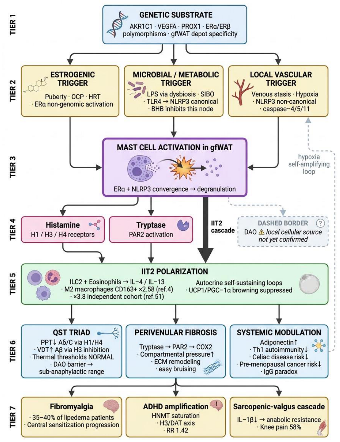
Figure 4.
Bidirectional lipedema-venous interaction and asymmetric lipedema as a natural controlled experiment. Pathway A (IIT2 → C1 to C2 telangiectasia, secondary); Pathway B (CEAP C3+ reflux → stasis/hypoxia → NLRP3 non-canonical → ipsilateral amplification). Asymmetric lipedema controls genetics/hormones/diet within-patient and isolates the local trigger (Prediction P7).
Figure 4.
Bidirectional lipedema-venous interaction and asymmetric lipedema as a natural controlled experiment. Pathway A (IIT2 → C1 to C2 telangiectasia, secondary); Pathway B (CEAP C3+ reflux → stasis/hypoxia → NLRP3 non-canonical → ipsilateral amplification). Asymmetric lipedema controls genetics/hormones/diet within-patient and isolates the local trigger (Prediction P7).
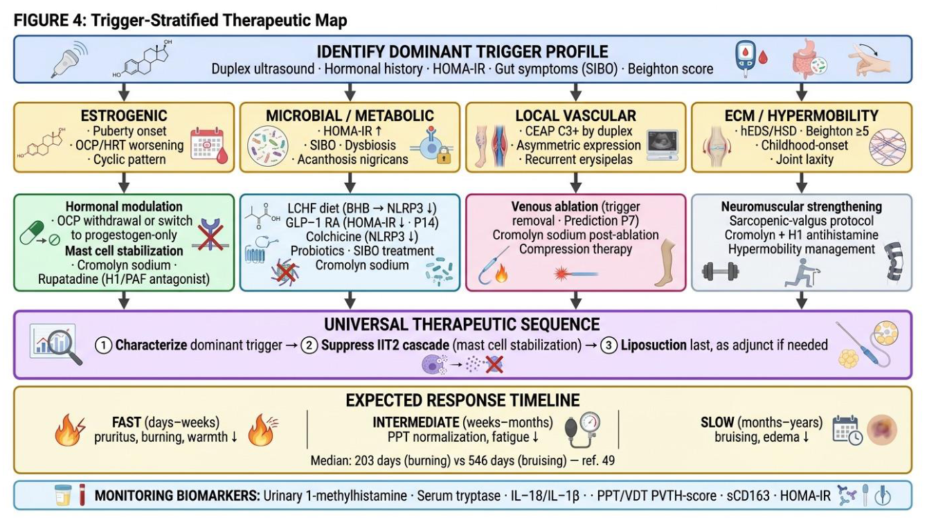
Figure 5.
Endotype- and trigger-stratified research map. Candidate stratification (hormonal-transition, microbial/metabolic, local vascular, ECM/hypermobility) and matched candidate interventions, all framed as hypotheses for stratified prospective trials with pain and limb volume (not body weight) as primary endpoints.
Figure 5.
Endotype- and trigger-stratified research map. Candidate stratification (hormonal-transition, microbial/metabolic, local vascular, ECM/hypermobility) and matched candidate interventions, all framed as hypotheses for stratified prospective trials with pain and limb volume (not body weight) as primary endpoints.
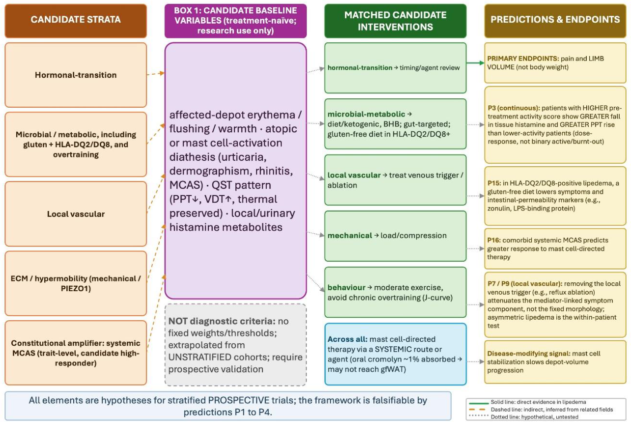
Table 1.
The gfWAT-IIT2 endotype framework positioned among existing models of lipedema.
| Framework | Main mechanism | Supporting evidence | Major limitation | Clinical features explained | Key testable prediction | Current evidence level |
| Vascular/lymphatic [60,61,62] | Primary microvascular/lymphatic dysfunction; adipose secondary | Capillary fragility; functional lymphatic changes; normal venous function | Does not explain QST selectivity, immunoglobulin paradox, FM/estrogen-dependence | Edema, bruising, telangiectasia | Endothelial barrier markers precede morphology | Moderate (direct human data) |
| Adipokine/metabolic-omics [9,17] | Altered adiponectin/leptin; OXPHOS/mitochondrial shift | Multi-omics; metabolically protected depot | Descriptive; bulk-tissue, composition-confounded; no unifying driver | Metabolic protection (insulin sensitivity increased) | Composition-controlled single-nucleus signature | Moderate |
| Adipo-vascular/ECM germline (4, 5; A.C.M. Amato, unpublished data) | Heritable adipo-vascular/ECM programme; gynoid-adiposity extreme | Replicated GWAS; depot heritability; no immune top loci | Case definition is a proxy; not lipedema-specific | Female limitation, depot specificity, diet-resistance | Polygenic adipo-vascular vs immune score predicts disease | moderate to strong (genetics) |
| Adipose stem-cell dysfunction [7,23] | ASC dysfunction; early interstitial fibrosis; M2-driven | In-vitro ASC causality; histology | Initiating signal uncertain; fibrosis-first vs -last debated | Fibrosis, nodularity, recurrence | Fibrosis temporally precedes adipocyte hypertrophy | Moderate |
| Evolutionary [63] | Ancestral gynoid energy store, now maladaptive | Adaptive coherence | Not molecular; not falsifiable as stated | Why gfWAT, why protected | (explanatory, not predictive) | Conceptual |
| gfWAT-IIT2 endotype (this work) | Type 2 / mast cell-activation endotype on an adipo-vascular substrate; per-cell histamine signalling | Tissue histamine 2.2×; QST triad; asymmetric-limb experiment; cromolyn response | Mast cell density not increased; bulk immune signal non-identifiable; many links indirect | QST triad, histamine/atopy features, trigger-stratified response, FM | QST normalizes with mast cell stabilization in the responsive subgroup (P3) | Hypothesis (graded; Table 2) |
Table 2.
Evidence levels for each component of the gfWAT-IIT2 endotype framework.
| Framework component | Direct evidence in lipedema | Indirect evidence (related fields) | Hypothetical (untested) |
| Adipo-vascular/ECM germline substrate | GWAS loci VEGFA, GRB14-COBLL1, RSPO3, ADAMTS9 [4,5] | gfWAT heritability, cardio-protection [5] | (none) |
| ERβ-dominant ASC receptor shift; pro-adipogenic estrogen response | ASC receptor profile; paradoxical adipogenic (PPARγ2) response [23]; no transcriptional brake (unpublished data) | Depot ER biology [18] | ERα density on gfWAT mast cells (P2) |
| Elevated tissue histamine † | 2.2× in preliminary metabolomic study, n=44/18 [14] | (none) | Histamine as primary local pain mediator |
| Mast cell numerical density | Not increased (CD117+ n.s.) [24] | (none) | Per-cell hyper-secretion (P3) |
| M2 polarization | M2 transcriptomic signature [7]; CD163+ enrichment [39] | Eosinophil/ILC2-maintained M2 in lean fat [38] | ILC2/eosinophil source in lipedema (P1) |
| NLRP3 inflammasome | Not directly measured; bulk transcripts reduced but composition-confounded (unpublished data) | NLRP3 in obesity; BHB inhibition [30] | Active inflammasome in tissue (P4) |
| Histaminergic QST mechanism | QST triad pattern itself [13]; PPT BMI-independent [41] | H1/H3/H4 fibre-class actions (general) | Receptor-fibre model in lipedema (P3, P6) |
| Tryptase→PAR2 fibrosis | Nodule histology, elevated RI [43] | Tryptase-PAR2-COX2 fibrosis [42] | Causal sequence in lipedema |
| Hormonal-transition trigger | Onset/exacerbation epidemiology [27] | Non-genomic ERα degranulation in cell lines [25] | gfWAT mast cell ER mechanism (P2) |
| Asymmetric lipedema / local trigger † | Case series, within-patient (Amato et al., asymmetric-lipedema case series, under review) | NLRP3 non-canonical (general) | Trigger removal disease-modifying (P7) |
| Fibromyalgia continuum | Comorbidity 35 to 40% [15,16] | Central sensitization, MRGPRX2 [48] | Mediator-tracked progression (P8) |
† Framework components currently supported predominantly by the authors' own group, for which independent external replication is not yet available: the elevated tissue-histamine finding, the asymmetric-lipedema / trigger-removal observations. These are flagged in the text where they appear and should be read as hypothesis-generating pending independent confirmation.
Table 3.
Predictions, experiments, readouts, and falsifying thresholds.
(Tier: C = core, E = exploratory.).
Table 3.
Predictions, experiments, readouts, and falsifying thresholds.
(Tier: C = core, E = exploratory.).
| # | Tier | Cascade node | Experiment | Primary readout | Falsifying threshold |
| P1 | C | ILC2/eosinophil source | scRNA/snRNA-seq, lipedema vs control gfWAT | ILC2/eosinophil frequency ↑ | ILC2 <0.1% CD45+ and indistinguishable from control (p>0.05, power ≥0.80) |
| P2 | C | gfWAT mast cell ER | IHC/flow of tryptase+ cells | ERα present, ERα>ERβ | ERβ-dominant or ER-absent |
| P3 | C | Histamine→QST | 8-wk trial, systemic mast cell-directed agent, baseline-activity-stratified | PPT rise scales with baseline activity, alongside tissue histamine ↓ | No activity-to-response relationship (with confirmed depot delivery) |
| P4 | C | BHB→NLRP3 | Ketogenic vs isocaloric low-fat RCT | IL-18/IL-1β/tryptase ↓ ∝ pain | Pain ↓ without biomarker ↓ |
| P5 | E | Histamine→ADHD | 12-wk mast cell stabilizer, ASRS-18 | ADHD score ↓≥30% with metabolite ↓ | No change despite metabolite ↓ |
| P6 | E | H3 inverse agonism | Pitolisant, 12 wk, VDT+CAARS | VDT normalized + ADHD ↓ | VDT unchanged |
| P7 | E | Local trigger | Venous ablation in CEAP C3+ asymmetric | Asymmetry ratio ↓; ipsilateral PPT ↑ | No asymmetry change after confirmed trigger removal |
| P8 | E | IgG paradox/FM | Anti-neuronal IgG, FM±lipedema; passive-transfer model [65] | Low/absent IgG; no transfer hyperalgesia | IgG elevated as in non-lipedema FM |
| P9 | E | Trigger removal | Early vs late ablation, CEAP C3+ | Lower recurrence; tryptase ↓ | No recurrence difference |
| P10 | E | TLS | Multiplex IHC, gfWAT vs abdominal SAT | CD20+/CXCL13+/HEV clusters | Absent despite IIT2 markers |
| P11 | E | Browning suppression [66,67] | Paired biopsies, cromolyn 12 wk | UCP1/PGC-1α/PRDM16 ↑ | No increase despite stabilization |
| P12 | E | Cellulite continuum | Paired biopsies, grade-3 cellulite | Intermediate tryptase/histamine | Identical to controls |
| P13 | E | H3/HNMT/DAT | Lisdexamfetamine dose before/after stabilization | Lower dose ∝ metabolite ↓ | No dose change |
| P14 | E | GLP-1 stratification | GLP-1 RA by HOMA-IR tertile | Greatest benefit in high HOMA-IR | Equal across strata |
| P15 | E | Gluten/HLA trigger | Gluten-free vs gluten diet, HLA-DQ2/DQ8+ lipedema | Symptoms and permeability markers fall in HLA+ on a gluten-free diet | No symptom or marker change in HLA+ subgroup |
| P16 | E | Systemic MCAS amplifier | Mast cell-directed therapy, MCAS-positive vs MCAS-negative lipedema | Greater symptom reduction in MCAS-positive | Equal response regardless of MCAS status |
Disclaimer/Publisher’s Note: The statements, opinions and data contained in all publications are solely those of the individual author(s) and contributor(s) and not of MDPI and/or the editor(s). MDPI and/or the editor(s) disclaim responsibility for any injury to people or property resulting from any ideas, methods, instructions or products referred to in the content. |
© 2026 by the author. Licensee MDPI, Basel, Switzerland. This article is an open access article distributed under the terms and conditions of the Creative Commons Attribution (CC BY) license.
Copyright: This open access article is published under a Creative Commons CC BY 4.0 license, which permit the free download, distribution, and reuse, provided that the author and preprint are cited in any reuse.