

Submitted:
11 May 2026
Posted:
12 May 2026
You are already at the latest version
Abstract
This prospective case series evaluated the short-term outcomes following percutaneous cementoplasty as the sole palliative treatment for appendicular osteosarcoma in 10 dogs. Synthetic self-hardening calcium-phosphate bone substitute was injected into the osseous defect under fluoroscopic guidance after curettage of the bone tumor. Clinician assessment included a numerical rating score for lameness, offloading, and ease of lifting the contra-lateral limb as well as the 4A-VET post-operative pain scale. Owner assessment was obtained using three descriptive questionnaires, the Helsinki Chronic Pain Index (HCPI), the Canine Brief Pain Inventory (CBPI) and the Canine Symptom Assessment Scale (CSAS). Measures were recorded preoperatively and at 2-, 4-, 8-, and 12-weeks following surgery. Early improvement in the 4A-Vet score was noted at the 2-, 4-, 8-, and 12-week time points for all major pain and function metrics. Similarly, the CBPI pain severity and interference scores demonstrated early postoperative improvement during the 2- and 4-week time points with partial attenuation by 8- and 12-weeks. Panting, difficulty sleeping, whining/moaning, and lack of appetite were significantly reduced when assessed via the CSAS. Cementoplasty as a monotherapy, affording early pain relief and improved structural integrity, supports its role as a limb-sparing option for dogs unable to undergo amputation.
Keywords:
calcium phosphate cement
; dog
; osteosarcoma
; limb-sparing
; cementoplasty
; palliation
1. Introduction
Recent advancements in the clinical management of canine appendicular osteosarcoma have been centered primarily on the prevention or treatment of metastatic disease following primary tumor removal [1,2,3,4,5,6]. Although widely accepted as the singular life-limiting factor, ongoing research is required to understand specific primary tumor features that dictate differing metastatic patterns [7]. Non-metastatic related death, such as development of a pathologic fracture resulting in subsequent euthanasia, represents a noteworthy subset of the disease-bearing population [8,9]. Technological improvements in diagnostic and therapeutic techniques for canine appendicular osteosarcoma have generated new opportunities for the evaluation of safe, feasible, and effective local therapies for dogs that are unable to withstand limb amputation [10,11]. Immediate and sustained analgesia is among the many salient features driving palliative limb preservation in pets [12]. Insomuch that the extension of life in this population may be attributed to the mitigation of pain, lameness, and the risk of pathologic fracture rather than delaying the progression of disease [13,14,15].
Local therapies for primary appendicular osseous neoplasia in dogs have historically been limited to either surgery or radiation therapy (RT) [10,11,16]. A variety of factors including lifestyle, existing comorbidities, and financial considerations influence the choice of local therapy. Ultimately, the success of limb salvage procedures is reliant on tumor location, disease burden, and procedure accessibility [16,17,18]. The wide range of reported response rates (74-92%) and duration of response rates (53 to 95 days) highlights the high degree of variability in treatment outcomes following RT [17,19,20,21,22,23,24,25]. Pain and risk of pathologic fracture remain at the forefront of managing the primary tumor regardless of RT protocol [9,21,23,24,25].
Bisphosphonates have been used to arrest osteoclastic activity and slow osteolysis in dogs with primary bone tumors [26]. Single agent intravenous pamidronate is reported to alleviate bone pain in 28% of dogs treated for a median of 231 days in one study [27]. Although neither bisphosphonate, pamidronate nor zoledronate, nor timing of the drug (concurrent or following RT) had measurable impact on survivability [19]. In dogs that develop pathologic fractures, surgical excision and implant stabilization is possible in bone tumors affecting the distal radius, however, the reported risk of surgical site infection and implant-associated complications is 40-70% [16,18,28,29,30].
Calcium phosphate bone cement has advantageous attributes such as good biocompatibility, injectability, and osteoconductivity and has been used for intraosseous bone augmentation [30]. Injection of polymethylmethacrylate (PMMA) or calcium phosphate cement allows for stabilization of the bone mitigating the nociception and mechanotransduction driven by cortical thinning, microfracture, and endosteal stimulation observed following the development of osteosarcoma [31,32]. In contrast with a previous study evaluating percutaneous cementoplasty using PMMA which identified deep wound infections, intraarticular cement leakage, and venous thrombosis as the main complications, calcium phosphate cements circumvent the activation of a typical necrotic and inflammatory process attributed to the exothermic polymerization of PMMA [12]. Cementoplasty using a tricalcium phosphate-based cement in conjunction with microwave ablation resulted in pain-free mobility in a dog with stable disease 18 months after its initial diagnosis of stage-1 osteoblastic appendicular osteosarcoma [33]. Results from a pilot clinical trial supported cementoplasty as a viable, safe, and effective limb-sparing option using a calcium phosphate bone substitute in 12 dogs [13]. While previous studies evaluate the use of cementoplasty as part of a multimodal approach, further investigation is required to evaluate cementoplasty as a sole treatment option for palliation in dogs with appendicular osteosarcoma that are not candidates for adjunctive therapies. This distinction allows for identification of the isolated palliative effects of cementoplasty as a standalone therapy. Hence, cementoplasty may be a desirable option for owners with the therapeutic goal of high-function survival via relief of weight-bearing pain during palliation without the addition of other disease-directed therapies.
We hypothesize that the therapeutic effects of percutaneous calcium phosphate cement (BIOCERA-VET®, TheraVet, Gosselies, Belgium) injection used as a monotherapy for treatment of appendicular osteosarcoma in dogs would allow for short-term improvement in limb function, pain level, and owner-perceived quality of life. We further hypothesized that improvement in symptom burden following cementoplasty could be maintained for the duration of the follow-up period in this subset of dogs despite the absence of anti-tumoral activity.
2. Materials and Methods
2.1. Inclusion Criteria
All dogs presenting to a single veterinary referral hospital which had histologically confirmed appendicular osteosarcoma via Jamshidi bone core biopsy between February of 2024 and 2025 were given the option to prospectively enroll in the study if amputation was categorically declined by the owner or contraindicated. All interventions were performed as part of standard clinical care and no experimental strategies were introduced. Written informed client consent was obtained prior to inclusion. Dogs were included if owners declined bisphosphonate therapy, adjuvant chemotherapy, microwave ablation, radiotherapy, targeted therapy, or vaccine/immunotherapy following preoperative counselling. Dogs with radiographic abnormalities consistent with pulmonary metastasis were excluded. Substantial orthopedic comorbidities detected during palpation or radiographically within the remaining three limbs resulted in exclusion. Dogs receiving adjunctive oral pain control were considered eligible for enrollment provided that the dog’s pain and lameness was localizable to the neoplastic limb. All dogs underwent a complete blood count and serum chemistry prior to general anesthesia. Enrollment in the study required owner follow up at predetermined timepoints for the 12-week duration of the study.
2.2. Procedure
Access points for all dogs were identified using anatomical landmarks based on radiographic images obtained prior to surgery. Dogs were pre-medicated with hydromorphone (0.08 mg/kg IM) combined with acepromazine maleate (0.04 mg/kg IM) and anesthesia was induced with propofol (4 mg/kg IV). General anesthesia was maintained with an inhalant mixture of isoflurane and oxygen delivered by endotracheal intubation. Perioperative antimicrobial prophylaxis consisted of cefazolin (22 mg/kg IV) given at induction and repeated every 90 minutes during surgery. The procedure was performed by a single ACVS Diplomate assisted by a surgery resident. Dogs were placed in lateral recumbency with a hanging limb preparation of the affected limb. A 3 cm incision was made through the skin and soft tissues to expose the underlying bone. Under fluoroscopic guidance, a Kirschner wire was used to mark the access point on the cortical bone 1-3 cm from Codman’s triangle on radiography. A high-speed pneumatic burr was used to create a 0.5 cm cortical defect. The bone cavity was subsequently debrided with a Volkmann’s curette and suctioned. A 7G cannula was inserted through the cortical defect and its position and angulation were confirmed under fluoroscopy. Slow injection filling of the synthetic calcium-phosphate bone substitute was performed in multiple directions until the access point outer limit was reached and at least 50% of the cavity adjacent to the intact cortical bone was filled. The access point was filled with cement until the product was level with the adjacent cortical bone. The surgical site was lavaged thoroughly and closed routinely with monofilament absorbable sutures. The minimum duration of hospitalization was 24 hours post-operative to allow for complete hardening and maximal early mechanical support of the cement. All dogs received perioperative analgesia consisting of hydromorphone (0.08 mg/kg IV) as injectable boluses every 4-6 hours and carprofen (2.2 mg/kg PO q 12h) with gabapentin (8-10 mg/kg PO q 8h) postoperatively. Postoperatively, cefpodoxime (5–10 mg/kg PO once daily) was administered for 10 days. Complications were classified as minor if the complication did not necessitate additional surgical intervention and did not significantly alter limb function. Revision surgery, amputation, or pathologic fracture were classified as a major complication.
2.3. Veterinary Assessment
A single ACVS Diplomate performed standardized physical and orthopedic examinations on each dog prior to and following surgery at each designated time point. A numerical rating score for lameness was assigned during gait analysis prior to surgery and at 2-, 4-, 8-, 12-weeks post-operatively. The numerical scores were defined as: clinically sound/no lameness (0), mild or slight weight-bearing lameness (1), obvious or moderate weight-bearing lameness (2), severe weight-bearing lameness (3), intermittent non–weight bearing lameness (4), and continuous non–weight bearing lameness (5). Offloading was assessed during standing using a standardized ordinal scale (0-2), defined as no offloading of the affected limb (0), intermittent offloading (1), and complete offloading (2). Ease of lifting the contralateral limb was recorded using a similar scale (0-2), defined as maintaining lift comfortably for >5 seconds with stable posture (0), maintaining lift for several seconds but unstable (1), lifting briefly and immediately unloading (2), and inability to lift the contralateral limb (3). Eight preoperative radiographic projections were obtained including a right lateral, left lateral, and ventrodorsal thorax as well as a hip-extended ventrodorsal, a right and left mediolateral and orthogonal cranial to caudal view of the affected and contralateral limbs. Following cementoplasty, all dogs underwent radiographic monitoring immediately postoperatively, 4- and 8-weeks post-operatively (Figure 1). All dogs were evaluated using the total French Association for Animal Anesthesia and Analgesia pain-scoring system (4A-VET) score. This multidimensional tool uses the sum of six subscale scores comprising an overall subjective assessment of the dog’s pain level preoperatively and was assessed at 2-, 4-, 8-, and 12-weeks postoperatively [34]. This score described the cumulative effect of a dog’s general behavior and response to handling of the operative limb as well as the intensity of the reaction following palpation.
2.4. Owner Assessment
Owners were asked to use three outcome assessment tools to quantify the severity and impact of their dog’s pain prior to surgery and at 2-, 4-, 8-, 12-weeks post-operatively. The Canine Brief Pain Inventory (CBPI) is a two-part instrument that assesses the severity of pain as well as how the pain interferes with the dog’s lifestyle. The pain severity score (PSS) is the mean value of four items scored on an eleven-point numerical scale. Similarly, the pain interference score (PIS) is the mean value of six items. An increased mean value score correlates to an increased level of pain and interference with daily activity, respectively. There is one global quality of life score calculated at the end of the instrument. The second tool was the Helsinki Chronic Pain Index (HCPI) which contains eleven questions regarding activity, behavior, and mood evaluated via Likert scale (0-4) to which the value for each score was added to give an overall instrument score. The third tool was the Canine Symptom Assessment Scale (CSAS) which contains 12 questions related to the subjective physical disturbance dogs experienced during the course of daily living as assessed by means of proxy reports of owners.
2.5. Statistical Analysis
Descriptive statistics were calculated. Continuous variables were expressed as median values and ranges while categoric variables were expressed as frequencies and percentages for each group. Statistical analyses were performed using JMP® Pro 18. Differences among scores over time were evaluated using linear mixed-effects models with repeated measures on the same animal. The individual animal was included as a random effect, and a first-order autoregressive AR(1) covariance structure was specified to account for correlation among repeated observations within animals. Postoperative time points were modeled as fixed effects. Least-squares mean comparisons were conducted using Dunnett’s multiple-comparison procedure, with baseline measurements treated as the control. Statistical significance was set at α = 0.05.
3. Results
Ten large and giant-breed dogs, including seven males (70%) and three females (30%) met the inclusion criteria for the study (Table 1). The mean weight was 59.1 kg (44-87 kg) and the mean age was 8 years and 2 months (62-146 months). The tumor locations treated were the proximal humerus (n=3), distal radius (n=5) and proximal tibia (n=2). The mean elapsed time from OSA diagnosis to surgery was 5 days (range: 1-9 days). The degree of cortical lysis and size of the tumoral defect dictated the volume of bone cement injected (median: 13; range: 10-18 mL). All dogs underwent filling of the tumoral defect until overflow was noted at the access point. Greater than 50% of the tumoral defects were filled in all dogs and confirmed via fluoroscopy. The mean anesthesia time for cementoplasty was 83.9 minutes. The mean operative time was 49.6 minutes for radial defects, 53.3 minutes for humeral defects, and 41.5 minutes for tibial defects. The mean operative time for all dogs was 49.1 minutes. The duration of hospitalization was 2 days for all dogs.
6 dogs completed the 12-week study period while 4 of the dogs withdrew from the study. Disenrollment was due to initiation of chemotherapy (n=2), pathological fracture of the cementoplasty site (n=1) and owner-elected amputation of the limb (n=1). During a four week recheck, Dog 9 was diagnosed with stage IIIa lymphoma following the discovery of generalized lymph node enlargement. Dog 1 withdrew from the study at 4 weeks post-operatively to begin chemotherapy. For the duration of the study period, two major complications were observed. Dog 7 sustained a fracture proximal to the cementoplasty site along the distal radius which was diagnosed at four weeks post-operatively (Figure 2 and Figure 3). The dog was managed with external coaptation until euthanasia at 6 months post-operatively. Amputation was elected in four dogs due to infection of the cementoplasty site (dogs 2 and 8) and persistent and unrelenting pain (dogs 4 and 10). However, three of the four dogs were followed to 12 weeks post-cementoplasty prior to undergoing amputation (Figure 4). Of these two dogs, culture and sensitivity were performed prior to amputation. Microbiological isolates for dog 2 included multi-drug resistant Klebsiella pneumoniae and Escherichia coli. At 2.5 months post-cementoplasty, dog 8 developed three equal-sized 2 cm erosions along the dorsal aspect of the peritumoral swelling. A culture revealed growth of multi-drug-resistant isolates, Klebsiella pneumoniae and Methicillin-resistant Staphylococcus pseudintermedius. Four minor complications were recorded including peri-incisional swelling and edema in dogs 1, 2, and 8 which were resolved with appropriate supportive care. All three dogs had radial lesions. The fourth minor complication was reported in dog 6 in which the dog developed a temporary, acute, non-ambulatory paraparesis following a subcutaneous injection of bedinvetmab (Librela®, Zoetis). The adverse neurological event spontaneously resolved after 48 hours.
Three dogs developed suspected or confirmed pulmonary and distant metastasis (dogs 2, 4, 8) following the end of the study period. In dog 2, an osteolytic lesion developed in the contralateral proximal humerus at 177 days postoperatively. A nasal biopsy was performed in dog 4 following the development of epistaxis. The biopsy returned as a poorly differentiated sarcoma 322 days postoperatively. Bilateral hyperechogenic renal masses with secondary hydronephrosis as well as mesenteric mass with associated mesenteric nodules consistent with sarcoma were discovered in dogs 8, 100 days postoperatively. All three dogs underwent limb amputation following the conclusion of the study. The average time to amputation following cementoplasty was 166 days (116-350). No dogs were euthanized during the 12-week study period. The median time for euthanasia was 179 days following surgery.
The effect of time point on lameness score differed for dogs evaluated at a walk versus a trot. For lameness at a walk, the least-squares mean scores (95% CI) were 2.00 (1.57–2.43) preoperatively, 1.40 (0.97–1.83) at 2 weeks, 1.20 (0.77–1.63) at 4 weeks, 1.62 (1.14–2.10) at 8 weeks, and 1.78 (1.27–2.29) at 12 weeks which demonstrated that the time point significantly affected the walk lameness score (P=0.011). Compared to preoperative values, the walk lameness scores were significantly lower at 2- (P=0.03) and 4- (P=0.003) weeks post operative but not at 8- or 12 weeks. For evaluation at a trot, the preoperative 2.20 (1.72–2.68), 2 weeks 1.70 (1.22–2.18), 4 weeks 1.70 (1.22–2.18), 8 weeks 2.00 (1.43–2.57), and 12 weeks 2.21 (1.59–2.82) time points did not significantly affect lameness score (P=0.3). Least-squares mean offloading of the affected limb improved following cementoplasty decreasing from 1.60 preoperatively to 1.00 at 2-, and 0.90 at 4 weeks postoperatively. An increase to 1.21 at 8- and 1.64 at 12 weeks postoperatively as noted. In comparison to baseline, offloading scores were significantly lower at 2- and 4 weeks postoperatively (P=0.012) but not at 8- or 12 weeks. In contrast, lifting of the contralateral limb did not differ significantly from baseline at any postoperative time point (P=0.3).
The 4A-Vet least-squares mean ± SE scores were the highest preoperatively (7.40 ± 0.58) and were lower at all postoperative time points: 2 weeks (3.60 ± 0.58), 4 weeks (3.40 ± 0.58), 8 weeks (2.96 ± 0.64), and 12 weeks (3.28 ± 0.70). The improvement in score was identified early at 2 weeks and maintained throughout the study period at 4, 8, and 12 weeks (Table 2). Mean differences from preoperative values were −3.80 at 2 weeks (95% CI: −4.93 to −2.67), −4.00 at 4 weeks (95% CI: −5.42 to −2.58), −4.44 at 8 weeks (95% CI: −6.16 to −2.71), and −4.12 at 12 weeks (95% CI: −6.06 to −2.18); as scores dropped by an average of 4 points compared to preoperative values with all comparisons being statistically significant (P < 0.001).
The Canine Brief Pain Inventory was divided into pain severity score (intensity of pain) and pain interference score (degree to which pain disrupts normal function and daily activity). The postoperative time point significantly affected all CBPI pain severity measures including when pain was reported to be at its lowest (P=0.012), at its highest (P=0.0009), at the time of assessment (P=0.0004), and on average (P=0.0071). Scores for pain at its highest were lower than preoperative values at 2 and 4 weeks but not different at 8 or 12 weeks (Figure 5). Pain at its least and average pain was similar with significantly lower scores at 2 and 4 weeks postoperatively compared with preoperative values, while differences at 8 and 12 weeks were not significant. Pain interference scores were significantly reduced at 2, 4, and 8 weeks postoperatively but did not differ from baseline at 12 weeks (Figure 6). The postoperative time point significantly affected pain interference when assessing general activity (P=0.034), enjoyment of life (P=0.005), ability to walk (P=0.013), ability to run (P=0.015), and ability to climb stairs or curbs (P=0.026). Pain interference with regard to the ability to rise from lying down did not reach statistical significance (0.069). For these actions, pain interference was significantly reduced at 2 and 4 weeks postoperatively, however, this was not observed at 8 or 12 weeks. Pain interference for enjoyment of life was significantly lower than preoperatively at 2 and 4 weeks and remained even lower at 8 weeks but did not differ at 12 weeks. The overall quality of life score was significantly affected by time point, where a higher score was identified at 2, 4, and 8 weeks postoperatively as opposed to no difference at 12 weeks.
When assessing individual symptom burden and functional impact on dogs, postoperative time point had a significant effect on reported pain (P<0.0001). The presence of panting (P=0.004), difficulty sleeping (P=0.0039), sleepiness (P=0.022), and whining/moaning/groaning (P=0.018) were significantly improved across all time points. Lack of appetite was worse at 2 weeks but by 8-12 weeks scores were similar or better than preoperative scores (P=0.013). In contrast, scores for pacing, energy level, coughing, yelping/crying out, vomiting, and diarrhea remained uniformly low across all time points without detectable change from preoperative scores. The severity of symptoms, when present, were significantly lower than preoperative values for panting (P=0.018), difficulty sleeping (P=0.005), lack of appetite (0.017), and sleepiness (P=0.009) at all time points. Overall mean CSAS scores demonstrating the evolution of symptom burden for each time point are depictured in Figure 7.
Parameters from the Helsinki Chronic Pain Index identified functional and behavioral worsening during the initial postoperative period (2 and 4 weeks) in the form of willingness to walk (P=0.001) and willingness to play/interact (P=0.042). These abilities both recovered to baseline levels at 8 and 12 weeks postoperative. Vocalization/discomfort behaviors such as whining, yelping, and licking were observed to be significantly reduced by 2 weeks postoperative (P=0.004), however, at later time points were not different from baseline (Figure 8). Reports of attitude/mood improvements were seen over time (P=0.047), however, a consistently improved shift at a specific week was not recognized. No consistent pattern of improvement could be identified with regard to the ability/willingness to use stairs, run, or jump, as well as ease of lying down, movement during or after exercise or after long rest, and rising from a down position.
4. Discussion
Evaluation of percutaneous cementoplasty using calcium phosphate cement injected into an appendicular osteosarcoma tumoral cavity yielded early improvement in pain, function, and owner-perceived quality of life, as well as a reduction in symptom interference with daily activities. The results of this study demonstrate that based on multiple validated owner- and clinician reported outcome measurement scales, a robust treatment effect exists for pain mitigation, improvement in limb use, as well as willingness to engage in a variety of behaviors that were otherwise diminished or absent prior to surgery. Using the 4A-VET score, clinician assessed pain and functional impairment identified scores significantly lower than preoperatively across all time points. This consistent reduction of approximately 4 points from baseline emphasizes the demonstrable positive impact of cementoplasty, as this scale reflects broad functional states in which each singular point represents a substantial improvement in the dog’s postoperative experience [35]. The observed rapid improvement in 4A-VET scores suggests a decrease in both spontaneous pain and pain elicited from manipulation. The precipitous drop in score (3.8–4.4 points) across each postoperative time point substantiates improved mobility for all dogs. This finding would suggest that cementoplasty did not just reduce pain but also facilitated functional normalization as dogs quickly transitioned from compensatory or guarding behaviors to more consistent limb use at 2 weeks postoperatively.
Numerical lameness scores at a walk improved significantly at 2- and 4 weeks with no difference from preoperative scores observed at 8- and 12 weeks postoperatively. In contrast, trot lameness scores did not differ significantly across time points which highlights that subtle residual lameness may be more apparent at a faster pace despite overt improvement while walking. The lack of statistical improvement at later time points may reflect the burden of increased activity and functional demands at 8- and 12 weeks rather than true clinical deterioration of the dog in spite of treatment similarly to previous reports following palliative radiation therapy [22,23]. The functional demand as activity restrictions are lifted have been reported in studies evaluating radiation therapy for osteosarcoma palliation in that improvements in comfort and limb use may diminish over time [19,21,23,24,25,26,27,28,29,30,31,32,33,34,35]. Offloading scores provide additional functional data that supports improved limb loading up to 4 weeks post-operatively followed by a partial return to baseline at 8 weeks. This is consistent with the proposed biomechanical mechanisms of cementoplasty increasing compressive strength and structural integrity thereby reducing pain ascribed to micromotion in lytic bone [36,37]. Conversely, no significant overall time effect was noted for offloading to affected limb across all time points. This finding suggests that although cementoplasty improves offloading and lameness scores, its application is insufficient to support full loading of the operated limb in this study. Behavioral factors may influence limb lifting to a greater extent than observations of lameness and offloading, as painful dogs may demonstrate avoidance and resistance to manipulation due to an aversion to handling in general [38]. This metric may function independently of measurable biomechanical dysfunction [39].
The multiple owner-reported assessments were utilized to fill key gaps in our knowledge of symptom burden and interference with daily activity in dogs having undergone limb sparing surgery. Osteosarcoma-associated pain is multifactorial, driven by cortical destruction, microfracture formation, periosteal disruption, and endosteal nociceptor activation [31,32]. Progressive osteolysis promotes loss of structural integrity, both of which contribute greatly to increased nociception and risk of pathologic fracture [40,41,42]. As such, reporting pain reduction and improved limb use following limb-sparing surgery as singular findings is incomplete without investigating whether these changes translate to a diminished symptom burden on the dog and primary caregiver. In this study, CBPI pain severity scores improved significantly across all domains (pain at its worst, pain at its least, pain on average, and current pain) at 2 and 4 weeks postoperatively, capturing changes in baseline and breakthrough pain [43]. This assessment provides a clinically relevant measure of real-time suffering [44] and this trend is comparable to previous studies evaluating palliative radiation therapy where 75-96% of pet owners report measurable pain relief within weeks of treatment [17,19,21,22,23]. The attenuation of these scores at 8 and 12 weeks may have resulted from a combination of factors such as increased activity during later convalescence or owner hyperawareness of pain as a consequence of detection bias. However, despite this finding, overall quality of life scores remained significantly improved through 8 weeks, suggesting that individual owner-perceived fluctuations in pain at later time points did not affect the overarching welfare gains for this population of dogs.
The interference scores allowed for broader contextualization of these findings in that significant improvement in the dog’s general activity, walking, running, stair climbing, and enjoyment of life improve in the early postoperative period (2 and 4 weeks) [44]. A pattern emerged in which clear improvements at 8 and 12 weeks could not be proven statistically across all survey instruments. The varied owner perceptions at later time points may reflect the normalization of expectations following intervention along with the withdrawal of activity restriction and oral pain management, known external influences of pain control. Although the parameter of rising from lying down did not reach statistical significance in this study, it is likely that its measurement is less sensitive than others to the presence or absence of pain alone and more likely to be influenced by orthopedic or neurologic comorbidities. Of the domains related to willingness to walk, play, and interact, the findings of the Helsinki Chronic Pain Index align closely with the CBPI and NRS-lameness findings in that significant improvements were observed early during the postoperative period.
The Canine Symptom Assessment Scale provided valuable insight into the burden of the disease beyond pain reporting alone. The results of this scale delineated a marked and sustained postoperative reduction in panting, difficulty sleeping, appetite changes, and vocalization behaviors. Respiratory (coughing) and gastrointestinal signs (vomiting and diarrhea), were identified as infrequent and static across all time points suggesting that for dogs in this study, observed improvements were specifically related to pain and discomfort behaviors. This further supports the effect of cementoplasty on alleviating symptoms more closely attributable to the diagnosis of appendicular osteosarcoma rather than being a result of conflated reporting. The rapid improvement in stability and return to weight bearing resulted in improved owner-perceived comfort which is perhaps more valuable to owners seeking palliation than marginal changes in survival time [8,45,46].
A multimodal outcome assessment strategy was utilized in this study to control differences in timing and sensitivity of the instruments. Despite their differences by design, the results converged upon a common conclusion in that percutaneous cementoplasty resulted in substantial early improvement in pain and function with later time points more likely representing a clinical plateau rather than true regression. The concordance between clinician-based and owner-reported instruments demonstrates that a sustained reduction in pain-related behaviors and functional impairment was recognized throughout the recovery phase by a trained observer (ACVS Diplomate) as well as within the home setting.
The small sample size used in this study limits our ability to identify small differences between time points or within symptom domains. Outcome measures as reported by the primary caregiver are inherently influenced by perception, expectations after their financial and emotional investment, as well as the level of activity modification and frequency of adjunctive pain control over the study period [47]. Follow-up was limited to 12 weeks post operative and as such, a longer-term assessment may be valuable at distinguishing additional clinical benefit or late-emerging side effects and complications. In addition, the 4 dogs that withdrew from the study limits generalizability but does not automatically overrepresent favorable responders. Three out of the four withdrawals were not due to treatment failure but rather changes in owner treatment goals, particularly in pursuit of life-prolonging therapies [48]. This reflects the reality of osteosarcoma palliation, where owner decision-making will likely evolve over time [49,50] With respect to these limitations, the consistency and convergence of findings across multiple instruments provides compelling evidence that percutaneous cementoplasty as a monotherapy for the treatment of appendicular osteosarcoma allowed for immediate short-term improvement in limb function, pain level, symptom burden, and owner-perceived quality of life. However, data garnered from subjective outcome measures are influenced by expectation bias, particularly from pet owners as demonstrated in a study identifying placebo effect in caregivers assessing lameness [51]. While the use of multiple validated instruments mitigates this effect, objective measures such as force plate gait analysis or continuous activity monitoring would strengthen future investigations [52,53,54,55].
The findings of this study support our hypotheses by demonstrating that cementoplasty used as a monotherapy provided early improvement in pain, limb function, and owner-perceived quality of life while maintaining improvement in several symptom burden domains despite lacking direct anti-tumoral activity. These results illustrate that cementoplasty provides a suitable limb-sparing option for owners who are exclusively interested in palliation. It is important to note the distinction between survivability and delaying disease progression in this specific cohort. The attrition rate reflects the reality of osteosarcoma palliation in dogs and as such, we highlight the role cementoplasty can play in preservation of the dog’s clinical status, thus aligning with the primary goals of the pet owner for a unique subset of dogs. Importantly, this study was not designed to asses survival outcomes or disease progression and thus the results should be interpreted within the framework of local palliation.
5. Conclusions
Cementoplasty as a monotherapy for appendicular osteosarcoma in dogs is an intervention that resulted in substantial early postoperative improvement in multiple pain, function, and symptom parameters as assessed by the clinician and owner. This study utilized several validated multidimensional pain and function metrics which revealed clinically meaningful improvement of discomfort behaviors in dogs that underwent cementoplasty for up to 12 weeks postoperatively. This intervention produced a measurable positive welfare impact by providing short-term improvement in pain, function, and owner-perceived quality of life in selected dogs with appendicular osteosarcoma. Future studies are necessary to explore objective activity measurement and gait analysis with an emphasis on long-term trajectories of dogs undergoing this procedure, especially as part of multimodal palliation with radiation and systemic therapies. Furthermore, additional work should investigate procedural benefits over established full limb-sparing segmental resection with endoprosthesis.
Supplementary Materials
The following supporting information can be downloaded at: Preprints.org, Figure S1: title; Table S1: title; Video S1: title.
Author Contributions
Conceptualization, S.M. and A.V.-C.; methodology, S.M., K.D., J.C., and A.V.-C.; validation, S.M., K.D., D.L., and A.V.-C; formal analysis, S.M., K.D., D.L., and J.C.; investigation, S.M.; data curation, S.M. and K.D.; writing—original draft preparation, S.M.; writing—review and editing, S.M., K.D., and D.L.; project administration, J.C. All authors have read and agreed to the published version of the manuscript.
Funding
This research received no external funding.
Institutional Review Board Statement
Ethical review and approval were waived by the Institutional Animal Care and Use Committee because all procedures performed were part of routine clinical care, and no experimental interventions were introduced. Written informed owner consent was obtained for all enrolled dogs.
Informed Consent Statement
Informed consent was obtained for all client-owned animals in this study.
Data Availability Statement
The raw data supporting the conclusions of this article will be made available by the authors on request.
Acknowledgments
The authors would like to thank Billy Price for his administrative and technical support.
Conflicts of Interest
The authors declare no conflicts of interest.
References
- Bryan, J.N. Updates in osteosarcoma. Vet. Clin. North Am. Small Anim. Pract. 2024, 54, 523–539. [CrossRef]
- Dornbusch, J.A.; Wavreille, V.A.; Dent, B.; Fuerst, J.A.; Green, E.M.; Selmic, L.E. Percutaneous microwave ablation of solitary presumptive pulmonary metastases in two dogs with appendicular osteosarcoma. Vet. Surg. 2020, 49, 1174–1182. [CrossRef]
- Regan, D.P.; Chow, L.; Das, S.; et al. Losartan blocks osteosarcoma-elicited monocyte recruitment, and combined with the kinase inhibitor toceranib, exerts significant clinical benefit in canine metastatic osteosarcoma. Clin. Cancer Res. 2022, 28, 662–676. [CrossRef]
- Oblak, M.L.; Boston, S.E.; Higginson, G.; Patten, S.G.; Monteith, G.J.; Woods, J.P. The impact of pamidronate and chemotherapy on survival times in dogs with appendicular primary bone tumors treated with palliative radiation therapy. Vet. Surg. 2012, 41, 430–435. [CrossRef]
- Marconato, L.; Buracco, P.; Polton, G.A.; et al. Timing of adjuvant chemotherapy after limb amputation and effect on outcome in dogs with appendicular osteosarcoma without distant metastases. J. Am. Vet. Med. Assoc. 2021, 259, 749–756. [CrossRef]
- LeBlanc, A.K.; Mazcko, C.N.; Cherukuri, A.; et al. Adjuvant sirolimus does not improve outcome in pet dogs receiving standard-of-care therapy for appendicular osteosarcoma: A prospective, randomized trial of 324 dogs. Clin. Cancer Res. 2021, 27, 3005–3016. [CrossRef]
- Silver, K.I.; Patkar, S.; Mazcko, C.; Berger, E.P.; Beck, J.A.; LeBlanc, A.K. Patterns of metastatic progression and association with clinical outcomes in canine osteosarcoma: A necropsy study of 83 dogs. Vet. Comp. Oncol. 2023, 21, 646–655. [CrossRef]
- Boston, S.E.; Bacon, N.J.; Culp, W.T.; et al. Outcome after repair of a sarcoma-related pathologic fracture in dogs: A Veterinary Society of Surgical Oncology retrospective study. Vet. Surg. 2011, 40, 431–437. [CrossRef]
- Altwal, J.; Martin, T.W.; Thamm, D.H.; Séguin, B. Configuration of pathologic fractures in dogs with osteosarcoma following stereotactic body radiation therapy: A retrospective analysis. Vet. Comp. Oncol. 2023, 21, 131–137. [CrossRef]
- Griffin, M.A.; Martin, T.W.; Thamm, D.H.; Worley, D.R. Partial ulnar ostectomy, stereotactic body radiation therapy, and palliative radiation therapy as local limb sparing treatment modalities for ulnar tumors in dogs. Front. Vet. Sci. 2023, 10, 1172139. [CrossRef]
- Salyer, S.A.; Wavreille, V.A.; Fenger, J.M.; Jennings, R.N.; Selmic, L.E. Evaluation of microwave ablation for local treatment of dogs with distal radial osteosarcoma: A pilot study. Vet. Surg. 2020, 49, 1396–1405. [CrossRef]
- Böttcher, P.; Krastel, D.; Hierholzer, J.; et al. Percutaneous cementoplasty in the palliative, multimodal treatment of primary bone tumors of the distal aspect of the radius in four dogs. Vet. Surg. 2009, 38, 888–901. [CrossRef]
- Molle, C.; Villamonte-Chevalier, A.; Carabalona, J.; et al. Pilot clinical trial to evaluate in situ calcium phosphate cement injection for conservative surgical management of appendicular osteosarcoma in dogs. Animals 2024, 14, 1460. [CrossRef]
- Bhandal, J.; Boston, S.E. Pathologic fracture in dogs with suspected or confirmed osteosarcoma. Vet. Surg. 2011, 40, 423–430. [CrossRef]
- Rubin, J.A.; Suran, J.N.; Brown, D.C.; Agnello, K.A. Factors associated with pathological fractures in dogs with appendicular primary bone neoplasia: 84 cases (2007–2013). J. Am. Vet. Med. Assoc. 2015, 247, 917–923. [CrossRef]
- Liptak, J.M.; Dernell, W.S.; Ehrhart, N.; Lafferty, M.H.; Monteith, G.J.; Withrow, S.J. Cortical allograft and endoprosthesis for limb-sparing surgery in dogs with distal radial osteosarcoma: A prospective clinical comparison of two different limb-sparing techniques. Vet. Surg. 2006, 35, 518–533. [CrossRef]
- Martin, T.W.; Griffin, L.; Custis, J.; et al. Outcome and prognosis for canine appendicular osteosarcoma treated with stereotactic body radiation therapy in 123 dogs. Vet. Comp. Oncol. 2021, 19, 284–294. [CrossRef]
- Séguin, B.; Walsh, P.J.; Ehrhart, E.J.; Hayden, E.; Lafferty, M.H.; Selmic, L.E. Lateral manus translation for limb-sparing surgery in 18 dogs with distal radial osteosarcoma. Vet. Surg. 2019, 48, 247–256. [CrossRef]
- Ringdahl-Mayland, B.; Thamm, D.H.; Martin, T.W. Retrospective evaluation of outcome in dogs with appendicular osteosarcoma following hypofractionated palliative radiation therapy with or without bisphosphonates: 165 cases (2010–2019). Front. Vet. Sci. 2022, 9, 892297. [CrossRef]
- Nolan, M.W.; Green, N.A.; DiVito, E.M.; Lascelles, B.D.X.; Haney, S.M. Impact of radiation dose and pre-treatment pain levels on survival in dogs undergoing radiotherapy with or without chemotherapy for presumed extremity osteosarcoma. Vet. Comp. Oncol. 2020, 18, 538–547. [CrossRef]
- Coomer, A.; Farese, J.; Milner, R.; Liptak, J.; Bacon, N.; Lurie, D. Radiation therapy for canine appendicular osteosarcoma. Vet. Comp. Oncol. 2009, 7, 15–27. [CrossRef]
- Knapp-Hoch, H.M.; Fidel, J.L.; Sellon, R.K.; Gavin, P.R. An expedited palliative radiation protocol for lytic or proliferative lesions of appendicular bone in dogs. J. Am. Anim. Hosp. Assoc. 2009, 45, 24–32. [CrossRef]
- Mueller, F.; Poirier, V.; Melzer, K.; Nitzl, D.; Roos, M.; Kaser-Hotz, B. Palliative radiotherapy with electrons of appendicular osteosarcoma in 54 dogs. In Vivo 2005, 19, 713–716.
- Green, E.M.; Adams, W.M.; Forrest, L.J. Four fraction palliative radiotherapy for osteosarcoma in 24 dogs. J. Am. Anim. Hosp. Assoc. 2002, 38, 445–451. [CrossRef]
- Ramirez, O.; Dodge, R.K.; Page, R.L.; et al. Palliative radiotherapy of appendicular osteosarcoma in 95 dogs. Vet. Radiol. Ultrasound 1999, 40, 517–522.
- Norquest, C.J.; Rogic, A.; Gimotty, P.A.; Maitz, C.A.; Rindt, H.; Ashworth, H.L.; Bryan, J.N.; Donnelly, L.L.; McCleary-Wheeler, A.L.; Flesner, B.K. Effects of neoadjuvant zoledronate and radiation therapy on cell survival, cell cycle distribution, and clinical status in canine osteosarcoma. Front. Vet. Sci. 2024, 11, 1237084. [CrossRef]
- Fan, T.M.; de Lorimier, L.P.; O’Dell-Anderson, K.; Lacoste, H.I.; Charney, S.C. Single-agent pamidronate for palliative therapy of canine appendicular osteosarcoma bone pain. J. Vet. Intern. Med. 2007, 21, 431–439. [CrossRef]
- Mitchell, K.E.; Boston, S.E.; Kung, M.; et al. Outcomes of limb-sparing surgery using two generations of metal endoprosthesis in 45 dogs with distal radial osteosarcoma: A Veterinary Society of Surgical Oncology retrospective study. Vet. Surg. 2016, 45, 36–43. [CrossRef]
- Boston, S.E.; Vinayak, A.; Lu, X.; et al. Outcome and complications in dogs with appendicular primary bone tumors treated with stereotactic radiotherapy and concurrent surgical stabilization. Vet. Surg. 2017, 46, 829–837. [CrossRef]
- Ma, S.; Wei, Y.; Sun, R.; et al. Calcium phosphate bone cements incorporated with black phosphorus nanosheets enhanced osteogenesis. ACS Biomater. Sci. Eng. 2023, 9, 292–302. [CrossRef]
- Burr, D.B.; Radin, E.L. Microfractures and microcracks in subchondral bone: Are they relevant to osteoarthrosis? Rheum. Dis. Clin. North Am. 2003, 29, 675–685. [CrossRef]
- Mantyh, P.W. Mechanisms that drive bone pain across the lifespan. Br. J. Clin. Pharmacol. 2019, 85, 1103–1113. [CrossRef]
- Sayag, D.; Jacques, D.; Thierry, F.; et al. Combination of CT-guided microwave ablation and cementoplasty as a minimally invasive limb-sparing approach in a dog with appendicular osteosarcoma. Animals 2023, 13, 3804. [CrossRef]
- Rialland, P.; Authier, S.; Guillot, M.; et al. Validation of orthopedic postoperative pain assessment methods for dogs: A prospective, blinded, randomized, placebo-controlled study. PLoS One 2012, 7, e49480. [CrossRef]
- Reid, J.; Nolan, A.M.; Scott, E.M. Measuring pain in dogs and cats using structured behavioral observation. Vet. J. 2018, 236, 72-79. [CrossRef]
- Wang Q.; Dong J.F.; Fang X.; et al. Application and modification of bone cement in vertebroplasty: A literature review. Jt Dis Relat Surg. 2022, 33, 467-478.
- Hong S.J.; Park Y.K.; Kim J.H.; et al. The biomechanical evaluation of calcium phosphate cements for use in vertebroplasty. J Neurosurg Spine. 2006, 4, 154-9.
- Kwik J.; De Keuster T.; Bosmans T.; et al. Detection of maladaptive pain in dogs referred for behavioral complaints: challenges and opportunities. Front Behav Neurosci. 2025, 19, 1569351.
- Mills D.S.; Demontigny-Bédard I.; Gruen M.; et al. Pain and Problem Behavior in Cats and Dogs. Animals (Basel). 2020, 10, 318. [CrossRef]
- Lascelles, B.D.X.; Brown, D.C.; Maixner, W. Mechanisms of bone cancer pain in dogs and humans. Vet. Comp. Oncol. 2019, 17, 261–269.
- Sabino, M.A.; Mantyh, P.W. Pathophysiology of bone cancer pain. J. Support. Oncol. 2005, 3, 15–24.
- Sevcik, M.A.; Ghilardi, J.R.; Peters, C.M.; et al. Anti-NGF therapy profoundly reduces bone cancer pain and the accompanying increase in markers of peripheral and central sensitization. Clin. Cancer Res. 2005, 11, 4031–4039. [CrossRef]
- Brown D.C.; Boston R.; Coyne J.C.; et al. A novel approach to the use of animals in studies of pain: validation of the canine brief pain inventory in canine bone cancer. Pain Med. 2009, 10, 133-42. [CrossRef]
- Brown D.C.; Boston R.; Coyne J.C.; et al. Ability of the canine brief pain inventory to detect response to treatment in dogs with osteoarthritis. J Am Vet Med Assoc. 2008, 233, 1278-83. [CrossRef]
- Bacon N.J.; Ehrhart N.P.; Dernell W.S.; et al. Use of alternating administration of carboplatin and doxorubicin in dogs with microscopic metastases after amputation for appendicular osteosarcoma: 50 cases (1999-2006). J Am Vet Med Assoc 2008, 232, 1504-10. [CrossRef]
- Spodnick G.J.; Berg J.; Rand W.M.; et al. Prognosis for dogs with appendicular osteosarcoma treated by amputation alone: 162 cases (1978-1988). J Am Vet Med Assoc 1992, 200, 995-9.
- Essner A.; Högberg H.; Zetterberg L.; et al. Investigating the probability of response bias in owner-perceived pain assessment in dogs with osteoarthritis. Topics in Companion Animal Medicine 2020, 39, 100407. [CrossRef]
- Withrow S.J.; Thrall D.E.; Straw R.C.; et al. Intra-arterial cisplatin with or without radiation in limb-sparing for canine osteosarcoma. Cancer. 1993, 71, 2484-90. [CrossRef]
- Monteiro BP, de Lorimier LP, Moreau M, et al. Pain characterization and response to palliative care in dogs with naturally-occurring appendicular osteosarcoma: an open label clinical trial. PLoS One. 2018;13(12):e0207200. [CrossRef]
- Stoewen DL, Coe JB, MacMartin C, Dewey CE. Identification of illness uncertainty in veterinary oncology: implications for service. Front Vet Sci. 2019;6:147. [CrossRef]
- Conzemius M.G.; Evans R.B. Caregiver placebo effect for dogs with lameness from osteoarthritis. J Am Vet Med Assoc. 2012, 241, 1314-9. [CrossRef]
- Conzemius, M.G.; Evans, R.B. Assessing treatment response in canine orthopedic conditions: Objective force plate analysis. Vet. Surg. 2007, 36, 119–127.
- Voss, K.; Damur, D.M.; Guerrero, T.; et al. Force plate gait analysis at the walk and trot in dogs with low-grade hindlimb lameness. Vet. Comp. Orthop. Traumatol. 2007, 20, 299–304.
- Brown, D.C.; Boston, R.C.; Farrar, J.T. Use of an activity monitor to detect response to treatment in dogs with osteoarthritis. J. Am. Vet. Med. Assoc. 2010, 237, 66–70. [CrossRef]
- Hansen, B.D.; Lascelles, B.D.X.; Keene, B.W.; et al. Evaluation of an accelerometer for at-home monitoring of spontaneous activity in dogs. Am. J. Vet. Res. 2007, 68, 468–475. [CrossRef]
Figure 1.
Serial radiographic evaluation of the right proximal humerus (Dog 6) at the time of diagnosis (A); immediately postoperative (B); 4 weeks postoperative (C); and 8 weeks postoperative (D).
Figure 1.
Serial radiographic evaluation of the right proximal humerus (Dog 6) at the time of diagnosis (A); immediately postoperative (B); 4 weeks postoperative (C); and 8 weeks postoperative (D).
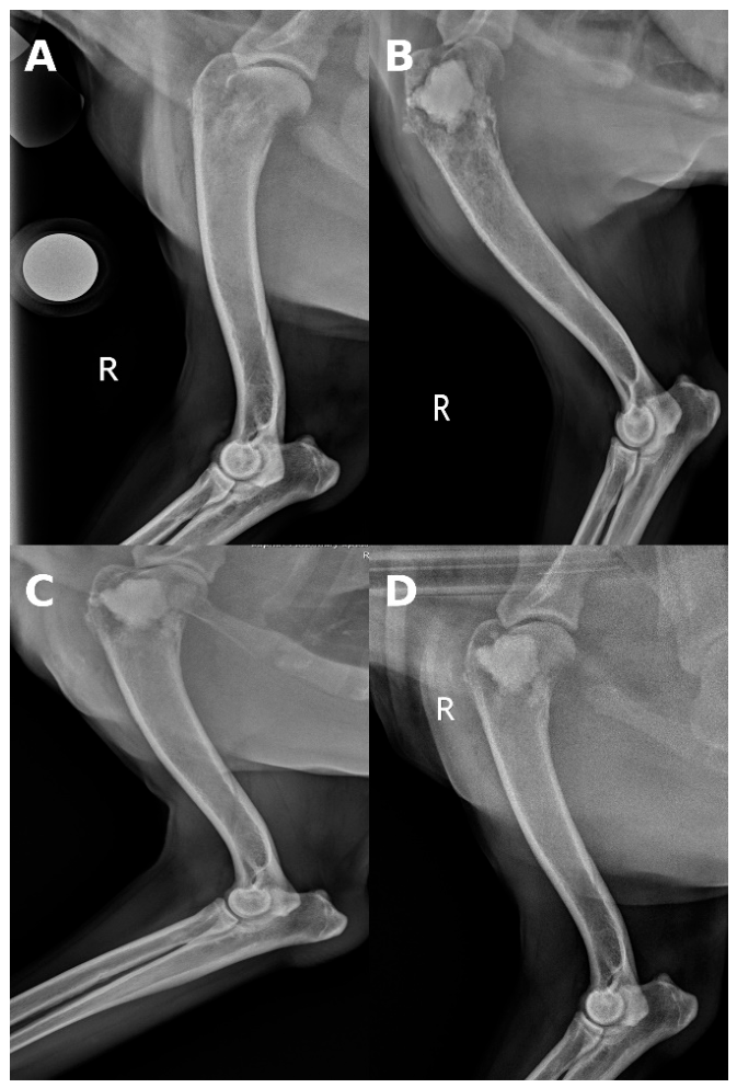
Figure 2.
Craniocaudal serial radiographs of the left distal radius and manus in dog 7. Images are arranged chronologically from left to right and top to bottom. (A) Preoperative; (B) Immediate postoperative; (C) 4 weeks postoperative; (D) 8 weeks postoperative; (E) 12 weeks postoperative; (F) 16 weeks postoperative. Progressive remodeling of the distal radial metaphysis and loss of cortical integrity are evident over time, with maintained cement position and persistent carpal instability.
Figure 2.
Craniocaudal serial radiographs of the left distal radius and manus in dog 7. Images are arranged chronologically from left to right and top to bottom. (A) Preoperative; (B) Immediate postoperative; (C) 4 weeks postoperative; (D) 8 weeks postoperative; (E) 12 weeks postoperative; (F) 16 weeks postoperative. Progressive remodeling of the distal radial metaphysis and loss of cortical integrity are evident over time, with maintained cement position and persistent carpal instability.
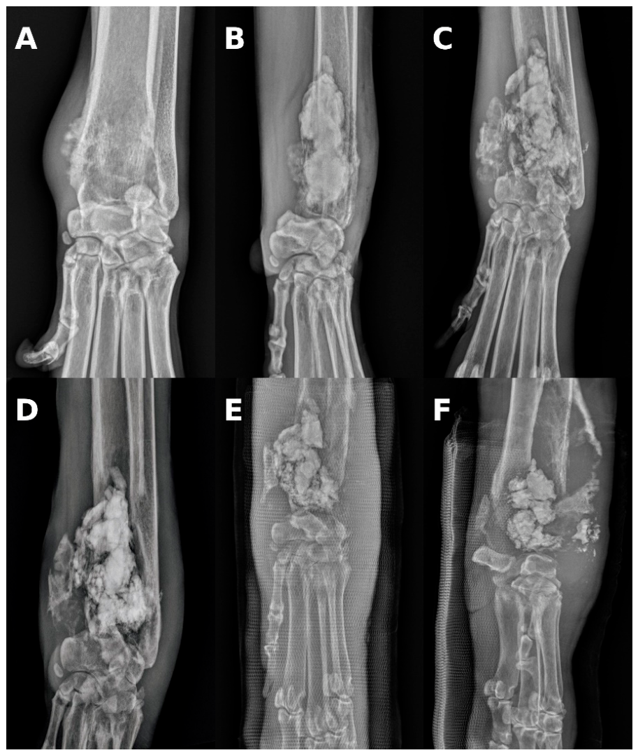
Figure 3.
Lateral serial radiographis of the left distal radius and manus in dog 7. Images are arranged chronologically from left to right and top to bottom. (A) Preoperative; (B) Immediate postoperative; (C) 4 weeks postoperative; (D) 8 weeks postoperative; (E) 12 weeks postoperative; (F) 16 weeks postoperative. There is an aggressive lesion centered in the distal radius characterized by cortical thinning and lysis followed by a increased volume of amorphous mineral opacity occupying the distal radial lesion with progressive irregularity of the cement/bone interface at later timepoints accompanied by extensive soft tissue swelling.
Figure 3.
Lateral serial radiographis of the left distal radius and manus in dog 7. Images are arranged chronologically from left to right and top to bottom. (A) Preoperative; (B) Immediate postoperative; (C) 4 weeks postoperative; (D) 8 weeks postoperative; (E) 12 weeks postoperative; (F) 16 weeks postoperative. There is an aggressive lesion centered in the distal radius characterized by cortical thinning and lysis followed by a increased volume of amorphous mineral opacity occupying the distal radial lesion with progressive irregularity of the cement/bone interface at later timepoints accompanied by extensive soft tissue swelling.
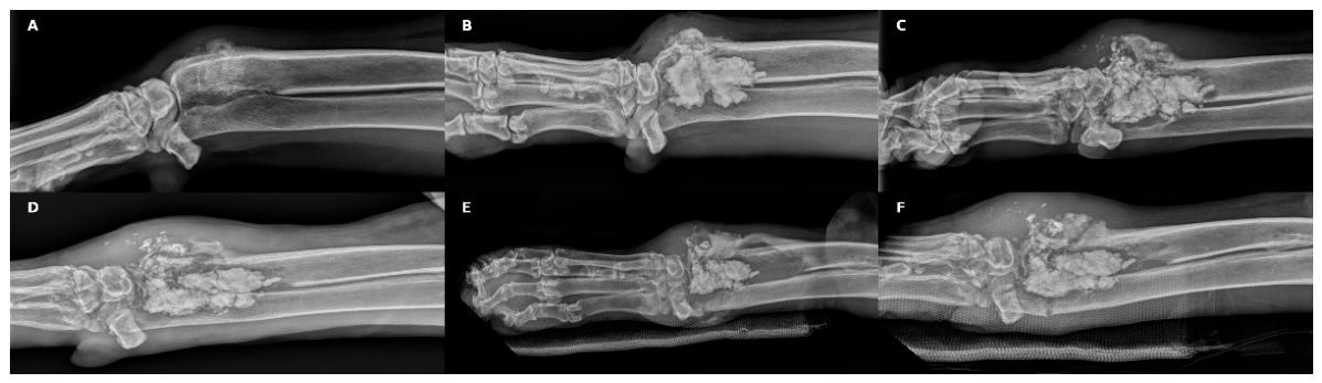
Figure 4.
Lateral radiographs of the distal radius in dogs 2, 4, and 8 prior to undergoing amputation following the end of the 12-week study period. (A) 109 days post-cementoplasty; (B) 303 days post-cementoplasty; (C) 86 days post-cementoplasty. There is irregular cortical lysis and thinning, loss of medullary architecture, and irregular periosteal new bone formation with extensive soft tissue swelling.
Figure 4.
Lateral radiographs of the distal radius in dogs 2, 4, and 8 prior to undergoing amputation following the end of the 12-week study period. (A) 109 days post-cementoplasty; (B) 303 days post-cementoplasty; (C) 86 days post-cementoplasty. There is irregular cortical lysis and thinning, loss of medullary architecture, and irregular periosteal new bone formation with extensive soft tissue swelling.

Figure 5.
Sequential CBPI Pain Severity scores for 10 dogs undergoing cementoplasty for appendicular osteosarcoma. Values are presented as mean ± SE. The CBPI Pain Severity score was calculated for each dog at each assessment period, with higher scores indicating greater owner-perceived pain severity. Preoperative scores were compared with postoperative scores at 2, 4, 8, and 12 weeks to evaluate changes in owner-reported pain burden following cementoplasty.
Figure 5.
Sequential CBPI Pain Severity scores for 10 dogs undergoing cementoplasty for appendicular osteosarcoma. Values are presented as mean ± SE. The CBPI Pain Severity score was calculated for each dog at each assessment period, with higher scores indicating greater owner-perceived pain severity. Preoperative scores were compared with postoperative scores at 2, 4, 8, and 12 weeks to evaluate changes in owner-reported pain burden following cementoplasty.
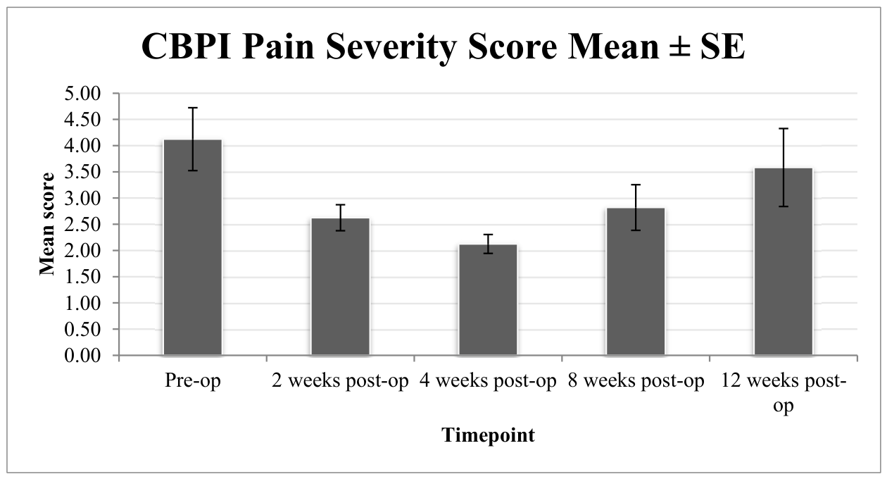
Figure 6.
Sequential CBPI Pain Interference scores for 10 dogs undergoing cementoplasty for appendicular osteosarcoma. Values are presented as mean ± SE. The CBPI Pain Interference score was calculated for each dog at each assessment period, with higher scores indicating greater owner-perceived pain-related functional interference. Preoperative scores were compared with postoperative scores at 2, 4, 8, and 12 weeks to evaluate changes in owner-reported pain-associated interference with daily activity following cementoplasty.
Figure 6.
Sequential CBPI Pain Interference scores for 10 dogs undergoing cementoplasty for appendicular osteosarcoma. Values are presented as mean ± SE. The CBPI Pain Interference score was calculated for each dog at each assessment period, with higher scores indicating greater owner-perceived pain-related functional interference. Preoperative scores were compared with postoperative scores at 2, 4, 8, and 12 weeks to evaluate changes in owner-reported pain-associated interference with daily activity following cementoplasty.
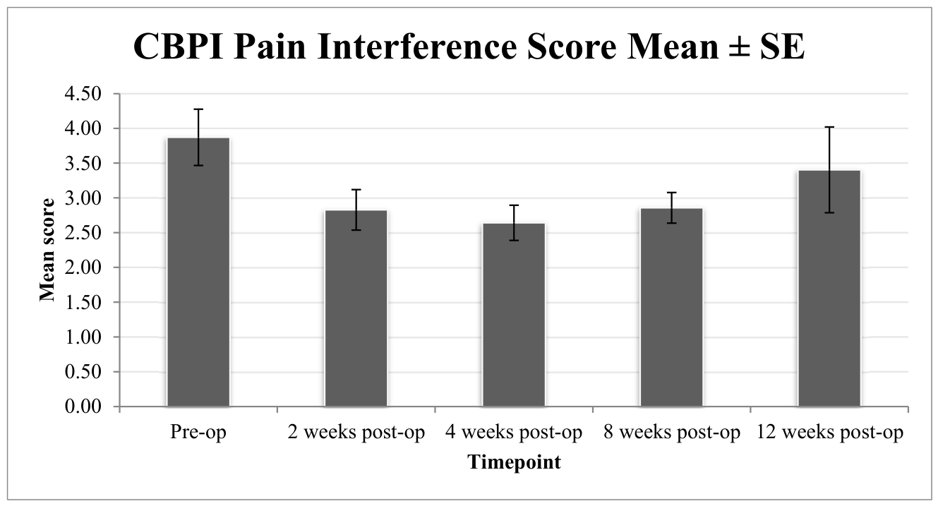
Figure 7.
Sequential CSAS scores for 10 dogs undergoing cementoplasty for appendicular osteosarcoma. Values are presented as mean ± SE. The CSAS score was calculated for each dog at each time point, with higher scores indicating greater owner-reported symptom burden. Preoperative scores were compared with postoperative scores at 2, 4, 8, and 12 weeks to evaluate changes in owner-perceived symptom burden following cementoplasty.
Figure 7.
Sequential CSAS scores for 10 dogs undergoing cementoplasty for appendicular osteosarcoma. Values are presented as mean ± SE. The CSAS score was calculated for each dog at each time point, with higher scores indicating greater owner-reported symptom burden. Preoperative scores were compared with postoperative scores at 2, 4, 8, and 12 weeks to evaluate changes in owner-perceived symptom burden following cementoplasty.
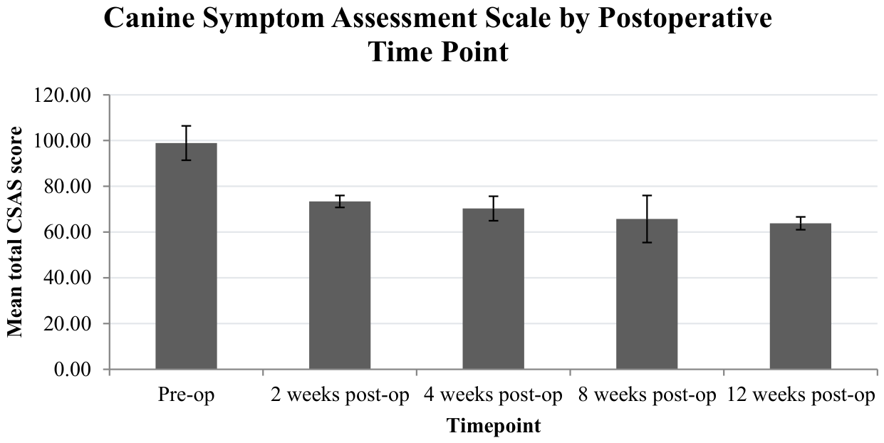
Figure 8.
Summary of Helsinki Chronic Pain Index scores by postoperative time point. Values are presented as mean ± SE. The total Helsinki Chronic Pain Index scores are calculated from the sum of the survey item responses for each dog at each time point. Higher scores indicate greater owner-perceived chronic pain or functional impairment.
Figure 8.
Summary of Helsinki Chronic Pain Index scores by postoperative time point. Values are presented as mean ± SE. The total Helsinki Chronic Pain Index scores are calculated from the sum of the survey item responses for each dog at each time point. Higher scores indicate greater owner-perceived chronic pain or functional impairment.
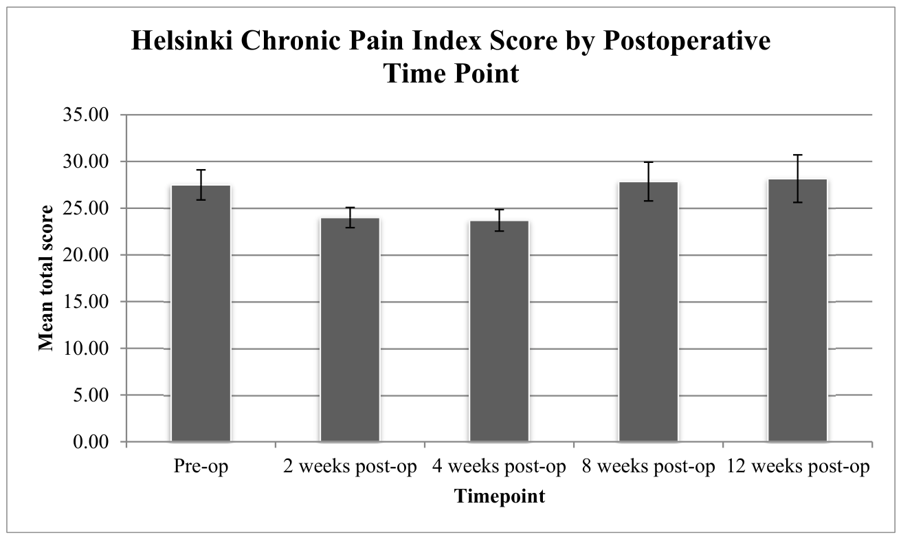

Table 1.
Clinical information for 10 dogs undergoing cementoplasty for appendicular osteosarcoma.
| Dog | Breed | Sex | Age (months) | Weight (kg) | Location of OSA | Withdrawal reason | Metastasis (days) | Euthanasia (days) |
|---|---|---|---|---|---|---|---|---|
| 1 | Great Dane | MN | 97 | 70.9 | R distal radius | Chemotherapy - 4 weeks | N/A | Unavailable |
| 2 | Anatolian Shepherd | MN | 90 | 59.1 | R distal radius | N/A | 177 | 185 |
| 3 | Husky | MN | 81 | 36.4 | R proximal tibia | N/A | N/A | 251 |
| 4 | Great Dane | MN | 115 | 69 | R distal radius | N/A | 346 | 350 |
| 5 | Great Dane | MN | 109 | 56.4 | R proximal humerus | N/A | N/A | 41 |
| 6 | Pitbull | FS | 146 | 28.6 | R proximal humerus | N/A | N/A | 96 |
| 7 | English Mastiff | FS | 62 | 82.8 | L distal radius | Fracture at 4 weeks | N/A | 173 |
| 8 | English Mastiff | MN | 90 | 87 | R distal radius | N/A | 116 | 258 |
| 9 | Golden Retriever | MN | 114 | 44 | L proximal tibia | Chemotherapy - 4 weeks | N/A | 115 |
| 10 | Cane Corso | F | 67 | 57 | R proximal humerus | Amputation - 8 weeks | N/A | Unavailable |
Table 2.
Sequential 4A-VET scores for 10 dogs undergoing cementoplasty for appendicular osteosarcoma demonstrating partial attenuation from baseline (pre-op) for up to 8 weeks postoperatively. The decrease in 4A-VET scores from preoperative values to postoperative time points indicates improvement in clinician-assessed pain.
Table 2.
Sequential 4A-VET scores for 10 dogs undergoing cementoplasty for appendicular osteosarcoma demonstrating partial attenuation from baseline (pre-op) for up to 8 weeks postoperatively. The decrease in 4A-VET scores from preoperative values to postoperative time points indicates improvement in clinician-assessed pain.
| Dog | Pre-op | 2 weeks | 4 weeks | 8 weeks | 12 weeks |
|---|---|---|---|---|---|
| 1 | 7 | 5 | 3 | 2 | 3 |
| 2 | 5 | 2 | 3 | 2 | 2 |
| 3 | 6 | 3 | 2 | 2 | 3 |
| 4 | 7 | 4 | 4 | 3 | 4 |
| 5 | 6 | 3 | 2 | 2 | 1 |
| 6 | 11 | 5 | 4 | 3 | 4 |
| 7 | 7 | 3 | 5 | 6 | N/A |
| 8 | 9 | 3 | 5 | N/A | N/A |
| 9 | 12 | 6 | 4 | N/A | N/A |
| 10 | 4 | 2 | 2 | N/A | N/A |
Disclaimer/Publisher’s Note: The statements, opinions and data contained in all publications are solely those of the individual author(s) and contributor(s) and not of MDPI and/or the editor(s). MDPI and/or the editor(s) disclaim responsibility for any injury to people or property resulting from any ideas, methods, instructions or products referred to in the content. |
© 2026 by the authors. Licensee MDPI, Basel, Switzerland. This article is an open access article distributed under the terms and conditions of the Creative Commons Attribution (CC BY) license (http://creativecommons.org/licenses/by/4.0/).
Copyright: This open access article is published under a Creative Commons CC BY 4.0 license, which permit the free download, distribution, and reuse, provided that the author and preprint are cited in any reuse.


