

Submitted:
11 May 2026
Posted:
12 May 2026
You are already at the latest version
Abstract
This research explores the aromatase-inhibitory and estrogen-agonistic/antagonistic properties of two natural naphthoquinones, α- and β-lapachone, which are known for their anticancer effects. Initial tests showed that both lapachones inhibit aromatase in the sub-micromolar range, with IC50(β) = 0.78 ± 0.06 μM and IC50(α) = 10.6 ± 2.4μM, similar to the steroidal aromatase inhibitor Exemestane (IC50: 0.02-0.2 μM). A molecular docking study comparing these compounds with androstenedione, one of the native aromatase substrates, identified their binding sites and specific interactions with the enzyme. The Yeast Estrogen Screening assay indicated that both compounds lacked hERα-agonistic activity but exhibited antagonistic effects, similar to 4-Hydroxytamoxifen (Afimoxifene; IC50 = 0.81 ± 0.65 μM). The IC50 values were 0.33 ± 0.24 μM for β-lapachone and 48.3 ± 18.9 μM for α-lapachone. Overall, the study propose unexplored mechanism of action and highlights the dual role of α- and β-lapachones: inhibiting estrogen synthesis and serving as potent, selective estrogen receptor modulators, emphasizing their potential in cancer treatment, especially for hormone-dependent cancers.
Keywords:
lapachone
; naphtoquinone
; cancer
; aromatase inhibitor
; estrogen
; SERM
1. Introduction
Natural products account for approximately 35% of approved pharmaceuticals and remain a vital resource in drug discovery [1,2,3,4,5]. Compared with conventional therapies, natural substances are often viewed as safer and more accessible due to their typically lower side effects and wider availability. Although synthetic small-molecule libraries can aid in identifying novel lead compounds, natural products offer unique advantages and are essential to pioneering drug development, especially in the battle against cancer, the second-leading cause of mortality worldwide that affects millions of individuals annually [6,7,8].
Natural products are increasingly acknowledged as a valuable reservoir of bioactive phytochemicals with diverse mechanisms of action toward malignant tumors. Evidence from epidemiological, preclinical, and early clinical investigations indicates that phytochemicals derived from fruits, vegetables, and medicinal plants can modulate oxidative stress, inflammation, cell signaling pathways, and detoxification processes. This underscores their potential as promising agents for cancer prevention, adjunctive therapy, and the reduction of treatment-related toxicity [9,10,11,12,13,14].
The inner bark and heartwood of red lapacho (Tabebuia avellanedae) contain cycloalkyl and benzene derivatives, flavonoids, quinones, furanonaphthoquinones, naphthoquinones, and anthraquinones, to name just a few [15]. Notably, three naphthoquinones — lapachol, α-lapachone, and β-lapachone (Figure 1) — have garnered scientific interest in recent decades mainly for their anticancer properties [16,17,18,19,20,21]. Lapachol was the first naphthoquinone studied for this purpose, which prompted extensive research into its therapeutic potential, mechanisms of action, and toxicological profile. Although it exhibited promising antiproliferative activity across cancer cell lines, notable toxicity concerns in mammalian systems delaying or terminating some clinical investigations [22,23]. Nonetheless, two of its isomers — α- and β-lapachone (see Figure 1) — have emerged as promising candidates in this area.
β-Lapachone (β-lap) is an ortho-naphthoquinone originally isolated from the heartwood of Handroanthus impetiginosus and can also be synthesized from lapachol. β-Lap is among the most extensively studied natural naphthoquinones in recent years due to its diverse pharmacological effects [24,25]. This natural compound demonstrates activity against various malignant tumors, including pancreatic cancer, breast cancer, hepatocellular carcinoma, and others [26,27,28,29,30,31,32]. Additionally, this ortho-naphthoquinone exhibits antifungal and antibacterial activities, emphasizing its efficacy against resistant microorganisms, and offers anti-inflammatory, anti-obesity, antioxidant, neuroprotective, nephroprotective, and wound-healing properties [24]. It is also noteworthy that β-lapachone itself and its pro-drug ARQ-761 passed phase I/II of clinical studies for solid tumors and another synthetic β-lapachone derivative (MB12066) was a subject for verification of its therapeutic effects in metabolic syndrome, obesity and alcoholic fatty liver disease, underscoring translational interest in β-lapachone–related chemistries [33,34,35,36].
α-Lapachone (α-lap), a para-naphthoquinone, is another structural isomer of lapachol that has been studied less extensively than β-lap. Although it is also known for its antitumor activity, there are few publications on it, and its mechanism of action remains poorly understood [37,38,39]. Notably, β-lap can convert to α-lap, especially in acidic environment or when irradiated by either UV or visible light (e.g., sunlight) [40,41], emphasizing the need to study both compounds together. Although the pH-dependent isomerization of β-lap to α-lap in humans has not yet been confirmed [42], examining their effects on the same targets remains crucial. Doing so can reveal similarities or differences that sustain the choice of the more efficient agent when considering other influential factors, such as stability, pharmacokinetics, or bioavailability.
Unlike α-lap, whose anticancer activity is attributed to its ability to inhibit the initial non-covalent binding of topoisomerase II to DNA and to induce the religation of DNA breaks before dissociating the enzyme from DNA [37,38], the anticancer effects of β-lap are complex and depend on various interconnected mechanisms [24]. It is widely accepted that β-lap’s antitumor activity primarily relies on a unique bioactivation process involving NQO1, an enzyme overexpressed in many solid tumors [43,44,45,46,47,48,49]. Unlike standard quinones, which are reduced by a single electron to form unstable semiquinones, β-lap undergoes a two-electron reduction cycle. This process consumes a considerable amount of cellular NAD(P)H and oxygen, thereby increasing the production of ROS such as superoxide and hydrogen peroxide. The induced oxidative stress causes extensive single-strand DNA breaks, which hyperactivate PARP-1. This overactivation depletes NAD+ and ATP pools, resulting in necroptosis, a form of programmed necrosis, rather than the typical caspase-dependent apoptosis. Another mechanism involves β-lap’s effect on the cellular glutathione pool, particularly its reduced form, GSH [50,51,52,53]. Research indicates that β-lap decreases GSH levels by acting as a potent oxidizing agent and by inhibiting GSH synthesis. GSH serves as the primary antioxidant, safeguarding cells against ROS; therefore, these mechanisms shift the redox balance toward oxidation, increasing the GSSG/GSH ratio in both tumor and normal cells. In doing so, β-lap effectively compromises the antioxidant defenses of cancer cells and restores their sensitivity to chemotherapeutic agents. Other studies also suggest that β-lap exerts its cytotoxic effects through interactions with DNA topoisomerases (I and II) [37,54], indoleamine 2,3-dioxygenase 1 (IDO1) [55], telomerase [56], heat shock protein 90 (Hsp90) [28], and serine-threonine protein kinase (AKT1) [30].
Based on the above reasoning, it is not irrational to conclude that unexplored mechanisms of action may exist for proven multi-target natural compounds such as α- and β-lap. Considering that: (i) numerous compounds, including naphthoquinones, exert anticancer effects by either inhibiting estrogen synthesis (Aromatase Inhibitors, AIs) or disrupting estrogen signaling through blocking estrogen receptors (Selective Estrogen Receptor Modulators, SERMs) [57,58,59,60,61]; (ii) α- and β-lap are redox-active species [62,63]; and (iii) β-lap is known as a glucocorticoid antagonist that competes for the steroid-binding site of the glucocorticoid receptor [64]; we hypothesized that α- and β-lap could also inhibit aromatase, a steroid oxidoreductase enzyme, and compete with estrogens for their receptor. While the anticancer properties of α- and β-lap are widely recognized, these two possible mechanisms of action remain unexplored for them. To verify the validity of our hypothesis, we first evaluate the capacity of α- and β-lap to inhibit aromatase activity and performed molecular docking analyses to identify the binding sites and interactions of each of them with aromatase. Additionally, by using the Yeast Estrogen Screening (YES) assays, we studied their agonistic/antagonistic properties toward the estrogen receptor (hERα). Overall, this research focuses on uncovering unknown mechanisms of action and determining whether α- and β-lap can serve as dual-function agents— AIs and SERMs—thereby supporting their potential for treating hormone-dependent cancers.
2. Results and Discussion
Given that the natural hormone 17β-estradiol (E2) induces tumors in various organs of animals and humans [65], and that many tumors are physiologically dependent on estrogen for proliferation, therapeutic intervention focuses on either depleting the systemic supply of the hormone or blocking its signaling pathways. Indeed, aromatase inhibitors and estrogen receptor antagonists constitute the cornerstone of endocrine therapy for hormone-receptor-positive (HR+) cancers [57,58,59,60,61]. Aromatase inhibitors achieve this by inhibiting the enzyme responsible for converting androgens into estrogens, effectively lowering circulating and intracellular levels in patients of any age and gender. Conversely, estrogen receptor antagonists competitively bind to the estrogen receptor to prevent transcriptional activation of oncogenic pathways. Together, these classes of drugs have significantly reduced recurrence rates and improved overall survival by precisely targeting the endocrine drivers of malignancy.
2.1. Aromatase Inhibitory Activity
The results of the aromatase inhibition assay, as illustrated in the dose-response curves (Figure 2), demonstrate that both α- and β-lap are effective inhibitors of the human CYP19A1 enzyme; however, the data also reveals a significant disparity in potency between the two structural isomers. β-Lap exhibits a high inhibitory activity with an IC50 of 0.78 ± 0.06 μM. This sub-micromolar value places β-lap in a high-potency category, as it can suppress 50% of enzyme activity at a concentration an order of magnitude lower than its alpha counterpart. While α-lap also inhibits the enzyme, its IC50 = 10.63 ± 2.44 μM indicates a more moderate inhibitory profile. This difference underscores the structural sensitivity of the aromatase active site; the ortho-quinone arrangement of β-lap likely allows for a more favorable geometric or electronic fit within the heme-containing catalytic pocket compared to the para-quinone structure of α-lap. While the positive control Letrozole (a non-steroidal third-generation inhibitor) remains more potent with an IC50 of approximately 0.01 μM, the activity of the β-isomer is similar to Exemestane, a steroidal “suicide” inhibitor widely used in HR+ breast cancer treatment with reported IC50 values between 0.02 and 0.2 μM [66,67]. This suggests that the naphthoquinone scaffold, especially the ortho-isomer, has promising pharmacological potential for developing non-steroidal aromatase inhibitors (NSAIs).
2.2. Molecular Docking
Molecular docking studies were conducted to identify the binding site of α- and β-lap and to analyze their specific interactions with the aromatase enzyme. The crystal structure used (PDB ID: 3EQM) is of a human placental aromatase cytochrome P450 complexed with androstenedione (A4) [68]. This structure was selected for its high resolution (2.90 Å) and because binding to the endogenous substrate A4 offers detailed insights into specific interactions and accurate tertiary structure. The results are presented in Figure 3 A-C.
Aromatase is a heme-dependent enzyme where the heme-iron complex plays a crucial role in the oxidative removal of the C19-methyl group during estrogen production. As shown in Figure 3A, in the natural enzyme-substrate complex, A4 is bound within a hydrophobic pocket in the enzyme’s active site (docking score = -13.0 eV), with the C19-methyl group directed toward the iron (Fe) atom at about 4 Å distance. The amide group (-NH-) of Met374 forms a hydrogen bond with the 17-keto oxygen atom, while amino acid residues as given in Figure 3A stabilize the enzyme-substrate complex through van der Waals interactions, aligning with crystallographic data [68,69]. α- and β-Lap also exhibit high binding affinity to the enzyme’s active site, with docking scores of -9.9 eV and -9.6 eV, respectively. Although these scores are slightly lower than the endogenous substrate A4 (-13.0 eV), they still reflect strong stabilization within the catalytic pocket. Both isomers position themselves close to the heme group, physically blocking the substrate binding site. While it was initially expected that the carbonyl oxygen atoms in the lapachone isomers would form hydrogen bonds with residues like Met374, Ser478, or Trp224—similar to A4—the orientations suggest these oxygens are not ideally positioned for such interactions. Instead, their inhibitory effect largely stems from their conjugated ring systems, which enable strong π-alkyl interactions with key hydrophobic residues: Leu477 and Ile133 for α-lapachone, and Ile133, Val370, and Leu477 for β-lapachone. These π-alkyl interactions are more energetically favorable than simple alkyl-alkyl interactions seen with A4. Consequently, the structural similarity to the natural substrate, combined with these enhanced electronic interactions, explains the high affinity of the lapachone isomers for the active site and offer a mechanistic explanation for their experimentally observed aromatase inhibitory activity
2.3. Anti-Estrogenic Activities [70]
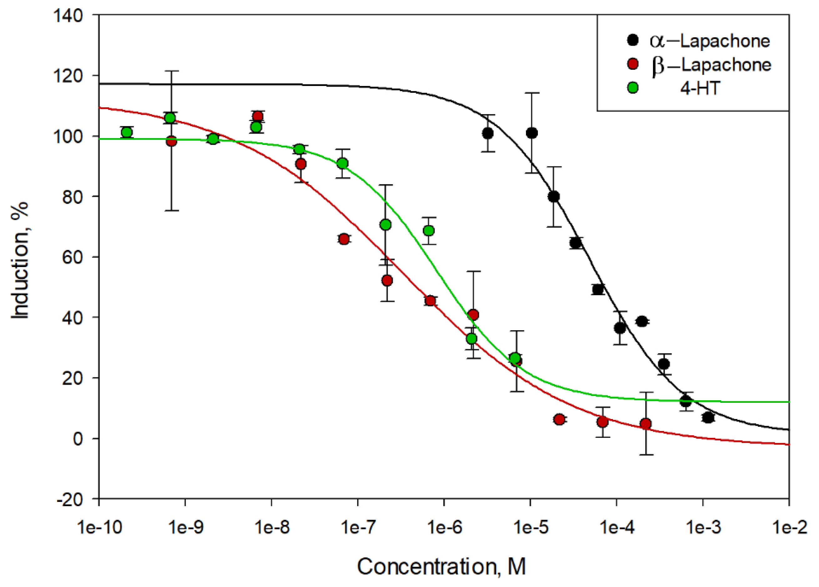
The preliminary conducted YES assays showed that neither α- nor β-lap possesses agonist activity; however, both compounds exhibit inherent antagonistic effects toward the hERα receptor. The dose-response curves in Figure 4 offer a comparative quantitative view of their anti-estrogenic activity in relation to the standard reference, the active metabolite of tamoxifen 4-hydroxytamoxifen (4-HT). As can be seen, all three tested compounds (α-lap, β-lap, and 4-HT) followed a classic sigmoidal dose-response relationship, where β-galactosidase production —a marker of ER transcriptional activity—decreased proportionally with increasing concentrations. At concentrations above 1 mM, all compounds converged toward maximal inhibition, effectively neutralizing the 100% induction effect of the most potent estrogen estradiol.
A more detailed analysis of the reduction in β-galactosidase induction additionally underscores the compound’s potent ability to inhibit estrogenic signaling pathways. The steepness of the curves obtained for β-lap (red curve) compared to 4-HT (green curve) suggests a robust inhibitory response within the nanomolar to micromolar range (10−7 to 10−6 M), where β-lap consistently maintains lower induction levels than the reference drug. The principal finding is that β-lap may functions as a highly efficacious ER antagonist, with an IC50 value of 0.33 ± 0.24 μM, achieving near-complete inhibition (approaching 0% induction) at concentrations exceeding 10 μM. Furthermore, the results are comparable to those observed with 4-HT, with IC50 of 0.81 ± 0.65 μM. In contrast, α-lap exhibits markedly lower potency (IC50 of 48.3 ± 18.8 μM) and achieves near-complete inhibition at concentrations above 1 mM – two orders of magnitude higher than that demonstrated by the beta-isomer. Consequently, similarly to the trend demonstrated for the aromatase-inhibitory potential, the observed significant difference in activity between the two isomers highlights the critical importance of the ortho-naphthoquinone orientation (found in β-lap) versus the para-orientation (found in α-lap) for optimal interaction with the estrogen receptor’s ligand-binding domain. Nevertheless, despite requiring much higher pharmacological doses to be effective, α-lap can still interfere with the estrogen signaling
3. Materials and Methods
3.1. General
All chemicals used in this study were purchased from Sigma-Aldrich (FOT, Sofia, Bulgaria). The organic solvents were of analytical grade and were used without further purification. XenoScreen XL YES assay kits and XenoScreen YES strain were obtained from Xenometrix AG (Allschwil, Switzerland) and the Aromatase (CYP19A) Inhibitor Screening Kit from BioVision (BioVision Inc., Milpitas, CA, USA). α- and β-Lap were synthesized by us as described below. NMR spectra were recorded on a Bruker Avance III HD (Bruker BioSpin GmbH, Rheinstetten, Germany, 500 MHz and 126 MHz for 1H and 13C, respectively) using CDCl3 as a solvent. The chemical shifts (δ) are given in ppm and J values are reported in Hz. Biological assessment were performed on 800TS (Biotek Instruments, Inc, ELTA90, Sofia, Bulgaria) and Synergy H1 (Biotek Instruments, Inc, ELTA90, Sofia, Bulgaria) ELISA microplate readers. Dose–response curves were fitted using the SigmaPlot version 12.5 (Systat Software Inc. San Jose, CA, USA).
3.2. Synthesis and Characterization of α- and β-Lapachone
α- and β-Lap were synthesized as described elsewhere [71,72]. Тhe procedure involves two synthetic steps. Shortly, in round-bottom flask 1g (5.74 mmol) of lawsone (2-hydroxy-1,4-naphthoquinone) was dissolved in 10 ml dry DMF, and then 1.05 g KI (6.32 mmol) and 0.88 ml (6.32 mmol) triethylamine were added. The reaction mixture was stirred for an hour at room temperature and 0.86 g (5.74 mmol) 4-bromo-2-methyl-2-butene were added. The reaction mixture was heated at 40 °C for 2 h and monitored by TLC. At the end of the reaction 50 ml water were added and the resultant lapachol was extracted with ethyl acetate (EtOAc), washed with 5% NaHCO3 and water to pH = 7. The organic layer was dried with Na2SO4, and the solvent was evaporated. Lapachol was isolated via column chromatography (benzene/acetone = 99/1 + formic acid). Lapachol, 1g (4.13 mmol) was reacted with 10 ml conc.H2SO4 for 15 min. at room temperature. The reaction mixture was poured in ice water and filtered. α- and β-Lapachones were isolated after column chromatography (benzene/acetone = 95/5) and their structure was confirmed by NMR spectroscopy. The data obtained is consistent with those available the literature [73,74].
α-Lapachone: 1H NMR (500 MHz, CDCl3): δ = 8.08 (2H, t, J = 7.7 Hz, ArH), 7.72–7.62 (2H, m, ArH), 2.62 (2H, t, J = 6.6 Hz, CH2), 1.82 (2H, t, J = 6.6 Hz, CH2), 1.43 (6H, s, CH3). 13C NMR (126 MHz, CDCl3): δ = 184.40 (C), 180.01 (C), 154.64 (C), 133.86 (CH), 132.92 (CH), 132.11 (C), 131.20 (C), 126.33 (CH), 125.98 (CH), 120.16 (C), 78.16 (C), 31.44 (CH2), 26.52 (CH3), 16.75 (CH2).
β-Lapachone: 1H NMR (500 MHz, CDCl3): δ = 8.05 (1H, d, J = 6.7 Hz, ArH), 7.80 (1H, d, J = 7.8 Hz, ArH), 7.63 (1H, td, J = 7.7, 1.1 Hz, ArH), 7.49 (1H, t, J = 7.6 Hz, ArH), 2.56 (2H, t, J = 6.7 Hz, CH2), 1.85 (2H, t, J = 6.6 Hz, CH2), 1.46 (6H, s, CH3). 13C NMR (126 MHz, CDCl3): δ = 179.88 (C), 178.59 (C), 162.03 (C), 134.78 (CH), 132.65 (C), 130.66 (CH), 130.17 (C), 128.57 (CH), 124.07 (CH), 112.74 (C), 79.28 (C), 31.62 (CH2), 26.78 (CH3), 16.18 (CH2).
3.3. Aromatase Inhibition Assay
The assay for evaluating anti-aromatase activity relies on converting a fluorogenic substrate into a fluorescent product (Ex/Em = 488/527 nm) through aromatase catalysis, enabling enzyme activity monitoring under various conditions. To assess the inhibitory effects of α- and β-lap, twelve concentrations of each compound were prepared—0.226 to 18.1 mM for α-lap and 0.025 to 10.0 mM for β-lap—in acetonitrile, facilitating dose-response curve creation and IC50 calculation. These solutions were diluted with aromatase buffer to produce 1% acetonitrile stock solutions. Letrozole served as a positive control for 100% inhibition, a no-inhibitor control indicated 0% inhibition, and a background control lacked the fluorogenic substrate. According to protocol, test compounds and controls were incubated for 10 minutes at 37 °C with the aromatase enzyme. Post-incubation, the fluorogenic substrate and NADP+ system were added to all wells except the background control. Fluorescence was then measured kinetically at Ex/Em = 488/527 nm every minute for 60 minutes. All measurements were duplicated.
3.4. Molecular Docking
Crystal structure of human placental aromatase cytochrome P450 in complex with androstenedione (PDB: 3EQM) [68], was obtained from RCSB protein data bank (http://www.pdb.org). AutoDock Tools v. 1.5.7 [75] was used for the preparation of the enzyme. All non-protein components were removed: phosphate groups, androstenedione and water molecules, with exception of the heme group. Polar hydrogen atoms and Kollman charges were added. The grid box coordinates were x = 83.427, y = 50.113, z= 46.367, grid size: 40: 40: 40. The ligands, α- and β-lap were obtained in SDF format from the Zinc20 database [76] and were then converted into PDBQT file format using Open Babel software v2.3.1 [77]. Molecular Docking was performed via Autodock Vina v1.1.2 [78]. Analysis of the docking results and interactions between the ligands and aromatase were acquired by importing the docked results into PyMOL v.3.1 [79] and BIOVIA Discovery Studio Visualizer v25.1.0.24284 [80]. To validate the calculations, re-docking with A4 was performed, and the conformation of the resulting pose was compared with the crystallographic data.
3.5. XenoScreen YES Assay
The estrogenic agonistic/antagonistic activities of α- and β-lap were measured with the XenoScreen XL YES Assay kit according to the manufacturer protocol (Xenometrix, XenoScreen XL YES, Instructions for Use Version 3.11). Growing yeast cells (Saccharomyces cerevisiae) were exposed to serial dilutions of the test compound (α- or β-lap) in 96-well plates. After 18 h of incubation at 31 ˚C, the yeast cell growth was assessed by measuring the optical density at 690 nm (OD690), to calculate the growth factor for each well (sample wells compared to solvent control wells). After the growth measurement, the lysis buffer containing lyticase and CPRG was added. After 1 h of incubation at 31 ˚C, β-galactosidase secretion into the media was quantified colorimetrically at 570 nm, by measuring the conversion of the yellow substrate (CPRG) into a red product. The red colour development at 570 nm was corrected for diffraction by the cells and debris by simultaneous measurements again of the OD690. Calculated growth factors < 0.5 were considered to be cytotoxic effects. The calculation of induction or inhibition was corrected according to the growth factor for each well, and therefore the activity of the β-galactosidase was not influenced by inhibited growth of yeast cells. The calculations of the induction and inhibition are described in the manufacturer protocol. Estrogenic antagonistic activity was measured by evaluating β-galactosidase signal reduction in yeast cells in the presence of an agonist (3.3 × 10⁻¹⁰M) E2 throughout the plate. Serial dilutions of α- and β-lap were then assessed for their ability to inhibit their response to agonist in the medium. To determine antagonistic activity, 4-hydroxytamoxifen (4-HT) (1×10⁻³ M) was used as positive control (100% inhibition) for the YES assay. After preliminary experiments for approximate estimation of IC50 values, α-and β-lap were dissolved in DMSO and measured at twelve concentrations (3.20 µM to 1.14 mM for α-lap and 0.69 nM to 218 µM for β-lap ). Experiments were conducted in two independent experiments in triplicates.
3.6. Data Analysis
Dose–response curves were fitted using the SigmaPlot version 12.5 (Systat Software Inc. San Jose, CA, USA), Where the compounds exhibited a complete dose–response curve and IC50 values were calculated. A sample dilution that inhibited negative control activity (medium with agonist) by at least 50% was considered to have antagonistic endocrine activity in antagonist assay.
4. Conclusions
This study reveals a novel dual mechanism of action for the natural naphthoquinones α- and β-lap, showing that both isomers act as potent AIs and SERMs. Notably, β-lap proved more effective, showing sub-micromolar inhibition of aromatase (IC 50 = 0.78 μM) and the human estrogen receptor hERα (IC 50 = 0.33 μM), with these values comparable to those of drugs like Exemestane and Afimoxifene. Molecular docking confirmed these compounds fit into the enzyme’s catalytic pocket, forming stable π-alkyl interactions with key hydrophobic residues through their conjugated ring systems. Although α-lap has lower activity, its ability to modulate estrogen signaling remains important, especially given the potential for pH-dependent isomerization between the two forms.
The identification of these dual-function agents carries substantial clinical significance. In postmenopausal women, clinical data have demonstrated that AIs are highly effective in managing estrogen-dependent diseases; however, in premenopausal women, estrogen synthesis is only partially inhibited, resulting in a reflex increase in gonadotropin levels that stimulates ovarian aromatase and counteracts treatment. By concurrently depleting estrogen production and blocking receptor activation within a single molecular framework, α- and β-lap provide a comprehensive therapeutic strategy. This dual mechanism may overcome the limitations associated with current AI monotherapy in premenopausal patients and diminish the risk of treatment resistance in HR+ breast cancers, as influencing multiple cellular processes simultaneously constrains the tumor’s capacity to develop escape mechanisms. Finally, the anti-estrogenic effects of these naphthoquinones are anticipated to synergize with the aforementioned effects on the cell’s redox status and signaling pathways. This coordinated action is expected to simultaneously inhibit hormone-driven pathologies and restore the essential cellular environment necessary for organism-wide repair and longevity.
Despite the demonstrated potential, challenges persist concerning the clinical application of these compounds. The pharmacokinetic profiles of α- and β-lap are complex, particularly with respect to their bioavailability, metabolism, and toxicity. Moreover, although their potency is considerable for natural products, it generally remains inferior to that of standard synthetic therapies such as letrozol or fulvestrant. Nonetheless, these naphthoquinones may be employed in combination with standard drugs to investigate potential synergistic effects. Such combinations could enhance overall therapeutic efficacy while allowing lower concentrations of synthetic agents, thereby reducing systemic toxicity and delaying the development of resistance. Ultimately, although α- and β-lap demonstrate promising anticancer activity via these previously unexamined endocrine pathways, comprehensive in vitro and in vivo pharmacological investigations are required to thoroughly evaluate their suitability and safety for clinical oncological application.
Author Contributions
Conceptualization, M.G.B. and G.D.; methodology, M.G.B. and S.S.; resources, M.G.B. and G.D.; data curation, S.S. and F.N.; investigation, M.G.B., S.S. and F.N.; writing—original draft preparation, M.G.B., S.S. and F.N.; writing—review and editing, M.G.B. and G.D. All authors have read and agreed to the published version of the manuscript.
Funding
This research received no external funding.
Data Availability Statement
Any inquiries can be directed to the corresponding author.
Conflicts of Interest
Author Georgi Dinkov was employed by the company (IdeaLabs, LLC). The remaining authors declare that the research was conducted in the absence of any commercial or financial relationships that could be construed as a potential conflict of interest.
References
- Calixto, J. The role of natural products in modern drug discovery. An. Da Acad. Bras. De Ciências 2019, 91, e20190105. [CrossRef]
- Newman, D.; Cragg, G. Natural Products as Sources of New Drugs over the Nearly Four Decades from 01/1981 to 09/2019. J. Nat. Prod. 2020, 83 (3), 770-830. [CrossRef]
- Chaachouay, N.; Zidane, L. Plant-Derived Natural Products: A Source for Drug Discovery and Development. Drugs and Drug Candidates 2024, 3 (1), 184-207. [CrossRef]
- Najmi, A.; Javed, S.; Al Bratty, M.; Alhazmi, H. Modern Approaches in the Discovery and Development of Plant-Based Natural Products and Their Analogues as Potential Therapeutic Agents. Molecules 2022, 27 (2), 349. [CrossRef]
- Chihomvu, P.; Ganesan, A.; Gibbons, S.; Woollard, K.; Hayes, M. Phytochemicals in Drug Discovery—a Confluence of Tradition and Innovation. Int. J. Mol. Sci. 2024, 25 (16), 8792-8792. [CrossRef]
- Filho, A.; Laversanne, M.; Ferlay, J.; Colombet, M.; Piñeros, M.; Znaor, A.; Parkin, D.; Soerjomataram, I.; Bray, F. The GLOBOCAN 2022 Cancer Estimates: Data Sources, Methods, and a Snapshot of the Cancer Burden Worldwide. Int. J. Cancer 2024, 156 (7). [CrossRef]
- Force, L.; Kocarnik, J.; May, M et al. The Global, Regional, and National Burden of Cancer, 1990–2023, with Forecasts to 2050: A Systematic Analysis for the Global Burden of Disease Study 2023. The Lancet 2025, 406 (10512), 1565-1586. [CrossRef]
- Thompson, J. B.; Hawkins, J. A. Phylogeny and Bioprospecting: The Diversity of Medicinal Plants Used in Cancer Management. Plants, People. Planet 2024, 7 (1), 147-158. [CrossRef]
- Rawat, D.; Singh, R. Editorial: Plant Derived Secondary Metabolites as Anti-Cancer Agents. Anti-Cancer Agents Med. Chem. 2013, 13 (10), 1551-1551. [CrossRef]
- Naeem, A.; Hu, P.; Yang, M.; Zhang, J.; Liu, Y.; Zhu, W.; Zheng, Q. Natural Products as Anticancer Agents: Current Status and Future Perspectives. Molecules 2022, 27 (23), 8367. [CrossRef]
- Muhammad, N.; Usmani, D.; Tarique, M.; Naz, H.; Ashraf, M.; Raliya, R.; Tabrez, S.; Zughaibi, T.; Alsaieedi, A.; Hakeem, I.; Suhail, M. The Role of Natural Products and Their Multitargeted Approach to Treat Solid Cancer. Cells 2022, 11 (14), 2209. [CrossRef]
- Shi, A.; Liu, L.; Li, S.; Qi, B. Natural Products Targeting the MAPK-Signaling Pathway in Cancer: Overview. J. Cancer Res. Clin. Oncol. 2024, 150 (1). [CrossRef]
- Ali Abdalla, Y.; Subramaniam, B.; Nyamathulla, S.; Shamsuddin, N.; Arshad, N.; Mun, K.; Awang, K.; Nagoor, N. Natural Products for Cancer Therapy: A Review of Their Mechanism of Actions and Toxicity in the Past Decade. J. Trop. Med. 2022, 2022, 1-20. [CrossRef]
- Dong, S.; Guo, X.; Han, F.; He, Z.; Wang, Y. Emerging Role of Natural Products in Cancer Immunotherapy. Acta Pharm. Sin. B 2022, 12 (3), 1163-1185. [CrossRef]
- Gómez Castellanos, R.; Prieto, J.; Heinrich, M. Red Lapacho (Tabebuia Impetiginosa)—a Global Ethnopharmacological Commodity? J. Ethnopharmacol. 2009, 121 (1), 1-13. [CrossRef]
- Balassiano, I.; De Paulo, S.; Silva, N.; Cabral, M.; Carvalho M. Demonstration of the lapachol as a potential drug for reducing cancer metastasis. Oncol. Rep. 2005, 13 (2), 329-333.
- Epifano, F.; Genovese, S.; Fiorito, S.; Mathieu, V.; Kiss, R. Lapachol and Its Congeners as Anticancer Agents: A Review. Phytochem. Rev. 2013, 13 (1), 37-49. [CrossRef]
- Hussain, H.; Green, I. Lapachol and Lapachone Analogs: A Journey of Two Decadesof Patent Research(1997-2016). Expert Opin. Ther. Pat. 2017, 27 (10), 1111-1121. [CrossRef]
- Zhang, Z.; Bai, L.; Lu, C.; Li, X.; Wu, Y.; Zhang, X.; Shen, Y. Lapachol Inhibits the Growth of Lung Cancer by Reversing M2-like Macrophage Polarization via Activating NF-ΚB Signaling Pathway. Cell. Signal. 2023, 112, 110902. [CrossRef]
- Amparo, T.; da Anunciação, K.; Almeida, T.; de Oliveira, A.; da Silva, G.; Brandão, G. Lapachol Interferes with the Cell Cycle and Inhibits Proliferation and Migration of Bladder Tumor Cells with Effects on NcRNA Expression. Sci. Rep. 2025, 15 (1). [CrossRef]
- Kurniawan, R.; Ashari, A.; Yogi Saputra, M.; Indarto, I.; Ahmadi, P.; Suhartati, T. The Lapachol Derivatives from Tabebuia Aurea: Distribution and Cytotoxic Evaluation against Human Cancer Cells. J. Appl. Pharm. Sci. 2026, 162-168. [CrossRef]
- Block, J.; Serpick, A.; Miller, W.; Wiernik, P. Early clinical studies with lapachol (NSC-11905). Cancer Chemother. Rep. 1974; 4, 27-28.
- Morrison, R.; Brown, D.; Oleson, J.; Cooney, D. Oral Toxicology Studies with Lapachol. Toxicol. Appl. Pharmacol. 1970, 17 (1), 1-11. [CrossRef]
- Gong, Q.; Jiabao, H.; Wang, P.; Li, X.; Zhang, X. A Comprehensive Review on β-Lapachone: Mechanisms, Structural Modifications, and Therapeutic Potentials. Eur. J. Med. Chem. 2021, 210, 112962-112962. [CrossRef]
- Motta, K.; França, L.; Rissino, J.; Vale, V.; Vanessa, E.; Dolabela, M.; Nagamachi, C.; Pieczarka, J. Anticancer Potential and Safety Profile of β-Lapachone in Vitro. Molecules 2024, 29 (6), 1395-1395. [CrossRef]
- Li, Y.; Sun, X.; LaMont, J.; Pardee, A.; Li, C. Selective killing of cancer cells by β-lapachone: Direct checkpoint activation as a strategy against cancer. Proc. Natl. Acad. Sci. U.S.A. 2003, 100 (5) 2674-2678, 10.1073/pnas.0538044100.
- Ough, M.; Lewis, A.; Bey, E.; Gao, J.; Ritchie, J.; Bornmann, W.; Boothman, D.; Oberley, L.; Joseph C. Efficacy of beta-lapachone in pancreatic cancer treatment: Exploiting the novel, therapeutic target NQO1. Cancer Biol. Ther. 2005, 4 (1), 102-109, . [CrossRef]
- Wu, Y.; Wang, X.; Chang, S.; Lu, W.; Liu, M.; Pang, X. B-Lapachone Induces NAD(P)H:Quinone Oxidoreductase-1- and Oxidative Stress-Dependent Heat Shock Protein 90 Cleavage and Inhibits Tumor Growth and Angiogenesis. J. Pharmacol. Exp. Ther. 2016, 357 (3), 466-475. [CrossRef]
- Silvers, M.; Deja, S.; Singh, N.; Egnatchik, R.; Sudderth, J.; Luo, X.; Beg, M.; Burgess, S.; DeBerardinis RJ.; Boothman, D.; Merritt, M. The NQO1 bioactivatable drug, β-lapachone, alters the redox state of NQO1+ pancreatic cancer cells, causing perturbation in central carbon metabolism. J. Biol. Chem. 2017, 292 (44), 18203-18216. [CrossRef]
- Du, P.; Li, Y.; Han, A.; Wang, M.; Liu, J.; Piao, Y.; Chen, L. β-Lapachone Suppresses Carcinogenesis of Cervical Cancer via Interaction with AKT1. Front. Pharmacol. 2025, 16, 1509568. [CrossRef]
- Coelho, L.; Vianna, M.; da Silva, D.; Gonzaga, B.; Ferreira, R.; Coutinho, C.; Magalhães, F.; Maria, E.; de Oliveira, R.; Manso, P.; de Carvalho, M.; Vargas, F.; Garzoni, L. β-Lapachone Impairs Viability, Migration, and Epithelial–Mesenchymal Transition in Mammary Tumor Spheroids. BMC Cancer 2025, 25 (1) 1826. [CrossRef]
- Amparo, T.; Anunciação, K.; Almeida, T.; Silva, G.; Brandão, G. Beta-Lapachone: Effects on Proliferation, Survival, Migration, Cell Cycle, and lncRNA Modulation in Bladder Cancer Cells With Distinct TP53 Profiles. Drug Dev. Res. 2025, 86 (5), e70128. [CrossRef]
- Khong, H.; Dreisbach, L.; Kindler, H.; Trent, D.; Jeziorski, K.; Bonderenko, I.; Popiela, T.; Yagovane, D.; Dombal, G. A phase 2 study of ARQ 501 in combination with gemcitabine in adult patients with treatment naïve, unresectable pancreatic adenocarcinoma, J. Clin. Oncol. 2007, 25, 15017.
- Kawecki, A.; Adkins, D.; Cunningham, C.; Vokes, E.; Yagovane, D.; Dombal, G.; Koralewski, P.; Hotko, Y.; Vladimirov, V. A phase II study of ARQ 501 in patients with advanced squamous cell carcinoma of the head and neck. J. Clin. Oncol. 2007, 25, 16509.
- Gerber, M.; Beg, M.; Fattah, F.; Frankel, A.; Fatunde, O.; Arriaga, Y.; Dowell, J.; Bisen, A.; Leff, R.; Meek, C.; Putnam, W.; Kallem, R.; Subramaniyan, I.; Dong, Y.; Bolluyt, J.; Sarode, V.; Luo, X.; Xie, Y.; Schwartz, B.; Boothman, D. Phase 1 study of ARQ 761, a b-lapachone analogue that promotes NQO1-mediated programmed cancer cell necrosis. Br. J. Canc. 2018, 119 (8), 928-936.
- Kim, S.; Lee, S.; Cho, J..; Yoon, S.; Jang, I.; Yu, K. Pharmacokinetics and tolerability of MB12066, a b-lapachone derivative targeting NAD(P)H: quinone oxidoreductase 1: two independent, double-blind, placebocontrolled, combined single and multiple ascending dose first-in-human clinical trial. Drug Des. Dev. Ther. 2017, 11, 3187-3195.
- Krishnan, P.; Bastow, K. Novel Mechanism of Cellular DNA Topoisomerase II Inhibition by the Pyranonaphthoquinone Derivatives α-Lapachone and β-Lapachone. Cancer Chemother. Pharmacol. 2001, 47 (3), 187–198. [CrossRef]
- Salustiano, E.; Netto, C.; Fernandes, R. et al. Comparison of the cytotoxic effect of lapachol, α-lapachone and pentacyclic 1,4-naphthoquinones on human leukemic cells. Invest. New Drugs 2010, 28, 139-144. [CrossRef]
- Ferreira, D.; Tapsoba, I.; Arbault, S.; Bouret, Y.; Alexandre Moreira, M.; Ventura Pinto, A.; Goulart, M. Amatore, C. Ex vivo Activities of β-Lapachone and α-Lapachone on Macrophages: A Quantitative Pharmacological Analysis Based on Amperometric Monitoring of Oxidative Bursts by Single Cells. ChemBioChem 2009, 10 (3), 528-538. [CrossRef]
- Ravelo, A.; Estevez-Braun, A.; Perez-Sacau, E. The chemistry and biology of lapachol and related natural products α and β-lapachones. Stud. Nat. Prod. Chem. 2003, 29, 719-760. [CrossRef]
- Du, L.; Li, M.; Zhang, Y.; Xue, J.; Zhang, X.; Zhu, R.; Cheng, S.; Li, X.; Phillips, D. Photoconversion of β-Lapachone to α-Lapachone via a Protonation-Assisted Singlet Excited State Pathway in Aqueous Solution: A Time-Resolved Spectroscopic Study. J. Org. Chem. 2015, 80 (15), 7340-7350. [CrossRef]
- Lee, K.; Gwon, M.-R.; Lee, H.; Seong, S.; Yoon, Y.-R. The Possibility of Low Isomerization of β-Lapachone in the Human Body. Transl. Clin. Pharmacol. 2021, 29 (3), 160–160. [CrossRef]
- Planchon, S.; Pink, J.; Tagliarino, C.; Bornmann, W.; Varnes, M.; Boothman, D. β-Lapachone-Induced Apoptosis in Human Prostate Cancer Cells: Involvement of NQO1/Xip3. Exp. Cell Res. 2001, 267 (1), 95-106. [CrossRef]
- Pardee, A.; Li, Y.; Li, C. Cancer Therapy with Beta-Lapachone. Curr. Cancer Drug Targets 2002, 2 (3), 227-242. [CrossRef]
- Bey, E.; Bentle, M.; Reinicke, K.; Dong, Y.; Yang, C.; Girard, L.; Minna, J.; Bornmann, W.; Gao, J.; Boothman, D. An NQO1- and PARP-1-mediated cell death pathway induced in non-small-cell lung cancer cells by β-lapachone. Proc. Natl. Acad. Sci. U.S.A. 2007, 104 (28), 11832-11837. [CrossRef]
- Chakrabarti, G.; Silvers, M.; Ilcheva, M. et al. Tumor-selective use of DNA base excision repair inhibition in pancreatic cancer using the NQO1 bioactivatable drug, β-lapachone. Sci. Rep. 2015, 5, 17066. [CrossRef]
- Yang, Y.; Zhou, X.; Xu, M.; Piao, J.; Zhang, Y.; Lin, Z.; Chen, L. β-Lapachone Suppresses Tumour Progression by Inhibiting Epithelial-To-Mesenchymal Transition in NQO1-Positive Breast Cancers. Sci. Rep. 2017, 7 (1). [CrossRef]
- Zhao, W.; Jiang, L.; Fang, T.; Fang, F.; Liu, Y.; Sentis, L.; You, Y.; Zhou, H.; Su, X.; Wang, J.; Liu, S.; Chen, Y.; Wan, J.; Huang, X. β-Lapachone Selectively Kills Hepatocellular Carcinoma Cells by Targeting NQO1 to Induce Extensive DNA Damage and PARP1 Hyperactivation. Front. Oncol. 2021, 11, 747282. [CrossRef]
- Qadir, M. I.; Iqbal, M. S.; Khan, R. β-Lapachone: A Promising Anticancer Agent with a Unique NQO1 Specific Apoptosis in Pancreatic Cancer. Curr. Cancer Drug Targets 2022, 22, 537-540. [CrossRef]
- Steinmeier, J.; Kube, S.; Karger, G.; Ehrke, E.; Dringen, R. β-Lapachone Induces Acute Oxidative Stress in Rat Primary Astrocyte Cultures that is Terminated by the NQO1-Inhibitor Dicoumarol. Neurochem. Res. 2020, 45 (10), 2442-2455. [CrossRef]
- Zhao, M.; Liu, Q.; Gong, Y.; Xu, X.; Zhang, C.; Liu, X.; Zhang, C.; Guo, H.; Zhang, X.; Gong, Y.; Shao, C. GSH-dependent antioxidant defense contributes to the acclimation of colon cancer cells to acidic microenvironment. Cell cycle 2016, 15 (8), 1125-1133. [CrossRef]
- Niu, B.; Liao, K.; Zhou, Y.; Wen, T.; Quan, G.; Pan, X.; Wu, C. Application of glutathione depletion in cancer therapy: Enhanced ROS-based therapy, ferroptosis, and chemotherapy. Biomaterials 2021, 277, 121110. [CrossRef]
- Kennedy, L.; Sandhu, J.; Harper, M.; Cuperlovic-Culf, M. Role of Glutathione in Cancer: From Mechanisms to Therapies. Biomolecules 2020, 10 (10), 1429. [CrossRef]
- Li, C.; Averboukh, L.; Pardee, A. b-Lapachone, a novel DNA topoisomerase I inhibitor with a mode of action different from camptothecin, J. Biol. Chem. 1993, 268, 22463-22468.
- Yamamoto, R.; Yamamoto, Y.; Imai, S.; Fukutomi, R.; Ozawa, Y.; Abe, M.; Matuo, Y.; Saito, K. Effects of various phytochemicals on indoleamine 2,3-dioxygenase 1activity: galanal is a novel, competitive inhibitor of the enzyme, PloS One 2014, 9, e88789.
- Lee, J.; Cheong, Y.; Park, Y.; Choi, Y. Down-regulation of cyclooxygenase-2 and telomerase activity by b-lapachone in human prostate carcinoma cells. Pharmacol. Res. 2005, 51, (6), 553-560.
- Sapiezynski, J.; Taratula, O.; Rodriguez-Rodriguez, L.; Minko, T. Precision Targeted Therapy of Ovarian Cancer. J. Control. Release 2016, 243, 250-268. [CrossRef]
- Begam, A.; Jubie, S.; Nanjan, M. Estrogen Receptor Agonists/Antagonists in Breast Cancer Therapy: A Critical Review. Bioorg. Chem. 2017, 71, 257-274. [CrossRef]
- Saha, T.; Makar, S.; Swetha, R.; Gutti, G.; Singh, S. Estrogen Signaling: An Emanating Therapeutic Target for Breast Cancer Treatment. Eur. J. Med. Chem. 2019, 177, 116-143. [CrossRef]
- Pereyra, C.; Dantas, R.; Ferreira, S.; Gomes, L.; Silva-Jr, F. The Diverse Mechanisms and Anticancer Potential of Naphthoquinones. Cancer Cell Int. 2019, 19 (1). [CrossRef]
- Oliveira, V.; Muxfeldt, M.; Muniz da Paz, M.; Silva Coutinho, M.; Eduardo Dos Santos, R.; Diniz da Silva Ferretti, G.; Ferraz da Costa, D.; Fonseca Regufe, P.; Lelis Gama, I.; da Costa Santos Boechat, F.; Silva Lima, E.; Ferreira, V.; de Moraes, M.; Bastos Vieira de Souza, M.; Netto Batalha, P.; Pereira Rangel, L. Naphthoquinone-Quinolone Hybrids with Antitumor Effects on Breast Cancer Cell Lines-From the Synthesis to 3D-Cell Culture Effects. Int. J. Mol. Sci. 2024, 25 (12), 6490. [CrossRef]
- Li, D.; Li, M.; He, Q.; Wei, A.; Chen, C.; Mo, S.; Wang, X.; Zhao, L.; Li, J. Cascade-Driven β-Lapachone Cycle in a Photothermal/Self-Oxygenating MXene Nanoplatform for Synergistic Tumor Therapy. Bioorg. Chem. 2026, 109936. [CrossRef]
- Vera-Guzmán, M.; Trigos, Á.; Jerezano, A.; Medina, M. Theoretical Study on the Prooxidant Activity of α- and β-Lapachones in Aqueous Media. J. Phys. Org. Chem. 2025, 38 (9), e70030. [CrossRef]
- Schmidt, T.; Miller-Diener, A.; Litwack, G. Beta-Lapachone, a Specific Competitive Inhibitor of Ligand Binding to the Glucocorticoid Receptor. J. Biol. Chem. 1984, 259, (15), 9536-9543.
- Liehr, J. Is estradiol a genotoxic mutagenic carcinogen? Endocr. Rev. 2000, 21, (1), 40-54. [CrossRef]
- Wang, X.; Chen, S. Aromatase destabilizer: novel action of exemestane, a food and drug administration-approved aromatase inhibitor. Cancer Res. 2006, 66, (21), 10281-10286. [CrossRef]
- Zafar, H.; Anis, R.; Hafeez, S.; Atia-tul-Wahab, N.; Khan, M.; Basha, F.; Maslennikov, I.; Choudhary, M. Identification of Non-Steroidal Aromatase Inhibitors via in Silico and in Vitro Studies. Med. Chem. 2023, 19 (10), 986-1001. [CrossRef]
- Ghosh, D.; Griswold, J.; Erman, M.; Pangborn, W. Structural basis for androgen specificity and oestrogen synthesis in human aromatase. Nature 2009, 457 (7226), 219-223. [CrossRef]
- Ghosh, D.; Lo, J.; Morton, D.; Valette, D.; Xi, J.; Griswold, J.; Hubbell, S.; Egbuta, C.; Jiang, W.; An, J.; Davies, H. Novel aromatase inhibitors by structure-guided design. J. Med. Chem. 2012, 55 (19), 8464-8476. [CrossRef]
- Routledge, E. Sumpter, J. Estrogenic activity of surfactants and some of their degradation products assessed using a recombinant yeast screen. Environ. Toxicol. Chem. 1996, 15 (3), 241-248. [CrossRef]
- Wang, S.; Lo, C.; Gwo, Z.; Lin, H.; Chen, L.; Kuo, C.; Wu, J. Synthesis and Biological Evaluation of Lipophilic 1,4-Naphthoquinone Derivatives against Human Cancer Cell Lines. Molecules 2015, 20 (7), 11994-12015. [CrossRef]
- Sacau, E.; Estévez-Braun, S.; Ravelo, A.; Ferro, E.; Tokuda, H.; Mukainaka, T.; Nishino, H. Inhibitory effects of lapachol derivatives on epstein-barr virus activation, Bioorg. med. chem. 2003, 11, 483-488. [CrossRef]
- E. Kaye, B. irabi, I. Lopez-Miranda, K. Dissanayake, U. Banerjee, M. Austin, M. Lautens, A. Winter, A. Beharry, Photo-Uncaging by C(sp3)–C(sp3) Bond Cleavage Restores β-Lapachone Activity. J. Am. Chem. Soc. 2023, 145 12518-12531. [CrossRef]
- Forno, G.; Latocheski, E.; Machado, A.; Becher, J.; Dunsmore, L.; John, A.; Oliveira, B.; Navo, C.; Jiménez-Osés, G.; Fior, R.; Domingos, J.; Bernardes, G. Expanding Transition Metal-Mediated Bioorthogonal Decaging to Include C–C Bond Cleavage Reactions, J. Am. Chem. Soc. 2023, 145 10790-10799. [CrossRef]
- AutoDock Tools. Available online: https://ccsb.scripps.edu/mgltools/downloads (accessed on 10 September 2025).
- Zinc20 database. Available online: https://zinc.docking.org (accessed on 10 September 2025.).
- Open Babel. Available online: http://openbabel.org (accessed on 10 September 2025).
- Autodock Vina. Available online: https://vina.scripps.edu/downloads/ (accessed on 10 September 2025).
- PyMOL. Available online: https://www.pymol.org/ (accessed on 10 September 2025).
- BIOVIA Discovery Studio Visualizer. Available online: https://discover.3ds.com/discovery-studio-visualizer-download (accessed on 10 October 2025).
Figure 1.
Chemical structure of lapachol, α-lapachone, and β-lapachone.
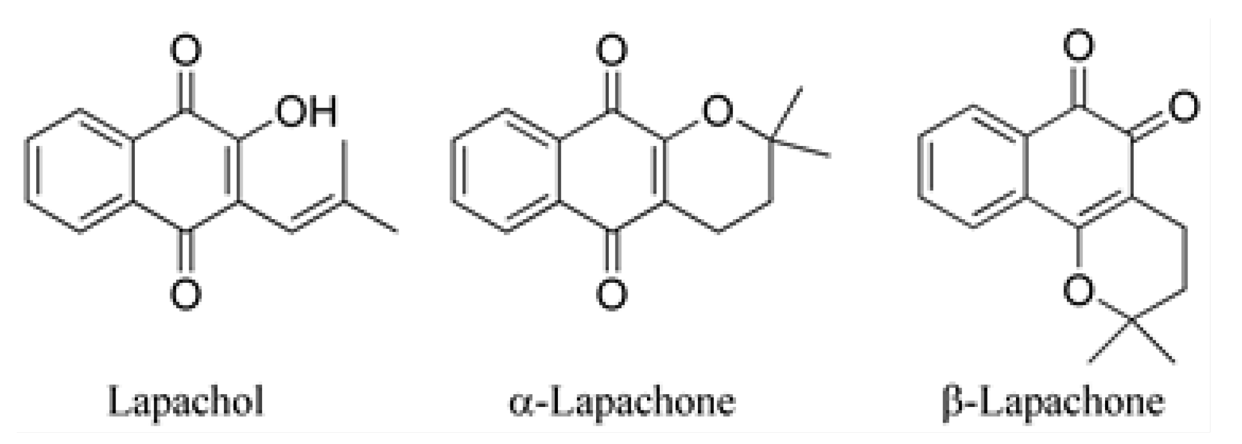
Figure 2.
Sigmoidal dose-response curves for the aromatase inhibition potential of α-lap (black) and β-lap (red).
Figure 2.
Sigmoidal dose-response curves for the aromatase inhibition potential of α-lap (black) and β-lap (red).
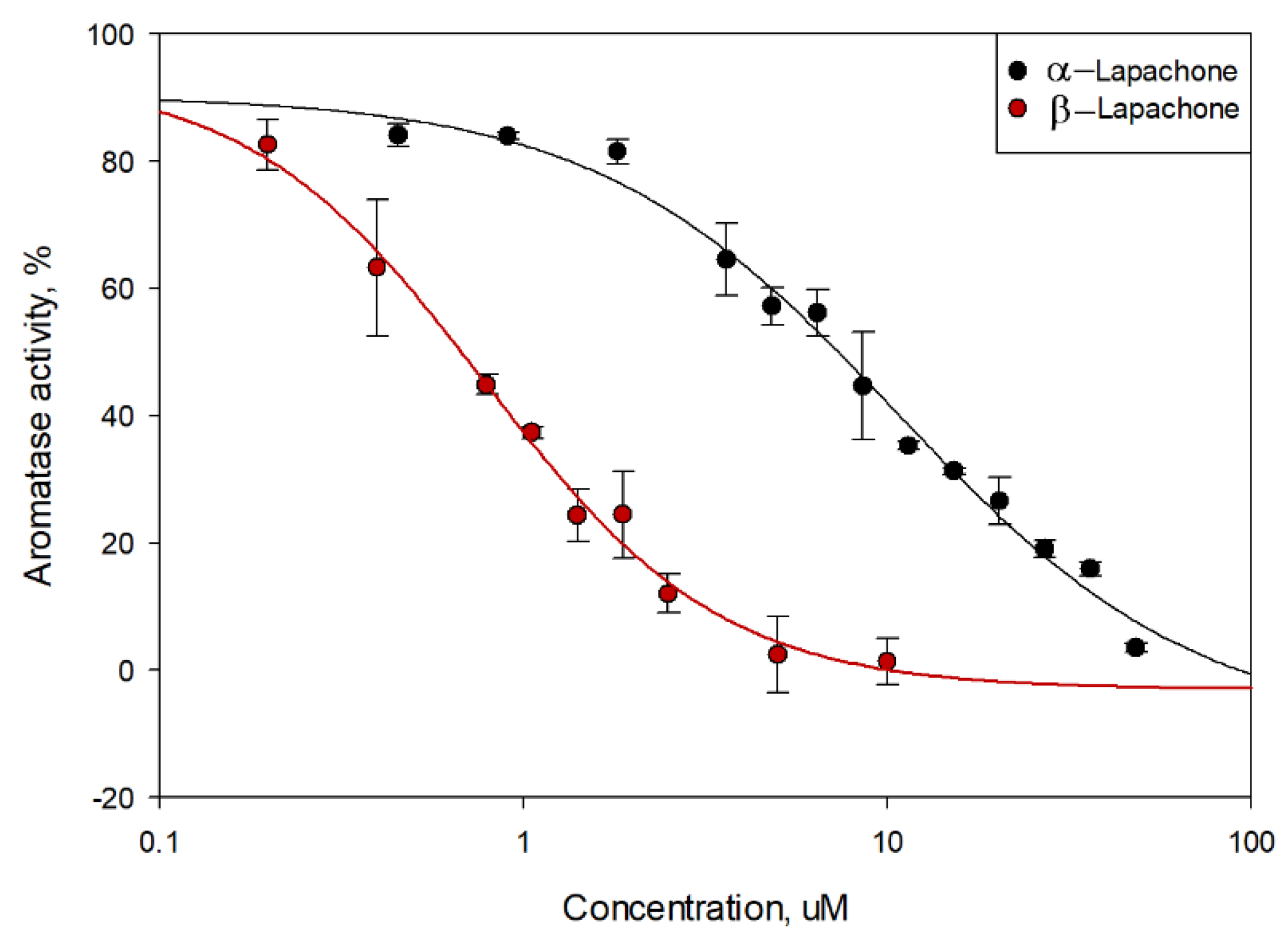
Figure 3.
Three- and 2D visualizations of docking poses possessing the lowest binding energy (highest binding affinity) and specific interactions within the active center for the natural substrate Androstenedione (A), α-lapachone (B) and β-lapachone (C) with human placental aromatase cytochrome P450 (PDB ID: 3EQM) [68]. The grid box coordinates are x = 83.427, y = 50.113, z= 46.367, grid size: 40: 40: 40. Results indicate that both isomers are located in a hydrophobic pocket near the heme group, physically obstructing the substrate-binding site, and that an additional π-alkyl interaction with key hydrophobic residues further enhances the inhibitory effect.
Figure 3.
Three- and 2D visualizations of docking poses possessing the lowest binding energy (highest binding affinity) and specific interactions within the active center for the natural substrate Androstenedione (A), α-lapachone (B) and β-lapachone (C) with human placental aromatase cytochrome P450 (PDB ID: 3EQM) [68]. The grid box coordinates are x = 83.427, y = 50.113, z= 46.367, grid size: 40: 40: 40. Results indicate that both isomers are located in a hydrophobic pocket near the heme group, physically obstructing the substrate-binding site, and that an additional π-alkyl interaction with key hydrophobic residues further enhances the inhibitory effect.
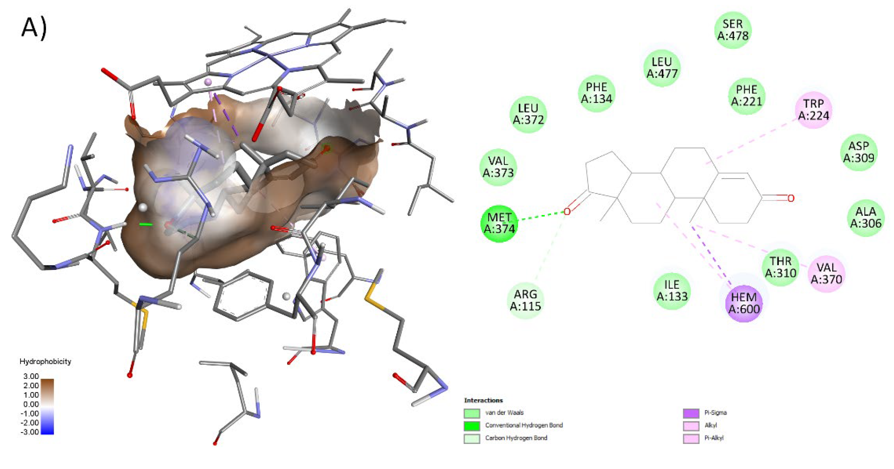
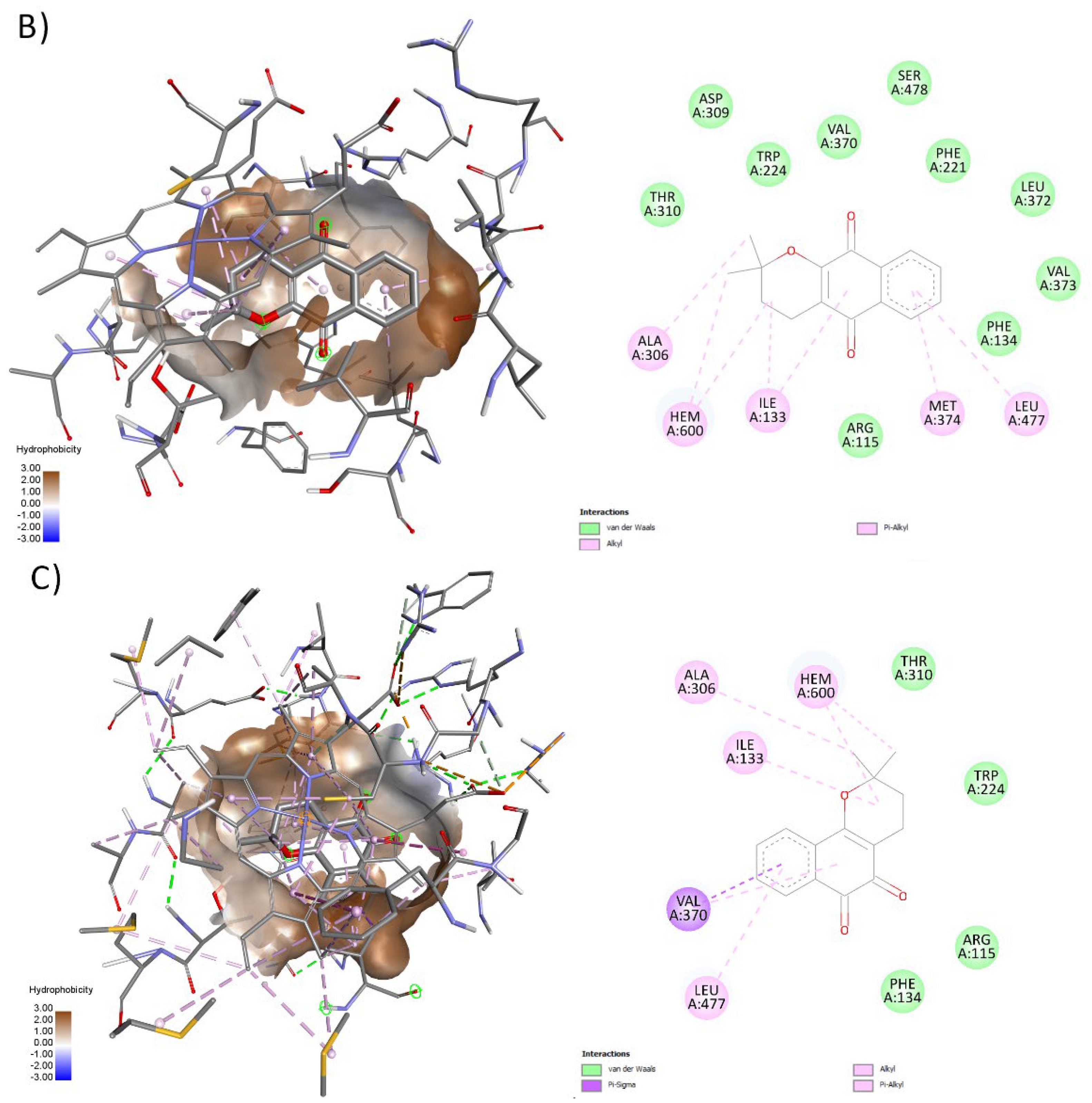
Figure 4.
Sigmoidal dose-response curves for the antagonistic potential on estrogen receptor (hERα) of α-lap (black), β-lap (red), and 4-HT (green).
Figure 4.
Sigmoidal dose-response curves for the antagonistic potential on estrogen receptor (hERα) of α-lap (black), β-lap (red), and 4-HT (green).
Disclaimer/Publisher’s Note: The statements, opinions and data contained in all publications are solely those of the individual author(s) and contributor(s) and not of MDPI and/or the editor(s). MDPI and/or the editor(s) disclaim responsibility for any injury to people or property resulting from any ideas, methods, instructions or products referred to in the content. |
© 2026 by the authors. Licensee MDPI, Basel, Switzerland. This article is an open access article distributed under the terms and conditions of the Creative Commons Attribution (CC BY) license.
Copyright: This open access article is published under a Creative Commons CC BY 4.0 license, which permit the free download, distribution, and reuse, provided that the author and preprint are cited in any reuse.


