

Submitted:
07 July 2026
Posted:
08 July 2026
You are already at the latest version
Abstract
Ergothioneine (ET) is a chemically-stable, tasteless, odourless, highly water-soluble diet-derived compound that is avidly absorbed and retained by the human body using a selective transporter, organic cation transporter novel 1, OCTN1 (often called the ET transporter, ETT). A substantial and growing body of evidence supports a role for ET in maintaining human health and protecting against age-related diseases, especially neurodegenerative diseases, and multiple studies indicate that low blood/plasma/serum ET levels increase the risk of developing age-related diseases. Despite the growing interest in ET, much fundamental work remains to be done to investigate its metabolism, actions (if any) on the genome, lipidome, metabolome and proteome, intracellular and intercellular transport (especially in the brain), precise mechanisms of cytoprotection, interactions with the microbiome, mycobiome and human pathogens, and identifying the factors that control body ET levels. This narrative review explores these issues and suggests what research needs to be done to improve our understanding of ET biology.
Keywords:
ergothioneine
; OCTN1
; antioxidant
; mitochondria
; mycobiome
; microbiome
; M. tuberculosis
1. Introduction
Ergothioneine (ET; structure shown in Figure 1) is a compound first discovered in 1909; it was thought until recently that it can only be made by fungi and some bacteria, including cyanobacteria, and a few yeasts [1,2,3,4,5,6,7,8,9,10,11]. However, a recent report [12] suggests that ET can also be made by certain mosses and gymnosperm plants, although not by angiosperms, a category that includes most plants eaten by humans, such as asparagus, corn, wheat and rye [12]. An interesting evolutionary question would be to understand why angiosperms lost the capacity to make ET during their evolution [12], a topic mentioned again in Section 6 below.
Several researchers investigated the properties of ET in the 1940s and 1950s, mostly studying its antioxidant abilities. It was then largely ignored for decades, as reflected by the low number of published papers recorded on PubMed (Figure 2). Melville [8] provided an excellent review of this early work. When humans consume ET, it is rapidly absorbed by a selective transporter (OCTN1, organic cation transporter novel 1, often now called the ET transporter, ETT) present in the small intestine (and in many other organs; Figure 3), distributed to all body tissues, avidly retained (half-life in the body estimated at several weeks) and renally reabsorbed; very little is excreted in urine but some in faeces (discussed further in Section 6 below) [1,2,3,4,9,13,14]. ET also passes from mother to child [2,3,9,15,16] since it is present in breast milk and the mammy gland shows increased levels of OCTN1 during lactation [9]. In addition, OCTN1 has been detected in human placenta (reviewed in ref [17], also see ref [18]). These facts strongly suggest that ET is important to the human body: selective transporters are usually only present to take up essential vitamins (e.g. vitamin C [19]) and minerals such as iron [20]; many other dietary compounds with antioxidant properties (e.g. most polyphenols) are poorly absorbed and rapidly metabolised, so that their concentrations in human tissues and body fluids are very low (reviewed in [2,21,22]). By contrast, blood and tissue concentrations of ET in humans and other animals are usually much higher [1,2,3,4,21,22,23,24,25]; some specific values for ET concentrations in humans are given in Section 3 below. Indeed, this potential importance of ET was recognised as long ago as 1959, by Melville [8], who wrote “the avidity with which dietary ET is incorporated into tissues of all animals, the tenacity with which it is held there, and its characteristic non-uniform pattern of distribution in these tissues (see Figure 3) are all facts which hint that there is a purpose in its presence”.
2. Where Does ET Come from in the Human Diet?
The major dietary source of ET in most populations is mushrooms [6,7,9,10,11,22,23,24,25], although smaller amounts are found in other foods such as asparagus, corn, wheat, other plants, Spirulina, meat, fish, eggs, milk and even Beluga whales [6,7,9,10,11,22,23,24,25,26,27,28,29]. Feeding hens with the ET-producing fungus Neurospora crassa was observed to increase the ET content of their eggs [29]. To quote references [23] and [30], “Ergothioneine intake, which derives almost totally from mushrooms, was evaluated in different countries and age groups, such as Belgium (average intake of 0.062 mg/kg body weight/day in children), France (0.03 mg/kgbw/day in adults), Ireland (0.053 mg/kgbw/day in adults), Italy, Finland (0.069 mg/kgbw/day in adults), and the United States. Higher average intakes were reported for Italian children and adults, with both being 0.067 mg/kg bw/day. On the other hand, the lowest intakes were reported for US adults and children, with average daily consumption of ergothioneine at 0.016 mg/kg bw/day and 0.014 mg/kg bw/day, respectively” [23,30]. Children were defined as age range 2.5–6.5 years, adults defined as over 18 (Italy and Ireland), 15 (France) or 25 (Finland) [30]. Beelman et al [31] attributed the high Italian intakes to frequent consumption of ET-rich Porcini mushrooms. Dietary intakes across the various regions of China varied widely, with a mean value estimated as 0.043 mg/kgbw/day [32]. By contrast, in the Inuit population, who appear to have exceptionally high blood ET concentrations (Section 3 below), the main sources of ET seem to be meat and marine foods including Beluga whales [26,27,28].
3. Why Has Interest in ET Increased?
The recent growing interest in ET, as revealed by more publications (Figure 2), was sparked by two factors. One was the identification of OCTN1, encoded by the gene slc22a4, as the ET transporter [33,34,35]. ET is highly water soluble [as well as tasteless and odourless] so a transporter is needed to take it across the hydrophobic interior of cell membranes. OCTN1 can transport a few other substrates in vitro [17,36] but studies of its transport kinetics reveal that ET is likely to be the major, if not the only, substrate in vivo [33,34,35,37,38]. Hence, OCTN1 is often now simply referred to as the ET transporter [34,35]. However, OCTN1 can also transport selenoneine [39], an analogue of ET in which the sulphur is replaced by selenium [Se]. Selenoneine is found mostly in foods of marine origin, although there have been few studies as yet on its role in vivo and its concentrations in most populations are much lower than those of ET [26,27,28,39].
A second factor raising interest in ET was the growing realisation, first pointed out in 2016 [40,41], that low blood/plasma/serum ET concentrations in humans are strongly associated with an increased risk of developing age-related diseases such as cognitive impairment, cardiovascular disease, dementia, frailty, Parkinson’s disease and eye disorders, including cataract and macular degeneration [2,3,4,22,40,41,42,43,44,45,46,47,48,49,50,51,52,53,54,55,56,57,58] (Figure 4), as well as erectile dysfunction [59], chronic renal disease [60] and pre-eclampsia [61]. Indeed, ET has been suggested to be a compound critical to implantation of the fertilised egg into the uterus wall and hence for pregnancy maintenance [62]. As an illustration, Figure 4 shows the range of plasma ET concentrations in an Asian (Singaporean, mostly Chinese) population [2,42,43,44]; a concentration below 810 nanomolar (nM), 0.81 micromolar (µM), is associated with increased risk of age-related diseases. Similarly, in an Australian population median serum ET levels were 0.79-1.35 µM but 24% lower in subjects with psychological stress [54]. Most ET in human blood is present in erythrocytes, which can have millimolar (mM) intracellular ET concentrations (reviewed in [63]), so whole blood concentrations of ET are much greater than plasma levels. For example, in a healthy Singaporean population mean plasma ET concentration was about 1µM whereas whole blood levels were around 152 µM [14]. Similarly, in an Arab (Saudi Arabian) population whole blood levels were around 122-161µM [64]. Although different research groups have measured plasma, serum or whole blood ET concentrations in humans, we [40,56] and others [65] have shown that plasma, serum, whole blood and erythrocyte levels are strongly correlated. By contrast, whole blood ET concentrations in an Inuit population were reported as around 400 µM whereas the control group (laboratory members in Quebec, Canada) had whole blood concentrations close to 100µM [28], broadly similar to the Saudi, Australian and Singaporean populations described above [14,64].
Of course, correlations between disease incidence and ET concentrations, however strong, do not prove causation, as explained in Figure 5. However, a cause-consequence relationship between low ET concentrations and the above age-related diseases is strongly suggested by, first, the predictive value of low ET concentrations in identifying increased disease risk or disease progression (e.g. refs [47,58,59]. For example, in a Singaporean population, healthy subjects with low plasma ET concentrations [Figure 4] are at increased risk of developing cognitive impairment, dementia and other diseases [2,40,42,43,44]. Similarly, in a cohort of Japanese subjects aged 69-79, the mean serum ET levels were 0.639µM (below the “healthy threshold”, Figure 4) and 273 of the 1344 subjects went on to develop dementia [45].
Second, further evidence for a cause-consequence relationship is provided by a large and growing number of studies on laboratory animals, human induced pluripotent stem cells (iPSC), primary neurons, other cell types, organoids, brain slices and other systems showing that ET is highly neuroprotective in models of dementia, stroke, eye disease, hearing loss, neuropathy, depression, sleep disorders and Parkinson’s Disease, among others (reviewed in [1,2,3,22,44], also see [66,67,68,69,70,71,72,73,74,75,76,77,78,79,80,81,82,83,84,85,86,87,88,89,90]). Several pilot human clinical trials have indicated significant neuroprotective effects of ET [91,92,93]. For example, in the pilot study by Yau et al [91] administration of ET (25 mg 3 times per week) for a year or more to Asian subjects aged 60 or above with mild cognitive impairment (MCI) raised mean plasma ET concentrations from around 0.8 µM to about 3.96 µM, improved cognitive function, and prevented increases in the plasma concentrations of neurofilament light protein, a biomarker of neuronal damage. In the study by Zajac et al [93] in an Australian population, consumption of ET (10mg or 25mg daily) raised plasma levels from a median of 1.154 µM to 9 µM (10mg ET daily) or 16 µM (25mg ET daily) after 16 weeks and improved sleep and some aspects of cognition. Many other clinical trials with ET are underway, including a trial on kidney dialysis patients, who have low ET levels because washout of ET during dialysis is aggravated by its impaired reabsorption by the dysfunctional kidneys [94,95]. For example, in a United States (USA) cohort [96] subjects on full dialysis had mean ±SD plasma ET concentrations of 0.47 ± 0.20 µM compared with controls of 2.1 ± 1.6 µM. Peritoneal dialysis subjects had concentrations of 0.67 ± 0.30 µM ET [96]. Both levels would seem to fall in the “at risk” range (Figure 4). Indeed, it has been suggested that patients with chronic kidney disease should eat more mushrooms [97].
As a result of the above discoveries, ET is becoming increasingly popularised on social media, podcasts, and talk shows as a “healthy longevity” agent. The number of published papers on it, although still small (e.g. as compared to papers on the possible health benefits of flavonoids, carotenoids or vitamins C and E), is rapidly increasing (Figure 2), although overall awareness of ET among the public and medical professionals is still low. ET is now being sold by itself, or as a constituent of several “longevity supplements”, through multiple online platforms. Pure ET appears safe for human consumption and has been approved for human usage by the European and USA Food and Supplement Safety Regulatory Agencies (reviewed in [2,3]). Of course, a major problem revealed by analyses of multiple commercial supplements of different nutrients is poor quality control (QC); a recent example reports that many of the formulations of the popular supplement nicotinamide mononucleotide (NMN) being sold do not match the contents claimed on the labels [98]. Hence, ET for human use should be obtained from a reputable source that has established and validated (ideally externally validated) QC protocols.
Although the number of published papers describing ET is increasing (Figure 2), many of them are reviews or meta-analyses (for some recent examples see [99,100,101,102,103,104,105,106]) and many new ones continue to appear. Very few of these publications, and indeed few research groups globally, are investigating in depth the fundamental biochemistry and molecular biology underlying the actions of ET, and their relationship to human health and nutrition. This lack of basic research into ET is unfortunate and needs to be addressed. Let us discuss the areas that the author believes need attention.
4. Ergothioneine Chemistry and Metabolism Have Scarcely Been Explored, Especially in the Brain
The chemistry of ET (Figure 1) in relation to biological systems has only been explored to a very limited extent [107,108,109], although all agree that ET is a water-soluble, tasteless, odourless and chemically-stable molecule that resists heat and does not degrade on prolonged storage, allowing its accurate analysis in stored biological materials. However, repeated freeze-thawing of biological samples should be avoided as limited loss of ET may sometimes occur (our unpublished data and ref [110]. This stability helps to facilitate the use of ET as a food preservative [111].
Several studies report that ET’s unique chemical properties can improve the light-harvesting efficiency of solar cells [112,113,114]—but the mechanism(s) by which this occurs and their possible relevance to biological systems have not been elucidated. Three metabolites of ET have been identified [2,3], namely hercynine, ET sulphonate and S-methyl ET (Figure 1) but there has been no systematic search for others. S-methyl ET presumably arises by the action of a methyltransferase, but this enzyme has not been identified. None of these three compounds is present in mice lacking OCTN1, who have no ET in their tissues [2], suggesting that all three derive from ET. A role for ET in drug metabolism has been suggested: incubation of liver microsomes from several animals with ET and NADPH led to production of ET conjugates with added paracetamol, raloxifene, diclofenac, clozapine, carbamazepine, and tamoxifen, among other drugs [115,116]. The significance of this in vivo remains to be discovered, although a raloxifene—ET conjugate has been detected in vivo in rats [115].
It has been suggested that when ET acts as an antioxidant by scavenging various oxygen radicals and other reactive oxygen species (ROS, a term explained in detail in ref [21]) the sulphonate and hercynine are produced, and thus could be biomarkers of ROS generation [117,118,119], i.e. their concentrations in plasma or serum might provide evidence that ET has antioxidant properties in vivo (discussed further in section 5 below). However, this needs to be validated in vivo in humans by comparison with established biomarkers of ROS generation and oxidative damage, as reviewed in [14,21,120,121]. Oxidation products of ET exposed to various ROS in vitro also include a thiyl radical (ES), ET disulphide and 5-oxoET (Figure 1) [108,109]. The enzymes that act upon ET in humans and other animals and how ET influences the cell and tissue lipidomes, metabolomes and transcriptomes have not been elucidated, although some preliminary proteomic and metabolomic studies have been published in rodents [122,123]. Some of the oxidized forms of ET can be reduced by the enzyme glutathione reductase plus its co-substrate reduced glutathione (GSH) or by thioredoxin reductase enzymes [109] but the importance of this in vivo needs more investigation. ET has also been reported to inhibit γ-glutamyl transpeptidase, an enzyme on cell surfaces that catalyses the hydrolysis of GSH, although fairly high concentrations of ET are required (Ki ~170 µM) [124]. A claim that nM concentrations of ET increase the activities of certain human carbonic anhydrase enzymes [125] remains to be confirmed. Nor have the potential biological roles of the three known ET metabolites (Figure 1) (and any other metabolites yet to be identified) been investigated. Do they contribute to ET’s cytoprotective effects? When ET enters cells, some of it is taken up by mitochondria [5,126], but where the rest of it goes is unknown, nor has the subcellular distribution of OCTN1 been accurately mapped, apart from its presence on plasma membranes [35]. Nor has ET’s mechanism of entry into mitochondria been elucidated [126]. OCTN1 appears to be involved, but there seem to be additional transporter(s).
This lack of knowledge of ET distribution, metabolism and any biological effects of its metabolites is especially true for the brain. ET has been reported to be present in human and other animal brains (and in human cerebrospinal fluid) in multiple studies [2,3,9,14,44,48,65,127,128,129,130,131,132,133,134], and orally supplementing laboratory animals with ET increases their brain levels of this compound [130,132]. However, no one has yet identified OCTN1 in intact human blood-brain barrier [2,35] although it is present in human brain microvascular endothelial cells and several other regions of the human brain (Figure 3) (reviewed in [2,35]. It thus remains to be discovered how ET enters the human brain, as it clearly does.
OCTN1 is not the only molecule capable of transporting ET. Another human transporter, SLC22A15, encoded by the slc22a15 gene, which is not involved in the intestinal absorption of ET but is widespread in the brain (Figure 3), can also transport ET, albeit with much lower affinity (Km ~ 400 µM) and lower Vmax compared with OCTN1, Km ~ 21 µM [9,35,135,136]; the major substrate of SLC22A15 seems to be creatine [137]. Mitochondria from OCTN1-knockout mice can still take up ET, albeit at a slower rate than mitochondria from wild-type mice, and the additional transport mechanism(s) involved have not yet been identified [126].
5. The Mechanism(s) of Action of Ergothioneine Is(Are) Unclear and Are Likely to Be Multifactorial
ET is frequently described as an antioxidant, although determinations of its redox potential (0.45 volts) show it to be a weaker reducing agent than GSH, cysteine or ascorbate [138]. Nevertheless, in vitro studies have shown that ET is capable of scavenging several biologically-important ROS, including hydroxyl radical (OH), hypochlorous acid (HOCl), hypobromous acid (HOBr), ozone (O3), peroxynitrite (ONOO−/ONOOH), singlet oxygen [118,139,140,141,142,143] and the pro-oxidant higher haem oxidation states (ferryl haem) of haemoglobin and myoglobin [144,145]. ET can also render pro-oxidant transition metal ions (Fe2+, Cu+) less redox active by chelating them [139,146]. It can scavenge methylglyoxal, acrolein, and possibly other cytotoxic aldehydes that result from lipid peroxidation [147,148].
In addition to direct scavenging of ROS, several studies have presented evidence that ET can activate the “master regulator” of endogenous antioxidant defences, Nrf2, and so raise endogenous antioxidant defence levels in vivo [71,149,150,151,152,153,154,155]. However, how ET activates Nrf2 is unclear: does it increase the phosphorylation of keap1 through activation of various kinase enzymes [150,153,155,156,157,158], or does ET interact directly with the thiol groups on keap1, or both; these are the two usual mechanisms by which Nrf2 is activated [157,159]? The role of these two established mechanisms of Nrf2 activation needs to be elucidated for ET, although in silico studies have suggested that ET might bind to the active site of Nrf2 to prevent its proteasomal degradation and thereby allow it to promote the transcription of genes encoding antioxidant defences [152].
Despite its demonstrated antioxidant abilities, evidence that ET exerts significant antioxidant effects in vivo in humans remains limited, as discussed in [2,14], although if ET only acts as an antioxidant at specific sites where it accumulates in the human body, this would be hard to detect by measuring systemic biomarkers of oxidative stress (reviewed in [21,120,121,159]. Indeed, tissue injury, which usually leads to increased oxidative damage for a variety of reasons, as reviewed in [21,160], can lead to increases in the levels of OCTN1, resulting in higher concentrations of ET in the injured tissue [161,162]. This elevation of OCTN1 has been proposed to be an adaptive cytoprotective mechanism, bringing in more ET to protect the injured tissue [161,162], although any protection by the extra ET need not necessarily be by antioxidant action, of course.
Several other lines of evidence suggest that the antioxidant properties of ET may not be its only, or even its major, mechanism of action in vivo. First, antioxidant supplements have generally shown only limited effectiveness in the treatment or prevention of human diseases [21,159,163,164,165,166,167,168] whereas ET seems more promising (as reviewed in Section 3 above), although this remains to be fully validated by large-scale double blind placebo-controlled clinical trials. Second, ET biosynthesis has been identified in anaerobes, suggesting that ET production may have evolved for reasons other than antioxidant properties, although no metabolic roles of ET specific to anaerobes have yet been discovered [169,170,171]. However, this is not a conclusive argument, because it remains possible that anaerobes evolved antioxidants (including ET perhaps) to protect against free radicals generated by radiation damage, which was a major problem in the “pre-oxygen” world because of the lack of Earth’s ozone layer [21,170]). Indeed, several papers have described radioprotective effects of ET [172,173,174]. These antioxidants might have later allowed anaerobes to survive transient exposures to O2 as O2 levels rose in the atmosphere due to the evolution of photosynthesis [21,175,176,177]. In any case, these studies [169,170,171] indicate that ET had an early evolutionary origin, perhaps as an antioxidant, but as life evolved it may have taken on other roles. Interestingly, ET has been shown to play a role in biosynthesis of lincosamide antibiotics (such as lincomycin A) in a few microorganisms [178,179].
Indeed, multiple mechanisms of ET action other than antioxidant ones have been suggested (Figure 6) and their relative importance has not been elucidated. It may, of course, vary depending on the organism, organ, tissue, cell, organelle and the injurious agent. For example, the protective effect of ET against the development of fatty liver in a mouse model was attributed to inhibition of expression of the enzyme phosphatidylethanolamine cytidyltransferase 2, thus decreasing the accumulation of toxic phosphatidylethanolamine species [180]. The anti-inflammatory and anti-fibrotic properties of ET described in multiple animal studies and attributed to variety of mechanisms [60,61,76,88,156,158,181,182,183,184,185,186,187,188,189,190,191] may be of especial importance in prevention and treatment of age-related diseases, including neurodegenerative diseases, in which inflammation is intimately involved, as it is in ageing generally (“inflammaging”) [192,193]. The proposed abilities of ET to promote neurogenesis and neuronal differentiation and enhance the actions of neurotrophic factors [194,195,196,197,198,199] may also be very important in protection against and treatment of neurodegeneration. A role in early childhood brain development is also possible, since ET passes from mother to baby [2,3,9,15,16]. To date, these studies are mostly based on work with cell cultures, although human studies are beginning to appear [198]—more studies are needed to establish the importance of ET in vivo in enhancing neurogenesis, synaptogenesis, neuronal differentiation and the beneficial effects of neurotrophic factors, and the mechanisms by which ET is acting. It cannot be over-emphasised that, as proposed in [44], a brain-penetrant agent safe for human consumption, that not only slows neurodegeneration but also promotes the genesis of new neurons and supports the functions of new and existing neurons, would be of immense value in the slowing and prevention of age-related diseases of the nervous system, especially for dementia, in the world’s rapidly-ageing populations; time will tell if ET can deliver on this potential. This is especially true given the growing evidence that neurogenesis and neuronal development contribute to cognitive resilience in the brains of patients with Alzheimer’s disease [199,200] and ET may help to promote cognitive resilience [58,201].
Established mechanisms (good evidence)
|
Likely Mechanisms (evidence is accumulating)
|
Suggested mechanisms (evidence for a role in vivo is limited as yet)
|
Comments:
- Ferroptosis is a mode of cell death that involves iron ions and lipid peroxidation [286], both of which play key roles in oxidative damage in vivo [21,160,286,287]. Preliminary studies in vitro suggest that ET can inhibit ferroptosis (Chester Drum, Irwin Cheah and Ong Wei Yi, personal communications).
- Several papers have suggested that ET might inhibit cell senescence [61,288,289], a term which refers to persistent cell cycle arrest due to stressful events such as telomere shortening, oxidative damage, activation of oncogenes etc [290]. However, ET failed to exert anti-senescence effects in a model of replication – stress associated senescence (Prof. Marie-Veronique Clement and Dr. Le Luo, personal communication, also ref [291]), or in etoposide-induced cellular senescence (Prof. Rachel Watson and Drs Keith Tan, Oliver Dreesen and Selwyn Loh, personal communication). In the cases where ET did protect, it seemed to be acting by diminishing oxidative stress [288,289].
Several papers have suggested that ET could modulate autophagy [61,189,292] but the importance of this in vivo and the mechanism(s) by which ET could do this are unclear as yet.
Returning to the mechanisms of action of ET, two papers published in Cell Metabolism in 2025 proposed that the actions of ET in mitochondria are its major cytoprotective role, suggesting that the generation of hydrogen sulphide (H2S) from ET is a key mechanism [122,202]. However, they reached different conclusions as to how this is achieved. One paper [122] reported that ET binds to and activates the enzyme 3-mercaptopyruvate sulphotransferase, but the other [202] that ET acts as a substrate for cystathionine gamma-lyase, leading to increases in NAD+ concentrations. Indeed, there is a voluminous literature claiming that raising tissue NAD+ concentrations promotes healthy longevity [98,203] although there are some conflicting data (reviewed in [203,204]). These discrepancies between references [122] and [202] need to be resolved. In any case, is H2S likely to explain all the biological actions of ET? This could be the case, but this author is sceptical. Low levels of H2S can indeed be neuroprotective [205,206,207,208,209,210,211], but higher H2S levels can cause tissue damage [210,212,213,214,215], as illustrated by studies on breast cancer [215], human stroke and animal models of stroke [212,213]. Indeed, to quote a recent paper [214] on the mechanisms used by the nematode C. elegans to avoid exposure to H2S: “H2S is an energy source, a toxin and a gasotransmitter”. The biological effects of the widely used “antioxidant” drug N-acetylcysteine (NAc) have also been suggested, at least in part, to be due to H2S generation [121,216,217], yet NAc has both protective and damaging effects in vivo, including promoting cancer development under some circumstances (reviewed in [159]). By contrast, no one has yet found pro-neurodegenerative or other toxic effects of ET, even at high doses, in any animal model or in the clinical trials conducted to date [2,3].
6. Interaction of ET with the Human Microbiome Has Scarcely Been Studied
It is not only humans and other animals that avidly take up ET; several (and possibly all, most have not been studied) higher plants do so as well, absorbing ET generated by fungi and bacteria in the soil through their roots [6,218,219,220]. Was the loss of ET biosynthesis in higher plants described in the Introduction (Section 1) perhaps related to the ease with which angiosperms can obtain it from fungi and bacteria in the soil [6,218,219,220], saving them the energy cost of making it? This is perhaps analogous to the argument that early humans lost the capacity to make vitamin C because they ate a diet rich in natural fruits and vegetables [21,22]. Hence, it has been suggested that modern agricultural practices that disrupt fungi and bacteria in soil could decrease amounts of ET in dietary plants, leading to potential health risks if body ET concentrations fall too low [6,219,220]. The ET content of plants grown hydroponically is also likely to be low or zero.
Many bacteria also have transporters that allow them to take up and accumulate ET [221,222,223,224,225,226]. ET is efficiently absorbed in the small intestine, but some passes through to the colon and small amounts can be measured in human faeces [2] although very little in urine even after supplementation, probably due to efficient renal reabsorption [14,227]. Presumably if subjects consume ET-rich diets or supplements containing ET, more will end up in the colon. Since the body will have absorbed the ET it requires in the small intestine, uptake and metabolism of ET by the colonic microbiome would not be expected to deprive the human body of ET, but there are other potential concerns. Research into interactions of ET with the microbiome is still in its early stages [58,228,229,230,231], but one concern that has been raised is that some colonic bacteria can degrade ET using the enzyme ergothionase to eventually produce trimethylamine (TMA), which is absorbed from the gut and converted by the liver to trimethylamine oxide (TMAO) [232,233,234]. TMAO is, epidemiologically, the exact opposite of ET: in most (but not all) studies high plasma concentrations of TMAO are associated with increased risk of developing several diseases, including cognitive impairment and cardiovascular disease [232,233,234]. Hence could too much dietary ET be deleterious by raising TMAO levels? Evidence from the absence of increases in plasma TMAO levels in human subjects supplemented with ET, and the lack of correlations of circulating ET and TMAO concentrations in large patient cohorts, suggest that ET is not a significant source of TMAO in humans [93,235], which is consistent with the literature on the safety and health-promoting aspects of ET reviewed above. Another study even suggested that TMA, the precursor of TMAO, is beneficial to human health [236]. Although increased expression of genes encoding ergothionase has been reported in the colonic microbiome of patients with colorectal cancer [230], ET actually appears to be toxic to colorectal cancer cells, based on in vitro studies [237]. It would be good to confirm this action in vivo. Nevertheless, other pathways of ET metabolism by the colonic microbiome are rapidly being discovered [58,229,230,231,238,239] and the potential health effects of the products of these pathways, whether beneficial or deleterious, is an area ripe for further investigation.
The stomach and small intestine also have a microbiome [240,241]. The duodenal/small intestinal microbiome is poorly characterised and how it might take up or metabolise ET and thus affect its uptake into the human body is largely unknown. However, one constituent of the small intestine microbiome is Lactobacillus reuteri (Limosilactobacillus reuteri); strains of this organism have been widely promoted as probiotics to promote human gut health [242,243]. Some strains of L. reuteri avidly take up ET [221] – whether or not these bacteria could absorb enough ET to compete with its human intestinal update is unknown. It presumably would depend on their abundance in vivo; something to think about when selecting probiotics, perhaps – should we even preload them with ET to prevent them taking it from our diet and possibly (given the cytoprotective effects of ET in bacteria described in Section 7 below) to promote their survival in the gastrointestinal tract?
However, an important component of the gastric microbiome is Helicobacter pylori [240,244]. H. pylori cannot synthesize ET, but takes it up avidly, and ET helps to protect this pathogen against the antimicrobial effects of ROS generated by human phagocytes as part of the immune response [222,223,224,225,244]. Indeed, the high efficiency of ET uptake was illustrated in a recent paper [245] describing conjugation of an anti-microbial compound with ET as a vehicle to deliver that compound to H. pylori and eradicate this pathogen. Along similar lines, an ET-coated imaging agent has been used to image kidney disease: the agent bound to OCTN1 in the kidney [246]. Unfortunately, H. pylori can trigger chronic gastritis, in the worst-case scenario leading to gastric cancer [244]. Could uptake of dietary ET by H. pylori aggravate this? We do not know – it has not been studied. As discussed in Section 5 above, ET has anti-inflammatory effects, which should help to ameliorate risk. Epidemiological evidence suggests that consumption of mushrooms, a major dietary source of ET, is negatively associated with risk of gastritis and gastric cancer [247,248]. Of course, mushrooms contain multiple compounds that could be protective against cancer [247,248] and so these studies do not prove that ET is responsible for the protection. An interesting recent observation is that consumption of antacids by a Caucasian population in Rotterdam was associated with worse cognition, correlated with lower plasma ET levels [58]. Raising gastric pH increases the size of the gastric microbiome [240] – perhaps the various organisms can then absorb sufficient ET in the stomach to reduce its bioavailability?
7. ET and Other Human Pathogens
Several bacteria can themselves synthesise ET, and some are human pathogens, including Burkholderia pseudomallei, the causative agent of human melioidosis, a serious and sometimes lethal infection by this organism [249]. One of the most studied ET-synthesising pathogens is Mycobacterium tuberculosis, the agent responsible for human tuberculosis (TB). This organism synthesises two sulphur-containing compounds, ET and mycothiol, to help protect itself against ROS generated by the human immune system, thereby hindering its eradication and facilitating its persistence in the human body [250,251,252]. Indeed, ET concentrations may have increased during evolution of M. tuberculosis to enhance its persistence [250]. Indeed, there has been some work on inhibitors of ET synthesis as potential therapeutic agents [253,254]. Could consuming ET facilitate TB development in infected subjects? M. tuberculosis and other mycobacteria do not possess an uptake transporter for ET, so presumably they cannot accumulate it from their surroundings [223]. However, B. pseudomallei can (ref [249] and our unpublished results). Several mycobacteria, including M. tuberculosis, release some of the ET that they synthesise into their surrounding environment, suggesting that external ET could help to protect them against ROS generated by phagocytes as part of the human immune response [255,256,257]. In addition, H. pylori, Streptococcus pneumoniae, Listeria monocytogenes, Clostridium difficile, Enterococcus faecalis and Staphyloccus aureus are among the pathogenic bacteria that can take up ET and use it for cytoprotection [223,225]. Could dietary ET enhance the pathogenicity of these organisms and of M. tuberculosis by raising ET levels in body fluids so as to protect them against ROS generated by the immune system? Another gap in our knowledge.
8. Can ET Be Synthesized in the Human Body?
The consensus view, based on multiple old and a few recent experiments, is that ET cannot be made in animals [1,2,3,4,8,13,258,259], and our experiments with laboratory mice (reviewed in [2]) confirm that view. All known ET biosynthetic pathways begin with the amino acid histidine [260], so we simply fed mice with isotopically-labelled histidine (N15-histidine) and looked for N15-labelled ET, by mass spectrometry, which we never convincingly found. However, bioinformatic analyses of the gut microbiota in humans and other animals confirm that all the genes necessary to make ET are present [221] but of course they may not be being transcribed and translated to produce active enzymes, or present together in a single organism. A recent paper [260,261] suggested that some gut microorganisms (Cyanobacteria and related species) in mice could make ET, although biosynthesis was not directly demonstrated and such bacteria are quite rare in the human gut microbiome [262,263] – more research on this is needed.
However, the human gut not only has a microbiome, it has a mycobiome, a collection of fungi [264,265]. Although fungi are usually less than 0.1% of detected microbial reads or genes in the human gut, they can exert effects on the body by producing bioactive metabolites and interacting with bacteria and the host immune system [265]. Many fungi [including mushrooms of course!] can make ET [8,9,13,266]; could this ever happen in vivo and, if so, would the fungi retain ET or release it into the human body? We don’t know, but a few studies suggest that ET might sometimes be made. For example, ET can be synthesized by several established pathogenic fungi that can reside in the human body, including Cryptococci and Candida species and Aspergillus fumigatus [266,267,268,269]. Could they perhaps sometimes generate and release ET wherever in the human body the fungus is present? The author believes that it is more likely that they will keep it to themselves, but it could be released when the fungi die, e.g. as a result of treatment with anti-fungal medications.
9. Why Some Humans Have Low ET Levels Is Unclear
Low plasma/serum/whole blood ET concentrations in humans are strongly associated with higher risk of age-related diseases, as reviewed in Section 3 above (also see Figure 4). But why do some people have low levels? An obvious explanation is a diet low in sources of ET (hence the frequent suggestion to eat more mushrooms), but evidence suggests that there is more to it than that, e.g. in a Singaporean population with low ET no change in mushroom consumption was observed [40]. For example, poor kidney function [e.g. due to diabetes] could impair reabsorption of ET from the renal ultrafiltrate [94,95,96,187]. The slc22a4 gene that encodes OCTN1 has several polymorphisms [17,35,270,271,272,273,274,275,276,277], that could lead to amino acid substitutions which affect the transport of ET by OCTN1 [271,272]. In our study of ET uptake by healthy young Singaporean subjects [14], we found that plasma ET concentrations after supplementation were higher in subjects who already had higher plasma ET concentrations, suggesting variations in intestinal uptake efficiency. The population frequency in different countries and precise effects of these polymorphisms on the kinetics of ET transport in vivo remain to be studied in detail, one of the many important aspects of ET biology that need to be explored in more depth. For example, the 503F variant of OCTN1 has a lower Km for ET but also a lower maximal transport velocity (Vmax), resulting in a 50% higher initial transport capacity of ET at low concentrations (≤ 10µM) [271] but perhaps less effective transport at higher ET concentrations. Some other variants were reported to have impaired ET transport capacity [272]. Such kinetic studies need to be extended to all the variants, and their frequencies in different populations examined. In addition, a mutation in slc22a4 has been linked to human hearing loss [273,276]. This mutation was reported to severely decrease the efficiency of ET uptake by OCTN1 [273]. Indeed ET has been shown to protect against age-related hearing loss [68] and hearing loss due to treatment with the chemotherapeutic agent cis-platin [151], both in mice, suggesting a role of ET in auditory function.
Many other aspects of ET biology have been hinted at in the literature. For example, a mouse study reported that there are circadian changes in the intestinal expression of OCTN1 that could influence ET uptake from diet [278] and a rat study suggested that administration of 1α,25-dihydroxyvitamin D3 can modulate OCTN1 levels [279]; the relevance of either to humans has not been studied. The interaction of ET with other dietary nutrients, and their potential effects on ET uptake in the gastrointestinal tract or by the body tissues, have also not been studied. Nor have possible interactions of ET with medications been examined in detail: an early suggestion that ET could influence the pharmacokinetics of the drug gabapentin does not seem to be a significant effect [280,281]. One possible drug interaction could be with the calcium channel blocker verapamil, which inhibits OCTN1 [33] and is often used in the laboratory to examine the role of this transporter in the actions of ET (e.g. [37,70,76]. An effect of antacids on ET concentrations has been described [59], as discussed in Section 6 above. Another issue worth further exploration is species differences. For example, OCTN1 expression is high in rat and mouse livers and human foetal liver [35], but much lower in adult human liver (Figure 3). When mice consume ET, a lot enters the liver [130], but this may not be true in humans [35,282]. The distributions of OCTN1 in the various cell types of mouse and human brain are also different [35].
10. Conclusion
Evidence is rapidly growing that ET is a valuable component of the human diet and may even be essential for healthy longevity [1,2,3,4,8,42,44]; the late Professor Bruce Ames called it a “longevity vitamin” [283]. Indeed, increased OCTN1 activity has been proposed as an early evolutionary trait in Neolithic farmers to decrease the risk of ET deficiency, since many of the plants they first domesticated were low in ET [284]. There is a threshold blood level of ET, below which the risk of multiple age-related diseases increases [2] (Figure 4), but is the optimal level of ET needed to maintain health greater than this? An analogy could perhaps be with vitamin C; only low intakes are needed to prevent overt symptoms of deficiency (scurvy), but larger amounts of vitamin C seem necessary for optimal health [19]. So should we all be consuming ET supplements, or just eating more mushrooms? Many aspects of ET biology need to be investigated before making firm recommendations. I hope that this narrative review will stimulate more research in the field. In studies of a very large number of human subjects, we have never found anyone completely lacking ET: does this mean that OCTN1 and ET are essential for early human development and human life?
Acknowledgments
I am grateful to (in alphabetical order) Marie-Veronique Clement, Oliver Dreesen, Chester Drum, Stavroula Hatzios, Selwyn Loh, Le Luo, Niranjan Nagarajan, Wei Yi Ong, Keith Tan, Rachel Watson and Sabine Zachgo for sharing their data and advice with me. I am especially grateful to Irwin Cheah for his great help with data collection and suggestions to improve the manuscript. The author (BH) is solely responsible for the design, writing and final content of this manuscript and read and approved the final version.
Abbreviations
| ET | Ergothioneine |
| ET disulphide | Ergothioneine disulphide |
| ET sulphonate | Ergothioneine sulphonate |
| ETT | Ergothioneine transporter |
| GSH | Reduced glutathione |
| H2S | Hydrogen sulphide |
| iPSC | Induced pluripotent stem cells |
| Ki | Inhibition constant – the concentration of the inhibitor required to occupy half of the enzyme binding sites |
| Km | Michaelis constant |
| MCI | Mild cognitive impairment |
| Mg/kgbw/day | Milligrams per kilogram body weight per day |
| mM | Millimolar |
| NAc | N-acetylcysteine |
| NAD+ | Nicotinamide adenine dinucleotide |
| NMN | Nicotinamide mononucleotide |
| Nrf2 | Nuclear factor erythroid 2 – related factor 2 |
| nM | Nanomolar |
| OCTN1 | Organic cation transporter novel 1 |
| 5-oxoET | 5-oxoergothioneine |
| QC | Quality Control |
| ROS | Reactive oxygen species |
| Se | Selenium |
| Slc22a4 gene | Gene encoding solute carrier – family 22 member 4 |
| Slc22a15 gene | Gene encoding solute carrier – family 22 member 15 |
| S-methyl ET | S-methylergothioneine |
| TMA | Trimethylamine |
| TMAO | Trimethylamine oxide |
| TB | Tuberculosis |
| µM | Micromolar |
| Vmax | Maximal velocity |
References
- Borodina, I.; Kenny, L.C.; McCarthy, C.M.; Paramasivan, K.; Pretorius, E.; Roberts, T.J.; et al. The biology of ergothioneine, an antioxidant nutraceutical. Nutr. Res. Rev. 2020, 1–28. [Google Scholar]
- Halliwell, B.; Tang, R.M.Y.; Cheah, I.K. Diet-Derived Antioxidants: The Special Case of Ergothioneine. Annu Rev. Food Sci. Technol. 2023, 14, 323–45. [Google Scholar] [CrossRef] [PubMed]
- Cheah, I.K.; Halliwell, B. Ergothioneine, recent developments. Redox Biol. 2021, 42, 101868. [Google Scholar] [CrossRef] [PubMed]
- Paul, B.D. Ergothioneine: A Stress Vitamin with Antiaging, Vascular, and Neuroprotective Roles? Antioxid. Redox Signal 2022, 36(16-18), 1306–17. [Google Scholar] [CrossRef] [PubMed]
- Paul, B.D.; Snyder, S.H. The unusual amino acid L-ergothioneine is a physiologic cytoprotectant. Cell Death Differ. 2010, 17(7), 1134–40. [Google Scholar] [PubMed]
- Beelman, R.B.; Phillips, A.T.; Richie, J.P., Jr.; Ba, D.M.; Duiker, S.W.; Kalaras, M.D. Health consequences of improving the content of ergothioneine in the food supply. FEBS Lett. 2022, 596(10), 1231–40. [Google Scholar] [CrossRef] [PubMed]
- Kalaras, M.D.; Richie, J.P.; Calcagnotto, A.; Beelman, R.B. Mushrooms: A rich source of the antioxidants ergothioneine and glutathione. Food Chem. 2017, 233, 429–33. [Google Scholar] [CrossRef] [PubMed]
- Melville, D.B. Ergothioneine. In Vitamins & Hormones; Academic Press: 17, 1959; pp. 155–204. [Google Scholar]
- Halliwell, B.; Cheah, I.K.; Tang, R.M.Y. Ergothioneine - a diet-derived antioxidant with therapeutic potential. FEBS Lett. 2018, 592(20), 3357–66. [Google Scholar] [CrossRef] [PubMed]
- Pfeiffer, C.; Bauer, T.; Surek, B.; Schömig, E.; Gründemann, D. Cyanobacteria produce high levels of ergothioneine. Food Chem. 2011, 129, 1766–9. [Google Scholar] [CrossRef]
- Tian, X.; Thorne, J.L.; Moore, J.B. Ergothioneine: an underrecognised dietary micronutrient required for healthy ageing? Br. J. Nutr. 2023, 129(1), 104–14. [Google Scholar] [CrossRef] [PubMed]
- Kock, C.; Gutsche, N.; Walter, S.; Zachgo, S. Plants synthesize ergothioneine, showing a link to abiotic stress. Plant Biol. (Stuttg) 2026. [Google Scholar] [CrossRef] [PubMed]
- Cheah, I.K.; Halliwell, B. Ergothioneine; antioxidant potential, physiological function and role in disease. Biochim Biophys. Acta 2012, 1822(5), 784–93. [Google Scholar] [CrossRef] [PubMed]
- Cheah, I.K.; Tang, R.M.Y.; Yew, T.S.; Lim, K.C.; Halliwell, B. Administration of pure ergothioneine to healthy human subjects; Uptake, metabolism and effects on biomarkers of oxidative damage and inflammation. Antioxid. Redox Signal. 2017, 26(5), 193–206. [Google Scholar] [CrossRef] [PubMed]
- Brustad, N.; Olarini, A.; Kim, M.; Chen, L.; Ali, M.; Wang, T.; et al. Diet-associated vertically transferred metabolites and risk of asthma, allergy, eczema, and infections in early childhood. Pediatr. Allergy Immunol. 2023, 34(2), e13917. [Google Scholar] [PubMed]
- Olarini, A.; Ernst, M.; Gurdeniz, G.; Kim, M.; Brustad, N.; Bonnelykke, K.; et al. Vertical Transfer of Metabolites Detectable from Newborn’s Dried Blood Spot Samples Using UPLC-MS: A Chemometric Study. Metabolites 2022, 12(2), 94. [Google Scholar] [PubMed]
- Guo, Y.; Zhou, K.; Zhang, J.; Tao, Z. Organic cation transporter novel 1 (OCTN1): beyond an ergothioneine transporter. Cell Commun. Signal 2026. [Google Scholar] [CrossRef] [PubMed]
- Li, J.; Chen, S.; Wu, P.; Li, Y. Maternal transfer of bisphenol analogues and their conjugated metabolites. J. Hazard Mater. 2026, 508, 141941. [Google Scholar] [CrossRef] [PubMed]
- Padayatty, S.J.; Levine, M. Vitamin C: the known and the unknown and Goldilocks. Oral Dis. 2016, 22(6), 463–93. [Google Scholar] [CrossRef] [PubMed]
- Enko, D. Physiology of Iron Metabolism. Clin. Lab. 2025, 71(3). [Google Scholar] [CrossRef] [PubMed]
- Halliwell, B.; Gutteridge, J.M. Free Radicals in Biology and Medicine (Fifth Edition); Clarendon Press: Oxford, 2015. [Google Scholar]
- Tng, T.J.W.; Cheah, I.K.; Halliwell, B.; Lim, K.-L. Potential Protection Against Parkinson’s Disease by Ergothioneine—Nature’s Multifactorial Neuroprotectant. Antioxidants 2026, 15(4), 519. [Google Scholar] [CrossRef] [PubMed]
- Michelini, C.; Rosi, A.; Bragazzi, N.; Del Rio, D.; Mena, P. Dietary Intake of Plant Bioactives. Annu Rev. Nutr. 2026. [Google Scholar] [CrossRef]
- Ey, J.; Schomig, E.; Taubert, D. Dietary sources and antioxidant effects of ergothioneine. J. Agric. Food Chem. 2007, 55(16), 6466–74. [Google Scholar] [CrossRef] [PubMed]
- Beelman, R.B.; Kalaras, M.D.; Phillips, A.T.; Richie, J.P., Jr. Is ergothioneine a ‘longevity vitamin’ limited in the American diet? J. Nutr. Sci. 2020, 9, e52. [Google Scholar] [CrossRef] [PubMed]
- Barrette, A.B.; Archambault, P.; Lemire, M.; Zinflou, C.; Ouellet, N.; Dumas, P.; et al. Selenoneine and ergothioneine in beluga skin and tissues as key antioxidants in the Inuit diet. Sci. Rep. 2025, 15(1), 35819. [Google Scholar] [CrossRef] [PubMed]
- Fernandez-Bautista, T.; Gomez-Gomez, B.; Gracia-Lor, E.; Perez-Corona, T.; Madrid, Y. Investigating the Presence of Selenoneine, Ergothioneine, and Selenium-Containing Biomolecules in Fish and Fish-Derived Commercial Products. J. Agric. Food Chem. 2024, 72(47), 26155–64. [Google Scholar] [CrossRef] [PubMed]
- Ayotte, P.; Lemire, M.; Dumas, P.; Achouba, A.; Yedjenou, M.; Barrette, A.B.; et al. Wild foods as drivers of blood ergothioneine and selenoneine concentrations among Inuit living in Nunavik: results from the cross-sectional Qanuilirpitaa? 2017 survey. Am. J. Clin. Nutr. 2025, 122(2), 582–92. [Google Scholar] [CrossRef] [PubMed]
- Nguyen, H.; Ebbole, D.J.; Walzem, R.L.; Rezenom, Y.H.; Watanabe, C.M.H. Applied Research Note: Pilot demonstration of feasibility of Neurospora crassa (rco-3; csp-2) mycelium feeding to increase L-ergothioneine in boiled egg white. J. Appl. Poult. Res. 2026, 35, 100748. [Google Scholar] [CrossRef]
- Ramirez-Martinez, A.; Wesolek, N.; Yadan, J.-C.; Moutet, M.; Roudot, A.-C. Intake assessment of L-ergothioneine in some European countries and in the United States. Hum. Ecol. Risk Assess. 2016, 22(3), 667–77. [Google Scholar]
- Beelman, R.B.; Kalaras, M.D.; Richie, J.P.J. Micronutrients and Bioactive Compounds in Mushrooms: A Recipe for Healthy Aging? Nutr. Today 2019, 54(1), 16–22. [Google Scholar] [CrossRef]
- Ma, S.; Ma, X.; Hao, L.; Yong, L.; Ou, T.; Xiao, X.; et al. Dietary Exposure and Risk Assessment for L-Ergothioneine in China. Foods 2026, 15(5). [Google Scholar] [CrossRef] [PubMed]
- Grigat, S.; Harlfinger, S.; Pal, S.; Striebinger, R.; Golz, S.; Geerts, A.; et al. Probing the substrate specificity of the ergothioneine transporter with methimazole, hercynine, and organic cations. Biochem Pharmacol. 2007, 74(2), 309–16. [Google Scholar] [CrossRef] [PubMed]
- Grundemann, D.; Harlfinger, S.; Golz, S.; Geerts, A.; Lazar, A.; Berkels, R.; et al. Discovery of the ergothioneine transporter. Proc. Natl. Acad. Sci. U S A 2005, 102(14), 5256–61. [Google Scholar] [CrossRef] [PubMed]
- Grundemann, D.; Hartmann, L.; Flogel, S. The ergothioneine transporter (ETT): substrates and locations, an inventory. FEBS Lett. 2022, 596(10), 1252–69. [Google Scholar] [PubMed]
- Pochini, L.; Galluccio, M.; Console, L.; Scalise, M.; Eberini, I.; Indiveri, C. Inflammation and Organic Cation Transporters Novel (OCTNs). Biomolecules 2024, 14(4), 392. [Google Scholar] [CrossRef] [PubMed]
- Tucker, R.A.J.; Cheah, I.K.; Halliwell, B. Specificity of the ergothioneine transporter natively expressed in HeLa cells. Biochem Biophys. Res. Commun. 2019, 513(1), 22–7. [Google Scholar] [CrossRef] [PubMed]
- Tsang, Y.P.; Rodriguez, A.G.; Warren, M.S.; Unadkat, J.D. Identification of selective substrates and inhibitors of the major human renal uptake transporters. Drug Metab. Dispos. 2025, 53(3), 100046. [Google Scholar] [CrossRef] [PubMed]
- Yamashita, M.; Yamashita, Y.; Suzuki, T.; Kani, Y.; Mizusawa, N.; Imamura, S.; et al. Selenoneine, a novel selenium-containing compound, mediates detoxification mechanisms against methylmercury accumulation and toxicity in zebrafish embryo. Mar. Biotechnol. (NY) 2013, 15(5), 559–70. [Google Scholar] [CrossRef] [PubMed]
- Cheah, I.; Feng, L.; Tang, R.M.Y.; Lim, K.H.M.; Halliwell, B. Ergothioneine levels in an elderly population decrease with age and incidence of cognitive decline; a risk factor for neurodegeneration? Biochem Biophys. Res. Commun. 2016, 478(1), 162–7. [Google Scholar] [CrossRef] [PubMed]
- Hatano, T.; Saiki, S.; Okuzumi, A.; Mohney, R.P.; Hattori, N. Identification of novel biomarkers for Parkinson’s disease by metabolomic technologies. J. Neurol. Neurosurg. Psychiatry 2016, 87(3), 295–301. [Google Scholar] [PubMed]
- Wu, L.Y.; Kan, C.N.; Cheah, I.K.; Chong, J.R.; Xu, X.; Vrooman, H.; et al. Low Plasma Ergothioneine Predicts Cognitive and Functional Decline in an Elderly Cohort Attending Memory Clinics. Antioxidants 2022, 11(9), 1717. [Google Scholar] [CrossRef] [PubMed]
- Wu, L.Y.; Cheah, I.K.; Chong, J.R.; Chai, Y.L.; Tan, J.Y.; Hilal, S.; et al. Low plasma ergothioneine levels are associated with neurodegeneration and cerebrovascular disease in dementia. Free Radic. Biol. Med. 2021, 177, 201–11. [Google Scholar] [CrossRef] [PubMed]
- Halliwell, B.; Cheah, I. Are age-related neurodegenerative diseases caused by a lack of the diet-derived compound ergothioneine? Free Radic. Biol. Med. 2024, 217, 60–7. [Google Scholar] [CrossRef] [PubMed]
- Meng, X.; Ohara, T.; Nishioka, K.; Shibata, M.; Katsube, M.; Tateishi, N.; et al. Serum ergothioneine and risk of dementia in a general older Japanese population: the Hisayama Study. Psychiatry Clin. Neurosci. 2025, 79(12), 808–16. [Google Scholar] [CrossRef] [PubMed]
- Teruya, T.; Chen, Y.J.; Kondoh, H.; Fukuji, Y.; Yanagida, M. Whole-blood metabolomics of dementia patients reveal classes of disease-linked metabolites. Proc. Natl. Acad. Sci. U S A 2021, 118(37). [Google Scholar] [CrossRef] [PubMed]
- Oka, T.; Matsuzawa, Y.; Tsuneyoshi, M.; Nakamura, Y.; Aoshima, K.; Tsugawa, H.; et al. Multiomics analysis to explore blood metabolite biomarkers in an Alzheimer’s Disease Neuroimaging Initiative cohort. Sci. Rep. 2024, 14(1), 6797. [Google Scholar] [CrossRef] [PubMed]
- Novotny, B.C.; Fernandez, M.V.; Wang, C.; Budde, J.P.; Bergmann, K.; Eteleeb, A.M.; et al. Metabolomic and lipidomic signatures in autosomal dominant and late-onset Alzheimer’s disease brains. Alzheimers Dement. 2023, 19(5), 1785–99. [Google Scholar] [CrossRef] [PubMed]
- Kameda, M.; Teruya, T.; Yanagida, M.; Kondoh, H. Frailty markers comprise blood metabolites involved in antioxidation, cognition, and mobility. Proc. Natl. Acad. Sci. U S A 2020, 117(17), 9483–9. [Google Scholar] [CrossRef] [PubMed]
- Kondoh, H.; Teruya, T.; Kameda, M.; Yanagida, M. Decline of ergothioneine in frailty and cognition impairment. FEBS Lett. 2022, 596(10), 1270–8. [Google Scholar] [CrossRef] [PubMed]
- Ishimoto, T.; Kato, Y. Ergothioneine in the brain. FEBS Lett. 2022, 596(10), 1290–8. [Google Scholar] [CrossRef] [PubMed]
- Smith, E.; Ottosson, F.; Hellstrand, S.; Ericson, U.; Orho-Melander, M.; Fernandez, C.; et al. Ergothioneine is associated with reduced mortality and decreased risk of cardiovascular disease. Heart 2020, 106(9), 691–7. [Google Scholar] [PubMed]
- Servillo, L.; D’Onofrio, N.; Balestrieri, M.L. Ergothioneine Antioxidant Function: From Chemistry to Cardiovascular Therapeutic Potential. J. Cardiovasc Pharmacol. 2017, 69(4), 183–91. [Google Scholar] [CrossRef] [PubMed]
- Sotgia, S.; Mangoni, A.A.; Zoroddu, S.; Di Lorenzo, B.; Zinellu, A.; Carru, C.; et al. Higher scores of the Kessler Psychological Distress Scale (K10) are associated with lower serum ergothioneine and higher serum asymmetric dimethyl-l-arginine concentrations in a cohort of middle-aged and older adults. Clin. Nutr. ESPEN 2024, 64, 107–13. [Google Scholar] [CrossRef] [PubMed]
- Dinesh, D.; Kuntz, T.; Morgan, X.; Scott, T.M.; Garelnabi, M.; Tucker, K.L.; et al. Stool and Plasma Metabolites and Cognitive Function in the Boston Puerto Rican Health Study. Alzheimer’s Dement. 2025, 21, e105357. [Google Scholar] [CrossRef]
- Cheah, I.K.; Fong, Z.W.; Chen, L.; Tang, R.M.Y.; Zhou, L.; Yanagi, Y.; et al. Ergothioneine as a potential protective agent against macular degeneration and other eye disorders. Sci. Rep. 2026, 16, 17891. [Google Scholar] [CrossRef] [PubMed]
- Eslick, S.; Kee-Mun, I.C.; Chatterjee, P.; Jerome, S.; Halliwell, B.; Martins, R. The Relationship between Ergothioneine, Allantoin and Neocortical Amyloid Load. Aging Dis. 2026. [Google Scholar] [CrossRef] [PubMed]
- Ahmad, S.; Wu, T.; Arnold, M.; Hankemeier, T.; Ghanbari, M.; Roshchupkin, G.; et al. The blood metabolome of brain health in midlife and influences of genes, microbiome and exposome. Nat. Aging 2026. [Google Scholar] [CrossRef] [PubMed]
- Xu, R.; Liu, S.; Li, L.Y.; Bu, Y.; Bai, P.M.; Luo, G.C.; et al. Exploring the causal association between serum metabolites and erectile dysfunction: a bidirectional Mendelian randomisation study. Int. J. Impot Res. 2025, 37(8), 601–11. [Google Scholar] [PubMed]
- Shinozaki, Y.; Furuichi, K.; Toyama, T.; Kitajima, S.; Hara, A.; Iwata, Y.; et al. Impairment of the carnitine/organic cation transporter 1-ergothioneine axis is mediated by intestinal transporter dysfunction in chronic kidney disease. Kidney Int. 2017, 92(6), 1356–69. [Google Scholar] [CrossRef] [PubMed]
- Kell, D.B.; Kell, L.; Kenny, L.C.; Merriel, A.; Moore, J.B.; Pretorius, E. The roles of placental senescence, autophagy and senotherapeutics in the development and prevention of pre-eclampsia: A focus on ergothioneine. J. Reprod. Immunol. 2025, 171, 104621. [Google Scholar] [CrossRef] [PubMed]
- Yoshida, N.; Murata, H.; Ide, K.; Tanaka, M.; Mori, K.; Futani, K.; et al. Regulatory Mechanism of Human Endometrial Stromal Cell Decidualization by Ergothioneine. Nutraceuticals 2025, 5, 16. [Google Scholar] [CrossRef]
- Thomas, T.A.; Francis, R.O.; Zimring, J.C.; Kao, J.P.; Nemkov, T.; Spitalnik, S.L. The Role of Ergothioneine in Red Blood Cell Biology: A Review and Perspective. Antioxidants 2024, 13(6), 717. [Google Scholar] [CrossRef] [PubMed]
- Kumosani, T.A. L-ergothioneine level in red blood cells of healthy human males in the Western province of Saudi Arabia. Exp. Mol. Med. 2001, 33(1), 20–2. [Google Scholar] [CrossRef] [PubMed]
- Nagana Gowda, G.A.; Zhu, W.; Pascua, V.; McMillen, T.; Tian, R.; Raftery, D. Identification and Distribution of the Dietary Antioxidant Ergothioneine in Humans and Animal Models Combining NMR, RANSY, and MS Methods. Anal. Chem. 2025, 97(35), 19313–20. [Google Scholar] [CrossRef] [PubMed]
- Li, H.; Wang, X.; Xu, Y.; Cheng, Z.; Li, R.; Xiong, J.; et al. Ergothioneine and lactoferrin exert synergistic neuroprotective effects through the Tnfrsf12a/NF-κB signaling axis. Food Biosci. 2026, 79, 108819. [Google Scholar] [CrossRef]
- Yuzawa, S.; Nakashio, M.; Ichimura, S.; Shimoda, M.; Nakashima, A.; Marukawa-Hashimoto, Y.; et al. Ergothioneine Prevents Neuronal Cell Death Caused by the Neurotoxin 6-Hydroxydopamine. Cells 2024, 13(3), 230. [Google Scholar] [CrossRef] [PubMed]
- Bauer, M.A.; Bazard, P.; Acosta, A.A.; Bangalore, N.; Elessaway, L.; Thivierge, M.; et al. L-Ergothioneine slows the progression of age-related hearing loss in CBA/CaJ mice. Hear. Res. 2024, 446, 109004. [Google Scholar] [CrossRef] [PubMed]
- Cadile, F.; Ratto, D.; Rastelli, G.; Ferraro, O.E.; Temporini, C.; Kumar, S.; et al. Ergothioneine supplementation improves pup phenotype and survival in a murine model of spinal muscular atrophy. FEBS Lett. 2025, 599, 3086–102. [Google Scholar] [CrossRef] [PubMed]
- Leow, D.M.; Cheah, I.K.; Chen, L.; Ng, Y.K.; Yeo, C.J.; Halliwell, B.; et al. Ergothioneine-Mediated Neuroprotection of Human iPSC-Derived Dopaminergic Neurons. Antioxidants 2024, 13(6), 693. [Google Scholar] [CrossRef] [PubMed]
- Gu, S.; Wu, S.; Lin, Z.; Han, Z.; Mo, K.; Huang, H.; et al. Screening and evaluation of antioxidants for retinal pigment epithelial cell protection: L-ergothioneine as a novel therapeutic candidate through NRF2 activation. Exp. Eye Res. 2024, 242, 109862. [Google Scholar] [CrossRef] [PubMed]
- Wang, Z.; Ma, J.; Miao, Z.; Sun, Y.; Dong, M.; Lin, Y.; et al. Ergothioneine inhibits the progression of osteoarthritis via the Sirt6/NF-kappaB axis both in vitro and in vivo. Int. Immunopharmacol. 2023, 119, 110211. [Google Scholar] [CrossRef] [PubMed]
- Tng, T.J.W.; Leow, D.M.K.; Goh, G.; Wang, Z.; Liu, Y.; Tang, R.M.Y.; et al. Ergothioneine Treatment Ameliorates the Pathological Phenotypes of Parkinson’s Disease Models. J. Neurochem. 2025, 169(7), e70168. [Google Scholar] [CrossRef] [PubMed]
- Gao, W.; Wang, Y.; Wang, F.; Wu, X.; Lu, F.; Liu, F. Ergothioneine exerts neuroprotective effects in Parkinson’s disease: Targeting alpha-synuclein aggregation and oxidative stress. Food Res. Int. 2025, 201, 115590. [Google Scholar] [CrossRef] [PubMed]
- Whitmore, C.A.; Haynes, J.R.; Behof, W.J.; Rosenberg, A.J.; Tantawy, M.N.; Hachey, B.C.; et al. Longitudinal Consumption of Ergothioneine Reduces Oxidative Stress and Amyloid Plaques and Restores Glucose Metabolism in the 5XFAD Mouse Model of Alzheimer’s Disease. Pharmaceuticals 2022, 15(6), 742. [Google Scholar] [CrossRef] [PubMed]
- Leow, D.M.; Cheah, I.K.; Fong, Z.W.; Halliwell, B.; Ong, W.Y. Protective Effect of Ergothioneine against 7-Ketocholesterol-Induced Mitochondrial Damage in hCMEC/D3 Human Brain Endothelial Cells. Int. J. Mol. Sci. 2023, 24(6), 5498. [Google Scholar] [CrossRef] [PubMed]
- Ong, W.Y.; Kao, M.H.; Cheung, W.M.; Leow, D.M.; Cheah, I.K.; Lin, T.N. Protective Effect of Ergothioneine Against Stroke in Rodent Models. Neuromolecular Med. 2023, 25(2), 205–16. [Google Scholar] [PubMed]
- Meng, X.; Meng, H.; Hou, S.; Yin, Z.; Wang, X.; Gong, K.; et al. L-Ergothioneine ameliorates 1-methyl-4-phenyl-1,2,3,6-tetrahydropyridine-induced Parkinson’s disease in C57BL/6J mice by activating DJ-1. Food Sci. Hum. Wellness 2025, 14, 9250068. [Google Scholar] [CrossRef]
- Duan, R.; Pan, H.; Li, D.; Liao, S.; Han, B. Ergothioneine improves myocardial remodeling and heart function after acute myocardial infarction via S-glutathionylation through the NF-kB dependent Wnt5a-sFlt-1 pathway. Eur. J. Pharmacol. 2023, 950, 175759. [Google Scholar] [CrossRef] [PubMed]
- Kimura, K.; Tanaka, M.T.; Miki, Y.; Furukawa, T.; Kasai, S.; Ozaki, T.; et al. Intranasal administration of ergothioneine improves memory in a mouse model of multiple system atrophy. Biochem Biophys. Res. Commun. 2025, 756, 151550. [Google Scholar] [CrossRef] [PubMed]
- Song, T.Y.; Lin, H.C.; Chen, C.L.; Wu, J.H.; Liao, J.W.; Hu, M.L. Ergothioneine and melatonin attenuate oxidative stress and protect against learning and memory deficits in C57BL/6J mice treated with D-galactose. Free Radic. Res. 2014, 48(9), 1049–60. [Google Scholar] [CrossRef] [PubMed]
- Zhu, X.; Zhang, W.; Sheng, X.; Feng, Y.; Lin, N.; Li, L.; et al. Ergothioneine Alleviates Motor Dysfunction in Parkinson’s Disease Mouse Model by Restoring Neural Oscillations. In Food Science and Human Wellness; 2026; p. 15. [Google Scholar]
- Vallianatou, T.; Lin, W.; Bechet, N.B.; Correia, M.S.; Shanbhag, N.C.; Lundgaard, I.; et al. Differential regulation of oxidative stress, microbiota-derived, and energy metabolites in the mouse brain during sleep. J. Cereb. Blood Flow Metab. 2021, 41(12), 3324–38. [Google Scholar] [CrossRef] [PubMed]
- Matsuda, Y.; Ozawa, N.; Shinozaki, T.; Wakabayashi, K.I.; Suzuki, K.; Kawano, Y.; et al. Ergothioneine, a metabolite of the gut bacterium Lactobacillus reuteri, protects against stress-induced sleep disturbances. Transl. Psychiatry 2020, 10(1), 170. [Google Scholar] [CrossRef] [PubMed]
- Nakamichi, N.; Nakayama, K.; Ishimoto, T.; Masuo, Y.; Wakayama, T.; Sekiguchi, H.; et al. Food-derived hydrophilic antioxidant ergothioneine is distributed to the brain and exerts antidepressant effect in mice. Brain Behav. 2016, e00477. [Google Scholar] [PubMed]
- Yamada, T.; Iwasawa, T.; Tsuchiya, R.; Ito, T.; Kato, K. Ergothioneine Attenuates Oxaliplatin-Induced Peripheral Neuropathy Without Compromising Antitumor Efficacy. Int. J. Mol. Sci. 2025, 26(21), 10263. [Google Scholar] [CrossRef] [PubMed]
- Mayayo-Vallverdú, C.; López de Heredia, M.; Prat, E.; González, L.; Espino Guarch, M.; Vilches, C.; et al. The antioxidant l-Ergothioneine prevents cystine lithiasis in the Slc7a9(-/-) mouse model of cystinuria. Redox Biol. 2023, 64, 102801. [Google Scholar] [CrossRef] [PubMed]
- Li, G.; Fang, Q.; Lin, Z.; Zhang, C.; Gao, C.; Zhu, D.; et al. Ergothioneine prevents ovariectomy-induced osteoporosis by suppressing NF-kappaB/p65 signalling to attenuate osteoclastogenesis and bone marrow inflammation. Bone Jt. Res. 2026, 15(3), 248–63. [Google Scholar] [CrossRef]
- Li, X.; Lin, J.; Wu, M.; Han, F.; Chen, S.; Ke, H.; et al. Ergothioneine rescues obesity-induced testicular dysfunction via dual restoration of steroidogenesis and mitochondrial redox homeostasis. Redox Biol. 2026, 91, 104090. [Google Scholar] [CrossRef] [PubMed]
- Mayayo-Vallverdu, C.; Vecino-Perez, M.; Prat, E.; Cutanda-Tesouro, S.; Ormazabal, A.; Artuch, R.; et al. L-Ergothioneine Attenuates Nephrolithiasis by Modulating Redox Signaling and Mitochondrial Function in Cystine and Calcium Oxalate Models. Antioxid. Redox Signal. 2026, 44(16-18), 878–91. [Google Scholar] [CrossRef] [PubMed]
- Yau, Y.F.; Cheah, I.K.; Mahendran, R.; Tang, R.M.; Chua, R.Y.; Goh, R.E.; et al. Investigating the efficacy of ergothioneine to delay cognitive decline in mild cognitively impaired subjects: A pilot study. J. Alzheimers Dis. 2024, 102(3), 841–54. [Google Scholar] [CrossRef] [PubMed]
- Okumura, H.; Araragi, Y.; Nishioka, K.; Yamashita, R.; Suzuki, T.; Watanabe, H.; et al. Estimation and Validation of an Effective Ergothioneine Dose for Improved Sleep Quality Using Physiologically Based Pharmacokinetic Model. Food Sci. Nutr. 2025, 13(6), e70382. [Google Scholar] [CrossRef] [PubMed]
- Zajac, I.T.; Kakoschke, N.; Kuhn-Sherlock, B.; May-Zhang, L.S. The Effect of Ergothioneine Supplementation on Cognitive Function, Memory, and Sleep in Older Adults with Subjective Memory Complaints: A Randomized Placebo-Controlled Trial. Nutraceuticals 2025, 5, 15. [Google Scholar] [CrossRef]
- Suba, J.K.; Keo, L.S.; Sirich, T.L. Depletion by Hemodialysis of the Antioxidant Ergothioneine. Kidney360 2025, 6(2), 265–71. [Google Scholar] [CrossRef] [PubMed]
- Lapczuk-Romanska, J.; Hybiak, J.; Piotrowska, K.; Marchelek-Mysliwiec, M.; Wilk, A.; Slojewski, M.; et al. Expression of drug transporters in human allogenic transplanted kidneys in acute rejection. Toxicol. Appl. Pharmacol. 2026, 508, 117735. [Google Scholar] [CrossRef] [PubMed]
- Keo, L.S.; Suba, J.K.; Pham, N.M.; Abra, G.E.; Yu, M.K.; Sirich, T.L. Ergothioneine Depletion in Peritoneal Dialysis. Kidney360 2026, 7(4), 842–8. [Google Scholar] [PubMed]
- Calvo, M.S.; Uribarri, J. Underappreciated Health Benefits of Edible Mushrooms in the Dietary Management of Chronic Kidney Disease. J. Ren. Nutr. 2026, 36(2), 187–200. [Google Scholar] [CrossRef] [PubMed]
- Sandalova, E.; Li, H.; Guan, L.; Raj, S.D.; Lim, T.G.; Tian, E.; et al. Testing the amount of nicotinamide mononucleotide and urolithin A as compared to the label claim. Geroscience 2024, 46(5), 5075–83. [Google Scholar] [CrossRef] [PubMed]
- Lei, Z.; Wang, Z.; Zhang, H.; Jia, Y.; Zhang, D.; Shi, R.; et al. Ergothioneine as a promising natural antioxidant: bioactivities, therapeutic potential, and industrial applications. Food Funct. 2025, 16(19), 7473–90. [Google Scholar] [CrossRef] [PubMed]
- Fu, Y.; Guan, H.; Yu, M.; Jiang, P.; He, X.; Bao, J.; et al. The Current Situation and Future Trends of Ergothioneine in Biology and Medical Research: A Bibliometric Analysis. J. Multidiscip. Healthc. 2025, 18, 6127–49. [Google Scholar] [CrossRef] [PubMed]
- Gede, A.; Gu, Q.; Phukhatmuen, P.; Xiong, J.; Zhang, S.; Yi, M.; et al. Advances and prospects of ergothioneine in the treatment of cognitive frailty. Ann. Med. 2025, 57(1), 2555742. [Google Scholar] [CrossRef] [PubMed]
- Syahputra, R.A.; Ahmed, A.; Asriadi; Sitorus, A.G.B.; Putri, S.K.; Tan, M.W.; et al. Ergothioneine as a functional nutraceutical: Mechanisms, bioavailability, and therapeutic implications. J. Nutr. Biochem. 2025, 145, 110006. [Google Scholar] [CrossRef] [PubMed]
- Liang, L.; Shan-Shan, X.; Yan-Jun, J. Ergothioneine biosynthesis: The present state and future prospect. Synth. Syst. Biotechnol. 2025, 10(1), 314–25. [Google Scholar] [CrossRef] [PubMed]
- Chen, K.; Chen, X.; He, W.-S.; Deng, W.; Wu, M.; Li, Haimei; et al. Ergothioneine: A Multifunctional Dietary Antioxidant with Emerging Therapeutic Potential. Food Res. Int. 2025, 41(9), 2993–3015. [Google Scholar] [CrossRef]
- Wazny, V.K.; Grgic, J.; Valenzuela, P.L.; Feng, L.; Maier, A.B. Ergothioneine and exercise: A match made in (cognitive) heaven? Ageing Res. Rev. 2026, 114, 102993. [Google Scholar] [CrossRef] [PubMed]
- Xiong, K.; Xue, S.; Guo, H.; Dai, Y.; Ji, C.; Dong, L.; et al. Ergothioneine: new functional factor in fermented foods. Crit. Rev. Food Sci. Nutr. 2024, 64(21), 7505–16. [Google Scholar] [PubMed]
- Yadan, J.C. Matching chemical properties to molecular biological activities opens a new perspective on l-ergothioneine. FEBS Lett. 2022, 596(10), 1299–312. [Google Scholar] [CrossRef] [PubMed]
- Asmus, K.D.; Bensasson, R.V.; Bernier, J.L.; Houssin, R.; Land, E.J. One-electron oxidation of ergothioneine and analogues investigated by pulse radiolysis: redox reaction involving ergothioneine and vitamin C. Biochem J. 1996, 315 Pt 2)(Pt 2, 625–9. [Google Scholar] [CrossRef] [PubMed]
- Jenny, K.A.; Mose, G.; Haupt, D.J.; Hondal, R.J. Oxidized Forms of Ergothioneine Are Substrates for Mammalian Thioredoxin Reductase. Antioxidants 2022, 11(2), 185. [Google Scholar] [CrossRef] [PubMed]
- Achouba, A.; Dumas, P.; Ayotte, P. Simultaneous determination of ergothioneine, selenoneine, and their methylated metabolites in human blood using ID-LC-MS/MS. Anal. Bioanal. Chem. 2023, 415(29-30), 7259–67. [Google Scholar] [CrossRef] [PubMed]
- Kitsanayanyong, L.; Ohshima, T. Ergothioneine: a potential antioxidative and antimelanosis agent for food quality preservation. FEBS Lett. 2022, 596(10), 1330–47. [Google Scholar] [CrossRef] [PubMed]
- Gidi, Y.; Ramos-Sanchez, J.; Lovell, T.C.; Glembockyte, V.; Cheah, I.K.; Schnermann, M.J.; et al. Superior Photoprotection of Cyanine Dyes with Thio-imidazole Amino Acids. J. Am. Chem. Soc. 2023, 145(36), 19571–7. [Google Scholar] [CrossRef] [PubMed]
- Yeh, C.H.; Hsu, H.C.; Tsao, J.C.; Wu, H.T.; Lin, T.P.; Wu, C.T.; et al. Suppressing Interface Defects in Perovskite Solar Cells via Introducing a Plant-Derived Ergothioneine Self-Assembled Monolayer. Materials 2024, 17(23), 5739. [Google Scholar] [CrossRef] [PubMed]
- Qi, Z.; Li, J.; Zhang, X.; Dou, J.; Guo, Q.; Zhao, Y.; et al. Healing the Buried Interface by a Plant-Derived Green Passivator for Carbon-Based CsPbIBr(2) Perovskite Solar Cells. ACS Appl. Mater. Interfaces 2024, 16(12), 14974–83. [Google Scholar] [CrossRef] [PubMed]
- Rao, A.; Putluru, S.; Eapen, P.; Kumar, H.; Landge, R.; Bairwa, K.; et al. Identification and characterization of a novel ergothioneine conjugate of raloxifene. Drug Metab. Dispos. 2025, 53(2), 100034. [Google Scholar] [CrossRef] [PubMed]
- Wang, J.; Chen, L.; Mitra, K. Evaluation of the Effectiveness of l-Ergothioneine in Conjunction with Reactive Intermediates Derived from Model Pharmaceuticals. Chem. Res. Toxicol. 2025, 38(6), 1113–21. [Google Scholar] [CrossRef] [PubMed]
- Servillo, L.; D’Onofrio, N.; Casale, R.; Cautela, D.; Giovane, A.; Castaldo, D.; et al. Ergothioneine products derived by superoxide oxidation in endothelial cells exposed to high-glucose. Free Radic. Biol. Med. 2017, 108, 8–18. [Google Scholar] [CrossRef] [PubMed]
- Servillo, L.; Castaldo, D.; Casale, R.; D’Onofrio, N.; Giovane, A.; Cautela, D.; et al. An uncommon redox behavior sheds light on the cellular antioxidant properties of ergothioneine. Free Radic. Biol. Med. 2015, 79, 228–36. [Google Scholar] [CrossRef] [PubMed]
- Ando, C.; Morimitsu, Y. A proposed antioxidation mechanism of ergothioneine based on the chemically derived oxidation product hercynine and further decomposition products. Biosci. Biotechnol. Biochem. 2021, 85(5), 1175–82. [Google Scholar] [CrossRef] [PubMed]
- Seet, R.C.; Lee, C.Y.; Loke, W.M.; Huang, S.H.; Huang, H.; Looi, W.F.; et al. Biomarkers of oxidative damage in cigarette smokers: which biomarkers might reflect acute versus chronic oxidative stress? Free Radic. Biol. Med. 2011, 50(12), 1787–93. [Google Scholar] [CrossRef] [PubMed]
- Murphy, M.P.; Bayir, H.; Belousov, V.; Chang, C.J.; Davies, K.J.A.; Davies, M.J.; et al. Guidelines for measuring reactive oxygen species and oxidative damage in cells and in vivo. Nat. Metab. 2022, 4(6), 651–62. [Google Scholar] [CrossRef] [PubMed]
- Sprenger, H.G.; Mittenbuhler, M.J.; Sun, Y.; Van Vranken, J.G.; Schindler, S.; Jayaraj, A.; et al. Ergothioneine controls mitochondrial function and exercise performance via direct activation of MPST. Cell Metab. 2025, 37(4), 857–69 e9. [Google Scholar] [CrossRef] [PubMed]
- Peng, J.; Chen, J.; Wu, M.; He, T.; Guo, X.; Ao, Q.; et al. Proteomic analysis reveals the potential mechanism of ergothioneine in preventing acute kidney injury to chronic kidney disease transition. Arch. Biochem Biophys. 2025, 772, 110534. [Google Scholar] [CrossRef] [PubMed]
- Brancaccio, M.; Russo, M.; Masullo, M.; Palumbo, A.; Russo, G.L.; Castellano, I. Sulfur-containing histidine compounds inhibit gamma-glutamyl transpeptidase activity in human cancer cells. J. Biol. Chem. 2019, 294(40), 14603–14. [Google Scholar] [CrossRef] [PubMed]
- Mollica, A.; Macedonio, G.; Stefanucci, A.; Carradori, S.; Akdemir, A.; Angeli, A.; et al. Five- and Six-Membered Nitrogen-Containing Compounds as Selective Carbonic Anhydrase Activators. Molecules 2017, 22(12), 2178. [Google Scholar] [CrossRef] [PubMed]
- Fong, Z.W.; Tang, R.M.Y.; Cheah, I.K.; Leow, D.M.K.; Chen, L.; Halliwell, B. Ergothioneine and mitochondria: An important protective mechanism? Biochem Biophys. Res. Commun. 2024, 726, 150269. [Google Scholar] [CrossRef] [PubMed]
- Novotny, B.C.; Fernandez, V.; Budde, J.P.; Bergmann, K.; Morris, J.C.; Bateman, R.J.; et al. Profiling the metabolic landscape of AD. Alzheimer’s Dement. 2022, 17(S3), e050086. [Google Scholar]
- Graham, S.F.; Turkoglu, O.; Kumar, P.; Yilmaz, A.; Bjorndahl, T.C.; Han, B.; et al. Targeted Metabolic Profiling of Post-Mortem Brain from Infants Who Died from Sudden Infant Death Syndrome. J. Proteome Res. 2017, 16(7), 2587–96. [Google Scholar] [CrossRef] [PubMed]
- Wang, C.; Yang, C.; Western, D.; Ali, M.; Wang, Y.; Phuah, C.L.; et al. Genetic architecture of cerebrospinal fluid and brain metabolite levels and the genetic colocalization of metabolites with human traits. Nat. Genet. 2024, 56(12), 2685–95. [Google Scholar] [CrossRef] [PubMed]
- Tang, R.M.Y.; Cheah, I.K.; Yew, T.S.K.; Halliwell, B. Distribution and accumulation of dietary ergothioneine and its metabolites in mouse tissues. Sci. Rep. 2018, 8(1), Article 1601. [Google Scholar] [CrossRef] [PubMed]
- Masvosva, W.; Lehtonen, M.; Martiskainen, M.; Tiihonen, J.; Karhunen, P.J.; Hanhineva, K.; et al. Metabolic Alterations in Human Post-Mortem Frontal Cortex and Cerebrospinal Fluid Associated With High Levels of Nicotine Metabolite Cotinine. Addict Biol. 2025, 30(6), e70064. [Google Scholar] [CrossRef] [PubMed]
- Behof, W.J.; Whitmore, C.A.; Haynes, J.R.; Rosenberg, A.J.; Tantawy, M.N.; Peterson, T.E.; et al. A novel antioxidant ergothioneine PET radioligand for in vivo imaging applications. Sci. Rep. 2021, 11(1), 18450. [Google Scholar] [CrossRef] [PubMed]
- Crossland, J.; Mitchell, J.; Woodruff, G.N. The presence of ergothioneine in the central nervous system and its probable identity with the cerebellar factor. J. Physiol. 1966, 182(2), 427–38. [Google Scholar] [CrossRef] [PubMed]
- Wang, A.; Xu, L.; Xue, F.; Lu, H.; Liu, X.; Sun, H.; et al. Metabolomic Profiling of Three Body Fluids Differentiates UWS and MCS in Disorders of Consciousness. BioMed Chromatogr. 2025, 39(5), e70079. [Google Scholar] [CrossRef] [PubMed]
- Yee, S.W.; Buitrago, D.; Stecula, A.; Ngo, H.X.; Chien, H.C.; Zou, L.; et al. Deorphaning a solute carrier 22 family member, SLC22A15, through functional genomic studies. FASEB J. 2020, 34(12), 15734–52. [Google Scholar] [CrossRef] [PubMed]
- Yee, S.W.; Giacomini, K.M. Emerging Roles of the Human Solute Carrier 22 Family. Drug Metab. Dispos. 2021, 50(9), 1193–210. [Google Scholar] [PubMed]
- Flogel, S.; Strater, M.; Fischer, D.; Grundemann, D. A creatine efflux transporter in oligodendrocytes. FEBS J. 2025, 292(5), 1124–40. [Google Scholar] [CrossRef] [PubMed]
- Cao, C.T.; Hondal, R.J.; Geiger, W.E. The redox potential of ergothioneine Revisited: Determination by spectroscopic and electrochemical methods. Free Radic. Biol. Med. 2026, 248, 143–9. [Google Scholar] [CrossRef] [PubMed]
- Akanmu, D.; Cecchini, R.; Aruoma, O.I.; Halliwell, B. The antioxidant action of ergothioneine. Arch. Biochem Biophys. 1991, 288(1), 10–6. [Google Scholar] [CrossRef] [PubMed]
- Hartman, P.E. Ergothioneine as antioxidant. Methods Enzymol. 1990, 186, 310–8. [Google Scholar] [CrossRef] [PubMed]
- Hartmann, L.; Seebeck, F.P.; Schmalz, H.G.; Grundemann, D. Isotope-labeled ergothioneine clarifies the mechanism of reaction with singlet oxygen. Free Radic. Biol. Med. 2023, 198, 12–26. [Google Scholar] [CrossRef] [PubMed]
- He, Q.C.; Krone, K.; Scherl, D.; Kotler, M.; Tavakkol, A. The use of ozone as an oxidizing agent to evaluate antioxidant activities of natural substrates. Skin. Pharmacol. Physiol. 2004, 17(4), 183–9. [Google Scholar] [CrossRef] [PubMed]
- Asahi, T.; Wu, X.; Shimoda, H.; Hisaka, S.; Harada, E.; Kanno, T.; et al. A mushroom-derived amino acid, ergothioneine, is a potential inhibitor of inflammation-related DNA halogenation. Biosci. Biotechnol. Biochem. 2016, 80(2), 313–7. [Google Scholar] [CrossRef] [PubMed]
- Aruoma, O.I.; Whiteman, M.; England, T.G.; Halliwell, B. Antioxidant action of ergothioneine: assessment of its ability to scavenge peroxynitrite. Biochem Biophys. Res. Commun. 1997, 231(2), 389–91. [Google Scholar] [CrossRef] [PubMed]
- Arduini, A.; Mancinelli, G.; Radatti, G.L.; Hochstein, P.; Cadenas, E. Possible mechanism of inhibition of nitrite-induced oxidation of oxyhemoglobin by ergothioneine and uric acid. Arch. Biochem Biophys. 1992, 294(2), 398–402. [Google Scholar] [CrossRef] [PubMed]
- Zhu, B.Z.; Mao, L.; Fan, R.M.; Zhu, J.G.; Zhang, Y.N.; Wang, J.; et al. Ergothioneine prevents copper-induced oxidative damage to DNA and protein by forming a redox-inactive ergothioneine-copper complex. Chem. Res. Toxicol. 2011, 24(1), 30–4. [Google Scholar] [PubMed]
- Gu, Y.; Wang, Y.; Wen, R.; Lu, Y.; Lv, L. Effective Capture of Acrolein In Vivo Using the Combination of Ergothioneine and Aminobutyric Acid, or Mushrooms as a Carrier, in Both Mice and Human Models. J. Agric. Food Chem. 2025, 73(50), 32026–36. [Google Scholar] [CrossRef] [PubMed]
- Song, T.Y.; Yang, N.C.; Chen, C.L.; Thi, T.L.V. Protective Effects and Possible Mechanisms of Ergothioneine and Hispidin against Methylglyoxal-Induced Injuries in Rat Pheochromocytoma Cells. Oxid. Med. Cell Longev. 2017, 2017, 4824371. [Google Scholar] [CrossRef] [PubMed]
- Wang, H.; Wang, Y.; Li, X.; Ma, J.; Hu, Z.; Qu, Q.; et al. Ergothioneine regulates the paracrine of dermal papilla cells through SIRT1/Nrf2 pathway to antagonize oxidative stress and natural hair follicle aging. Free Radic. Biol. Med. 2025, 237, 570–84. [Google Scholar] [CrossRef] [PubMed]
- Li, Y.; Gao, J.; Liu, S.; Chen, S.; Wei, X.; Guan, Y.; et al. Ergothioneine Protects Against UV-Induced Oxidative Stress Through the PI3K/AKT/Nrf2 Signaling Pathway. Clin. Cosmet. Investig. Dermatol. 2024, 17, 1309–19. [Google Scholar] [CrossRef] [PubMed]
- Zhao, W.; Wu, F.; Hu, R.; Lou, J.; Chen, G.; Cai, Z.; et al. The Antioxidant Ergothioneine Alleviates Cisplatin-Induced Hearing Loss Through the Nrf2 Pathway. Antioxid. Redox Signal. 2025, 42(1-3), 97–114. [Google Scholar] [CrossRef] [PubMed]
- Dare, A.; Elrashedy, A.A.; Channa, M.L.; Nadar, A. Cardioprotective Effects and In-Silico Antioxidant Mechanism of L-Ergothioneine In Experimental Type-2 Diabetic Rats. Cardiovasc Hematol. Agents Med. Chem. 2021, 20, 133–47. [Google Scholar] [CrossRef]
- Salama, S.A.; Abd-Allah, G.M.; Mohamadin, A.M.; Elshafey, M.M.; Gad, H.S. Ergothioneine mitigates cisplatin-evoked nephrotoxicity via targeting Nrf2, NF-kappaB, and apoptotic signaling and inhibiting gamma-glutamyl transpeptidase. Life Sci. 2021, 278, 119572. [Google Scholar] [CrossRef] [PubMed]
- Ko, H.J.; Kim, J.; Ahn, M.; Kim, J.H.; Lee, G.S.; Shin, T. Ergothioneine alleviates senescence of fibroblasts induced by UVB damage of keratinocytes via activation of the Nrf2/HO-1 pathway and HSP70 in keratinocytes. Exp. Cell Res. 2021, 400(1), 112516. [Google Scholar] [CrossRef] [PubMed]
- Hseu, Y.-C.; Vudhya Gowrisankar, Y.; Chen, X.-Z.; Yang, Y.-C.; Yang, H.-L. The Antiaging Activity of Ergothioneine in UVA-Irradiated Human Dermal Fibroblasts via the Inhibition of the AP-1 Pathway and the Activation of Nrf2-Mediated Antioxidant Genes. Oxid. Med. Cell Longev. 2020, 2020, 2576823. [Google Scholar] [CrossRef] [PubMed]
- Li, A.; Liu, Y.; Yu, P.; Zhang, Z.; Huang, T.; Li, H.; et al. Ergothioneine attenuates psoriasis symptoms through modulation of M1/M2 macrophage polarisation via the NF-kappaB/JAK-STAT3 pathway. Front Pharmacol. 2025, 16, 1521743. [Google Scholar] [CrossRef] [PubMed]
- Chen, L.; Zhang, L.; Ye, X.; Deng, Z.; Zhao, C. Ergothioneine and its congeners: anti-ageing mechanisms and pharmacophore biosynthesis. Protein Cell. 2024, 15(3), 191–206. [Google Scholar] [PubMed]
- Sun, X.; Jin, G.; Li, H.; Ma, X. Ergothioneine promotes osteogenesis and angiogenesis through PI3K/AKT pathway and prevents glucocorticoid-induced osteonecrosis of the femoral head. Sci. Rep. 2026, 16(1), 11411. [Google Scholar] [CrossRef]
- Halliwell, B. Understanding mechanisms of antioxidant action in health and disease. Nat. Rev. Mol. Cell Biol. 2024, 25(1), 13–33. [Google Scholar] [PubMed]
- Halliwell, B. The wanderings of a free radical. Free Radic. Biol. Med. 2009, 46(5), 531–42. [Google Scholar] [CrossRef] [PubMed]
- Halliwell, B.; Cheah, I.K.; Drum, C.L. Ergothioneine, an adaptive antioxidant for the protection of injured tissues? A hypothesis. Biochem Biophys. Res. Commun. 2016, 470(2), 245–50. [Google Scholar] [CrossRef] [PubMed]
- Cheah, I.K.; Tang, R.; Ye, P.; Yew, T.S.; Lim, K.H.; Halliwell, B. Liver ergothioneine accumulation in a guinea pig model of non-alcoholic fatty liver disease. A possible mechanism of defence? Free Radic. Res. 2016, 50(1), 14–25. [Google Scholar] [PubMed]
- Gutteridge, J.M.; Halliwell, B. Antioxidants: Molecules, medicines, and myths. Biochem Biophys. Res. Commun. 2010, 393(4), 561–4. [Google Scholar] [CrossRef] [PubMed]
- Forman, H.J.; Zhang, H. Targeting oxidative stress in disease: promise and limitations of antioxidant therapy. Nat. Rev. Drug Discov. 2021, 20(9), 689–709. [Google Scholar] [CrossRef] [PubMed]
- Polidori, M.C.; Nelles, G. Antioxidant clinical trials in mild cognitive impairment and Alzheimer’s disease - challenges and perspectives. Curr. Pharm. Des. 2014, 20(18), 3083–92. [Google Scholar] [CrossRef] [PubMed]
- Al-Khudairy, L.; Flowers, N.; Wheelhouse, R.; Ghannam, O.; Hartley, L.; Stranges, S.; et al. Vitamin C supplementation for the primary prevention of cardiovascular disease. Cochrane Database Syst. Rev. 2017, 3(3), CD011114. [Google Scholar] [CrossRef] [PubMed]
- Casas, A.I.; Nogales, C.; Mucke, H.A.M.; Petraina, A.; Cuadrado, A.; Rojo, A.I.; et al. On the Clinical Pharmacology of Reactive Oxygen Species. Pharmacol. Rev. 2020, 72(4), 801–28. [Google Scholar] [CrossRef] [PubMed]
- Heimes, F.; Berendes, L.S.; Hannibal, L.; Park, J. Antioxidant therapy in inborn metabolic diseases. Mol. Genet Metab. 2025, 145(4), 109176. [Google Scholar] [CrossRef] [PubMed]
- Burn, R.; Misson, L.; Meury, M.; Seebeck, F.P. Anaerobic Origin of Ergothioneine. Angew. Chem. Int. Ed. Engl. 2017, 56(41), 12508–11. [Google Scholar] [CrossRef] [PubMed]
- Valachova, K.; Mach, M.; Dubovicky, M.; Soltes, L. The importance of ergothioneine synthesis in ancient time by organisms living in oxygen free atmosphere. Med. Hypotheses 2019, 123, 72–3. [Google Scholar] [CrossRef] [PubMed]
- Jones, G.W.; Doyle, S.; Fitzpatrick, D.A. The evolutionary history of the genes involved in the biosynthesis of the antioxidant ergothioneine. Gene 2014, 549(1), 161–70. [Google Scholar] [CrossRef] [PubMed]
- Ding, X.; Du, J.; Wang, Z.; Lu, L.; Fan, S. Ergothioneine attenuates whole-abdominal irradiation-induced multi-organ injury via the gut-heart-brain axis by modulating calcium voltage-gated channel subunit alpha1 C (Cacna1c) expression. Mol. Biomed. 2026, 7(1), 6. [Google Scholar] [CrossRef] [PubMed]
- Liu, Y.; Wang, C.; Liu, R.; Zhao, M.; Ding, X.; Zhang, T.; et al. Adhesive Ergothioneine Hyaluronate Gel Protects against Radiation Gastroenteritis by Alleviating Apoptosis, Inflammation, and Gut Microbiota Dysbiosis. ACS Appl. Mater. Interfaces 2023, 15(16), 19833–46. [Google Scholar] [CrossRef] [PubMed]
- Hartman, P.E.; Hartman, Z.; Citardi, M.J. Ergothioneine, histidine, and two naturally occurring histidine dipeptides as radioprotectors against gamma-irradiation inactivation of bacteriophages T4 and P22. Radiat. Res. 1988, 114(2), 319–30. [Google Scholar] [CrossRef] [PubMed]
- Slesak, I.; Kula, M.; Slesak, H.; Miszalski, Z.; Strzalka, K. How to define obligatory anaerobiosis? An evolutionary view on the antioxidant response system and the early stages of the evolution of life on Earth. Free Radic. Biol. Med. 2019, 140, 61–73. [Google Scholar] [CrossRef] [PubMed]
- Johnson, L.A.; Hug, L.A. Distribution of reactive oxygen species defense mechanisms across domain bacteria. Free Radic. Biol. Med. 2019, 140, 93–102. [Google Scholar] [CrossRef] [PubMed]
- Boyanova, L.; Boyanova, L.; Hadzhiyski, P.; Gergova, R.; Markovska, R. Oxygen tolerance in anaerobes as a virulence factor and a health-beneficial property. Anaerobe 2024, 89, 102897. [Google Scholar] [CrossRef] [PubMed]
- Zhang, D.; Tang, Z.; Liu, W. Biosynthesis of Lincosamide Antibiotics: Reactions Associated with Degradation and Detoxification Pathways Play a Constructive Role. Acc. Chem. Res. 2018, 51(6), 1496–506. [Google Scholar] [CrossRef] [PubMed]
- Huang, Z.; WAvd, Donk. An unexpected role for ergothioneine. Natl. Sci. Rev. 2015, 2, 382–4. [Google Scholar] [CrossRef]
- Ye, Rongyi; Wang, Yihan; Wang, Kebo; Huang, Nan; Liu, Guo; Gao, Dexin; et al. Ergothioneine alleviates hepatic steatosis by modulating PCYT2 to restore the phosphatidylethanolamine-ACOT8 homeostasis. Phytomedicine. Online ahead print. 2026. [CrossRef]
- Tang, Y.; Masuo, Y.; Sakai, Y.; Wakayama, T.; Sugiura, T.; Harada, R.; et al. Localization of Xenobiotic Transporter OCTN1/SLC22A4 in Hepatic Stellate Cells and Its Protective Role in Liver Fibrosis. J. Pharm. Sci. 2016, 105(5), 1779–89. [Google Scholar] [CrossRef] [PubMed]
- Repine, J.E.; Elkins, N.D. Effect of ergothioneine on acute lung injury and inflammation in cytokine insufflated rats. Prev. Med. 2012, 54 Suppl(Suppl), S79–82. [Google Scholar] [CrossRef] [PubMed]
- Cheah, I.K.; Halliwell, B. Could Ergothioneine Aid in the Treatment of Coronavirus Patients? Antioxidants 2020, 9(7), 595. [Google Scholar] [CrossRef] [PubMed]
- Zhang, T.; Lei, X.; Li, G.; Zheng, Z.; Yu, H.; Chen, L.; et al. Ergothioneine Attenuates CCl(4)-Induced Acute Liver Injury by Inhibiting Oxidative Stress and Apoptosis via Regulating LKB1/Nrf2 Pathway. FASEB J. 2025, 39(21), e71175. [Google Scholar] [CrossRef] [PubMed]
- Mao, Y.; Xie, Z.; Zhang, X.; Fu, Y.; Yu, X.; Deng, L.; et al. Ergothioneine Ameliorates Liver Fibrosis by Inhibiting Glycerophospholipids Metabolism and TGF-beta/Smads Signaling Pathway: Based on Metabonomics and Network Pharmacology. J. Appl. Toxicol. 2025, 45(3), 514–30. [Google Scholar] [PubMed]
- Shu, G.; Lei, X.; Li, G.; Zhang, T.; Wang, C.; Song, A.; et al. Ergothioneine suppresses hepatic stellate cell activation via promoting Foxa3-dependent potentiation of the Hint1/Smad7 cascade and improves CCl(4)-induced liver fibrosis in mice. Food Funct. 2023, 14(23), 10591–604. [Google Scholar] [CrossRef] [PubMed]
- Makiishi, S.; Furuichi, K.; Yamamura, Y.; Sako, K.; Shinozaki, Y.; Toyama, T.; et al. Carnitine/organic cation transporter 1 precipitates the progression of interstitial fibrosis through oxidative stress in diabetic nephropathy in mice. Sci. Rep. 2021, 11(1), 9093. [Google Scholar] [CrossRef] [PubMed]
- Iqbal, S.; Jabeen, F.; Aslam, N.; Manan, M. Anti-EMT properties of ergothioneine attenuate lipopolysaccharide-induced oxidative stress-mediated acute lung injury via modulating TGF-beta/smad/snail signaling pathway. Hum. Exp. Toxicol. 2023, 42, 9603271231178015. [Google Scholar] [CrossRef] [PubMed]
- Lv, X.; Nie, C.; Shi, Y.; Qiao, Q.; Gao, J.; Zou, Y.; et al. Ergothioneine ameliorates metabolic dysfunction-Associated Steatotic Liver Disease (MASLD) by enhancing autophagy, inhibiting oxidative damage and inflammation. Lipids Health Dis. 2024, 23(1), 395. [Google Scholar] [CrossRef] [PubMed]
- Chen, F.; Wang, B.; Sun, X.; Wang, Y.; Wang, R.; Li, K. Ergothioneine improves cognitive function by ameliorating mitochondrial damage and decreasing neuroinflammation in a D-galactose-induced aging model. Food Funct. 2024, 15(23), 11686–96. [Google Scholar] [CrossRef] [PubMed]
- McElwain, C.J.; Musumeci, A.; Manna, S.; McCarthy, F.P.; McCarthy, C.M. L-ergothioneine reduces mitochondrial-driven NLRP3 activation in gestational diabetes mellitus. J. Reprod. Immunol. 2024, 161, 104171. [Google Scholar] [CrossRef] [PubMed]
- Huerta, T.J.; Urbina-Munoz, V.; Urra-Alvarez, V.; Villablanca, C.; Gomez-Perez, L.S.; Saavedra, B.; et al. Cell-based immunotherapy for neurodegenerative disease: A promising avenue. Neural Regen. Res. 2026, 21(10), 4623–32. [Google Scholar] [PubMed]
- Hashimoto, K.; Gotoh, M.; Ikeshima-Kataoka, H. Astrocytic and microglial cell functions in neuroinflammatory diseases and their animal models. Front Cell Neurosci. 2025, 19, 1708775. [Google Scholar] [CrossRef] [PubMed]
- Shibagaki, F.; Hayashi, Y.; Matsumoto, S.; Nakamichi, N. Ergothioneine Suppresses Amyloid beta-Induced Tau Phosphorylation and Cytotoxicity by Inactivating Glycogen Synthase Kinase-3beta in Cultured Neurons. Curr. Mol. Pharmacol. 2024, 17, e18761429387340. [Google Scholar] [CrossRef] [PubMed]
- Ishimoto, T.; Masuo, Y.; Kato, Y.; Nakamichi, N. Ergothioneine-induced neuronal differentiation is mediated through activation of S6K1 and neurotrophin 4/5-TrkB signaling in murine neural stem cells. Cell Signal. 2019, 53, 269–80. [Google Scholar] [CrossRef] [PubMed]
- Nakamichi, N.; Kato, Y. Physiological Roles of Carnitine/Organic Cation Transporter OCTN1/SLC22A4 in Neural Cells. Biol. Pharm. Bull. 2017, 40(8), 1146–52. [Google Scholar] [CrossRef] [PubMed]
- Shi, C.; Asaba, S.; Nakamura, S.; Matsui, T. Ergothioneine Stimulates Ca2+-Mediated Brain-Derived Neurotrophic Factor Expression in NE-4C Nerve Cells. ACS Omega 2025, 10(7), 7004–12. [Google Scholar] [CrossRef] [PubMed]
- Ishimoto, T.; Yamashita, R.; Matsumoto, R.; Matsumoto, S.; Matsuo, Y.; Nakao, S.; et al. TrkB phosphorylation in serum extracellular vesicles correlates with cognitive function enhanced by ergothioneine in humans. npj Sci. Food 2024, 8(1), 11. [Google Scholar] [CrossRef] [PubMed]
- Disouky, A.; Sanborn, M.A.; Sabitha, K.R.; Mostafa, M.M.; Ayala, I.A.; Bennett, D.A.; et al. Human hippocampal neurogenesis in adulthood, ageing and Alzheimer’s disease. Nature 2026, 652(8112), 1264–73. [Google Scholar] [CrossRef] [PubMed]
- Tosoni, G.; Ayyildiz, D.; Snoeck, S.; Moreno-Jimenez, E.P.; Penning, A.; Santiago-Mujika, E.; et al. Transcriptional profiles of immature neurons in aged human hippocampus track Alzheimer’s pathology and cognitive resilience. Cell Stem Cell. 2026, 33(5), 763–83 e9. [Google Scholar] [CrossRef] [PubMed]
- Chong, J.R.; Cheah, I.K.; Tang, R.M.; Halliwell, B.; Chen, C.P.; Lai, M.K.P. Metabolism of the antioxidant micronutrient ergothioneine as a plasma biomarker of cognitive resilience in older people with Alzheimer’s disease amyloid pathology. Free Radic. Biol. Med. 2026, 254, 370–7. [Google Scholar] [CrossRef] [PubMed]
- Petrovic, D.; Slade, L.; Paikopoulos, Y.; D’Andrea, D.; Savic, N.; Stancic, A.; et al. Ergothioneine improves healthspan of aged animals by enhancing cGPDH activity through CSE-dependent persulfidation. Cell Metab. 2025, 37(2), 542–56. [Google Scholar] [CrossRef] [PubMed]
- Zhang, J.; Wang, H.L.; Lautrup, S.; Nilsen, H.L.; Treebak, J.T.; Watne, L.O.; et al. Emerging strategies, applications and challenges of targeting NAD(+) in the clinic. Nat. Aging 2025, 5(9), 1704–31. [Google Scholar] [CrossRef] [PubMed]
- Damgaard, M.V.; Treebak, J.T. What is really known about the effects of nicotinamide riboside supplementation in humans. Sci. Adv. 2023, 9(29), eadi4862. [Google Scholar] [CrossRef] [PubMed]
- Caliz-Molina, M.A.; Lopez-Fernandez-Sobrino, R.; Pino-Perez, I.; Panadero-Moron, C.; Vilches-Perez, M.D.C.; Camacho-Cabrera, M.; et al. Enhanced non-enzymatic H(2)S generation extends lifespan and healthspan in male mice. Cell Metab. 2025, 38(2), 350–69. [Google Scholar] [PubMed]
- Chakraborty, S.; Jamuna Tripathi, S.; Vazquez-Rosa, E.; Chaubey, K.; Fujioka, H.; Miller, E.; et al. Cystathionine gamma-lyase is a major regulator of cognitive function through neurotrophin signaling and neurogenesis. Proc. Natl. Acad. Sci. U S A 2025, 122(52), e2528478122. [Google Scholar] [CrossRef] [PubMed]
- Jurczyk, J.; Guzda, Z.; Skorkowska, A.; Broniowska, Z.; Piechaczek, M.; Wiecek, A.; et al. The mitochondria-targeted hydrogen sulfide donor AP39 reduces cortical stroke volume and improves motor function in a photothrombotic stroke model in mice in a sex-dependent manner. Neuropharmacology 2025, 279, 110602. [Google Scholar] [CrossRef] [PubMed]
- Slade, L.; Deane, C.S.; Szewczyk, N.J.; Etheridge, T.; Whiteman, M. Hydrogen sulfide supplementation as a potential treatment for primary mitochondrial diseases. Pharmacol. Res. 2024, 203, 107180. [Google Scholar] [CrossRef] [PubMed]
- Morris, J.L.; Lee, J.J.; Morris, R.E.; Miljkovic, J.L. Black gas, bright future: H(2)S based therapeutics for neurodegenerative disorders. Neurotherapeutics 2025, e00755. [Google Scholar] [CrossRef] [PubMed]
- Munteanu, C.; Galaction, A.I.; Onose, G.; Turnea, M.; Rotariu, M. The Janus Face of Oxidative Stress and Hydrogen Sulfide: Insights into Neurodegenerative Disease Pathogenesis. Antioxidants 2025, 14(3), 360. [Google Scholar] [CrossRef] [PubMed]
- Wei, H.J.; Tan, H.Y.; Cao, J.P.; He, J.; Zhang, Q.L.; Jiang, L.; et al. Therapeutic importance of hydrogen sulfide in cognitive impairment diseases. Brain Res. 2025, 1856, 149547. [Google Scholar] [CrossRef] [PubMed]
- Wong, P.T.; Qu, K.; Chimon, G.N.; Seah, A.B.; Chang, H.M.; Wong, M.C.; et al. High plasma cyst(e)ine level may indicate poor clinical outcome in patients with acute stroke: possible involvement of hydrogen sulfide. J. Neuropathol. Exp. Neurol. 2006, 65(2), 109–15. [Google Scholar] [CrossRef] [PubMed]
- Qu, K.; Chen, C.P.; Halliwell, B.; Moore, P.K.; Wong, P.T. Hydrogen sulfide is a mediator of cerebral ischemic damage. Stroke 2006, 37(3), 889–93. [Google Scholar] [CrossRef] [PubMed]
- Pu, L.; Zhao, L.; Wang, J.; Deleuze, C.; Nilsson, L.; Henriksson, J.; et al. Avoidance of hydrogen sulfide is modulated by external and internal states in C. elegans. eLife 2023, 12, RP92964. [Google Scholar]
- Luo, C.; Zhao, M.; Wang, Y.; Xu, Y.; Chen, S.; Liang, W.; et al. PGK1 Persulfidation Promotes the Proliferation and Metastasis of Breast Cancer. Antioxid. Redox Signal. 2026, 44(4-6), 164–81. [Google Scholar] [PubMed]
- Pedre, B.; Barayeu, U.; Ezerina, D.; Dick, T.P. The mechanism of action of N-acetylcysteine (NAC): The emerging role of H(2)S and sulfane sulfur species. Pharmacol. Ther. 2021, 228, 107916. [Google Scholar] [CrossRef] [PubMed]
- Ezerina, D.; Takano, Y.; Hanaoka, K.; Urano, Y.; Dick, T.P. N-Acetyl Cysteine Functions as a Fast-Acting Antioxidant by Triggering Intracellular H(2)S and Sulfane Sulfur Production. Cell Chem. Biol. 2018, 25(4), 447–59 e4. [Google Scholar] [PubMed]
- Carrara, J.E.; Lehotay, S.J.; Lightfield, A.R.; Sun, D.; Richie, Jr JP; Smith, A.H.; et al. Linking soil health to human health: Arbuscular mycorrhizae play a key role in plant uptake of the antioxidant ergothioneine from soils. Plants People Planet. 2023, 5(3), 449–58. [Google Scholar] [CrossRef]
- Das, S.; Omondi, E.C.; Hinson, P.O.; Nguyen, K.D.; Olson, R.; Beelman, R.; et al. Ergothioneine dynamics in a long-term farming systems trial: Effects of tillage, management, and oxidative stress on grain and soil ergothioneine. J. Agric. Food Res. 2026, 26, 102661. [Google Scholar] [CrossRef]
- Ravi, S.S.N.; Pipinos, A.; Insley, N.; Kaur, C.; Kan, J.; Heller, W.; et al. Exploring the role of ergothioneine and mycorrhizal fungi in shaping the wheat soil microbiome. Env. Microbiome 2026. [Google Scholar] [CrossRef] [PubMed]
- Cheah, I.K.; Lee, J.Z.; Tang, R.M.Y.; Koh, P.W.; Halliwell, B. Does Lactobacillus reuteri influence ergothioneine levels in the human body? FEBS Lett. 2022, 596(10), 1241–51. [Google Scholar] [CrossRef] [PubMed]
- Seminara, A.B.; Hatzios, S.K. An affinity-based depletion strategy for evaluating the effects of ergothioneine on bacterial physiology. Cell Chem. Biol. 2025, 32(3), 486–97 e7. [Google Scholar] [CrossRef] [PubMed]
- Zhang, Y.; Gonzalez-Gutierrez, G.; Legg, K.A.; Walsh, B.J.C.; Pis Diez, C.M.; Edmonds, K.A.; et al. Discovery and structure of a widespread bacterial ABC transporter specific for ergothioneine. Nat. Commun. 2022, 13(1), 7586. [Google Scholar] [CrossRef] [PubMed]
- Zhu, X.; Wu, Y.; Yao, H.; Wu, Z. OseR, a bacterial redox sensor, regulates ergothioneine uptake via a Cys thiol switch, enhancing oxidative stress resistance and virulence. Redox Biol. 2025, 86, 103790. [Google Scholar] [CrossRef] [PubMed]
- Dumitrescu, D.G.; Gordon, E.M.; Kovalyova, Y.; Seminara, A.B.; Duncan-Lowey, B.; Forster, E.R.; et al. A microbial transporter of the dietary antioxidant ergothioneine. Cell. 2022, 185(24), 4526–40 e18. [Google Scholar] [CrossRef] [PubMed]
- Edmonds, K.A.; Diaz-Rodriguez, K.; Giedroc, D.P. Expression and purification of the intact bacterial ergothioneine transporter EgtU. Protein Expr. Purif. 2025, 227, 106633. [Google Scholar] [CrossRef] [PubMed]
- Woolf, L.I. Ergothioneine in urine. The Lancet 1949, 253(6557), 757. [Google Scholar] [CrossRef]
- Li, N.; Wang, S.; Li, H.; Liu, Z.; Mo, H.; Luan, J.; et al. Gut Microbiota-Driven Metabolic Alterations Reveal the Gut-Brain Communication of Ergothioneine in Ameliorating Cognitive Impairment in APP/PS1 Mice. J. Agric. Food Chem. 2025, 73(27), 16933–48. [Google Scholar] [CrossRef] [PubMed]
- Hsu, B.M.; Chen, J.S.; Wang, W.Y.; Chen, C.J.; Fan, C.W.; Wu, C.C.; et al. An integral view of gut microbiome diversity and functional metabolic changes of a gut-brain axis associated with dementia based on metagenomic analysis. Physiol. Behav. 2025, 302, 115112. [Google Scholar] [CrossRef] [PubMed]
- Zhou, Z.; Jiang, A.; Jiang, X.; Hatzios, S.K. Metabolic cross-feeding of a dietary antioxidant enhances anaerobic energy metabolism by human gut bacteria. Cell Host Microbe 2025, 33(8), 1321–32 e9. [Google Scholar] [CrossRef] [PubMed]
- Syberg, S.C.; Rajakovich, L.J. One bacterium’s trash is another’s treasure. Cell Host Microbe 2025, 33(8), 1308–10. [Google Scholar] [CrossRef] [PubMed]
- Ren, Z.; Mo, L. Association between levels of trimethylamine N-oxide and cognitive dysfunction: a systematic review and meta-analysis. Peer J. 2025, 13, e20000. [Google Scholar] [CrossRef] [PubMed]
- Gatarek, P.; Kaluzna-Czaplinska, J. Trimethylamine N-oxide (TMAO) in human health. EXCLI J. 2021, 20, 301–19. [Google Scholar] [CrossRef] [PubMed]
- He, W.; Li, Q.; Wang, X.; Chen, J.; Yin, J.Y. Unraveling the Formation Mechanisms of Trimethylamine N-Oxide and Its Inhibition by Food Ingredients: Toward Cardioprotective Food Development. J. Agric. Food Chem. 2025, 73(49), 30975–89. [Google Scholar] [CrossRef] [PubMed]
- Cheah, I.K.; Wu, L.H.; Tang, R.M.; Rieprecht, U.; Pakkiri, L.S.; Richards, M.A.; et al. Is ergothioneine an important source of plasma trimethylamine N-oxide in humans? Antioxidants 2026, 15(7), 819. [Google Scholar] [CrossRef]
- Chilloux, J.; Brial, F.; Everard, A.; Smyth, D.; Andrikopoulos, P.; Zhang, L.; et al. Inhibition of IRAK4 by microbial trimethylamine blunts metabolic inflammation and ameliorates glycemic control. Nat. Metab. 2025, 7(12), 2531–47. [Google Scholar] [CrossRef] [PubMed]
- D’Onofrio, N.; Martino, E.; Balestrieri, A.; Mele, L.; Cautela, D.; Castaldo, D.; et al. Diet-derived ergothioneine induces necroptosis in colorectal cancer cells by activating the SIRT3/MLKL pathway. FEBS Lett. 2022, 596(10), 1313–29. [Google Scholar] [PubMed]
- Feng, C.; Yan, Q.; Li, X.; Zhao, H.; Huang, H.; Zhang, X. Discovery of a Gut Bacterial Pathway for Ergothioneine Catabolism. J. Am. Chem. Soc. 2025, 147(1), 257–64. [Google Scholar] [CrossRef] [PubMed]
- Zhou, Z.; Hatzios, S.K. Microbial metabolism of host-derived antioxidants. Curr. Opin. Chem. Biol. 2025, 84, 102565. [Google Scholar] [CrossRef] [PubMed]
- Nardone, G.; Compare, D. The human gastric microbiota: Is it time to rethink the pathogenesis of stomach diseases? United Eur. Gastroenterol. J. 2015, 3(3), 255–60. [Google Scholar] [CrossRef]
- Leite, G.; Barlow, G.M.; Rashid, M.; Hosseini, A.; Cohrs, D.; Parodi, G.; et al. Characterization of the Small Bowel Microbiome Reveals Different Profiles in Human Subjects Who Are Overweight or Have Obesity. Am. J. Gastroenterol. 2024, 119(6), 1141–53. [Google Scholar] [CrossRef] [PubMed]
- Rienzi, S.C.D.; Danhof, H.A.; Forshee, M.D.; Roberts, A.; Britton, R.A. Limosilactobacillus reuteri promotes the expression and secretion of enteroendocrine- and enterocyte-derived hormones. FASEB J. 2025, 39, e70408. [Google Scholar] [CrossRef] [PubMed]
- Zemla, M.; Kotowska-Babol, M.; Szajewska, H. An update on probiotics in paediatrics. Curr. Opin. Clin. Nutr. Metab. Care 2026, 29(3), 320–6. [Google Scholar] [PubMed]
- Snow, J.A.; Hatzios, S.K.; Salama, N.R. Managing dangerous liaisons: lessons from Helicobacter pylori for understanding bacterial carcinogenesis. J. Bacteriol. 2026, 208(2), e0045725. [Google Scholar] [CrossRef] [PubMed]
- Sharaf, M.; Khan, T.U.; Yu, Y.; Ahmed, B.; Chi, Z.; Liu, C.-G. Ultrasonic nanoengineering of biocompatible lignin-ergothioneine hybrids for targeted membrane disruption and sustained oral eradication of Helicobacter pylori. J. Drug Deliv. Sci. Technol. 2025, 114, 107545. [Google Scholar] [CrossRef]
- Xu, M.; Qi, Y.; Peng, Y.; Lu, H.; Chen, Y.; Wang, Z.; et al. Reno-protective CT contrast nanoagent targets proximal tubular epithelium for kidney disease imaging and repair in a mouse model. Nat. Commun. 2025, 16(1), 9346. [Google Scholar] [CrossRef] [PubMed]
- Ikram, A.; Ibrahim, N.A.; Arshad, M.T.; Fatima, A.; Taseer, A.A.; Hussain, M.F.; et al. Mushroom Bioactive Molecules as Anticancerous Agents: An Overview. Food Sci. Nutr. 2025, 13(7), e70580. [Google Scholar] [CrossRef] [PubMed]
- Liu, J.; Dai, Y.; Yang, W.; Chen, Z.Y. Role of Mushroom Polysaccharides in Modulation of GI Homeostasis and Protection of GI Barrier. J. Agric. Food Chem. 2025, 73(11), 6416–41. [Google Scholar] [CrossRef] [PubMed]
- Gamage, A.M.; Liao, C.; Cheah, I.K.; Chen, Y.; Lim, D.R.X.; Ku, J.W.K.; et al. The proteobacterial species Burkholderia pseudomallei produces ergothioneine, which enhances virulence in mammalian infection. FASEB J. 2018, fj201800716. [Google Scholar] [PubMed]
- Cheyne, A.; Oskoui, E.V.; Broda, A.; Krishnan, N.; Liu, Y.; Robertson, B.D.; et al. Metabolomics profiling reveals changes in peptidoglycan and redox metabolism between ancient and modern Mycobacterium tuberculosis. Microbiol. Spectr. 2026, e0288725. [Google Scholar] [CrossRef] [PubMed]
- Cumming, B.M.; Chinta, K.C.; Reddy, V.P.; Steyn, A.J.C. Role of Ergothioneine in Microbial Physiology and Pathogenesis. Antioxid. Redox Signal. 2018, 28(6), 431–44. [Google Scholar] [CrossRef] [PubMed]
- Sao Emani, C.; Williams, M.J.; Van Helden, P.D.; Taylor, M.J.C.; Carolis, C.; Wiid, I.J.; et al. Generation and characterization of thiol-deficient Mycobacterium tuberculosis mutants. Sci. Data 2018, 5, 180184. [Google Scholar] [CrossRef] [PubMed]
- Sudasinghe, T.D.; Banco, M.T.; Ronning, D.R. Inhibitors of Mycobacterium tuberculosis EgtD target both substrate binding sites to limit hercynine production. Sci. Rep. 2021, 11(1), 22240. [Google Scholar] [CrossRef] [PubMed]
- Prabhu, D.; Sureshan, M.; Nalina, V.; Rajamanikandan, S.; Padmavathi, A.R.; Vidhyavathi, R. Computational Identification of Inhibitors Targeting Hercynylcysteine Sulfoxide Lyase of Mycobacterium tuberculosis: Structure-Guided Virtual Screening and Molecular Dynamics Simulation Studies. J. Comput. Biophys. Chem. 2026, 25(11), 1711–30. [Google Scholar]
- Sao Emani, C.; Williams, M.J.; Wiid, I.J.; Hiten, N.F.; Viljoen, A.J.; Pietersen, R.D.; et al. Ergothioneine is a secreted antioxidant in Mycobacterium smegmatis. Antimicrob. Agents Chemother. 2013, 57(7), 3202–7. [Google Scholar] [CrossRef] [PubMed]
- Sao Emani, C.; Williams, M.J.; Wiid, I.J.; Baker, B.; Carolis, C. Compounds with Potential Activity against Mycobacterium tuberculosis. Antimicrob. Agents Chemother. 2018, 62(4), e02236–17. [Google Scholar] [CrossRef] [PubMed]
- Sao Emani, C.; Gallant, J.L.; Wiid, I.J.; Baker, B. The role of low molecular weight thiols in Mycobacterium tuberculosis. Tuberculosis (Edinb) 2019, 116, 44–55. [Google Scholar] [CrossRef] [PubMed]
- Saarinen, M.T.; Forssten, S.D.; Evans, K.; Airaksinen, K.; Telving, R.; Hornshøj, B.H.; et al. Effects of Betaine and Polydextrose on Intestinal Microbiota and Liver Ergothioneine in a High-Fat Diet-Fed Mouse Model and a Human Colonic Simulation Model. Nutrients 2024, 17(1), 109. [Google Scholar] [CrossRef] [PubMed]
- Kawano, H.; Otani, M.; Takeyama, K.; Kawai, Y.; Mayumi, T.; Hama, T. Studies on ergothioneine. VI. Distribution and fluctuations of ergothioneine in rats. Chem. Pharm. Bull. 1982, 30(5), 1760–5. [Google Scholar] [CrossRef]
- Stampfli, A.R.; Blankenfeldt, W.; Seebeck, F.P. Structural basis of ergothioneine biosynthesis. Curr. Opin. Struct. Biol. 2020, 65, 1–8. [Google Scholar] [CrossRef] [PubMed]
- Zheng, M.; Yan, H.; Hao, W.; An, H.; Chen, X.; Wu, Q.; et al. Gut microbiota-derived ergothioneine alleviates antipsychotic-induced synaptic and cognitive impairments. Cell Host Microbe 2026, 34(5), 959–74 e7. [Google Scholar] [CrossRef] [PubMed]
- Hu, C.; Rzymski, P. Non-Photosynthetic Melainabacteria (Cyanobacteria) in Human Gut: Characteristics and Association with Health. Life 2022, 12(4), 476. [Google Scholar] [CrossRef] [PubMed]
- Ghosh, S.; Pramanik, S. Structural diversity, functional aspects and future therapeutic applications of human gut microbiome. Arch. Microbiol. 2021, 203(9), 5281–308. [Google Scholar] [CrossRef] [PubMed]
- Carrillo-Serradell, L.; Liu-Tindall, J.; Planells-Romeo, V.; Aragon-Serrano, L.; Isamat, M.; Gabaldon, T.; et al. The Human Mycobiome: Composition, Immune Interactions, and Impact on Disease. Int. J. Mol. Sci. 2025, 26(15), 7281. [Google Scholar] [CrossRef] [PubMed]
- Octoman, A.; Arrieta, M.C. Early-life gut fungi as modulators of metabolic development. Trends Endocrinol. Metab. 2026. [Google Scholar] [CrossRef] [PubMed]
- Black, B.; Kussat, T.; Kronstad, J.W. Balancing act: thiol-based redox regulation drives fungal pathogenesis. Trends Microbiol. 2025, 33(12), 1317–30. [Google Scholar] [CrossRef] [PubMed]
- Sheridan, K.J.; Lechner, B.E.; Keeffe, G.O.; Keller, M.A.; Werner, E.R.; Lindner, H.; et al. Ergothioneine Biosynthesis and Functionality in the Opportunistic Fungal Pathogen, Aspergillus fumigatus. Sci. Rep. 2016, 6, 35306. [Google Scholar] [CrossRef] [PubMed]
- Ke, W.; Xie, Y.; Chen, Y.; Ding, H.; Ye, L.; Qiu, H.; et al. Fungicide-tolerant persister formation during cryptococcal pulmonary infection. Cell Host Microbe 2024, 32(2), 276–89 e7. [Google Scholar] [CrossRef] [PubMed]
- Yuan, X.; Li, X.; Pang, L.; Kang, Y.; Hei, G.; Zhang, X.; et al. Association between Purpureocillium, amino acid metabolism and cognitive function in drug-naive, first-episode schizophrenia. BMC Psychiatry 2025, 25(1), 524. [Google Scholar] [CrossRef] [PubMed]
- Toh, D.S.; Koo, S.H.; Limenta, L.M.; Yee, J.Y.; Murray, M.; Lee, E.J. Genetic variations of the SLC22A4 gene in Chinese and Indian populations of Singapore. Drug Metab. Pharmacokinet. 2009, 24(5), 475–81. [Google Scholar] [CrossRef] [PubMed]
- Taubert, D.; Grimberg, G.; Jung, N.; Rubbert, A.; Schomig, E. Functional role of the 503F variant of the organic cation transporter OCTN1 in Crohn’s disease. Gut 2005, 54(10), 1505–6. [Google Scholar] [CrossRef] [PubMed]
- Toh, D.S.; Cheung, F.S.; Murray, M.; Pern, T.K.; Lee, E.J.; Zhou, F. Functional analysis of novel variants in the organic cation/ergothioneine transporter 1 identified in Singapore populations. Mol. Pharm. 2013, 10(7), 2509–16. [Google Scholar] [CrossRef] [PubMed]
- Ben Said, M.; Grati, M.; Ishimoto, T.; Zou, B.; Chakchouk, I.; Ma, Q.; et al. A mutation in SLC22A4 encoding an organic cation transporter expressed in the cochlea strial endothelium causes human recessive non-syndromic hearing loss DFNB60. Hum. Genet. 2016, 135(5), 513–24. [Google Scholar] [CrossRef] [PubMed]
- Kawasaki, Y.; Kato, Y.; Sai, Y.; Tsuji, A. Functional characterization of human organic cation transporter OCTN1 single nucleotide polymorphisms in the Japanese population. J. Pharm. Sci. 2004, 93(12), 2920–6. [Google Scholar] [CrossRef] [PubMed]
- Tschirka, J.; Bach, M.; Kisis, I.; Lemmen, J.; Gnoth, M.J.; Grundemann, D. Transporter tandems: precise tools for normalizing active transporter in the plasma membrane. Biochem J. 2020, 477(21), 4191–206. [Google Scholar] [CrossRef] [PubMed]
- Souissi, A.; Ben Said, M.; Ben Ayed, I.; Elloumi, I.; Bouzid, A.; Mosrati, M.A.; et al. Novel pathogenic mutations and further evidence for clinical relevance of genes and variants causing hearing impairment in Tunisian population. J. Adv. Res. 2021, 31, 13–24. [Google Scholar] [CrossRef] [PubMed]
- Urban, T.J.; Yang, C.; Lagpacan, L.L.; Brown, C.; Castro, R.A.; Taylor, T.R.; et al. Functional effects of protein sequence polymorphisms in the organic cation/ergothioneine transporter OCTN1 (SLC22A4). Pharmacogenet Genom. 2007, 17(9), 773–82. [Google Scholar] [CrossRef]
- Wada, E.; Koyanagi, S.; Kusunose, N.; Akamine, T.; Masui, H.; Hashimoto, H.; et al. Modulation of peroxisome proliferator-activated receptor-alpha activity by bile acids causes circadian changes in the intestinal expression of Octn1/Slc22a4 in mice. Mol. Pharmacol. 2015, 87(2), 314–22. [Google Scholar] [CrossRef] [PubMed]
- Vo, D.K.; Nguyen, T.T.; Maeng, H.J. Effects of 1alpha,25-dihydroxyvitamin D(3) on the pharmacokinetics and biodistribution of ergothioneine, an endogenous organic cation/carnitine transporter 1 substrate, in rats. J. Pharm. Investig. 2022, 52(3), 341–51. [Google Scholar] [CrossRef] [PubMed]
- Toh, D.S.; Limenta, L.M.; Yee, J.Y.; Wang, L.Z.; Goh, B.C.; Murray, M.; et al. Effect of mushroom diet on pharmacokinetics of gabapentin in healthy Chinese subjects. Br. J. Clin. Pharmacol. 2014, 78(1), 129–34. [Google Scholar] [CrossRef] [PubMed]
- Shaheen, A.; Alam, S.M.; Azam, F.; Saleem, S.A.; Khan, M.; Hasan, S.S.; et al. Lack of impact of OCTN1 gene polymorphisms on clinical outcomes of gabapentinoids in Pakistani patients with neuropathic pain. PLoS ONE 2022, 17(5), e0266559. [Google Scholar] [CrossRef] [PubMed]
- Tamai, I.; Yabuuchi, H.; Nezu, J.; Sai, Y.; Oku, A.; Shimane, M.; et al. Cloning and characterization of a novel human pH-dependent organic cation transporter, OCTN1. FEBS Lett. 1997, 419(1), 107–11. [Google Scholar] [CrossRef] [PubMed]
- Ames, B.N. Prolonging healthy aging: Longevity vitamins and proteins. Proc. Natl. Acad. Sci. USA 2018, 115(43), 10836–44. [Google Scholar] [CrossRef] [PubMed]
- Huff, C.D.; Witherspoon, D.J.; Zhang, Y.; Gatenbee, C.; Denson, L.A.; Kugathasan, S.; et al. Crohn’s disease and genetic hitchhiking at IBD5. Mol. Biol. Evol. 2012, 29(1), 101–11. [Google Scholar] [PubMed]
- Yoshida, S.; Shime, H.; Funami, K.; Takaki, H.; Matsumoto, M.; Kasahara, M.; et al. The Anti-Oxidant Ergothioneine Augments the Immunomodulatory Function of TLR Agonists by Direct Action on Macrophages. PLoS ONE 2017, 12(1), e0169360. [Google Scholar] [CrossRef] [PubMed]
- Jiang, X.; Stockwell, B.R.; Conrad, M. Ferroptosis: mechanisms, biology and role in disease. Nat. Rev. Mol. Cell Biol. 2021, 22(4), 266–82. [Google Scholar] [CrossRef] [PubMed]
- Halliwell, B. Reflections of an aging free radical. Free Radic. Biol. Med. 2020, 161, 234–45. [Google Scholar] [CrossRef] [PubMed]
- Apparoo, Y.; Phan, C.W.; Kuppusamy, U.R.; Wei Chiang, E.C. Ergothioneine-rich Lentinula edodes mushroom extract restores mitochondrial functions in senescent HT22 cells. Neuroscience 2025, 565, 277–91. [Google Scholar] [PubMed]
- Bartosz, G.; Pienkowska, N.; Sadowska-Bartosz, I. Effect of Selected Antioxidants on the In Vitro Aging of Human Fibroblasts. Int. J. Mol. Sci. 2024, 25(3), 1529. [Google Scholar] [CrossRef] [PubMed]
- Moskalev, A.; Veselova, O. Potential dietary geroprotectors and their impact on key mechanisms of aging. Biogerontology 2025, 27(1), 8. [Google Scholar] [CrossRef] [PubMed]
- Luo, L.; Pervaiz, S.; Clement, M.V. A superoxide-driven redox state promotes geroconversion and resistance to senolysis in replication-stress associated senescence. Redox Biol. 2023, 64, 102757. [Google Scholar] [CrossRef] [PubMed]
- Hseu, Y.-C.; Zhang, Y.-Z.; Pandey, S.; Chen, S.-J.; Huang, D.-G.; Chen, Y.-C.; et al. The in vitro and in vivo whitening activity of ergothioneine by autophagy induction through organic cation transporter 1 into melanocytes and keratinocytes. Pharmacol. Res.-Nat. Prod. 2025, 8. [Google Scholar] [CrossRef]
- Kato, Y.; Kubo, Y.; Iwata, D.; Kato, S.; Sudo, T.; Sugiura, T.; et al. Gene knockout and metabolome analysis of carnitine/organic cation transporter OCTN1. Pharm. Res. 2010, 27(5), 832–40. [Google Scholar] [CrossRef] [PubMed]
- Bernardo, V.S.; Torres, F.F.; de Paula, C.P.; da Silva, J.; de Almeida, E.A.; da Cunha, A.F.; et al. Potential Cytoprotective and Regulatory Effects of Ergothioneine on Gene Expression of Proteins Involved in Erythroid Adaptation Mechanisms and Redox Pathways in K562 Cells. Genes 2022, 13(12), 2368. [Google Scholar] [CrossRef] [PubMed]
- Nakamura, T.; Sugiura, S.; Kobayashi, D.; Yoshida, K.; Yabuuchi, H.; Aizawa, S.; et al. Decreased proliferation and erythroid differentiation of K562 cells by siRNA-induced depression of OCTN1 (SLC22A4) transporter gene. Pharm. Res. 2007, 24(9), 1628–35. [Google Scholar] [CrossRef] [PubMed]
- Sakrak, O.; Kerem, M.; Bedirli, A.; Pasaoglu, H.; Akyurek, N.; Ofluoglu, E.; et al. Ergothioneine modulates proinflammatory cytokines and heat shock protein 70 in mesenteric ischemia and reperfusion injury. J. Surg. Res. 2008, 144(1), 36–42. [Google Scholar] [CrossRef] [PubMed]
- Bedirli, A.; Sakrak, O.; Muhtaroglu, S.; Soyuer, I.; Guler, I.; Riza Erdogan, A.; et al. Ergothioneine pretreatment protects the liver from ischemia-reperfusion injury caused by increasing hepatic heat shock protein 70. J. Surg. Res. 2004, 122(1), 96–102. [Google Scholar] [CrossRef] [PubMed]
Figure 1.
Chemical structures of the (a) L-ergothioneine thione-thiol tautomers (the thione form is heavily favoured) and the ET metabolites (b) hercynine, (c) S-methyl ergothioneine, (d) ergothioneine sulphonate. Oxidation products of ET include (e) 5-oxoET, (f) ET disulphide, and (g) a sulphur (thiyl) radical.
Figure 1.
Chemical structures of the (a) L-ergothioneine thione-thiol tautomers (the thione form is heavily favoured) and the ET metabolites (b) hercynine, (c) S-methyl ergothioneine, (d) ergothioneine sulphonate. Oxidation products of ET include (e) 5-oxoET, (f) ET disulphide, and (g) a sulphur (thiyl) radical.
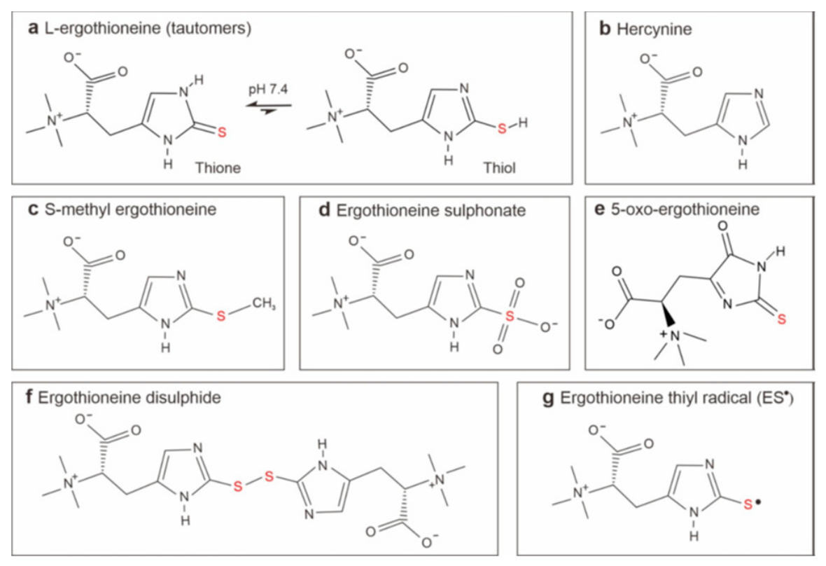
Figure 2.
Number of publications recorded on PubMed in 2025 containing the keyword “ergothioneine”.
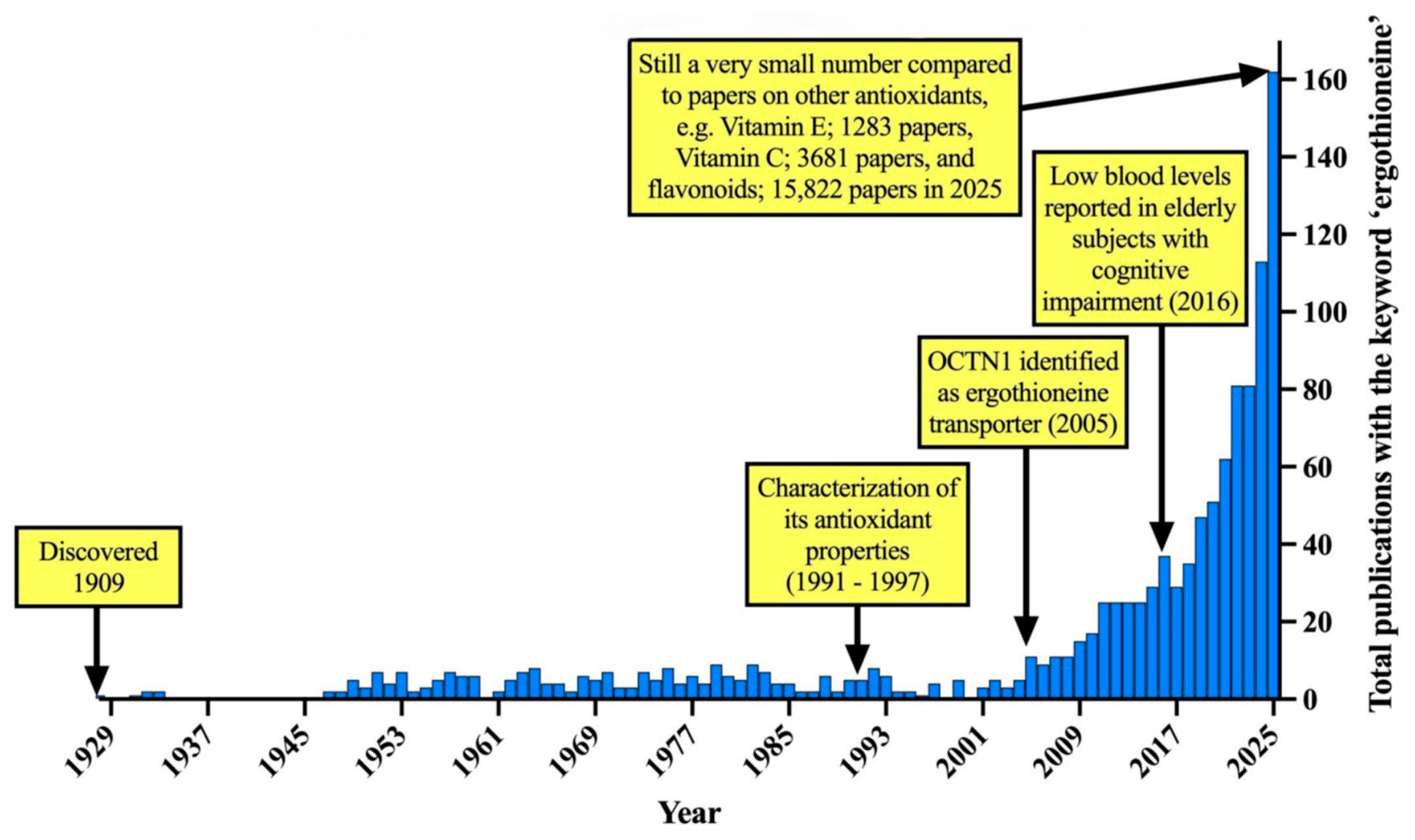
Figure 3.
Expression of transporters in the human brain and other tissues. Comparison of normalised mRNA expression of SLC22A4 (OCTN1) and SLC22A15 across a range of human tissues (data adapted from the Human Protein Atlas; https://www.proteinatlas.org/). SLC22A4 expression is highest in the small intestine, kidney and skeletal muscles, whereas SLC22A15 expression is markedly higher in the brain, retina, and skin. Both transporters are expressed abundantly in the bone marrow and are present in the adult human liver, albeit at lower levels. .
Figure 3.
Expression of transporters in the human brain and other tissues. Comparison of normalised mRNA expression of SLC22A4 (OCTN1) and SLC22A15 across a range of human tissues (data adapted from the Human Protein Atlas; https://www.proteinatlas.org/). SLC22A4 expression is highest in the small intestine, kidney and skeletal muscles, whereas SLC22A15 expression is markedly higher in the brain, retina, and skin. Both transporters are expressed abundantly in the bone marrow and are present in the adult human liver, albeit at lower levels. .
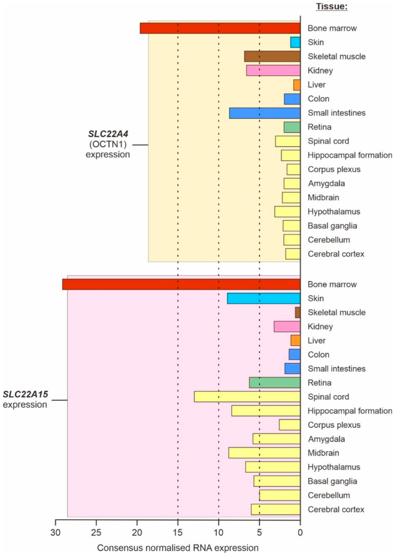
Figure 4.
Plasma ergothioneine (nM) levels in healthy controls (healthy individuals) versus unhealthy individuals. (a) Plasma ergothioneine (ET) levels in healthy controls (healthy Singaporean individuals, mostly but not entirely Chinese) versus individuals with a range of age-related disorders (unhealthy individuals). (b) These unhealthy individuals were patients with a range of disorders, including age-related macular degeneration (AMD), Parkinson’s disease (PD), mild cognitive impairment (MCI), Alzheimer’s disease (AD), and vascular dementia (VaD), a form of dementia more common in Asia than in the West. Significantly lower plasma ET levels were seen in the unhealthy individuals relative to age-matched healthy controls (**** p < 0.0001, *** p < 0.001, Mann-Whitney test). These data were used to establish a hypothetical threshold between healthy and unhealthy plasma ET levels (dotted line in panel a), with levels of plasma ET below ~810 nM defined as being in the unhealthy or at-risk range. Figure adapted from [2] with permission from the journal, based on copyright transfer agreement.
Figure 4.
Plasma ergothioneine (nM) levels in healthy controls (healthy individuals) versus unhealthy individuals. (a) Plasma ergothioneine (ET) levels in healthy controls (healthy Singaporean individuals, mostly but not entirely Chinese) versus individuals with a range of age-related disorders (unhealthy individuals). (b) These unhealthy individuals were patients with a range of disorders, including age-related macular degeneration (AMD), Parkinson’s disease (PD), mild cognitive impairment (MCI), Alzheimer’s disease (AD), and vascular dementia (VaD), a form of dementia more common in Asia than in the West. Significantly lower plasma ET levels were seen in the unhealthy individuals relative to age-matched healthy controls (**** p < 0.0001, *** p < 0.001, Mann-Whitney test). These data were used to establish a hypothetical threshold between healthy and unhealthy plasma ET levels (dotted line in panel a), with levels of plasma ET below ~810 nM defined as being in the unhealthy or at-risk range. Figure adapted from [2] with permission from the journal, based on copyright transfer agreement.
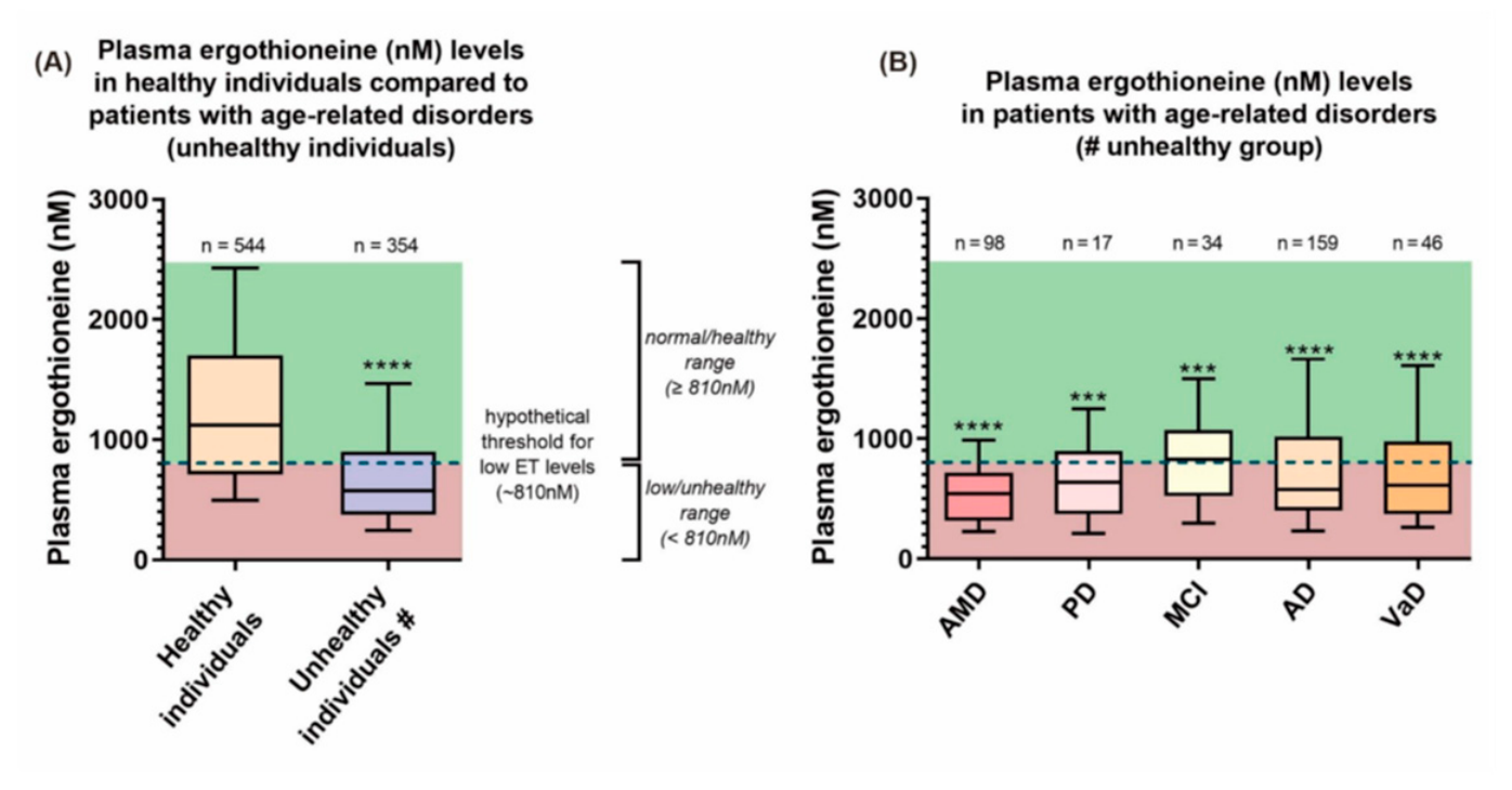
Figure 5.
The Cause—Consequence Issue. The healthy longevity/nutrition literature often assumes that correlation equates to consequence, but this can often not be the case.
Figure 5.
The Cause—Consequence Issue. The healthy longevity/nutrition literature often assumes that correlation equates to consequence, but this can often not be the case.
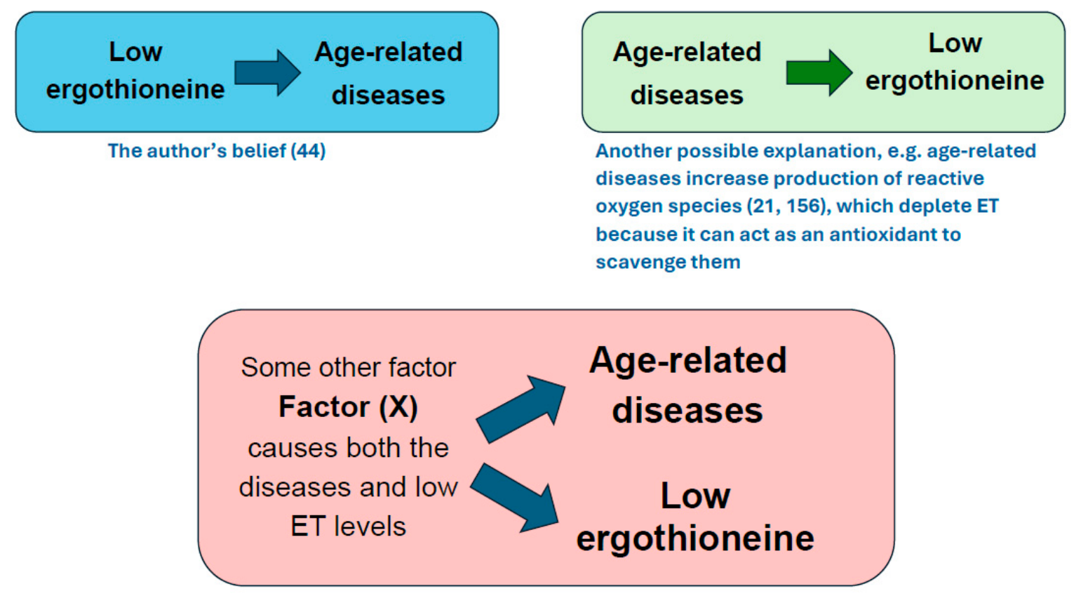
Figure 6.
What are the mechanisms of action of ergothioneine?
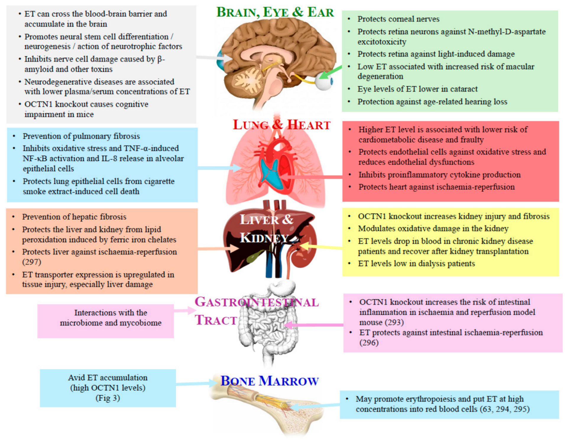
Disclaimer/Publisher’s Note: The statements, opinions and data contained in all publications are solely those of the individual author(s) and contributor(s) and not of MDPI and/or the editor(s). MDPI and/or the editor(s) disclaim responsibility for any injury to people or property resulting from any ideas, methods, instructions or products referred to in the content. |
© 2026 by the authors. Licensee MDPI, Basel, Switzerland. This article is an open access article distributed under the terms and conditions of the Creative Commons Attribution (CC BY) license (http://creativecommons.org/licenses/by/4.0/).
Copyright: This open access article is published under a Creative Commons CC BY 4.0 license, which permit the free download, distribution, and reuse, provided that the author and preprint are cited in any reuse.


