

Submitted:
29 March 2026
Posted:
30 March 2026
You are already at the latest version
Abstract
Background: Post-stroke cognitive impairment (PSCI) is a common complication affecting a substantial proportion of stroke survivors and is associated with reduced functional independence, poorer rehabilitation outcomes, and increased caregiver burden. Early identification of patients at risk remains challenging. Neurofilament Light Chain (NfL), a marker of neuroaxonal injury, has emerged as a potential blood-based biomarker in neurological disorders.Objective: To systematically evaluate prospective evidence on NfL as a predictor of cognitive impairment following ischemic or hemorrhagic stroke.Methods: A systematic review was conducted in accordance with PRISMA Guidelines. PubMed, Cochrane Library, and ScienceDirect were searched for prospective cohort studies assessing plasma or serum NfL levels and subsequent cognitive outcomes in adult stroke patients. Eligible studies evaluated cognitive outcomes using validated assessment tools during follow-up. Due to methodological heterogeneity across studies, findings were synthesized narratively due to methodological heterogeneity.Results: Seven prospective cohort studies involving 2,907 stroke patients were included. Most studies measured NfL within 24-48 hours after stroke onset and assessed cognitive outcomes between 3 and 12 months. Across studies, elevated baseline NfL levels were consistently associated with an increased risk of PSCI and lower cognitive scores. In multivariable analyses, NfL remained independently predictive after adjustment for age, stroke severity, and infarct volume. Reported discriminative performance ranged from AUC 0.672 to 0.865, indicating moderate to good predictive ability.Conclusion: Blood-based NfL measurement is consistently associated with post-stroke cognitive impairment. These findings support circulating Nfl as a promising biomarker for early risk stratification of PSCI in stroke survivors.
Keywords:
neurofilament light chain
; post-stroke cognitive impairment
; stroke
; biomaker
1. Introduction
Stroke remains a leading cause of mortality and long-term disability worldwide, representing a substantial burden for patients, families, and healthcare systems. Although advances in acute reperfusion therapies and secondary prevention have improved survival rates, a growing proportion of stroke survivors live with chronic neurological sequelae. Among these, post-stroke cognitive impairment (PSCI) has emerged as a particularly challenging complication. Epidemiological studies suggest that cognitive impairment develops in approximately one-third to one-half of patients within the first year after stroke, with variability depending on stroke severity, location, and pre-existing vascular risk factors.[1,2] Beyond its impact on memory, attention, and executive function, PSCI is strongly associated with reduced functional independence, poorer rehabilitation outcomes, increased caregiver burden, and elevated mortality risk.[3]
The mechanisms underlying PSCI are complex and extend beyond the focal lesion itself. Acute ischemic or hemorrhagic injury initiates a cascade of cellular and molecular events, including excitotoxicity, oxidative stress, inflammation, and blood–brain barrier disruption. These processes lead to axonal injury, demyelination, and network disconnection that may evolve over weeks to months after the initial insult.[4] In addition, pre-existing small vessel disease, white matter hyperintensities, and neurodegenerative pathology may interact with acute vascular injury, amplifying vulnerability to cognitive decline.[5] This multifactorial pathophysiology suggests that structural lesion volume alone may not fully capture the biological processes contributing to subsequent cognitive deterioration.
In recent years, there has been increasing interest in identifying blood-based biomarkers that reflect neuroaxonal injury and may assist in early risk stratification for PSCI. Neurofilament light chain (NfL) is a cytoskeletal protein highly expressed in large-caliber myelinated axons. When axons are damaged, NfL is released into the interstitial fluid and cerebrospinal fluid, eventually entering the systemic circulation. The development of ultrasensitive assay platforms, particularly single-molecule array (Simoa) technology, has enabled accurate detection of NfL in plasma and serum at picogram-per-milliliter concentrations.[6] This technological advance has significantly expanded the clinical applicability of NfL as a minimally invasive biomarker of neuroaxonal injury.
Accumulating evidence supports the role of circulating NfL as a sensitive indicator of neuronal damage across various neurological conditions. Elevated NfL concentrations have been documented in multiple sclerosis, traumatic brain injury, and neurodegenerative diseases, where levels correlate with disease activity and progression.[7,8] In ischemic stroke specifically, serum or plasma NfL levels have been shown to correlate with infarct volume, stroke severity, and short-term functional outcome measures.[9] These findings suggest that NfL reflects the magnitude of acute axonal disruption.
However, functional recovery assessed by scales such as the modified Rankin Scale does not necessarily equate to preserved cognitive function. Motor independence can coexist with significant impairment in executive function, memory, or processing speed. Therefore, understanding whether early circulating NfL levels predict long-term cognitive trajectories is of substantial clinical relevance. Several prospective cohort studies have recently explored the association between early post-stroke NfL concentrations and later cognitive performance, typically assessed using screening instruments such as the Montreal Cognitive Assessment (MoCA) or structured cognitive batteries.[10] These studies suggest that higher baseline NfL levels may independently predict PSCI after adjustment for age, stroke severity, and infarct characteristics.
Despite these emerging data, the existing literature remains heterogeneous. Studies vary in stroke subtype inclusion (ischemic versus hemorrhagic), timing of biomarker measurement, assay methodology, cognitive outcome definitions, and follow-up duration. Moreover, reported effect sizes and predictive thresholds differ across cohorts. To date, no comprehensive synthesis has systematically evaluated the prognostic role of blood-based NfL specifically for cognitive outcomes following stroke. Such a synthesis is essential to clarify the consistency of findings, identify methodological gaps, and determine whether circulating NfL has sufficient robustness to be considered a clinically meaningful predictor of PSCI.
Given the substantial burden of PSCI and the need for early risk stratification tools, a systematic evaluation of the available evidence is warranted. Therefore, this systematic review aims to synthesize prospective cohort studies investigating blood-based neurofilament light chain as a predictor of post-stroke cognitive impairment in adult patients with ischemic or hemorrhagic stroke.
2. Materials and Methods
2.1. Study Design and Reporting Standard
This systematic review was conducted in accordance with the Preferred Reporting Items for Systematic Reviews and Meta-Analyses (PRISMA) guidelines. The objective was to synthesize prospective evidence evaluating circulating neurofilament light chain (NfL) as a predictor of post-stroke cognitive impairment (PSCI) in adult patients with ischemic or hemorrhagic stroke.
2.2. Search Strategy
A comprehensive literature search was performed in three electronic databases: PubMed, Cochrane Library, and ScienceDirect. The search strategy combined terms related to stroke, neurofilament light chain, and cognitive outcomes. The following core concepts were used: “stroke” or “intracerebral hemorrhage,” combined with “neurofilament light chain” or “NfL,” and cognitive-related terms including “cognitive impairment,” “post-stroke cognitive impairment,” “PSCI,” “MoCA,” and “cognitive decline,” along with blood-based sampling terms such as “plasma” or “serum.”
The final search strategy was adapted to the syntax of each database. All databases were searched from inception to the most recent available date prior to manuscript preparation. No language restriction was initially applied during the search process; however, only studies published in English were included in the final synthesis.
2.3. Eligibility Criteria
Studies were included if they met the following criteria: adult participants aged 18 years or older with a diagnosis of ischemic or hemorrhagic stroke; measurement of neurofilament light chain in blood (plasma or serum) during the acute or subacute phase following stroke; evaluation of cognitive outcome during follow-up using validated cognitive instruments such as the Montreal Cognitive Assessment (MoCA), Telephone Interview of Cognitive Status (TICS), or equivalent structured assessments; and a prospective cohort design evaluating the prognostic association between early NfL levels and subsequent cognitive outcomes.
Studies were excluded if they did not assess cognitive outcomes, measured NfL only in cerebrospinal fluid without blood sampling, lacked a longitudinal follow-up design, included pediatric populations, or did not provide sufficient data to evaluate the association between NfL and cognitive performance.
2.4. Study Selection
All records identified through database searching were screened independently at the title and abstract level. Studies that were clearly irrelevant were excluded. Full-text articles were retrieved for potentially eligible studies and assessed in detail according to the predefined inclusion and exclusion criteria. Discrepancies in study selection were resolved through discussion and consensus.
The study selection process is illustrated in the PRISMA flow diagram. A total of 348 records were identified, of which 339 were excluded after title and abstract screening. Eight full-text articles were assessed for eligibility, and seven prospective cohort studies were ultimately included in the qualitative synthesis.
2.5. Data Extraction
Data were extracted from each included study using a standardized extraction framework. The following information was collected: first author and year of publication, country, study design, stroke subtype, sample size, participant demographics, timing of NfL measurement, biological fluid type, assay method, duration of follow-up, cognitive assessment instrument, definition of PSCI where applicable, statistical modeling approach, effect measures, covariates adjusted in multivariable analyses, main findings, and risk of bias assessment.
Where studies reported both continuous and categorical cognitive outcomes, both types of results were extracted. Particular attention was given to adjusted effect estimates and receiver operating characteristic (ROC) performance metrics when available.
2.6. Outcomes
The primary outcome of interest was post-stroke cognitive impairment assessed during follow-up. PSCI was typically defined using MoCA-based thresholds, although variations in cutoff values were noted. Secondary outcomes included continuous cognitive scores, longitudinal cognitive decline, and, in one study, long-term neurological or functional outcomes closely related to cognitive performance.
2.7. Risk of Bias Assessment
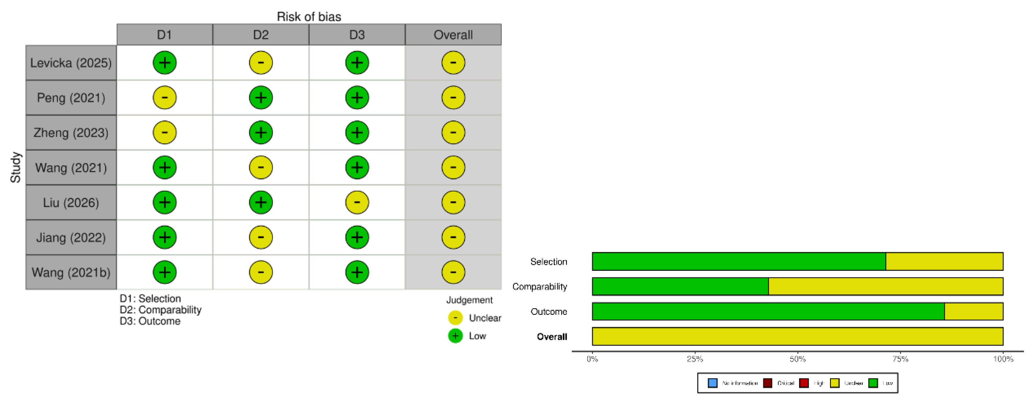
Methodological quality was evaluated using a structured appraisal approach appropriate for prognostic cohort studies. Domains assessed included study design, participant selection, measurement of exposure and outcomes, adequacy of follow-up, control of confounding variables, and statistical analysis. All included studies were prospective in design and employed multivariable regression modeling. In accordance with predefined assessment criteria, risk of bias was categorized as low or moderate, with ratings capped at moderate to reflect methodological limitations without overclassification.
2.8. Data Synthesis
Given the heterogeneity in cognitive assessment tools, PSCI cutoff definitions, timing of biomarker measurement, and statistical reporting formats, a narrative synthesis approach was performed. Findings were summarized according to stroke subtype, cognitive outcome type, and strength of association between early circulating NfL levels and subsequent cognitive performance. Where available, predictive performance metrics such as area under the ROC curve were also described to contextualize the potential clinical utility of NfL as a prognostic biomarker.

Table 1.
Characteristics and results of the included studies.
| First Author (Year) | Country | Study Design | Stroke Type (Ischemic / Hemorrhagic) | Sample Size (Total) | Mean/Median Age | % Male | Timing of NfL Measurement | Biological Fluid (Serum/Plasma/CSF) | Assay Method | Follow-up Duration | Cognitive Assessment Tool | Definition of PSCI (if applicable) | Statistical Model (Adjusted/Unadjusted) | Effect Measure (OR/HR/β/Correlation) | Covariates Adjusted | Summary of Findings | Risk of Bias | |
|---|---|---|---|---|---|---|---|---|---|---|---|---|---|---|---|---|---|---|
| Levicka (2025) | Slovakia | Prospective observational cohort | Ischemic stroke | 73 acute IS (59 completed follow-up) | 68.5 ± 10.7 years | 57.60% | 5–10 days post-stroke and 3 months | Plasma | SIMOA (NF-Light Advantage Kit, Quanterix HD-1 analyzer) | 3 months | MoCA | MoCA-based categorization (no formal PSCI diagnostic criteria stated) | Multiple regression (stepwise); correlation analysis; non-parametric tests | Correlation; regression coefficients (B); longitudinal change (Wilcoxon) | Age, prior stroke, anxiety (final model) | pNFL significantly decreased at 3 months; strongly correlated with infarct volume; no association between pNFL and cognitive performance; pNFL not correlated with OSA severity or CPAP adherence. Age, previous stroke, and anxiety predicted MoCA at 3 months. | Moderate | |
| Peng (2021) | USA (Spaulding Rehabilitation Hospital, Harvard affiliate) | Observational cohort | Acute ischemic stroke | 144 ischemic stroke (30 controls) | ~69 years | 61–67% male (varied by subgroup) | Within 1 week of admission (mean ~11 days post-stroke); subset repeated at 3 weeks | Serum | Simoa single-molecule array (Quanterix SR-X) | Discharge from inpatient rehabilitation (acute rehab phase) | FIM cognitive subscore (primary); FIM motor & total | No formal PSCI diagnosis; dichotomized by median FIM cognitive score | Spearman correlation; multivariable linear regression; ROC analysis | Correlation (r); β coefficients; AUC (ROC) | Age-adjusted analyses; multivariable models included log(NfL), infarct volume, WMH volume, translated mRS | Serum NfL markedly elevated vs controls (9-fold). NfL correlated with infarct volume (r ≈ 0.46) and negatively with FIM cognitive score (r ≈ −0.39). Independent predictor of cognitive outcome at discharge (AUC 0.672). Combination of NfL + infarct volume + WMH improved predictive value (AUC 0.760). | Moderate | |
| Zheng (2023) | China | Prospective cohort (Chinese Cerebral Hemorrhage Mechanisms and Intervention Study) | Hemorrhagic (Intracerebral hemorrhage) | 300 (MoCA & MRI subset n=26) | Median 59 years (27–84) | 68.00% | Within 24 h; repeated at days 7 and 14 | Plasma | Single-molecule array (Simoa) | 6 months (MoCA subset) | MoCA (6 months); MRI (FA, WMH, VBM) | Not formally defined (continuous MoCA analysis) | Spearman correlation (cognitive analysis) | r = −0.59 (day-7 NfL vs MoCA) | Not adjusted in MoCA correlation analysis | Day-7 NfL negatively correlated with 6-month MoCA (r = −0.59, p < 0.01) and reduced white matter integrity (FA); subacute NfL showed stronger cognitive association than hyperacute measurement. | Moderate | |
| Wang (2021) | China | Prospective longitudinal cohort | Acute ischemic stroke (AIS) with post-stroke subjective cognitive impairment (SCI) | 304 patients | 64.9 ± 9.3 years | 58% male (approx.) | 1 month after stroke onset | Serum | Simoa (single-molecule array) | 12 months | TICS-40 (Telephone Interview of Cognitive Status-40) | Cognitive decline defined as decreased TICS-40 score at 12 months; OCI defined as TICS-40 ≤20 | Logistic regression; multivariable linear regression; ROC analysis | Adjusted OR (multivariable); β coefficients; AUC | Age, sex, ApoE ε4, education, BMI, vascular risk factors, infarct volume (DWI), anxiety/depression, baseline TICS-40 | Baseline serum NfL significantly higher in progression group (75.6 ± 17.2 pg/ml) vs stable group (49.5 ± 24.1 pg/ml). NfL independently predicted longitudinal cognitive decline (adjusted OR ≈ 1.066 per pg/ml increase, p<0.001). ROC AUC = 0.865; cutoff 79.31 pg/ml (Sensitivity 0.653; Specificity 0.906). Higher NfL associated with greater decline in TICS-40 and worse endpoint cognitive performance. | Moderate | |
| Liu (2026) | China | Prospective observational cohort | First acute ischemic stroke | 128 patients | Not explicitly stated in abstract | Not stated in abstract | Within 24 hours of admission | Plasma | ELISA (for GFAP, Aβ1-40, Aβ1-42, p-Tau181, p-Tau217, Aβ42/40 ratio, NfL) | 3 months | MoCA | PSCI defined as MoCA < 22 at 3 months | Logistic regression; ROC analysis | OR; AUC | Not fully detailed in abstract (multivariate analysis reported) | PSCI incidence: 53.9% (69/128). Plasma NfL significantly higher in PSCI group. NfL independently associated with PSCI (OR 1.0406; 95% CI 1.0006–1.0806; p=0.025). ROC AUC for NfL = 0.809 (95% CI 0.733–0.885). Combined GFAP+NfL improved prediction (AUC 0.855). | Moderate | |
| Jiang (2022) | China | Prospective cohort | Posterior circulation acute ischemic stroke | 264 patients | Median 65 years (IQR 51–73) | 59.47% male | Within 48 hours of stroke onset (median 26 h) | Plasma | Simoa (single-molecule array) | 90 days (3 months) | MoCA | PSCI defined as MoCA < 26 at 90 days | Multivariable logistic regression; ROC analysis; Spearman correlation | Adjusted OR; AUC; correlation coefficients | Age, sex, education; NIHSS; infarct volume; TOAST classification | Median pNfL significantly higher in PSCI group (49.54 pg/mL) vs non-PSCI (30.12 pg/mL), p<0.001. pNfL independently associated with PSCI (Adjusted OR 1.035–1.044 per pg/mL increase, p<0.001). ROC AUC = 0.782 (95% CI 0.726–0.837); optimal cutoff 38.12 pg/mL (Sensitivity 78.2%, Specificity 66.9%). pNfL negatively correlated with MoCA (r = −0.323). | Moderate | |
| Wang (2021) | China | Prospective single-center cohort | Acute ischemic stroke (anterior circulation) | 1,694 patients | Median 64 years (IQR 16) | 52.7% male | Within 24 hours (≤48 h) of stroke onset | Plasma | Simoa (single-molecule array) | 90 days (3 months) | MoCA | PSCI defined as MoCA < 26 at 90 days | Multivariable logistic regression; ROC analysis | Adjusted OR; AUC | Age, sex, education, NIHSS score, TOAST classification, infarct volume | Median pNfL significantly higher in PSCI vs non-PSCI (55.96 vs 35.73 pg/mL, p < 0.001). pNfL independently predicted PSCI (adjusted OR 1.038–1.049 per pg/mL increase, p < 0.001). ROC AUC = 0.785 (95% CI 0.762–0.808). Optimal cutoff 46.12 pg/mL (Sensitivity 71.0%, Specificity 81.5%). | Moderate |
3. Results
3.1. Study Selection
The literature search yielded a total of 348 records across the three electronic databases, comprising 63 records from PubMed, 2 from the Cochrane Library, and 283 from ScienceDirect. After removal of clearly irrelevant articles through title and abstract screening, 339 records were excluded primarily due to unrelated outcomes, non-stroke populations, absence of neurofilament measurement, or non-cognitive endpoints. Nine duplicate records were identified during the screening process, and one duplicate was removed prior to full-text assessment.
Eight full-text articles were retrieved and assessed for eligibility. Of these, one study was excluded because its population characteristics and study design did not align with the predefined inclusion criteria, specifically lacking appropriate cognitive outcome assessment or prognostic evaluation of blood-based NfL. Ultimately, seven prospective cohort studies met all eligibility criteria and were included in the qualitative synthesis. The study selection process is illustrated in Figure 1.
Figure 1.
Diagram flow of literature search strategy for this systematic review.
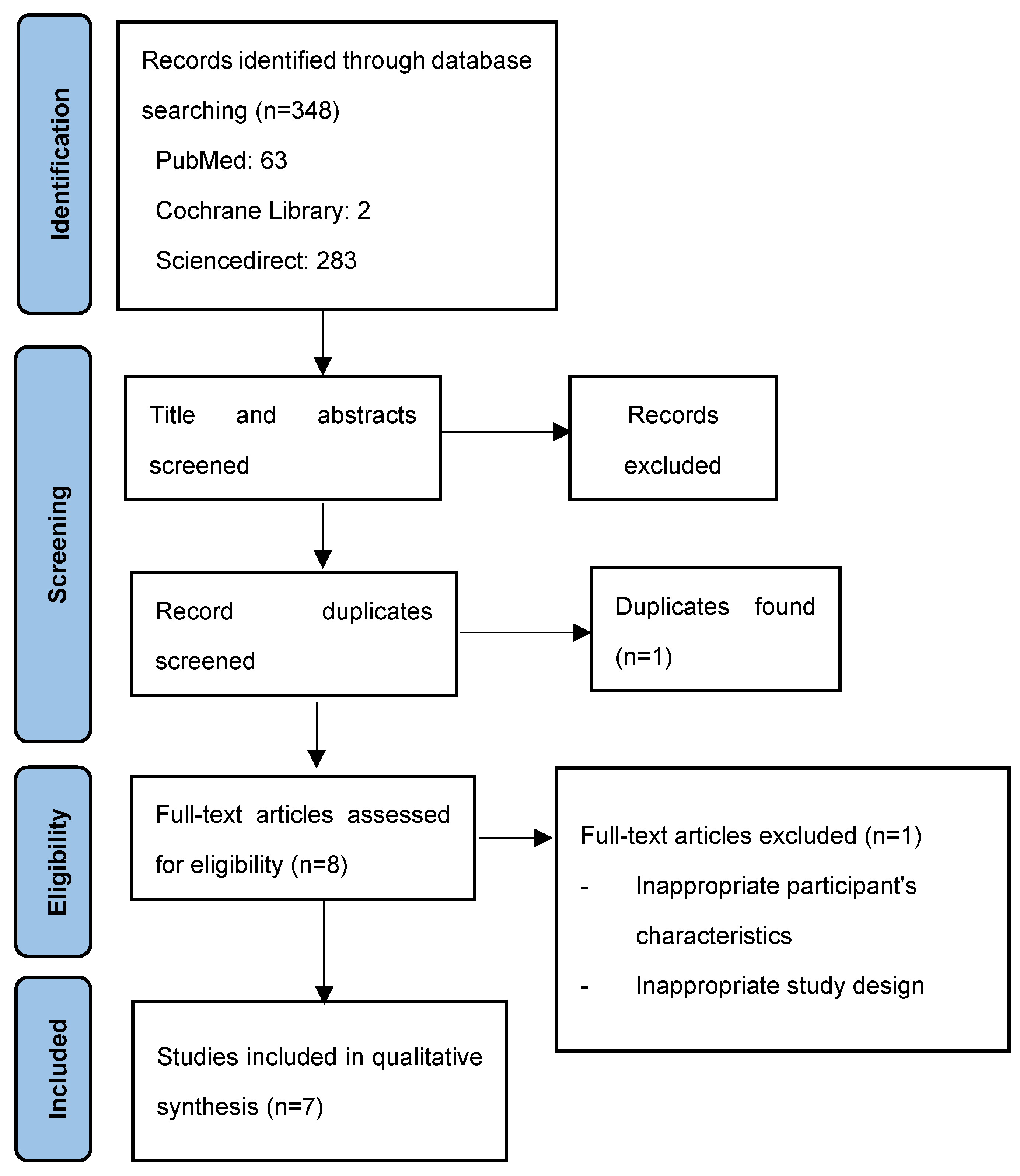
Figure 2.
Risk of bias assessment result using Newcastle Ottawa Scale (NOS).
The final dataset therefore reflects a focused body of longitudinal prognostic evidence evaluating circulating neurofilament light chain as a predictor of post-stroke cognitive outcomes.
3.2. Study Characteristics
All included studies employed prospective observational cohort designs, reflecting the prognostic nature of the research question. Publication years ranged from 2021 to 2026, indicating that this is a relatively recent and rapidly evolving research area. Six studies investigated patients with acute ischemic stroke, whereas one study included individuals with intracerebral hemorrhage. This distribution underscores the current predominance of ischemic stroke research in biomarker-driven cognitive prognostication.
Sample sizes varied considerably across studies, ranging from 128 participants in smaller biomarker cohorts to 1,694 patients in the largest anterior circulation ischemic stroke study. The majority of cohorts enrolled middle-aged to elderly adults, with mean or median ages generally in the mid-60s. Male participants constituted slightly more than half of the study populations in most cohorts, reflecting the known epidemiological distribution of stroke.
Neurofilament light chain was measured predominantly in plasma, although some studies analyzed serum concentrations. Most studies collected blood samples within 24 to 48 hours of stroke onset, emphasizing the early injury phase as the prognostic window of interest. One cohort assessed NfL during the subacute phase at one month post-stroke, allowing exploration of longer-term axonal injury dynamics. Assay methodology was largely standardized across studies, with most using highly sensitive single-molecule array (Simoa) technology. One study utilized enzyme-linked immunosorbent assay (ELISA), introducing a potential source of inter-study measurement variability.
Follow-up durations ranged from 3 months to 12 months, with the majority evaluating cognitive outcomes at 90 days. Six studies used the Montreal Cognitive Assessment (MoCA) as the primary cognitive screening instrument. Most defined post-stroke cognitive impairment as MoCA <26, although one study applied a lower threshold of <22, reflecting variability in diagnostic cutoffs. One cohort assessed cognitive function using the Telephone Interview of Cognitive Status (TICS-40) over 12 months, focusing on longitudinal decline. Another study evaluated cognitive and functional recovery during inpatient rehabilitation using validated functional cognitive scales.
Overall, the included studies provide a relatively consistent methodological framework centered on early NfL measurement and short- to intermediate-term cognitive follow-up, while differing in stroke subtype focus, cognitive definitions, and analytic models.
3.3. Association Between NfL and Post-Stroke Cognitive Impairment
Across ischemic stroke cohorts, elevated baseline plasma or serum NfL levels were consistently associated with an increased risk of subsequent cognitive impairment. Patients who developed PSCI demonstrated significantly higher early NfL concentrations compared to those who maintained preserved cognition. These associations remained statistically significant after multivariable adjustment in most studies, suggesting that NfL provides independent prognostic information beyond traditional clinical predictors.
Multivariable logistic regression models typically adjusted for age, sex, educational level, stroke severity as measured by NIHSS, infarct volume, and vascular risk factors. In the largest cohort, plasma NfL remained independently associated with PSCI even after controlling for infarct volume and TOAST classification, indicating that NfL may capture aspects of axonal injury not fully explained by structural lesion burden alone.
Several studies also evaluated continuous cognitive outcomes. Higher NfL concentrations were negatively correlated with MoCA scores at follow-up, reinforcing the graded relationship between axonal injury biomarker levels and cognitive performance. In the longitudinal TICS-based cohort, baseline NfL levels predicted progressive cognitive decline over a 12-month period, suggesting that NfL may reflect ongoing neurodegenerative or secondary injury processes rather than purely acute structural damage.
Although the intracerebral hemorrhage study primarily focused on functional and mortality outcomes, elevated NfL levels were strongly associated with long-term neurological deterioration. While direct cognitive testing was not uniformly reported in that cohort, the findings support the broader biological plausibility of NfL as a marker of axonal injury severity influencing long-term brain function.
Collectively, the direction of association was remarkably consistent: higher early circulating NfL levels were associated with worse cognitive or neurological outcomes across different stroke subtypes and study settings.
3.4. Predictive Performance of NfL
Beyond statistical association, several studies evaluated the discriminative ability of NfL using receiver operating characteristic analyses. Reported area under the curve values ranged approximately from the high 0.7 range to above 0.8, indicating moderate to good predictive performance for PSCI. These findings suggest that NfL has potential clinical utility as a prognostic biomarker, although it is not sufficient as a standalone diagnostic tool.
Importantly, predictive accuracy improved when NfL was incorporated into multivariable models alongside established clinical predictors such as age, stroke severity, and infarct volume. In one study, combining NfL with additional biomarkers further enhanced discrimination. This pattern indicates that NfL may function most effectively within multimodal prognostic models rather than in isolation.
The largest cohort study provided a clinically interpretable cutoff value with acceptable sensitivity and specificity, suggesting translational relevance. However, variability in cutoff thresholds across studies, influenced by assay platform and population characteristics, highlights the need for standardization before routine clinical application.
3.5. Hemorrhagic Stroke Findings
The study focusing on intracerebral hemorrhage demonstrated that plasma NfL levels exhibited a dynamic trajectory following hemorrhagic injury, with early peaks corresponding to acute axonal damage. Elevated NfL levels were independently associated with poor long-term neurological outcomes and mortality at 6 and 12 months. Although formal cognitive instruments such as MoCA were not the primary endpoints in that study, functional outcomes are closely intertwined with cognitive capacity in severe hemorrhagic stroke.
The inclusion of hemorrhagic stroke data broadens the applicability of NfL beyond ischemic injury, supporting its role as a generalized marker of neuroaxonal damage severity. Nevertheless, the limited number of hemorrhagic cohorts prevents definitive conclusions regarding stroke subtype–specific differences in cognitive prognostication.
3.6. Heterogeneity Across Studies
Several sources of heterogeneity were identified. First, cognitive outcome definitions varied, including different MoCA thresholds and alternative instruments such as TICS or functional cognitive scales. Second, the timing of NfL measurement ranged from hyperacute (within 24 hours) to subacute (one month), potentially capturing different biological processes. Third, assay methodologies differed between Simoa and ELISA platforms, which may influence absolute NfL values and cutoff thresholds.
Stroke subtype also varied, with some cohorts restricted to anterior or posterior circulation ischemic stroke, potentially affecting lesion location–related cognitive outcomes. Additionally, follow-up duration ranged from 3 months to 12 months, limiting direct comparability across timepoints.
Despite these methodological differences, the consistency in directionality of findings across cohorts strengthens confidence in the observed association between elevated circulating NfL and subsequent cognitive impairment.
3.7. Risk of Bias Assessment
All included studies were prospective in design and employed structured cognitive assessments with predefined follow-up intervals. Most studies implemented multivariable regression modeling to adjust for major confounders, including age, stroke severity, infarct volume, and vascular risk factors. These methodological features reduce the likelihood of major selection or measurement bias.
However, several limitations were noted. Most cohorts were single-center studies, potentially limiting external validity. Cognitive assessment relied predominantly on screening instruments rather than comprehensive neuropsychological batteries, which may underestimate subtle domain-specific impairment. Variability in PSCI cutoff definitions and lack of standardized biomarker thresholds further contribute to inter-study heterogeneity. Residual confounding cannot be entirely excluded.
In accordance with predefined criteria and the decision to cap ratings at moderate severity, all studies were judged to have at most moderate risk of bias.
4. Discussion
4.1. Principal Findings
This systematic review synthesizes prospective cohort evidence evaluating circulating neurofilament light chain as a prognostic biomarker for post-stroke cognitive impairment. Across the seven included studies, a consistent pattern emerged: higher plasma or serum NfL concentrations measured in the acute or early subacute phase following stroke were associated with worse cognitive outcomes during follow-up. Importantly, this association persisted even after adjustment for established clinical predictors such as age, stroke severity, infarct volume, and vascular risk factors in most cohorts.
These findings extend prior knowledge regarding NfL as a marker of global neuroaxonal injury. While earlier studies predominantly focused on functional outcomes or mortality after stroke,[9,11,12,13,14] the present synthesis highlights cognitive endpoints, which represent a distinct and often underrecognized dimension of stroke recovery. Given that PSCI affects up to half of stroke survivors in certain populations,[1,2] the identification of a blood-based biomarker capable of early risk stratification is of considerable clinical importance.
Moreover, several included studies demonstrated moderate to good discriminative ability of NfL for predicting PSCI, with area under the curve values frequently approaching 0.80. Although these values do not support NfL as a standalone diagnostic tool, they indicate clinically meaningful prognostic potential when integrated with conventional clinical parameters.
4.2. Pathophysiological Interpretation
The biological plausibility of NfL as a predictor of PSCI is grounded in its fundamental role in axonal structure. Neurofilament light chain forms part of the cytoskeletal framework of myelinated axons, and its release into extracellular fluid occurs in response to axonal disruption.[8] In acute ischemic stroke, axonal injury extends beyond the immediate infarct core due to mechanisms such as excitotoxicity, oxidative stress, inflammatory cascades, and retrograde degeneration along connected white matter tracts.[4] Similar mechanisms operate in intracerebral hemorrhage, where mass effect, perihematomal edema, and secondary injury processes contribute to widespread neural damage.
Importantly, cognitive impairment after stroke is rarely attributable to a single focal lesion. Rather, it reflects disruption of distributed neural networks supporting executive function, attention, and memory. White matter hyperintensities and pre-existing small vessel disease may amplify vulnerability to cognitive decline.[5] In this context, circulating NfL likely captures a composite measure of axonal injury burden, integrating both focal damage and diffuse secondary neurodegeneration.
The independence of NfL from infarct volume in multivariable models is particularly noteworthy. Although NfL correlates with stroke severity and lesion size,[9,12,14] several studies included in this review demonstrated that its association with PSCI persisted after adjustment for these factors. This suggests that NfL may reflect microstructural damage or delayed axonal degeneration not fully visualized by routine neuroimaging. Such processes may be especially relevant for cognitive networks, where subtle white matter disconnection can significantly impair higher-order processing.
Analogous patterns have been observed in neurodegenerative disorders. In Alzheimer’s disease, longitudinal increases in serum NfL precede overt clinical decline and correlate with progressive neurodegeneration.[7] Similarly, cerebrospinal fluid NfL levels have been associated with cognitive performance across various dementia syndromes.[15] These cross-disease observations reinforce the concept that NfL is a sensitive indicator of cumulative neuroaxonal injury, irrespective of the underlying etiology.
4.3. Comparison with Existing Stroke Biomarker Literature
Previous stroke biomarker research has explored inflammatory mediators, oxidative stress markers, and metabolic indicators as predictors of cognitive decline. However, these biomarkers often reflect systemic processes and lack specificity for neuronal injury. By contrast, NfL directly indexes structural axonal damage.[8]
Several earlier studies established that serum NfL levels correlate with acute ischemic stroke severity and functional outcomes.[9,11,12,13,14] Uphaus et al. demonstrated that NfL levels measured after ischemic stroke were predictive of long-term functional outcome,[11] while Pedersen et al. described a temporal profile of circulating NfL following ischemic injury.[12] Gendron et al. further showed that plasma NfL predicted mortality after stroke.[13] However, these investigations primarily focused on global disability or survival rather than cognitive domains.
The present synthesis builds upon this foundation by consolidating evidence specifically targeting cognitive outcomes. In light of epidemiological data showing high rates of PSCI,[1,3] and mechanistic frameworks emphasizing delayed and network-level degeneration,[4] the association between early NfL elevation and subsequent cognitive impairment appears both biologically coherent and clinically meaningful.
4.4. Clinical Implications
The early identification of patients at risk for PSCI carries substantial implications. Cognitive impairment may compromise rehabilitation engagement, medication adherence, financial decision-making, and social reintegration.[3] Despite this, routine cognitive risk stratification in the acute stroke setting remains limited.
A blood-based biomarker such as NfL offers several practical advantages. Blood sampling is minimally invasive and widely available. When measured within the first 24 to 48 hours after stroke, NfL levels could inform early prognostic counseling and prompt intensified cognitive monitoring in high-risk individuals. Integration of NfL into multivariable predictive models alongside age, NIHSS score, and imaging findings may enhance discrimination beyond clinical parameters alone.
Nevertheless, several challenges remain before translation into routine practice. Standardization of assay platforms is required, as differences in measurement techniques may influence absolute concentration values. Furthermore, consensus regarding clinically actionable cutoff thresholds is lacking. Given that NfL reflects global axonal injury rather than cognition-specific pathways, it should be interpreted within a broader clinical context rather than as a definitive diagnostic indicator.
4.5. Heterogeneity and Methodological Considerations
Although the direction of association between elevated NfL and worse cognitive outcome was consistent, heterogeneity across studies warrants careful consideration. Differences in stroke subtype inclusion, timing of biomarker measurement, cognitive assessment tools, and follow-up duration may influence effect estimates.
Most studies relied on screening instruments such as MoCA rather than comprehensive neuropsychological batteries. While MoCA is validated and sensitive for mild cognitive impairment, it may not detect domain-specific deficits. Additionally, variations in PSCI cutoff thresholds could affect classification rates.
The majority of cohorts were single-center, potentially limiting external validity. Residual confounding remains possible despite multivariable adjustment. Pre-existing neurodegenerative pathology, microvascular disease burden, or socioeconomic factors may contribute both to baseline NfL levels and later cognitive outcomes.
4.6. Future Research Directions
Future studies should prioritize multicenter validation in diverse populations, incorporating standardized NfL measurement protocols and harmonized cognitive assessment frameworks. Longitudinal designs assessing serial NfL levels may clarify whether dynamic changes over time provide incremental predictive value compared to single early measurements.
Moreover, combining NfL with imaging markers of white matter integrity and other biomarkers may enable the development of integrated risk models. Advanced analytical approaches, including machine-learning–based prediction algorithms, may further refine risk stratification.
Ultimately, interventional trials are required to determine whether early identification of high-risk patients based on NfL levels can modify cognitive trajectories through targeted rehabilitation, vascular risk optimization, or neuroprotective strategies.
5. Conclusions
In summary, this systematic review demonstrates that elevated circulating neurofilament light chain levels measured in the acute or early subacute phase after stroke are consistently associated with subsequent cognitive impairment. Across prospective cohort studies, higher plasma or serum NfL concentrations independently predicted worse cognitive outcomes at follow-up, even after adjustment for stroke severity and infarct characteristics. These findings support the role of NfL as a biologically plausible marker of neuroaxonal injury relevant to cognitive trajectories.
Although promising, the clinical implementation of NfL requires further validation in multicenter cohorts with standardized assay protocols and harmonized cognitive definitions. Future research should explore longitudinal NfL dynamics and multimodal prediction models integrating imaging and clinical variables. Circulating NfL represents a potential step toward early biomarker-driven risk stratification for post-stroke cognitive impairment.
Author Contributions
Conceptualization, S.R.A, H.N.D, R.R .; methodology, S.R.A ; data curation H.N.D, X.X.; writing—original draft preparation, S.R.A, H.N.D, R.R writing—review and editing, S.R.A, H.N.D, R.R.; visualization, S.R.A.; supervision,.
Funding
This research received no external funding.
Conflicts of Interest
The authors declare no conflict of interest.
References
- Pendlebury ST, Rothwell PM, Oxford Vascular Study. Incidence and prevalence of dementia associated with transient ischaemic attack and stroke: analysis of the population-based Oxford Vascular Study. Lancet Neurol. 2019 Mar;18(3):248–58. PubMed PMID: 30784556; PubMed Central PMCID: PMC6390174. [CrossRef]
- Mellon L, Brewer L, Hall P, Horgan F, Williams D, Hickey A, et al. Cognitive impairment six months after ischaemic stroke: a profile from the ASPIRE-S study. BMC Neurol. 2015 Mar 12;15:31. PubMed PMID: 25879880; PubMed Central PMCID: PMC4359388. [CrossRef]
- Sun JH, Tan L, Yu JT. Post-stroke cognitive impairment: epidemiology, mechanisms and management. Ann Transl Med. 2014 Aug;2(8):80. 2014.08.05 PubMed PMID: 25333055; PubMed Central PMCID: PMC4200648. [CrossRef]
- Mok VCT, Lam BYK, Wong A, Ko H, Markus HS, Wong LKS. Early-onset and delayed-onset poststroke dementia - revisiting the mechanisms. Nat Rev Neurol. 2017 Mar;13(3):148–59. PubMed PMID: 28211452. [CrossRef]
- de Kort FAS, Coenen M, Weaver NA, Kuijf HJ, Aben HP, Bae HJ, et al. White Matter Hyperintensity Volume and Poststroke Cognition: An Individual Patient Data Pooled Analysis of 9 Ischemic Stroke Cohort Studies. Stroke. 2023 Dec;54(12):3021–9. PubMed PMID: 37901947; PubMed Central PMCID: PMC10664782. [CrossRef]
- Disanto G, Adiutori R, Dobson R, Martinelli V, Dalla Costa G, Runia T, et al. Serum neurofilament light chain levels are increased in patients with a clinically isolated syndrome. J Neurol Neurosurg Psychiatry. 2016 Feb;87(2):126–9. PubMed PMID: 25716934. [CrossRef]
- Dominantly Inherited Alzheimer Network, Preische O, Schultz SA, Apel A, Kuhle J, Kaeser SA, et al. Serum neurofilament dynamics predicts neurodegeneration and clinical progression in presymptomatic Alzheimer’s disease. Nat Med. 2019 Feb;25(2):277–83. [CrossRef]
- Khalil M, Teunissen CE, Lehmann S, Otto M, Piehl F, Ziemssen T, et al. Neurofilaments as biomarkers in neurological disorders — towards clinical application. Nat Rev Neurol. 2024 May;20(5):269–87. [CrossRef]
- Tiedt S, Duering M, Barro C, Kaya AG, Boeck J, Bode FJ, et al. Serum neurofilament light: A biomarker of neuroaxonal injury after ischemic stroke. Neurology. 2018 Oct 2;91(14). [CrossRef]
- Wang Z, Wang R, Li Y, Li M, Zhang Y, Jiang L, et al. Plasma Neurofilament Light Chain as a Predictive Biomarker for Post-stroke Cognitive Impairment: A Prospective Cohort Study. Front Aging Neurosci. 2021 Feb 19;13:631738. [CrossRef]
- Uphaus T, Bittner S, Gröschel S, Steffen F, Muthuraman M, Wasser K, et al. NfL (Neurofilament Light Chain) Levels as a Predictive Marker for Long-Term Outcome After Ischemic Stroke. Stroke. 2019 Nov;50(11):3077–84. PubMed PMID: 31537188. [CrossRef]
- Pedersen A, Stanne TM, Nilsson S, Klasson S, Rosengren L, Holmegaard L, et al. Circulating neurofilament light in ischemic stroke: temporal profile and outcome prediction. J Neurol. 2019 Nov;266(11):2796–806. PubMed PMID: 31375988; PubMed Central PMCID: PMC6803587. [CrossRef]
- Gendron TF, Badi MK, Heckman MG, Jansen-West KR, Vilanilam GK, Johnson PW, et al. Plasma neurofilament light predicts mortality in patients with stroke. Sci Transl Med. 2020 Nov 11;12(569):eaay1913. PubMed PMID: 33177179; PubMed Central PMCID: PMC9534269. [CrossRef]
- Nielsen HH, Soares CB, Høgedal SS, Madsen JS, Hansen RB, Christensen AA, et al. Acute Neurofilament Light Chain Plasma Levels Correlate With Stroke Severity and Clinical Outcome in Ischemic Stroke Patients. Front Neurol. 2020;11:448. PubMed PMID: 32595585; PubMed Central PMCID: PMC7300211. [CrossRef]
- Olsson B, Portelius E, Cullen NC, Sandelius Å, Zetterberg H, Andreasson U, et al. Association of Cerebrospinal Fluid Neurofilament Light Protein Levels With Cognition in Patients With Dementia, Motor Neuron Disease, and Movement Disorders. JAMA Neurol. 2019 Mar 1;76(3):318–25. PubMed PMID: 30508027; PubMed Central PMCID: PMC6440232. [CrossRef]
Disclaimer/Publisher’s Note: The statements, opinions and data contained in all publications are solely those of the individual author(s) and contributor(s) and not of MDPI and/or the editor(s). MDPI and/or the editor(s) disclaim responsibility for any injury to people or property resulting from any ideas, methods, instructions or products referred to in the content. |
© 2026 by the authors. Licensee MDPI, Basel, Switzerland. This article is an open access article distributed under the terms and conditions of the Creative Commons Attribution (CC BY) license (http://creativecommons.org/licenses/by/4.0/).
Copyright: This open access article is published under a Creative Commons CC BY 4.0 license, which permit the free download, distribution, and reuse, provided that the author and preprint are cited in any reuse.