Submitted:
24 March 2026
Posted:
24 March 2026
You are already at the latest version
Abstract
Although Phyllanthus species possess a rich history of ethnomedicinal use, their potential in managing neurodegenerative disorders remains under-explored. This study screened ten Phyllanthus species for antioxidant profiles and monoamine oxidase (MAO) inhibition to identify candidates for neuroprotection. Among the tested species, P. emblica emerged as the most potent candidate, exhibiting superior radical scavenging activity, reducing power, and dual MAO-A/B inhibition. These bioactivities were strongly correlated with its high content of phytoconstituents, including total phenolics, flavonoids, and phenylpropanoids. Notably, P. urinaria displayed distinctive selectivity as an MAO-B inhibitor. In a rotenone-induced Parkinson’s disease (PD) model using SH-SY5Y cells, both species significantly mitigated neurotoxicity by attenuating oxidative stress. Mechanistically, treatments with both Phyllanthus species reduced intracellular reactive oxygen species (ROS) and malondialdehyde (MDA) accumulation while preserving glutathione (GSH) levels and restoring superoxide dismutase (SOD) and catalase activities. Collectively, these findings demonstrate that P. emblica and P. urinaria confer neuroprotection through a multi-target mechanism involving direct antioxidant action, enhancement of endogenous defenses, and enzyme modulation, highlighting their potential as therapeutic agents.
Keywords:
Phyllanthus species
; radical scavenging capacity
; monoamine oxidase inhibition
; neuroprotection
; rotenone
; Parkinson’s disease
1. Introduction
The genus Phyllanthus (Euphorbiaceae), comprising over 1,065 species (retrieved from “The Plant List” (http://www.theplantlist.org/)), is widely distributed in tropical and subtropical regions of South America, Asia, and Africa [1]. Various Phyllanthus species have long been used as herbal drugs in China, India, Brazil, and Southeast Asia to treat diverse ailments, including hepatic and digestive disorders [1,2,3]. Modern research has validated many of these traditional uses, confirming a range of pharmacological activities, including antioxidant, anti-inflammatory, anti-aging, chemoprotective, neuroprotective, analgesic, antipyretic, antimicrobial, hypoglycaemic, anti-atherosclerotic, immunomodulatory, and antineoplastic effects [1,2,3]. Over 535 bioactive compounds—such as phenols, flavonoids, tannins, terpenoids, lignans, and alkaloids—have been identified from the genus [1,3,4,5]. The most studied species are P. amarus (P. niruri), P. emblica, P. urinaria, and P. acidus, primarily for their hepatoprotective properties which are attributed to their rich polyphenol, lignan and flavonoid contents [3,6,7,8,9,10]. Additionally, P. amarus and P. emblica have demonstrated neuroprotective potential, highlighting their promise in treating neurodegenerative diseases especially Parkison’s disease (PD) and ischemic stroke [11,12].
Oxidative stress and mitochondrial dysfunction are central to the pathology of Parkinson’s disease (PD). Characterized by an imbalance between the production of reactive oxygen species (ROS) and the brain’s limited antioxidant defenses, oxidative stress contributes to the progressive degeneration of dopaminergic neurons in the substantia nigra (SN). Furthermore, mitochondrial dysfunction exacerbates ROS generation, leading to metabolic dysregulation, lipid peroxidation, and DNA damage [13,14]. Current therapeutic strategies, such as dopamine replacement (L-DOPA) and dopaminergic agonists, primarily focus on symptom management but fail to halt disease progression and are often associated with severe side effects [15]. Increasing attention has turned to antioxidant-based interventions, including dietary polyphenols, glutathione (GSH) precursors, and synthetic compounds aimed at restoring redox balance [16]. On the other hand, monoamine oxidase (MAO)-B inhibitors such as selegiline and rasagiline have also attracted much attention because they not only reduce dopamine metabolism to increase dopamine concentration in the brain but also possess antioxidant properties to decrease neuronal damage [17,18]. With the increasing prevalence of PD worldwide, the development of antioxidants and MAO-B inhibitors represents both a challenge and an opportunity in PD therapeutics. In vitro models using neuronal cell lines exposed to oxidative agents such as 6-hydroxydopamine (6-OHDA), rotenone and 1-methyl-4-phenyl-1,2,3,6-tetrahydropyridine (MPTP) provide valuable platforms for mechanistic studies and drug screening [19]. Rotenone, a lipophilic isoflavonoid-derived pesticide from the Fabaceae family, readily traverses the blood–brain barrier (BBB) and selectively accumulates in the nigrostriatal dopaminergic pathway, where it inhibits mitochondrial complex I of the electron transport chain. This disruption leads to impaired ATP production, excessive ROS generation and mitochondrial dysfunction. This pathological cascade culminates in dopaminergic neuronal degeneration, triggering a distinct array of molecular mechanisms that underlie the symptomatic characteristics of PD. Therefore, rotenone is considered to be an experimental model that can be used to develop PD preventive/therapeutic drugs and explore their action mechanism [20].
According to the Plants of Taiwan database (https://tai2.ntu.edu.tw/), there are approximately 31 Phyllanthus species recorded in Taiwan. Among these, 7 are native, 3 naturalized, 2 cultivated, and the remaining 19 introduced. As mentioned above, Phyllanthus species are recognized to be functional health remedies with liver-protective effects [3]. Lee et al. [21] found that P. acidus and P. urinaria had better hepatoprotective effects against CCl4-induced acute hepatic damage in mice among 11 species collected in Taiwan. However, no systematic study has been conducted to compare the antioxidant, MAOs inhibitory and neuroprotective effects of Phyllanthus species. This study utilized 10 Phyllanthus species provided by Lee et al. (excluding P. amarus). Based on the rich phytochemical profile (total phenolic, flavonoid, and phenylpropanoid) of Phyllanthus species, we quantified their antioxidant phytochemical contents (total phenolics, flavonoids, and phenylpropanoids) and assessed their radical scavenging activities against both non-biological radicals (2,2-azinobis-(3-ethylbenzothiazoline-6-sulphonate) (ABTS)) and 2,2-diphenyl-1-picryl-hydrazyl (DPPH)) and specific ROS in vitro. On the other hand, Ibitoye et al. [22] found that one Phyllanthus species reduced the oxidative stress and MAO activity to protect against ciprofloxacin-neurotoxicity in rats. Furthermore, we screened their inhibitory effects on MAO-A and MAO-B in vitro. Based on these screenings, two standout species — P. emblica and P. urinaria — were selected to evaluate their neuroprotective efficacy and underlying mechanisms in a rotenone-induced SH-SY5Y cellular PD model.
2. Results
2.1. Antioxidant Phytochemical Contents
In qualification method for total phenol, flavonoids, and phenylpropanoid, the calibration curves for catechin (0-250 μg/mL, y=0.0211x+0.0471), quercetin (0-100 μg/mL, y=0.0282x+0.0238), and verbascoside (0-100 μg/mL, y=0.0015x+ 0.0035) demonstrated strong linearity (R2 = 0.997, 0.999, and 0.998, respectively). The absorbance values of all the methanolic extracts of Phyllanthus species at the used concentration (1 or 5 mg/mL) fell within the linear ranges. As shown in Figure 1, P. emblica exhibited the highest total phenolics, flavonoids, and phenylpropanoids. P. myrtifolius also ranked highly, followed by P. hookeri and P. multiflorus. Conversely, P. acidus displayed the lowest phenolics and phenylpropanoids, while P. embergeri showed the lowest flavonoid content.
2.2. Radical Scavenging Capacities Against Non-Biological Radicals In Vitro
(+)-Catechin (10–100 μg/mL) showed linear DPPH radical scavenging at 517 nm (IC50 = 69.49 ± 2.90 μg/mL; R² = 0.991). All methanolic extracts of Phyllanthus species (0.5–5.0 mg/mL) also displayed linear dose-dependence (R2: 0.983–0.995). P. emblica exhibited the highest CEDSC values, followed by P. myrtifolius and P. hookeri, with P. acidus showing the lowest (Figure 2(A)). Trolox (2.5–50 μM) exhibited linear ABTS radical scavenging at 734 nm (IC50 = 48.12 ± 1.20 μM; R2 = 0.999). All methanolic extracts of Phyllanthus species (0.5–5.0 mg/mL) showed similar linearity (R2: 0.975–0.996). TEAC values (Figure 2(B)) confirmed P. emblica had the strongest capacity, followed by P. myrtifolius and P. embergeri, while P. acidus remained the weakest.
2.3. Radical Scavenging Capacities Against Biological Radicals In Vitro
Superoxide and H₂O₂ scavenging activities were assessed using xanthine-xanthine oxidase (X–XO) and H2O2-horseradish peroxidase (HRPase) systems, respectively. Reference standards superoxide dismutase (SOD, 25–500 mU/mL) and Trolox (50–300 µM) demonstrated showed linear and effective scavenging with IC50 values of 208.38 ± 13.99 mU/mL and 234.21 ± 9.19 µM, respectively (R² = 0.977 and 0.995). All methanolic extracts of Phyllanthus species (0.025–2.5 mg/mL) also displayed linear dose-dependence (R2: 0.955–0.988 and R2: 0.974–0.997, respectively). As shown in Figure 3(A-B), P. emblica consistently displayed the strongest scavenging capacity across all two assays. Conversely, P. acidus was the weakest scavenger for superoxide and H₂O₂. Focusing on hydroxyl radicals scavenging activity with Fenton–2-deoxyribose system, all methanolic extracts of Phyllanthus species (0.25–2.5 mg/mL) exhibited concentration-dependent scavenging capacities. As shown in Figure 3(C) based on the calculation method published by Halliwell et al. [23], P. emblica demonstrated the strongest activity, followed by P. multiflorus and P. myrtifolius, whereas P. debilis was the least effective.
2.4. Relative Reducing Power (RRP) Values In Vitro
Ascorbic acid (0 – 20 µM) showed linear correlation (R² = 0.998). Similarly, all methanolic extracts of Phyllanthus species demonstrated a concentration-dependent linear relationship across 0–1.0 mg/mL, with R2 values ranging from 0.993 to 0.999. As shown in Figure 2(C), P. emblica showed the highest reducing power, followed by P. myrtifolius and P. urinaria subsp. nudicarpus, while P. acidus had the lowest.
2.5. MAO Inhibitory Activities In Vitro
MAO-A and MAO-B inhibitory activities were assessed using a fluorometric selective functional assay with kynuramine substrate and selective inhibitors (MAO-B inhibitor - pargyline or MAO-A inhibitor – clorgyline, respectively). A strong linear relationship was observed between the concentration of 4-hydroxyquinoline (0–100 µM) and its fluorescence intensity (R² = 0.999). Seven methanolic extracts of Phyllanthus species (0–25 mg/mL) displayed concentration-dependent inhibition of both MAO-A and MAO-B with high linearity (R²: 0.988–0.999 and R2: 0.984–0.998, respectively). As shown in Figure 4, P. emblica exhibited the most potent inhibition against both isoforms. In terms of selectivity, P. urinaria showed the highest preference for MAO-B inhibition, whereas P. embergeri was most selective for MAO-A. Conversely, P. urinaria and P. multiflorus were the weakest inhibitors of MAO-A and MAO-B, respectively.
2.6. Protective Effects Against Rotenone-Induced PD Model in SH-SY5Y Cells
Based on their respective strengths in free radical scavenging (P. emblica) and selective MAO-B inhibition (P. urinaria), two Phyllanthus plants were selected to investigate their neuroprotective effects and underlying mechanisms in cell experiment. Rotenone exposure (0.1–5 μM) significantly reduced SH-SY5Y cell viability in a concentration-dependent manner, cell viability at 1.0 μM decreased to 48.9 ± 6.1% (Figure 5(A), P < 0.001). P. emblica and P. urinaria (10–100 μg/mL) effectively attenuated rotenone-induced neurotoxicity in a concentration-dependent manner (Figure 5(B), P < 0.001), consistent with their above observed radical scavenging and MAO-B inhibitory activities.
2.7. Intracellular ROS Levels and Oxidative Parameters in Rotenone-Induced PD Model in SH-SY5Y Cells
Under normal conditions, intracellular ROS levels are tightly regulated by antioxidant defenses systems to maintain cellular function and viability. Given that excessive ROS generation is a key intermediate event in rotenone-induced neurotoxicity, intracellular ROS levels were measured using the non-fluorescent probe 2ʹ,7ʹ-dichloro-dihydrofluorescein diacetate (DCFH-DA). Forty-eight hours after rotenone exposure, fluorescence intensity in SH-SY5Y cells increased to 456.5 ± 10.8% compared to the control (Figure 6, P < 0.001). P. emblica (1–100 μg/mL) significantly reduced ROS levels in a concentration-dependent manner, with 10 μg/mL reducing fluorescence by approximately 50% (P < 0.001). Similarly, P. urinaria (10–100 μg/mL) attenuated ROS accumulation in a concentration-dependent manner (Figure 6, P < 0.001).
2.8. Intracellular ROS Levels and Oxidative Parameters in Rotenone-Induced PD Model in SH-SY5Y Cells
Under normal conditions, intracellular ROS levels are tightly regulated by antioxidant defenses systems to maintain cellular function and viability. Given that excessive ROS generation is a key intermediate event in rotenone-induced neurotoxicity, intracellular ROS levels were measured using the non-fluorescent probe DCFH-DA. Forty-eight hours after rotenone exposure, fluorescence intensity in SH-SY5Y cells increased to 456.5 ± 10.8% compared to the control (Figure 7, P < 0.001). P. emblica (1–100 μg/mL) significantly reduced ROS levels in a concentration-dependent manner, with 10 μg/mL reducing fluorescence by approximately 50% (P < 0.001). Similarly, P. urinaria (10–100 μg/mL) attenuated ROS accumulation in a concentration-dependent manner (Figure 7, P < 0.001).
3. Discussion
The selective loss of SN dopaminergic neurons, a hallmark of PD, is primarily driven by oxidative stress and redox imbalance resulting from the inherent metabolism of dopamine and environmental toxins such as herbicides and pesticides. The intrinsic metabolism of dopamine, particularly its oxidative deamination by MAO-B, produces reactive and toxic species such as dopamine-quinone, hydrogen peroxide, and aldehydes. These byproducts promote ROS accumulation and neuronal damage [13,14]. Environmental toxins like the herbicide paraquat and the pesticide rotenone further exacerbate oxidative stress, inducing PD-like pathology [20,24]. Consequently, antioxidants and selective MAO-B inhibitors are considered promising therapeutic strategies for PD [18,25].
Plants are significant sources of natural antioxidants, largely due to their secondary metabolites synthesized via the shikimic acid pathway, including phenolic acids, flavonoids, and phenylpropanoids. The chemical structure of these compounds, featuring phenyl rings with hydroxyl groups and conjugated double bonds, confers potent antioxidant and diverse biological activities, including anti-inflammatory, neuroprotective, hepatoprotective anti-inflammatory properties. Therefore, assessing total phenolic, flavonoid, and phenylpropanoid content, alongside antioxidant activity using both non-biological radical assays (DPPH, ABTS) and biologically relevant ROS-generating systems, is essential for validating a plant’s therapeutic potential [26,27].
Among ten Phyllanthus species analysed, P. emblica showed the highest levels of total phenolics, flavonoids and phenylpropanoids, along with the strongest scavenging capacity against both non-biological radicals and ROS. Widely recognized for the nutritional and medicinal value of its fruits, various parts of P. emblica have been studied, which leaf extracts shown to exhibit moderate antioxidant activity due to its phenolic acids (e.g., gallic acid, caffeic acid) and flavonoids (e.g., rutin, kaempferol) [10,21,28]. Well-known bioactive constituents present in P. emblica, such as gallic acid, caffeic acid, rutin, and kaempferol, possess strong antioxidant capacities primarily due to their phenolic hydroxyl groups. Therefore, the findings of this study align with previous research, confirming that P. emblica exhibits remarkable antioxidant activity attributable to its abundant phenolic, flavonoid and phenylpropanoid content, and further underscore P. emblica as the most promising health-promoting species. Next, P. myrtifolius, P. multiflorus, and P. hookeri exhibited superior antioxidant phytochemical contents and activities. P. myrtifolius ranked second in total phenolic and phenylpropanoid content and demonstrated strong scavenging capacity against non-biological radicals and H2O2. To date, research on this species has primarily focused on phytochemical isolation, hepatoprotective properties, and antiviral activities. Its major phytochemicals include phenolic acids (gallic acid and its derivates) and lignans [1,21]. This study is the first to highlight its notable antioxidant activity and rich phytochemical profile. P. multiflorus ranked second in flavonoid content and showed strong scavenging capacity against hydroxyl radicals. Research on P. multiflorus remains limited, only with reported phytochemicals including phenolic acids (e.g., gallic acid) and sesquiterpenes (e.g., roseoside) [1,21]. This study is also the first to highlight its hydroxyl radical scavenging capacity and high flavonoid content. P. hookeri ranked third in total phenolic and phenylpropanoid and exhibited strong scavenging capacity against superoxide radicals and H2O2. Similar to P. multiflorus, research on P. hookeri is scarce. Its reported phytochemicals include phenolic acids (e.g., gallic acid) [21]. This study is the first to report its ROS scavenging capacity. Collectively, these finding identify that P. myrtifolius, P. multiflorus, and P. hookeri as promising candidates for further pharmacological evaluation and phytochemical characterization. The antioxidant phytochemical content and activities of P. acidus, P. debilis, and P. tenellus were lower than other analysed Phyllanthus species, despite previous reports indicating that these three species possess antioxidant activity [29,30]. These differences may be attributed to variations in the plant parts analysed, the extraction solvents and methods used, as well as the collection area and season. Then, Pearson correlation analysis was performed to assess the relationship between the antioxidant phytochemical contents and the radical scavenging capacities among ten Phyllanthus plants examined. As illustrated in Table 1, radical scavenging capacity exhibited a significant positive correlation with total phenolic and phenylpropanoid content (P < 0.01, P < 0.001), except hydroxyl radical scavenging capacity vs total phenylpropanoid content (P > 0.05). A strong positive correlation was also observed between radical scavenging capacity and RRP (P < 0.001), except hydroxyl radical scavenging capacity vs total phenylpropanoid content (P < 0.05). Furthermore, RRP demonstrated positive correlations with both total phenolic content and phenylpropanoid content (P < 0.01 and P < 0.001, respectively). Therefore, drawn from the preceding results and correlation analyses, the radical scavenging capacity of the Phyllanthus plants examined appears to be closely correlated with its total phenolic content. This suggests an underlying electron transfer mechanism, as primarily evidenced by the methodological and ingredients correlations.
Previous studies have reported indicated that two Phyllanthus species reduced MAO activity on models of aluminium- or ciprofloxacin-induced neurotoxicity in Drosophila or rats [22,31]. However, to date, no literature has documented the MAO inhibitory potential of Phyllanthus species in vitro. Among seven Phyllanthus species (excluding the three species with lowest antioxidant activity), P. emblica exhibited the strongest inhibitory activity against both MAO-A and MAO-B. P. embergeri showed the second-highest inhibitory activity against MAO-A. P. urinaria ranked the second on MAO-B inhibition and demonstrated the greatest selectivity for MAO-B inhibition. This is the first report to reveal the MAOs inhibitory activity and selectivity of Phyllanthus species, suggesting that P. emblica and P. urinaria are promising candidates for MAO inhibitors. Notably, no significant correlation was observed between MAO inhibitory activity and antioxidant phytochemical content (P > 0.05). The broad-spectrum MAO inhibitory activity of P. emblica can be attributed to its complex phytochemical composition, which includes the selective MAO-A inhibitor kaempferol [32] alongside the MAO-B inhibitor caffeic acid [33]. This diverse profile results in remarkable efficacy but low selectivity. In contrast, the targeted MAO-B selectivity of P. urinaria is likely driven by its specific bioactive constituents—namely protocatechuic acid, ellagic acid, and syringin—all of which are established potent selective MAO-B inhibitors [34,35,36].
Building upon the aforementioned findings, we further explored the neuroprotective potential of P. emblica and P. urinaria. Guided by the mitochondrial oxidative stress hypothesis of PD, we employed SH-SY5Y neuroblastoma cell models of PD induced by rotenone, a mitochondrial complex I inhibitor known to elicit neuronal damage. Our findings revealed that both Phyllanthus species exerted concentration-dependent neuroprotective effects against rotenone-induced neuronal damage. Consistent with established literature [20], rotenone exposure markedly elevated intracellular ROS, concomitantly reducing the levels of the non-enzymatic antioxidant GSH, suppressing the activities of key antioxidant enzymes—SOD and catalase, and increasing a terminal lipid peroxidation product malondialdehyde (MDA), thereby precipitating neuronal damage. Notably, treatment with either P. emblica or P. urinaria effectively restored redox homeostasis by attenuating ROS accumulation and reestablishing antioxidant defenses, ultimately mitigating neuronal damage. The neuroprotective activity of P. emblica against rotenone-induced PD-like neuronal damage is likely mediated by its diverse phytochemical constituents, including kaempferol, rutin, gallic acid, and caffeic acid. These compounds have been extensively shown to exert protective effects in various neurotoxin-induced PD models (such as 6-OHDA, MPP+, paraquat, and rotenone) in vitro and in vivo [37,38,39,40,41,42,43]. Similarly, the efficacy of P. urinaria is conferred by its specific bioactive constituents—namely protocatechuic acid, ellagic acid, and syringin—which exhibit comparable broad-spectrum neuroprotection [44,45,46]. Furthermore, the therapeutic mechanisms of these bioactive constituents are primarily driven by the activation of Nrf2/HO-1, Akt/AMPK/mTOR, and MEK/ERK-dependent pathways [38,40,43,44,45]. Building upon these mechanistic insights, our future research will aim to establish the comprehensive HPLC fingerprints of P. emblica and P. urinaria, quantify their principal bioactive compounds, and thoroughly elucidate the intracellular signalling cascades responsible for their neuroprotective efficacy.
4. Materials and Methods
4.1. Collection, Authentication and Preparation
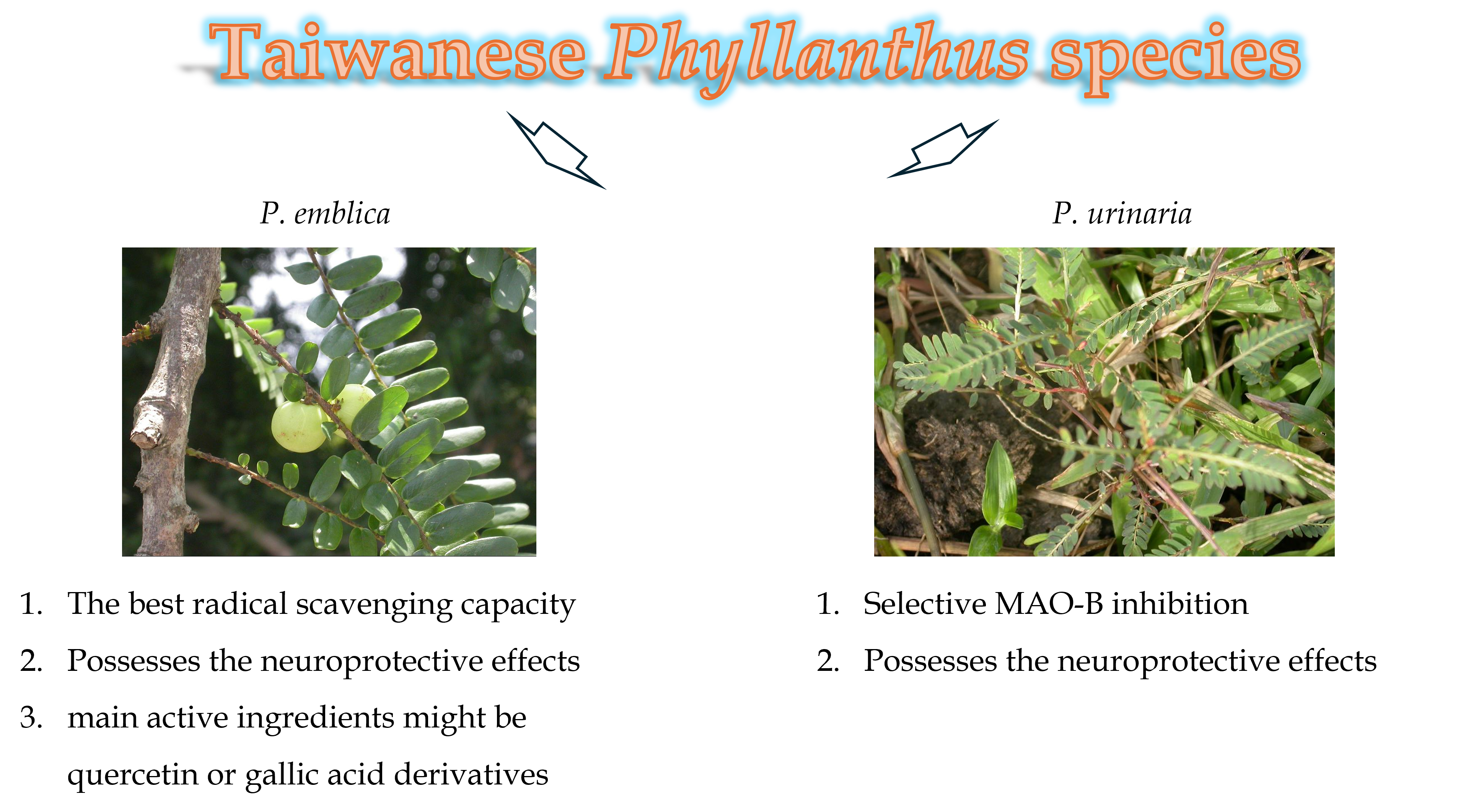
Ten Phyllanthus species (P. acidus, P. debilis, P. embergeri, P. emblica, P. hookeri, P. multiflorus, P. myrtifolius, P. tenellus, P. urinaria, P. urinaria subsp. nudicarpus) were collected and authenticated by Prof. Chao-Ying Lee from School of Pharmacy (China Medical University, Taiwan). The physical appearance of ten Phyllanthus species were shown in Figure 8. Collection details and voucher specimen information are consistent with previous Lee’s report [21].
The dried leaf powder (1 g) of each species was extracted with 10 mL of methanol via sonication for 90 min. The extracts were filtered with 0.45 μm filter and adjusted to a total volume of 10 mL to obtain a methanolic extract with a concentration of 100 mg/mL. To assess the antioxidant phytochemical content and radical scavenging activity, the methanolic extract was diluted with distilled water. For cell culture experiments, the extracts were dried under nitrogen and reconstituted in Dulbecco’s modified Eagles medium (DMEM).
4.2. Antioxidant Phytochemical Contents
The levels of antioxidant phytochemical contents, including total phenolics, flavonoids, and phenylpropanoids in Phyllanthus species, were quantified in triplicate using a microtiter spectrophotometric reader (PowerWave X340, Bio-Tek Inc., Winooski, VT, USA) following detailed procedures described in our previous report [16]. The used concentration of the methanolic extract was 1 or 5 mg/mL, respectively. Total phenolics were measured at 725 nm using Folic-Ciocalteu’s phenol (FCP) reagent and (+)-catechin standard (0–250 µg/mL). Total flavonoids were assessed at 415 nm using aluminium nitrate and quercetin standard (0–100 µg/mL). Total phenylpropanoids were determined at 525 nm using Arnow’s reagent (a mixture of sodium molybdate and sodium nitrite) and verbascoside standard (0–100 µg/mL). Total phenolic, flavonoids, or phenylpropanoids level of Phyllanthus species is expressed as the equivalent amount of (+)-catechin, quercetin, or verbascoside per gram of plant extract (mg catechin, quercetin or verbascoside /g sample), respectively.
4.3. Radical Scavenging Capacities Against Non-Biological Radicals In Vitro
The radical scavenging capacity of Phyllanthus species were evaluated using stable non-biological radicals such as DPPH and ABTS in triplicate, following our previous report [16]. The methanolic extracts of Phyllanthus species were used at six concentrations ranging from 0 to 5 mg/mL. DPPH scavenging activity was measured at 517 nm using (+)-catechin (0–100 µg/mL) as a reference standard and expressed in terms of mg catechin equivalents per gram of plant extract (abbreviated as CEDSC). ABTS scavenging activity was measured at 734 nm using trolox (0–400 µM) as a reference standard and expressed in terms of mmol trolox equivalents per gram of plant extract (abbreviated as TEAC).
4.4. Radical Scavenging Capacities Against Specific ROS In Vitro
ROS scavenging capacities of Phyllanthus species were evaluated using in vitro models that simulate ROS generation, including superoxide, hydrogen peroxide, and hydroxyl radicals. Their detailed procedures for these assays were described in our previous report [47]. Each assay was repeated in triplicate. The methanolic extract was used at six concentrations from 0 to 2.5 mg/mL. For superoxide scavenging capacity assay, an in vitro X-XO enzymatic system was used to generate superoxide. Nitroblue tetrazolium (NBT) was used as a chromogenic probe with a peak absorbance at 560 nm. SOD (0–500 mU/mL) served as a reference standard. The superoxide scavenging capacity of Phyllanthus species is expressed in terms of units of SOD equivalents per gram of plant extract (SOD-equivalent, U SOD/g sample). For H2O2 scavenging capacity assay, an in vitro H2O2-HRPase enzymatic system was used. Homovanillic acid (HVA) was used as a fluorescent probe and the fluorescence intensity was measured using a fluorescence spectrometric reader (FLX800, Bio-Tek Inc., Winooski, VT, USA) at an excitation wavelength of 315 nm and an emission wavelength of 425 nm. Trolox (0–300 µM) served as a reference standard. The H2O2 scavenging capacity of Phyllanthus species is expressed in terms millimole trolox equivalents per gram of plant extract (trolox-equivalent, mmol trolox/g sample). For hydroxyl radical scavenging capacity assay, an in vitro Fenton reaction system was used to generate hydroxyl radical. The end product produced by the generated hydroxyl radicals, after attacking a substrate 2-deoxyribose, can react with thiobarbituric acid (TBA) to produce TBA reactive substances (TBARS). TBARS has a peak absorbance at 532 nm. According to the calculation method published by Halliwell [23], multiplying the slope of concentration and absorbance value by the reaction rate between 2-deoxyribose and hydroxyl radical (3 × 10⁹ M-1 S-1) represents the hydroxyl radical scavenging rate of plant extract.
4.5. Relative Reducing Power (RPP) In Vitro
RRP values of Phyllanthus species was measured followed the procedure described in Vasyliev’s report [48]. Each assay was repeated in triplicate. The methanolic extracts were used at six concentrations from 0 to 1.0 mg/mL. In RRP assay, a reference standard ascorbic acid (0–20 µM) was used and potassium ferricyanide and ferric chloride were used as a chromogenic probe with a peak absorbance wavelength at 700 nm. The RRP value of Phyllanthus species is expressed in terms of micromole ascorbate equivalents per gram of plant extract (µmol ascorbate/g sample).
4.6. MAO Inhibitory Activities In Vitro
MAO inhibitory activities of Phyllanthus species were measured using a modified version of the procedure described in Haraguchi’s report [49]. Each assay was repeated in triplicate. The methanolic extract was used at six concentrations from 0 to 1.0 mg/mL. MAO enzyme solutions were prepared by homogenizing rat brain tissue in ice-cold 50 mM PBS solution (pH 7.4) containing 250 mM sucrose and 0.5 mM EDTA, followed by centrifugating at 14,000 rpm for 30 min using a standard sucrose gradient method. The resulting mitochondrial homogenates, containing active MAOs, were pre-incubated with either MAO-B inhibitor pargyline (2 µM) or MAO-A inhibitor clorgyline (2 µM) to establish selective assay models for MAO-A or MAO-B inhibitory activity, respectively. Homogenates pre-incubated with pargyline were considered to represent 100% MAO-A activity, while those pre-incubated with clorgyline represented 100% MAO-B activity. Kynuramine solution was used as the substrate for MAOs and its metabolite, 4-hydroxyquinoline, was quantified to generate a calibration curve at 0–100 µM by measuring the fluorescence intensity at an excitation wavelength of 315 nm and an emission wavelength of 380 nm. The IC50 values of Phyllanthus species against MAO-A or MAO-B were determined based on the inhibition of 4-hydroxyquinoline formation caused by the various concentrations of plant extract.
4.7. Rotenone-Induced PD Model in SH-SY5Y Cells
Human neuroblastoma SH-SY5Y cells, obtained from the American Type Culture Collection (ATCC; Manassas, Virginia, USA), are commonly used as an in vitro neuronal model for developing neuroprotective drugs against PD, due to their catecholaminergic neuronal properties, including the expression of enzymes involved in noradrenaline synthesis such as tyrosine hydroxylase and dopamine β-hydroxylase [19]. The cells were cultured in 25 cm3 culture flask with DMEM fortified with 10% FBS, penicillin (100 U/mL), and streptomycin (100 μg/mL). The cells were kept at 37 °C in a humidified incubator with 5% CO2. Upon reaching 70−80% confluency, the cells were trypsinized with fresh 0.25% trypsin solution (containing 0.53 mM EDTA) and then seeded into 96-well sterile clear-bottom plates (2 × 10⁴ cells/well), or 90-mm sterile clear-bottom dishes (4 × 10⁶ cells/dish) for the subsequent experiments. Twenty-four h after seeding, cells were exposed to rotenone (0.1–5 μM) for 48 h [20]. Then, the cell viability was assessed using 3-(4,5-dimethyl-thiazol-2-yl)-2,5-diphenyl-tetrazolium bromide (MTT) assay, following the detailed protocols described in our previous report [50]. Methanolic extracts of P. emblica or P. urinaria (1–100 μg/mL) were administered 2 h prior to rotenone exposure to evaluate their neuroprotective effects.
4.8. Intracellular ROS Levels in SH-SY5Y Cells
ROS-sensitive cell-permeant fluorophore DCFH-DA was used to label intracellular ROS. Once inside the cell, DCFH-DA is hydrolysed by intracellular esterases to form non-fluorescent DCFH, which is subsequently oxidized by intracellular ROS to generate the fluorescent compound DCF. Forty-eight h after rotenone treatment (with or without pretreatment with methanolic extracts of P. emblica or P. urinaria) in SH-SY5Y cells, the cultured medium in 96-well plate was replaced with 100 μM DCFH-DA solution. Cells were incubated at 37 °C in the dark for 30 min, DCF fluorescence was measured at Ex 485/Em 530 nm using a fluorescent microplate reader [50]. The data are expressed as a percentage relative to untreated cells, which served as the control group (designated as 100%).
4.9. Intracellular Oxidative Parameters in SH-SY5Y Cells
The oxidative parameters (including SOD, catalase, GSH and MDA) were assessed following the detailed protocol described in our previous report [50]. Forty-eight h after rotenone treatment (with or without pretreatment with methanolic extracts of P. emblica or P. urinaria), SH-SY5Y cells were harvested, suspended in ice-cold PBS, and centrifuged at 4 °C for 15 min. The resulting supernatant was then used to assess oxidative parameters.
4.10. Statistical Analysis
All data (including phytochemical contents, radical scavenging capacities, MAO inhibitory activities) are expressed as mean ± standard deviation (SD) from three repeated experiments. Similarly, the data on cell experiments (including cell viability, intracellular ROS levels, and the levels of oxidative parameters) are also presented as mean ± SD from four repeated experiments. Statistical analysis was conducted utilizing SPSS 20 for Windows (IBM Corporation, New York, USA). Data acquired were analysis using the one-way analysis of variance (ANOVA), and subsequent contrasts among groups were done using Turkey’s test. The mean difference was statistically significant when Probability (P) values were less than 0.05.
5. Conclusions
In conclusion, this comprehensive in vitro evaluation of ten Phyllanthus species extracts highlights their significant potential as neuroprotective agents. It provides compelling evidence that specific Phyllanthus species—namely P. emblica and P. urinaria—exert potent neuroprotective effects against rotenone-induced PD-like cellular damage. While P. emblica extract operates as a broad-spectrum antioxidant and dual MAO inhibitor, P. urinaria extract offers a targeted therapeutic approach via selective MAO-B inhibition. Both Phyllanthus species effectively mitigated rotenone-induced neurotoxicity in SH-SY5Y cells by restoring redox homeostasis, suppressing lipid peroxidation, and bolstering endogenous antioxidant defenses. These findings not only validate the ethnopharmacological use of these Phyllanthus plants but also position them as promising botanical resources for managing PD.
Author Contributions
Conceptualization, S.E.Y. and C.R.W.; methodology, C.R.W.; validation, C.R.W.; investigation, Y.C.W.; data curation, Y.C.W.; writing—original draft preparation, S.E.Y. and Y.C.W.; writing—review and editing, C.R.W.; project administration, J.C.L.; funding acquisition, J.C.L. All authors have read and agreed to the published version of the manuscript.
Funding
This research was funded by China Medical University, grant number CMU112-S-34 and CMU113-S-41.
Acknowledgments
The authors are deeply grateful to Prof. Chao-Ying Lee of School of Pharmacy at China Medical University for assisting in the collection and authentication of Phyllanthus plants, which greatly facilitated the progress of the study.
Conflicts of Interest
The authors declare no conflict of interest.
References
- Mao, X.; Wu, L.F.; Guo, H.L.; Chen, W.J.; Cui, Y.P.; Qi, Q.; Li, S.; Liang, W.Y.; Yang, G.H.; Shao, Y.Y.; et al. The Genus Phyllanthus: An Ethnopharmacological, Phytochemical, and Pharmacological Review. Evid Based Complement Alternat Med 2016, 2016, 7584952. [CrossRef]
- Bhattacharyya, J.; Saikia, L.; Kalita, V.; Dutta, P.P. An Updated Review on the Anti-Inflammatory Potential of Phyllanthus Genus. Chem Biodivers 2025, e202402483. [CrossRef]
- Pratima, H.; Shiraguppi, A.; Joojagar, P.; Shah, K.; Cheeraladinni, S.S.; Singh, P.S.; Mendem, S.K.; Chauhan, N.S. Phytochemical Profile and Hepatoprotective Potentiality of Phyllanthus Genus: a Review. J Pharm Pharmacol 2025, 77, 189-205. [CrossRef]
- Calixto, J.B.; Santos, A.R.; Cechinel Filho, V.; Yunes, R.A. A Review of the Plants of the Genus Phyllanthus: their Chemistry, Pharmacology, and Therapeutic Potential. Med Res Rev 1998, 18, 225-258. [CrossRef]
- Nisar, M.F.; He, J.; Ahmed, A.; Yang, Y.; Li, M.; Wan, C. Chemical Components and Biological Activities of the Genus Phyllanthus: A Review of the Recent Literature. Molecules 2018, 23. [CrossRef]
- Liu, J.; Lin, H.; McIntosh, H. Genus Phyllanthus for chronic hepatitis B virus infection: a systematic review. J Viral Hepat 2001, 8, 358-366. [CrossRef]
- Patel, J.R.; Tripathi, P.; Sharma, V.; Chauhan, N.S.; Dixit, V.K. Phyllanthus amarus: ethnomedicinal uses, phytochemistry and pharmacology: a review. J Ethnopharmacol 2011, 138, 286-313. [CrossRef]
- Geethangili, M.; Ding, S.T. A Review of the Phytochemistry and Pharmacology of Phyllanthus urinaria L. Front Pharmacol 2018, 9, 1109. [CrossRef]
- Tan, S.P.; Tan, E.N.; Lim, Q.Y.; Nafiah, M.A. Phyllanthus acidus (L.) Skeels: A review of its traditional uses, phytochemistry, and pharmacological properties. J Ethnopharmacol 2020, 253, 112610. [CrossRef]
- Prananda, A.T.; Dalimunthe, A.; Harahap, U.; Simanjuntak, Y.; Peronika, E.; Karosekali, N.E.; Hasibuan, P.A.Z.; Syahputra, R.A.; Situmorang, P.C.; Nurkolis, F. Phyllanthus emblica: a comprehensive review of its phytochemical composition and pharmacological properties. Front Pharmacol 2023, 14, 1288618. [CrossRef]
- Enemali, F.U.; Iteire, K.A.; Uweigho, R.E.; Blessing, O.; Judah, G.T. Aqueous leaf extract of Phyllanthus amarus protects against oxidative stress and misfiring of dopaminergic neurons in Paraquat-induced Parkinson's disease-like model of adult Wistar rats. J Chem Neuroanat 2024, 135, 102365. [CrossRef]
- Sarmah, D.; Verma, G.; Datta, A.; Vadak, N.; Chaudhary, A.; Kalia, K.; Bhattacharya, P. Phyllanthus emblica L. Regulates BDNF/PI3K Pathway to Modulate Glutathione for Mitoprotection and Neuroprotection in a Rodent Model of Ischemic Stroke. Cent Nerv Syst Agents Med Chem 2022, 22, 175-187. [CrossRef]
- Liu, T.; Kong, X.; Qiao, J.; Wei, J. Decoding Parkinson's Disease: The interplay of cell death pathways, oxidative stress, and therapeutic innovations. Redox Biol 2025, 85, 103787. [CrossRef]
- Kaur, M.; Aran, K.R. Unraveling the role of Nrf2 in dopaminergic neurons: a review of oxidative stress and mitochondrial dysfunction in Parkinson's disease. Metab Brain Dis 2025, 40, 123. [CrossRef]
- You, H.; Mariani, L.L.; Mangone, G.; Le Febvre de Nailly, D.; Charbonnier-Beaupel, F.; Corvol, J.C. Molecular basis of dopamine replacement therapy and its side effects in Parkinson's disease. Cell Tissue Res 2018, 373, 111-135. [CrossRef]
- Zhao, Z.W.; Chang, H.C.; Ching, H.; Lien, J.C.; Huang, H.C.; Wu, C.R. Antioxidant effects and phytochemical properties of seven Taiwanese Cirsium species extracts. Molecules 2021, 26. [CrossRef]
- Jost, W.H. A critical appraisal of MAO-B inhibitors in the treatment of Parkinson's disease. J Neural Transm (Vienna) 2022, 129, 723-736. [CrossRef]
- Tan, Y.Y.; Jenner, P.; Chen, S.D. Monoamine Oxidase-B Inhibitors for the Treatment of Parkinson's Disease: Past, Present, and Future. J Parkinsons Dis 2022, 12, 477-493. [CrossRef]
- Ioghen, O.C.; Ceafalan, L.C.; Popescu, B.O. SH-SY5Y cell line in vitro models for Parkinson disease research-old practice for new trends. J Integr Neurosci 2023, 22, 20. [CrossRef]
- Ibarra-Gutierrez, M.T.; Serrano-Garcia, N.; Orozco-Ibarra, M. Rotenone-induced model of Parkinson's disease: Beyond mitochondrial complex I inhibition. Mol Neurobiol 2023, 60, 1929-1948. [CrossRef]
- Lee, C.Y.; Peng, W.H.; Cheng, H.Y.; Chen, F.N.; Lai, M.T.; Chiu, T.H. Hepatoprotective effect of Phyllanthus in Taiwan on acute liver damage induced by carbon tetrachloride. Am J Chin Med 2006, 34, 471-482. [CrossRef]
- Ibitoye, O.B.; Aliyu, N.O.; Ajiboye, T.O. Protective Influence of Phyllanthus Muellarianus on Ciprofloxacin-Induced Neurotoxicity in Male Rats. J Diet Suppl 2020, 17, 321-335. [CrossRef]
- Halliwell, B.; Gutteridge, J.M.; Aruoma, O.I. The deoxyribose method: a simple "test-tube" assay for determination of rate constants for reactions of hydroxyl radicals. Anal Biochem 1987, 165, 215-219. [CrossRef]
- Sharma, P.; Mittal, P. Paraquat (herbicide) as a cause of Parkinson's Disease. Parkinsonism Relat Disord 2024, 119, 105932. [CrossRef]
- Duarte-Jurado, A.P.; Gopar-Cuevas, Y.; Saucedo-Cardenas, O.; Loera-Arias, M.J.; Montes-de-Oca-Luna, R.; Garcia-Garcia, A.; Rodriguez-Rocha, H. Antioxidant Therapeutics in Parkinson's Disease: Current Challenges and Opportunities. Antioxidants (Basel) 2021, 10. [CrossRef]
- Salem, M.A.; Khalil, H.M.A.; Manaa, E.G.; Bass, A.K.A.; Osama, N.; Samaka, R.M.; Ibrahim, M.T.; Hamdan, D.I. Antioxidant Potential of Selected Apiaceae Plant Extracts: A Study Focused on the Chemical Composition and Neuroprotective Effect of Coriandrum sativum L. Extract Against Lead (Pb)-Induced Neurotoxicity in Rats. Biol Trace Elem Res 2025, 10.1007/s12011-025-04627-9. [CrossRef]
- May, N.; de Sousa Alves Neri, J.L.; Clunas, H.; Shi, J.; Parkes, E.; Dongol, A.; Wang, Z.; Jimenez Naranjo, C.; Yu, Y.; Huang, X.F.; et al. Investigating the Therapeutic Potential of Plants and Plant-Based Medicines: Relevance to Antioxidant and Neuroprotective Effects. Nutrients 2023, 15. [CrossRef]
- Tahir, I.; Khan, M.R.; Shah, N.A.; Aftab, M. Evaluation of phytochemicals, antioxidant activity and amelioration of pulmonary fibrosis with Phyllanthus emblica leaves. BMC Complement Altern Med 2016, 16, 406. [CrossRef]
- Jain, N.K.; Singhai, A.K. Protective effects of Phyllanthus acidus (L.) Skeels leaf extracts on acetaminophen and thioacetamide induced hepatic injuries in Wistar rats. Asian Pac J Trop Med 2011, 4, 470-474. [CrossRef]
- Perera, D.; Soysa, P.; Wijeratne, S. Polyphenols contribute to the antioxidant and antiproliferative activity of Phyllanthus debilis plant in-vitro. BMC Complement Altern Med 2016, 16, 339. [CrossRef]
- Adedayo, B.C.; Ogunsuyi, O.B.; Akinniyi, S.T.; Oboh, G. Effect of Andrographis paniculata and Phyllanthus amarus leaf extracts on selected biochemical indices in Drosophila melanogaster model of neurotoxicity. Drug Chem Toxicol 2022, 45, 407-416. [CrossRef]
- Gidaro, M.C.; Astorino, C.; Petzer, A.; Carradori, S.; Alcaro, F.; Costa, G.; Artese, A.; Rafele, G.; Russo, F.M.; Petzer, J.P.; et al. Kaempferol as Selective Human MAO-A Inhibitor: Analytical Detection in Calabrian Red Wines, Biological and Molecular Modeling Studies. J Agric Food Chem 2016, 64, 1394-1400. [CrossRef]
- Chavarria, D.; Benfeito, S.; Soares, P.; Lima, C.; Garrido, J.; Serrao, P.; Soares-da-Silva, P.; Remiao, F.; Oliveira, P.J.; Borges, F. Boosting caffeic acid performance as antioxidant and monoamine oxidase B/catechol-O-methyltransferase inhibitor. Eur J Med Chem 2022, 243, 114740. [CrossRef]
- Liang, S.; Zhao, Z.; Liu, L.; Zhang, Y.; Liu, X. Research Progress on the Mechanisms of Protocatechuic Acid in the Treatment of Cognitive Impairment. Molecules 2024, 29. [CrossRef]
- Qian, Q.; Pan, J.; Yang, J.; Wang, R.; Luo, K.; Wu, Z.; Ma, S.; Wang, Y.; Li, M.; Gao, Y. Syringin: a naturally occurring compound with medicinal properties. Front Pharmacol 2024, 15, 1435524. [CrossRef]
- Oh, J.M.; Jang, H.J.; Kang, M.G.; Song, S.; Kim, D.Y.; Kim, J.H.; Noh, J.I.; Park, J.E.; Park, D.; Yee, S.T.; et al. Acetylcholinesterase and monoamine oxidase-B inhibitory activities by ellagic acid derivatives isolated from Castanopsis cuspidata var. sieboldii. Sci Rep 2021, 11, 13953. [CrossRef]
- Thong-Asa, W.; Wassana, C.; Sukkasem, K.; Innoi, P.; Dechakul, M.; Timda, P. Neuroprotective effect of gallic acid in mice with rotenone-induced neurodegeneration. Exp Anim 2024, 73, 259-269. [CrossRef]
- Chandrasekhar, Y.; Phani Kumar, G.; Ramya, E.M.; Anilakumar, K.R. Gallic Acid Protects 6-OHDA Induced Neurotoxicity by Attenuating Oxidative Stress in Human Dopaminergic Cell Line. Neurochem Res 2018, 43, 1150-1160. [CrossRef]
- Zaitone, S.A.; Ahmed, E.; Elsherbiny, N.M.; Mehanna, E.T.; El-Kherbetawy, M.K.; ElSayed, M.H.; Alshareef, D.M.; Moustafa, Y.M. Caffeic acid improves locomotor activity and lessens inflammatory burden in a mouse model of rotenone-induced nigral neurodegeneration: Relevance to Parkinson's disease therapy. Pharmacol Rep 2019, 71, 32-41. [CrossRef]
- Dos Santos Nunes, R.G.; Pereira, P.S.; Elekofehinti, O.O.; Fidelis, K.R.; da Silva, C.S.; Ibrahim, M.; Barros, L.M.; da Cunha, F.A.B.; Lukong, K.E.; de Menezes, I.R.A.; et al. Possible involvement of transcriptional activation of nuclear factor erythroid 2-related factor 2 (Nrf2) in the protective effect of caffeic acid on paraquat-induced oxidative damage in Drosophila melanogaster. Pestic Biochem Physiol 2019, 157, 161-168. [CrossRef]
- Park, S.E.; Sapkota, K.; Choi, J.H.; Kim, M.K.; Kim, Y.H.; Kim, K.M.; Kim, K.J.; Oh, H.N.; Kim, S.J.; Kim, S. Rutin from Dendropanax morbifera Leveille protects human dopaminergic cells against rotenone induced cell injury through inhibiting JNK and p38 MAPK signaling. Neurochem Res 2014, 39, 707-718. [CrossRef]
- Filomeni, G.; Graziani, I.; De Zio, D.; Dini, L.; Centonze, D.; Rotilio, G.; Ciriolo, M.R. Neuroprotection of kaempferol by autophagy in models of rotenone-mediated acute toxicity: possible implications for Parkinson's disease. Neurobiol Aging 2012, 33, 767-785. [CrossRef]
- Enogieru, A.B.; Haylett, W.; Hiss, D.C.; Ekpo, O.E. Regulation of AKT/AMPK signaling, autophagy and mitigation of apoptosis in Rutin-pretreated SH-SY5Y cells exposed to MPP(). Metab Brain Dis 2021, 36, 315-326. [CrossRef]
- Wang, Q.; Botchway, B.O.A.; Zhang, Y.; Liu, X. Ellagic acid activates the Keap1-Nrf2-ARE signaling pathway in improving Parkinson's disease: A review. Biomed Pharmacother 2022, 156, 113848. [CrossRef]
- Fu, R.H.; Hong, S.Y.; Chen, H.J. Syringin Prevents 6-Hydroxydopamine Neurotoxicity by Mediating the MiR-34a/SIRT1/Beclin-1 Pathway and Activating Autophagy in SH-SY5Y Cells and the Caenorhabditis elegans Model. Cells 2023, 12. [CrossRef]
- Krzysztoforska, K.; Mirowska-Guzel, D.; Widy-Tyszkiewicz, E. Pharmacological effects of protocatechuic acid and its therapeutic potential in neurodegenerative diseases: Review on the basis of in vitro and in vivo studies in rodents and humans. Nutr Neurosci 2019, 22, 72-82. [CrossRef]
- Wu, C.R.; Lin, W.H.; Hseu, Y.C.; Lien, J.C.; Lin, Y.T.; Kuo, T.P.; Ching, H. Evaluation of the antioxidant activity of five endemic Ligustrum species leaves from Taiwan flora in vitro. Food Chem 2011, 127, 564-571. [CrossRef]
- Vasyliev, G.S.; Vorobyova, V.I.; Linyucheva, O.V. Evaluation of reducing ability and antioxidant activity of fruit pomace extracts by spectrophotometric and electrochemical methods. J Anal Methods Chem 2020, 2020, 8869436. [CrossRef]
- Haraguchi, H.; Tanaka, Y.; Kabbash, A.; Fujioka, T.; Ishizu, T.; Yagi, A. Monoamine oxidase inhibitors from Gentiana lutea. Phytochemistry 2004, 65, 2255-2260. [CrossRef]
- Wu, C.R.; Chang, H.C.; Cheng, Y.D.; Lan, W.C.; Yang, S.E.; Ching, H. Aqueous extract of Davallia mariesii attenuates 6-hydroxydopamine-induced oxidative damage and apoptosis in B35 cells through inhibition of caspase cascade and activation of PI3K/AKT/GSK-3beta pathway. Nutrients 2018, 10. [CrossRef]
Figure 1.
The antioxidant phytochemical contents of the methanolic extracts of ten Phyllanthus species. (A) total phenol contents, (B) total flavonoid contents, (C) total phenylpropanoid contents. Results are expressed as the mean ± SD (n = 3).
Figure 1.
The antioxidant phytochemical contents of the methanolic extracts of ten Phyllanthus species. (A) total phenol contents, (B) total flavonoid contents, (C) total phenylpropanoid contents. Results are expressed as the mean ± SD (n = 3).

Figure 2.
The radical scavenging capacities of the methanolic extracts of ten Phyllanthus species. (A) DPPH assay, (B) TEAC assay, (C) RRP test. CEDSC: (+)-catechin-equivalent of DPPH radical scavenging capacity, DPPH: 1,1-diphenyl-2-picryhydrazyl, RRP: relative reducing power, TEAC: trolox equivalent antioxidant capacity. Results are expressed as the mean ± SD (n = 3).

Figure 3.
The ROS scavenging capacities of the methanolic extracts of ten Phyllanthus species. (A) O2- scavenging assay, (B) H2O2 scavenging assay, (C) OH- scavenging assay. ROS: reactive oxygen species. Results are expressed as the mean ± SD (n = 3).
Figure 3.
The ROS scavenging capacities of the methanolic extracts of ten Phyllanthus species. (A) O2- scavenging assay, (B) H2O2 scavenging assay, (C) OH- scavenging assay. ROS: reactive oxygen species. Results are expressed as the mean ± SD (n = 3).

Figure 4.
The MAO inhibitory activities of the methanolic extracts of ten Phyllanthus species. (A) MAO-A inhibitory activities, (B) MAO-B inhibitory activities, (C) Ratio of IC50 in MAO-B and IC50 in MAO-A. MAO: monoamine oxidase. Results are expressed as the mean ± SD (n = 3).
Figure 4.
The MAO inhibitory activities of the methanolic extracts of ten Phyllanthus species. (A) MAO-A inhibitory activities, (B) MAO-B inhibitory activities, (C) Ratio of IC50 in MAO-B and IC50 in MAO-A. MAO: monoamine oxidase. Results are expressed as the mean ± SD (n = 3).

Figure 5.
Cell viability of SH-SY5Y cells. (A) Treatment with rotenone (0.1-5.0 μM) alone. (B) Pretreatment with the methanolic extracts of two Phyllanthus plants (1, 10, 100 μg/mL) in rotenone (1.0 μM)-treated cells. Results are expressed as the mean ± SD (n = 4). * P < 0.05 and *** P < 0.001, compared with VEH/rotenone group. VEH: vehicle.

Figure 6.
Effects of the methanolic extracts of two Phyllanthus plants (1, 10, 100 μg/mL) on Intracellular ROS levels in rotenone (1.0 μM)-treated SH-SY5Y cells. Results are expressed as the mean ± SD (n = 4). *** P < 0.001, compared with VEH/rotenone group. VEH: vehicle.
Figure 6.
Effects of the methanolic extracts of two Phyllanthus plants (1, 10, 100 μg/mL) on Intracellular ROS levels in rotenone (1.0 μM)-treated SH-SY5Y cells. Results are expressed as the mean ± SD (n = 4). *** P < 0.001, compared with VEH/rotenone group. VEH: vehicle.

Figure 7.
Effects of the methanolic extracts of two Phyllanthus plants (1, 10, 100 μg/mL) on (A) SOD, (B) catalase, (C) GSH, and (E) MDA in rotenone (1.0 μM)-treated SH-SY5Y cells. Results are expressed as the mean ± SD (n = 4). ** P < 0.01, and *** P < 0.001, compared with VEH/rotenone group. VEH: vehicle.

Figure 8.
The physical appearance of Phyllanthus species. (A) P. acidus, (B) P. debilis, (C) P. embergeri, (D) P. emblica, (E) P. hookeri, (F) P. multiflorus, (G) P. myrtifolius, (H) P. tenellus, (I) P. urinaria, (J) P. urinaria subsp. nudicarpus.
Figure 8.
The physical appearance of Phyllanthus species. (A) P. acidus, (B) P. debilis, (C) P. embergeri, (D) P. emblica, (E) P. hookeri, (F) P. multiflorus, (G) P. myrtifolius, (H) P. tenellus, (I) P. urinaria, (J) P. urinaria subsp. nudicarpus.


Table 1.
Pearson correlation coefficients (r) between the contents of antioxidant phytoconstituents and radical scavenging capacities of Phyllanthus plants.
Table 1.
Pearson correlation coefficients (r) between the contents of antioxidant phytoconstituents and radical scavenging capacities of Phyllanthus plants.
| TF | TPP | DPPH | TEAC | O2˙ | H2O2 | OH˙ | RRP | |
|---|---|---|---|---|---|---|---|---|
| TP | 0.783** | 0.815** | 0.972*** | 0.903*** | 0.922*** | 0.956*** | 0.907*** | 0.860** |
| TF | 0.367 | 0.691* | 0.528 | 0.595 | 0.670* | 0.846** | 0.491 | |
| TPP | 0.896*** | 0.936*** | 0.872** | 0.862** | 0.605 | 0.934*** | ||
| DPPH | 0.967*** | 0.961*** | 0.981*** | 0.81** | 0.948*** | |||
| TEAC | 0.932*** | 0.966*** | 0.69* | 0.978*** | ||||
| O2˙ | 0.964*** | 0.722* | 0.945*** | |||||
| H2O2 | 0.776** | 0.946*** | ||||||
| OH | 0.644* |
* p < 0.05, ** p < 0.01, *** p < 0.001. DPPH: 1,1-diphenyl-2-picryhydrazyl; RRP: relative reducing power; TEAC: trolox equivalent antioxidant capacity; TF: total flavonoids; TP: total phenolics; TPPs: total phenylpropanoids.
Disclaimer/Publisher’s Note: The statements, opinions and data contained in all publications are solely those of the individual author(s) and contributor(s) and not of MDPI and/or the editor(s). MDPI and/or the editor(s) disclaim responsibility for any injury to people or property resulting from any ideas, methods, instructions or products referred to in the content. |
© 2026 by the authors. Licensee MDPI, Basel, Switzerland. This article is an open access article distributed under the terms and conditions of the Creative Commons Attribution (CC BY) license (http://creativecommons.org/licenses/by/4.0/).
Copyright: This open access article is published under a Creative Commons CC BY 4.0 license, which permit the free download, distribution, and reuse, provided that the author and preprint are cited in any reuse.


