

Submitted:
19 February 2026
Posted:
27 February 2026
You are already at the latest version
Abstract
Cytochrome P450 (CYP) 2B7 is currently classified as a pseudogene due to the presence of a premature stop codon in exon 7 that is predicted to result in a truncated protein. We hypothesized that in some individuals, CYP2B7 may code for a full length protein with enzyme activity similar to CYP2B6, as the result of a DNA sequence polymorphism in the CYP2B7 premature stop codon. Genomic DNA was prepared from our human liver bank (2 Hispanic, 4 African-Americans, and 47 Caucasians) and blood obtained from 20 African-Americans undergoing an unrelated clinical study. PCR was used to amplify and sequence the genomic region surrounding the CYP2B7 premature stop codon. All European-American and Hispanic DNA showed no variation of the stop codon sequence (TGA). In 9 of 24 African Americans, the first nucleotide of the stop codon was heterozygous (T/C), and in one of 24 African-American this nucleotide was homozygous (C/C) resulting in an arginine (codon CGA) instead of the stop codon. We next cloned the CYP2B7-R378 full-length cDNA by reverse transcription PCR from human liver mRNA. However, in addition to the CYP2B7-R378 cDNA, we also cloned a chimeric CYP2B6/CYP2B7-R378, which like CYP2B7-R378 was also predicted to code for a full-length protein. Both CYP2B7-R378 and CYP2B6 amino acid sequences could be aligned to the rabbit CYP2B4 crystal structure indicating that CYP2B7-R378 contained no mutations that would disrupt protein structure. Expression of cDNAs encoding CYP2B7-R378, CYP2B6/CYP2B7-R378, and CYP2B6 in 3T3 fibroblast cells showed immunodetectable CYP2B protein for each, but minimal (CYP2B6) or no bupropion hydroxylation activity. However, by use of the baculovirus-insect cell expression system we successfully showed bupropion hydroxylation activity in cells infected with CYP2B7-R378 and CYP2B6, but not in negative controls (including CYP2B7-X378). No activity was found for chimeric CYP2B6/CYP2B7-R378, although this result needs to be substantiated by further study. Expression of CYP2B7 mRNA was also demonstrated in most human tissues that also contained CYP2B6 mRNA, with particularly high expression in lung, liver and intestine. In conclusion, results indicate that CYP2B7 is expressed as an active cytochrome P450 in some individuals, particularly those of African-American ancestry. The presence of this polymorphic enzyme may explain some important differences in drug response and carcinogen activation related to race and/or ethnicity.
Keywords:
CYP2B7
; cytochrome P450 2B7
; CYP2B6
; pseudogene reactivation
; premature stop codon polymorphism
1. Introduction
Previous in vitro and in vivo studies in this and other laboratories indicates that there is substantial interindividual variability in the oxidation of drugs and other compounds by cytochrome P450 2B6. Various factors have been suggested to explain this large variability, including genetic factors or environmental influences. Genetic factors including single nucleotide polymorphisms (SNPs) in the CYP2B6 gene and effects on aberrant pre-mRNA splicing were investigated in Chapter 2. The role of alcohol as a CYP2B inducing agent was investigated in Chapter 3. However, CYP2B6 may not be the only CYP2B isoform contributing to the oxidation of drugs in people. A second CYP2B gene (CYP2B7P) has been identified in the human genome, and several studies have identified CYP2B7P mRNA in human and lung tissues indicating that this gene is transcriptionally functional (Yamano et al., 1989). However currently this gene is currently designated a pseudogene (as indicated by the “P” in CYP2B7) since all transcripts evaluated to date include the codon “tga” at the predicted amino acid position 378 that codes for a stop codon (X378) instead of the codon “cga” found at this same position in the CYP2B6 cDNA which codes for an arginine (R378). Consequently CYP2B7P, although present as a mRNA in tissues is predicted to result in a truncated (and presumably inactive) protein lacking the heme-binding domain in the C-terminal end of the protein (Yamano et al., 1989). However, apart from the premature stop codon at position 378, no other deleterious mutations have been found in the CYP2B7 cDNA. Consequently we hypothesized that one of the nucleotides in codon position 378 was polymorphic such that there are some individuals with an amino acid rather than a stop codon at this position. If so, CYP2B7 might code for an active CYP enzyme in these individuals with functional properties similar to CYP2B6, and contributing to the oxidation of drugs and other compounds normally attributed to CYP2B6.
2. Materials and Methods
2.1. Reagents
Bupropion hydrochloride and hydroxybupropion were kindly provided by Glaxo-Wellcome (Research Triangle Park, NC) and trazodone was provided by Mead Johnson (Evansville, IN). The antipeptide antibody to CYP2B6 used for immunoblotting (cat. # A143) was purchased from BD-Gentest (Woburn, MA). Chemical reagents were provided by their manufacturers or purchased from commercial sources. NADP+, isocitrate dehydrogenase, DL-isocitrate, and 50 mM potassium phosphate buffer (pH 7.5) were purchased from Sigma (St. Louis, MO).
Liver samples from donors with no known liver disease were obtained from the National Disease Research Interchange (Philadelphia, PA), the Liver Tissue Procurement and Distribution Service (Minneapolis, MN), or the International Institute for the Advancement of Medicine (Jessup, PA). The protein concentration of microsome samples was determined using the bicinchoninic acid protein assay (Pierce, Rockford, IL). Bovine serum albumin was used as a standard.
Cell homogenates of insect cells infected with baculovirus constructs containing the cDNA encoding CYP2B6 were purchased from BD-Gentest (Woburn, MA). Recombinant CYP2B6 was stored at -80°C, and thawed on ice immediately before use. Microsomal protein concentration and CYP2B6 content was provided by the manufacturer. RNA samples extracted from various human tissues and cancer cell lines were purchased from BD-Clontech and Ambion.
2.2. Sequencing of the CYP2B7 Gene Exon 7 Region
For this study, the CYP2B7 reference sequence was the gene sequence contained in the NCBI GenBank file NM_000767. Nucleotide position is given relative to the first nucleotide of the predicted translation start code ATG in exon 1. Genomic DNA was isolated from the same human liver tissues used to prepare microsomes and RNA with DNAzol (Invitrogen, Carlsbad, CA). The presence of possible nucleotide polymorphisms in the exon 7 region containing the predicted premature stop codon (X378) in the CYP2B7 gene were determined by polymerase chain reaction (PCR) amplification using primers 313 and 314 (Table 1) and direct sequence analysis. Primer homology and specificity were checked by use of the alignment and oligonucleotide analysis tools BLASTn (www.ncbi.nlm.nih.gov) and Vector NTI Suite 7.0 (Invitrogen, Carlsbad, CA) and confirmed by comparisons of derived DNA sequences with CYP2B6 and CYP2B7 gene sequences. PCR amplification was carried out in a 25 µl reaction mixture that contained Platinum Taq PCR supermix (Invitrogen, Carlsbad, CA), 0.2 µM of each primer and 50 ng of genomic DNA template. Denaturation was at 94 °C for 30 seconds, followed by annealing for 30 seconds with temperature specific to primer pairs, and extending at 72 °C for 1 minute with 40 cycles. These PCR products were purified using ExoSAP-IT PCR purification Kit (USB, Cleveland, OH) and then subjected to direct sequencing with the same primers used for PCR amplification (ABI PRISM 3100, Applied Biosystems, Foster City, CA).
2.3. Amino Acid Sequence Alignment of CYP2B7-R378 with Other Human CYPs
All amino acid sequences for human CYPs were obtained from the NCBI (National Center for Biotechnology Information) Genbank database. Amino acid sequences of CYP2B7 and other CYPs were compared using the Vector NT1 program. Further comparisons were performed by using conserved domain databases (CD, NCBI; ScanProsite database). Using the PROSITE motif and conserved domain (CD) databases, primary sequence conservation within the heme-ligand domain of CYP2B6 and CYP2B7 compared with other CYPs were evaluated.
2.4. Cloning of CYP2B6, CYP2B7-X378 and CYP2B7-R378 cDNA
Primers were designed specific to CYP2B6 and CYP2B7-X378 cDNAs incorporating the complete coding region. The nucleotide sequence “CACC” was added to the 5’ end of forward primer to allow for directional cloning into the pENTR cloning vector. The full-length cDNA of both genes were amplified by PCR using primers 373 and 216 for CYP2B6 and primers 217 and 346 for CYP2B7. CYP2B7 (X278) was amplified by PCR from cDNA generated by reverse transcription of total RNA isolated from our human liver bank, while CYP2B6 was subcloned by PCR from a plasmid generously donated by Dr Masa Negishi (NIEHS, NIH), each using the proofreading DNA polymerase enzyme Pfu Turbo (Stratagene). The PCR product was mixed with a directional TOPO pENTR vector (pENTR/D-TOPO cloning kit (Cat. No. K2400-20, Invitrogen) as described in the manual and transformed into E. coli. Transformants were selected on agar plates (50 ug/mL kanamycin). The identity and fidelity of isolated clones was verified by direct DNA sequencing.
After successfully cloning CYP2B7-X378, site-directed mutagenesis on the pENTR plasmid was then performed using primers 319 and 320 to generate CYP2B7-R378 using the QuikChange Site-directed Mutagenesis Kit (Stratagene). We chose to generate CYP2B7-R378 by this method rather than direct cloning from liver since our human liver bank is primarily from European-Americans which do not express CYP2B7-R378. Successful mutagenesis was verified by sequencing.
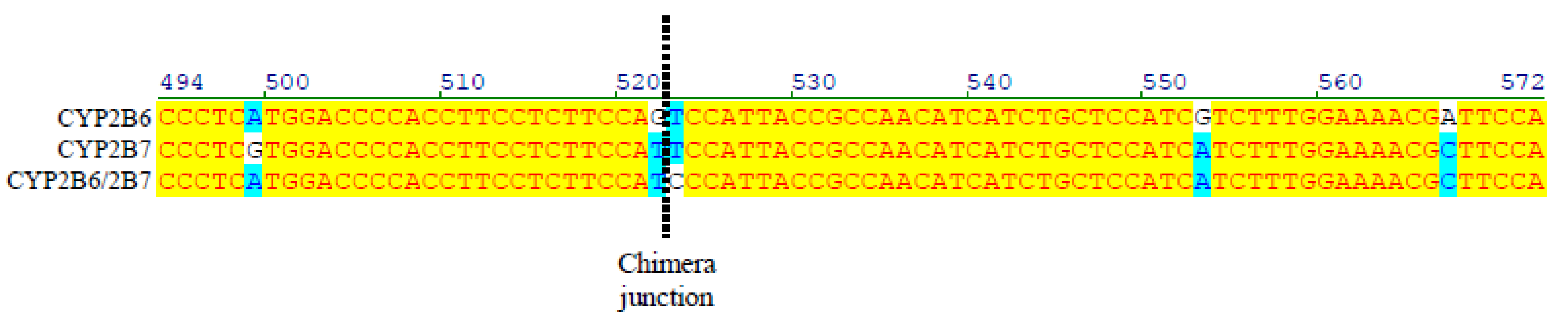
In the process of cloning CYP2B7, several clones were identified that appeared to be naturally occurring chimeras of CYP2B6 and CYP2B7 containing the 5’-end of CYP2B6 and the 3’-end of CYP2B7 as well as the stop codon at position 378 (Figure 3). One of these clones was selected and mutated at position 378 to create CYP2B6/2B7-R378 that was included in further analyses.
The resultant CYP2B6, CYP2B7-X378, CYP2B7-R378, and CYP2B6/2B7-R378 clones in pENTR vectors were then purified by miniprep (Qiagen).
2.5. Subcloning into pDEST47 Mammalian Expression Vector for Transfection of 3T3 Cells
The pENTR vector was used for initial cloning since it allows easy transfer to multiple types of expression vectors by simple recombination reactions. To enable expression in mammalian cells, pENTR vector inserts were subcloned into the pDEST47 vector (Invitrogen) using the LR Clonase enzyme mix (Invitrogen) followed by incubation at room temperature overnight. The next day, proteinase K solution was added and incubated at 37°C for 10 min then transformed in competent E. coli (DH5alpha). Transformants were selected on agar containing 100 ug/ml ampicillin. Large amounts (up to 100 ug) of each plasmid for transient transfection studies were generated by maxiprep (Qiagen).
The 3T3 cell line is a fibroblast cell line established from disaggregated tissue of an albino Swiss mouse (Mus musculus) embryo that is commonly used to express recombinant protein since it is easy to grow and transfect. This cell line was plated into 6 well plates in complete growth medium with antibiotics. When the cells reached nearly 40% confluency, the medium was changed to remove antibiotics. The next day, cells were transfected with 6 µg plasmid using the Lipofectamine 2000 transfection reagent according to the manufacturer's protocol (Cat#11668-019, Invitrogen). After 24 hr, the medium was changed and antibiotic added. The cells were allowed to grow in complete growth medium for a further 72 hr and then harvested, washed 3 times in phosphate buffer saline, lysed by sonication, and analyzed for expression of CYP2B protein by immunoblotting and bupropion hydroxylation activity by HPLC.
2.6. Subcloning into pDEST8 for Baculovirus-Insect Cell Expression
The Bac-to-Bac® Baculovirus Expression System was used as described in the manual. Briefly, pENTR clones were subcloned into the insect cell expression vector pDEST8 plasmid vector using the LR clonase enzyme mix with incubation at room temperature over night. The next day, proteinase K solution was added and incubated at 37C for 10 min and then transformed into competent E. coli (DH5a) cells. Transformants were selected on agar plates (100 ug/ml ampicillin) and inserts verified by colony PCR. Positive colonies were sequenced to confirm identity and then plasmid minipreps were obtained (Qiagen). Bacmids were then generated by transforming these plasmids into DH10Bac competent cells followed by selection on agar plates containing 50ug/ml kanamycin, 7ug/ml gentamicin, 10ug/ml tetracycline, 100ug/ml X gal and 100 ug/ml IPTG. After 48 hours incubation at 37C0, white colonies were screened for the presence of an insert by colony PCR. One positive colony for each was used for plasmid miniprep (Qiagen). Finally, the resultant bacmids were transfected into Sf9 cells on 6-well plates using Cellfectin reagent. Following incubation of insect cells for 7 days, 2 mL of P1 viral stock (media) was obtained and 1 mL of this was used to infect newly plated Sf9 cells. This was repeated 2 more times to obtain a high titer P3 viral stock. 100 uL of this P3 viral stock was then used to infect insect cells grown in 75 square cm flasks. Cells were harvested after 48 hr, washed 3 times in phosphate buffered saline, sonicated 3 times to disrupt cell membranes and then stored at -80°C until assay for bupropion hydroxylation activity.
2.7. Immunoblotting for CYP2B Proteins
Cell lysates were denatured for 5 minutes in 100 mM Tris buffer containing 10% glycerol, 2% beta-mercaptoethanol, 2% SDS and 5 µg ml-1 pyronin Y. (pH 6.8) at 100ºC. Protein was separated by SDS-PAGE in precast 7.5% polyacrylamide gels (Biorad, Hercules, CA). Samples were run at 100 V for one hour in 25 mM tris buffer/ 0.192M glycine/ 0.1% SDS buffer. Then samples were transferred to Immobilon-P (PVDF membrane) (Millipore) for one hour at 100 V in 25 mM Tris buffer/ 20% methanol. Blots were blocked with 0.5% powdered nonfat milk in TBS-Tween (0.15 M NaCl, 0.04 M TrisCl pH 7.7 and 0.06% Tween 20) for one hour at room temperature. Blots were then incubated with a 1:500 dilution of a polyclonal antipeptide CYP2B6 antibody (Stresser and Kupfer, 1999) (BD-Gentest) in TBS-Tween containing 0.1% BSA for one hour at room temperature. After washing in TBS-Tween, blots were incubated with a 1:500 dilution of HRP-conjugated goat anti-rabbit IgG (BD-Gentest) in 0.5% powdered nonfat in TBS-Tween for one hour at room temperature. Blots were rinsed with TBS-Tween and the Super Signal CL-HRP Substrate System (Pierce) was used for enhanced chemiluminescence detection with imaging using the ImageStation system (Kodak, Rochester, NY).
2.8. Bupropion Hydroxylation Assay
Hydroxylation of bupropion (Figure 1) was used to measure CYP2B enzyme function. Solutions of bupropion hydrochloride, hydroxybupropion were prepared in methanol. 1000 uM (final concentration) of bupropion was added to the incubation tubes and solvent evaporated to dryness in a 45°C vacuum oven. Samples using cell lysate of 3T3 transfected cells were incubated in duplicate. Incubation mixtures contained 50 mM potassium phosphate buffer (pH 7.5), 0.5 mM NADP+, an isocitrate/isocitric dehydrogenase regenerating system, and 5 mM MgCl2. Reactions were initiated by the addition of the protein and the final volume was 100 uL. A protein concentration of 0.5 mg ml-1 was used for the incubations (for cell lysate and cDNA-expressed CYP2B6 (Gentest). Incubations were performed in a shaking water bath for 20 minutes at 37°C, and terminated by addition of 50 ul of 1N HCl. Trazodone (25 uL of 125 uM solution in methanol) was added as an internal standard. The mixture was vortex mixed and spun at 14,000g for 10 minutes. Supernatants were injected into the HPLC for analysis. HPLC apparatus (Agilent 1100, Agilent Technologies, Palo Alto, CA) included a 250 x 4.6-mm C18 column (Synergy, Phenomenex, Torrence, CA) connected to a UV absorbance detector set at 214 nm. The HPLC mobile phase consisted of 20 mM phosphate buffer, pH 4.5, in water (solution A) and acetonitrile (B). The gradient program was 10% B (0–5 min), 10 to 20% B (5–10 min), 20 to 30 % B (10–15 min), 30 to 10% B (15–22 min), at a flow rate of 1 ml/min. The hydroxybupropion peak was identified by comparing the peak retention time of that obtained from the reaction mixture to that of the pure standard. In addition, incubations with uninfected insect cells, untransfected 3T3 cells, and without added protein were used as negative controls.
2.9. Quantitation of CYP2B6 and CYP2B7 mRNA in Different Human Tissues
RNA samples were treated with DNAse to remove contamination genomic DNA and reverse transcribed in a total volume of 20 uL including random hexamer primers, dNTP mix, 0.1 M DTT, Rnase inhibitor and Omniscript reverse transcriptase according to the manufacturer’s protocol (Qiagen). Reverse transcription proceeded at 37°C for 60 minutes and the resultant cDNA was stored at -20°C prior to quantitative PCR amplification. First strand cDNA was generated from total RNA (1 µg) with random hexamer (0.1 µg), and then diluted 10-fold with TE buffer and 10 µl of this used in real-time PCR amplification (7300 Real-Time PCR System, Applied Biosystems, Foster City, CA). Primer pairs specific to cDNA sequences for CYP2B6 mRNA, CYP2B7 mRNA and 18S rRNA (for normalization) are given in Table 1. Each 25 µl reaction mixture contained 12.5 µl of SYBR Green 2X master mix (Applied Biosystems, Foster City, CA), 0.2 µM of forward and reverse primers, and 0.5 µl of distilled water. The PCR conditions were as follows: initial denaturation was at 95°C for 10 min followed by 40 cycles of denaturation at 95°C (15 sec), annealing and extension at 60°C (1 min). Specificity of product formation was validated by purifying and sequencing PCR products when the primers were validated for the first time, and by evaluating melting temperature curves following each quantitative PCR run.
For each tissue, the following formula was used to determine the amount of each gene transcript being measured (mRNA) relative to the 18S rRNA control gene:
mRNA = A (CtmRNA – Ct18SrRNA)
where CtmRNA is the real-time PCR threshold cycle determined using the primer set for the particular gene transcript being assayed, Ct18SrRNA is the real-time PCR threshold cycle for the 18SrRNA primers, and A is the PCR amplification efficiency, which was set at 1.8 corresponding to an average amplification efficiency of 80% as determined experimentally.
2.10. Three-Dimensional (3D) Structure Prediction of CYP2B6 and CYP2B7 Primary Amino Acid Sequences by Comparative Modeling
The 3D crystal structure of rabbit CYP2B4 has been previously solved and visualized (Cn3D version 4.0, NCBI; Swiss-PdbViewer version 3.7, Swiss-Model). The 3D models of CYP2B6 and CYP2B7 were therefore constructed by submitting the primary amino acid sequences (targets) to the Swiss-Model protein alignment program (http://swissmodel.expasy.org//SWISS-MODEL.html). The CYP2B4 crystal structure was used as a template to align and match with the target proteins (CYP2B6 and CYP2B7). The next step was construction of a framework, which was computed by averaging the position of each atom in the target sequence, based on the location of the corresponding atoms in the template followed by loops and side chain predictions. After the construction was completed, the model was refined to check the bond geometry and to remove unfavorable non-bonded contacts by energy minimization (repair distorted geometries in the model).
The accuracies of the CYP2B6 and CYP2B7 3D models were evaluated by use of the WhatCheck program data generated from the Swiss-Model program output. The program generates root mean square (RMS) Z-scores for each criterion that is used in the evaluation. The RMS Z-scores represent the average statistical deviation from ideal stereochemical values for bond lengths and angles as determined by the WhatCheck program. RMS Z-scores close to 1.0 are considered ideal (Hooft et al., 1996).
3. Results
3.1. Sequence of the Genomic Region Surrounding the Premature Stop Codon (X378) in Exon 8 of the CYP2B7 Gene
Exon 8 of the CYP2B7 gene containing the premature stop codon was sequenced using genomic DNA obtained from our human liver bank (2 Hispanic, 4 African-Americans, and 47 Caucasians) and also blood obtained from 20 African-Americans undergoing an unrelated clinical study. All Caucasian and Hispanic DNA showed no variation of the stop codon sequence (codon 378, TGA = X378) (Figure 2A). However, in 9 of 24 African-Americans, the first nucleotide of the stop codon was heterozygous (T/C) (Figure 2B), and in one of 24 African-Americans this nucleotide was homozygous (C/C) resulting in an arginine (codon 378, CGA = R378) (Figure 2C) instead of the stop codon.
3.2. Alignment of Amino Acid Sequence of Human CYP2B7-R378 and Chimeric CYP2B6/2B7-R378 with Other CYPs
Alignment of the predicted amino acid sequence of CYP2B7-R378 with chimeric CYP2B6/2B7-R378, CYP2B6 and other human family 2 and 3 CYPs, as well as with rabbit CYP2B4 is shown in Figure 3. A chimeric CYP2B6/2B7-R378 cDNA was identified during cloning of CYP2B7 from pooled human liver RNA and based on the nucleotide sequence alignment shown in Figure 4 and amino acid sequence alignment shown in Figure 3 contains nucleotide sequence from the 5’ end of CYP2B6 mRNA and the 3’ end of CYP2B7 mRNA. Rabbit CYP2B4 is the only 2B subfamily CYP that has a publicly available solved crystal structure.
Figure 3.
Amino acid sequence alignment of CYP2B7 (X378R) with other human CYP2 and CYP3 family isoenzymes. Also included is rabbit CYP2B4 which is in the same subfamily as CYP2B6 and CYP2B7 and also has publicly available crystal structure data. Highly conserved residues are indicated by boxes. The CYP2B7 X378R site and the heme-ligand binding domain are indicated by arrows.
Figure 3.
Amino acid sequence alignment of CYP2B7 (X378R) with other human CYP2 and CYP3 family isoenzymes. Also included is rabbit CYP2B4 which is in the same subfamily as CYP2B6 and CYP2B7 and also has publicly available crystal structure data. Highly conserved residues are indicated by boxes. The CYP2B7 X378R site and the heme-ligand binding domain are indicated by arrows.
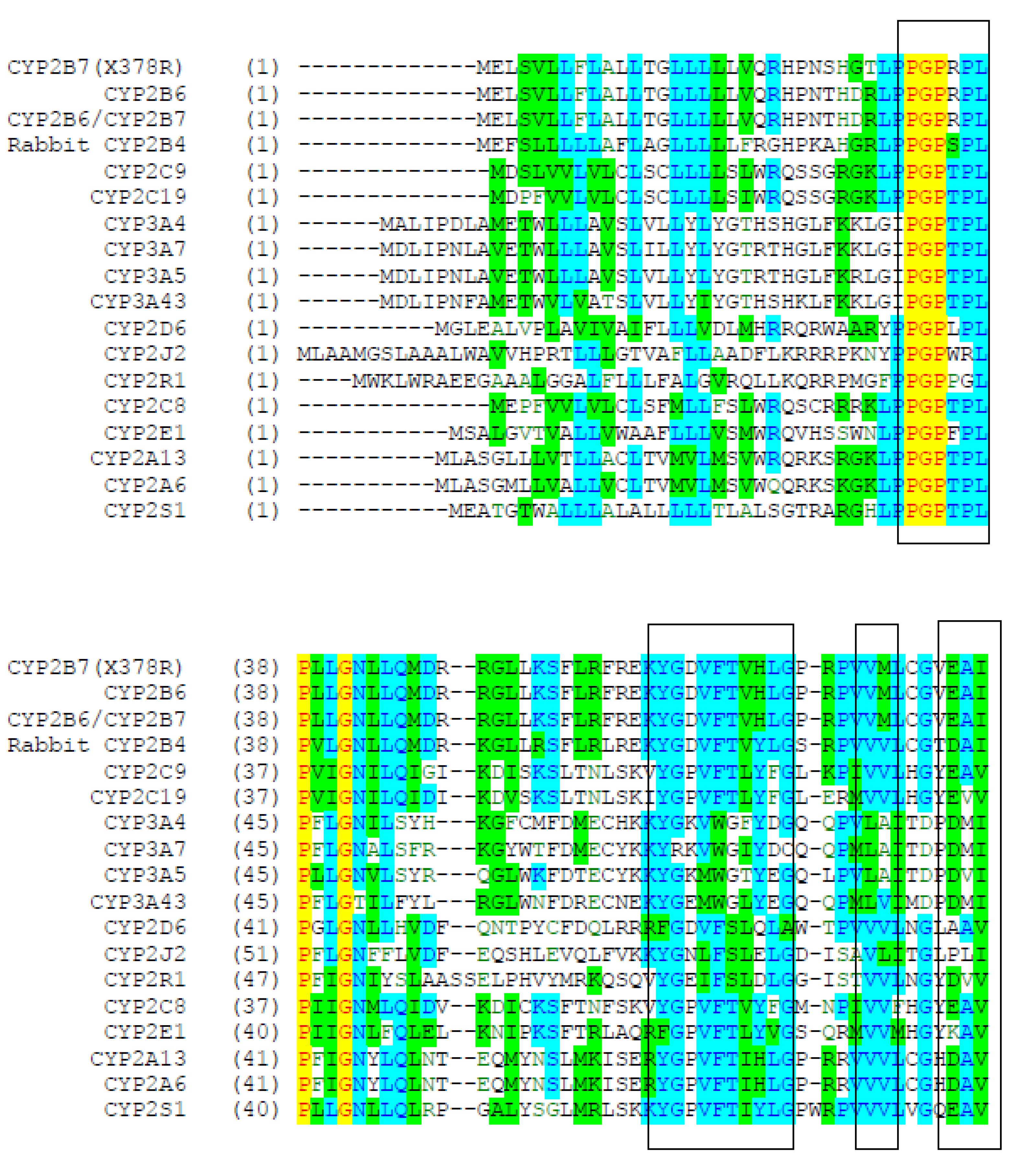
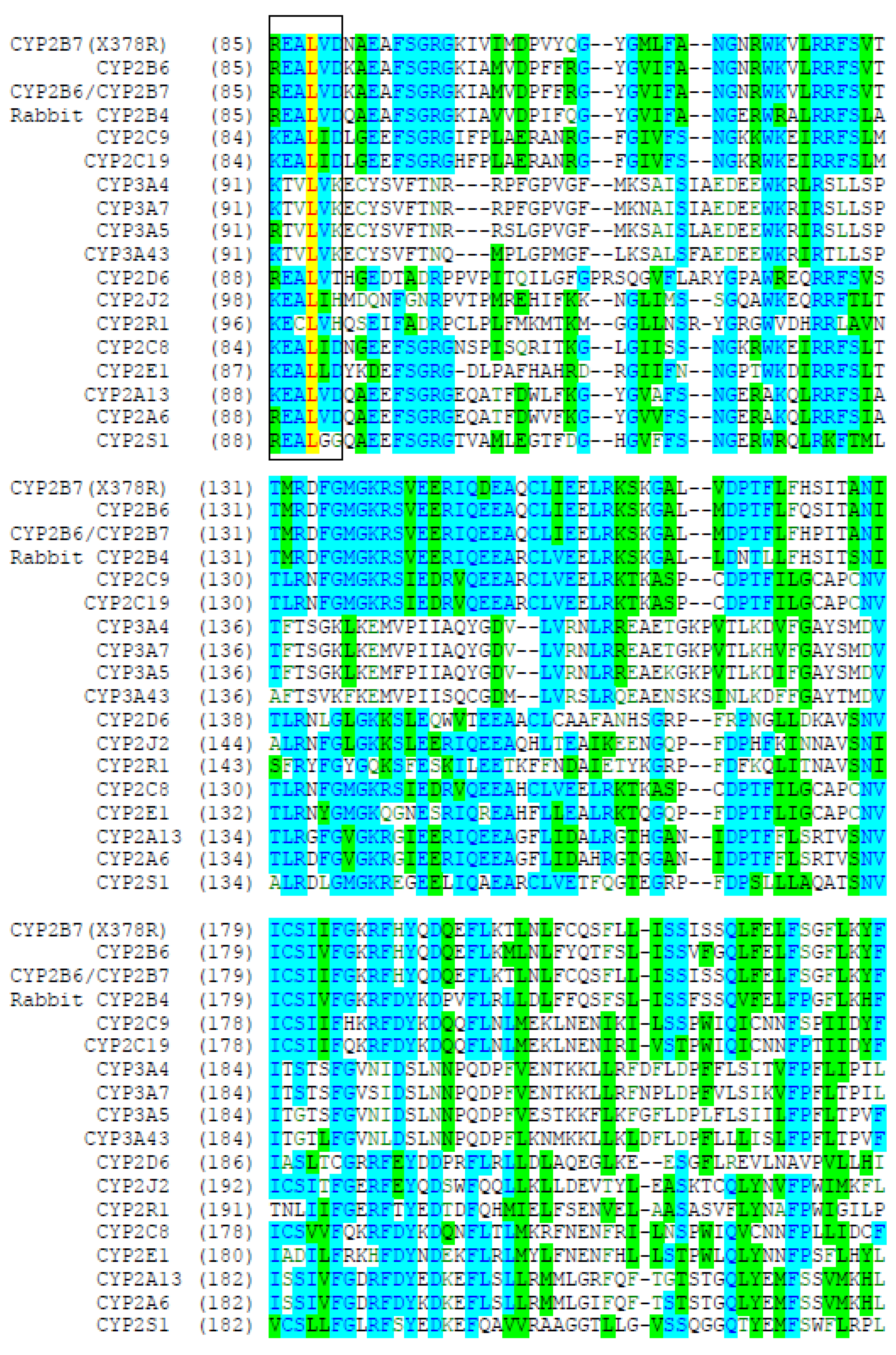
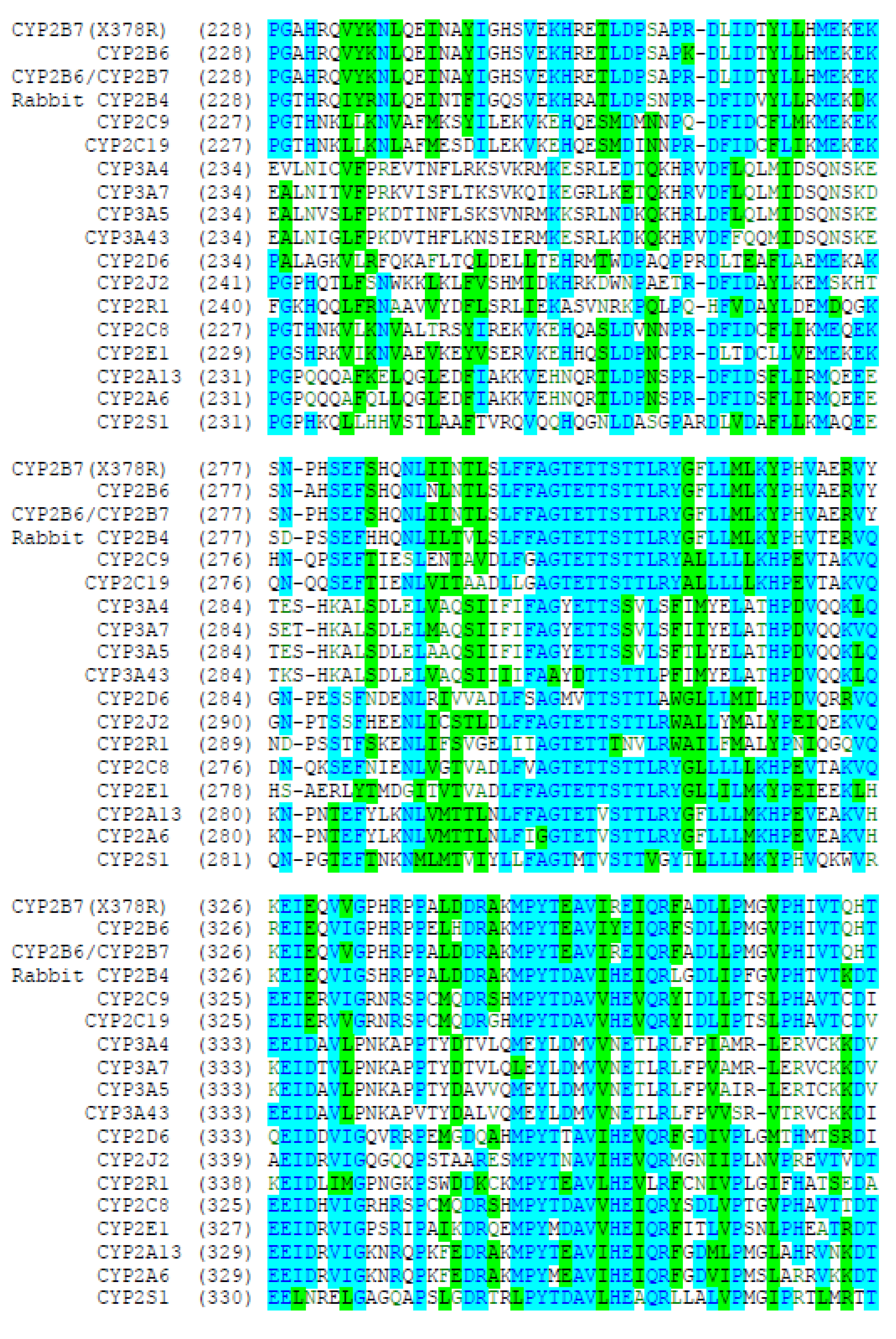
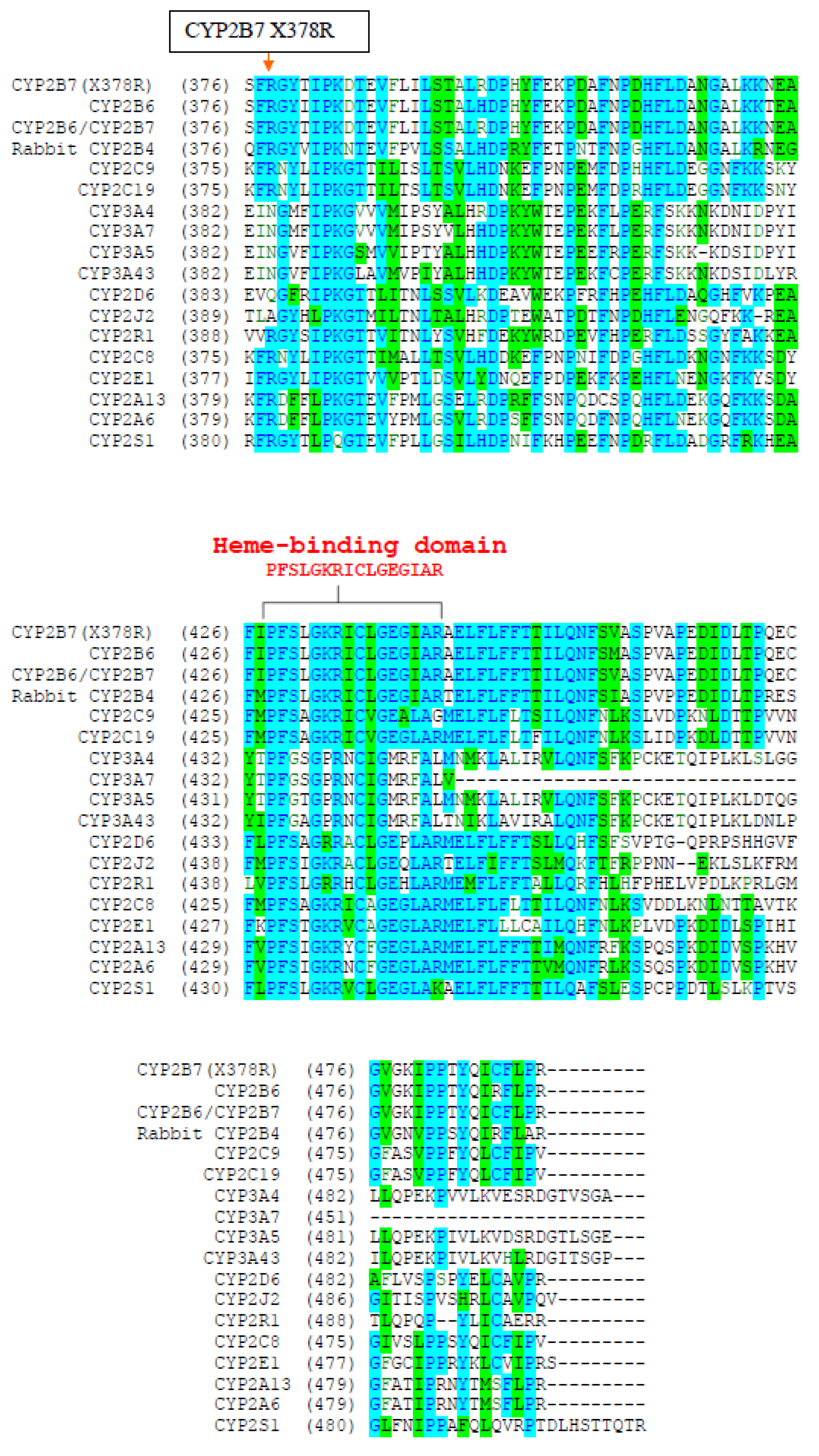
Figure 4.
Nucleotide sequence alignment of CYP2B6/CYP2B7 chimeric cDNA with cDNAs for CYP2B6 and CYP2B7. The dotted line indicates the apparent junction between the 5’-CYP2B6 and 3’-CYP2B7 portions of the cDNA.
Figure 4.
Nucleotide sequence alignment of CYP2B6/CYP2B7 chimeric cDNA with cDNAs for CYP2B6 and CYP2B7. The dotted line indicates the apparent junction between the 5’-CYP2B6 and 3’-CYP2B7 portions of the cDNA.
The analysis indicated that there was very high amino acid identity (92.3%) and similarity (96.7%) of CYP2B7-R378 with CYP2B6. Of note, the amino acid created at position 378 by the CYP2B7 polymorphism (arginine) as well as several surrounding residues (FRGY) appear to be absolutely conserved in CYP2B6, CYP2E1, CYP2S1 and rabbit CYP2B4. Furthermore, the sequence including the C-terminal heme-ligand binding domain (PFSLGKRICLGEGIAR) was absolutely conserved in CYP2B6 and rabbit CYP2B4. This region is lacking in CYP2B7-X378 as the result of the premature stop codon.
3.3. Prediction of a 3D Model for CYP2B7-R378 Compared with CYP2B6
As shown in Figure 5, structural models could be predicted for CYP2B7-R378 and CYP2B6 by 3-dimensional alignment of the primary amino acid to the published crystal structure of rabbit CYP2B4. As shown in Table 2, RMS Z-scores for all except one of the criteria evaluated (B-factor distribution) were between 0.5 and 2.0 indicating reasonable fit of the model to the data. RMS Z-score values for CYP2B7-R378 were identical to values obtained for CYP2B6. Furthermore, the predicted structure of the heme-ligand binding domain for CYP2B7-R378 was identical to that of CYP2B6 (indicated in yellow in Figure 5).
3.4. Expression of CYP2B7-R378 and Chimeric CYP2B6/CYP2B7-R378 in 3T3 Cells
3T3 cells were transfected with plasmids containing cDNAs encoding CYP2B7-R378 and CYP2B6/2B7-R378 along with CYP2B6 (as positive control). As seen in Figure 6, immunoblots probed with CYP2B6 antibody of cell homogenates transfected with CYP2B6, CYP2B7-R378 and CYP2B6/2B7-R378R showed bands of a similar size to CYP2B6-Gentest (positive blotting control), while no bands were detected for untransfected 3T3 cells. However, when these cell homogenates were screened for bupropion hydroxylation enzyme activity (Figure 7) only very low activity was found for transfected CYP2B6 compared with CYP2B6–Gentest (positive activity control), while no activity was detected for other cell homogenates.
3.5. Expression of CYP2B7-R378 and Chimeric CYP2B6/CYP2B7-R378 in Sf9 Insect Cells
Insect cells were infected with baculovirus constructs containing cDNAs encoding CYP2B7-R378, CYP2B7-X378, and CYP2B6/2B7-R378 along with CYP2B6 (as positive control). Cells were harvested after 48 hr and homogenates screened for bupropion hydroxylation activity. Recombinant CYP2B6 from Gentest was used as a positive activity control. As shown in Figure 8, HPLC peaks with retention times similar to hydroxybupropion standard were identified in CYP2B6-Gentest as well as cells infected with CYP2B6 and CYP2B7-R378, but not CYP2B7-X378 and CYP2B6/2B7-R378. Negative control incubations with uninfected insect cells and also without any added cell homogenate also showed no hydroxybupropion peak. Optimization studies conducted to determine the optimum amount of virus to add to insect cell cultures also showed differences in bupropion hydroxylation activity that was proportional to the amount of virus added (data not shown).
3.6. Expression of CYP2B6 and CYP2B7 mRNA in Different Human Tissues
CYP2B6 and CYP2B7 mRNA levels were quantified using RNA isolated from a range of human tissues and cancer cell lines. As seen in Figure 9, CYP2B6 mRNA was found in the highest levels in the liver followed by small intestine, colon, prostate, lung, colon adenocarcinoma, uterus, trachea, kidney, and heart (10 highest in descending order). In contrast, CYP2B7 mRNA showed highest expression in the lung followed by liver, small intestine, prostate, uterus, trachea, testis, colon, kidney, colon adenocarcinoma, and pancreas (10 highest in descending order). CYP2B7 mRNA appeared to be more highly expressed (>2 times) than CYP2B6 mRNA in lung, trachea, uterus and testis, while CYP2B6 mRNA was more highly expressed in colon and heart. In addition, CYP2B7 mRNA expression was detected in salivary gland, spleen, thymus and ovary without detectable expression of CYP2B6 mRNA, while CYP2B6 mRNA was only detected in adipose tissue, and skeletal muscle. Finally, expression of CYP2B6 and CYP2B7 was not detected in thyroid, breast, breast cancer, stomach, fetal brain, and placenta.
4. Discussion
The objective of this study was to determine whether CYP2B7 might contribute to oxidation of drugs and other compounds known to be metabolized by CYP2B6. Although the CYP2B7 gene contains a premature stop codon, we had hypothesized that one of the nucleotides in this stop codon is polymorphic and may code for an amino acid in some people. This hypothesis was proven correct in that a T/C polymorphism was identified in the first nucleotide position of stop codon resulting in the amino acid arginine. Arginine is likely the ancestral amino acid since both CYP2B6 and rabbit CYP2B4 have arginine at this position.
Although we now had a predicted amino acid sequence identical in length to that of CYP2B6 (491 amino acids), this did not prove that CYP2B7 was a functional enzyme since other amino acid differences in CYP2B7 might inactivate the enzyme. Consequently we used several approaches to prove that CYP2B7-R378 was functional. Firstly, we performed a 3-dimensional alignment of the CYP2B7 amino acid sequence to the solved crystal structure of CYP2B4. This was compared to similar alignment of CYP2B6 to CYP2B4. The results indicated that CYP2B7 fit equally well as CYP2B6 to the CYP2B4 crystal structure. In particular, none of the amino acid differences between CYP2B6 and CYP2B7 appeared to be sufficient to adversely affect the model fit. Next, cDNAs encoding CYP2B7-R378 were expressed in 3T3 cells. Although we were able to identify bands consistent with recombinant CYP2B7 protein by immunoblotting, there was not detectable bupropion hydroxylation activity. However, since 3T3 cells transfected with CYP2B6 showed very low activity (compared with recombinant CYP2B6 from a commercial source), this suggested that the 3T3 cell system was not ideal for expressing CYPs. It is possible that 3T3 cells do not express sufficient amounts of accessory proteins such as NADPH-oxidoreductase. Consequently, our next step was to use the baculovirus-insect cell system, which is used extensively to express human CYPs since large amounts of protein can be produced and insect cells have high amounts of endogenous NAPDH oxidoreductase. Using this system we were successful in producing insect cell homogenates from CYP2B7-R378 cDNA that clearly had bupropion hydroxylation activity. Since these activity studies were conducted using an initial screening culture, studies are currently being performed to scale-up the insect cell culture to enable measurement of P450 concentration by the reduced carbon monoxide spectrum since this technique requires much larger amounts of recombinant protein. Also planned are evaluation of reactivity of CYP2B7 towards other CYP2B6 substrates such as efavirenz, nicotine, and NNK (tobacco carcinogen).
In the process of cloning CYP2B7 from pooled human liver cDNA, we also identified clones that appeared to be chimeras of CYP2B6 and CYP2B7. Several possibilities may explain these clones. Firstly they may be an artifact of the PCR used to amplify the cDNA. This seems unlikely since such artifacts usually produce deletions and insertions. Secondly, they may be the result of trans-splicing of CYP2B6 and CYP2B7 pre-mRNA. This has been described previously for CYP3A4 and CYP3A5. Thirdly it may represent a genomic rearrangement in some individuals resulting in fusion of the CYP2B6 and CYP2B7 genes. This possibility (versus trans-splicing) could be excluded by genomic PCR across the chimera junction. Initial evaluation of the CYP2B6/2B7-R378 in 3T3 cells showed detectable protein, but expression in insect cells did not result in bupropion hydroxylation activity. These insect cell expression studies should be repeated, and reduced carbon monoxide P450 spectrum obtained to verify this result. Furthermore it should be possible to design quantitative RT-PCR primers to determine whether CYP2B6/2B7 mRNA is expressed in all or only some livers in our liver bank, and also whether the amount of CYP2B6/2B7 is comparable to CYP2B6 and CYP2B7 mRNA.
Comparative evaluation of the expression of CYP2B6 and CYP2B7 mRNA in a panel of human tissues indicate that there is similar distribution of the CYPs with some notable exceptions. CYP2B7 appears to be more highly expressed in the airway tissues including lung and trachea as well as reproductive tissues including uterus and testis than CYP2B6. CYPs are important in the activation of carcinogens and it is possible that people with the CYP2B7-R378 polymorphism may be more prone to cancer of the airways, uterus, and testis than those with the inactivating variant CYP2B7-X378.
Perhaps the most interesting outcome of this work was the finding of the CYP2B7-R378 activating mutation in African-Americans but not in European-Americans. This finding needs to be confirmed and extended to other ethnic groups by genotyping of DNA from a larger number of representative individuals. Several diseases and genetic predispositions might be attributed to this difference between African-Americans and other ethnic groups. Firstly, prostate cancer is almost twice as prevalent in African-Americans compared with other ethnic groups, and both CYP2B6 and CYP2B7 are highly expressed in prostate. An activating mutation would result in greater carcinogen formation in the prostate. Secondly, cigarette smoking and related cancers (particularly lung cancer) is also high amongst African-Americans and this has been attributed in part to higher metabolism of nicotine by CYP including CYP2B6. An activating mutation of CYP2B7 would promote higher nicotine metabolism and higher cigarette consumption to maintain nicotine levels. CYP2B7 may also directly influence lung cancer by carcinogen activation in the lung. Furthermore, faster metabolism of bupropion (a smoking cessation treatment) may result in lower bupropion blood levels and less effective treatment in African-Americans trying to quite smoking with this drug.
In conclusion, results of this study indicate that CYP2B7 is likely an active drug metabolizing enzyme particularly in a significant number of African-American individuals that carry the CYP2B7-R378 polymorphism. The presence of this polymorphism is likely to be a useful genetic marker identifying individuals at higher risk for certain cancers, as well as individuals with altered metabolism of drugs metabolized by CYP2B7.
Ethics Approval and Consent to Participate
Human liver tissues from donors with no known liver disease were obtained from established tissue procurement organizations, and blood samples were obtained from African-American participants undergoing an unrelated clinical study. All human materials were handled in accordance with applicable ethical standards and regulations. Approval by the relevant ethics committee/institutional review board and written informed consent (where applicable) were obtained for use of these samples.
Consent for Publication
Not applicable.
Availability of Data and Materials
All data generated or analyzed during this study are included in this manuscript and its figures/tables. Any additional materials referenced (e.g., primer sequences and constructs) are described within the Methods and Tables.
Competing Interests
The author(s) declare that they have no competing interests.
Author Contributions
A.H.A. conceived and designed the study. A.H.A. performed the experiments,
analyzed and interpreted the data, and wrote the manuscript. All authors read and approved the final
manuscript.
Funding
This research received no external funding.
Acknowledgments
The authors thank Glaxo-Wellcome for providing bupropion hydrochloride and hydroxybupropion and Mead Johnson for providing trazodone. The authors also acknowledge BD-Gentest for providing reagents and commercial recombinant CYP2B6 material used as a positive control, and the tissue procurement organizations for providing donor liver samples.
Clinical Trial Registration
Not applicable.
Permissions / Licenses for Reused Material
All figures, tables, and text in this manuscript are original to the authors, unless otherwise stated. Where proprietary reagents or materials were obtained from commercial sources, they are cited in the Methods.
Use of Artificial Intelligence
No artificial intelligence tools were used to generate the research data, results, or conclusions reported in this manuscript.
References
- Yamano, S.; Nhamburo, P. T.; Aoyama, T.; Meyer, U. A.; Inaba, T.; Kalow, W.; Gelboin, H. V. cDNA cloning and sequence and expression of human cytochrome P450 IIB1. Journal of Biological Chemistry 1989, 264, 8688–8694. [Google Scholar]
- Rendic, S.; Guengerich, F. P. Survey of human oxidoreductases and cytochrome P450 enzymes involved in the metabolism of xenobiotic and endogenous compounds. Chemical Research in Toxicology 2015, 28, 38–42. [Google Scholar] [CrossRef]
- Zanger, U. M.; Schwab, M. Cytochrome P450 enzymes in drug metabolism: Regulation of gene expression, enzyme activities, and impact of genetic variation. Pharmacology & Therapeutics 2013, 138, 103–141. [Google Scholar] [CrossRef]
- Wang, H.; Tompkins, L. M. CYP2B6: New insights into a historically overlooked cytochrome P450 isozyme. Current Drug Metabolism 2008, 9, 598–610. [Google Scholar] [CrossRef]
- Hesse, L. M.; He, P.; Krishnaswamy, S.; Hao, Q.; Hogan, K.; von Moltke, L. L.; Greenblatt, D. J. Pharmacogenetic determinants of interindividual variability in bupropion hydroxylation by cytochrome P450 2B6. Pharmacogenetics 2004, 14, 225–238. [Google Scholar] [CrossRef]
- Faucette, S. R.; Hawke, R. L.; Lecluyse, E. L.; Shord, S. S.; Yan, B.; Laethem, R. M.; Lindley, C. M. Validation of bupropion hydroxylation as a selective marker of human cytochrome P450 2B6 catalytic activity. Drug Metabolism and Disposition 2000, 28, 1222–1230. [Google Scholar] [CrossRef]
- Turpeinen, M.; Zanger, U. M.; Pelkonen, O. Functional expression, inhibition and induction of CYP2B6. Current Drug Metabolism 2006, 7, 705–714. [Google Scholar] [CrossRef]
- Stresser, D. M.; Kupfer, D. Monospecific antipeptide antibody to cytochrome P450 2B6. Drug Metabolism and Disposition 1999, 27, 517–525. [Google Scholar] [CrossRef]
- Ekins, S.; de Groot, M. J.; Jones, J. P. Pharmacophore and three-dimensional quantitative structure activity relationship methods for modeling cytochrome P450 active sites. Drug Metabolism and Disposition 2001, 29, 936–944. [Google Scholar]
- Scott, E. E.; White, M. A.; He, Y. A.; Johnson, E. F.; Stout, C. D.; Halpert, J. R. Structure of mammalian cytochrome P450 2B4 complexed with 4-(4-chlorophenyl)imidazole. Journal of Biological Chemistry 2004, 279, 27294–27301. [Google Scholar] [CrossRef]
- Hooft, R. W. W.; Vriend, G.; Sander, C.; Abola, E. E. Errors in protein structures. Nature 1996, 381, 272. [Google Scholar] [CrossRef]
- Guengerich, F. P. Cytochrome P450 and chemical toxicology. Chemical Research in Toxicology 2008, 21, 70–83. [Google Scholar] [CrossRef]
- Ingelman-Sundberg, M. Pharmacogenetics of cytochrome P450 and its applications in drug therapy. Nature Reviews Drug Discovery 2004, 3, 859–870. [Google Scholar]
- Sim, S. C.; Ingelman-Sundberg, M. The human cytochrome P450 allele nomenclature website. Methods in Molecular Biology 2010, 578, 307–318. [Google Scholar]
- Meyer, U. A. Pharmacogenetics—five decades of therapeutic lessons from genetic diversity. Nature Reviews Genetics 2004, 5, 669–676. [Google Scholar] [CrossRef]
- Zhou, S. F.; Liu, J. P.; Chowbay, B. Polymorphism of human cytochrome P450 enzymes and its clinical impact. Drug Metabolism Reviews 2009, 41, 89–295. [Google Scholar] [CrossRef]
- Nelson, D. R. The cytochrome P450 homepage. Human Genomics 2009, 4, 59–65. [Google Scholar] [CrossRef]
- Gufford, B. T.; Lu, J. B.; Metzger, I. F.; Jones, D. R.; Desta, Z.; Skaar, T. C. Bupropion metabolism by CYP2B6 and CYP2C19 in human liver microsomes. Journal of Pharmacology and Experimental Therapeutics 2016, 356, 491–499. [Google Scholar]
- Zhang, H.; Sridar, C.; Kenaan, C.; Amunugama, H.; Ballou, D. P.; Hollenberg, P. F. Polymorphic variants of CYP2B6. Archives of Biochemistry and Biophysics 2011, 507, 240–248. [Google Scholar]
- Yamazaki, H.; Inui, Y.; Wrighton, S. A.; Guengerich, F. P.; Shimada, T. Human lung cytochrome P450 2B6. Archives of Biochemistry and Biophysics 1995, 322, 305–313. [Google Scholar]
- Shimada, T.; Yamazaki, H.; Mimura, M.; Inui, Y.; Guengerich, F. P. Interindividual variations in human liver cytochrome P450 enzymes. Journal of Pharmacology and Experimental Therapeutics 1994, 270, 414–423. [Google Scholar] [CrossRef]
- Rodriguez-Antona, C.; Ingelman-Sundberg, M. Cytochrome P450 pharmacogenetics and cancer. Oncogene 2006, 25, 1679–1691. [Google Scholar] [CrossRef]
- Guengerich, F. P.; Shimada, T. Oxidation of toxic and carcinogenic chemicals by human cytochrome P450 enzymes. Chemical Research in Toxicology 1998, 11, 139–145. [Google Scholar]
- Hukkanen, J.; Jacob, P.; Benowitz, N. L. Metabolism and disposition kinetics of nicotine. Pharmacological Reviews 2005, 57, 79–115. [Google Scholar] [CrossRef]
- Mwenifumbo, J. C.; Tyndale, R. F. Genetic variability in CYP2B6 and smoking behavior. Pharmacogenomics 2007, 8, 1385–1408. [Google Scholar] [CrossRef]
- Tyndale, R. F.; Sellers, E. M.; Hodgkinson, C. A. Gene–environment interactions in smoking. Journal of Psychiatry & Neuroscience 2003, 28, 181–191. [Google Scholar]
- Nelson, D. R.; Koymans, L.; Kamataki, T.; Stegeman, J. J.; Feyereisen, R.; Waxman, D. J.; Nebert, D. W. P450 superfamily: Update on new sequences, gene mapping, accession numbers, and nomenclature. Pharmacogenetics 1996, 6, 1–42. [Google Scholar] [CrossRef]
- Lewis, D. F. V. Human cytochromes P450 associated with the metabolism of drugs and xenobiotics. Drug Metabolism Reviews 2003, 35, 1–18. [Google Scholar] [CrossRef]
- Rendic, S.; Di Carlo, F. J. Human cytochrome P450 enzymes: A status report. Drug Metabolism Reviews 1997, 29, 413–580. [Google Scholar] [CrossRef]
- Zanger, U. M.; Raimundo, S.; Eichelbaum, M. Cytochrome P450 2B6: Genetics, regulation, and function. Clinical Pharmacokinetics 2004, 43, 1–19. [Google Scholar]
Figure 1.
Chemical structures of bupropion and its major in vivo metabolites: hydroxybupropion, threohydrobupropion, and erythrohydrobupropion. Hydroxylation of bupropion is used as a specific index of CYP2B6 function in vivo and in vitro.
Figure 1.
Chemical structures of bupropion and its major in vivo metabolites: hydroxybupropion, threohydrobupropion, and erythrohydrobupropion. Hydroxylation of bupropion is used as a specific index of CYP2B6 function in vivo and in vitro.
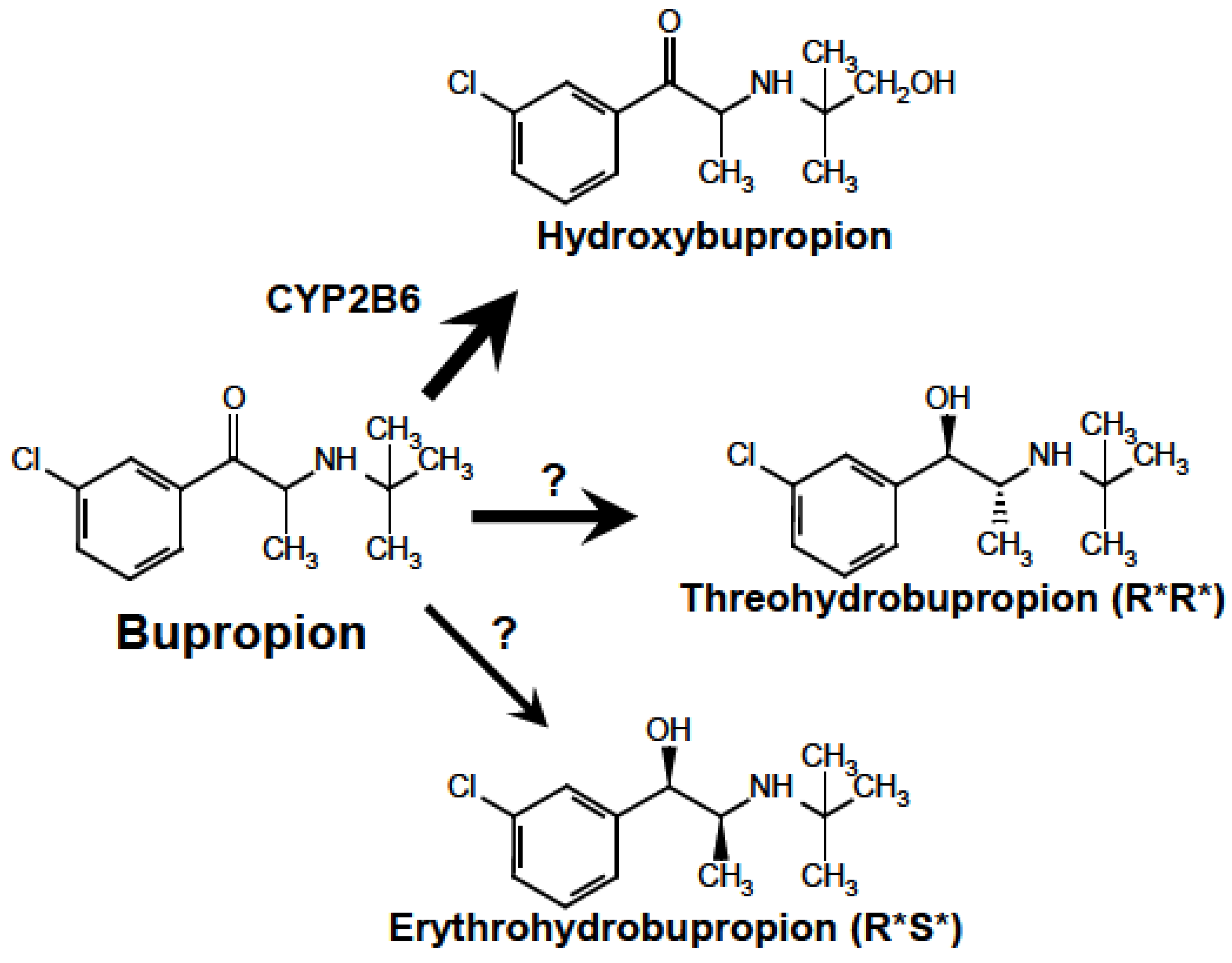
Figure 2.
Representative DNA sequence chromatograms obtained from PCR product generated by amplification of the exon 7 region of the CYP2B7 gene in three African-American individuals. Indicated are the 3 nucleotides comprising codon 378 which is either TGA coding for a stop (A), CGA coding for an arginine (B), or heterozygous TGA/CGA coding for a stop on one allele and arginine on the other allele. Note that the sequence chromatogram is from the antisense (complementary) strand.
Figure 2.
Representative DNA sequence chromatograms obtained from PCR product generated by amplification of the exon 7 region of the CYP2B7 gene in three African-American individuals. Indicated are the 3 nucleotides comprising codon 378 which is either TGA coding for a stop (A), CGA coding for an arginine (B), or heterozygous TGA/CGA coding for a stop on one allele and arginine on the other allele. Note that the sequence chromatogram is from the antisense (complementary) strand.
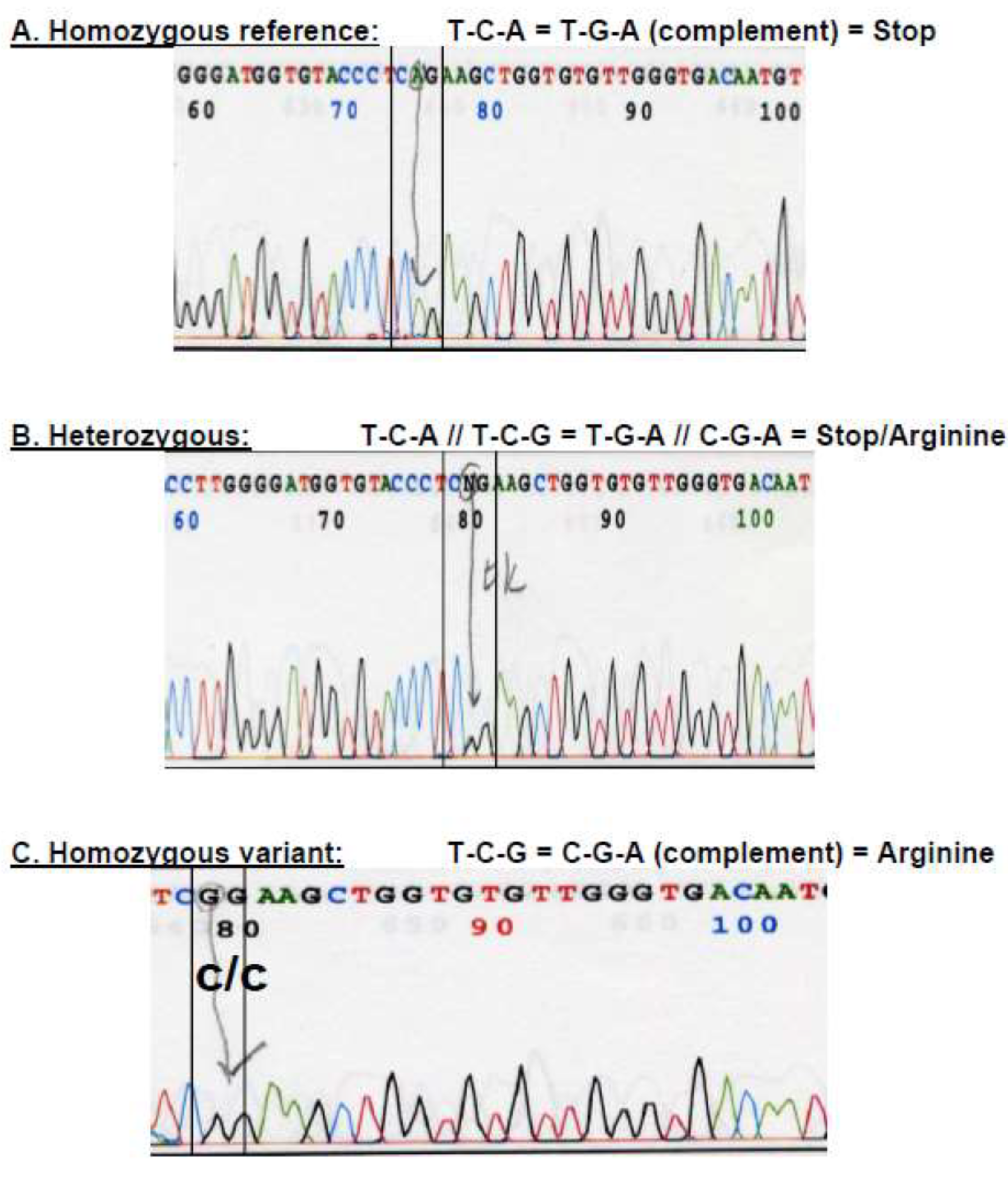
Figure 5.
Views of the predicted three-dimensional structures of CYP2B6 (A) and CYP2B7-378R (B) including the heme-ligand binding domain (indicated in yellow).
Figure 5.
Views of the predicted three-dimensional structures of CYP2B6 (A) and CYP2B7-378R (B) including the heme-ligand binding domain (indicated in yellow).
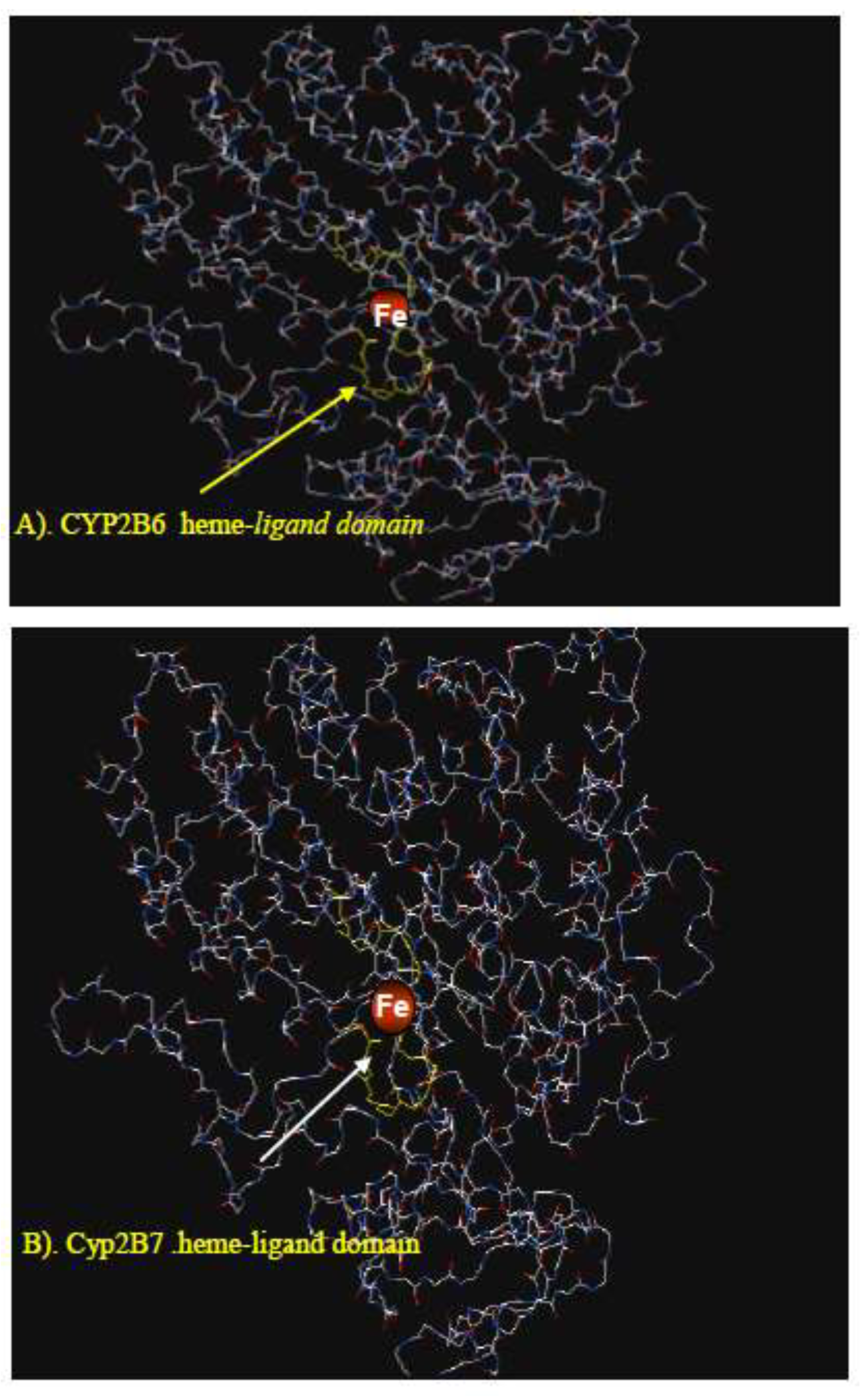
Figure 6.
Immunoblot of CYP2B isoforms expressed in 3T3 mouse cell line. Shown are insect cell expressed recombinant CYP2B6 obtained from BD-Gentest as a blotting positive control (+ve C), and homogenates of 3T3 cells transfected with plasmids containing cDNA encoding CYP2B6, CYP2B7-R378, natural chimeric CYP2B6/2B7-R378, and untransfected cells (-C).
Figure 6.
Immunoblot of CYP2B isoforms expressed in 3T3 mouse cell line. Shown are insect cell expressed recombinant CYP2B6 obtained from BD-Gentest as a blotting positive control (+ve C), and homogenates of 3T3 cells transfected with plasmids containing cDNA encoding CYP2B6, CYP2B7-R378, natural chimeric CYP2B6/2B7-R378, and untransfected cells (-C).
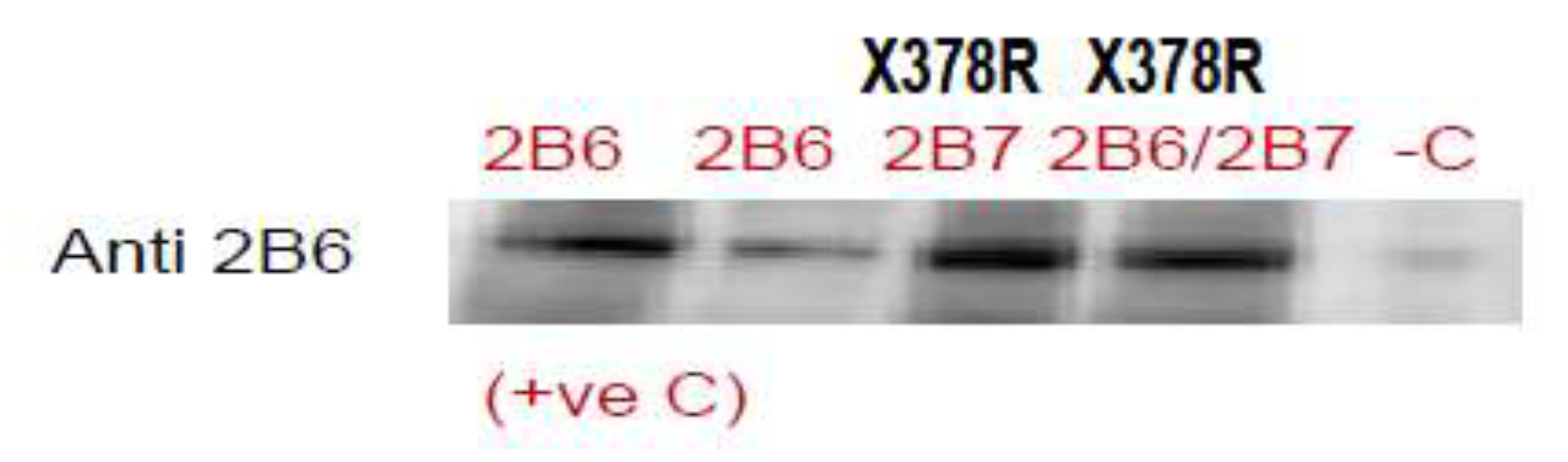
Figure 7.
Representative HPLC chromatograms of bupropion hydroxylation by 3T3 cells transfected with mammalian expression vector containing cDNAs for CYP2B6 and CYP2B7-R378. HBUP=hydroxybupropion (metabolite); IS=internal standard (trazodone); BUP=bupropion (substrate).
Figure 7.
Representative HPLC chromatograms of bupropion hydroxylation by 3T3 cells transfected with mammalian expression vector containing cDNAs for CYP2B6 and CYP2B7-R378. HBUP=hydroxybupropion (metabolite); IS=internal standard (trazodone); BUP=bupropion (substrate).
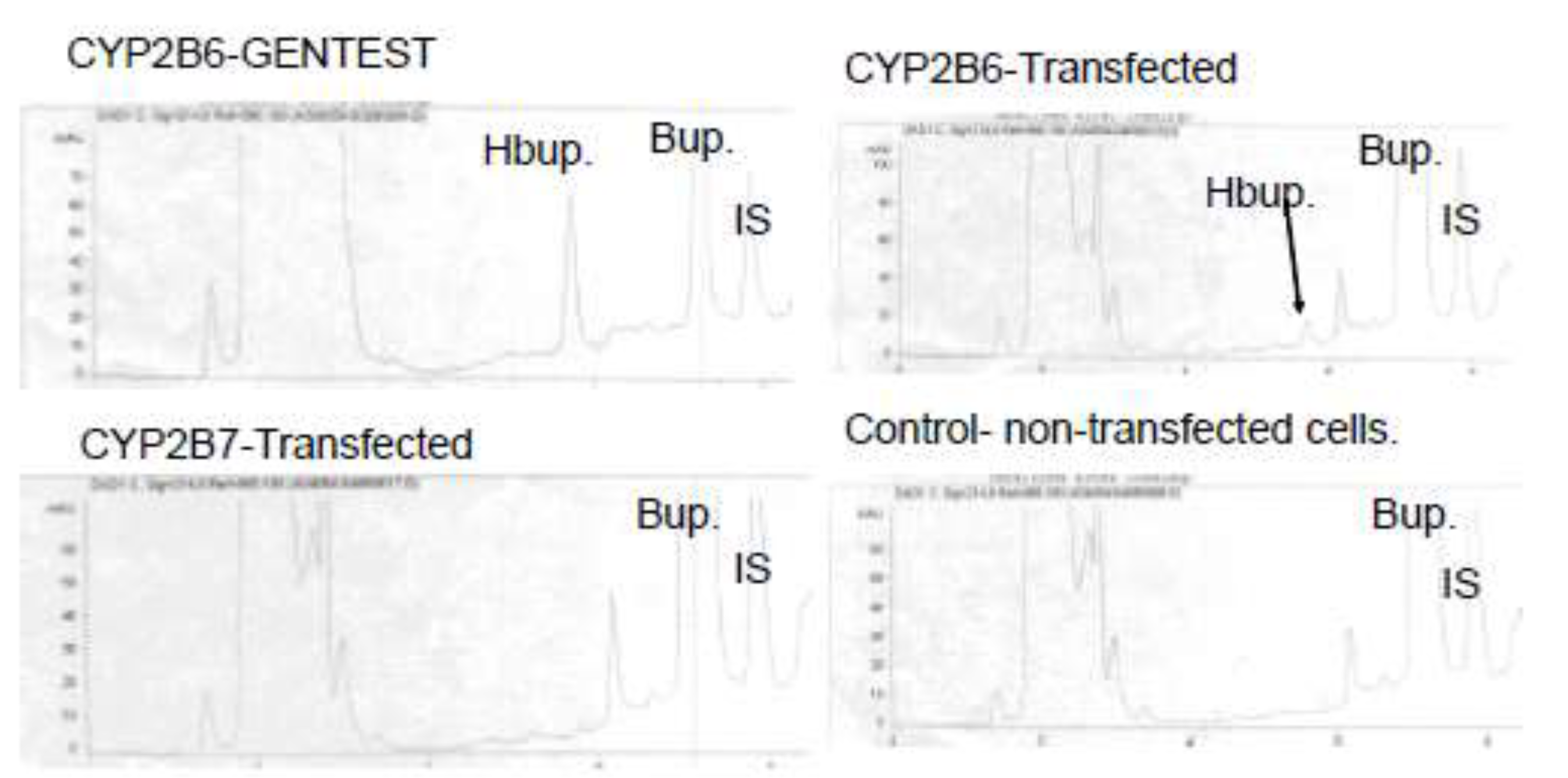
Figure 8.
Representative HPLC chromatograms of bupropion hydroxylation by insect cells infected with baculovirus containing cDNAs for CYP2B6, CYP2B7-R378, CYP2B7-X378, and CYP2B6/2B7-R378. HBUP=hydroxybupropion (metabolite); IS=internal standard (trazodone); BUP=bupropion (substrate).
Figure 8.
Representative HPLC chromatograms of bupropion hydroxylation by insect cells infected with baculovirus containing cDNAs for CYP2B6, CYP2B7-R378, CYP2B7-X378, and CYP2B6/2B7-R378. HBUP=hydroxybupropion (metabolite); IS=internal standard (trazodone); BUP=bupropion (substrate).
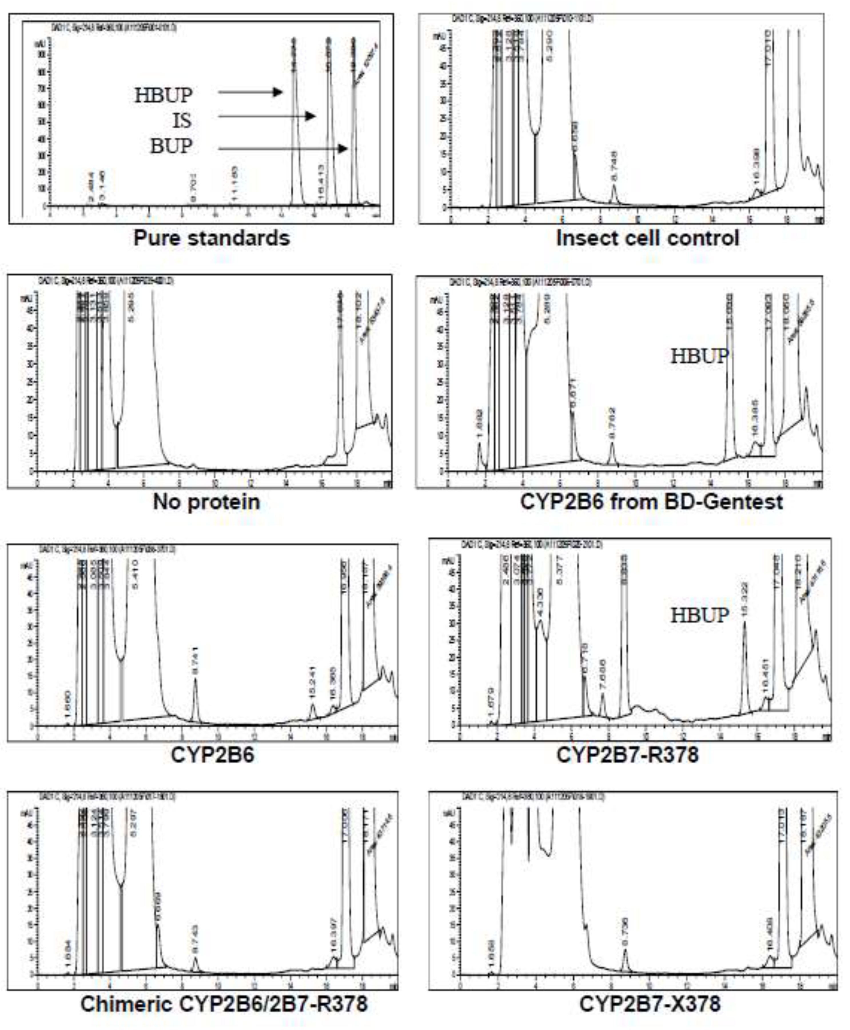
Figure 9.
Expression of CYP2B6 and CYP2B7 mRNA in different human tissues. Values (average of duplicate determinations) were measured by quantitative RT-PCR and are given in arbitrary units (x10-4) relative to amounts of 18S rRNA measured in each sample. RNA was purchased from commercial sources including Clontech and Ambion.
Figure 9.
Expression of CYP2B6 and CYP2B7 mRNA in different human tissues. Values (average of duplicate determinations) were measured by quantitative RT-PCR and are given in arbitrary units (x10-4) relative to amounts of 18S rRNA measured in each sample. RNA was purchased from commercial sources including Clontech and Ambion.
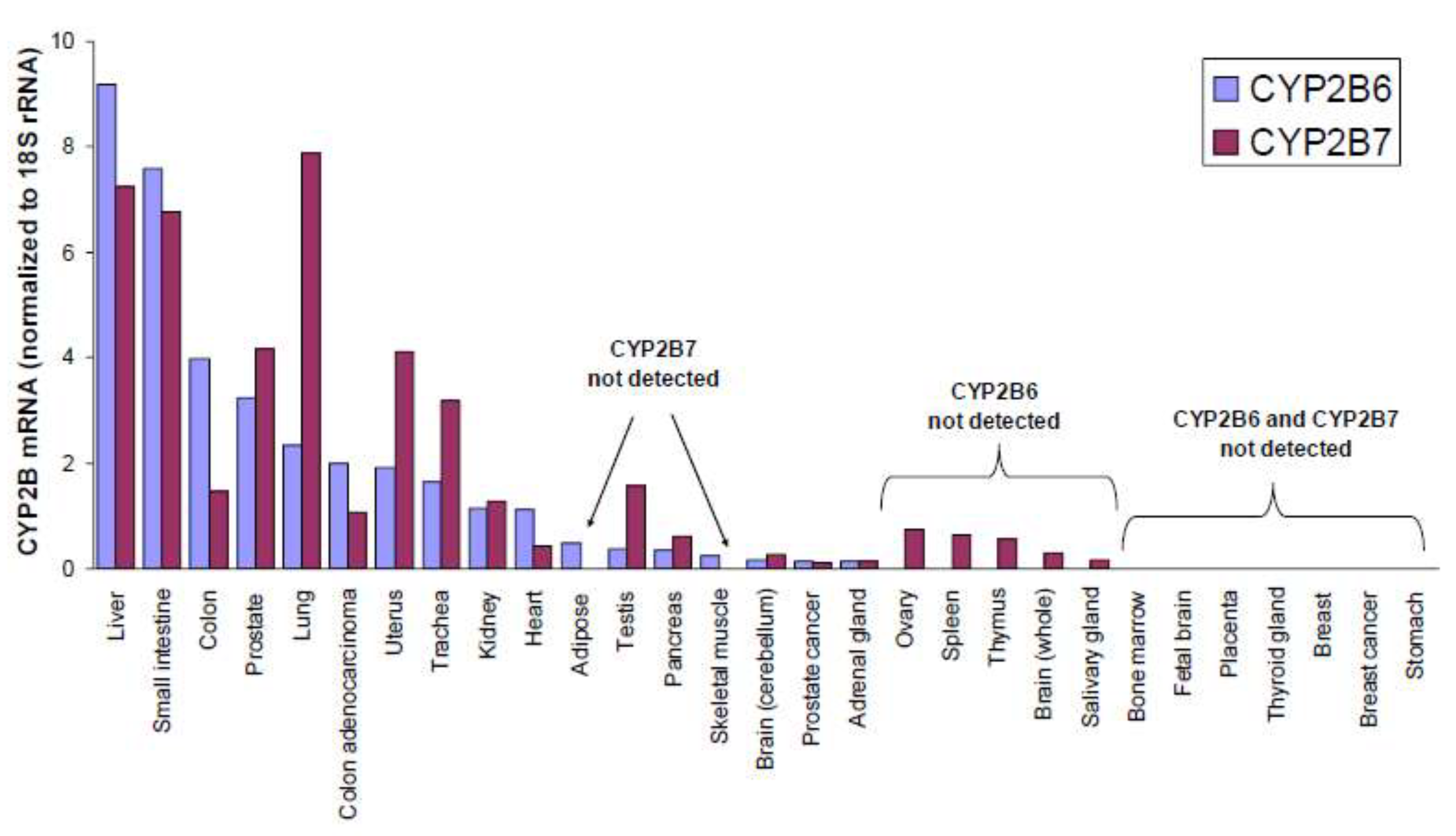

Table 1.
Sequences of oligonucleotide primers used in this study.
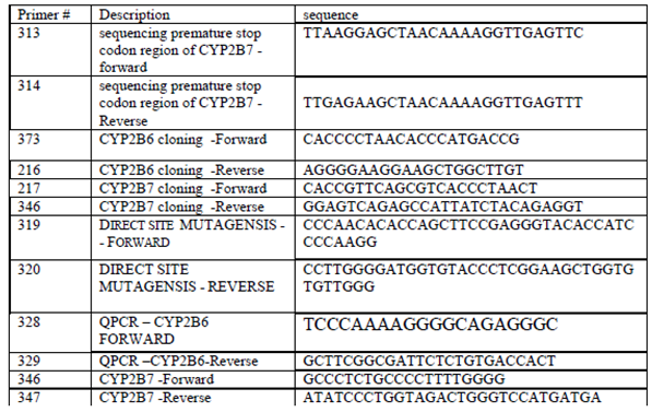 |
Table 2.
Root mean square (RMS) Z-scores indicative of the goodness of fit of the predicted 3-dimensional
structural models of CYP2B6 and CYP2B7-R378 (shown in Figure 9) to the rabbit CYP2B4 crystal structure. The
RMS Z-scores represent the deviation from ideal stereochemical values for bond lengths and angles as
determined by the WhatCheck program. RMS Z-scores close to 1.0 are considered ideal..
Table 2.
Root mean square (RMS) Z-scores indicative of the goodness of fit of the predicted 3-dimensional
structural models of CYP2B6 and CYP2B7-R378 (shown in Figure 9) to the rabbit CYP2B4 crystal structure. The
RMS Z-scores represent the deviation from ideal stereochemical values for bond lengths and angles as
determined by the WhatCheck program. RMS Z-scores close to 1.0 are considered ideal..
|
Disclaimer/Publisher’s Note: The statements, opinions and data contained in all publications are solely those of the individual author(s) and contributor(s) and not of MDPI and/or the editor(s). MDPI and/or the editor(s) disclaim responsibility for any injury to people or property resulting from any ideas, methods, instructions or products referred to in the content. |
© 2026 by the authors. Licensee MDPI, Basel, Switzerland. This article is an open access article distributed under the terms and conditions of the Creative Commons Attribution (CC BY) license (http://creativecommons.org/licenses/by/4.0/).
Copyright: This open access article is published under a Creative Commons CC BY 4.0 license, which permit the free download, distribution, and reuse, provided that the author and preprint are cited in any reuse.