

Submitted:
25 August 2026
Posted:
26 August 2026
You are already at the latest version
Abstract
Background: Musculoskeletal disorders such as osteoarthritis, chronic low back pain, radicular syndromes and osteoporosis produce major clinical burdens, and noninvasive therapies capable of reaching deep anatomical structures are increasingly needed. Nuclear magnetic resonance therapy (molecular biophysical stimulation therapy; NMRT MBST) applies resonance-based magnetic fields to deliver uniform biophysical stimulation independent of tissue depth. This review synthesizes clinical and mechanistic evidence to evaluate the therapeutic potential of NMRT MBST and contextualize it within emerging deep-tissue biophysical treatment strategies. Methods: A systematic search of PubMed, Ovid Embase and peer-reviewed, published reviews identified 15 studies, including randomized trials, imaging investigations, observational cohorts, long-term follow-ups and blinded veterinary work. Experimental literature examining cellular, metabolic and molecular responses to NMRT MBST was reviewed to align biological effects with clinical outcomes. Results: NMRT MBST is consistently reported as safe, with no treatment-emergent adverse events. Clinical findings indicate improvements in pain, function and, in selected studies, imaging or densitometric parameters across osteoarthritis, spine-related pain, radicular syndromes and osteoporosis. Placebo-controlled effects were demonstrated in finger-joint osteoarthritis and radicular pain. Mechanistic studies have shown anti-inflammatory, mitochondrial, redox, anabolic, neurotrophic, epigenetic and circadian effects that closely parallel near-infrared photobiomodulation, supporting the concept of NMRT MBST as a deep-penetrating analogue capable of reaching tissues inaccessible to light. Conventional pulsed electromagnetic field therapy is physically the closest comparator but shows a narrower mechanistic overlap. This positions NMRT MBST within a broader therapeutic framework in which biophysical stimulation may modulate metabolic–inflammatory–regenerative axes in deep musculoskeletal and central nervous system (CNS) structures. Conclusions: NMRT/MBST appears to be a safe and biologically coherent deep-tissue biophysical therapy with promising clinical effects. Larger trials, optimized dosing, mechanistic biomarkers and head-to-head comparisons with established modalities are needed to define its therapeutic role and to clarify how deep-acting biophysical interventions may be integrated into future musculoskeletal and CNS care.
Keywords:
nuclear magnetic resonance therapy
; deep-tissue biophysical stimulation
; mitochondrial bioenergetics & redox signalling
; musculoskeletal regeneration
; anti-inflammatory modulation
1. Introduction
Musculoskeletal and skeletal disorders – including osteoarthritis (OA), chronic low back pain, lumbar radicular syndromes and osteoporosis – are leading causes of pain, disability and healthcare utilization, especially in aging societies [1,2,3,4]. Conservative management commonly combines exercise therapy, education, analgesics/anti-inflammatory medication, injections and, when needed, surgery [5,6,7]. However, long-term pharmacological strategies carry cumulative risk (e.g., gastrointestinal, renal, cardiovascular, metabolic) [8,9,10], and invasive interventions are not suitable or desired for many patients [3,11,12]. As a result, noninvasive physical medicine modalities have become increasingly relevant as adjuncts or alternatives within multimodal care pathways, particularly when they can reduce pain, improve function and potentially influence underlying tissue biology [13,14,15,16].
Despite the breadth of these options, current standards of care remain limited in ways that are increasingly acknowledged at guideline level. In osteoarthritis, recommended pharmacological treatments provide only modest and often short-lived symptomatic relief; oral and topical NSAIDs are constrained by gastrointestinal, renal and cardiovascular toxicity in the older, comorbid populations most affected; intra-articular injections confer only transient benefit; and no agent has been shown to reliably modify structural disease progression, while joint arthroplasty, although effective, is invasive, has a finite implant lifespan and is neither suitable nor desired for every patient [3,6,9]. In chronic low back pain and lumbar radicular syndromes, guidelines increasingly emphasize the limited and short-lived efficacy of analgesic pharmacotherapy, caution against long-term opioid use, and note that injections and surgery benefit only selected patients, leaving many with persistent symptoms despite guideline-concordant care [2,5,8,11]. In osteoporosis, antiresorptive and anabolic agents reduce fracture risk but are limited by suboptimal long-term adherence, rare but serious adverse effects and a persistent treatment gap, and they do not fully restore the compromised bone micro-architecture underlying fragility [4,7,10,12]. A common thread across these conditions is that established care is largely symptom-oriented rather than tissue-modifying, is frequently constrained by cumulative safety concerns, and is often unable to reach or repair the deep structural compartments in which much of the underlying pathology resides.
A central practical limitation of many established noninvasive modalities is depth dependence. Techniques based on optical energy (e.g., low-level laser therapy/photobiomodulation therapy, PBMT; near-infrared laser therapy, NILT) are constrained by absorption and scattering, which markedly reduce fluence with increasing depth of the target tissues and structures [17,18,19]. Mechanical modalities such as extracorporeal shock wave therapy (ESWT) can be highly effective for selected indications but are often focal, may be uncomfortable and can be technically constrained for deep anatomical targets [13,20,21]. Conventional pulsed electromagnetic field therapy (PEMFT) can reach deeper than light, but frequently produce heterogeneous fields, and are not inherently designed around resonance coupling to ubiquitous biological nuclei [22,23,24,25]. These constraints matter because key pathological compartments in OA and spine disorders – including subchondral bone, deep cartilage zones, vertebral endplates, intervertebral discs and nerve-root environments – are often located beyond the effective reach of surface-directed energy delivery [26,27,28].
Nuclear magnetic resonance therapy, also referred to as molecular biophysical stimulation therapy (NMRT/MBST), is positioned within this landscape as a noninvasive modality derived from nuclear magnetic resonance principles but operated at field strengths and radiofrequency (RF) energies far below those used for diagnostic magnetic resonance imaging (MRI) and far below thresholds associated with heating or ionizing effects [29,30]. In NMRT/MBST, static magnetic fields are combined with low-frequency sweep fields and RF signals tuned to the resonance frequency of hydrogen nuclei [30]. Hydrogen protons – abundant in water and organic molecules – enter transient resonance states in which minuscule amounts of electromagnetic energy are absorbed and re-emitted. This resonance-based interaction is commonly described in the NMRT/MBST context as adiabatic fast passage [31,32], emphasizing the controlled traversal through resonance without thermal loading or mechanical impact.
A defining clinical feature of NMRT/MBST is that magnetic fields and the relevant low-frequency RF components are not meaningfully attenuated by biological tissues at therapeutic settings [33,34,35]. Consequently, NMRT/MBST can expose superficial and deep structures to comparatively uniform field penetration, enabling treatment of anatomical regions that are difficult to access with photonic or mechanical modalities. From a clinical “reach” perspective, this characteristic makes NMRT/MBST conceptually attractive for deep joints (e.g., hip), axial spine targets (e.g., discs, endplates, nerve roots) and skeletal compartments relevant to osteoporosis.
At the same time, contemporary rehabilitation medicine increasingly expects that noninvasive modalities should not only exert symptomatic benefits but also act through plausible biological mechanisms that align with tissue homeostasis: controlling inflammation, stabilizing cellular bioenergetics and supporting anabolic repair signaling [3,11,14,36,37]. PBMT/NILT has an extensive mechanistic literature in this respect: photon absorption by mitochondrial chromophores (often highlighted: cytochrome c oxidase) can modulate respiration, transient redox signaling, inflammatory transcription factors and growth-factor pathways [38,39,40]. Importantly, the responsiveness of cytochrome c oxidase to PBMT/NILT is not confined to a single wavelength. Early experimental work demonstrated robust activation at one investigated wavelength (670 nm; [38]), whereas later research showed that comparable effects occur across multiple distinct red and near-infrared wavelengths (660 nm, 830 nm and 905 nm; [39]). Together, these findings indicate that the key mechanistic determinant is adequate photon penetration rather than wavelength specificity per se – an important clarification for the present comparison. This raises an important translational question: can a resonance-based electromagnetic modality such as NMRT/MBST converge on similar intracellular endpoints – particularly those linked to mitochondrial function, redox balance and inflammatory regulation – while bypassing the depth limitations of light?
Comparative analysis of NMRT/MBST and PBMT/NILT indicates substantial overlap in downstream biological effects across multiple domains relevant to musculoskeletal pathology: (i) preservation of cell viability with pro-proliferative tendencies in selected connective-tissue cell types such as chondrocytes and osteoblasts, alongside matrix-modulatory but non-proliferative responses in fibroblasts [41,42,43,44,45,46,47,48], (ii) anti-inflammatory modulation including attenuation of NF-κB–linked responses and suppression of cytokine-induced catabolic gene programs such as matrix metalloproteinases in chondrocytes [40,42,43,44,49,50], (iii) metabolic “recharging”, with improved cellular energy charge, reduced reliance on glycolysis and stabilization of ATP levels under stress, together with normalization of hypoxia-linked metabolic signalling [44,45,51,52,53,54,55,56,57], and (iv) regenerative signaling extending to neuro-supportive actions including enhanced Schwann-cell–derived trophic support and neurite outgrowth in peripheral neural models [52,53,58,59,60]. Critically, this mechanistic comparison does not depend on enumerating specific PBMT/NILM wavelengths: while the initiating physics differ (photon–chromophore excitation versus magnetic-resonance/radical-pair mechanisms [54,61,62]), the canonical targets of PBMT/NILT – such as cytochrome c oxidase – are activated across a physiologically broad red/NIR spectrum [38,39]. Therefore, wavelength specification is not mechanistically essential when comparing PBMT/NILT with NMRT/MBST at the level of downstream biology. Both modalities appear capable of shifting stressed tissues away from catabolic and inflammatory trajectories toward conditions supportive of repair and functional recovery [40,41,42,43,44,45,51,52,53,54,55,63].
This biological convergence is clinically relevant. Many of the most burdensome musculoskeletal disorders involve a coupled cycle of inflammation, metabolic stress and impaired matrix maintenance. In OA, inflammatory cytokines drive catabolic enzyme expression, reduce anabolic transcriptional programs and can impair chondrocyte energy homeostasis – mechanisms that align with pain, stiffness and progressive structural deterioration [36]. In chronic spinal pain and radicular syndromes, peripheral nociception is shaped not only by mechanical compression but also by neuroinflammatory and metabolic factors within deep spinal compartments [64]. In osteoporosis, skeletal integrity is determined by bone remodeling balance within deep trabecular structures [4], where noninvasive stimulation is challenging. A modality that can reach deep tissues and plausibly modulate inflammation-metabolism-regeneration axes could therefore fill an unmet niche – provided clinical outcomes support meaningful patient benefit and safety.
This study addresses the same overarching topic – NMRT/MBST as a noninvasive therapy for musculoskeletal and skeletal disease – but from complementary vantage points. One synthesizes clinical outcomes across indications, emphasizing efficacy signals, durability and safety in human and veterinary studies (summarized in Table 1). The other compares molecular and cellular mechanisms of NMRT/MBST (summarized in Table 2) with those of PBMT/NILT to clarify potential mechanistic overlap and to motivate the concept of NMRT/MBST as a “deep-penetrating” analog to PBMT/NILT-like stimulation in tissues inaccessible to light.
Accordingly, the objective of this integrated study is to unify the clinical evidence on NMRT/MBST across musculoskeletal and skeletal indications, and the mechanistic rationale emerging from comparative cellular biology, into a single coherent narrative. Specifically, we aim to (i) summarize the breadth and structure of the clinical evidence base (trial designs, indications, outcomes, follow-up horizons and safety); (ii) align clinical endpoints (pain, function, imaging, densitometry, work-related outcomes) with plausible biological mechanisms (inflammation control, metabolic stabilization, tissue-regenerative signaling); and (iii) identify research gaps and the next experimental/clinical steps needed to validate the role of NMRT/MBST – alone or in combination with PBMT/NILT and other rehabilitation modalities – within conservative management pathways for deep and superficial musculoskeletal (as well as central nervous system) ailments.
2. Methods
A systematic search was performed in PubMed and Ovid/Embase (including all resources) using the terms “MBST”, “MBST therapy”, “molecular biophysical stimulation therapy”, “NMRT”, “nuclear magnetic resonance therapy”, “tNMR” and “therapeutic nuclear magnetic resonance” from database inception through December 28, 2025, in accordance with the 2020 PRISMA (Preferred Reporting Items for Systematic Reviews and Meta-Analyses) guidelines [65]. The assessment strategy for the identified records is summarized in Figure 1.
In PubMed, the searches retrieved 62 records for “MBST”, 28 for “MBST therapy”, 2 for “molecular biophysical stimulation therapy”, 26 for “NMRT”, 10 for “nuclear magnetic resonance therapy”, 16 for “tNMR” and 8 for “therapeutic nuclear magnetic resonance.” In Ovid/Embase, the same searches retrieved 178, 62, 4, 72, 35, 35 and 16 records, respectively. Across both databases, this yielded a total of 554 records, from which 367 duplicates were removed prior to screening. No automated tools were used to flag records as ineligible.
The remaining 187 records were screened, and 159 were excluded because they were unrelated to NMRT/MBST; this substantial number reflects the common use of the abbreviations “MBST”, “NMRT” and “tNMR” in unrelated scientific fields (details in the Appendix). All 28 records sought for retrieval were obtained either from the E-media library of LMU Munich (Munich, Germany) or other sources and were subsequently assessed for eligibility. Publications in languages other than English were translated using ChatGPT (version 5.2; OpenAI OpCo, San Francisco, CA, USA).
Among the 28 retrieved records, five were conference abstracts and were excluded. The remaining publications consisted of 19 original articles and four peer-reviewed reviews [66,67,68,69]. Screening the reference lists of these 19 original articles and four reviews identified seven additional peer-reviewed original articles. The final dataset therefore comprised 26 peer-reviewed original articles, including 11 clinical studies, three veterinary clinical studies, one experimentally induced animal model study and 11 in vitro studies. None of these studies were excluded from the systematic review based on study outcome.
Given the heterogeneity of clinical indications addressed in the included clinical studies, no statistical meta-analysis was performed.
3. Results
3.1. Clinical Outcomes Across Indications
3.1.1. Knee Osteoarthritis
Clinical investigation of NMRT/MBST in knee OA includes early structural MRI studies [70], one randomized controlled trial [71] and long-term observational follow-up [72,73]. Initial pre–post MRI work demonstrated statistically significant increases in cartilage thickness and volume following NMRT/MBST, with changes observed in both patellar and tibial compartments [70]. Resonance-based stimulation could therefore influence cartilage morphology, although interpretation is limited by the absence of a control group in [70].
A subsequent randomized, double-blind, placebo-controlled trial provided a more conservative perspective [71]. Over a 12-week treatment period, both active and sham groups demonstrated improvements in pain, functional scores and imaging parameters assessed by ultrasound and MRI-based scoring systems. No significant differences between groups were detected at the primary endpoint. This trial, however, warrants strong reservation, because the two groups differed significantly at baseline in the primary outcome: the NMRT/MBST group reported significantly higher pain than the sham group (VAS 6.4 versus 5.5; p = 0.045), an imbalance that was not accounted for in the analysis. Because both groups reached almost identical pain scores at 12 weeks (VAS 3.8 versus 3.6), the more severely affected NMRT/MBST group underwent the larger absolute improvement (a mean reduction of 2.6 versus 1.9 points), so that a baseline-adjusted analysis, such as analysis of covariance, could have yielded a different conclusion from the unadjusted between-group comparison reported. Importantly, treatment was well tolerated, and no safety concerns emerged [71]. Short-term symptomatic improvement can occur therefore in knee OA without clear between-group separation, a pattern consistent with the substantial placebo responsiveness known in this condition [74,75,76].
Preclinical evidence in knee OA also supports early-phase NMRT/MBST-mediated modulation of degenerative processes. In a blinded, randomized, rabbit anterior cruciate ligament (ACL)-transection model of post-traumatic OA, NMRT/MBST initiated 6 weeks after ACL transection resulted in significantly lower macroscopic OA severity compared with sham-treated controls [77]. Improvements were primarily driven by reduced osteophyte formation, fewer fibrillations of the articular cartilage and decreased joint effusion. When NMRT/MBST was initiated later, at 12 weeks post-injury, no macroscopic differences were observed, and in both timing groups histologic Mankin scores were not altered. Hence, NMRT/MBST may preferentially influence early inflammatory and structural responses preceding more advanced cartilage matrix deterioration [77].
3.1.2. Finger-Joint Osteoarthritis
Evidence for small-joint osteoarthritis demonstrates a clearer treatment signal. In a randomized, placebo-controlled trial, patients with finger-joint osteoarthritis who received NMRT/MBST experienced significant and sustained reductions in pain alongside marked improvements in hand function [78]. These benefits persisted through mid-term follow-up. In contrast, placebo-treated patients showed minimal improvement and, in some functional domains, progressive deterioration over time [78]. The magnitude and durability of benefit observed in this setting indicate a robust therapeutic effect of NMRT/MBST in small-joint degenerative disease.
3.1.3. Chronic Low Back Pain
Chronic low back pain has been evaluated in a randomized, placebo-controlled, inpatient rehabilitation setting [79,80]. In [79], patients receiving NMRT/MBST in addition to standardized inpatient rehabilitation demonstrated greater and more durable reductions in pain intensity than those treated with sham therapy. Improvements extended beyond pain reduction and included improved sleep quality as well as increased daily activities. Differences between groups became more pronounced during follow-up, suggesting delayed or cumulative biological effects that persisted beyond the immediate treatment period. Treatment acceptance was high, and no adverse effects were reported [79].
In [80], the same trial was reported with a focus on VAS pain ratings and disability outcomes. Both the active NMRT/MBST and placebo groups showed reductions in pain shortly after treatment, but NMRT/MBST produced more consistent advantages in pain under stress and in several domains of functional disability. The NMRT/MBST group demonstrated more frequent improvements and fewer deteriorations in measures such as personal care and overall disability, whereas some domains – such as sleep and walking – improved similarly in both groups at follow-up [80].
Taken together, this study [79,80] indicated that, while both NMRT/MBST and placebo groups show notable short-term improvements – consistent with the effects of standardized inpatient rehabilitation – MBST confers additional, domain-specific and in some cases longer-lasting benefits, particularly for pain under stress, personal care and overall disability. The two reports differ in emphasis, with [79] describing broader and more uniformly favorable NMRT/MBST-specific effects, whereas [80] documents a more differentiated pattern in which certain functional areas improved similarly across groups and others showed clear NMRT/MBST advantages.
These controlled findings are reinforced by a large observational dataset in which patients with degenerative spinal and rheumatic conditions reported sustained improvements in pain and functional disability across several months following NMRT/MBST [73]. Although observational in nature, these data provide insight into real-world responses across diverse care settings.
3.1.4. Lumbar Disc Herniation and Radicular Syndromes
The strongest controlled evidence for NMRT/MBST in spinal pathology comes from a double-blind, randomized trial in patients with lumbar disc herniation and associated radicular syndromes [81,82]. In this study, NMRT/MBST produced clinically meaningful reductions in pain and significantly reduced sick-leave duration compared to placebo, indicating tangible functional benefit. These differences were most pronounced at early follow-up intervals and persisted over several months.
Magnetic resonance imaging demonstrated improvement in disc morphology in both treatment and placebo groups, consistent with natural recovery processes, but no significant between-group differences were detected [82]. This dissociation between functional recovery and short-term structural imaging changes suggests that NMRT/MBST primarily influences inflammatory, metabolic and neurophysiological aspects of radicular pathology rather than directly accelerating disc resorption. The consistent reduction in sick-leave days [81] underscores the practical relevance of these effects.
3.1.5. Osteoporosis
Two clinical investigations provided insight into the potential role of NMRT/MBST in osteoporosis [83,84]. In a prospective densitometric study, patients demonstrated statistically significant increases in bone mineral density at the lumbar spine and hip one year after treatment, accompanied by favourable changes in biochemical markers of bone formation [83], suggesting a shift toward anabolic bone metabolism. A larger retrospective cohort study monitored patients for several years after NMRT/MBST treatment and documented multiple episodes of severe trauma that resulted in substantial soft-tissue injury but no fractures [84]. The unexpectedly low fracture incidence during long-term follow-up suggests that NMRT/MBST may enhance bone quality or structural resilience in ways not fully captured by standard densitometric measures. Experimental evidence of enhanced osteoblast proliferation [41] and improved cellular energy homeostasis [43] provides a biologically plausible framework for these clinical observations.
3.1.6. Veterinary Evidence
Veterinary investigations provide an additional perspective that is largely unaffected by placebo effects. A long-term case report described sustained functional improvement in a dog with severe hip osteoarthritis following repeated annual cycles of NMRT/MBST, with mobility maintained for nearly a decade [85]. More rigorously, a double-blind controlled trial in dogs with elbow osteoarthritis employed objective gait analysis, and demonstrated measurable improvements in locomotor parameters after NMRT/MBST treatment [86]. Similarly, a randomized, double-blind, placebo-controlled veterinary trial in dogs with osteoarthritis [87] showed that NMRT/MBST produced significantly greater improvements in gait symmetry than placebo at three months post-treatment, accompanied by directional advantages in lameness scores and overall clinical response. These between-group differences diminished by six months, and no sustained effects were observed thereafter. Collectively, these findings support both the biological efficacy of NMRT/MBST and the feasibility of blinded evaluation in non-human models.
3.1.7. Safety and Tolerability
Across all clinical and veterinary investigations, NMRT/MBST has been consistently reported as safe and well tolerated. No treatment-emergent adverse events, therapy-related complications, discontinuations from intolerance or delayed negative outcomes have been described. This favourable safety profile is consistent across repeated treatment cycles, long-term follow-up and diverse patient populations.
3.2. Cellular and Molecular Responses to NMRT/MBST
3.2.1. Proliferation and Viability of Musculoskeletal Cells
NMRT/MBST promotes proliferation of human chondrocytes, osteoblasts and fibroblasts without inducing cytotoxicity or apoptosis. Early investigations showed marked increases in cell numbers following intermittent exposure, with viability assays confirming preserved membrane integrity and metabolic activity [41]. Subsequent studies confirmed that NMRT/MBST supports regulated cell growth and preserves chondrocyte homeostasis under inflammatory or stress conditions [42,43,44], indicating stimulation of physiological rather than pathological proliferation. Importantly, not all musculoskeletal cell types responded with increased proliferation: in human dermal fibroblasts, NMRT/MBST did not alter proliferation or viability but instead modulated extracellular matrix (ECM) composition and collagen organization, indicating cell-type–specific and non-mitogenic regulatory effects [45]. These effects mirror those observed with PBMT/NILT, in which connective-tissue cells exhibit increased proliferation and survival under metabolic or inflammatory stress [55,63]. The consistency across modalities supports the interpretation that NMRT/MBST activates conserved cellular repair and maintenance programmes.
3.2.2. Anti-Inflammatory and Immunomodulatory Effects
NMRT/MBST exerts pronounced anti-inflammatory effects in musculoskeletal cells exposed to inflammatory stimuli. In osteoarthritic chondrocytes, NMRT/MBST suppressed activation of key inflammatory transcription factors and reduced expression of matrix-degrading enzymes central to cartilage destruction [42,43,44]. At the same time, it preserved or restored anabolic regulators essential for ECM maintenance [42,43,44]. These coordinated effects shift the cellular environment from catabolic degradation toward tissue preservation. Comparable anti-inflammatory profiles are well established for PBMT/NILT [40,55,88], suggesting that NMRT/MBST engages shared inflammatory control pathways that are central to degenerative musculoskeletal disease.
3.2.3. Mitochondrial Function, Metabolism and Redox Balance
NMRT/MBST improves mitochondrial function and cellular energy homeostasis, characterized by increased ATP availability, normalization of redox balance, reduced reliance on glycolysis and stabilization of mitochondrial respiration [43,44,52,53,54]. NMRT/MBST normalizes pathological redox imbalance by reducing excessive NADH levels and restoring a healthier NAD⁺/NADH ratio [44]. This redox correction supported efficient electron transport [52,53], stabilized mitochondrial respiration [54] and enhanced cellular oxidative homeostasis [44,54]. Under hypoxic or inflammatory conditions, NMRT/MBST prevented maladaptive metabolic responses and normalized hypoxia-related signalling pathways [43,44,51,52,53,54,61]. These metabolic effects closely parallel those induced by PBMT/NILT [55,57,89] and provide a mechanistic explanation for improved tissue resilience, reduced fatigue and enhanced regenerative capacity observed clinically.
3.2.4. Chondrogenic, Osteogenic and Matrix-Regulatory Effects
In chondrocytes, NMRT/MBST suppressed catabolic gene expression while restoring growth-factor signalling pathways and preserving key regulators of ECM synthesis [42,44]. In osteoblasts, NMRT/MBST induced a marked increase in cell proliferation while preserving viability, consistent with stimulation of bone-forming processes [41]. These findings align closely with PBMT/NILT research demonstrating enhanced collagen synthesis, proteoglycan production, osteoblast differentiation and mineralization [63,89,90]. The convergence of these effects provides a biological basis for clinical observations of symptom improvement in osteoarthritis and increased bone density in osteoporosis.
3.2.5. Neurotrophic and Neuroregenerative Effects
In neuronal models, NMRT/MBST promoted neurite outgrowth and enhanced Schwann-cell–mediated neuro-supportive signalling, resulting in improved neuronal growth and metabolic support [52,53]. These findings, which are also known for PBMT/NILT [58,91], are particularly relevant for radicular pain syndromes and nerve irritation, where metabolic stress and inflammation impair neural function. Although no clinical applications to primary neurological disorders have been published, the experimental data suggest broader neurobiological relevance.
3.2.6. Epigenetic and Circadian Regulation
Beyond immediate signaling pathways, NMRT/MBST modulates gene regulation at the epigenetic level. It restored dysregulated miRNAs involved in inflammation and matrix regeneration, altered histone-modifying enzyme activity such as HDAC4 and reshaped expression patterns of core circadian clock genes across several model systems [44,51,54,61,62]. Collectively, these epigenetic, metabolic and circadian alterations, which align closely with PBMT/NILT research [92,93,94], indicate that NMRT/MBST may impart more lasting adjustments to cellular homeostasis, thereby modifying how cells respond to metabolic or inflammatory stressors over time [43,44,51,54,61,62].
NMRT/MBST and PBMT/NILT converge mechanistically on cryptochrome-associated signaling, but do so through fundamentally distinct biophysical entry points with different consequences for cellular timing and redox control. Whereas PBMT/NILT with 810 nm near-infrared light directly triggered ubiquitination-dependent degradation of the circadian clock protein cryptochrome 1 (CRY1) – rapidly lowering nuclear CRY1 levels and derepressing downstream osteogenic pathways [93] – NMRT/MBST operates primarily through magnetic-field–sensitive radical-pair processes that influence cryptochrome indirectly via changes in intracellular reactive oxygen species (ROS) partitioning [62]. This magnetic-field–driven modulation alters CRY1, CRY2 and CLOCK1 steady-state levels in a distinctly nonlinear, window-dependent fashion, without inducing proteasomal degradation or direct photoreceptor activation [61]. Related studies demonstrated that the resulting shifts in redox balance feed into HIF-1α expression, glycolytic flux, mitochondrial respiration and time-of-day–dependent “on/off” effects on Per2-driven circadian oscillations [54,62]. Moreover, cross-species comparisons indicate that mammalian CRY-linked radical-pair pathways are considerably more responsive to NMRT/MBST than those of zebrafish [51], pointing to organism-specific susceptibilities in cryptochrome-mediated magnetic sensing.
Together, these findings suggest that while both NMRT/MBST and PBMT/NILT act on cryptochrome-centered regulatory networks, NMRT/MBST does so by reconfiguring the redox-sensitive circadian and hypoxia-responsive machinery rather than by inducing direct CRY1 turnover. This distinction highlights NMRT/MBST as a modality capable of influencing the temporal organization of cellular metabolism and stress responsiveness through persistent adjustments in ROS signaling, HIF-1α dynamics and cryptochrome-dependent gene regulation – mechanisms that may contribute to its reported therapeutic benefits in inflammatory, degenerative and ischemia-related conditions.
3.2.7. Radical-Pair Mechanisms and Redox Signalling
Mechanistic studies indicate that NMRT/MBST operates through redox-sensitive radical-pair processes consistent with cryptochrome-mediated magnetosensing [54,61,62]. NMRT/MBST reshaped intracellular ROS signalling in a tightly regulated manner, and these effects critically depend on intact radical intermediates rather than nonspecific oxidative stress [54]. This mechanism closely parallels the controlled ROS signalling induced by PBMT/NILT [40,95], providing a unifying framework to understand how distinct physical stimuli converge on shared biological outcomes.
3.2.8. Penetration Depth and Therapeutic Reach
A defining distinction between NMRT/MBST and PBMT/NILT (or optical therapies in general) lies in tissue penetration. Near-infrared light is limited by absorption and scattering, restricting its effective range to superficial structures [17,18,19]. NMRT/MBST, by contrast, employs magnetic and low-frequency radiofrequency fields that are not meaningfully attenuated by biological tissue [33,34,35]. As a result, entire joints, spinal segments and deep skeletal structures can be exposed uniformly, regardless of anatomical depth or complexity.
This unrestricted penetration provides a coherent explanation for the effectiveness of NMRT/MBST in conditions dominated by deep tissue pathology, including radicular syndromes, deep-joint osteoarthritis and osteoporosis. It also positions NMRT/MBST as a complementary modality capable of extending PBMT/NILT-like biological effects to regions beyond the reach of light-based therapies.
4. Discussion
4.1. A Shifting Therapeutic Landscape and the Role of Deep Biophysical Treatments
The management of chronic musculoskeletal and skeletal disorders is undergoing a conceptual evolution. Historically, treatment pathways have relied heavily on pharmacological symptom control, mechanical unloading, physical therapy and, when conservative options fail, surgical intervention [5,6,7]. Yet millions of patients live with conditions that do not progress to surgical indications, remain symptomatic despite medication or suffer adverse effects from long-term drug use. Against this backdrop, noninvasive biophysical therapies have emerged as compelling adjuncts [13,14,15,16]. Among them, PBMT/NILT has gained meaningful clinical and mechanistic support [15,38,39,40]. However, the inherent depth limitation of PBMT/NILT means that its most robust effects are restricted to surface tissues and shallow musculoskeletal structures [17,18,19].
This constraint has left a substantial gap: the need for a noninvasive therapy capable of safely delivering meaningful biological modulation to deep tissues such as intervertebral discs, subchondral bone, trabecular structures and large weight-bearing joints. NMRT/MBST enters this landscape as a modality designed explicitly to address this unmet need. Its mode of action – based on low-intensity magnetic fields and radiofrequency signals configured to induce magnetic-resonance interactions – differs fundamentally from optical and mechanical therapies. Crucially, the energy delivery of NMRT/MBST is not attenuated by tissue depth or composition [33,34,35], enabling homogeneous exposure of entire anatomical regions irrespective of size, shape or depth. This distinctive property provides not only a technical advantage but also a theoretical basis for why NMRT/MBST may exert clinically meaningful effects in conditions where surface-limited therapies have shown inconsistent benefit.
Preclinical translational data further reinforce this perspective. In a blinded, randomized ACL-transection rabbit model, NMRT/MBST initiated at an early post-injury stage produced significantly lower macroscopic OA severity compared with sham treatment, driven by fewer fibrillations, reduced osteophyte formation and less joint effusion. When initiated later, these macroscopic benefits were absent, and histologic Mankin scores were unchanged in both treatment windows [77]. This timing-dependent effect suggests that NMRT/MBST acts most effectively during the early inflammatory and metabolic phases of joint degeneration before irreversible matrix breakdown develops [77].
4.2. Broadening the Biological Model for Noninvasive Regenerative Therapies
Modern biophysical therapies (especially PBMT/NILT) can be conceptualized as modulators of cellular bioenergetics, inflammation and tissue homeostasis [40,55,57]. Over the last two decades, PBMT/NILT has established a comprehensive mechanistic framework describing how controlled photonic energy influences mitochondrial function, redox biology, inflammatory transcription factors and growth-factor signalling [38,39,40,55,57]. Interestingly, these pathways represent precisely the domains in which NMRT/MBST exerts experimentally verified effects [41,42,43,44,45,52,53,54,61,62] – even though the initiating stimulus is entirely different.
This convergence invites a broader theoretical model in which noninvasive therapies modulate shared intracellular systems through distinct external inputs. Under this model, PBMT/NILT and NMRT/MBST are not competing or contradictory approaches but rather parallel routes into the same regulatory architecture of the cell. If PBMT/NILT represents a light-driven mechanism to enhance mitochondrial and metabolic equilibrium, NMRT/MBST can be conceptualized as a resonance-driven mechanism capable of producing comparable downstream outcomes without optical penetration restrictions.
Such a model helps explain the remarkable consistency observed across mechanistic domains while also recognizing the unique advantages and boundaries of each therapy.
4.3. Mechanistic Convergence: Parallels Between NMRT/MBST and PBMT/NILT
Table 3 offers a consolidated comparison of the presently established biological effects of NMRT/MBST and PBM/NILT, summarizing points of mechanistic convergence and divergence across both modalities.
4.3.1. Shared Modulation of Mitochondrial Bioenergetics
Both NMRT/MBST and PBMT/NILT significantly increase mitochondrial efficiency, although by different physical mechanisms [38,39,43,44,52,53,54,55,57,62]. PBMT/NILT operates by photon absorption in red and near-infrared wavelengths, particularly in cytochrome c oxidase [38,39]. This interaction enhances electron transport, increases ATP production and improves the membrane potential of mitochondria [38,39,55,57]. NMRT/MBST, by contrast, appears to influence electron transport and energy homeostasis through resonance-dependent interactions with radical pairs and magnetosensitive flavoproteins, particularly cryptochrome [54,61,62].
Despite these fundamentally different initiating events, the cellular consequences show overlap: improved ATP availability, reduced ADP/ATP ratios, more stable oxidative phosphorylation and reduced reliance on glycolysis [38,39,43,44,54,55,57,62]. These effects are pivotal in tissues with high metabolic demands and limited vascularity – such as cartilage, intervertebral discs or regions of trabecular bone – where metabolic resilience strongly influences pain, function and degeneration [96,97,98].
4.3.2. Parallel Effects on Redox Signalling and Controlled Dynamics of Reactive Oxygen Species
Modern research increasingly recognizes that therapeutic modulation of ROS must be neither excessive nor suppressive but optimally balanced to activate adaptive cellular pathways [99,100,101]. PBMT/NILT induces a brief, controlled burst of mitochondrial ROS, which serves as a signalling trigger for antioxidant upregulation (e.g., Nrf2 activation), growth-factor release and reduced chronic inflammation [38,39,40,55,57]. NMRT/MBST appears to mirror this dynamic, producing shifts in peroxide and superoxide compartmentalization via radical-pair mechanisms. These changes influence redox-sensitive transcription factors, including those governing inflammation, cell survival and matrix turnover [42,43,44,54,61,62]. This parallel suggests that both NMRT/MBST and PBMT/NILT harness low-level ROS as a signalling intermediate rather than as a destructive force, activating restorative and defensive programmes that outlast the initial stimulus.
4.3.3. Inflammatory Control and Transcriptional Modulation
In a wide range of cell types – chondrocytes, osteoblasts, fibroblasts and neural cells – PBMT/NILT downregulates NF-κB activity, reduces inflammatory cytokine expression and rebalances the inflammatory milieu toward resolution [40,55,88]. NMRT/MBST demonstrates nearly identical anti-inflammatory signatures, including suppression of catabolic cytokine-driven genes, preservation of anabolic and hypoxia-adaptive transcription factors such as HIF-1α, and stabilization of ECM metabolism [42,43,44,51,52,54,61]. These responses help explain clinical findings in osteoarthritis and radiculopathy, where inflammation – not structural deformation alone – drives pain and dysfunction [102,103,104,105,106].
4.3.4. Stimulation of Regeneration in Musculoskeletal Tissues
PBMT/NILT stimulates collagen synthesis, proteoglycan production, fibroblast proliferation, osteoblast differentiation and neurite extension [58,63,89,90,91]. NMRT/MBST activates analogous pathways: it enhances osteoblast and chondrocyte proliferation, preserves matrix-production capacity under inflammatory stress and stimulates neurite extension via neurotrophic signalling [41,42,43,44,52,53]. This implies that NMRT/MBST and PBMT/NILT activate comparable regenerative programmes, but NMRT/MBST can do so in deep tissues where light cannot effectively penetrate.
4.3.5. Epigenetic and Circadian Regulation
Both NMRT/MBST and PBMT/NILT influence epigenetic modifiers such as histone acetylation, HDAC regulation and miRNA expression [44,51,54,61,62,92,93,94]. They also modulate circadian genes [44,51,54,61,62,92,93,94] – an emerging area in musculoskeletal biology [107,108,109,110,111] – implicating deeper regulatory systems that influence metabolism, repair and inflammation across time. NMRT/MBST has demonstrated changes in the oscillatory amplitude of clock genes [54,62], aligning with PBMT/NILT findings that link circadian regulation to enhanced tissue recovery [92,93,94]. Together, these parallels reinforce the conceptualization of NMRT/MBST as a deep-tissue analogue of PBMT/NILT, accessing the same intracellular regulatory pathways via a resonance-based physical mechanism.
The timing-sensitive effects observed in the ACL-transection rabbit model [77] support this mechanistic interpretation. NMRT/MBST produced macroscopic improvements only when delivered in an early post-injury window, paralleling in vitro findings that the modality most effectively modulates inflammatory, redox and metabolic stress responses before irreversible matrix deterioration occurs. The absence of histologic differences at later initiation points underscores that NMRT/MBST influences upstream biological processes that precede overt structural degeneration [77].
4.4. Clinical Interpretation: Understanding the Variability Across Indications
The expanded clinical evidence reveals a nuanced picture: certain indications show strong, consistent improvements [73,78,79,81,82,85,86], while others present mixed results depending on trial design, outcome measurements and biological considerations [70,71,72].
4.4.1. Osteoarthritis: Structural vs. Functional Outcomes
Finger-joint osteoarthritis has shown the clearest controlled evidence of superiority over placebo [78]. This success may relate to the small joint environment, where inflammatory and metabolic processes influence symptoms more directly than large-joint biomechanical load [112]. In knee osteoarthritis, structural heterogeneity, high placebo responsiveness and slower cartilage turnover complicate short-term detection of treatment effects [74,75,76,113,114]. Nevertheless, real-world observational data and long-term follow-up suggest that NMRT/MBST may exert meaningful influence on symptom trajectories [72,73].
4.4.2. Spinal Pain and Radiculopathy: Biological vs. Structural Correlates
In radicular syndromes, the dissociation between early functional improvements and unchanged short-term imaging aligns with contemporary understanding of spine pain [64,115]. Symptoms often arise from neuroinflammation, metabolic dysfunction within nerve tissues and microenvironmental stress, rather than from static structural encroachment [116,117]. NMRT/MBST’s anti-inflammatory and neurotrophic effects thus offer a plausible mechanism for these clinical improvements.
4.4.3. Osteoporosis: Beyond Bone Mineral Density
In addition to the observed increases in bone mineral density [83], the relevance of NMRT/MBST for osteoporosis is underscored by anecdotal reports of higher trauma tolerance without fracture following treatment [84]. Bone mineral density alone does not reliably predict fracture risk [118,119], and the demonstrated biological effects of NMRT/MBST on osteoblast proliferation, cellular energy metabolism and redox regulation [41,42,43,44,52,53,54,61,62] position this modality closer in concept to established anti-fracture medications [120]. If validated in further studies, NMRT/MBST could represent a rare non-pharmacological intervention with the potential to enhance both bone mass and bone resilience.
4.4.4. Veterinary Medicine: Independent Confirmation
Evidence from veterinary models – particularly objective gait analysis in controlled studies – provides additional support that NMRT elicits biological effects independent of expectation or placebo mechanisms [86,87]. The multi-year effectiveness observed in long-term veterinary cases further highlights NMRT’s potential durability.
4.5. Theoretical Implications: Toward a Unified Model of Deep Bioenergetic Modulation
The mechanistic parallels between NMRT/MBST and PBMT/NILT, combined with the depth-independent penetration of NMRT/MBST, support the following unified conceptual framework: noninvasive biophysical therapies exert their therapeutic effects by modulating mitochondrial, redox, inflammatory and regenerative networks. PBMT/NILT operates through photon–chromophore interactions; NMRT/MBST operates through magnetic-resonance interactions with radical pairs and magnetosensitive proteins. The downstream consequences converge.
Under this model, NMRT/MBST is not merely an electromagnetic analogue of PBMT/NILT; it is an extension of the same biological logic into the deep anatomical domain. This framing has several implications. First, deep musculoskeletal tissues – including discs, subchondral bone, deep-joint cartilage and central joint spaces – may be amenable to noninvasive bioenergetic therapy for the first time. Second, combined therapy strategies may become possible – PBMT/NILT for superficial tissues; NMRT/MBST for deep structures – harmonizing both modalities in a comprehensive regenerative approach. Third, mechanistic biomarkers may help personalize therapy. Redox state, mitochondrial profile, inflammatory signatures and circadian markers could guide optimal dosing and candidate selection [40,55,88,121,122,123,124,125,126,127].
4.6. Current Limitations
While the integrated evidence is compelling, several limitations temper final conclusions. First, controlled trials employing NMRT/MBST remain few, with modest sample sizes in several indications. Second, structural imaging endpoints have not consistently mirrored symptomatic improvements, reflecting the complex relationship between imaging and clinical outcomes. Third, optimal treatment parameters – frequency, field strength, duration and cumulative dose – remain to be rigorously defined. Fourth, comparative studies directly evaluating NMRT/MBST vs. PBMT/NILT across tissues of differing depth have not yet been performed. Fifth, the impact on bone quality, circadian regulation and epigenetic dynamics requires dedicated longitudinal investigation. Priorities for future research include larger randomized trials, mechanistically informed biomarkers, head-to-head comparisons with PBMT/NILT and exploration of synergistic combination protocols.
4.7. Broader Biophysical Horizons: What PBMT/NILT Mechanisms Reveal About the Untapped Mechanistic Landscape of NMRT/MBST in Musculoskeletal Disorders
While the present review summarizes the molecular and cellular mechanisms of NMRT/MBST that are currently supported by experimental data, this likely represents only a partial view of the modality’s broader biophysical potential. The historical trajectory of PBMT/NILT offers a relevant perspective: decades of research have gradually revealed multiple mechanistic pathways far beyond the initially hypothesized mitochondrial chromophore activation. This evolution in understanding underscores how emerging biophysical therapies often begin with a narrow mechanistic model that later expands as more diverse cellular and neurophysiological effects are uncovered. Importantly, none of the mechanistic or clinical studies discussed below investigated NMRT/MBST. However, they illustrate the range of biological actions that become conceivable if NMRT/MBST were to share – even partially – the non-thermal, low-energy biophysical mechanisms described for PBMT/NILT, without being limited by the shallow penetration depth of PBMT/NILT.
Across musculoskeletal medicine, several well-characterized PBMT/NILT-based effects illustrate this potential. Deep nociceptor modulation demonstrated in early neurophysiological work [128] and anti-inflammatory suppression of prostaglandin E2 in human tendinopathy [129] highlight how PBMT/NILT can influence both pain-processing networks and local inflammatory mediators – mechanistic domains that are of broad relevance for NMRT/MBST, which aims to modulate musculoskeletal tissue physiology and symptom generation throughout entire anatomical volumes rather than only superficial layers. Likewise, the attenuation of trauma-induced NF-κB and iNOS activity and prevention of fibrosis in structurally injured skeletal muscle [130] expands the concept of PBMT/NILT from symptomatic relief to true modulation of tissue healing. If NMRT/MBST were capable of producing analogous anti-nociceptive, anti-inflammatory or anti-fibrotic responses in injured muscles – but at far greater tissue depth – it could synergize with ESWT in ways surpassing the regeneration observed in animal models of structural muscle injury [131], and potentially improve clinical outcomes beyond what was achieved in ESWT-based protocols for functional and structural muscle injuries in professional athletes [132,133].
Two additional clinical domains further illustrate this horizon. In controlled studies on myofascial pain syndrome, PBMT/NILT and dry needling reduced pain and improved pressure thresholds [134,135], yet both interventions are constrained by the inaccessibility of deep or anatomically hazardous trigger points – most notably the psoas major muscle, whose myofascial trigger points are now recognized as a major contributor to low-back, hip and groin pain and a key driver of lumbopelvic dysfunction [136], while also being exceptionally difficult to access safely given its deep location and proximity to visceral and neurovascular structures. A non-invasive whole-tissue modality such as NMRT/MBST, not restricted by the millimeter-range penetration of optical wavelengths, could theoretically modulate deep myofascial dysfunction in regions entirely beyond the reach of PBMT/NILT. Similarly, in whiplash-associated disorders, high-power laser therapy has demonstrated meaningful reductions in pain and faster return to work compared to conventional rehabilitation [137], but again only within the constrained envelope of optical penetration. If NMRT/MBST shares any of the same downstream biological mechanisms, yet acts volumetrically across the cervical musculoligamentous complex, it could offer a broader therapeutic impact in patients whose symptoms arise from tissues situated beyond the effective depth of PBMT/NILT delivery.
Together, these mechanistic and clinical examples demonstrate how PBMT/NILT has progressively uncovered multiple layers of non-thermal biological action – nociceptive, inflammatory, metabolic and transcriptional – yet remains fundamentally limited by its shallow penetration. NMRT/MBST, if it engages comparable pathways at clinically meaningful depths, may ultimately enable therapeutic effects in musculoskeletal disorders that exceed both the reach and the scope of PBMT/NILT-based interventions.
4.8. Broader Biophysical Horizons: Exploring PBMT/NILT-Informed Mechanistic Pathways Potentially Accessible to NMRT/MBST in Neurodegenerative and Neuroinflammatory Diseases
In the context of neurodegenerative diseases, PBMT/NILT has long been proposed to counteract progressive neuronal dysfunction through mechanisms such as mitochondrial stabilization, improved oxidative balance, modulation of neuroinflammation and support of synaptic maintenance and plasticity [138]. These mechanistic hypotheses generated substantial expectations, especially the idea that low-energy, non-thermal stimulation might not only slow degenerative decline but potentially enhance endogenous repair pathways within vulnerable neuronal populations. However, the translation of these concepts into clinically meaningful outcomes has been consistently limited by the fundamental physical constraints of light penetration: even at near-infrared wavelengths, even superficial cortical regions cannot be reached at therapeutic intensities. This depth barrier has been highlighted repeatedly, including in recent experimental work suggesting that externally applied photonic energy cannot produce measurable changes in cortical tissue in a reliable or physiologically significant manner [19]. These observations underscore a central limitation of PBMT/NILT: promising biochemical and cellular mechanisms exist, yet their practical application is constrained by tissue optics rather than biological potential.
Against this backdrop, NMRT/MBST represents a physically distinct class of biophysical stimulation that is not subject to optical attenuation and thus offers an opportunity to test whether deeper neural structures might respond to NMRT/MBST in ways conceptually analogous to those proposed for PBMT/NILT. Early in vitro observations provide tentative but encouraging indications. In experiments on peripheral neuronal and glial cultures, NMRT/MBST exposure enhanced neurite outgrowth, promoted neuronal survival and influenced glial support functions under controlled conditions, including increased release of neurotrophic factors that support regeneration [52]. In additional studies, such stimulation modulated the secretory activity of supportive glial cells, elevating levels of key neurotrophins without inducing unwanted sensitization responses in nociceptive neurons [53]. Although these effects remain confined to peripheral and in vitro preparations, they illustrate that NMRT/MBST can influence cellular programs that are highly relevant for neurodegenerative disorders, especially as they involve axonal vulnerability, impaired trophic support and glial dysfunction.
A further illustration of this PBMT/NILT-derived blueprint is provided by multiple sclerosis (MS), for which recent experimental and clinical work has shown that PBMT/NILT can modulate several pathophysiological hallmarks of the disease – including suppression of pro-inflammatory cytokines, attenuation of microglial and astroglial activation, reduction of oxidative and nitrosative stress, and partial preservation of oligodendrocyte lineage cells – while improving motor, sensory and functional scores in patients and experimental autoimmune encephalitis (EAE) models alike [139]. Such findings might be especially relevant during the progressive disease stages where treatment options are still limited. These findings also reinforce that low-energy biophysical stimulation can engage neuroimmune, mitochondrial and anti-apoptotic pathways highly relevant to demyelinating disease, yet PBMT/NILT remains physically unable to deliver these effects. If NMRT/MBST could access even a subset of these mechanisms at depth – particularly those related to glial modulation, redox stabilization and trophic support – it could theoretically target deep periventricular and spinal white-matter regions that remain inaccessible to photonic approaches. Although entirely untested at present, MS thus represents a prototypical condition in which PBMT/NILT-identified mechanisms intersect with the depth-independent delivery profile of NMRT/MBST, providing a concrete rationale for future mechanistic exploration.
At the same time, it is essential to recognize that the mechanistic understanding of NMRT/MBST effects on the nervous system remains at a very early stage. No data currently address central neurons, neural networks, microglial states or interactions within the neurovascular unit. The limited evidence available is restricted to peripheral glia and sensory neurons [52,53], and therefore does not yet permit conclusions about potential effects within the brain or spinal cord. Nevertheless, the combination of (i) the mechanistic blueprint provided by PBMT/NILT research; (ii) the absence of a penetration-depth barrier for NMRT/MBST; and (iii) the preliminary in vitro evidence for neurotrophic and pro-regenerative actions together offer a scientifically grounded rationale for further exploration. Should future studies reveal that NMRT/MBST can safely modulate neural tissue at meaningful depths, this modality could potentially address therapeutic targets in neurodegenerative and neuroinflammatory conditions that have remained inaccessible to light-based approaches. Such a trajectory remains speculative at present but represents a worthwhile and increasingly actionable avenue for rigorous mechanistic and translational research.
4.9. Conventional PEMFT as a Comparator: Physical Proximity Does Not Imply Mechanistic Equivalence
Conventional pulsed electromagnetic field therapy (PEMFT) represents an important comparator when positioning NMRT/MBST within the broader landscape of non-thermal, non-mechanical biophysical therapies. At first sight, PEMFT may appear more closely related to NMRT/MBST than PBMT/NILT because both employ magnetic fields, whereas PBMT/NILT is based on optical radiation. However, similarity at the level of the externally applied physical stimulus should not be conflated with similarity of either the primary tissue interaction or the subsequent biological mechanism. Conventional PEMFT employs time-varying magnetic fields that generate induced electric fields and currents in conductive tissue, whereas NMRT/MBST combines a static magnetic field with controlled sweep fields and low-frequency radiofrequency excitation under nuclear magnetic resonance conditions. PBMT/NILT differs physically from both, relying on photon absorption by endogenous photoacceptors. Thus, all three modalities are non-invasive and non-thermal under therapeutic conditions, but they introduce physical energy into biological systems through fundamentally different routes.
These physical distinctions also produce different constraints on target exposure. PBMT/NILT has the clearest intrinsic depth limitation because photon fluence decreases markedly through absorption and scattering in intervening tissue, which becomes particularly relevant for deep cartilage, subchondral bone, intervertebral discs and central neural structures [17,18,19]. Conventional PEMFT avoids this optical attenuation and can reach deeper tissues, but this should not be interpreted as depth-independent or spatially uniform biological exposure. Magnetic-field strength and, particularly, the induced electric field depend on coil geometry, distance, orientation, waveform and rate of field change; consequently, conventional PEMFT replaces the photon-penetration problem of PBMT/NILT with a spatial electromagnetic-dosimetry problem [22,23,24,25]. NMRT/MBST differs again: its static, sweep and radiofrequency fields are configured to establish resonance conditions throughout a defined treatment volume and are not progressively depleted by absorption or scattering in intervening biological tissue [33,34,35]. These physical differences are important for device characterization, dosimetry and safety, but physical similarity alone does not determine which modality represents the most informative biological comparator.
At the mechanistic level, the distinction becomes more consequential. Conventional PEMFT has accumulated substantial experimental evidence for membrane-associated signal transduction [140,141,142], modulation of Ca²⁺-dependent pathways [143,144,145] and, particularly, A2A and A3 adenosine-receptor signalling [146,147,148], with subsequent effects on cAMP [141,146,147], kinases [149,150,151], NF-κB [145,147,152], vascular responses [147,150,153], extracellular-matrix regulation [149,150,154] and cellular differentiation [149,150,155]. Mitochondrial and redox changes have also been reported [143,144,152], but they are more commonly positioned downstream within this signalling architecture than as the primary biological transducer. By contrast, PBMT/NILT has developed the broadest mechanistic framework around mitochondrial photobiology, bioenergetics, transient redox signalling, nitric oxide and subsequent transcriptional regulation [38,39,40,55,57]. Importantly, the NMRT/MBST evidence summarized in the present review shows unexpectedly extensive convergence with this PBMT/NILT downstream biology despite fundamentally different initiating physics: both modalities modulate mitochondrial energy metabolism, redox homeostasis, Ca²⁺ signalling, inflammatory pathways, gene and epigenetic regulation, extracellular-matrix turnover, differentiation, hypoxia adaptation, circadian regulation and neuroregenerative signalling [41,42,43,44,45,51,52,53,54,61,62]. The mechanistic rationale for using PBMT/NILT as the principal biological comparator in this review therefore rests not on physical similarity, but on this unusually broad convergence of experimentally demonstrated downstream cellular responses.
PEMFT nevertheless provides important independent support for the broader concept that weak, non-thermal electromagnetic exposure can produce biologically and clinically meaningful effects. Its most established historical application is skeletal regeneration, particularly delayed fracture healing and nonunion, and subsequent research has extended to osteoarthritis, osteoporosis, spinal disorders, wound healing, tendinopathies and neurological applications [22,23,24,25,156,157,158]. However, “PEMF” encompasses an exceptionally heterogeneous signal space, including substantial differences in field strength, frequency, waveform, pulse structure, applicator geometry and treatment schedule [159,160,161]. Consequently, evidence obtained with one PEMF device cannot automatically be generalized to another, and still less can it be transferred directly to NMRT/MBST merely because both modalities employ magnetic fields. This distinction is especially relevant when comparing conventional induced-electric-field mechanisms with the resonance-based exposure paradigm of NMRT/MBST.
From a clinical perspective, the three modalities should therefore not be ranked according to physical resemblance but evaluated indication by indication. Differences in the volume of published evidence partly reflect historical adoption: PEMFT developed particularly strongly around bone healing [162,163,164], whereas PBMT/NILT became established across numerous superficial or optically accessible indications. Such differences do not necessarily imply biological exclusivity. Conversely, mechanistic evidence may occasionally suggest genuine differences in therapeutic suitability, emphasizing that shared downstream endpoints do not guarantee equivalent effects in every disease. For NMRT/MBST, the most informative comparator therefore depends on the level of analysis: PEMFT is the more obvious comparator in terms of the applied physical stimulus, whereas PBMT/NILT shows the more extensive experimentally demonstrated overlap in downstream molecular and cellular responses. Neither modality should be regarded as a mechanistic surrogate for NMRT/MBST, and ultimately only direct clinical evidence can establish patient benefit for a defined indication.
Conclusion
NMRT/MBST emerges as a safe, biologically coherent and clinically promising noninvasive therapy capable of modulating deep-tissue physiology in ways previously accessible only to optical or invasive modalities. Across musculoskeletal conditions – including OA, chronic low back pain, radicular syndromes and osteoporosis – studies report consistent signals of pain reduction, functional improvement and, in selected contexts, structural or densitometric change, with an exceptionally favourable safety profile and durable benefits.
Mechanistically, NMRT/MBST engages a broad regulatory network encompassing mitochondrial bioenergetics, redox signalling, inflammatory control, anabolic cartilage and bone pathways, neurotrophic activation, and epigenetic and circadian modulation. These effects closely parallel those of near-infrared PBMT/NILT, yet NMRT/MBST uniquely delivers depth-independent stimulation, enabling access to tissues not reachable by light-based therapies.
Together, the clinical evidence and mechanistic plausibility position NMRT/MBST as a meaningful addition to conservative musculoskeletal care. While current data are encouraging, larger randomized trials, optimized dosing studies and mechanistic biomarker-driven research remain essential to fully define its therapeutic role and long-term impact.
Author Contributions
Conceptualization, D.K., A.M., A.Y., A.B., R.A.S., P.H., M.K., T.M., A.S., N.M., C.S.; methodology, C.S.; validation, D.K., A.B., R.A.S., P.H., M.K., T.M., A.S.; formal analysis, C.S.; investigation, T.M., A.S., C.S.; resources, C.S.; data curation, C.S.; writing – original draft preparation, C.S.; writing – review and editing, D.K., A.M., A.Y., A.B., R.A.S., P.H., M.K., T.M., A.S., N.M.; supervision, P.H., N.M.; project administration, CS. All authors have read and agreed to the published version of the manuscript.
Data availability
The data underlying this article are available in the article.
Disclosures and Competing Interests
T.M. is Group Managing Director and a member of the Management Board of Lifco AB (Enköping, Sweden), the parent company of MedTec Medizintechnik (Gießen, Germany), the inventor, manufacturer and distributor of NMRT/MBST devices. In addition, T.M. is Chairman of the Board of MedTec Medizintechnik. A.S. is Chief Medical Officer of MedTec Medizintechnik. C.S. works as a consultant for MBST Health Limited (Grantham, Lincolnshire, UK), which also (partially) belongs to Lifco AB. Lifco AB, MedTec Medizintechnik and MBST Health Limited were not involved in study design, data collection, management, analysis or interpretation, and had no influence on the decision to prepare this manuscript. No other potential conflicts of interest related to this article were reported.
Appendix A Details of the Systematic Literature Search Performed on December 28, 2025

Table A1.
Search strategies: number of searches by search terms and databases used (as referenced in Table A2).
Table A1.
Search strategies: number of searches by search terms and databases used (as referenced in Table A2).
| Search terms | PubMed | Embase/OVID |
| MBST | 1 | 8 |
| MBST therapy | 2 | 9 |
| NMRT | 3 | 10 |
| tNMR | 4 | 11 |
| nuclear magnetic resonance therapy | 5 | 12 |
| therapeutic nuclear magnetic resonance | 6 | 13 |
| molecular biophysical stimulation therapy | 7 | 14 |
Table A2.
Detailed results of the systematic literature search. Abbreviations: N1, consecutive study number; N2, consecutive number within the respective category; T, type of study (O, original study; R, review; C, conference abstract; N, not relevant); Search, search strategy in Table A1 in which the corresponding study was identified. For illustration, the study by Gökşen et al. (consecutive study number 1; Eur J Phys Rehabil Med 2016;52(4):431–9) was identified using the search terms “nuclear magnetic resonance therapy” and “therapeutic nuclear magnetic resonance” in PubMed (Searches 5 and 6), and twice each with the same search terms in Embase/Ovid (Searches 12 and 13). The occurrence of duplicate hits in Embase/Ovid is attributable to the fact that the search was conducted across all available resource subsets within that database.
Table A2.
Detailed results of the systematic literature search. Abbreviations: N1, consecutive study number; N2, consecutive number within the respective category; T, type of study (O, original study; R, review; C, conference abstract; N, not relevant); Search, search strategy in Table A1 in which the corresponding study was identified. For illustration, the study by Gökşen et al. (consecutive study number 1; Eur J Phys Rehabil Med 2016;52(4):431–9) was identified using the search terms “nuclear magnetic resonance therapy” and “therapeutic nuclear magnetic resonance” in PubMed (Searches 5 and 6), and twice each with the same search terms in Embase/Ovid (Searches 12 and 13). The occurrence of duplicate hits in Embase/Ovid is attributable to the fact that the search was conducted across all available resource subsets within that database.
| N1 | N2 | T | Reference | Search |
| 1 | 1 | O | Gökşen et al. Magnetic resonance therapy for knee osteoarthritis: a randomized, double blind placebo controlled trial. Eur J Phys Rehabil Med 2016;52(4):431-9. | 5, 6, 12 (2x), 13 (2x) |
| 2 | 2 | O | Huels et al. Treatment of the clinical symptoms of osteoarthritis in the elbow joints of dogs using nuclear magnetic resonance therapy: a randomized, double-blinded trial. Front Vet Sci 2020;7:500278. | 1, 2, 5, 8(2x), 9 (2x), 12 (2x) |
| 3 | 3 | O | Jansen et al. Effects of low-energy NMR on posttraumatic osteoarthritis: observations in a rabbit model. Arch Orthop Trauma Surg 2011;131(6):863-8. | 1, 2, 8 (2x), 12 |
| 4 | 4 | O | Krpan and Kullich. Nuclear magnetic resonance therapy (MBST) in the treatment of osteoporosis. Case report study. Clin Cases Miner Bone Metab 2017;14(2):235-8. | 1, 2, 3, 5, 6, 8, ,9, 10, 12, 13 |
| 5 | 5 | O | Krpan et al. Non-pharmacological treatment of osteoporosis with nuclear magnetic resonance therapy (NMR-therapy). Period Biol 2015;117(1):161-165. | 8, 9, 12 |
| 6 | 6 | O | Kullich et al. Additional outcome improvement in the rehabilitation of chronic low back pain after nuclear resonance therapy. Rheumatologia 2006;20(1):7-12. | 8, 9 |
| 7 | 7 | O | Kullich et al. One-year-survey with multicenter data of more than 4,500 patients with degenerative rheumatic diseases treated with therapeutic nuclear magnetic resonance. J Back Musculoskelet Rehabil 2013;26(1):93-104. | 6, 13 (2x) |
| 8 | 8 | O | Kullich et al. The effect of MBST-NuclearResonanceTherapy with a complex 3-dimensional electromagnetic nuclear resonance field on patients with low back pain. J Back Musculoskelet Rehabil 2006;19(2-3):79-87. | 8, 9 |
| 9 | 9 | O | Mann et al. Nuclear magnetic resonance treatment accelerates the regeneration of dorsal root ganglion neurons in vitro. Front Cell Neurosci 2022;16:859545. | 3, 5, 10 (2x), 12 (2x) |
| 10 | 10 | O | Mucha et al. Treatment of the clinical symptoms caused by osteoarthritis using nuclear magnetic resonance (MBST) in dogs a randomized trial-a pilot study. Vet Med Austria 2017;104:109. | 8, 9 |
| 11 | 11 | O | Oliva R, Jansen B, Benscheidt F, Sandbichler A.M, Egg M. Nuclear magnetic resonance affects the circadian clock and hypoxia-inducible factor isoforms in zebrafish. Biol Rhythm Res 2019;50(5):739-57. | 12 |
| 12 | 12 | O | Rad et al. Nuclear magnetic resonance treatment induces ß-NGF release from schwann cells and enhances the neurite growth of dorsal root ganglion neurons in vitro. Cells 2024;13(18):1544. | 3, 5, 10 (2x), 12 (2x) |
| 13 | 13 | O | Salfinger et al. Nuclear magnetic resonance therapy in lumbar disc herniation with lumbar radicular syndrome: effects of the intervention on pain intensity, health-related quality of life, disease-related disability, consumption of pain medication, duration of sick leave and MRI analysis. Eur Spine J 2015;24(6):1296-308. | 4, 5, 6, 11 (2x), 12 (2x), 13 (2x) |
| 14 | 14 | O | Steinecker-Frohnwieser et al. Nuclear magnetic resonance therapy modulates the miRNA profile in human primary OA chondrocytes and antagonizes inflammation in Tc28/2a cells. Int J Mol Sci 2021;22(11):5959. | 3, 5, 10 (2x), 12 (2x) |
| 15 | 15 | O | Steinecker-Frohnwieser et al. The therapeutic nuclear magnetic resonance changes the balance in intracellular calcium and reduces the interleukin-1β induced increase of NF-κB activity in chondrocytes. Clin Exp Rheumatol 2018;36(2):294-301. | 3, 6, 10 (2x), 12, 13 (2x) |
| 16 | 16 | O | Temiz-Artmann A, Linder P, Kayser P, Digel I, Artmann GM, Lücker P. NMR in vitro effects on proliferation, apoptosis, and viability of human chondrocytes and osteoblasts. Methods Find Exp Clin Pharmacol 2005;27(6):391-4. | 1, 2, 5, 8 (3x), 9 (3x), 12 (3x) |
| 17 | 17 | O | Thoeni et al. Therapeutic nuclear magnetic resonance and intermittent hypoxia trigger time dependent on/off effects in circadian clocks and confirm a central role of superoxide in cellular magnetic field effects. Redox Biol 2024;72:103152. | 4, 6, 11 (2x), 13 (2x) |
| 18 | 18 | O | Thöni et al. Quantum based effects of therapeutic nuclear magnetic resonance persistently reduce glycolysis. iScience 2022;25(12):105536. | 4, 6, 11, 13 |
| 19 | 19 | O | Thöni et al. Therapeutic nuclear magnetic resonance affects the core clock mechanism and associated hypoxia-inducible factor-1. Chronobiol Int 2021;38(8):1120-34. | 4, 6, 11 (2x), 13 (2x) |
| 20 | 1 | R | Krpan. A new concept of integrated holistic approch in treatment of chronic musculoskeletal diseases the “BAR” method. Period Biol 2015;117(1):119-24. | 12 |
| 21 | 2 | R | Krysiak-Zielonka. Regeneration of bone and cartilage tissue - new treatment and rehabilitation strategy. Ortop Traumatol Rehabil 2024;26(5):225-32. | 1, 2, 3, 5, 8 (2x), 9 (2x), 10 (2x), 12 (2x) |
| 22 | 3 | R | Schmidt et al. Magnetic resonance therapy in the treatment of osteoarthritis: A scoping review. Radiography 2021;27(3):968-75. | 1, 2, 7, 8 (2x), 9 (2x), 14 (2x) |
| 23 | 4 | R | Žnidarič et al. Potential of molecular biophysical stimulation therapy in chronic musculoskeletal disorders: a narrative review. Eur J Transl Myol 2023;33(4):11894. | 1, 2, 5, 7, 8 (2x), 9 (2x), 12 (2x), 14 (2x) |
| 24 | 1 | C | Kullich and Schwann. MBST (R) nuclear resonance therapy improves rehabilitation outcome in patients with low back pain. Ann Rheum Dis 2005;64;(Suppl. 3):519. | 8, 9 |
| 25 | 2 | C | Lohberger et al. Changes in the miRNA profile and hypoxic behaviour of human chondrocytes by therapeutic nuclear magnetic resonance therapy (NMRT). Ann Rheum Dis 2019;78(Suppl. 2):1514. | 10 (2x), 12 (2x) , 13 (2x) |
| 26 | 3 | C | Mohr et al. Beneficial effects of nuclear magnetic resonance therapy on liver regeneration. Gastroenterol 2021;59(1):e27. | 10, 12 |
| 27 | 4 | C | Peehal et al. Nuclear magnetic resonance therapy for knee joint osteoarthrosis: is there any clinical or radiological beneficial effect? A double blind randomised control study. J Bone Joint Surg Br 2011;93-B:308. | 12 |
| 28 | 5 | C | Steinecker-Frohnwieser al. Influence of NMR therapy on metabolism of osteosarcoma- and chondrosarcoma cell lines. Bone 2009;44(Suppl. 2):S295. | 8, 9, 10, 12 (2x) |
| 29 | 1 | N | A trial of mindfulness-based skills training groups versus a waiting list control period for parents of obsessive-compulsive disorder (OCD)-affected youth. Clinicaltrials.gov 2017;no pagination. | 8, 9 |
| 30 | 2 | N | Abbo et al. Mechanism-based-susceptibility testing (MBST) using disc diffusion assays (DDA) to guide treatment of multidrug- and extensively drug-resistant pseudomonas aeruginosa (MDR-XDR-PA) in a cystic fibrosis (CF) lung transplant recipient; Are we ready for combination therapy vs. MDR-XDR-PA? Open Forum Infect Dis 2018;5(Suppl. 1):S714. | 8 (3x), 9 (3x) |
| 31 | 3 | N | Abdi et al., The effect of repeated flexion-based exercises versus extension-based exercises on the clinical outcomes of patients with lumbar disk herniation surgery: a randomized clinical trial. Neurol Res 2023;45(1):28-40. | 1, 2, 8 (2x) |
| 32 | 4 | N | Abramova and Belichenko. The clinical aspect of the joint use of magnetic resonance tomography of the brain and magnetic resonance angiography of the extra- and intracranial arteries in patients with arterial hypertension. [in Russian] Ter Arkh 1996;68(9):26-31. | 3, 10 (3x) |
| 33 | 5 | N | Akbari et al. Systems thinking, causal loop diagram, and systems dynamic in public health challenges: navigating long COVID syndrome and sense of smell in LGBTQIA+ communities. Public Health Chall 2024;3(3):e70004. | 1, 8 (3x) |
| 34 | 6 | N | Aksoy et al. Proposal for an expanded “R” classification: impact of positive surgical margin length on biochemical recurrence after robotic radical prostatectomy. J Clin Med 2025;14(12):4310. | 4 |
| 35 | 7 | N | Aqeele et al. Determination of the effective dose of curcumin alone and in combination with antimicrobial peptide CM11 on promastigote forms of Iranian strain of L. major (MRHO / IR / 75 / ER). Arch Razi Inst 2019;74(4):413-22. | 1, 2 |
| 36 | 8 | N | Aqeele et al. Evaluation of curcumin and CM11 peptide alone and in combination against amastigote form of Iranian strain of L. major (MRHO/IR75/ER) in vitro. Exp Parasitol 2021;229:108151. | 1, 2 |
| 37 | 9 | N | Arasaradnam et al. TNM&R staging in colorectal cancer: has the two week wait for patients made any difference? Gut 2002;50;(Suppl. 2):A48. | 11 (2x) |
| 38 | 10 | N | Aue W.P. Topical nuclear magnetic resonance--a non-invasive probe for biochemical measurements in living organisms. [in German] Radiologe 1983;23(8):357-60. | 4, 11 (2x) |
| 39 | 11 | N | Aupaix et al. Evaluation of a new protocol for rapid identification of Streptococcus pneumoniae in blood cultures using the modified bile solubility test: Gram staining is still standing. J Clin Microbiol 2025;63(1):e0122224. | 1, 8 (2x) |
| 40 | 12 | N | Austerberry et al. The effect of charge mutations on the stability and aggregation of a human single chain Fv fragment. Eur J Pharm Biopharm 2017;115:18-30. | 11 |
| 41 | 13 | N | Badoux et al. Change of carrier density at the pseudogap critical point of a cuprate superconductor. Nature 2016;531(7593):210-4. | 11 |
| 42 | 14 | N | Bahcivan et al. A single-session mindfulness-based swinging technique vs. cognitive disputation intervention among women with breast cancer: A pilot randomised controlled study examining the efficacy at 8-week follow-up. Front Psychol 2022;13:1007065. | 1, 2, 8, 9 |
| 43 | 15 | N | Bahcivan et al. Efficacy of new mindfulness-based swinging technique intervention: a pilot randomised controlled trial among women with breast cancer. Front Psychol 2022;13:863857. | 1, 2, 8, 9 |
| 44 | 16 | N | Baumgarten et al. Analysis of surgeon biometrics during open and robotic radical cystectomy with electromyography and motion capture analysis. Int Braz J Urol 2020;46(1):138. | 1, 2, 8 |
| 45 | 17 | N | Begum et al. Active Rho kinase (ROK-alpha) associates with insulin receptor substrate-1 and inhibits insulin signaling in vascular smooth muscle cells. J Biol Chem 2002;277(8):6214-22. | 8 |
| 46 | 18 | N | Belopitova et al. Glioma of the optical nerve diagnostic and differential diagnostic problems. Pediatriya 1997;36(3):50-2+5. | 10 |
| 47 | 19 | N | Belopitova et al. Nuclear magnetic resonance tomography in resistant epileptic syndromes in childhood - New possibilities for the disclosure of cerebral pathology. Pediatriya 1998;38(3):27-30. | 10 |
| 48 | 20 | N | Beng et al. Mindfulness-based supportive therapy (MBST): proposing a palliative psychotherapy from a conceptual perspective to address suffering in palliative care. Am J Hosp Palliat Care 2015;32(2):144-60. | 1, 2, 8 (3x), 9 (3x) |
| 49 | 21 | N | Bialik et al. Multifocal aseptic bone necrosis after COVID-19: a clinical case. Aging Clin Exp Res 2022;34(Suppl. 1):S353-4. | 12 (3x) |
| 50 | 22 | N | Bilderback et al. Phosphoinositide 3-kinase regulates crosstalk between Trk A tyrosine kinase and p75NTR-dependent sphingolipid signaling pathways. J Neurochem 2001;76(5):1540-51. | 8 |
| 51 | 23 | N | Burton et al. Identification of an ordered compact structure within the recombinant bovine fibrinogen alpha C-domain fragment by NMRT. Biochemistry 2006;45;(7):2257-66. | 10 |
| 52 | 24 | N | Cainelli et al. Chemo- and enzyme-catalyzed reactions revealing a common temperature-dependent dynamic solvent effect on enantioselectivity. Helv Chim Acta 2003;86(11):3548-59. | 11 |
| 53 | 25 | N | Cainelli et al. Chiral aldehydes in hydrocarbons: diastereoselective nucleophilic addition, NMR, and CD spectroscopy reveal dynamic solvation effects. Chirality 2004;16(1):50-6. | 4, 11 |
| 54 | 26 | N | Cainelli et al. Dynamic solvation effects on the endo/exo selectivity of the Diels-Alder reaction. Tetrahedron Lett 2003;44(1):93-6. | 11 (2x) |
| 55 | 27 | N | Cao et al. MBST-Driven 4D-CBCT reconstruction: leveraging swin transformer and masking for robust performance. Comput Methods Programs Biomed 2025:262:108637. | 1, 8 (2x) |
| 56 | 28 | N | Cheetham et al. How do nuclear medicine radiographers and technologists understand the concept of patient care? Nucl Med Commun 2021;42(10):1172-3. | 10 |
| 57 | 29 | N | Chen et al. Artificial cavernosa-like tissue based on multibubble matrigel and a human corpus cavernous fibroblast scaffold. Asian J Androl 2024;26(3):260-7. | 8, 9 |
| 58 | 30 | N | Chen et al. Tunable chiral magneto-transport through band structure engineering in magnetic topological insulators Mn(Bi1-x Sb x )2Te4. Sci Adv 2025;11(20):eadt6084. | 8 (2x) |
| 59 | 31 | N | Cheung et al. Carcinoma of the breast: measurement and the management of treatment. I. The value of the data. Br J Radiol 1991;64(757):29-36. | 1, 2, 8 (4x), 9 (4x) |
| 60 | 32 | N | Cornil et al. Organizing effects of sex steroids on brain aromatase activity in quail. PLoS ONE 2011;6(4):e19196. | 1, 8 (2x) |
| 61 | 33 | N | da Silva Temperini et al. Insecticidal activity in vitro of the essential oil of Pogostemon cablin against Ctenocephalides felis felis. Braz J Vet Med 2022;44:e003422. | 1 |
| 62 | 34 | N | Dickens et al. Acute stress differentially affects aromatase activity in specific brain nuclei of adult male and female quail. Endocrinology 2011;152(11):4242-51. | 8 |
| 63 | 35 | N | Dickens et al. Brain aromatase and circulating corticosterone are rapidly regulated by combined acute stress and sexual interaction in a sex-specific manner. J Neuroendocrinol 2012;24(10):1322-34. | 8 |
| 64 | 36 | N | Dickens et al. Neurochemical control of rapid stress-induced changes in brain aromatase activity. J Neuroendocrinol 2013;25(4):329-39. | 8 |
| 65 | 37 | N | Dogan Kuday et al. Relationship between emotional intelligence and disaster response self-efficacy: A comparative study in nurses. Int Emerg Nurs 2023;70:101319. | 3, 10 (2x) |
| 66 | 38 | N | Efficacy of mindfulness based swinging technique (MBST) for improving anxiety, stress, hopelessness and self efficacy among breast cancer patients in treatment. Clinicaltrials.gov 2019;no pagination. | 8, 9 |
| 67 | 39 | N | El-Dawlatly et al. Anesthetic implications for video assisted thoracoscopic thymectomy in myasthenia gravis. Middle East J Anesthiol 2005;18(2):339-45. | 3, 10 (3x) |
| 68 | 40 | N | El-Fayoumy et al. Evaluation of antioxidant and anticancer activity of crude extract and different fractions of Chlorella vulgaris axenic culture grown under various concentrations of copper ions. BMC Complement Med Ther 2021;21(1):51. | 3, 10 (2x) |
| 69 | 41 | N | El-Tahan and Regal. Target-controlled infusion of remifentanil without muscle relaxants allows acceptable surgical conditions during thoracotomy performed under sevoflurane anesthesia. J Cardiothorac Vasc Anesth 2015;29(6):1557-66. | 3, 10 (2x) |
| 70 | 42 | N | Espin et al. Creating a progress-monitoring system in reading for middle-school students: tracking progress toward meeting high-stakes standards. LDRP 2010;25(2):60-75. | 8 |
| 71 | 43 | N | Evtushenko and Derevianko. The role of nuclear magnetic resonance monitoring in early clinical-instrumental diagnosis of multiple sclerosis. [in Russian] Zh Nevrol Psikhiatr Im S S Korsakova 1998;98(12):29-31. | 3, 10 (3x) |
| 72 | 44 | N | Farag et al. “TNMR” versus “TNM” in the staging of colorectal cancer. Colorectal Dis 2014;16(Suppl. 3):67. | 11 (2x) |
| 73 | 45 | N | Faridi et al. Experimental assessment of microwave ablation computational modeling with MR thermometry. Med Phys 2020;47(9):3777-88. | 10 |
| 74 | 46 | N | Festinger. The significanee of difference between means without reference to the frequency distribution function. Psychometrika 1946;11(2):97-106. | 8 (2x) |
| 75 | 47 | N | Fu et al. BST-2/Tetherin is involved in BAFF-enhanced proliferation and survival via canonical NF-[kappa]B signaling in neoplastic B-lymphoid cells. Exp Cell Res 2021;398(1):112399. | 8 |
| 76 | 48 | N | Gerek et al. The effectiveness of fiberoptic endoscopic swallow study and modified barium swallow study techniques in diagnosis of dysphagia [in Turkish]. Kulak Burun Bogaz Ihtis Derg 2005;15(5-6):103-11. | 1, 8 (2x) |
| 77 | 49 | N | Giratallah et al. Nicotine metabolite ratio: comparison of the three urinary versions to the plasma version and nicotine clearance in three clinical studies. Drug Alcohol Depend 2021;223:108708. | 10 (2x) |
| 78 | 50 | N | Goychuk. Viscoelastic subdiffusion: from anomalous to normal. Phys Rev E Stat Nonlin Soft Matter Phys 2009;80(4 Pt 2):046125. | 3, 10 (2x) |
| 79 | 51 | N | Greco et al. Mind-body skills training and supportive counseling for depression in SLE: positive effects in a randomized controlled trial. Arthritis Rheum 2016;68(Suppl. 10):1386-1387. | 8, 9 |
| 80 | 52 | N | Healthy relationships training study: comparing the efficacy of a mindfulness-based skills training to a psychoeducational intervention at reducing dating violence on college campuses. Clinicaltrials.gov 2019;no pagination. | 8, 9 |
| 81 | 53 | N | Hu et al. Effect of freeze-thaw cycles on mechanical performance of loess soil stabilized with nano magnesium oxide. PLoS ONE 2025;20(4):e0319909. | 3, 10 (2x) |
| 82 | 54 | N | Hwang and Lee. Comparison of efficacy of reverse moxifloxacin-based sequential therapy and moxifloxacin-based sequential therapy as first-line eradication regimen for helicobacter pylori infection. Digestion 2017;95(1):100-101. | 8 (3x), 9 (3x) |
| 83 | 55 | N | Hwang et al. Comparison of efficacy of bismuth containing quintet therapy and moxifloxacinbased sequential therapy as first- line eradication regimen for Helicobacter Pylori infection. Helicobacter 2016;21(Suppl. 1):144. | 8, 9 |
| 84 | 56 | N | Hwang et al. Efficacy of moxifloxacin-based sequential and hybrid therapy for first-line Helicobacter pylori eradication. World J Gastroenterol 2015;21(35):10234-10241. | 1, 2, 8 (2x), 9 (2x) |
| 85 | 57 | N | Jeong et al. Neuromuscular retraining therapy combined with preceding botulinum toxin A injection for chronic facial paralysis. Acta Oto-Laryngol 2023;143(5):446-51. | 3, 10 (2x) |
| 86 | 58 | N | Jhou et al. The mesopontine rostromedial tegmental nucleus: A structure targeted by the lateral habenula that projects to the ventral tegmental area of Tsai and substantia nigra compacta. J Comp Neurol 2009;513(6):566-96. | 8 |
| 87 | 59 | N | Jia et al. Ethylene-induced hydrogen sulfide negatively regulates ethylene biosynthesis by persulfidation of ACO in tomato under osmotic stress. Front Plant Sci 2018;9:1517. | 1, 8 |
| 88 | 60 | N | Johnson et al. The management of individual breast cancers. Breast 1995;4(2):100-11. | 8 (2x), 9 (2x) |
| 89 | 61 | N | Joshi et al. Effect of a single-session mindfulness-based swinging technique (MBST) intervention on emotional distress in cancer patients undergoing PET-CT scan: a randomized controlled trial. J Integr Complement Med 2024;30(10):961-9. | 1, 2, 8 (2x) |
| 90 | 62 | N | Kazemi et al. Comparing the effectiveness of mindfulness based schema therapy and transdiagnostic intervention on cognitive fusion with illness and posttraumatic avoidance in women with breast cancer: a semi-experimental study. J Mazandaran Univ Med Sci 2023;33(220):54-65. | 8, 9 |
| 91 | 63 | N | Kebert et al. Ectomycorrhizal fungi modulate pedunculate oak’s heat stress responses through the alternation of polyamines, phenolics, and osmotica content. Plants 2022;11(23):3360. | 3 |
| 92 | 64 | N | Khalid et al. Performance evaluation of membrane-based septic tank and its reuse potential for irrigating crops. Water Environ Res 2017;89(8):744-51. | 1, 8 (2x) |
| 93 | 65 | N | Kim and Speece. Reactor configuration-part II comparative process stability and efficiency of thermophilic anaerobic digestion. Environ Technol 2002;23(6):643-54. | 4, 11 (3x) |
| 94 | 66 | N | Kim et al. Comparison of efficacy of bismuth-containing quintet therapy and moxifloxacin-based sequential therapy as first-line eradication regimen for Helicobacter pylori infection. United Eur Gastroenterol J 2019;7(Suppl. 8):731. | 8, 9 |
| 95 | 67 | N | Kim et al. Comparing efficacy of Helicobacter pylori eradication regimens: bismuth containing quintet therapy vs moxifloxacin based sequential therapy as first-line eradication regimen. Helicobacter 2020;25(Suppl. 1): no pagination. | 8, 9 |
| 96 | 68 | N | Lange and Seyyedi. Evidence of a Lyme borreliosis infection from the viewpoint of laboratory medicine. Int J Med Microbiol 2002;291(Suppl 33):120-4. | 1, 8 (2x) |
| 97 | 69 | N | Lee et al. Comparison of efficacy of bismuth containing quintet therapy and moxifloxacin-based sequential therapy as first-line eradication regimen for helicobacter pylori infection. Digestion 2018;97(1):120. | 8, 9 |
| 98 | 70 | N | Lee et al. Comparison of efficacy of bismuth containing quintet therapy and moxifloxacin-based sequential therapy as first-line eradication regimen for helicobacter pylori infection. Dig Endosc 2020;32(Suppl. 1):197. | 8, 9 |
| 99 | 71 | N | Lennon-Maslin and Quaiser-Pohl. “It’s different for girls!” The role of anxiety, physiological arousal, and subject preferences in primary school children’s math and mental rotation performance. Behav Sci 2024;14(9):809. | 3, 10 |
| 100 | 72 | N | Li et al. Seed transmission of three viruses in two pear rootstock species Pyrus betulifolia and P. calleryana. Viruses 2022;14(3):599. | 3, 10 (2x) |
| 101 | 73 | N | Löhrke et al. Spontaneous tumors and lifespan of female NMRI mice of the outbred stock Sut:NMRT during a lifetime study. J Cancer Res Clin Oncol 1984;108(2):192-6. | 3, 10 (2x) |
| 102 | 74 | N | Loughmiller et al. An evaluation of differences in mean body surface temperature with infrared thermography in growing pigs fed different dietary energy intake and concentration. J Appl Anim Res 2005;28;(2):73-80. | 8 |
| 103 | 75 | N | Loughmiller et al. Relationship between mean body surface temperature measured by use of infrared thermography and ambient temperature in clinically normal pigs and pigs inoculated with Actinobacillus pleuropneumoniae. Am J Vet Res 2001;62(5):676-81. | 1, 8 (3x) |
| 104 | 76 | N | Lusk Monagle et al. New graduate nurse experiences in clinical judgment: what academic and practice educators need to know. Nurs Educ Perspect 2018;39(4):201-7. | 1 |
| 105 | 77 | N | Malham et al. Porous cage macro-topography improves early fusion rates in anterior cervical discectomy and fusion. Surg Res Pract 2024;2024:8452050. | 3, 10 (2x) |
| 106 | 78 | N | Matsukawa et al. Changes in the density of activated neurons in the medial part of the bed nucleus of stria terminalis following electrical stimulation in the mice olfactory bulb. Neurosci Res 2010;68(Suppl. 1):e389. | 8 |
| 107 | 79 | N | Matsukawa et al. Rose odor can innately counteract predator odor. Brain Res 2011;1381:117-23. | 1, 8 (3x) |
| 108 | 80 | N | Matsumoto et al. In vivo pharmacokinetic analysis utilizing non-targeted and targeted mass spectrometry and in vitro assay against transient receptor potential channels of maobushisaishinto and its constituent asiasari radix. Molecules 2020;25(18):4283. | 1, 2, 8 (2x) |
| 109 | 81 | N | McGinty et al. Emerging, reemerging, and forgotten brain areas of the reward circuit: Notes from the 2010 Motivational Neural Networks conference. Behav Brain Res 2011;225(1):348-57. | 8 |
| 110 | 82 | N | McLaughlin et al. Comparison of performance, clinical chemistry, and carcass characteristics of finishing lambs treated with recombinant ovine or bovine somatotropins. J Anim Sci 1993;71(6):1453-63. | 1 |
| 111 | 83 | N | Mendes and Silveira PF. The interrelationship between leukotriene B4 and leukotriene-A4-hydrolase in collagen/adjuvant-induced arthritis in rats. Biomed Res Int 2014;2014:730421. | 1 |
| 112 | 84 | N | Menezes et al. Deep Q-Managed: a new framework for multi-objective deep reinforcement learning. Front Artif Intell 2025;8:1683323. | 1, 8 |
| 113 | 85 | N | Miller et al. Hybrid positron emission tomography/magnetic resonance imaging in arrhythmic mitral valve prolapse. JAMA Cardiol 2020;5(9):1000-5. | 11 |
| 114 | 86 | N | Mind body therapy for the treatment of chronic pain. Clinicaltrials.gov 2019;no pagination. | 8, 9 |
| 115 | 87 | N | Minelli et al. Neuro-motor reaction time in young non-athletes in age of development. [in Italian] Med Sport 1998;51(1):1-11. | 10 (2x) |
| 116 | 88 | N | Miranda et al. Contactless body temperature assessment for signalling humane endpoints in a mouse model of sepsis. Anim Welf 2025;34:e13. | 1, 8 (2x) |
| 117 | 89 | N | Monini et al. Combined protocol for treatment of secondary effects from facial nerve palsy. Acta Otolaryngol 2011;131(8):882-6. | 3, 10 (2x) |
| 118 | 90 | N | Mura et al. Organizational effects of DDE on brain vasotocin system in male Japanese quail. Neurotoxicol 2009;30(3):479-84. | 8 |
| 119 | 91 | N | Murakami et al. Stress-related activities induced by predator odor may become indistinguishable by hinokitiol odor. Neuroreport 2012;23(18):1071-1076. | 8 (2x) |
| 120 | 92 | N | Ni et al. 4D-CBCT reconstruction with mask-based swin transformer network for improving image quality. Med Phys 2024;51(9):6722. | 8 |
| 121 | 93 | N | Nuclear magnetic resonance therapy in knee osteoarthrosis: a double blind ranomised controlled trial. Clinicaltrials.gov 2012;no pagination. | 8, 9, 10, 12 |
| 122 | 94 | N | Nuesslein-Hildesheim and Schmidt I. Is the circadian core temperature rhythm of juvenile rats due to a periodic blockade of thermoregulatory thermogenesis? Pflugers Arch 1994;427(5-6):450-4. | 4, 11 (3x) |
| 123 | 95 | N | Oku. Network analysis of kampo formulas based on crude drug composition and indications. Tradit Kampo Med 2019;6(3):139-47. | 8 |
| 124 | 96 | N | Olmos et al. Comparison of two coprological methods for the diagnosis of Eurytrema ssp. in cattle and sheep. J Helminthol 2022;96:e53. | 1, 8 (2x) |
| 125 | 97 | N | Omonkhua and Onoagbe. Effect of irvingia gabonensis total saponin fractions on blood glucose, lipid profile and oxidative status of normal male rats. Planta Med 2014;80(16):P2B10. | 11 |
| 126 | 98 | N | Otake et al. Colloidal particle in suspensions of maobushisaishinto extract granules enhances drug intestinal penetration. Chem Pharm Bull 2024;72(12):1055-60. | 1, 8 (2x) |
| 127 | 99 | N | Patel et al. Analysis of surgeon biometrics during open and robotic radical cystectomy. J Endourol 2017;31(Suppl. 2):A248-9. | 8 |
| 128 | 100 | N | Peters et al. Changes in mean body surface temperature (MBST) of the Neonate After Feeding+ 1097. Pediatr Res 1998;43(4):188. | 8 (3x) |
| 129 | 101 | N | Petrovs’kyĭ et al. Kliniko-renthenolohichna otsinka zmin stopy u khvorykh na tsukrovyĭ tiabet [Clinico-radiological estimation of the foot changes in patients with diabetes mellitus]. Klin Khir 2001;(8):37-9. | 3, 10 (2x) |
| 130 | 102 | N | Phelps et al. Individual differences in vasopressin V1A receptor expression, promoter structure and social behavior. Soc Neurosci Abstract 2002;2002:89.1. | 8 |
| 131 | 103 | N | Pijoan. Secretion of hydrogen peroxide by some common pig mycoplasmas. Vet Rec 1974;95(10):216-7. | 8 |
| 132 | 104 | N | Pourmomeny et al. Neuromuscular retraining versus BTX-A injection in subjects with chronic facial nerve palsy, a clinical trial. Iran J Otorhinolaryngol 2021;33(116):151-5. | 3, 10 (2x) |
| 133 | 105 | N | Purohit et al. Case series: transient neonatal macular retinoschisis - a developmental phenomenon in term infants. Eye 2025;39(18):3359-64. | 4, 11 (2x) |
| 134 | 106 | N | Raji et al. IL-7R α polymorphisms in 60 Iranian multiple sclerosis patients. Iran J Neurol 2012;11(1):6-11. | 1, 8 |
| 135 | 107 | N | Read et al. Relationships between field-based measures of strength and power and golf club head speed. J Strength Cond Res 2013;27(10):2708-13. | 1, 8 (4x) |
| 136 | 108 | N | Rebuffat et al. Tricholongins BI and BII 19-residue peptaibols from Trichoderma-longibrachiatum solution structure from two-dimensional NMR spectroscopy. Eur J Biochem 1991;201;(3):661-74. | 10 |
| 137 | 109 | N | Reducing depressive symptoms in systemic lupus erythematosus. Clinicaltrials.gov 2010;no pagination. | 8, 9 |
| 138 | 110 | N | Rieger-Hug and Stirm. Comparative study of host capsule depolymerases associated with Klebsiella bacterio phages. Virology 1981;113;(1):363-78. | 8 |
| 139 | 111 | N | Romcevich et al. Mind-body skills training for resident wellness: a pilot study of a brief mindfulness intervention. J Med Educ Curric Dev 2018;5:2382120518773061. | 1, 8 |
| 140 | 112 | N | Safaei et al. A simple and accurate PCR method for detection of genetically modified rice. J Environ Health Sci Eng 2019;17(2):847-51. | 1, 8 |
| 141 | 113 | N | Saint-Jalmes et al. Virtual phantom magnetic resonance imaging (ViP MRI) on a clinical MRI platform. Med Phys 2018;45(1):250-7. | 11 |
| 142 | 114 | N | Samborski et al. Adaptation of fracture mechanics methods for quality assessment of tungsten carbide cutting inserts. Materials 2021;14(13):3441. | 4, 11 |
| 143 | 115 | N | Sasidharanpillai. Triborate formation constants and polyborate speciation under hydrothermal conditions by Raman spectroscopy using a titanium/sapphire flow cell. J Phys Chem B 2019;123(24):5147-59. | 1, 8 (2x) |
| 144 | 116 | N | Sato et al. [Effects of half-sized secretory leukocyte protease inhibitor and Chinese traditional medicines, yokuinin and mao-bushi-saishin-to, on therapeutic efficacies of benzoxazinorifamycin KRM-1648 against Mycobacterium avium complex infection induced in mice]. Kekkaku 1998;73(8):501-6. Japanese. | 1, 2, 8 (2x), 9 (2x) |
| 145 | 117 | N | Sayag and Yahr. Unilateral lesions of the sexually dimorphic area SDA of the gerbil hypothalamus disrupt male sexual behavior when combined with contralateral lesions of the medial amygdala-amgydalohippocampal area MA-AHI or medial bed nucleus of the stria terminalis MBST. Soc Neurosci Abstracts 1991;17;(1-2):1061. | 8 |
| 146 | 118 | N | Shen et al. Strong deaggregating effect of a novel polyamino resorcinarene surfactant on gold nanoaggregates under microwave irradiation. Langmuir 2008;24(22):13161-7. | 4, 11 |
| 147 | 119 | N | Shibata et al. [Effect of mao-bushi-saishin-to (MBST), a formula of Chinese medicines, on 48 hr homologous passive cutaneous anaphylaxis in rats]. Arerugi 1995;44(9):1167-75. Japanese. | 1, 8 |
| 148 | 120 | N | Shibata et al. [Effects of mao-bushi-saishin-to (MBST) on experimental allergic models in rats]. Arerugi 1995;44(10):1234-40. Japanese. | 1, 2, 8 (3x) |
| 149 | 121 | N | Shibata et al. Effect of mao-bushi-saishin-to (MBST), a formula of Chinese medicines, on 48 hr homologous passive cutaneous anaphylaxis in rats. Jap J Allergol 1995;44;(9):1167-75. | 8 (2x) |
| 150 | 122 | N | Shibata et al. l-ephedrine is a major constituent of mao-bushi-saishin-to, one of the formulas of Chinese medicine, which shows immediate inhibition after oral administration of passive cutaneous anaphylaxis in rats. Inflamm Res 2000;49(8):398-403. | 1, 8 (3x) |
| 151 | 123 | N | Shimizu et al. [Effects of the Chinese traditional medicines “mao-bushi-saishin-to” and “yokuinin” on the antimycobacterial activity of murine macrophages against Mycobacterium avium complex infection]. Kekkaku 1999;74(9):661-6. Japanese. | 1, 2, 8 (2x) |
| 152 | 124 | N | Shimizu et al. [Therapeutic effects of benzoxazinorifamycin KRM-1648 administered alone or in combination with glycyrrhizin against Mycobacterium avium complex infection in mice]. Kekkaku 1999;74(8):617-21. Japanese. | 1, 2, 8 (2x) |
| 153 | 125 | N | Shimizu et al. Effects of the Chinese traditional medicine mao-bushi-saishin-to on therapeutic efficacy of a new benzoxazinorifamycin, KRM-1648, against Mycobacterium avium infection in mice. Antimicrob Agents Chemother 1999;43(3):514-9. | 1, 2, 8 (3x) |
| 154 | 126 | N | Shirmohammad et al. Evaluation of nanonanoliposomal curcumin on cutaneous leishmaniasis skin lesions caused by Leishmania major in BALB/c mice. Iran J Parasitol 2024;19(2):238-46. | 1 |
| 155 | 127 | N | Simkin et al. Meditation/mindfulness: use in clinical practice. J Am Acad Child Adolesc Psychiatry 2017;56(10):S128. | 8, 9 |
| 156 | 128 | N | Slavov et al. Anesthetic considerations for H-reflex monitoring during surgical treatment of spastic disorders. Anesthesiology Abstracts of Scientific Papers Annual Meeting 2002;(2000):Abstract No. 287. | 11 |
| 157 | 129 | N | Spees et al. Rat brain global ischemia-induced diffusion changes revisited: biophysical modeling of the water and NAA MR “diffusion signal”. Magn Reson Med 2022;88(3):1333-46. | 11 |
| 158 | 130 | N | Swierczek-Jereczek et al. Time-scale synchronisation of oscillatory responses can lead to non-monotonous R-tipping. Sci Rep 2023;13(1):2104. | 3, 10 (2x) |
| 159 | 131 | N | Taghavi et al. Ultrasound super-resolution imaging with a hierarchical Kalman tracker. Ultrasonics 2022;122:106695. | 1 |
| 160 | 132 | N | Taki et al. Prediction of final tumor response to preoperative chemotherapy by Tc-99m MIBI imaging at the middle of chemotherapy in malignant bone and soft tissue tumors: comparison with Tl-201 imaging. J Orthop Res 2008;26(3):411-8. | 1, 2, 8 (3x) |
| 161 | 133 | N | Tan et al. Mindfulness: a new paradigm of psychosocial care in the palliative care setting in southeast asia. Ann Acad Med Singap 2017;46(9):339-46. | 1, 2, 8 (2x), 9 (2x) |
| 162 | 134 | N | Tan et al. Mindfulness-based supportive therapy on reducing suffering in patients with advanced cancer: randomised controlled trial. BMJ Support Palliat Care 2024;13(e3):e1117-25. | 1, 2, 8 (3x), 9 (3x) |
| 163 | 135 | N | Tanemura et al. A case of successful fetal therapy for congenital chylothorax by intrapleural injection of OK-432. Ultrasound Obstet Gynecol 2001;18(4):371-5. | 4 |
| 164 | 136 | N | Target-controlled infusion of remifentanil without muscle relaxant allows acceptable surgical conditions during thoracotomy. Clinicaltrials.gov 2014;no pagination. | 10 |
| 165 | 137 | N | The effect of fatigue caused by search and rescue efforts in the debris on the quality of cardiopulmonary resuscitation: a randomized controlled trial. Clinicaltrials.gov 2024;no pagination. | 10 |
| 166 | 138 | N | The immediate effect of mindfulness-based supportive therapy (MBST) vs supportive listening on palliating suffering in palliative care cancer patients: a randomised controlled trial. Clinicaltrials.gov 2021;no pagination. | 8, 9 |
| 167 | 139 | N | The mindfulness, incontinence and sexual function treatment study. Clinicaltrials.gov 2016;no pagination. | 8, 9 |
| 168 | 140 | N | Tian et al. Synthesis of [3α-(3) H] 17α-hydroxy pregnenolone and [3α-(3) H] pregnenolone. J Labelled Comp Radiopharm 2014;57(1):1-11. | 4 |
| 169 | 141 | N | Trageser et al. Outcomes of dogs with thymoma treated with intensity modulated stereotactic body radiation therapy or non-modulated hypofractionated radiation therapy. Vet Comp Oncol 2022;20(2):491-501. | 3, 10 (4x) |
| 170 | 142 | N | Tsunomura and Tokumoto. Man-made electromagnetic noises causing difficulty in geomagnetic and geoelectric observations in city area. Biomed Pharmacother 2005;59(Suppl 1):S15-9. | 4 |
| 171 | 143 | N | Vahdani et al. Quantitative and qualitative analysis of three DNA extraction methods from soybean, maize, and canola oils and investigation of the presence of genetically modified organisms (GMOs). Food Chem 2024;8:100201. | 1, 8 (2x) |
| 172 | 144 | N | Van Den Bergh et al. Muscle glycogen recovery after exercise during glucose and fructose intake monitored by 13C-NMR. J Appl Physiol 1996;81(4):1495-500. | 4, 11 |
| 173 | 145 | N | Vassilev et al. The occlusion of secondary small branches arising in region of coronary bifurcation lesions increases restenosis rates after bifurcation stenting. The link between periprocedural ischemia and instent. Eur Heart J 2013;34(Suppl. 1):216. | 8 |
| 174 | 146 | N | Voldstad et al. The effect of mindfulness interventions on couple relationship satisfaction: a systematic review and meta-analysis. J Consult Clin Psychol 2025;93(6):427-42. | 8, 9 |
| 175 | 147 | N | Voss et al. Mindfulness training in students leads to a reduction in physiological stress levels. Biomed Tech 2019;64(2):S75. | 8 |
| 176 | 148 | N | Voss et al. Mindfulness-based student training improves vascular variability associated with sustained reductions in physiological stress response. Front Public Health 2022;10:863671. | 1, 2, 8 (2x) |
| 177 | 149 | N | Voss et al. Mindfulness-based student training leads to a reduction in physiological evaluated stress. Front Psychol 2020;11:645. | 1, 8 |
| 178 | 150 | N | Wakabayashi et al. Prognostic value of 99mTc-MIBI performed during middle course of the preoperative chemotherapy in patients with malignant bone and soft tissue tumors. Eur J Nucl Med Mol Imaging 2010;37(Suppl. 2):S252-S253. | 8 |
| 179 | 151 | N | Wakabayashi et al. Prognostic value of Tc-99m-MIBI performed during middle course of preoperative chemotherapy in patients with malignant bone and soft-tissue tumors. Clin Nucl Med 2012;37(1):1-8. | 1, 2, 8 (4x) |
| 180 | 152 | N | Wang and Li. Effect of internal interface layer on dielectric properties of doped Ba0.6Sr0.4TiO3thin films and its simulation in filters. J Phys Condens Matter 2023;35(36):no pagination. | 1, 8 (2x) |
| 181 | 153 | N | Wang and Li. Effect of zinc and magnesium ion doping on leakage current behavior of Ba0.6Sr0.4TiO3 thin film. RSC Adv 2024;14(43):31746-31755. | 1, 8 |
| 182 | 154 | N | Xie et al. Achieving high energy density and low loss in PVDF/BST nanodielectrics with enhanced structural homogeneity. ACS Appl Mater Interfaces 2018;10(34):29038-47. | 1, 8 (2x) |
| 183 | 155 | N | Yao et al. A sensitive nested multiplex RT-PCR assay for the simultaneous detection of three common viruses infecting pear plants. J Virol Methods 2019;263:105-110. | 3, 10 (2x) |
| 184 | 156 | N | Yeh et al. Lectin-triggered aggregation of glyco-gold nanoprobes for activity-based sensing of hydrogen peroxide by the naked eye. Chem Asian J 2021;16(21):3462-8. | 1, 8 (2x) |
| 185 | 157 | N | Yotova et al. Diagnostics and treatment of resistant forms of epilepsy in childhood. Pediatriya 1997;36(3):39-43+4. | 10 |
| 186 | 158 | N | Yousefi et al. Molecular detection of Anaplasma phagocytophilum as a zoonotic agent in owned and stray dogs in Tehran, Iran. Arch Razi Inst 2019;74(1):33-8. | 1, 8 (3x) |
| 187 | 159 | N | Zheng et al. Concomitant chemoradiation followed by radical surgery for locally advanced cervical cancer patients: Preliminary results from a randomized, controlled trial. Chin J Evid Based Med 2017;17(1):1-6. | 10 |
References
- GBD 2019 Diseases and Injuries Collaborators. Global burden of 369 diseases and injuries in 204 countries and territories, 1990-2019: a systematic analysis for the Global Burden of Disease Study 2019. Lancet 2020, 396(10258), 1204–22. [Google Scholar] [CrossRef] [PubMed]
- Buchbinder, R.; van Tulder, M.; Öberg, B.; Costa, L.M.; Woolf, A.; Schoene, M.; Croft, P. Lancet Low Back Pain Series Working Group. Low back pain: a call for action. Lancet 2018, 391(10137), 2384–8. [Google Scholar] [CrossRef] [PubMed]
- Hunter, D.J.; Bierma-Zeinstra, S. Osteoarthritis. Lancet 2019, 393(10182), 1745–59. [Google Scholar] [CrossRef] [PubMed]
- Compston, J.E.; McClung, M.R.; Leslie, W.D. Osteoporosis. Lancet 2019, 393(10169), 364–76. [Google Scholar] [CrossRef] [PubMed]
- Bernstein, I.A.; Malik, Q.; Carville, S.; Ward, S. Low back pain and sciatica: summary of NICE guidance. BMJ 2017, 356, i6748. [Google Scholar] [CrossRef] [PubMed]
- Kolasinski, S.L.; Neogi, T.; Hochberg, M.C.; Oatis, C.; Guyatt, G.; Block, J.; Callahan, L.; Copenhaver, C.; Dodge, C.; Felson, D.; Gellar, K.; Harvey, W.F.; Hawker, G.; Herzig, E.; Kwoh, C.K.; Nelson, A.E.; Samuels, J.; Scanzello, C.; White, D.; Wise, B.; Altman, R.D.; DiRenzo, D.; Fontanarosa, J.; Giradi, G.; Ishimori, M.; Misra, D.; Shah, A.A.; Shmagel, A.K.; Thoma, L.M.; Turgunbaev, M.; Turner, A.S.; Reston, J. 2019 American College of Rheumatology/Arthritis Foundation guideline for the management of osteoarthritis of the hand, hip, and knee. Arthritis Care Res. 2020, 72(2), 149–62. [Google Scholar] [CrossRef] [PubMed]
- Kanis, J.A.; Harvey, N.C.; McCloskey, E.; Bruyère, O.; Veronese, N.; Lorentzon, M.; Cooper, C.; Rizzoli, R.; Adib, G.; Al-Daghri, N.; Campusano, C.; Chandran, M.; Dawson-Hughes, B.; Javaid, K.; Jiwa, F.; Johansson, H.; Lee, J.K.; Liu, E.; Messina, D.; Mkinsi, O.; Pinto, D.; Prieto-Alhambra, D.; Saag, K.; Xia, W.; Zakraoui, L.; Reginster, J.-. Algorithm for the management of patients at low, high and very high risk of osteoporotic fractures. Osteoporos. Int. 2020, 31(1), 1–12. [Google Scholar] [CrossRef] [PubMed]
- Maher, C.; Underwood, M.; Buchbinder, R. Non-specific low back pain. Lancet 2017, 389(10070), 736–47. [Google Scholar] [CrossRef] [PubMed]
- Bannuru, R.R.; Osani, M.C.; Vaysbrot, E.E.; Arden, N.K.; Bennell, K.; Bierma-Zeinstra, S.M.A.; Kraus, V.B.; Lohmander, L.S.; Abbott, J.H.; Bhandari, M.; Blanco, F.J.; Espinosa, R.; Haugen, I.K.; Lin, J.; Mandl, L.A.; Moilanen, E.; Nakamura, N.; Snyder-Mackler, L.; Trojian, T.; Underwood, M.; McAlindon, T.E. OARSI guidelines for the non-surgical management of knee, hip, and polyarticular osteoarthritis. Osteoarthr. Cartil. 2019, 27(11), 1578–89. [Google Scholar] [CrossRef] [PubMed]
- Eastell, R.; Rosen, C.J.; Black, D.M.; Cheung, A.M.; Murad, M.H.; Shoback, D. Pharmacological management of osteoporosis in postmenopausal women: an endocrine society clinical practice guideline. J. Clin. Endocrinol. Metab. 2019, 104(5), 1595–622. [Google Scholar] [CrossRef] [PubMed]
- Foster, N.E.; Anema, J.R.; Cherkin, D.; Chou, R.; Cohen, S.P.; Gross, D.P.; Ferreira, P.H.; Fritz, J.M.; Koes, B.W.; Peul, W.; Turner, J.A.; Maher, C.G. Lancet Low Back Pain Series Working Group. Prevention and treatment of low back pain: evidence, challenges, and promising directions. Lancet 2018, 391(10137), 2368–83. [Google Scholar] [CrossRef] [PubMed]
- Ensrud, K.E.; Crandall, C.J. Osteoporosis. Ann. Intern Med. 2024, 177(1), ITC1–16. [Google Scholar] [CrossRef] [PubMed]
- Schmitz, C.; Császár, N.B.; Milz, S.; Schieker, M.; Maffulli, N.; Rompe, J.D.; Furia, J.P. Efficacy and safety of extracorporeal shock wave therapy for orthopedic conditions: a systematic review on studies listed in the PEDro database. Br. Med. Bull. 2015, 116(1), 115–38. [Google Scholar] [CrossRef] [PubMed]
- Wuerfel, T.; Schmitz, C.; Jokinen, L.L.J. The effects of the exposure of musculoskeletal tissue to extracorporeal shock waves. Biomedicines 2022, 10(5), 1084. [Google Scholar] [CrossRef] [PubMed]
- Clijsen, R.; Brunner, A.; Barbero, M.; Clarys, P.; Taeymans, J. Effects of low-level laser therapy on pain in patients with musculoskeletal disorders: a systematic review and meta-analysis. Eur. J. Phys. Rehabil. Med. 2017, 53(4), 603–10. [Google Scholar] [CrossRef] [PubMed]
- Hartley, G.W.; Roach, K.E.; Nithman, R.W.; Betz, S.R.; Lindsey, C.; Fuchs, R.K.; Avin, K.G. Physical therapist management of patients with suspected or confirmed osteoporosis: a clinical practice guideline from the academy of geriatric physical therapy. J. Geriatr. Phys. Ther. 2022, 44(2), E106-19. [Google Scholar] [CrossRef] [PubMed]
- Kaub, L.; Schmitz, C. More than ninety percent of the light energy emitted by near-infrared laser therapy devices used to treat musculoskeletal disorders is absorbed within the first ten millimeters of biological tissue. Biomedicines 2022, 10(12), 3204. [Google Scholar] [CrossRef] [PubMed]
- Kaub, L.; Schmitz, C. Comparison of the penetration depth of 905 nm and 1064 nm laser light in surface layers of biological tissue ex vivo. Biomedicines 2023, 11(5), 1355. [Google Scholar] [CrossRef] [PubMed]
- Tittelmeier, J.; Kaub, L.; Milz, S.; Kugelmann, D.; Hof, P.R.; Schmitz, C.; Nussbaum-Krammer, C. Insufficient low-level near infrared light penetration challenges the efficacy of transcranial photobiomodulation. Brain Stimul. 2025, 18(4), 1220–3. [Google Scholar] [CrossRef] [PubMed]
- Wang, C.J. Extracorporeal shockwave therapy in musculoskeletal disorders. J. Orthop. Surg. Res. 2012, 7, 11. [Google Scholar] [CrossRef] [PubMed]
- Moya, D.; Ramón, S.; Schaden, W.; Wang, C.J.; Guiloff, L.; Cheng, J.H. The role of extracorporeal shockwave treatment in musculoskeletal disorders. J. Bone Jt. Surg. Am. 2018, 100(3), 251–63. [Google Scholar] [CrossRef] [PubMed]
- Markov, M.S. Magnetic field therapy: a review. Electromagn. Biol. Med. 2007, 26(1), 1–23. [Google Scholar] [CrossRef] [PubMed]
- Cianni, L.; Di Gialleonardo, E.; Coppola, D.; Capece, G.; Libutti, E.; Nannerini, M.; Maccauro, G.; Vitiello, R. Current evidence using pulsed electromagnetic fields in osteoarthritis: a systematic review. J. Clin. Med. 2024, 13(7), 1959. [Google Scholar] [CrossRef] [PubMed]
- Lang, S.; Ma, J.; Gong, S.; Wang, Y.; Dong, B.; Ma, X. Pulse electromagnetic field for treating postmenopausal osteoporosis: a systematic review and meta-analysis of randomized controlled trials. Bioelectromagnetics 2022, 43(6), 381–93. [Google Scholar] [CrossRef] [PubMed]
- Sun, X.; Huang, L.; Wang, L.; Fu, C.; Zhang, Q.; Cheng, H.; Pei, G.; Wang, Y.; He, C.; Wei, Q. Efficacy of pulsed electromagnetic field on pain and physical function in patients with low back pain: A systematic review and meta-analysis. Clin. Rehabil. 2022, 36(5), 636–49. [Google Scholar] [CrossRef] [PubMed]
- Brighton, C.T.; Wang, W.; Seldes, R.; Zhang, G.; Pollack, S.R. Signal transduction in electrically stimulated bone cells. J. Bone Jt. Surg. Am. 2001, 83(10), 1514–23. [Google Scholar] [CrossRef] [PubMed]
- Adams, M.A.; Roughley, P.J. What is intervertebral disc degeneration, and what causes it? Spine 2006, 31(18), 2151–61. [Google Scholar] [CrossRef] [PubMed]
- Hunter, D.J.; Schofield, D.; Callander, E. The individual and socioeconomic impact of osteoarthritis. Nat. Rev. Rheumatol. 2014, 10(7), 437–41. [Google Scholar] [CrossRef] [PubMed]
- Kupce, E. Applications of adiabatic pulses in biomolecular nuclear magnetic resonance. Methods Enzymol. 2001, 338, 82–111. [Google Scholar] [CrossRef] [PubMed]
- Schreiner, A.; Welsch, G. Kernspinresonanz-Therapie. sportärztezeitung 2025, 1, 10–17. [Google Scholar]
- Norris, D.G. Adiabatic radiofrequency pulse forms in biomedical nuclear magnetic resonance. Concepts Magn. Res. 2002, 2, 89–101. [Google Scholar] [CrossRef]
- Baranowski, M.; Woźniak-Braszak, A.; Jurga, K. Adiabatic fast passage application in solid state NMR study of cross relaxation and molecular dynamics in heteronuclear systems. J. Magn. Reson 2016, 262, 27–32. [Google Scholar] [CrossRef] [PubMed]
- Schenck, J.F. Safety of strong, static magnetic fields. J. Magn. Reson Imaging 2000, 12(1), 2–19. [Google Scholar] [CrossRef]
- Adair, R.K. Biophysical limits on athermal effects of RF and microwave radiation. Bioelectromagnetics 2003, 24(1), 39–48. [Google Scholar] [CrossRef] [PubMed]
- Foster, K.R.; Glaser, R. Thermal mechanisms of interaction of radiofrequency energy with biological systems with relevance to exposure guidelines. Health Phys. 2007, 92(6), 609–20. [Google Scholar] [CrossRef] [PubMed]
- Goldring, M.B.; Goldring, S.R. Osteoarthritis. J. Cell Physiol. 2007, 213(3), 626–34. [Google Scholar] [CrossRef] [PubMed]
- Zhang, L.; Zheng, Y.L.; Wang, R.; Wang, X.Q.; Zhang, H. Exercise for osteoporosis: a literature review of pathology and mechanism. Front Immunol. 2022, 13, 1005665. [Google Scholar] [CrossRef] [PubMed]
- Wong-Riley, M.T.; Bai, X.; Buchmann, E.; Whelan, H.T. Light-emitting diode treatment reverses the effect of TTX on cytochrome oxidase in neurons. Neuroreport 2001, 12(14), 3033–7. [Google Scholar] [CrossRef] [PubMed]
- Albuquerque-Pontes, G.M.; Vieira, R.P.; Tomazoni, S.S.; Caires, C.O.; Nemeth, V.; Vanin, A.A.; Santos, L.A.; Pinto, H.D.; Marcos, R.L.; Bjordal, J.M.; de Carvalho Pde, T.; Leal-Junior, E.C. Effect of pre-irradiation with different doses, wavelengths, and application intervals of low-level laser therapy on cytochrome c oxidase activity in intact skeletal muscle of rats. Lasers Med. Sci. 2015, 30(1), 59–66. [Google Scholar] [CrossRef] [PubMed]
- Hamblin, M.R. Mechanisms and applications of the anti-inflammatory effects of photobiomodulation. AIMS Biophys. 2017, 4(3), 337–61. [Google Scholar] [CrossRef] [PubMed]
- Temiz-Artmann, A.; Linder, P.; Kayser, P.; Digel, I.; Artmann, G.M.; Lücker, P. NMR in vitro effects on proliferation, apoptosis, and viability of human chondrocytes and osteoblasts. Methods Find. Exp. Clin. Pharmacol. 2005, 27(6), 391–94. [Google Scholar] [CrossRef] [PubMed]
- Steinecker-Frohnwieser, B.; Weigl, L.; Weberhofer, G.; Kullich, W.; Kress, H.G. The influence of nuclear magnetic resonance therapy (NMRT) and interleukin IL-1β stimulation on Cal-78 chondrosarcoma cells and C28/I2 chondrocytes. J. Orthop. Rheumatol. 2014, 1(3), 9. [Google Scholar] [CrossRef]
- Steinecker-Frohnwieser, B.; Kullich, W.; Mann, A.; Kress, H.G.; Weigl, L. Therapeutic nuclear magnetic resonance changes the balance in intracellular calcium and reduces the interleukin-1β-induced increase of NF-κB activity in chondrocytes. Clin. Exp. Rheumatol. 2018, 36(2), 294–301. [Google Scholar] [PubMed]
- Steinecker-Frohnwieser, B.; Lohberger, B.; Eck, N.; Mann, A.; Kratschmann, C.; Leithner, A.; Kullich, W.; Weigl, L. Nuclear magnetic resonance therapy modulates the miRNA profile in human primary OA chondrocytes and antagonizes inflammation in Tc28/2a cells. Int. J. Mol. Sci. 2021, 22(11), 5959. [Google Scholar] [CrossRef] [PubMed]
- Digel, I.; Kurulgan, E.; Linder, P.; Kayser, P.; Porst, D.; Braem, G.J.; Zerlin, K.; Artmann, G.M.; Artmann, A.T. Decrease in extracellular collagen crosslinking after NMR magnetic field application in skin fibroblasts. Med. Biol. Eng. Comput 2007, 45(1), 91–7. [Google Scholar] [CrossRef] [PubMed]
- Hang, N.L.T.; Aviña, A.E.; Chang, C.J.; Yang, T.S. Photobiomodulation in promoting cartilage regeneration. Int. J. Mol. Sci. 2025, 26(12), 5580. [Google Scholar] [CrossRef] [PubMed]
- Agas, D.; Hanna, R.; Benedicenti, S.; De Angelis, N.; Sabbieti, M.G.; Amaroli, A. Photobiomodulation by near-infrared 980-nm wavelengths regulates pre-osteoblast proliferation and viability through the PI3K/Akt/Bcl-2 pathway. Int. J. Mol. Sci. 2021, 22(14), 7586. [Google Scholar] [CrossRef] [PubMed]
- Chellini, F.; Tani, A.; Zecchi-Orlandini, S.; Giannelli, M.; Sassoli, C. In vitro evidences of different fibroblast morpho-functional responses to red, near-infrared and violet-blue photobiomodulation: clues for addressing wound healing. Appl. Sci. 2020, 10(21), 7878. [Google Scholar] [CrossRef]
- Sakata, S.; Kunimatsu, R.; Tsuka, Y.; Nakatani, A.; Hiraki, T.; Gunji, H.; Hirose, N.; Yanoshita, M.; Putranti, N.A.R.; Tanimoto, K. High-frequency near-infrared diode laser irradiation attenuates il-1β-induced expression of inflammatory cytokines and matrix metalloproteinases in human primary chondrocytes. J. Clin. Med. 2020, 9(3), 881. [Google Scholar] [CrossRef] [PubMed]
- Kunimatsu, R.; Nakatani, A.; Sakata, S.; Tanimoto, K. Effects of photobiomodulation on osteoarthritis from in vivo and in vitro studies: a narrative review. Int. J. Mol. Sci. 2025, 26(18), 8997. [Google Scholar] [CrossRef] [PubMed]
- Oliva, R.; Jansen, B.; Benscheidt, F.; Sandbichler, A.M.; Egg, M. Nuclear magnetic resonance affects the circadian clock and hypoxia-inducible factor isoforms in zebrafish. Biol. Rhythm Res. 2019, 50(5), 739–57. [Google Scholar] [CrossRef]
- Mann, A.; Steinecker-Frohnwieser, B.; Naghilou, A.; Millesi, F.; Supper, P.; Semmler, L.; Wolf, S.; Marinova, L.; Weigl, L.; Weiss, T.; Radtke, C. Nuclear magnetic resonance treatment accelerates the regeneration of dorsal root ganglion neurons in vitro. Front Cell Neurosci. 2022, 16, 859545. [Google Scholar] [CrossRef] [PubMed]
- Rad, A.; Weigl, L.; Steinecker-Frohnwieser, B.; Stadlmayr, S.; Millesi, F.; Haertinger, M.; Borger, A.; Supper, P.; Semmler, L.; Wolf, S.; Naghilou, A.; Weiss, T.; Kress, H.G.; Radtke, C. Nuclear magnetic resonance treatment induces β-NGF release from Schwann cells and enhances neurite growth of dorsal root ganglion neurons in vitro. Cells 2024, 13(18), 1544. [Google Scholar] [CrossRef] [PubMed]
- Thöni, V.; Dimova, E.Y.; Kietzmann, T.; Usselman, R.J.; Egg, M. Therapeutic nuclear magnetic resonance and intermittent hypoxia trigger time-dependent on/off effects in circadian clocks and confirm a central role of superoxide in cellular magnetic field effects. Redox Biol. 2024, 72, 103152. [Google Scholar] [CrossRef] [PubMed]
- de Freitas, L.F.; Hamblin, M.R. Proposed mechanisms of photobiomodulation or low-level light therapy. IEEE J. Sel. Top. Quantum Electron 2016, 22(3), 7000417. [Google Scholar] [CrossRef] [PubMed]
- Quirk, B.J.; Sannagowdara, K.; Buchmann, E.V.; Jensen, E.S.; Gregg, D.C.; Whelan, H.T. Effect of near-infrared light on in vitro cellular ATP production of osteoblasts and fibroblasts and on fracture healing with intramedullary fixation. J. Clin. Orthop. Trauma 2016, 7(4), 234–41. [Google Scholar] [CrossRef] [PubMed]
- Hamblin, M.R. Mechanisms and mitochondrial redox signaling in photobiomodulation. Photochem Photobiol. 2018, 94(2), 199–212. [Google Scholar] [CrossRef] [PubMed]
- Rosso, M.P.O.; Buchaim, D.V.; Kawano, N.; Furlanette, G.; Pomini, K.T.; Buchaim, R.L. Photobiomodulation therapy (PBMT) in peripheral nerve regeneration: a systematic review. Bioengineering 2018, 5(2), 44. [Google Scholar] [CrossRef] [PubMed]
- Fellin, C.R.; Steiner, R.C.; Buchen, J.T.; Anders, J.J.; Jariwala, S.H. Photobiomodulation and vascularization in conduit-based peripheral nerve repair: a narrative review. Photobiomodul Photomed. Laser Surg. 2024, 42(1), 1–10. [Google Scholar] [CrossRef] [PubMed]
- Li, B.; Wang, X. Photobiomodulation enhances facial nerve regeneration via activation of PI3K/Akt signaling pathway-mediated antioxidant response. Lasers Med. Sci. 2022, 37(2), 993–1006. [Google Scholar] [CrossRef] [PubMed]
- Thöni, V.; Oliva, R.; Mauracher, D.; Egg, M. Therapeutic nuclear magnetic resonance affects the core clock mechanism and associated hypoxia-inducible factor-1. Chronobiol. Int. 2021, 38(8), 1120–34. [Google Scholar] [CrossRef] [PubMed]
- Thöni, V.; Mauracher, D.; Ramalingam, A.; Fiechtner, B.; Sandbichler, A.M.; Egg, M. Quantum-based effects of therapeutic nuclear magnetic resonance persistently reduce glycolysis. iScience 2022, 25(12), 105536. [Google Scholar] [CrossRef] [PubMed]
- Hawkins, D.; Abrahamse, H. Effect of multiple exposures of low-level laser therapy on the cellular responses of wounded human skin fibroblasts. Photomed. Laser Surg. 2006, 24(6), 705–14. [Google Scholar] [CrossRef] [PubMed]
- Cohen, S.P.; Mao, J. Neuropathic pain: mechanisms and their clinical implications. BMJ 2014, 348, f7656. [Google Scholar] [CrossRef] [PubMed]
- Page, M.J.; McKenzie, J.E.; Bossuyt, P.M.; Boutron, I.; Hoffmann, T.C.; Mulrow, C.D.; Shamseer, L.; Tetzlaff, J.M.; Akl, E.A.; Brennan, S.E.; Chou, R.; Glanville, J.; Grimshaw, J.M.; Hróbjartsson, A.; Lalu, M.M.; Li, T.; Loder, E.W.; Mayo-Wilson, E.; McDonald, S.; McGuinness, L.A.; Stewart, L.A.; Thomas, J.; Tricco, A.C.; Welch, V.A.; Whiting, P.; Moher, D. The PRISMA 2020 statement: an updated guideline for reporting systematic reviews. BMJ 2021, 372, n71. [Google Scholar] [CrossRef] [PubMed]
- Krpan, D. A new concept of integrated holistic approch in treatment of chronic musculoskeletal diseases the “BAR” method. Period Biol. 2015, 117(1), 119–24. [Google Scholar]
- Schmidt, J.K.; Debess, J.E.; Møller, L. Magnetic resonance therapy in the treatment of osteoarthritis: a scoping review. Radiography 2021, 27(3), 968–75. [Google Scholar] [CrossRef] [PubMed]
- Žnidarič, M.; Kozinc, Z.; Škrinjar, D. Potential of molecular biophysical stimulation therapy in chronic musculoskeletal disorders: a narrative review. Eur. J. Transl. Myol. 2023, 33(4), 11894. [Google Scholar] [CrossRef] [PubMed]
- Krysiak-Zielonka, I. Regeneration of bone and cartilage tissue – new treatment and rehabilitation strategy. Ortop. Traumatol. Rehabil. 2024, 26(5), 225–232. [Google Scholar] [CrossRef] [PubMed]
- Froböse, I.; Eckey, U.; Reiser, M.; Glaser, C.; Engelmaier, F.; Assheuer, J.; Breitgraf, G.; Muntermann, A. Evaluation of the effectiveness of three-dimensional pulsating electromagnetic fields of the MultiBioSignal Therapy (MBST) with respect to cartilage regeneration]. In Orthop Prax; German, 2000; Volume 36, pp. 510–5. [Google Scholar]
- Gökşen, N.; Çaliş, M.; Doğan, S.; Çaliş, H.T.; Özgöçmen, S. Magnetic resonance therapy for knee osteoarthritis: a randomized, double-blind, placebo-controlled trial. Eur. J. Phys. Rehabil. Med. 2016, 52(4), 431–9. [Google Scholar] [PubMed]
- Levers, A.; Staat, M.; van Laack, W. Analyse der Langzeitwirkung der MBST®KernspinResonanzTherapie bei Gonarthrose [Analysis of the long-term effects of MBST® nuclear magnetic resonance therapy in knee osteoarthritis]. In Orthopädische Praxis; German, 2011; Volume 47, 11, pp. 536–543. [Google Scholar]
- Kullich, W.; Overbeck, K.; Spiegel, H.U. One-year survey with multicentre data of more than 4,500 patients with degenerative rheumatic diseases treated with therapeutic nuclear magnetic resonance. J. Back. Musculoskelet. Rehabil. 2013, 26(1), 93–104. [Google Scholar] [CrossRef] [PubMed]
- Zhang, W.; Robertson, J.; Jones, A.C.; Dieppe, P.A.; Doherty, M. The placebo effect and its determinants in osteoarthritis: meta-analysis of randomised controlled trials. Ann. Rheum. Dis. 2008, 67(12), 1716–23. [Google Scholar] [CrossRef] [PubMed]
- Doherty, M.; Dieppe, P. The “placebo” response in osteoarthritis and its implications for clinical practice. Osteoarthr. Cartil. 2009, 17(10), 1255–62. [Google Scholar] [CrossRef] [PubMed]
- Previtali, D.; Merli, G.; Di Laura Frattura, G.; Candrian, C.; Zaffagnini, S.; Filardo, G. The long-lasting effects of “placebo injections” in knee osteoarthritis: a meta-analysis. Cartilage 2021, 13((1_) suppl, 185S–96S. [Google Scholar] [CrossRef] [PubMed]
- Jansen, H.; Frey, S.P.; Paletta, J.; Meffert, R.H. Effects of low-energy NMR on posttraumatic osteoarthritis: observations in a rabbit model. Arch. Orthop. Trauma Surg. 2011, 131(6), 863–8. [Google Scholar] [CrossRef] [PubMed]
- Kullich, W.; Außerwinkler, M. Functional improvement in finger joint osteoarthritis with therapeutic use of nuclear magnetic resonance. Orthop. Pract. 2008, 44, 287–90. [Google Scholar]
- Kullich, W.; Schwann, H.; Walcher, J.; Machreich, K. The effect of MBST® nuclear resonance therapy with a complex three-dimensional electromagnetic nuclear resonance field on patients with low back pain. J. Back. Musculoskelet. Rehabil. 2006, 19, 79–87. [Google Scholar] [CrossRef]
- Kullich, W.; Schwann, H.; Machreich, K.; Ausserwinkler, M. Additional outcome improvement in the rehabilitation of chronic low back pain after nuclear resonance therapy. Rheumatologia 2006, 20(1), 7–12. [Google Scholar]
- Salomonowitz, G.; Salfinger, H.; Hahne, J.; Friedrich, M. Impact of magnetic resonance therapy on sickness absence in patients with nerve root irritation following lumbar disc disease. Z. Orthop. Unf. 2011, 149, 575–81. [Google Scholar] [CrossRef] [PubMed]
- Salfinger, H.; Salomonowitz, G.; Friedrich, K.M.; Hahne, J.; Holzapfel, J.; Friedrich, M. Nuclear magnetic resonance therapy in lumbar disc herniation with lumbar radicular syndrome: effects on pain intensity, health-related quality of life, disability, analgesic consumption, sick leave duration and MRI findings. Eur. Spine J. 2015, 24(6), 1296–308. [Google Scholar] [CrossRef] [PubMed]
- Krpan, D.; Stritzinger, B.; Lukenda, I.; Overbeck, J.; Kullich, W. Non-pharmacological treatment of osteoporosis with nuclear magnetic resonance therapy (NMR therapy). Period Biol. 2015, 117(1), 161–5. [Google Scholar]
- Krpan, D.; Kullich, W. Nuclear magnetic resonance therapy (NMRT) in the treatment of osteoporosis: a case report study. Clin. Cases Min. Bone Metab. 2017, 14(2), 235–8. [Google Scholar] [CrossRef]
- Krpan, D. MBST nuclear magnetic resonance therapy in the treatment of osteoarthritis: long-term follow-up case report. BioMed J. Sci. Tech Res. 2018, 11(2), 8373–75. [Google Scholar] [CrossRef]
- Huels, N.; Harms, O.; Keim, D.; Rohn, K.; Fehr, M. Treatment of clinical symptoms of elbow joint osteoarthritis in dogs using nuclear magnetic resonance therapy: a randomized, double-blind trial. Front Vet. Sci. 2020, 7, 500278. [Google Scholar] [CrossRef] [PubMed]
- Mucha, M.; Virac, I.; Lang, C.; Wittek, K.; Tichy, A.; Bockstahler, B. Treatment of the clinical symptoms caused by osteoarthritis using nuclear magnetic resonance (MBST®) in dogs – a randomized trial. Vet. Med. Austria 2017, 104, 109. [Google Scholar]
- Chung, H.; Dai, T.; Sharma, S.K.; Huang, Y.Y.; Carroll, J.D.; Hamblin, M.R. The nuts and bolts of low-level laser (light) therapy. Ann. BioMed Eng. 2012, 40(2), 516–33. [Google Scholar] [CrossRef] [PubMed]
- Crous, A.; Abrahamse, H. The signalling effects of photobiomodulation on osteoblast proliferation, maturation and differentiation: a review. Stem Cell Rev. Rep. 2021, 17(5), 1570–89. [Google Scholar] [CrossRef] [PubMed]
- Ucci, S.; Caradonna, E.; Aliberti, A.; Cusano, A. Photobiomodulation in fibroblasts: from light to healing through molecular pathways, omics and artificial intelligence. Front Bioeng. Biotechnol. 2025, 13, 1675619. [Google Scholar] [CrossRef] [PubMed]
- Cho, H.; Jeon, H.J.; Park, S.; Park, C.S.; Chung, E. Neurite growth of trigeminal ganglion neurons in vitro with near-infrared light irradiation. J. Photochem Photobiol. B 2020, 210, 111959. [Google Scholar] [CrossRef] [PubMed]
- Amini, A.; Ghasemi Moravej, F.; Mostafavinia, A.; Ahmadi, H.; Chien, S.; Bayat, M. Photobiomodulation therapy improves inflammatory responses by modifying stereological parameters, microRNA-21 and FGF2 expression. J. Lasers Med. Sci. 2023, 14, e16. [Google Scholar] [CrossRef] [PubMed]
- Peng, J.; Zhao, J.; Tang, Q.; Wang, J.; Song, W.; Lu, X.; Huang, X.; Chen, G.; Zheng, W.; Zhang, L.; Han, Y.; Yan, C.; Wan, Q.; Chen, L. Low intensity near-infrared light promotes bone regeneration via circadian clock protein cryptochrome 1. Int. J. Oral Sci. 2022, 14(1), 53. [Google Scholar] [CrossRef] [PubMed]
- de Farias Gabriel, A.; Wagner, V.P.; Correa, C.; Webber, L.P.; Pilar, E.F.S.; Curra, M.; Carrard, V.C.; Martins, M.A.T.; Martins, M.D. Photobiomodulation therapy modulates epigenetic events and NF-κB expression in oral epithelial wound healing. Lasers Med. Sci. 2019, 34(7), 1465–1472. [Google Scholar] [CrossRef] [PubMed]
- Amaroli, A.; Ravera, S.; Baldini, F.; Benedicenti, S.; Panfoli, I.; Vergani, L. Photobiomodulation with 808-nm diode laser light promotes wound healing of human endothelial cells through increased reactive oxygen species production stimulating mitochondrial oxidative phosphorylation. Lasers Med. Sci. 2019, 34(3), 495–504. [Google Scholar] [CrossRef] [PubMed]
- Kenkre, J.S.; Bassett, J. The bone remodelling cycle. Ann. Clin. Biochem 2018, 55(3), 308–27. [Google Scholar] [CrossRef] [PubMed]
- Ashinsky, B.G.; Bonnevie, E.D.; Mandalapu, S.A.; Pickup, S.; Wang, C.; Han, L.; Mauck, R.L.; Smith, H.E.; Gullbrand, S.E. Intervertebral disc degeneration is associated with aberrant endplate remodeling and reduced small molecule transport. J. Bone Min. Res. 2020, 35(8), 1572–81. [Google Scholar] [CrossRef] [PubMed]
- Wu, X.; Fan, X.; Crawford, R.; Xiao, Y.; Prasadam, I. The metabolic landscape in osteoarthritis. Aging Dis. 2022, 13(4), 1166–82. [Google Scholar] [CrossRef] [PubMed]
- Ristow, M. Unraveling the truth about antioxidants: mitohormesis explains ROS-induced health benefits. Nat. Med. 2014, 20(7), 709–11. [Google Scholar] [CrossRef] [PubMed]
- Schieber, M.; Chandel, N.S. ROS function in redox signaling and oxidative stress. Curr. Biol. 2014, 24(10), R453–62. [Google Scholar] [CrossRef] [PubMed]
- Nitti, M.; Marengo, B.; Furfaro, A.L.; Pronzato, M.A.; Marinari, U.M.; Domenicotti, C.; Traverso, N. Hormesis and oxidative distress: pathophysiology of reactive oxygen species and the open question of antioxidant modulation and supplementation. Antioxidants 2022, 11(8), 1613. [Google Scholar] [CrossRef]
- Albrecht, D.S.; Ahmed, S.U.; Kettner, N.W.; Borra, R.J.H.; Cohen-Adad, J.; Deng, H.; Houle, T.T.; Opalacz, A.; Roth, S.A.; Melo, M.F.V.; Chen, L.; Mao, J.; Hooker, J.M.; Loggia, M.L.; Zhang, Y. Neuroinflammation of the spinal cord and nerve roots in chronic radicular pain patients. Pain 2018, 159(5), 968–77. [Google Scholar] [CrossRef] [PubMed]
- Yang, H.; Zhang, Y.; Sun, G.; Zhao, D.; Ma, Y.; Hao, Y.; Guan, B.; Yang, Q. Correlation of inflammatory cytokines with radicular pain after lumbar intervertebral disc protrusion. Int. J. Clin. Exp. Med. 2019, 12(8), 10380–6. [Google Scholar]
- Knights, A.J.; Redding, S.J.; Maerz, T. Inflammation in osteoarthritis: the latest progress and ongoing challenges. Curr. Opin. Rheumatol. 2023, 35(2), 128–134. [Google Scholar] [CrossRef] [PubMed]
- Saxer, F.; Hollinger, A.; Bjurström, M.F.; Conaghan, P.G.; Neogi, T.; Schieker, M.; Berenbaum, F. Pain-phenotyping in osteoarthritis: Current concepts, evidence, and considerations towards a comprehensive framework for assessment and treatment. Osteoarthr. Cartil. Open 2024, 6(1), 100433. [Google Scholar] [CrossRef] [PubMed]
- Alad, M.; Yousef, F.; Epure, L.M.; Lui, A.; Grant, M.P.; Merle, G.; Eliopoulos, N.; Barralet, J.; Antoniou, J.; Mwale, F. Unraveling osteoarthritis: mechanistic insights and emerging therapies targeting pain and inflammation. Biomolecules 2025, 15(6), 874. [Google Scholar] [CrossRef] [PubMed]
- Dudek, M.; Meng, Q.J. Running on time: the role of circadian clocks in the musculoskeletal system. Biochem J. 2014, 463(1), 1–8. [Google Scholar] [CrossRef] [PubMed]
- Schroder, E.A.; Harfmann, B.D.; Zhang, X.; Srikuea, R.; England, J.H.; Hodge, B.A.; Wen, Y.; Riley, L.A.; Yu, Q.; Christie, A.; Smith, J.D.; Seward, T.; Wolf Horrell, E.M.; Mula, J.; Peterson, C.A.; Butterfield, T.A.; Esser, K.A. Intrinsic muscle clock is necessary for musculoskeletal health. J. Physiol. 2015, 593(24), 5387–404. [Google Scholar] [CrossRef] [PubMed]
- Luo, B.; Zhou, X.; Tang, Q.; Yin, Y.; Feng, G.; Li, S.; Chen, L. Circadian rhythms affect bone reconstruction by regulating bone energy metabolism. J. Transl. Med. 2021, 19(1), 410. [Google Scholar] [CrossRef] [PubMed]
- Rodríguez-Palma, E.J.; Loya-Lopez, S.; Allen, K.; Cruz-Almeida, Y.; Khanna, R. The contribution of clock genes BMAL1 and PER2 in osteoarthritis-associated pain. Neurobiol. Pain 2024, 17, 100177. [Google Scholar] [CrossRef] [PubMed]
- Møbjerg, A.; Pedersen, S.D.; Kjaer, M.; Yeung, C.C. Role of the tendon circadian clock in tendinopathy and implications for therapeutics. Int. J. Exp. Pathol. 2025, 106(3), e70001. [Google Scholar] [CrossRef] [PubMed]
- Wang, X.; Hunter, D.; Xu, J.; Ding, C. Metabolic triggered inflammation in osteoarthritis. Osteoarthr. Cartil. 2015, 23(1), 22–30. [Google Scholar] [CrossRef] [PubMed]
- Primorac, D.; Molnar, V.; Rod, E.; Jeleč, Ž.; Čukelj, F.; Matišić, V.; Vrdoljak, T.; Hudetz, D.; Hajsok, H.; Borić, I. Knee osteoarthritis: a review of pathogenesis and state-of-the-art non-operative therapeutic considerations. Genes 2020, 11(8), 854. [Google Scholar] [CrossRef] [PubMed]
- Rezuş, E.; Burlui, A.; Cardoneanu, A.; Macovei, L.A.; Tamba, B.I.; Rezuş, C. From pathogenesis to therapy in knee osteoarthritis: bench-to-bedside. Int. J. Mol. Sci. 2021, 22(5), 2697. [Google Scholar] [CrossRef] [PubMed]
- Brinjikji, W.; Luetmer, P.H.; Comstock, B.; Bresnahan, B.W.; Chen, L.E.; Deyo, R.A.; Halabi, S.; Turner, J.A.; Avins, A.L.; James, K.; Wald, J.T.; Kallmes, D.F.; Jarvik, J.G. Systematic literature review of imaging features of spinal degeneration in asymptomatic populations. AJNR Am. J. Neuroradiol. 2015, 36(4), 811–6. [Google Scholar] [CrossRef] [PubMed]
- Rydevik, B.; Brown, M.D.; Lundborg, G. Pathoanatomy and pathophysiology of nerve root compression. Spine 1984, 9(1), 7–15. [Google Scholar] [CrossRef] [PubMed]
- Samuelly-Leichtag, G.; Eisenberg, E.; Zohar, Y.; Andraous, M.; Eran, A.; Sviri, G.E.; Keynan, O. Mechanism underlying painful radiculopathy in patients with lumbar disc herniation. Eur. J. Pain 2022, 26(6), 1269–81. [Google Scholar] [CrossRef] [PubMed]
- Cummings, S.R.; Bates, D.; Black, D.M. Clinical use of bone densitometry: scientific review. JAMA 2002, 288(15), 1889–97. [Google Scholar] [CrossRef] [PubMed]
- Felsenberg, D.; Boonen, S. The bone quality framework: determinants of bone strength and their interrelationships, and implications for osteoporosis management. Clin. Ther. 2005, 27(1), 1–11. [Google Scholar] [CrossRef] [PubMed]
- Sharma, S.; Shankar, V.; Rajender, S.; Mithal, A.; Rao, S.D.; Chattopadhyay, N. Impact of anti-fracture medications on bone material and strength properties: a systematic review and meta-analysis. Front Endocrinol. 2024, 15, 1426490. [Google Scholar] [CrossRef] [PubMed]
- Goldring, M.B.; Otero, M. Inflammation in osteoarthritis. Curr. Opin. Rheumatol. 2011, 23(5), 471–8. [Google Scholar] [CrossRef] [PubMed]
- Peek, C.B.; Affinati, A.H.; Ramsey, K.M.; Kuo, H.Y.; Yu, W.; Sena, L.A.; Ilkayeva, O.; Marcheva, B.; Kobayashi, Y.; Omura, C.; Levine, D.C.; Bacsik, D.J.; Gius, D.; Newgard, C.B.; Goetzman, E.; Chandel, N.S.; Denu, J.M.; Mrksich, M.; Bass, J. Circadian clock NAD+ cycle drives mitochondrial oxidative metabolism in mice. Science 2013, 342(6158), 1243417. [Google Scholar] [CrossRef] [PubMed]
- Cermakian, N.; Westfall, S.; Kiessling, S. Circadian clocks and inflammation: reciprocal regulation and shared mediators. Arch. Immunol. Ther. Exp. 2014, 62(4), 303–18. [Google Scholar] [CrossRef] [PubMed]
- Jacob, H.; Curtis, A.M.; Kearney, C.J. Therapeutics on the clock: Circadian medicine in the treatment of chronic inflammatory diseases. Biochem Pharmacol. 2020, 182, 114254. [Google Scholar] [CrossRef] [PubMed]
- Sies, H.; Jones, D.P. Reactive oxygen species (ROS) as pleiotropic physiological signalling agents. Nat. Rev. Mol. Cell Biol. 2020, 21(7), 363–83. [Google Scholar] [CrossRef] [PubMed]
- Ursini, F.; De Giorgi, A.; D’Onghia, M.; De Giorgio, R.; Fabbian, F.; Manfredini, R. Chronobiology and chronotherapy in inflammatory joint diseases. Pharmaceutics 2021, 13(11), 1832. [Google Scholar] [CrossRef] [PubMed]
- Zeng, Y.; Guo, Z.; Wu, M.; Chen, F.; Chen, L. Circadian rhythm regulates the function of immune cells and participates in the development of tumors. Cell Death Discov. 2024, 10(1), 199. [Google Scholar] [CrossRef] [PubMed]
- Mezawa, S.; Iwata, K.; Naito, K.; Kamogawa, H. The possible analgesic effect of soft-laser irradiation on heat nociceptors in the cat tongue. Arch. Oral Biol. 1988, 33(9), 693–4. [Google Scholar] [CrossRef] [PubMed]
- Bjordal, J.M.; Lopes-Martins, R.A.; Iversen, V.V. A randomised, placebo controlled trial of low level laser therapy for activated Achilles tendinitis with microdialysis measurement of peritendinous prostaglandin E2 concentrations. Br. J. Sports Med. 2006, 40(1), 76-80; discussion 76-80. [Google Scholar] [CrossRef] [PubMed]
- Assis, L.; Moretti, A.I.; Abrahão, T.B.; Cury, V.; Souza, H.P.; Hamblin, M.R.; Parizotto, N.A. Low-level laser therapy (808 nm) reduces inflammatory response and oxidative stress in rat tibialis anterior muscle after cryolesion. Lasers Surg. Med. 2012, 44(9), 726–35. [Google Scholar] [CrossRef] [PubMed]
- Langendorf, E.K.; Klein, A.; Drees, P.; Rommens, P.M.; Mattyasovszky, S.G.; Ritz, U. Exposure to radial extracorporeal shockwaves induces muscle regeneration after muscle injury in a surgical rat model. J. Orthop. Res. 2020, 38(6), 1386–97. [Google Scholar] [CrossRef] [PubMed]
- Morgan, J.P.M.; Hamm, M.; Schmitz, C.; Brem, M.H. Return to play after treating acute muscle injuries in elite football players with radial extracorporeal shock wave therapy. J. Orthop. Surg. Res. 2021, 16(1), 708. [Google Scholar] [CrossRef] [PubMed]
- Crupnik, J.; Silveti, S.; Wajnstein, N.; Rolon, A.; Wuerfel, T.; Stiller, P.; Morral, A.; Furia, J.P.; Maffulli, N.; Schmitz, C. Radial ESWT combined with a specific rehabilitation program (rESWT+RP) is more effective than sham rESWT+RP for acute hamstring muscle complex injury type 3b: a randomized, controlled trial. Br. Med. Bull. 2025, 155(1), ldaf009. [Google Scholar] [CrossRef] [PubMed]
- Tehrani, M.R.; Nazary-Moghadam, S.; Zeinalzadeh, A.; Moradi, A.; Mehrad-Majd, H.; Sahebalam, M. Efficacy of low-level laser therapy on pain, disability, pressure pain threshold, and range of motion in patients with myofascial neck pain syndrome: a systematic review and meta-analysis of randomized controlled trials. Lasers Med. Sci. 2022, 37(9), 3333–41. [Google Scholar] [CrossRef] [PubMed]
- Dalpiaz, A.; Kuriki, H.U.; Barbosa, R.A.P.; Diefenthaeler, F.; Marcolino, A.M.; Barbosa, R.I. Dry needling and photobiomodulation decreases myofascial pain in trapezius of women: randomized blind clinical trial. J. Manip. Physiol. Ther. 2021, 44(1), 61–71. [Google Scholar] [CrossRef] [PubMed]
- Ayyıldız, A.; Dede, B.T.; Temel, M.H.; Alyanak, B.; Yıldızgören, M.T.; Bağcıer, F. Importance of myofascial pain syndrome of the psoas major muscle and ultrasound-guided treatment algorithm. Clin. Med. Insights Arthritis Musculoskelet. Disord. 2025, 18, 11795441251351641. [Google Scholar] [CrossRef] [PubMed]
- Conforti, M.; Fachinetti, G.P. High power laser therapy treatment compared to simple segmental physical rehabilitation in whiplash injuries (1° and 2° grade of the Quebec Task Force classification) involving muscles and ligaments. Muscles Ligaments Tendons J. 2013, 3(2), 106–11. [Google Scholar] [CrossRef] [PubMed]
- Hamblin, M.R.; Salehpour, F. Photobiomodulation of the brain: shining light on Alzheimer’s and other neuropathological diseases. J. Alzheimers Dis. 2021, 83(4), 1395–7. [Google Scholar] [CrossRef] [PubMed]
- Oliveira de Andrade Filho, V.; Amarante, M.O.C.; Gonzalez-Lima, F.; Gomes da Silva, S.; Cardoso, F.D.S. Systematic review of photobiomodulation for multiple sclerosis. Front Neurol. 2024, 15, 1465621. [Google Scholar] [CrossRef] [PubMed]
- Pilla, A.A. Nonthermal electromagnetic fields: from first messenger to therapeutic applications. Electromagn. Biol. Med. 2013, 32(2), 123–36. [Google Scholar] [CrossRef] [PubMed]
- Luben, R.A. Effects of low-energy electromagnetic fields (pulsed and DC) on membrane signal transduction processes in biological systems. Health Phys. 1991, 61(1), 15–28. [Google Scholar] [CrossRef] [PubMed]
- Tsong, T.Y. Molecular recognition and processing of periodic signals in cells: study of activation of membrane ATPases by alternating electric fields. Biochim Biophys. Acta 1992, 1113(1), 53–70. [Google Scholar] [CrossRef] [PubMed]
- Franco-Obregón, A. Harmonizing magnetic mitohormetic regenerative strategies: developmental implications of a calcium-mitochondrial axis invoked by magnetic field exposure. Bioengineering 2023, 10(10), 1176. [Google Scholar] [CrossRef] [PubMed]
- Franco-Obregón, A.; Tai, Y.K.; Wu, K.Y.; Iversen, J.N.; Wong, C.J.K. The developmental implications of muscle-targeted magnetic mitohormesis: a human health and longevity perspective. Bioengineering 2023, 10(8), 956. [Google Scholar] [CrossRef] [PubMed]
- Ross, C.L.; Zhou, Y.; McCall, C.E.; Soker, S.; Criswell, T.L. The use of pulsed electromagnetic field to modulate inflammation and improve tissue regeneration: a review. Bioelectricity 2019, 1(4), 247–59. [Google Scholar] [CrossRef] [PubMed]
- Varani, K.; Vincenzi, F.; Ravani, A.; Pasquini, S.; Merighi, S.; Gessi, S.; Setti, S.; Cadossi, M.; Borea, P.A.; Cadossi, R. Adenosine receptors as a biological pathway for the anti-inflammatory and beneficial effects of low frequency low energy pulsed electromagnetic fields Regulation of inflammatory responses by pulsed electromagnetic fields. In Mediators Inflamm;Bioengineering; Kaadan, A., Salati, S., Cadossi, R., Aaron, R., Eds.; 2017; Volume 2017 12, 5. [Google Scholar] [CrossRef] [PubMed]
- Rosado, M.M.; Simkó, M.; Mattsson, M.O.; Pioli, C. Immune-modulating perspectives for low frequency electromagnetic fields in innate immunity. Front Public Health 2018, 6, 85. [Google Scholar] [CrossRef] [PubMed]
- Masante, B.; Gabetti, S.; Silva, J.C.; Putame, G.; Israel, S.; Bignardi, C.; Massai, D. Insights into bone and cartilage responses to pulsed electromagnetic field stimulation: a review with quantitative comparisons. Front Bioeng. Biotechnol. 2025, 13, 1557572. [Google Scholar] [CrossRef] [PubMed]
- Su, D.B.; Zhao, Z.X.; Yin, D.C.; Ye, Y.J. Promising application of pulsed electromagnetic fields on tissue repair and regeneration. Prog. Biophys. Mol. Biol. 2024, 187, 36–50. [Google Scholar] [CrossRef] [PubMed]
- Zhang, B.; Xie, Y.; Ni, Z.; Chen, L. Effects and mechanisms of exogenous electromagnetic field on bone cells: a review. Bioelectromagnetics 2020, 41(4), 263–78. [Google Scholar] [CrossRef] [PubMed]
- Funk, R.H.W. Coupling of pulsed electromagnetic fields (PEMF) therapy to molecular grounds of the cell. Am. J. Transl. Res. 2018, 10(5), 1260–72. [Google Scholar] [PubMed]
- Peng, L.; Fu, C.; Wang, L.; Zhang, Q.; Liang, Z.; He, C.; Wei, Q. The effect of pulsed electromagnetic fields on angiogenesis. Bioelectromagnetics 2021, 42(3), 250–8. [Google Scholar] [CrossRef] [PubMed]
- Galli, C.; Colangelo, M.; Pedrazzi, G.; Guizzardi, S. The response of osteoblasts and bone to sinusoidal electromagnetic fields: insights from the literature. Calcif. Tissue Int. 2019, 105(2), 127–47. [Google Scholar] [CrossRef] [PubMed]
- Flatscher, J.; Pavez Loriè, E.; Mittermayr, R.; Meznik, P.; Slezak, P.; Redl, H.; Slezak, C. Pulsed electromagnetic fields (PEMF)-physiological response and its potential in trauma treatment. Int. J. Mol. Sci. 2023, 24(14), 11239. [Google Scholar] [CrossRef] [PubMed]
- Mayer, Y.; Shibli, J.A.; Abu Saada, H.; Melo, M.; Gabay, E.; Barak, S.; Ginesin, O. Pulsed electromagnetic therapy: literature review and current update. Braz. Dent. J. 2024, 35, e24-6109. [Google Scholar] [CrossRef] [PubMed]
- Hu, H.; Yang, W.; Zeng, Q.; Chen, W.; Zhu, Y.; Liu, W.; Wang, S.; Wang, B.; Shao, Z.; Zhang, Y. Promising application of pulsed electromagnetic fields (PEMFs) in musculoskeletal disorders. BioMed Pharmacother. 2020, 131, 110767. [Google Scholar] [CrossRef] [PubMed]
- Markov, M.S. Expanding use of pulsed electromagnetic field therapies. Electromagn. Biol. Med. 2007, 26(3), 257–74. [Google Scholar] [CrossRef] [PubMed]
- Mansourian, M.; Shanei, A. Evaluation of pulsed electromagnetic field effects: a systematic review and meta-analysis on highlights of two decades of research in vitro studies. BioMed Res. Int. 2021, 2021, 6647497. [Google Scholar] [CrossRef] [PubMed]
- Greenebaum, B. Answers and questions: forty years in bioelectromagnetics. Bioelectromagnetics 2022, 43(1), 47–63. [Google Scholar] [CrossRef] [PubMed]
- Frey, A.H. Differential biologic effects of pulsed and continuous electromagnetic fields and mechanisms of effect. Ann. N Y Acad. Sci. 1974, 238(1), 273–9. [Google Scholar] [CrossRef] [PubMed]
- Picelli, A.; Di Censo, R.; Tomasello, S.; Scaturro, D.; Letizia Mauro, G.; Smania, N.; Filippetti, M. Effects of pulsed electromagnetic fields on bone fractures: a systematic review update. Eur. J. Phys. Rehabil. Med. 2024, 60(6), 989–94. [Google Scholar] [CrossRef] [PubMed]
- Wang, A.; Ma, X.; Bian, J.; Jiao, Z.; Zhu, Q.; Wang, P.; Zhao, Y. Signalling pathways underlying pulsed electromagnetic fields in bone repair. Front Bioeng. Biotechnol. 2024, 12, 1333566. [Google Scholar] [CrossRef] [PubMed]
- Caliogna, L.; Medetti, M.; Bina, V.; Brancato, A.M.; Castelli, A.; Jannelli, E.; Ivone, A.; Gastaldi, G.; Annunziata, S.; Mosconi, M.; Pasta, G. Pulsed electromagnetic fields in bone healing: molecular pathways and clinical applications. Int. J. Mol. Sci. 2021, 22(14), 7403. [Google Scholar] [CrossRef] [PubMed]
Figure 1.
| Systematic review flow chart of the literature search regarding NMRT/MBST, performed according to the PRISMA guidelines [65] on December 28, 2025.
Figure 1.
| Systematic review flow chart of the literature search regarding NMRT/MBST, performed according to the PRISMA guidelines [65] on December 28, 2025.
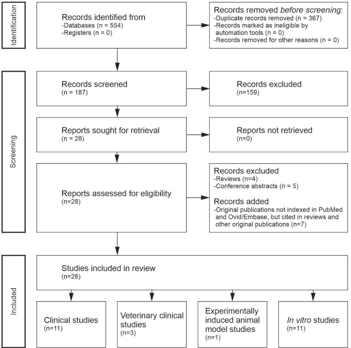
Table 1.
Characteristics and main outcomes of human and veterinary studies evaluating nuclear magnetic resonance therapy / molecular biophysical stimulation therapy (NMRT/MBST). Abbreviations: NT, number of patients in the treatment group; NC, number of patients in the control group; PEF, primary endpoint follow-up; LF, longest follow-up.; RCT, randomized controlled trial; ACL, anterior cruciate ligament. a, assessed one week after completion of the 7-day treatment series (i.e., 8 weeks post-ACL transection for early-treatment and matched control groups, and 14 weeks post-ACL transection for late-treatment and matched control groups.
Table 1.
Characteristics and main outcomes of human and veterinary studies evaluating nuclear magnetic resonance therapy / molecular biophysical stimulation therapy (NMRT/MBST). Abbreviations: NT, number of patients in the treatment group; NC, number of patients in the control group; PEF, primary endpoint follow-up; LF, longest follow-up.; RCT, randomized controlled trial; ACL, anterior cruciate ligament. a, assessed one week after completion of the 7-day treatment series (i.e., 8 weeks post-ACL transection for early-treatment and matched control groups, and 14 weeks post-ACL transection for late-treatment and matched control groups.
| Study | Design | Indication | Key Findings | NT | NC | PEF | LF | Risk of Bias Assessment |
| [70] | Pre–post MRI structural study | Knee osteoarthritis | Statistically significant increases in patellar cartilage thickness and volume and significant increases in multiple tibial cartilage parameters following NMRT/MBST. | 14 | 0 | 10 weeks | 10 weeks | High (non-controlled pre–post design; small sample; objective imaging endpoints mitigate but do not remove bias) |
| [71] | Randomized, double-blind, placebo-controlled trial | Knee osteoarthritis | Significant improvements occurred in both NMRT/MBST and placebo groups; NMRT/MBST did not demonstrate superiority over placebo at 12 weeks across pain/function measures and imaging endpoints (ultrasound cartilage thickness; MRI WORMS). | 50 | 50 | 12 weeks | 12 weeks | Low (strong RCT methodology with blinding and imaging endpoints; neutral result reduces risk of overestimation) |
| [72] | Long-term questionnaire-based follow-up survey | Knee osteoarthritis | Survey-based evidence of sustained improvement in pain and daily-life functional limitations up to four years after NMRT/MBST, with slight pain increase toward the end of the observation period. | 39 | 0 | 4 months | Up to 4 years | High (survey-based self-report and recall bias; no comparator; long follow-up provides valuable durability signal) |
| [73] | Large multicenter longitudinal observational survey | Degenerative rheumatic disease (osteoarthritis and spinal pain) | Sustained improvements in pain and function across multiple indications, with benefits persisting through 12 months. | ≈4500 | 0 | 3–12 months | 12 months | High (observational design without comparator; large sample and consistent longitudinal trends strengthen plausibility but confounding remains) |
| [77] | Randomized, blinded, sham-controlled in vivo animal study (ACL-transection rabbit model) | Post-traumatic knee osteoarthritis induced by bilateral ACL transection in rabbits | Early NMRT/MBST initiation (6 weeks post-transection) significantly reduced macroscopic OA severity compared with sham (p<0.01), driven by fewer fibrillations, reduced osteophyte formation and less joint effusion. No macroscopic improvement when treatment was initiated at 12 weeks. No histological improvement (Mankin score) at either timepoint. | 8+8 | 4+4 | One week after completion of treatmenta | 14 weeks after ACL transection (end of evaluation period) | Moderate risk of bias. Strengths include randomization, blinded macroscopic assessment and sham-treated controls. Limitations include small group sizes, differing exclusions between groups, absence of blinding for histology explicitly stated and potential confounding due to ongoing joint instability after ACL transection. |
| [78] | Randomized, placebo-controlled clinical trial | Finger-joint osteoarthritis | Significant and sustained improvements in pain and hand function in the NMRT/MBST group; placebo group showed no improvement and deterioration in some measures at 6 months. | 35 | 35 | 6 weeks | 6 months | Low-to-moderate (placebo-controlled RCT; modest sample size; outcome measures include validated pain/function scales) |
| [79] | Randomized, double-blind, placebo-controlled inpatient rehabilitation trial | Chronic low back pain | Greater durability of pain reduction in the NMRT/MBST group versus placebo at 3 months; improvements also observed in disability-related measures. | 31 | 31 | 3 months | 3 months | Moderate (controlled design supports internal validity; inpatient context and limited follow-up may influence generalizability) |
| [80] | Randomized, double-blind, placebo-controlled inpatient rehabilitation trial | Chronic low back pain | Active NMRT/MBST and placebo groups both improved under standardized inpatient rehabilitation; NMRT/MBST showed more consistent advantages in dynamic pain (pain under stress) and selected disability domains (e.g., personal care), with higher proportions reporting improvement and fewer deteriorations. Sleep and walking improved similarly across groups at follow-up. | 31 | 31 | One week and 3 months | 3 months | Moderate (controlled, blinded inpatient trial with clinically relevant outcomes; several domains show NMRT/MBST advantages, but improvements in both groups and inpatient context affect generalizability) |
| [81] | Randomized, double-blind, placebo-controlled trial | Nerve-root irritation following lumbar disc pathology | Significant reduction in sick-leave days in the NMRT/MBST group versus placebo, with group differences more apparent at later follow-up points. | 54 | 54 | 12 weeks | 12 weeks | Low-to-moderate (double-blind RCT; modest sample; pragmatic endpoint reduces subjectivity, but details may vary by subgroup) |
| [82] | Randomized, double-blind, placebo-controlled trial | Lumbar disc herniation with lumbar radicular syndrome | Both groups improved over time; NMRT/MBST showed a statistically significant advantage in pain at week 4 and significantly fewer sick-leave days at 3 months; MRI morphology improved similarly in both groups. | 54 | 54 | 12 weeks | 12 weeks | Low (double-blind RCT with sham control and predefined outcomes; some endpoints show limited between-group separation) |
| [83] | Prospective pre–post densitometric study | Osteoporosis | Statistically significant improvements in bone mineral density (T-scores) at lumbar spine and hip regions at 12 months. | 103 | 0 | 12 months | 12 months | Moderate (objective DXA outcomes; absence of control group limits causal attribution) |
| [84] | Retrospective longitudinal cohort with case descriptions | Osteoporosis | Long-term follow-up describing severe trauma events without fracture in selected cases after NMRT/MBST; cohort followed up to 5 years. | 450 | 0 | 1 to 3 months | Up to 5 years | High (retrospective uncontrolled design; potential selection/reporting bias; clinically meaningful endpoint but not systematically compared) |
| [85] | Long-term veterinary case report | Canine hip osteoarthritis secondary to dysplasia | Delayed but substantial functional improvement after NMRT/MBST, with reproducible benefit after retreatment and sustained mobility with yearly cycles over nine years. | 1 | 0 | ~3 months | 9 years | Very high (single uncontrolled case; narrative outcome assessment; nevertheless minimizes human placebo explanations) |
| [86] | Controlled veterinary trial with objective locomotor outcomes | Canine elbow osteoarthritis | Objective gait, lameness, pain, and range-of-motion measures were largely stable in both groups. The MBST/NMRT group showed numerically favorable trends—such as lower symmetry indices and improved range of motion at 3 months—while the placebo group tended to remain unchanged or decline. Although differences were not statistically significant, overall treatment-effectiveness patterns indicate that some MBST/NMRT-treated dogs showed clinically relevant improvements, suggesting a modest functional benefit in canine elbow osteoarthritis. | 14 | 14 | 3 months | 6 months | Moderate (controlled veterinary setting; objective endpoints strengthen reliability; sample size and heterogeneity may limit precision) |
| [87] | Randomized, double-blinded, placebo-controlled veterinary trial | Canine osteoarthritis (various joints) | NMRT/MBST produced significant within-group improvements in vertical impulse symmetry (SI_IFz) and lameness at 3 months, while placebo showed no comparable change. Between-group differences were not statistically significant, but overall treatment effectiveness scores favored MBST at all follow-up points. Improvements diminished by 6 months; pain scores did not differ between groups. | 15 | 15 | 3 months | 6 months | Moderate (rigorous blinding and objective gait metrics strengthen validity; small sample, dropouts, heterogeneous OA sites, and allowance of concomitant treatments limit interpretability). |
Table 2.
Summary of experimentally demonstrated molecular mechanisms of nuclear magnetic resonance therapy / molecular biophysical stimulation therapy across cell systems. Abbreviations: ECM, extracellular matrix; DRG, dorsal root ganglion; ROS, reactive oxygen species.
Table 2.
Summary of experimentally demonstrated molecular mechanisms of nuclear magnetic resonance therapy / molecular biophysical stimulation therapy across cell systems. Abbreviations: ECM, extracellular matrix; DRG, dorsal root ganglion; ROS, reactive oxygen species.
| Mechanistic Domain | Specific Effects Observed | Model Systems / Cell Types | Supporting Studies |
| Anti-inflammatory effects |
|
Osteoarthritic chondrocytes | [42,43,44] |
| Cell proliferation & viability |
|
Osteoblasts, chondrocytes | [41,42,43] |
| Anabolic/ECM maintenance pathways |
|
Chondrocytes | [42,43] |
| Extracellular matrix remodeling (non-proliferative) |
|
Human dermal fibroblasts | [45] |
| Neuronal growth & support |
|
DRG neurons, Schwann cells | [52,53] |
| Mitochondrial function / energy homeostasis |
|
Chondrocytes, Schwann cells, fibroblast clock reporters | [43,44,52,53,54] |
| Hypoxia-related signalling |
|
Zebrafish fibroblasts & larvae, chondrocytes, NIH3T3 clock cells | [44,51,52,54,61] |
| Redox regulation |
|
NIH3T3 fibroblasts (Per2:Luc), chondrocytes | [44,54] |
| Radical-pair / magnetosensitive mechanisms |
|
NIH3T3 cells, circadian clock systems | [54,61,62] |
| Epigenetic regulation |
|
Osteoarthritic chondrocytes | [44] |
| Circadian clock regulation |
|
NIH3T3 Per2:Luc cells, zebrafish cells & larvae | [51,54,61,62] |
Table 3.
Consolidated comparison of the presently established biological effects of nuclear magnetic resonance therapy / molecular biophysical stimulation (NMRT/MBST) and photobiomodulation / near-infrared laser therapy (PBM/NILT), summarizing points of mechanistic convergence and divergence across both modalities.
Table 3.
Consolidated comparison of the presently established biological effects of nuclear magnetic resonance therapy / molecular biophysical stimulation (NMRT/MBST) and photobiomodulation / near-infrared laser therapy (PBM/NILT), summarizing points of mechanistic convergence and divergence across both modalities.
| Effect/Outcome | NMRT/MBST | PBMT/NILT |
| Cell proliferation & viability |
|
|
| Anti-inflammatory effects | ||
| Mitochondrial function & metabolism | ||
| Chondrogenic & osteogenic stimulation | ||
| Neuronal regeneration & neuroprotection |
|
|
Disclaimer/Publisher’s Note: The statements, opinions and data contained in all publications are solely those of the individual author(s) and contributor(s) and not of MDPI and/or the editor(s). MDPI and/or the editor(s) disclaim responsibility for any injury to people or property resulting from any ideas, methods, instructions or products referred to in the content. |
© 2026 by the authors. Licensee MDPI, Basel, Switzerland. This article is an open access article distributed under the terms and conditions of the Creative Commons Attribution (CC BY) license.
Copyright: This open access article is published under a Creative Commons CC BY 4.0 license, which permit the free download, distribution, and reuse, provided that the author and preprint are cited in any reuse.