Preprint
Review

This version is not peer-reviewed.

Improvement of Adeno-Associated Virus (AAVs)- and Nano-Particle-Based Technologies by Cell Penetrating Penta-Peptides (CPP5s)

Submitted:

26 December 2025

Posted:

29 December 2025

You are already at the latest version

Abstract

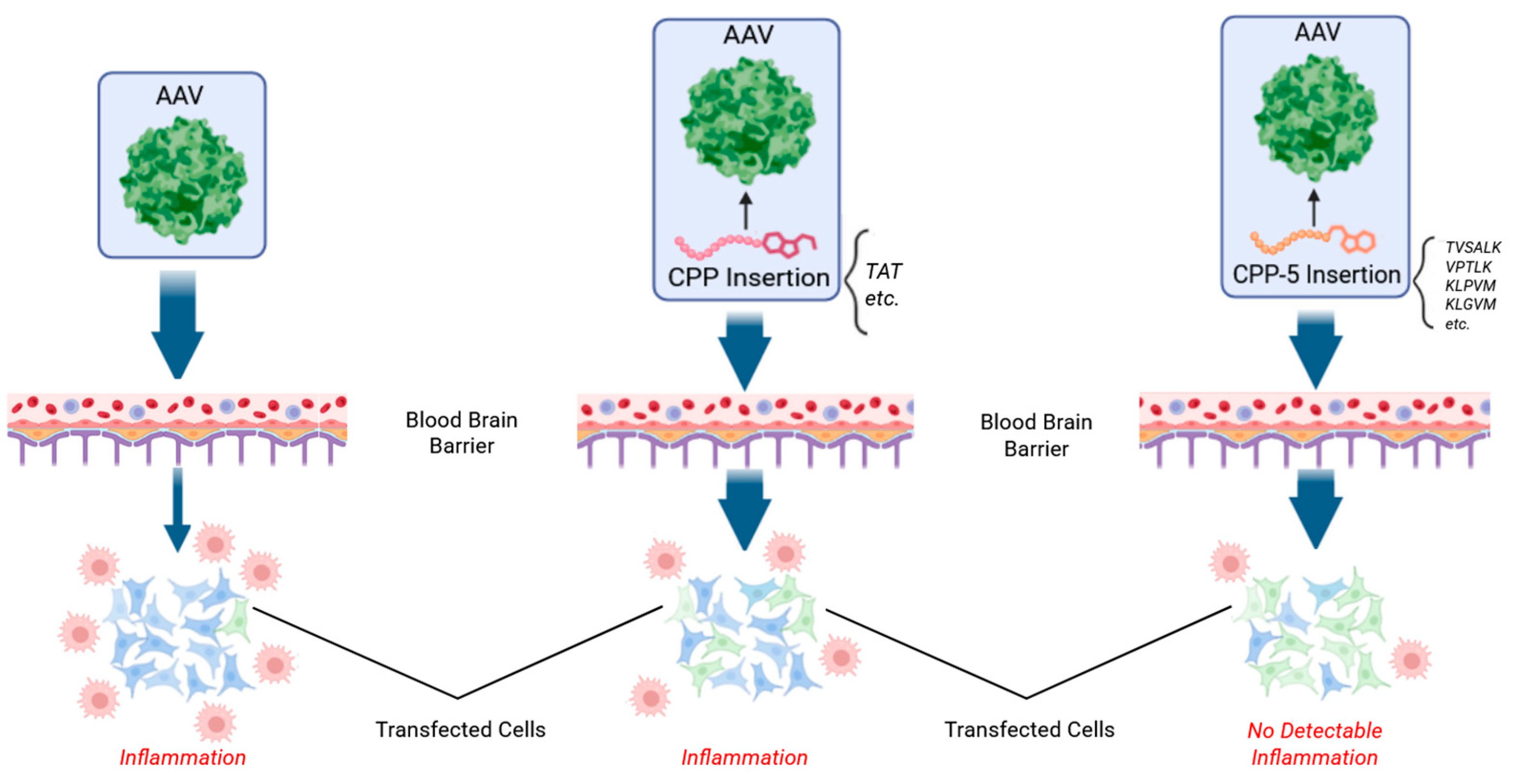
Adeno-Associated Viruses (AAVs) and nanoparticles have been used to deliver DNA or RNA to target cells for gene therapy technologies. These vectors are also useful for DNA- or RNA-based vaccines. Although AAVs and nanoparticles are promising technologies, two major technical problems remain. One problem is that the commonly used AAVs have a low efficiency to penetrate the blood-brain-barrier (BBB) and the blood-retina-barrier (BRB). Consequently, gene delivery to the nervous system has limitations. Another problem is that AAVs induce immune reactions that cause serious side effects. To avoid immune reactions, the AAV dose must be reduced to lower levels that may result in insufficient gene delivery. To overcome these problems, researchers searched for effective peptide sequences by modifying viral capsid proteins. As a result, Cell Penetrating Penta-Peptides (CPP5s) have been shown to be effective in improving the BBB/BRB penetration of AAV and the suppression immune reactions against AAV. CPP5s were originally developed from peptide sequences of Bax (a pro-apoptotic protein) binding domain of Ku70 (a DNA repair protein) and from negative control cell penetrating peptides without Bax-binding activity. This article will discuss the background science of CPP5 and the future direction of the use of CPP5 for AAV- and nanoparticle-mediated gene delivery to the nervous system as well as other organs.

Keywords: 
;  ;  ;  ;  ;  ;  ;  ;  

1. Introduction

Cell Penetrating Penta-Peptides (CPP5s) are a group of penta-peptides with the ability to deliver cargo molecules (e.g. proteins) into a cell, that was developed by our laboratory [1,2,3,4]. Recently, CPP5s have been re-discovered as useful tools to improve the efficacy and safety of Adeno-associated virus (AAV)-based gene therapy technology. In this manuscript, we will summarize the background science of CPP5s and review recent publications utilizing CPP5s for the improvement of AAV gene therapy-related technology including the intra-cellular delivery of DNA recombinases.
AAVs have become an FDA-approved delivery method for human gene therapy medicines [5,6,7,8]. These vehicles are increasingly being chosen for their low pathogenicity, ability to transduce non-dividing cells, and reduced immune responses [5,9,10]. Although AAVs have showed effective gene transfers in several organs such as the liver, skeletal muscle, and bone marrow, they are limited for neurological disorders due to the difficulties of viral delivery across the Blood-Brain-Barrier (BBB) and the Blood-Retina-Barrier (BRB) [11]. While certain serotypes such as AAV9 have been shown to cross the BBB from intravascular administration, its transduction efficiency remains lower compared to other tissues [12]. To improve efficacy, higher doses of AAVs are needed, but this approach increases toxicity by eliciting greater host immune response [13,14]. Notably, AAV gene therapy has been linked to 11 patient fatalities since 2023, leading to concerns for patient safety [15,16,17]. Current AAV research aims to improve tissue specificity, transduction efficiency, genome expression, and reduced host immune responses [18].
To improve the BBB penetration, researchers have been investigating the effects of the modification of the capsid surface proteins essential for the virus-host cell interaction [19,20,21,22,23]. As one of these attempts, a group of researchers examined Cell Penetrating Peptides (CPPs) to modify the AAV’s surface proteins [24,25,26,27]. CPPs are a group of short peptides ranging from 5 amino acids to over 40 amino acids that have been shown to have an activity to deliver cargo molecules (proteins (e.g. Immunoglobulin or Green Fluorescence Protein (GFP)), small molecules, and nucleic acids (DNA and RNA)) [28]. For their cargo delivery activity, CPPs are directly attached to the cargo (especially in the case of protein) or attached to the delivery-packaging tool such as liposomes [29]. These CPPs then facilitate cellular uptake by overcoming the cell membrane barrier [30,31,32,33,34], though the precise molecular mechanism of cell entry and cargo delivery has not been fully understood [4,35]. Previous studies suggest that host cells continuously internalize and recycle their cell surface area, facilitating the entry of CPPs through endocytosis pathways [36,37]. Although CPPs can generally facilitate the penetration of plasma membrane, the efficacy of cargo delivery is influenced by heterogeneity of the cell membrane of each cell types [38].
By engineering AAV viral capsids with CPPs, it has been expected to improve the poor tissue selectivity and low transduction efficiency of AAVs [39]. For example, AAVv128, a synthetic AAV8 enhanced transduction ability within retinal tissues [40]. THR peptide directly interacted with AAV8 to cross the BBB and enhance transduction in the brain [19]. PhP.B peptide enhanced BBB transduction 40-fold in the AAV9 serotype [41]. CPPs, such as TAT-HA2 or LAH4, incubated with AAV6 increased transduction efficiency in Jurkat T cells 10-fold [27]. Synthetic CPPs such as Tat-Y1068 improved AAV2 transduction efficiency in fibroblast and epithelial cells in a dose-dependent manner [42]. CPPs also have useful characteristics that reduce the immunogenicity of AAVs. For example, an anti-inflammatory CPP, KAFAK, penetrated the BBB and reduced the production of proinflammatory cytokines [43]. Another example of a prion protein derived CPP (Prp peptide) that have shown to exhibit anti-amyloid properties and re-direct pathways to reduce cell-toxic molecular structures in vivo [44]. Together, previous studies suggest that CPPs have potential in addressing the flaws (low efficiency, low penetration efficiency of BBB, immune response, and toxicities) of AAV delivery.

2. Cell-Penetrating Penta-Peptides (CPP5s)

2.1. Background History of the Discovery and Invention of CPP5s

Cell-penetrating penta-peptides (CPP5s) are a group of peptides comprised of five amino acids originally designed from Bax-binding domains of Ku70 of various species (human, mouse, rat, chicken, frog, etc.) and their negative control peptides, which retain CPP activity without Bax inhibiting activity (Table 1) [1,2,45]. CPP5s were developed by our laboratory, and this chapter will briefly explain the history and background of CPP5s.
Bax is a proapoptotic member of the Bcl-2 family of proteins that is an evolutionary conserved protein family regulating mitochondria-dependent cell death [46]. To identify proteins rescuing cells from Bax-induced cell death, we performed yeast-based functional screening [2]. In this system, yeast cells were genetically engineered to express cytotoxic levels of Bax (mouse Bax) under the galactose-inducible system [47]. To identify Bax suppressor genes, cDNA expression library generated from mouse brain was co-transfected to the yeast cells. As the result, Ku70 was identified as an inhibitor of Bax-induced cell death [46]. Previously, Ku70 has been known as an evolutionary conserved protein functioning as an essential factor for DNA double strand break repair mechanisms [48]. The cell death inhibition activity of Ku70 was confirmed in mammalian cells by our group and numerous other groups [1,3,45,49,50,51]. For example, our lab and others showed that anti-Bax activity of Ku70 is inactivated by post-translational modifications of Ku70 (ubiquitinylation and acetylation) [49,50,52,53]. Importantly, the ubiquitin-dependent Ku70- degradation can be suppressed by Akt survival kinase signals indicating that the survival kinase pathway maintains both cellular life and genomic integrity through the maintenance of Ku70 levels in the cells [49]. Furthermore, Bax gene knockout extended the lifespan of Ku70 knockout mice exhibiting accelerated aging [54,55]. These genetic studies suggest that the absence of Ku70 increases the occurrence of Bax-induced cell death which accelerate age-associated degenerative diseases. These studies support the hypothesis that Ku70 has an anti-Bax activity. To determine the molecular mechanism of Ku70-mediatred Bax inhibition, Bax binding domain of Ku70 has been identified, and penta-peptides mimicking the Bax binding domains of Ku70 were designed [1,56]. These peptides were named Bax Inhibiting Peptide (BIPs). Although BIPs showed cell death inhibition activity in certain conditions, it requires relatively high concentration (more than 200 uM) for cell death inhibition [3]. Due to this weak anti-cell death activity, the potential of BIP as a cell death therapeutic is not high. However, as explained later, BIPs did not show cytotoxicity even at 1.6 mM concentration suggesting that BIPs have a favorable “non-toxic” character as a CPPs to deliver cargo molecules [3].
Importantly, we also developed negative control peptides of BIP that have CPP activity but not Bax inhibiting activity [3]. To design negative control peptides, the amino acid sequence was flipped or randomized, and their cell entry activities were examined. Notably, KLPVM (one of the mutated peptides that do not inhibit Bax) showed the best cell-penetrating activity among the tested CPP5s [3]. We realized these negative control pentapeptides are valuable as CPPs, and we further designed additional pentapeptides based on these peptides. For example, based on KLPVM, we designed modified pentapeptides such as KLGVM. This KLGVM was recently rediscovered by Dr. Gao’s team, showing that KLGVM can enhance the gene delivery activity of in the retina as explained later [57].

2.2. Mechanism of Cell Penetration of CPP5s

The mechanisms of cell entry and cargo delivery activity of CPPs (including CPP5s) are not fully understood. It is plausible that different types of CPPs have their own specific mechanisms for cell entry and cargo delivery. As for CPP5s, by precise concentration measurements of isotope-labelled peptides inside of the cells using mass spectrometric analysis, we reported that CPP5s are likely to utilize a receptor-mediated cell entry mechanism [3]. At present, the receptor itself has not been identified yet. The efficiency of cell-penetrating activities of CPP5s is limited by the availability of the putative CPP5 receptor [3]. To utilize CPP5s to improve AAV- or nanoparticle-mediated drug delivery, the identification of the receptor (or mediator) will be necessary to further improve the drug delivery technologies [3].
In comparison with polycationic CPPs (Tat and poly-arginine peptides), CPP5 cell entry activity is relatively low when the activity was compared by using cell entry activities in cultured cells [3]. For example, the cell entry activity of CPP5 was approximately 10 times lower than that of Tat peptide [3]. Although CPP5s have lower cell entry activity than Tat peptide, CPP5s have much less cytotoxicity than Tat peptide [3]. Tat and poly-arginine peptides are known to show cytotoxicity when they are used at high doses, such as 100 uM or higher, likely due to their cationic properties [3]. On the other hand, CPP5s were not toxic even at 1.6 mM in cell culture [58]. Although Tat peptide is frequently used for bioactive peptide delivery, especially in the neuroscience field, it is important to remember that Tat peptide is a highly cationic peptide and it can bind negatively charged surfaces of any type of molecules, including proteins, nucleotides, and lipids. Due to this chemical character of the Tat peptide, the use of Tat peptide requires caution about the specificity and toxicity. On the other hand, CPP5s are not highly charged peptides (most of CPP5s have only one positively charged amino acid (Table 1), and they do not randomly neutralize negatively charged molecules in the cell [3].
The cell-penetrating activity of CPP5s were observed even in specific cells lacking proteoglycan (negatively charged molecule on the plasma membrane) [3,59]. Unlike cationic CPPs, most of CPP5s consist of four non-polar or uncharged amino acids and only one positively charged amino acid (e.g. K or E) (Table 1). Therefore, CPP5s are unlikely to be interacting with negatively charged cell surface molecules such as proteoglycans [60,61]. CPP5s also exhibited cell penetrating activity in Jurkat cells, which has very low caveolin-mediated endolytic activity [62]. Two CPP5s, KLPVM and VPTLK also displayed protein-transduction activity in Jurkat cells [1,3]. These unique features of CPP5s require further research to identify the primary receptor mediating mechanism for cell entry.

3. CPP5s Enhance AAV9 Delivery in the Brain of Mice and Non-Human Primates (Figure 1)

In 2022, Dr. Bei’s laboratory reported a groundbreaking article showing that utilization of VSALK, a CPP5, can improve gene delivery and BBB penetration of AAV9 [23]. They tested 14 CPPs to identify the best peptide sequence as a modifier of AAV capsid that can improve BBB penetration. As a result, they found that two CPP5s, VPALK and VSALK, showed positive results. Since these CPP5s were originally designed from Ku70 [1,2], the research team further tested TVSALK based on the original Ku70 amino acid sequence (The amino acid T is located on the N-terminus portion of VPALK in Ku70). The team found that TVSALK showed further improved gene transduction activity in comparison with VSALK, and they named this sequence (TVSALK) as CPP16. This peptide insert was further modified by adding F between L and K creating variant AAV.CPP.21, (CPP.21 has TVSLFK sequence). Intravenous injection of AAV.CPP.16 and AAV.CPP.21 showed increasing gene transduction efficiency of red fluorescent protein of up to 249-fold in comparison with the control AAV in four strains of adult mice brain cells. Consistently, AAV.CPP16 and AAV.CPP21 showed higher brain transduction than unmodified AAV9 intravenous injection, with no significant difference between AAV.CPP.16 and 21. AAV.CPP.16 was also capable of transducing 10-51% of total neurons, 20-49% of total astrocytes, 5-12% of total oligodendrocytes in comparison with non-modified AAVs [23].
Figure 1. Utilization of CPP5 increase the penetration of the Blood-Brain-Barrier (BBB) and minimize immune reaction [23]. Created in BioRender. Diener, A. (2025) https://BioRender.com/q17wmho.
Preprints 191628 g001
Furthermore, the authors confirmed that CPP5-containing AAV9 penetrated the BBB in primate brains. By using CPP.16 and 21, the efficiency of AAV9-mediated gene delivery to the brain cortex increased to 7.0-fold and 1.9-fold compared to the AAV9 without CPP, respectively. An up to 13.2-fold increase for AAV.CPP.16 and up to 20-fold increase in AAV.CPP.21 transduction efficiency in other brain regions were also established. At high doses, AAV.CPP.16 had greater transduction efficiency than AAV.CPP.21 in primates [23]. Previously it has been noted that higher doses of standard AAV vectors (without CPP.16 or CPP.21) carry the risk of leakage of the BBB, a pathological increase in membrane permeability. However, injection of dextran showed no leakage from the blood into the CNS and no difference in clearance between AAV.CPP.16 and AAV9 [23,63].

4. CPP5 Containing CPP.16 Enhances AAV Delivery in The Respiratory Tract

AAV.CPP.16 was further evaluated for transduction efficiency in airway and lung cell tissues [64]. In cultured human nasal epithelial cells, AAV.CPP.16 was 3.0-fold and 6.6-fold greater than AAV6 and AAV9 at delivering a GFP reporter respectively. Intranasal administration of AAV.CPP.16 into mouse nasal cavity, trachea, and lung all showed similar increases in GFP-derived fluorescence in comparison with wild-type AAV9 by 2.7-fold and AAV6 by 1.8-fold. Furthermore, AAV.CPP.16 penetrated deeper into lung and respiratory tissue, all with no-to-little transduction into other peripheral tissue such as the brain, retina, or muscle. In the nasal cavity, AAV.CPP.16 transduced 42.7% of goblet cells, while transduction of AAV6 was only 3.0% and AAV9 was 7.8%. In the trachea, AAV.CPP.16 transduced 71.0% of club cells, compared to 29.6% for AAV6 and 20.2% for AAV9. In the lung, AAV.CPP.16 transduced 13.6% of ciliated cells, compared to 2.8% for AAV6 and 1.9% for AAV9. In non-human primates, intranasal delivery of CPP.16 improved AAV9-mediated GFP gene delivery into the respiratory tract up to 10.5-fold compared to AAVs lacking CPP. Likewise, AAV.CPP.16 transduced GFP gene in 14-23% of goblet cells, 11-18% of club cells, and 27-33% of type I alveolar cells, while AAV9 without CPP resulted in little to no transduction. Notably, no AAV expression was found in peripheral tissues and no abnormal weight loss or adverse effects were noted in mice or non-human primates [64].
The AAV.CPP.16 was further modified to deliver a decoy receptor of TGF-β1 to act as a treatment for idiopathic pulmonary fibrosis. After inducing pulmonary fibrosis with bleomycin in mice, the decoy receptor expression by AAV.CPP.16 significantly reduced protein levels of TGF-β1 and VEGFα compared to non-treated mice. Furthermore, there were significantly reduced levels of mRNA for fibrogenic genes and inflammatory cytokines. This resulted in reduced expression of extracellular matrix proteins, decreased inflammatory cytokines, and reduced fibroblast-associated proteins in mouse lungs after CPP enhanced trap treatment [64].
CRISPR RNA endonuclease targeting viral RNA in SARS-CoV-2 was also modified with AAV.CPP.16 to measure prophylactic effectiveness [64]. In this mouse pseudovirus model, CasRx-gRNA.Niran and CasRx-gRNA.Interface were packaged into AAV.CPP.16, and Rdrp transcription of the pseudovirus was reduced by 50% and 60% respectively. Despite the pseudovirus inducing expression 50-300-fold in mouse respiratory tissues, AAV.CPP.16 mediated treatment had near-complete block of Rdrp transcription in the nostrils and was reduced 70% in the trachea and 67% in the lung [64]. These results suggest that CPP-integrated AAV may be able to improve the efficiency of CRISPR/Cas9-mediated gene therapy in the future.

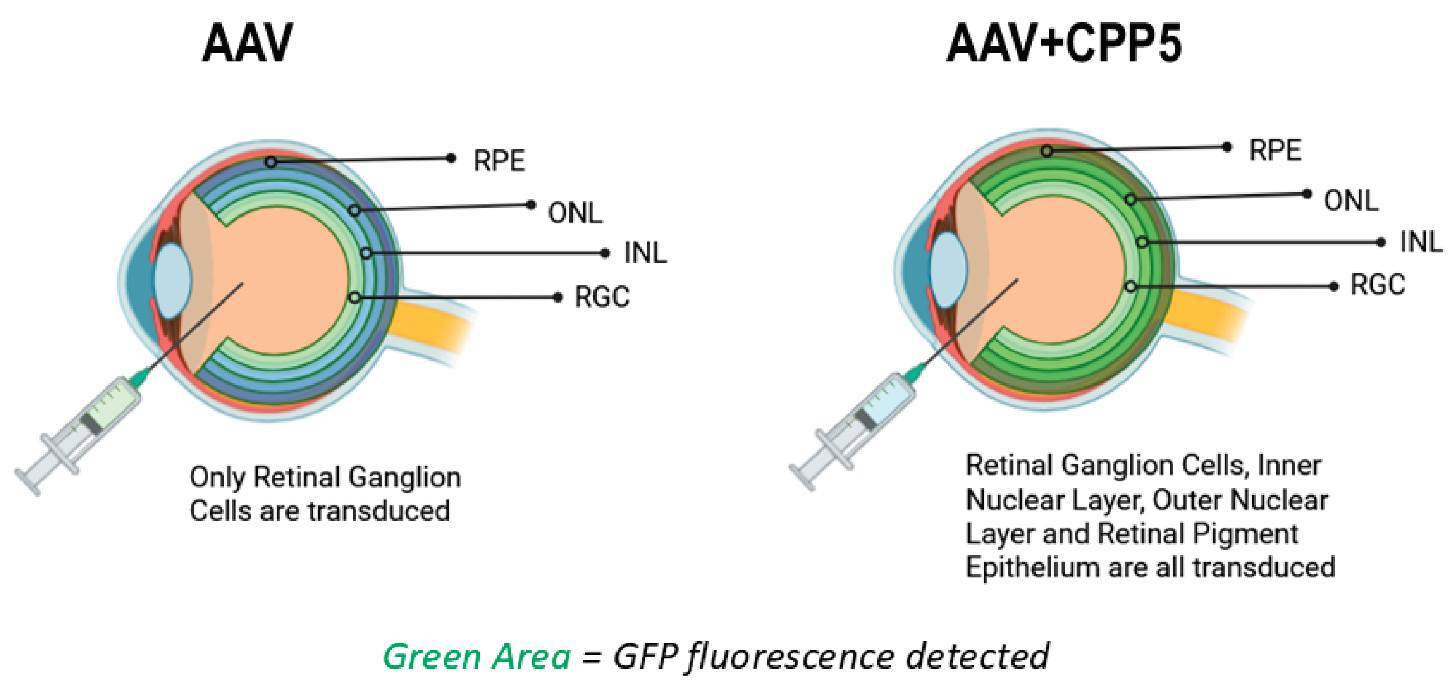
5. CPP5 Enhances AAV2 Delivery in the Retina (Figure 2)

He et al. examined whether CPP5 containing CPPs (CPP.16 and CPP.21) can improve gene delivery to the retinal cells by AAV2 using intravitreal injection route [65]. The authors found that AAV2.CPP.21 had the highest level of pan-retinal GFP’s fluorescence intensity (GFP cDNA was used as a reporter gene), with significant GFP expression in the outer nuclear layer (photoreceptor) and inner nuclear layer (bipolar cells). Non-modified AAV2 was found to primarily transduce cells in the retinal ganglion cell (RGC) layer, whereas AAV2.CPP.16 mediated fluorescence expression in deeper retinal cells. Notably, AAV2.CPP.21 was found to have the deepest penetration extending to the outermost RPE layer without noticeable retinal toxicity or transgene leakage in peripheral organs. Overall, CPP-integrated recombinant AAV2 showed improved efficiency of AAV2 delivery and targeting of the retina.
In rd1 mice (Pde6b gene mutation mouse model of retinitis pigmentosa), He et al. observed that AAV2.CPP.21 delivery of Pde6b gene rescued pupillary light reflex behavior and restored thickness of the retina when compared to AAV2 [65]. This was further confirmed by RNA sequencing and PCR, showing dramatically elevated gene expression of Pde6b and phototransduction in mice treated with AAV2.CPP.21 when compared to AAV2. The AAV2.CPP.21-treated group had a stronger visual perception ability and photo transduction genes, indicating that AAV.CPP.21 mediated gene delivery effectively attenuated photoreceptor degeneration in rd1 mice.
Figure 2. Utilization of CPP5 increase the penetration of the Blood-Retina-Barrier (BRB) [57,65]. RPE: Retinal Pigment Epithelial Cells, ONL: Outer Nuclear Layer (Photoreceptor), INL: Inner Nuclear layer (Bi-polar cells), RGC: Retinal Ganglion Cells. Created in BioRender. Diener, A. (2025) https://BioRender.com/glu4j9i.
Preprints 191628 g002

6. Mechanisms of How CPP.16 and CPP.21 Helps AAV for Gene Delivery

The precise molecular mechanism of how CPP.16 and CPP.21 enhance gene delivery of AAV is not fully understood yet. Since CPP.16 (TVSALK) is similar to the BIP designed from the Bax binding domain of Ku70, there is a possibility that CPP.16 increased the survival of host cells by inhibiting Bax-mediated cell death. However, this possibility is extremely low. BIP requires at least 200 uM concentration to achieve cell death inhibition [3], and the extracellular concentration of AAV.CPP16 in the target tissues (e.g. the brain, lung, and retina) would have been much lower than 200 uM in the experimental condition testing gene delivery in animal models. Notably, Dr. Bei’s team reported that they observed that AAV.CPP16 did not decrease Bax expression levels in HEK293T. Based on this result, they hypothesized that AAV.CPP16 does not function as a Bax inhibitor. However, there was a misunderstanding about the mechanism of Bax inhibition by Ku70 and Ku70-derived peptide in their study. Our original study showed that Ku70 directly binds and inhibits Bax without changing Bax expression levels [1,3,45,49,50,51]. The mechanism of Bax inhibition by Ku70 is to inhibit the Bax conformational change, but Ku70 does not alter Bax expression levels [3,45,49]. Furthermore, Ku70 has been reported to be expressed on the cell surface, and Ku70 functions as the receptor for extracellular molecules including parasitic organisms [66]. The authors of this manuscript speculate that AAV.CPP.16 may interact with Ku70 binding target molecules on the cell surface to enter the host cells.

7. KLGVM, a CPP5, Enhances AAV2 Delivery and Reduces Immunogenicity in the Retina (Figure 2)

Wang et al. reported that three versions of CPP5s (VPTLK, KLPVM, and KLGVM), significantly improved the gene transduction of AAV2 in the retina [57]. It has been known that the extracellular matrix of the retina has heparan sulfate proteoglycan which binds (traps) AAV2, and this interaction prevents the entry of AAV2 into the retinal cells [57]. To overcome this problem, Wang et al. generated a library of CPP-containing AAV2 and searched CPPs that can reduce the binding of AAV2 to heparin sulfate [57]. As the result, KLGVM was identified as the best CPP for this purpose after the screening of 1070 previously reported CPPs including TAT, poly-arginine, and other CPPs [57]. Other CPP5s, such as VPTLK and KLPVM also enhanced enrichment in the retina, but KLGVM was the most effective. They named the KLGVM-integrated AAV2 as AAV2.CPP1 [57]. The CPP5 sequence (KLGVM) was inserted within the VR-VIII region of AAV Capsid potentially interrupting the R585 and R588 binding motif of heparin sulfate proteoglycans [57].
Following intravitreal injection in mice, AAV2.CPP1 transduced reporter GFP gene in photoreceptors 2.5-fold greater than ssAAV2.7m8.CB6.GFP, a previously optimized AAV vector for retinal cell transduction. The transduction of AAV2.CPP1 was observed to be higher proportion in Müller cells, and present in the outer nuclear layer (the photoreceptor layer) to the retinal ganglion cell layer [57].
Previously, similarly to AAV.CPP1, AAV2.7m8 was developed as an AAV2 variant that has a low affinity to heparan sulfate proteoglycan [67]. Although AAV2.7m8 has an increased efficiency in gene transduction to the target tissue, AAV2.7m8 also increased immune response of the host tissue in comparison with the original AAV2. For example, AAV2.7m8 induced microglial cell migration into the inner plexiform layer more than the original AAV2 does. AAV2.CPP1, however, did not have an increased immune response in treated retinas. In fact, the RNA levels of inflammatory cytokines such as IL-6 were significantly lower in AAV2.CPP1 when compared to AAV2.7m8 [57].

8. Applications of CPP5s for Therapeutic Protein Delivery into Retinal Cells as Well as Other Cell Types

In addition to AAV-based technology, CPP5s as well as other CPPs have been used to deliver bioactive molecules into the target cells both in vitro and in vivo [68,69]. In 2024, Hołubowicz et al. repotted that CPPs were able to deliver Cre-DNA recombinase protein into the retinal photoreceptor in mouse when the CPP-conjugated Cre protein was administrated by intraocular injection [70]. In this study, three representative CPPs including CPP5, Tat, and ANTP were fused to Cre-recombinase protein, and all the tested CPPs effectively delivered Cre protein into the retina. The intracellular delivery of Cre was validated by Cre-recombinase-dependent GFP expression in the retina. Although CPP5 was able to deliver Cre-protein in the retinal cells in vivo, the study reported that the Cre-protein delivery activity was not detectable in vitro (cell culture system). We speculate that this negative result is due to the low concentration of CPP5-Cre protein in their experiment. In this study, only 1 uM concentration of CPP5-Cre was used [70]. Previously, we reported that cargo delivery activity of CPP5 became clearly detectable from 10 uM, but the activity was not detectable at 1 uM. Therefore, the negative result in Hołubowicz’s report is consistent with our previous observation. If CPP5-Cre was tested at 10 uM or higher concentration, the protein transduction activity would have been detected in their study. To be noted, Kang et al. (2021) reported that CPP5-Cre delivered Cre protein into the primary cultured porcine fibroblast and CPP5-Cre (4 uM concentration was used) achieved 90% efficiency of Cre-dependent gene recombination [71]. In this study by Kang et al, CPP5 showed the best gene recombination induction among three CPPs examined (CPP5, Tat, and R9) [71].
There are other successful applications of CPP5s for protein transduction both in vivo and in vitro. The examples of these applications are: (1) In mice, VPALR, one of CPP5s, was conjugated to Sulpiride (atypical antipsychotic medication in a benzamide class) and the CPP5 increased drug bioavailability, half-life, and increased transport across the BBB to enhance antidepressive effects [72]. (2) VPTLK enhances mini-chaperone activity of the peptides derived from alphaA-crystallin protein (amino acids 70-88) and prevented β-amyloid-induced cell death [73]. (3) The CPP5 conjugation to the AL3810 liposome attenuated the immune response induced by liposome and improved the delivery of anti-tumorigenic compound targeting glioma [74]. (4) QLPVM was found to allow transferrin-coated liposome to penetrate BBB in mouse model and QLPVM-conjugated liposome was able to deliver Doxorubicin (an anti-cancer drug) across BBB [75].

9. Summary and Future Direction

AAVs have been considered a relatively safe vector for gene therapy [76,77]. In ophthalmology, AAV-mediated gene delivery for a recessive genetic disease (PDE6b mutation-induced retinitis pigmentosa) has been approved by the FDA [78]. On the other hand, AAV-mediated gene therapy for Duchenne muscular dystrophy is now confronting serious risks due to the three fatal incidents of patients with an immune reaction against the AAV therapy [17,79,80]. Further improvements in safety and efficiency are warranted for more reliable AAV-based gene therapy.
In the case of neurological disorders including retinal diseases, the presence of the BBB and BRB are a major obstacle for gene delivery. As discussed in this review, CPP5s have been re-discovered as a beneficial tool to improve AAV penetration of the BBB [23] and BRB [57,65]. Furthermore, CPP5-integrated AAV showed much lower immune response in comparison with previously developed AAVs [57,65]. At present, the exact mechanism for how CPP5s facilitate cell entry is not well understood. In the case of KLPVM and KLGVM, these CPP5s seem to allow AAVs to bypass the proteoglycan barrier outside of the cell [57]. In the case of CPP.16 and CPP.21 [23], the mechanism of how these CPP can allow AAV to penetrate BBB and BRB is not fully understood. To develop more efficient and safe technologies for AAV-based gene therapy, the mechanism for CPP-mediated cell penetration requires further study.

Author Contributions

Conceptualization, C.W.G. and S.M.; writing—original draft preparation, C.W.G. and S.M.; writing—review and editing, C.W.G., S.M., and A.D.; visualization, A.D.; supervision, S.M.; project administration, S.M.; funding acquisition, S.M. All authors have read and agreed to the published version of the manuscript.

Funding

This work is partially supported by Foundation Fighting Blindness (FFB) Translational Research Acceleration Program to our laboratory.

Acknowledgments

We are grateful to the supports from the members in Matsuyama laboratory.

Conflicts of Interest

The authors declare no conflicts of interest.

Abbreviations

The following abbreviations are used in this manuscript:
CPP5 Cell penetrating penta-peptide
AAV Adeno-associated virus
BBB Blood brain barrier
BRB Blood retina barrier
CPP Cell-penetrating peptide
BIP Bax inhibiting peptide
GFP Green fluorescent protein
RPE Retinal pigment epithelium
RGC Retinal ganglion cell

References

  1. Yoshida, T.; Tomioka, I.; Nagahara, T.; Holyst, T.; Sawada, M.; Hayes, P.; Gama, V.; Okuno, M.; Chen, Y.; Abe, Y.; et al. Bax-Inhibiting Peptide Derived from Mouse and Rat Ku70. Biochem Biophys Res Commun 2004, 321, 961–966. [Google Scholar] [CrossRef]
  2. Gomez, J.A.; Gama, V.; Yoshida, T.; Sun, W.; Hayes, P.; Leskov, K.; Boothman, D.; Matsuyama, S. Bax-Inhibiting Peptides Derived from Ku70 and Cell-Penetrating Pentapeptides. Biochem Soc Trans 2007, 35, 797–801. [Google Scholar] [CrossRef]
  3. Gomez, J.A.; Chen, J.; Ngo, J.; Hajkova, D.; Yeh, I.-J.; Gama, V.; Miyagi, M.; Matsuyama, S. Cell-Penetrating Penta-Peptides (CPP5s): Measurement of Cell Entry and Protein-Transduction Activity. Pharmaceuticals 2010, 3, 3594–3613. [Google Scholar] [CrossRef]
  4. Gomez, J.; Matsuyama, S. Cell-Penetrating Penta-Peptides and Bax-Inhibiting Peptides: Protocol for Their Application. In Methods in Molecular Biology; Humana Press, 2011; Vol. 683, pp. 465–471. [Google Scholar]
  5. Pupo, A.; Fernández, A.; Low, S.H.; François, A.; Suárez-Amarán, L.; Samulski, R.J. AAV Vectors: The Rubik’s Cube of Human Gene Therapy. Molecular Therapy 2022, 30, 3515–3541. [Google Scholar] [CrossRef] [PubMed]
  6. Mendell, J.R.; Al-Zaidy, S.A.; Rodino-Klapac, L.R.; Goodspeed, K.; Gray, S.J.; Kay, C.N.; Boye, S.L.; Boye, S.E.; George, L.A.; Salabarria, S.; et al. Current Clinical Applications of In Vivo Gene Therapy with AAVs. Molecular Therapy 2021, 29, 464–488. [Google Scholar] [CrossRef]
  7. Byrne, B.J.; Flanigan, K.M.; Matesanz, S.E.; Finkel, R.S.; Waldrop, M.A.; D’Ambrosio, E.S.; Johnson, N.E.; Smith, B.K.; Bönnemann, C.; Carrig, S.; et al. Current Clinical Applications of AAV-Mediated Gene Therapy. Molecular Therapy 2025, 33, 2479–2516. [Google Scholar] [CrossRef]
  8. Liu, D.; Li, T.; Liu, L.; Che, X.; Li, X.; Liu, C.; Wu, G. Adeno-Associated Virus Therapies: Pioneering Solutions for Human Genetic Diseases. Cytokine Growth Factor Rev 2024, 80, 109–120. [Google Scholar] [CrossRef]
  9. Zaman, H.; Khan, A.; Khan, K.; Toheed, S.; Abdullah, M.; Zeeshan, H.M.; Hameed, A.; Umar, M.; Shahid, M.; Malik, K.; et al. Adeno-Associated Virus-Mediated Gene Therapy. Crit Rev Eukaryot Gene Expr 2023, 33, 87–100. [Google Scholar] [CrossRef] [PubMed]
  10. Zhou, X.; Liu, J.; Xiao, S.; Liang, X.; Li, Y.; Mo, F.; Xin, X.; Yang, Y.; Gao, C. Adeno-Associated Virus Engineering and Load Strategy for Tropism Modification, Immune Evasion and Enhanced Transgene Expression. Int J Nanomedicine 2024, Volume 19, 7691–7708. [Google Scholar] [CrossRef] [PubMed]
  11. Merkel, S.F.; Andrews, A.M.; Lutton, E.M.; Mu, D.; Hudry, E.; Hyman, B.T.; Maguire, C.A.; Ramirez, S.H. Trafficking of Adeno-Associated Virus Vectors across a Model of the Blood-Brain Barrier; a Comparative Study of Transcytosis and Transduction Using Primary Human Brain Endothelial Cells. J Neurochem 2017, 140, 216–230. [Google Scholar] [CrossRef]
  12. Sands, M.S. AAV-Mediated Liver-Directed Gene Therapy. In; 2012; pp. 141–157.
  13. Rabinowitz, J.; Chan, Y.K.; Samulski, R.J. Adeno-Associated Virus (AAV) Versus Immune Response. Viruses 2019, 11, 102. [Google Scholar] [CrossRef]
  14. Barnes, C.; Scheideler, O.; Schaffer, D. Engineering the AAV Capsid to Evade Immune Responses. Curr Opin Biotechnol 2019, 60, 99–103. [Google Scholar] [CrossRef]
  15. Wilton-Clark, H.; Yokota, T. Safety Concerns Surrounding AAV and CRISPR Therapies in Neuromuscular Treatment. Med 2023, 4, 855–856. [Google Scholar] [CrossRef]
  16. Whiteley, L.O. An Overview of Nonclinical and Clinical Liver Toxicity Associated With AAV Gene Therapy. Toxicol Pathol 2023, 51, 400–404. [Google Scholar] [CrossRef]
  17. Duan, D. Lethal Immunotoxicity in High-Dose Systemic AAV Therapy. Molecular Therapy 2023, 31, 3123–3126. [Google Scholar] [CrossRef]
  18. Srivastava, A. Rationale and Strategies for the Development of Safe and Effective Optimized AAV Vectors for Human Gene Therapy. Mol Ther Nucleic Acids 2023, 32, 949–959. [Google Scholar] [CrossRef] [PubMed]
  19. Zhang, X.; He, T.; Chai, Z.; Samulski, R.J.; Li, C. Blood-Brain Barrier Shuttle Peptides Enhance AAV Transduction in the Brain after Systemic Administration. Biomaterials 2018, 176, 71–83. [Google Scholar] [CrossRef] [PubMed]
  20. Moyer, T.C.; Hoffman, B.A.; Chen, W.; Shah, I.; Ren, X.-Q.; Knox, T.; Liu, J.; Wang, W.; Li, J.; Khalid, H.; et al. Highly Conserved Brain Vascular Receptor ALPL Mediates Transport of Engineered AAV Vectors across the Blood-Brain Barrier. Molecular Therapy 2025, 33, 3902–3916. [Google Scholar] [CrossRef] [PubMed]
  21. Shay, T.F.; Sullivan, E.E.; Ding, X.; Chen, X.; Ravindra Kumar, S.; Goertsen, D.; Brown, D.; Crosby, A.; Vielmetter, J.; Borsos, M.; et al. Primate-Conserved Carbonic Anhydrase IV and Murine-Restricted LY6C1 Enable Blood-Brain Barrier Crossing by Engineered Viral Vectors. Sci Adv 2023, 9. [Google Scholar] [CrossRef]
  22. Huang, Q.; Chen, A.T.; Chan, K.Y.; Sorensen, H.; Barry, A.J.; Azari, B.; Zheng, Q.; Beddow, T.; Zhao, B.; Tobey, I.G.; et al. Targeting AAV Vectors to the Central Nervous System by Engineering Capsid–Receptor Interactions That Enable Crossing of the Blood–Brain Barrier. PLoS Biol 2023, 21, e3002112. [Google Scholar] [CrossRef]
  23. Yao, Y.; Wang, J.; Liu, Y.; Qu, Y.; Wang, K.; Zhang, Y.; Chang, Y.; Yang, Z.; Wan, J.; Liu, J.; et al. Variants of the Adeno-Associated Virus Serotype 9 with Enhanced Penetration of the Blood–Brain Barrier in Rodents and Primates. Nat Biomed Eng 2022, 6, 1257–1271. [Google Scholar] [CrossRef]
  24. Váňová, J.; Hejtmánková, A.; Kalbáčová, M.H.; Španielová, H. The Utilization of Cell-Penetrating Peptides in the Intracellular Delivery of Viral Nanoparticles. Materials 2019, 12, 2671. [Google Scholar] [CrossRef]
  25. Feiner, R.C.; Kemker, I.; Krutzke, L.; Allmendinger, E.; Mandell, D.J.; Sewald, N.; Kochanek, S.; Müller, K.M. EGFR-Binding Peptides: From Computational Design towards Tumor-Targeting of Adeno-Associated Virus Capsids. Int J Mol Sci 2020, 21, 9535. [Google Scholar] [CrossRef]
  26. Váňová, J.; Hejtmánková, A.; Žáčková Suchanová, J.; Sauerová, P.; Forstová, J.; Hubálek Kalbáčová, M.; Španielová, H. Influence of Cell-Penetrating Peptides on the Activity and Stability of Virus-Based Nanoparticles. Int J Pharm 2020, 576, 119008. [Google Scholar] [CrossRef] [PubMed]
  27. Moço, P.D.; Dash, S.; Kamen, A.A. Enhancement of Adeno-associated Virus Serotype 6 Transduction into T Cells with Cell-penetrating Peptides. J Gene Med 2024, 26. [Google Scholar] [CrossRef]
  28. Cerrato, C.P.; Langel, Ü. An Update on Cell-Penetrating Peptides with Intracellular Organelle Targeting. Expert Opin Drug Deliv 2022, 19, 133–146. [Google Scholar] [CrossRef]
  29. Gestin, M.; Dowaidar, M.; Langel, Ü. Uptake Mechanism of Cell-Penetrating Peptides. In; 2017; pp. 255–264.
  30. Okafor, M.; Schmitt, D.; Ory, S.; Gasman, S.; Hureau, C.; Faller, P.; Vitale, N. The Different Cellular Entry Routes for Drug Delivery Using Cell Penetrating Peptides. Biol Cell 2025, 117. [Google Scholar] [CrossRef]
  31. Zahid, M.; Robbins, P. Cell-Type Specific Penetrating Peptides: Therapeutic Promises and Challenges. Molecules 2015, 20, 13055–13070. [Google Scholar] [CrossRef]
  32. Dowaidar, M. Uptake Pathways of Cell-Penetrating Peptides in the Context of Drug Delivery, Gene Therapy, and Vaccine Development. Cell Signal 2024, 117, 111116. [Google Scholar] [CrossRef] [PubMed]
  33. Taylor, R.E.; Zahid, M. Cell Penetrating Peptides, Novel Vectors for Gene Therapy. Pharmaceutics 2020, 12, 225. [Google Scholar] [CrossRef] [PubMed]
  34. Moreno-Vargas, L.M.; Prada-Gracia, D. Exploring the Chemical Features and Biomedical Relevance of Cell-Penetrating Peptides. Int J Mol Sci 2024, 26, 59. [Google Scholar] [CrossRef] [PubMed]
  35. Cleal, K.; He, L.; D. Watson, P.; T. Jones, A. Endocytosis, Intracellular Traffic and Fate of Cell Penetrating Peptide Based Conjugates and Nanoparticles. Curr Pharm Des 2013, 19, 2878–2894. [Google Scholar] [CrossRef]
  36. Steinman, R.M.; Mellman, I.S.; Muller, W.A.; Cohn, Z.A. Endocytosis and the Recycling of Plasma Membrane. J Cell Biol 1983, 96, 1–27. [Google Scholar] [CrossRef] [PubMed]
  37. Futaki, S.; Nakase, I. Cell-Surface Interactions on Arginine-Rich Cell-Penetrating Peptides Allow for Multiplex Modes of Internalization. Acc Chem Res 2017, 50, 2449–2456. [Google Scholar] [CrossRef]
  38. Thorén, P.E.G.; Persson, D.; Esbjörner, E.K.; Goksör, M.; Lincoln, P.; Nordén, B. Membrane Binding and Translocation of Cell-Penetrating Peptides. Biochemistry 2004, 43, 3471–3489. [Google Scholar] [CrossRef]
  39. Berry, G.E.; Asokan, A. Cellular Transduction Mechanisms of Adeno-Associated Viral Vectors. Curr Opin Virol 2016, 21, 54–60. [Google Scholar] [CrossRef]
  40. Luo, S.; Jiang, H.; Li, Q.; Qin, Y.; Yang, S.; Li, J.; Xu, L.; Gou, Y.; Zhang, Y.; Liu, F.; et al. An Adeno-Associated Virus Variant Enabling Efficient Ocular-Directed Gene Delivery across Species. Nat Commun 2024, 15, 3780. [Google Scholar] [CrossRef]
  41. Pietersz, K.L.; Du Plessis, F.; Pouw, S.M.; Liefhebber, J.M.; van Deventer, S.J.; Martens, G.J.M.; Konstantinova, P.S.; Blits, B. PhP.B Enhanced Adeno-Associated Virus Mediated-Expression Following Systemic Delivery or Direct Brain Administration. Front Bioeng Biotechnol 2021, 9. [Google Scholar] [CrossRef]
  42. Tabata, K.; Sugano, E.; Murakami, F.; Yamashita, T.; Ozaki, T.; Tomita, H. Improved Transduction Efficiencies of Adeno-Associated Virus Vectors by Synthetic Cell-Permeable Peptides. Biochem Biophys Res Commun 2016, 478, 1732–1738. [Google Scholar] [CrossRef]
  43. Yanamadala, Y.; Roy, R.; Williams, A.A.; Uppu, N.; Kim, A.Y.; DeCoster, M.A.; Kim, P.; Murray, T.A. Intranasal Delivery of Cell-Penetrating Therapeutic Peptide Enhances Brain Delivery, Reduces Inflammation, and Improves Neurologic Function in Moderate Traumatic Brain Injury. Pharmaceutics 2024, 16, 774. [Google Scholar] [CrossRef] [PubMed]
  44. Österlund, N.; Wärmländer, S.K.T.S.; Gräslund, A. Cell-Penetrating Peptides with Unexpected Anti-Amyloid Properties. Pharmaceutics 2022, 14, 823. [Google Scholar] [CrossRef]
  45. Li, Y.; Yokota, T.; Gama, V.; Yoshida, T.; Gomez, J.A.; Ishikawa, K.; Sasaguri, H.; Cohen, H.Y.; Sinclair, D.A.; Mizusawa, H.; et al. Bax-Inhibiting Peptide Protects Cells from Polyglutamine Toxicity Caused by Ku70 Acetylation. Cell Death Differ 2007, 14, 2058–2067. [Google Scholar] [CrossRef] [PubMed]
  46. Singh, R.; Letai, A.; Sarosiek, K. Regulation of Apoptosis in Health and Disease: The Balancing Act of BCL-2 Family Proteins. Nat Rev Mol Cell Biol 2019, 20, 175–193. [Google Scholar] [CrossRef] [PubMed]
  47. Matsuyama, S.; Xu, Q.; Velours, J.; Reed, J.C. The Mitochondrial F0F1-ATPase Proton Pump Is Required for Function of the Proapoptotic Protein Bax in Yeast and Mammalian Cells. Mol Cell 1998, 1, 327–336. [Google Scholar] [CrossRef]
  48. Downs, J.A.; Jackson, S.P. A Means to a DNA End: The Many Roles of Ku. Nat Rev Mol Cell Biol 2004, 5, 367–378. [Google Scholar] [CrossRef]
  49. Gama, V.; Gomez, J.A.; Mayo, L.D.; Jackson, M.W.; Danielpour, D.; Song, K.; Haas, A.L.; Laughlin, M.J.; Matsuyama, S. Hdm2 Is a Ubiquitin Ligase of Ku70-Akt Promotes Cell Survival by Inhibiting Hdm2-Dependent Ku70 Destabilization. Cell Death Differ 2009, 16, 758–769. [Google Scholar] [CrossRef]
  50. Cohen, H.Y.; Lavu, S.; Bitterman, K.J.; Hekking, B.; Imahiyerobo, T.A.; Miller, C.; Frye, R.; Ploegh, H.; Kessler, B.M.; Sinclair, D.A. Acetylation of the C Terminus of Ku70 by CBP and PCAF Controls Bax-Mediated Apoptosis. Mol Cell 2004, 13, 627–638. [Google Scholar] [CrossRef]
  51. Dumitru, R.; Gama, V.; Fagan, B.M.; Bower, J.J.; Swahari, V.; Pevny, L.H.; Deshmukh, M. Human Embryonic Stem Cells Have Constitutively Active Bax at the Golgi and Are Primed to Undergo Rapid Apoptosis. Mol Cell 2012, 46, 573–583. [Google Scholar] [CrossRef]
  52. Yurchenko, V.; Xue, Z.; Gama, V.; Matsuyama, S.; Sadofsky, M.J. Ku70 Is Stabilized by Increased Cellular SUMO. Biochem Biophys Res Commun 2008, 366, 263–268. [Google Scholar] [CrossRef] [PubMed]
  53. Gama, V.; Yoshida, T.; Gomez, J.A.; Basile, D.P.; Mayo, L.D.; Haas, A.L.; Matsuyama, S. Involvement of the Ubiquitin Pathway in Decreasing Ku70 Levels in Response to Drug-Induced Apoptosis. Exp Cell Res 2006, 312, 488–499. [Google Scholar] [CrossRef]
  54. Matsuyama, S.; Palmer, J.; Bates, A.; Poventud-Fuentes, I.; Wong, K.; Ngo, J.; Matsuyama, M. Bax-Induced Apoptosis Shortens the Life Span of DNA Repair Defect Ku70-Knockout Mice by Inducing Emphysema. Exp Biol Med 2016, 241, 1265–1271. [Google Scholar] [CrossRef]
  55. Ngo, J.; Matsuyama, M.; Kim, C.; Poventud-Fuentes, I.; Bates, A.; Siedlak, S.L.; Lee, H.; Doughman, Y.Q.; Watanabe, M.; Liner, A.; et al. Bax Deficiency Extends the Survival of Ku70 Knockout Mice That Develop Lung and Heart Diseases. Cell Death Dis 2015, 6, e1706–e1706. [Google Scholar] [CrossRef]
  56. Scott-McKean, J.J.; Matsuyama, M.; Guo, C.W.; Ni, L.; Sassouni, B.; Kurup, S.; Nickells, R.; Matsuyama, S. Cytoprotective Small Compound M109S Attenuated Retinal Ganglion Cell Degeneration Induced by Optic Nerve Crush in Mice. Cells 2024, 13, 911. [Google Scholar] [CrossRef] [PubMed]
  57. Wang, J.-H.; Cui, M.; Liu, H.; Guo, P.; McGowan, J.; Cheng, S.-Y.; Gessler, D.J.; Xie, J.; Punzo, C.; Tai, P.W.L.; et al. Cell-Penetrating Peptide-Grafted AAV2 Capsids for Improved Retinal Delivery via Intravitreal Injection. Mol Ther Methods Clin Dev 2025, 33, 101426. [Google Scholar] [CrossRef]
  58. Jensen, K.; WuWong, D.J.; Wong, S.; Matsuyama, M.; Matsuyama, S. Pharmacological Inhibition of Bax-Induced Cell Death: Bax-Inhibiting Peptides and Small Compounds Inhibiting Bax. Exp Biol Med 2019, 244, 621–629. [Google Scholar] [CrossRef]
  59. Sandgren, S.; Cheng, F.; Belting, M. Nuclear Targeting of Macromolecular Polyanions by an HIV-Tat Derived Peptide. Journal of Biological Chemistry 2002, 277, 38877–38883. [Google Scholar] [CrossRef]
  60. Joliot, A.; Prochiantz, A. Transduction Peptides: From Technology to Physiology. Nat Cell Biol 2004, 6, 189–196. [Google Scholar] [CrossRef]
  61. Hao, M.; Zhang, L.; Chen, P. Membrane Internalization Mechanisms and Design Strategies of Arginine-Rich Cell-Penetrating Peptides. Int J Mol Sci 2022, 23, 9038. [Google Scholar] [CrossRef] [PubMed]
  62. Rhee, M.; Davis, P. Mechanism of Uptake of C105Y, a Novel Cell-Penetrating Peptide. Journal of Biological Chemistry 2006, 281, 1233–1240. [Google Scholar] [CrossRef] [PubMed]
  63. Seitz, I.P.; Michalakis, S.; Wilhelm, B.; Reichel, F.F.; Ochakovski, G.A.; Zrenner, E.; Ueffing, M.; Biel, M.; Wissinger, B.; Bartz-Schmidt, K.U.; et al. Superior Retinal Gene Transfer and Biodistribution Profile of Subretinal Versus Intravitreal Delivery of AAV8 in Nonhuman Primates. Investigative Opthalmology & Visual Science 2017, 58, 5792. [Google Scholar] [CrossRef]
  64. Yang, Z.; Yao, Y.; Chen, X.; Madigan, V.; Pu, S.; Fan, X.; Pu, J.; Bei, F. Cross-Species Tropism of AAV.CPP.16 in the Respiratory Tract and Its Gene Therapies against Pulmonary Fibrosis and Viral Infection. Cell Rep Med 2025, 6, 102144. [Google Scholar] [CrossRef]
  65. He, X.; Fu, Y.; Xu, Y.; Ma, L.; Chai, P.; Shi, H.; Yao, Y.; Ge, S.; Jia, R.; Wen, X.; et al. A Penetrable AAV2 Capsid Variant for Efficient Intravitreal Gene Delivery to the Retina. Invest Ophthalmol Vis Sci 2025, 66, 6. [Google Scholar] [CrossRef]
  66. Martinez, J.J.; Seveau, S.; Veiga, E.; Matsuyama, S.; Cossart, P. Ku70, a Component of DNA-Dependent Protein Kinase, Is a Mammalian Receptor for Rickettsia Conorii. Cell 2005, 123, 1013–1023. [Google Scholar] [CrossRef]
  67. Cui, M.; Su, Q.; Yip, M.; McGowan, J.; Punzo, C.; Gao, G.; Tai, P.W.L. The AAV2.7m8 Capsid Packages a Higher Degree of Heterogeneous Vector Genomes than AAV2. Gene Ther 2024, 31, 489–498. [Google Scholar] [CrossRef]
  68. Ramsey, J.D.; Flynn, N.H. Cell-Penetrating Peptides Transport Therapeutics into Cells. Pharmacol Ther 2015, 154, 78–86. [Google Scholar] [CrossRef] [PubMed]
  69. Bolhassani, A.; Jafarzade, B.S.; Mardani, G. In Vitro and in Vivo Delivery of Therapeutic Proteins Using Cell Penetrating Peptides. Peptides (N.Y.) 2017, 87, 50–63. [Google Scholar] [CrossRef] [PubMed]
  70. Hołubowicz, R.; Du, S.W.; Felgner, J.; Smidak, R.; Choi, E.H.; Palczewska, G.; Menezes, C.R.; Dong, Z.; Gao, F.; Medani, O.; et al. Safer and Efficient Base Editing and Prime Editing via Ribonucleoproteins Delivered through Optimized Lipid-Nanoparticle Formulations. Nat Biomed Eng 2024, 9, 57–78. [Google Scholar] [CrossRef]
  71. Kang, Q.; Sun, Z.; Zou, Z.; Wang, M.; Li, Q.; Hu, X.; Li, N. Cell-Penetrating Peptide-Driven Cre Recombination in Porcine Primary Cells and Generation of Marker-Free Pigs. PLoS One 2018, 13, e0190690. [Google Scholar] [CrossRef]
  72. Liang, Y.; Yang, Y.; Huang, R.; Ning, J.; Bao, X.; Yan, Z.; Chen, H.; Ding, L.; Shu, C. Conjugation of Sulpiride with a Cell Penetrating Peptide to Augment the Antidepressant Efficacy and Reduce Serum Prolactin Levels. Biomedicine & Pharmacotherapy 2024, 174, 116610. [Google Scholar] [CrossRef] [PubMed]
  73. Raju, M.; Santhoshkumar, P.; Sharma, K.K. Cell-Penetrating Chaperone Peptide Prevents Protein Aggregation and Protects against Cell Apoptosis. Adv Biosyst 2018, 2. [Google Scholar] [CrossRef]
  74. Li, J.; Lu, J.; Guo, H.; Zhou, J.; Wang, S.; Jiang, K.; Chai, Z.; Yao, S.; Wang, X.; Lu, L.; et al. A Pentapeptide Enabled AL3810 Liposome-Based Glioma-Targeted Therapy with Immune Opsonic Effect Attenuated. Acta Pharm Sin B 2021, 11, 283–299. [Google Scholar] [CrossRef]
  75. Lakkadwala, S.; Dos Santos Rodrigues, B.; Sun, C.; Singh, J. Biodistribution of TAT or QLPVM Coupled to Receptor Targeted Liposomes for Delivery of Anticancer Therapeutics to Brain in Vitro and in Vivo. Nanomedicine 2020, 23, 102112. [Google Scholar] [CrossRef]
  76. Raguram, A.; Banskota, S.; Liu, D.R. Therapeutic in Vivo Delivery of Gene Editing Agents. Cell 2022, 185, 2806–2827. [Google Scholar] [CrossRef] [PubMed]
  77. Mingozzi, F.; Anguela, X.M.; Pavani, G.; Chen, Y.; Davidson, R.J.; Hui, D.J.; Yazicioglu, M.; Elkouby, L.; Hinderer, C.J.; Faella, A.; et al. Overcoming Preexisting Humoral Immunity to AAV Using Capsid Decoys. Sci Transl Med 2013, 5. [Google Scholar] [CrossRef]
  78. Palmowski-Wolfe, A.; Stingl, K.; Habibi, I.; Schorderet, D.; Tran, H. Novel PDE6B Mutation Presenting with Retinitis Pigmentosa – A Case Series of Three Patients. Klin Monbl Augenheilkd 2019, 236, 562–567. [Google Scholar] [CrossRef] [PubMed]
  79. Assaf, B.T. Systemic Toxicity of Recombinant Adeno-Associated Virus Gene Therapy Vectors. Toxicol Pathol 2024, 52, 523–530. [Google Scholar] [CrossRef]
  80. Laforet, G.A. Thrombotic Microangiopathy Associated with Systemic Adeno-Associated Virus Gene Transfer: Review of Reported Cases. Hum Gene Ther 2025, 36, 64–76. [Google Scholar] [CrossRef] [PubMed]
Table 1. List of CPP5s. Their cell penetration activity was tested and confirmed as previously reported [1,2,3,4].
Sequence Origin and background history
VPMLK Bax binding domain of human, monkey and dog Ku70
VPTLK Bax binding domain of mouse Ku70
VPALR Putative Bax binding domain of rat Ku70
VSALK Putative Bax binding domain of chicken Ku70
PMLKE Bax binding domain of human, monkey and dog Ku70
VPALK Putative Bax binding domain of Ccattle and African clawed frog Ku70
VSLKK Artificially designed CPP5
VSGKK Artificially designed CPP5
KLPVM Artificially designed CPP5
IPMIK Artificially designed CPP5
KLGVM Artificially designed CPP5
KLPVT Artificially designed CPP5
VPMIK Artificially designed CPP5
IPALK Artificially designed CPP5
IPMLK Artificially designed CPP5
VPTLQ Artificially designed CPP5
QLPVM Artificially designed CPP5
ELPVM Artificially designed CPP5
VPTLE Artificially designed CPP5
Amino acids with nonpolar side chains: A, G, C, V, P, L, M, F, W; Amino acids with uncharged side chains: N, Q, S, T, Y.
Disclaimer/Publisher’s Note: The statements, opinions and data contained in all publications are solely those of the individual author(s) and contributor(s) and not of MDPI and/or the editor(s). MDPI and/or the editor(s) disclaim responsibility for any injury to people or property resulting from any ideas, methods, instructions or products referred to in the content.
Copyright: This open access article is published under a Creative Commons CC BY 4.0 license, which permit the free download, distribution, and reuse, provided that the author and preprint are cited in any reuse.
Prerpints.org logo

Preprints.org is a free preprint server supported by MDPI in Basel, Switzerland.

Subscribe

Disclaimer

Terms of Use

Privacy Policy

Privacy Settings

© 2026 MDPI (Basel, Switzerland) unless otherwise stated