Submitted:
01 December 2025
Posted:
02 December 2025
You are already at the latest version
Abstract
The first 1,000 days of life, from conception through the second year, represents a uniquely sensitive period for neurodevelopment. During this time multiple physiological systems undergo rapid and coordinated maturation. Among these, the brain, gut, and sleep system form a tightly interconnected triad, exerting reciprocal influences on each other and playing a pivotal role in shaping lifelong cognitive, emotional, and behavioral trajectories. Disruptions in any one of these domains can reverberate across the others, amplifying developmental vulnerabilities. A key modifiable factor that can modulate this gut-brain-sleep triad is nutrition. In this review, we synthesize current evidence on the interconnected development of the brain, gut, and sleep systems, and examine the role of key nutrients in shaping these pathways. We also identify critical gaps in literature and highlight opportunities for future research to better understand how early-life nutritional interventions can optimize neurodevelopmental outcomes.
Keywords:
children
; infants
; gut
; brain
; sleep
; neurodevelopment
; nutrition
; HMO
; probiotics
; DHA
1. Introduction
The adult brain is a complex structure with more than 100 billion neurons [1]. The 3-millimeter neural tube’s transformation into a fully functional brain represents a rapid phase of growth and development [2]. Most of these processes begin within the first 1000 days of life, laying the foundation for brain development and later cognitive growth [3]. While intrinsic genetic factors drive much of this neurodevelopment, emerging evidence highlights the influence of extrinsic factors, particularly gut health, sleep architecture, and nutrition [4,5]. In early postnatal life, the infant gut microbiota is shaped by prenatal, perinatal, and postnatal exposures and typically stabilizes around 2–3 years of age [6]. However, disruptions in this process may result from factors such as cesarean delivery, antibiotic exposure, early-life stress, or diets lacking key nutrients and probiotics that favorably modulate the gut microbiota, thereby directly impacting neurodevelopmental outcomes. The gut microbiome and the developing brain evolve in parallel, with increasing evidence suggesting a bidirectional relationship that influences emotional regulation and cognitive function [7]. The gut also appears to modulate sleep, another foundational component of healthy neurodevelopment [8]. In infants and young children, sleep is essential for synaptic pruning, memory consolidation, attention, executive functioning, and emotional control. Disrupted sleep during this critical period may adversely affect long-term brain health [9].
Among the many influences on early development, nutrition stands out as the most modifiable factor, shaping the brain, gut, and sleep. Beyond neural development, early-life nutrition also shapes the gut microbiome which is discussed in detail later. Adequate nutrition during the first 1,000 days supports the establishment of a healthy gut microbiota, a cornerstone of long-term immune, metabolic, and neurodevelopmental health. Finally, emerging evidence highlights the bidirectional relationship between nutrition and sleep. Diet quality and specific nutrients modulate hormonal pathways that regulate sleep, which, in turn, influences total energy intake and food choices through biological and behavioral mechanisms [10,11].
2. Development of Brain: First 1000 Days
The maturation of the neural tube into a functional brain exemplifies the remarkable pace and intricacy of early human developmental processes [2]. It starts during the 3rd and 4th week of gestation [12]. Even, at this early stage, nutrition plays a pivotal role in supporting the development and differentiation of various organs. The rapidly developing embryo relies on the mother’s nutrient stores to fuel its growth [13]. Nutrients such as folic acid and iron are especially critical during this phase. Research shows that folic acid is essential for proper neural tube closure, and its deficiency can lead to neural tube defects (NTDs), which may impair cognitive development [14]. Similarly, deficiency in iron during this time has been associated with an increased risk of adverse pregnancy outcomes, such as premature delivery, low birth weight, small-for-gestational-age (SGA) neonates [15] and increased risk of neurodevelopmental disorders (NDDs) in children [16].
Physical and mental stressors experienced by the mother during this period, and in the later months can also impact fetal development by activating the hypothalamic-pituitary-adrenal (HPA) axis. This leads to increased cortisol production, which crosses the placenta and exposes the fetus to maternal stress signals [17]. Such exposure can disrupt fetal brain development, and may result in long-term cognitive, emotional, and behavioral consequences in the offspring [18]. Supporting this, a study conducted with pregnant women with singleton fetuses ranging in gestational age from 28 to 36 weeks, demonstrated that induced emotions in pregnant women affect movements of their fetuses. Increased fetal movements were noticed when pregnant women were being shown a happy film while the opposite was noticed with a sad film, suggesting a link between maternal emotions and stress and fetal response [19].
Overall, this is a time during which the brain displays remarkable plasticity, allowing for significant modification and refinement of synaptic connections. This period serves as a critical window of opportunity, where various mechanisms can shape neural development and ultimately cognitive and behavioral outcomes.
3. Development of Gut Microbiota: The First 1000 Days
As with the developing brain, the gut and its resident microbiome undergo accelerated and dynamic changes during the first 1,000 days of life. These changes have a significant role in establishing the composition and functional capacity of the gut microbiome, with long-term implications for metabolic, immunological, and mental development trajectories through the lifespan [20]. Gut microbes also play a key role in influencing neural development, modulating neurotransmitter systems, and impacting behavior [21,22]. The gut-brain interactions are particularly significant during the perinatal period as well as the first two years of postnatal life (Figure 1). Interactions during these windows contribute to the onset and/or progression of various neurodevelopmental, neuropsychiatric, and neurological disorders [23].
In the first two years after birth, the gut microbiota develops rapidly from a low-diversity state to a more complex composition. Colonization begins at birth [24] and is influenced by several factors discussed below. By 2–3 years of age, it typically resembles a more stable, adult-like state [25].
Factors Impacting Gut Microbiota in the First 1000 Days
Gestational age: Gestational age at birth is a critical factor affecting diversity of the gut microbiome of an infant. Premature infants may often have immature gastrointestinal tract, predisposing the infant to a vicious cycle of an underdeveloped immune system and gut epithelia leading to systemic inflammation or sepsis. This may require use of antibiotics, thereby promoting intestinal bacterial communities that are less diverse and enriched with potential pathogens [26]. The meconium of infants born prematurely (<35 weeks gestation) have shown to have a gut microbiota with reduced number of Bifidobacteria, key bacteria responsible for breaking down human milk oligosaccharides (HMOs) for energy and promoting immune development, and increased number of Enterobacter, Enterococcus, and Staphylococcus. This microbial imbalance heightens vulnerability to infections and disrupts healthy microbiome development compared with term infants [27,28].
Mode of delivery at birth: Several studies have noted that the mode of delivery influences the early infant gut microbiome. Infants born via caesarean section (C-section) demonstrate differences in their microbiota compared to those born through vaginal delivery. More specifically an enrichment of Bifidobacterium spp., Lactobacillus reuteri, Lactobacillus rhamnosus and reduction of opportunistic pathogens such as Enterococcus and Klebsiella spp. is observed in vaginally delivered infants [29,30]. The oral and nasal cavity and other exposed parts of an infant mimic the microflora of the maternal vagina, while that of C-section delivered infants is similar to that of mothers’ skin surface, dominated by Propionibacterium, Corynebacterium, and Staphylococcus spp. [31].This may heighten susceptibility to opportunistic infections. Given the critical role of C-section in safeguarding maternal and neonatal outcomes in high-risk pregnancies, attention to interventions such as supplementation with probiotics [32] and breastfeeding should be encouraged.
Exposure to antibiotics: Antibiotics are often administered during the caesarean birth process, further altering the gut microbiome of both the mother and the infant. Exposure to antibiotics during pregnancy is extremely common [33], generally prescribed for several reasons such as contexts of preterm labor, intrapartum fever during labor, and prevention of neonatal Group B Streptococcus infection. Around 70% of pregnant women use an antibiotic at least once during pregnancy, and antibiotics account for nearly 80% of all prescription medications used during pregnancy [33]. It is worth noting that studies have reported changes including reduced microbial diversity, changes in functional attributes of the microbiota, formation, and selection of antibiotic--resistant strains making hosts more susceptible to infection with pathogens. These alterations in microbial composition may persist for up to 12 weeks post-treatment [34]. Similar to C-section delivery, in cases where antibiotics are unavoidable, supplementation with probiotics may be a viable method to circumvent the microbiome disruption that may occur.
Breastfeeding: Breastfeeding exerts major influence on gut microbiome composition [35]. It can impact the gut microbiota both directly by exposure of the neonate to the mother’s milk microbiota and indirectly, via maternal milk factors that affect bacterial growth and metabolism such as HMOs, secretory immunoglobulin A (SIgA), and anti-microbial factors [36,37]. For instance, a study of 91 term infants that were either exclusively breast-fed or formula-fed, showed increase Bifidobacterium and Bacteroides and decreased Streptococcus and Enterococcus levels in the breast-fed group as compared to the formula fed group [38]. Studies in the past few years have also increasingly reported significant associations between exposure to HMOs in particular during the breastfeeding process and neurodevelopmental outcomes in infants [36]. In a randomized, double-blind trial, comparing formula-fed vs breastfed infants, inclusion of HMOs in the formula led to increased systemic levels of microbial-derived secondary bile acids, bringing those metabolite levels into alignment with those observed in breastfed infants [39]. Overall, this suggests that restoration of certain metabolites derived from microbial activity in the gastrointestinal tract is likely associated with HMOs. The timing of introduction of solid foods and the type is also an important factor in deciding the gut microbial composition [40]. Early introduction of solid foods, i.e. at or before 3 months of age can lead to changes in the levels of gut bacteria and bacterial byproducts. When infants are introduced to foods that are rich in complex carbohydrates, their gut microbiome tends to rapidly mimic an adult’s gut microbiome. Understanding the evolution of bacterial composition in the human gut from infancy and the impact of feeding modality and introduction of solids may help in the development of strategies to support early establishment of health-promoting microbiota. This may in turn have physiological benefits that last through the lifespan [41]
Figure 1- In the first 1000 days of life, gut microbiota development is shaped by factors such as gestational age, mode of delivery, antibiotic exposure, breastfeeding, and timing of solid food introduction. Premature birth and C-section delivery can disrupt microbial diversity and increase infection risk, while breastfeeding and exposure to HMOs promote beneficial bacteria and support immune and neurodevelopment.
Bi-directional Relationship of Gut-Brain: The impact of gut microbiome on the nervous system has been demonstrated in several rodent studies. The hypothesis of gut microbiome’s influence on brain was first demonstrated by Heitz et al. in 2011 where the authors showed that germ-free mice displayed increased motor activity (often associated with increased risk-taking behaviors that suggests impaired cognitive function), compared with specific pathogen free (SPF) mice with a normal gut microbiota [42]. In a subsequent study, fecal microbiota was transplanted from either young (3-4 months) or old (19-20 months) donor mice into aged recipient mice (19-20 months). The study concluded that the transplant of a microbiota from young donors reversed aging-associated differences in peripheral and brain immunity, as well as the hippocampal metabolome and transcriptome of aging recipient mice. Finally, the young donor-derived microbiota attenuated selective age-associated impairments in cognitive behavior when transplanted into an aged host [43]. Such studies, along with the colonization of the microbiome coinciding with the development of the nervous system in a, coordinated manner [44] clearly demonstrate the interdependence of a healthy microbiome of brain health and cognitive function.
However, environmental disruptions during prenatal life including nutritional stressors can interfere with this early developmental process, weakening the connection and leading to both clinical and subclinical outcomes, from altered stress responses to a range of NDDs [21,45]. These effects are mediated via several different pathways including neural pathways (vagus nerve), endocrine impact, immune molecules, and humoral links (cytokines, short-chain and long-chain fatty acids) [46,47,48] shown in Figure 2.
a. Vagal Nerve Pathway
The vagal nerve allows for direct bidirectional communication within the microbiota-gut-brain axis (MGBA), constituting the primary conduit for microbiota-derived signals to interface with the brain. Although the vagal nerve itself is not in direct contact with the gut microbiota, it serves as a receptor for signals transmitted via microbial metabolites, inflammatory processes, or neuroendocrine cells which are mediated by gut microbiota [49,50]. Certain neurotoxic metabolites generated by the gut microbiota can stimulate the vagus nerve and may impair brain function, disrupt sleep [51], and alter stress responses [52]. To this effect, animal studies have demonstrated that treatment with Lactobacillus rhamnosus can reduce stress-induced corticosterone levels and alleviate anxiety and depression-like behaviors in rats [53]. Additionally, the neuroendocrine pathway serves as a key mechanism through which the gut microbiome modulates the central nervous system and the HPA axis, primarily by influencing the secretion of serotonin, cortisol, and melatonin [54]. Serotonin is a critical neurotransmitter involved in regulating behavior, mood, and memory. Specifically, gut-derived serotonin can activate the vagal afferent system and transmitting signals to the specific regions of the brain; cortisol orchestrates the body’s stress response and is often associated with a high vagal tone; and melatonin which plays an essential role in governing the circadian rhythm and sleep–wake cycle is directly affected by vagus stimulation. A physiological balance within this interconnected axis of neuroendocrine mediators is essential for emotional and cognitive well-being through the lifespan.
b. Gut Hormones and Neurotransmitters
Microbiota can either produce neurotransmitter precursors, catalyze the synthesis of neurotransmitters through dietary metabolism, or in combination [21]. For example, the gut microbiota can produce neurotransmitters such as Gamma-Aminobutyric Acid (GABA), dopamine, and serotonin [55]. More than 90% of body’s serotonin, a key neurotransmitter of the brain-gut axis is synthesized in the gut [56]. Bacteria such as Streptococcus spp., Enterococcus spp., Escherichia spp., L. plantarum, Klebsiella pneumonia, and Morganella morganii all have the ability to produce serotonin [56]. Although gut microbes are capable of synthesizing dopamine, this neurotransmitter does not traverse the blood–brain barrier. Consequently, it has been proposed that the gut microbiota modulate brain function through indirect mechanisms, which in turn may influence mood regulation and sleep architecture [57].
c. Short-Chain Fatty Acids (SCFAs)
Gut bacteria ferment dietary fibers and produce short-chain fatty acids (SCFAs), essential chemical compounds such as butyrate, acetate, and propionate. SCFAs, when transported through the stomach, may directly influence vagal afferent nerves, which play crucial roles in regulating satiety, stress responses, and mood. Moreover, microbiota SCFAs may cross the blood-brain barrier (BBB) and enter the bloodstream and cerebrospinal fluid (CSF) within the brain. This potential transport mechanism could directly impact the levels of neurotrophic factors, which govern the development and differentiation of synapses and neurons within the brain [58]. Recent research highlights that SCFAs, such as butyrate, promote the production of brain-derived neurotrophic factor (BDNF), facilitate neurogenesis, and contribute to the consolidation of long-term memory [59].
e. Blood-Brain Barrier (BBB) Integrity
The BBB is a selective semi-permeable membrane between the blood and the interstitium of the brain, that plays a critical role in controlling the influx and efflux of biological substances essential for the brain’s metabolic activity as well as neuronal function [60,61]. The BBB appears to develop as soon as cerebral microvasculature begins to form during early embryonic development. Preclinical data suggests that gut microbiota dysbiosis is associated with increased BBB permeability impacting brain health. For example, Braniste reported that germ-free mice, beginning from intrauterine life, displayed increased BBB permeability compared to pathogen-free mice with a normal gut flora. The increased BBB permeability was maintained in germ-free mice after birth and during adulthood and was associated with reduced expression of the tight junction proteins occludin and claudin-5, which are known to regulate barrier function in endothelial tissues [62].
d. Immune System Modulation
The interplay between the commensal microbiota and the immune system development and function includes multifold interactions [63]. The gut microbiota plays an important role in the regulation of immune responses by either stimulating the innate immunity through the lymphoid tissue located in the intestine system, or through the interaction between bacterial fragments and receptors placed on the surface of epithelial and immune cells that activate specific systemic and local immune responses [64].
Figure 2- The gut microbiome and brain are deeply interconnected, influencing each other through neural, hormonal, immune, and metabolic pathways. Key mechanisms include the vagus nerve, neurotransmitter production, short-chain fatty acids, immune modulation, and blood-brain barrier integrity.
4. Development of Sleep: The First 1000 Days
Sleep architecture also undergoes rapid changes during early childhood (i.e., infants require the most sleep, toddlers less sleep than infants, and so on until late adolescence when adult-like levels of sleep are recommended) [65]. During early childhood, sleep appears to be both influenced by and a modulator of gut microbial composition and function. Disruptions in early microbial development have been associated with alterations in sleep-wake cycles, circadian rhythm consolidation, and behavioral outcomes. Given that both sleep and gut health are critical during the first 1000 days, understanding their interplay may offer new insights into strategies for optimizing neurodevelopmental outcomes through microbiome-targeted interventions.
While fetal eye movements (EMs) can be observed by ultrasonography from 15 weeks gestation [66], infants are born with an immature circadian system [67]. By 10–12 weeks after birth, circadian rhythms begin to emerge, enabling longer nighttime sleep. Total sleep duration decreases from 16–17 hours in newborns to 14–15 hours by 16 weeks, and 13–14 hours by 6 months. As daytime sleep declines, nighttime sleep increases, shifting toward a predominantly nocturnal pattern by the end of the first year [68]. This period of infancy is crucial to establish sleep patterns and through complex interactions involving microbial metabolites (e.g., SCFAs), neuroactive compounds (e.g., serotonin, GABA), and modulation of systemic inflammation, the gut microbiome exerts significant influence on sleep regulation as well.
A longitudinal study identified a compelling association between gut microbiota diversity and infant sleep patterns. The study investigated the interplay between sleep habits, gut microbiota, and behavioral development in 162 healthy infants at 3, 6, and 12 months of age, with a follow-up behavioral assessment at 24 months. The cohort consisted of term-born, vaginally delivered, and breastfed infants, with no critical confounding health factors. Across the first year of life, Bifidobacterium and Bacteroides were the predominant bacterial genera. Notably, the gut microbiome underwent a more pronounced compositional shift between 6 and 12 months than between 3 and 6 months. Two primary enterotypes emerged: enterotype A, dominated by Bifidobacterium, and enterotype B, dominated by Bacteroides. The majority of infants transitioned from enterotype A to B during the second half of the first year. Specifically, higher alpha diversity was linked to fewer and shorter daytime naps, suggesting more mature or “advanced” sleep behavior. This association was the strongest at 3 months of age and diminished thereafter. Additionally, infants with a more mature gut microbiota exhibited increased nighttime activity and awakenings [69]. Comparable associations were observed in another randomized double-blind trial with 161 infants who received cow’s milk-based infant formula (Control) or similar formula with an added prebiotic blend (polydextrose and galactooligosaccharides [PDX/GOS]) from 14–35 to 112 days of age. The study concluded faster consolidation of daytime waking state in infants receiving prebiotics [70]. Similarly, the Norwegian Mother, Father and Child Cohort Study (MoBa), concluded that children with reported colic were more likely to sleep less than recommended (22%) and to have more frequent night awakenings (14%) than usual for their age (6 months to 5 years) [71]. A double-blind, placebo-controlled randomized trial was conducted in Chengdu, China to evaluate the impact of Bifidobacterium animalis subsp. lactis BB-12® on sleep in breastfed infants with colic. The study enrolled 192 full-term infants younger than 3 months of age who met the ROME III criteria for colic. Following a 1-week run-in, infants were randomly assigned to receive either BB-12 (1×10⁹ cfu/day) or placebo for 3 weeks. Sleep outcomes were captured through 24-hour structured diaries completed by caregivers. At the end of the intervention, infants supplemented with BB-12 showed a significant improvement in daily sleep duration compared with those receiving placebo, with mean increases of 60.7±104.0 minutes versus 31.9±102.7 minutes per day, respectively (P<0.001). In addition to sleep, BB-12 supplementation was associated with reductions in crying/fussing duration and frequency, suggesting that improved sleep may be linked to alleviation of colic symptoms [72]. Together, these studies highlight emerging evidence that gut microbiome modulation can shape infant sleep, with consistent links to nap patterns, sleep consolidation, and night awakenings.
While direct studies that manipulate sleep to examine its effects on the microbiome and subsequent cognitive development in infants and young children remain limited, this area is gaining growing scientific attention. Evidence from studies in older children and adults indicates that healthy sleep is critical for normal brain development and cognitive functioning, supporting key processes such as memory consolidation, attention, executive functioning, and emotional regulation [68,73].
During a typical night, one’s sleep alternates through cycles of Rapid Eye Movement (REM) and Non-Rapid Eye Movement (NREM) sleep about every 90 minutes [74]. Deprivation of REM sleep has been shown to significantly influence neuronal excitation, a process essential for evaluating potential threats and processing threat-related stimuli. In contrast, NREM sleep deprivation impairs the normal release of certain neurotransmitters, potentially disrupting receptor recovery and sensitivity. The absence of these critical sleep stages ultimately leads to diminished cognitive functioning [75,76]. This concept is relatively straightforward and easy to interpret in individuals with established sleep architecture, such as adults. Drawing clear conclusions in infants is more challenging, as they lack mature circadian rhythms and well-defined REM and NREM sleep cycles. However, a few studies have suggested a potential link between early sleep characteristics and subsequent development of cognitive functions, memory [77] and language skills in infants and young children. For instance, a study was conducted to assess the association of frequency of nighttime awakenings and cognition; it was concluded that frequent nighttime awakenings were associated with poor cognitive function in toddlers. However, a nonlinear association between nighttime awakenings and cognitive performance was found among infants. It is important to note that total sleep duration was not associated with any developmental indices in both infants and toddlers [78]. In one of the first experimental demonstrations of sleep’s role in memory consolidation, 6 and 12-month-old infants who napped for at least 30 minutes within 4 hours of learning showed significantly better declarative memory for novel actions after both 4 and 24-hour delays, compared to non-napping peers and control groups. Additionally, memory performance was significantly enhanced in the nap group after the 24-hour delay [79]. Similar results have been reported for the impact of sleep on language. An interesting study conducted with twins in Quebec reported that children with language delays at 60 months had less mature sleep consolidation at both 6 and 18 months than children without delays and those with transient early delays [80]. In another longitudinal study infants were assessed at 8 and 14 months, on sleep, cognitive and language skills, and cortisol levels. Findings showed that optimal sleep at 8 months modestly predicted better cognitive and language outcomes at 14 months, highlighting the importance of early sleep patterns for developmental outcomes [81]. Similar results were reported in “The Beijing Longitudinal Study”, where high scores on the Bayley infant development assessment at 6 months predicted less nocturnal awakenings at 1 year of age and insufficient nocturnal sleep at 1 year predicted poor fine motor development at 2 years [82]. The results of the study carried out by Schoch were in line with the other studies. Schoch concluded that both sleep habits and gut microbiota composition were associated with behavioral development, particularly at 3 months. The study also established that sleep patterns were more closely linked to personal-social developmental domains, while gut microbiota composition was more strongly associated with motor development [69] alluding to the impact of sleep on cognition in young children.
Taken together, these studies provide compelling evidence that early-life sleep patterns play a critical role in shaping cognitive, memory, language, and behavioral development in infants and young children. While the architecture of infant sleep is immature and distinct from that of adults, disruptions such as frequent nighttime awakenings or insufficient consolidation have been consistently linked to poorer developmental outcomes. Conversely, timely and sufficient sleep including naps appears to enhance memory consolidation and support language acquisition. Longitudinal findings further emphasize that early sleep quality predicts later cognitive, motor, and social-emotional functioning, underscoring sleep as a foundational process in neurodevelopment. Emerging data also highlights the interplay between sleep, gut microbiota, and developmental trajectories, suggesting that sleep not only reflects but may actively shape broader biological and behavioral systems. Together, these findings position early sleep regulation as a critical window of opportunity for fostering optimal brain development and long-term child wellbeing.
5. Nutritional Needs: The First 1000 Days
Nutritional input during the first 1,000 days of life is a primary modifiable determinant of the coordinated maturation of the brain, gut, and sleep systems mentioned earlier in this review (Figure 3). Nutrients such as omega-3 fatty acids, choline, folate, iodine, vitamin B12, iron, and vitamin D are essential for optimal neurodevelopment [83,84]. Similarly, nutrients like omega-3 fatty acids, iron and vitamin D can support better sleep, while probiotics and HMOs are known to support a healthy microbiome and potentially lay the foundation for the rest of the lifespan. In this section, the role of key nutrients during the first 1,000 days of life is evaluated, along with recommendations provided by expert bodies for some nutrients (Table 1), as well as the existing gaps in guidance and future strategies that should be considered to support healthy maternal–fetal outcomes (Table 2).
5.1. Omega-3 Fatty Acids
The human brain is nearly 60 percent fat [85], and Docosahexaenoic Acid (DHA) is the major n-3 long-chain polyunsaturated fatty acid (PUFAs) in brain gray matter representing about 15% of all fatty acids in the human frontal cortex [86]. The human body can endogenously synthesize most fatty acids; however, it lacks the necessary enzymes for the de novo production of omega-3 and omega-6 PUFAs. Therefore, it relies on dietary intake of their essential precursors, α-linolenic acid (ALA) and linoleic acid (LA) or supplementation with DHA and EPA [87]. Enhanced maternal dietary intake of DHA increases fetal supply and results in higher DHA concentrations in cord blood [88]. DHA supplementation during pregnancy and infancy may offer beneficial effects on the development of visual acuity, cognitive functions and other domains of neurodevelopment [89,90], maturity of sleep patterns, spontaneous motor activity and immune phenotypes [91]. In the Kansas University DHA Outcomes Study (KUDOS), mothers were randomized in a double-blind fashion, to receive either 600 mg/d of DHA or a placebo beginning at 14.5 weeks of gestation until delivery. Children from those pregnancies were evaluated for cognitive and behavioral impact from 10 months through 6 years of age. The study reported a dramatic reduction in early preterm birth and improved visual attention in infancy was identified in the supplemented group, along with a favorable brain response in the Go/No-Go testing at 5.5 years. However, no other pronounced cognitive benefit was seen after controlling for socioeconomic status (SES) [92]. In another triple-blind randomized controlled trial in 150 pregnant women the effects of fish oil supplementation (120 mg DHA and 180 mg EPA) versus placebo were evaluated from the 20th week of pregnancy to 30 days postpartum. The children of mothers who received fish oil supplements showed higher mean scores on all Ages and Stages Questionnaire (ASQ) domains compared to the placebo group, and a statistically significant improvement was specifically observed in the communication domain at 4 months [93]. Although clinical trials on prenatal omega supplementation and subsequent cognitive outcomes in children are limited, clinical studies such as those listed above suggest a positive directional trend supporting its cognitive benefits.
DHA also has an essential role in photoreceptor cells in the retina and is crucial for the growth and survival of photoreceptor cells in the eye. While many studies show that DHA improves vision, the molecular mechanisms are still not fully understood [94]. A double blind, prospective, randomized, and controlled study aimed to test whether maternal DHA supplementation (200 mg/d) during pregnancy improves visual development in healthy term infants, measured through visual evoked potentials (VEPs). One hundred women received either DHA-rich fish oil or a placebo from the 15th week of pregnancy until delivery. The study concluded that infants with higher DHA status at birth showed more mature visual pathway responses. However, maternal DHA supplementation during pregnancy did not enhance VEP maturation in healthy term infants [95]. Another study investigated whether DHA supplementation (200 mg/day) in breastfeeding mothers affects infant brain and visual development. Mothers took DHA or a placebo for 4 months postpartum. Supplementation significantly increased DHA levels in maternal milk (to 0.3% of total fatty acids) and infant plasma. The notable benefit was a higher Psychomotor Development Index at 30 months in the DHA group. There were no significant differences between groups in infant visual functions or general neurodevelopment at earlier timepoints [96].
A large observational study of 11,875 pregnant women found a significant association between low maternal seafood consumption (less than 340 g per week) and suboptimal neurocognitive outcomes in their children [97]. Recognizing the importance of dietary sources of DHA in fetal development, the 2020–2025 Dietary Guidelines for Americans (DGA) recommend that pregnant and breastfeeding women consume 8 to 12 ounces of a variety of seafood per week [98]. The guidelines, being largely based on food sources, introduces a key paradox during pregnancy as some shellfish and fish may also be high in mercury and therefore not recommended for pregnant women [99]. Additionally, women following vegan or vegetarian diets often face challenges in meeting DHA requirements through food alone. Therefore, to support future maternal and fetal health, women planning pregnancy should consume at least 250 mg/day of combined DHA and EPA from diet or supplements. Pregnant women should aim at achieving an average intake of at least 200 mg DHA/d till breastfeeding [91]. Low maternal DHA intake or blood levels are associated with increased risk of preterm and early preterm birth and therefore, women at a higher risk should consider supplementing with 600–1000 mg/day of DHA ± EPA [100].
Maternal DHA status directly influences the DHA content of breast milk and should be adequate to meet infant’s requirements [101]. However, as discussed earlier, deficiencies can arise for various reasons, including maternal dietary restrictions such as strict vegan or vegetarian diets. In such cases, direct DHA supplementation for the infant may be necessary to support optimal neurodevelopment. In a dose-dependent, double-masked, randomized trial [DHA Intake And Measurement Of Neural Development-(DIAMOND Study)] 343 healthy, term, formula-fed infants (1–9 days old) were assigned to one of four formulas varying only in DHA content: 0% (control), 0.32% (17 mg/100 kcal of infant formula), 0.64% (34 mg/100 kcal), or 0.96% DHA (51 mg/100 kcal) all in combination with 0.64% AA. Visual acuity at 12 months was assessed in 244 infants via visual evoked potential. The study concluded that infants who were fed the control formula had significantly poorer visual acuity than those fed any DHA-supplemented formula (P < 0.001). However, increasing DHA levels beyond 0.32% did not yield further improvements [102].
A decline in blood levels of omega-3 fatty acids, particularly DHA, is observed between 6 and 12 months of age. This is primarily due to decreasing maternal DHA stores and the introduction of DHA-poor solid foods, which gradually replace human milk as the main source of nutrition. In a randomized clinical trial, at 6 months of age, breast-fed infants were randomly assigned to receive 1 jar (113 g)/day of baby food containing egg yolk enriched with DHA (115 mg DHA/100 g food) or control baby food. Gravimetric measures were used to estimate the supplemental DHA intake, which was 83 mg DHA/d in the supplemented group and 0 mg/d in controls. Although many infants in both groups continued to breast-feed for a mean of 9 months, red blood cell (RBC) DHA levels decreased significantly between 6 and 12 months (from 3.8 to 3.0 g/100 g total fatty acids) in control infants, whereas RBC DHA levels increased by 34% from 4.1 to 5.5 g/100 g by 12 months in supplemented infants. In DHA-supplemented infants, VEP acuity was 0.48 minimum angle of resolution (logMAR) at 6 months and matured to 0.14 logMAR at 12 months (1.5 lines on the eye chart better than controls). At 12 months, the difference corresponded to 1.5 lines on the eye chart. This trial demonstrated that the visual maturation of healthy infants is improved by continued supplies of DHA from both human milk and DHA-enriched baby foods well into 1 year of life [103].
Beyond its neurodevelopmental benefits, research indicates that DHA plays a critical role in the regulation of melatonin and serotonin.. Evidence from infant studies suggests that maternal DHA supplementation is positively associated with improved sleep quality in offspring [104]. For instance, in a study where maternal plasma phospholipid DHA levels ranging from 1.91% to 4.5% of total fatty acids were categorized as high (>3.0%) or low (≤3.0%), infants of high-DHA mothers demonstrated a lower active sleep (AS) to quiet sleep (QS) ratio, reduced AS, fewer sleep–wake transitions, and greater wakefulness on postnatal day 2. These associations were further supported by consistent correlations of maternal DHA status with infant sleep states. Additionally, a higher maternal n-6:n-3 fatty acid ratio was inversely associated with QS and positively associated with arousals on day 1, and with increased AS, sleep–wake transition, and AS:QS ratio on day 2, suggesting that maternal DHA status may be an important determinant of early sleep architecture [105]. A randomized, double-blind, placebo-controlled longitudinal study examined the impact of prenatal DHA supplementation via a cereal-based functional food (300 mg DHA, 92 kcal, consumed ~5 days/week) on early neurobehavioral development, specifically infant sleep patterning within the first 48 postnatal hours. Healthy pregnant women aged 18–35 years (n=27 DHA, n=21 placebo) began the intervention at 24 weeks gestation until delivery (38–40 weeks). Infant sleep/wake states were recorded on postnatal days 1 and 2 using a pressure-sensitive mattress that tracked respiration and body movements. After controlling for ethnic variation, significantly fewer arousals in quiet sleep on both days (P=0.006 and P=0.011) and in active sleep on day 1 (P=0.012) was reported in the DHA group compared to placebo [106].
In another study of 135 pregnant women, maternal PUFA status specifically the balance between anti-inflammatory DHA and pro-inflammatory AA was linked to gestational length. Women with a lower DHA:AA ratio exhibited shorter gestation and a higher risk of preterm birth, with both inflammatory pathways and maternal sleep quality identified as potential mediators of this relationship. Therefore, concluding better sleep quality in pregnant women with higher DHA:AA ratio [107]. Taken together, these findings highlight a dual role for maternal DHA: beyond supporting maternal sleep quality, its balance with proinflammatory AA appears to influence gestational length through inflammatory pathways. Given that gestational age is a critical determinant of infant microbiome establishment (discussed earlier in the paper), the convergence of these factors underscores a modifiable window through maternal nutrition. Optimizing maternal PUFA status particularly by improving the DHA status may represent a powerful strategy not only to enhance maternal sleep and reduce inflammation, but also to promote healthier gestational outcomes with downstream benefits for infant neurodevelopment and microbiome maturation. Additionally, in the absence of standardized, mandatory guidelines for omega-3 supplementation beyond 6 months of age, there is a compelling need to promote the inclusion of omega-3 rich foods in the complementary diet during this period [108].
5.2. Choline
Choline plays a vital physiological role in lipid metabolism and supports the proper functioning of the brain, liver, and muscles [109]. Although humans can synthesize small amounts of choline via the hepatic phosphatidylethanolamine N-methyltransferase pathway (PEMT) pathway, dietary intake is essential to meet physiological needs and prevent deficiency [110]. Choline is required for the development and activity of fetal progenitor cells involved in differentiation, migration, proliferation, and apoptosis [109]. Animal studies suggest that choline supplementation during pregnancy contributes to changes in neurological function in the fetus and an improvement in postnatal cognitive and behavioral tests. There is also evidence that choline deficiency leads to decrements in some measures of learning and memory [111,112]. In the United States, women eating diets that are lower in choline content (150 mg/day) are at significantly greater risk for having a baby with a NTDs [113], or an orofacial cleft [114] than are women eating diets higher in choline content. A prospective cohort study on the effect of choline during pregnancy and on child cognition determined that higher gestational choline intake (> 328 mg/day) was associated with better child visual memory at age 7 years [115]. In another study, pregnant women in their third trimester were randomly assigned to consume either 480 mg or 930 mg of choline daily until delivery. Infants’ cognitive development was assessed at 4, 7, 10, and 13 months. Infants of mothers who consumed 930 mg choline/day had significantly faster information processing speeds compared to those whose mothers consumed 480 mg/day. Additionally, within the 480 mg group, longer exposure to choline was associated with faster reaction times, indicating a dose–response relationship. These findings suggest that increasing maternal choline intake during late pregnancy can positively impact infant cognitive development [116]. In a 7 year follow-up of Caudill’s study, it was concluded that children in the 930 mg/d group showed superior performance (vs. 480 mg/d group) on the primary endpoint, sustained attention test (SAT) and a superior ability to maintain correct signal detections (hits) across the 12-min session, indicative of improved sustained attention thereby reinforcing the benefit of choline supplementation during prenatal period [117].
The adequate intake for choline was first established in 1998; however, reliable biomarkers to detect choline deficiency are still lacking, making early identification of inadequate intake difficult [118]. Despite its importance, most women in the U.S. do not meet the recommended intake of choline during pregnancy [119]. This gap is further exacerbated among women following vegetarian or vegan diets, as they rely solely on plant-based sources, which tend to be low in choline. As a result, choline supplementation becomes critical, particularly during pregnancy. Yet, most prenatal vitamins either lack choline entirely or provide insufficient amounts [120]. According to the DGA and American College of Obstetricians and Gynecologists (ACOG), a daily intake of 450 mg of choline is recommended during pregnancy [121] to support maternal and fetal health.
In addition to the above recommendations for pregnancy and fetal outcomes, it is important to further study the effects of choline supplementation in infants directly. In one double-blind RCT conducted in the UK, infants aged 1 to 18 months with suspected cerebral palsy (CP) were enrolled through child development centers. Participants received daily supplementation or placebo for two years. The active supplement was a multi-nutrient formulation containing Choline (10.5 mg in treatment group versus 1.38 mg in control) along with other nutrients known to support neurodevelopment (DHA-1% of estimated total daily fatty acid intake), EPA, AA, uridine monophosphate, cytidine monophosphate, vitamin B12, zinc, and iodine). While the study did not demonstrate a statistically significant neurodevelopmental advantage for the intervention group compared to controls, the treatment group showed cognitive and language improvements of a clinically meaningful magnitude [122]. Efforts such as improved diagnostic tools to detect choline deficiency early on and the implementation of well-designed randomized controlled trials (RCTs) in pregnant populations and in children are essential to generate high-quality evidence, furth guide clinical recommendations.
5.3. Folate
Folate is crucial for optimal brain functioning and plays an important role in mental and emotional health [123]. It helps in the production of DNA and RNA, the body’s genetic material, especially when cells and tissues are growing rapidly, such as during infancy, adolescence, and pregnancy. Folate works closely with vitamin B12 to make red blood cells and helps iron function properly in the body [124]. Mammals cannot synthesize folate and depend on supplementation to maintain normal levels. Low folate status may be caused by low dietary intake, poor absorption of ingested folate, and/or alteration of folate metabolism due to genetic defects or drug interactions. Adequate folate intake is especially crucial during pregnancy, as a woman’s folate needs increases by 5 to 10 times compared to when she is not pregnant. This heightened demand is essential to ensure proper growth and development of both maternal and fetal tissues [125]. While the link between folate deficiency during the first 1000 days and an increased risk of NTDs is well documented, the role of folate as it relates to cognition, there is a critical need for long-term, well-designed trials [126,127,128].
A large study (n= 3445) examined the relationship between maternal folic acid supplementation during pregnancy and cognitive outcomes in children at age four. The study indicated that children whose mothers began taking folic acid supplements before conception had significantly higher language-social developmental quotients (DQ) compared to those whose mothers did not use supplements at any point during pregnancy. Additionally, children of mothers who initiated folic acid supplementation within the first 12 weeks of gestation showed significantly better outcomes in both cognitive-adaptive and language-social DQ domains compared to non-users. In contrast, no significant association was found between levels of dietary folate intake from preconception to early pregnancy and any cognitive domain when comparing intake levels of 200–400 µg or ≥400 µg to the reference group (<200 µg). Therefore, establishing that early prenatal folic acid supplementation, rather than dietary folate intake alone, is positively linked to improved cognitive development in 4-year-old children [129]. Interestingly, continued supplementation of folic acid beyond the first trimester has shown better cognitive outcomes on tests of word reasoning and cognition. When compared with a nationally representative sample of British children at 7 years, Wechsler Preschool and Primary Scale of Intelligence- III (WPPSI- III) test scores were higher in children from folic acid treated mothers for verbal IQ (p < 0.001), performance IQ (p = 0.035), general language (p = 0.002), and full scale IQ (p = 0.001), whereas comparison of the placebo group with British children showed smaller differences in scores for verbal IQ (p = 0.034) and full scale IQ (p = 0.017) [130]. Findings from other studies [131] underscore the importance of folic acid supplementation during the first trimester and beyond for improved cognitive outcomes in offspring. However, higher doses do not necessarily confer additional benefits; in fact, supplementation exceeding 1000 mcg has been associated with adverse effects in children [132]. These findings highlight the need for appropriate dosing. Accordingly, the DGA [133] recommends a daily intake of 400 micrograms (mcg) of folic acid for all women of childbearing age, including those planning a pregnancy whereas The United States Preventive Services Task Force (USPSTF) recommends that all women who are planning or capable of becoming pregnant take a daily supplement containing 400–800 mcg of folic acid [121].
Dietary supplements may contain either folic acid or 5-methyltetrahydrofolate (5-MTHF), the biologically active form of folate. Folic acid undergoes a multi-step enzymatic conversion to l-methylfolate, with the final step catalyzed by methyltetrahydrofolate reductase (MTHFR). Individuals with certain MTHFR gene polymorphisms exhibit reduced enzymatic activity, which can impair folic acid metabolism and result in suboptimal blood folate levels, high homocysteine concentrations and may have higher requirements for folate and riboflavin [134]. In contrast, 5-MTHF bypasses these enzymatic steps, offering an alternative, particularly for those with compromised MTHFR function. Supporting this, the European Food Safety Authority (EFSA) concluded in 2022 that 5-MTHF is more bioavailable than folic acid at a daily intake of 400 mcg [135]. While mechanistic rationale supports its use, clinical studies directly linking 5-MTHF to NTD prevention is limited and more research is needed.
5.4. Iodine
The most damaging effect of iodine deficiency is on the developing brain [136], specifically when myelination of the central nervous system is most active during the perinatal period, and during fetal and early postnatal development. As a result, iodine deficiency is associated with intellectual disability, which in some cases can be severe, and result in impaired cognitive development, mental retardation, hypothyroidism, goiter, cretinism (neurological damage from fetal hypothyroidism), and other varying degrees of growth and developmental abnormalities [137]. A review of the effects of iodine deficiencies in pregnancy and infancy by Zimmermann [138] reported that iodine supplementation reduces infant mortality in severely iodine deficient populations. In addition, iodine deficiency may impair cognitive and neurological function in the offspring of iodine deficient women. Another meta-analysis also suggested that chronic moderate-to-severe iodine deficiency reduced expected average IQ by about 13.5 points [139]. Another review showed that iodine deficiency early in pregnancy is potentially damaging to fetal brain development and is irreversible by mid-gestation unless timely interventions are initiated to correct the accompanied maternal hypothyroxinemia [140]. The iodine intake of a pregnant woman needed to increase by about 50% to produce enough thyroid hormones to meet both her own and her baby’s requirements [141]. A series of studies in Papua New Guinea and in Andean regions, where endemic goiter with cretinism occurs, showed pregnant women were unable to respond to the onset of pregnancy with an increase in circulating thyroxine. It was also shown that interventions to correct iodine deficiency were required very early in pregnancy, which resulted in preventing cretinism and avoiding the lowered intelligence quotient suffered by the inhabitants of the areas [140].
Another observational study followed 1,040 mother-child pairs of the Avon Longitudinal Study of Parents and Children (ALSPAC) by measuring iodine concentration in stored spot-urine samples from the first trimester of pregnancy to investigate the relationship of those values to the child’s cognitive performance at 8-9 years of age. Verbal, performance and total IQ was assessed at 8 years of age, using an abbreviated form of the Weschler Intelligence Scale for Children (WISC-III), with reading speed, accuracy, and comprehension being assessed at age 9 by trained psychologists, using the Neale Analysis of Reading Ability (NARA II). Results of this study showed a higher percentage of children born to women with a lower iodine status during pregnancy, including those classified as having a mild-to-moderate deficiency, had suboptimal cognitive outcomes, meanwhile, a significant trend towards a lower risk of suboptimal classification with increasing maternal iodine status was also observed [142].
Adequate iodine intake is important among women of child-bearing age, and some studies have shown that pregnant women who had adequate iodine intake prior to conception showed better status of thyroid hormones during pregnancy. A study in Italy showed that mothers who regularly consumed iodized salt for two years before conception had better thyroid hormones status, higher UIC concentrations (115 µg/L vs. 63 µg/L), and less thyroid dysfunctions (6.4% vs. 36.8%), compared to those who started consuming iodized salt from the beginning of their pregnancy [143]. There is increasing evidence that even mild gestational iodine deficiency (GID) results in adverse neurocognitive impacts on offspring. In one study, researchers followed a unique cohort (Gestational Iodine Cohort, n = 266) where gestation occurred during a period of mild population iodine deficiency, with children subsequently growing up in an iodine replete environment. The study evaluated whether associations between mild GID and reductions in literacy outcomes, observed at age 9-years, persisted into adolescence. Comparisons were made between offspring of mothers with gestational urinary iodine concentrations (UICs) ≥ 150 mcg/L and < 150 mcg/L. Educational outcomes were measured using Australian National Assessment Program Literacy and Numeracy (NAPLAN) tests. The result of the study indicated that children whose mothers had UICs < 150 mcg/L exhibited persistent reductions in spelling from Year 3 (10%, −41.4 points (95% Confidence Interval −65.1 to −17.6, p = 0.001)) to Year 9 (5.6%, −31.6 (−57.0 to −6.2, p = 0.015)) compared to children whose mothers had UICs ≥ 150 mcg/L [139]. A similar study was conducted with 851 mother-child pairs in Norway. The aim of this study was to explore the association between maternal iodine status in pregnancy measured by UIC and child neurodevelopment at age 6, 12 and 18 months in a population-based cohort. The study concluded that having a low UIC (µg/L) in pregnancy (lower than ~100 µg/L) was significantly associated with poorer skills in language domains (receptive and expressive) in infancy and toddlerhood [144]. In a clinical study conducted in 2012, the association between maternal UIC during early pregnancy and executive functioning in children at 4 years of age was examined. In addition, the modification of this association by maternal diet and thyroid function was investigated. During pregnancy, UIC and thyroid hormone concentrations in 1156 women were measured. In 692 of their children, impairment of executive functioning was assessed by the Behavior Rating Inventory of Executive Function. Five hundred mothers of Dutch national origin completed an FFQ. Analyses were performed by using regression models. The children of mothers with low UIC showed higher scores on the problem scales of inhibition [β = 0.05 (95% CI: 0.01, 0.10), P = 0.03] and working memory [β = 0.07 (95% CI: 0.02, 0.12), P = 0.003]. Thus, concluding that low maternal UIC during pregnancy is associated with impaired executive functioning in children [145]. The Japan Environment and Children’s Study investigated the association between maternal iodine intake during pregnancy and neurodevelopmental delay in offspring at 1 and 3 years of age using a nationwide birth cohort. They assessed dietary iodine intake during pregnancy using a food frequency questionnaire and child neurodevelopment using the Japanese translation of the ASQ. The risk of delay (score below the cut-off value) for fine motor domain at one year of age was increased in the lowest quintile iodine intake group (≤40 µg/Day) compared with the fourth quintile iodine intake group (176–276 µg/Day). The risk of delay for problem-solving at 1 year of age was increased in the lowest and second quintile iodine intake group (41–123 µg/Day) and decreased in the highest quintile iodine intake group (≥277 µg/Day). The risk of delay for communication, fine motor, problem-solving, and personal–social domains at three years of age was increased in the lowest and second quintile iodine intake group compared with the fourth quintile iodine intake group, while the risk of delay for fine motor and problem solving domains was decreased in the highest quintile iodine intake group [146]. In a supplementation study in which the aim of the study was to evaluate the psychological development of infants aged 3 to 18 months whose mothers had received 300 mcg of potassium iodide during the first trimester of their pregnancy and compared with infants whose mothers had received no iodine supplements. The study included 133 women who had received 300 mcg of potassium iodine and 61 women who had received no iodine supplements. The neuropsychological status of the children was evaluated with the Bayley Scales of Infant Development, and measurements were made of thyroid stimulating hormone (TSH), free T3, free T4, and UIC. The study reported that those children whose mothers had received an iodine supplement of 300 mcg had a more favorable psychometric assessment than those of the other group of mothers. They had higher scores on the Psychomotor Development Index (P = 0.02) and the Behavior Rating Scale. The study concluded that dietary iodine supplements have a beneficial effect on the neurodevelopment of children [147]. Several other studies and review articles help establish a positive association between adequate iodine maternal levels and cognitive outcomes in offsprings [138,148,149].
There is a broad consensus on the importance of adequate iodine intake for improving maternal and fetal outcomes. Women who are pregnant or planning to conceive should consume 220 mcg/day of iodine, starting from the preconception period and continuing throughout pregnancy [133]. Childbearing women should be encouraged to use iodized salt and choose prenatal supplements that contain iodine. Breast milk is the primary source of iodine for infants in this category. Maternal iodine intake directly influences the iodine concentration in breast milk. Consequently, it is recommended that breastfeeding mothers maintain the same iodine intake as during pregnancy to ensure that the infant receives an adequate iodine supply throughout the first six months of life.
5.5. Vitamin B12
Vitamin B12 is an essential vitamin for the human body. Adequate intake reduces the incidence of neurological diseases, birth defects and chronic disorders, and is vital for maintaining the brain health [150]. Vitamin B12 is a critical nutrient, alongside folate, riboflavin and vitamin B6, as these interact as cofactors within the one-carbon metabolism, which is a network of interrelated cellular pathways that influence offspring development through roles in biosynthesis of DNA, amino acids, and other molecules, epigenetic modification, and the remodeling of placental function [151,152]. Pregnancy may add another layer of complexity leading to a steady decline of vitamin B12 due to increased fetal demand, hemodilution, and changes in vitamin B12 binding proteins [153]. Research demonstrates that maternal vitamin B12 stores have been linked to positive birth outcomes. In the ECLIPSES prospective cohort study of 434 mother–infant pairs from northern Spain, researchers investigated the association between maternal vitamin B12 status during pregnancy and infant neurodevelopment at 40 days postpartum. Maternal vitamin B12 levels were measured in the first and third trimesters, and infants were assessed using the Bayley Scales of Infant Development-III (BSID-III). After adjusting for relevant covariates, infants whose mothers had moderate first-trimester B12 levels (312–408 pg/mL; tertile 2) showed significantly better motor, gross motor, language, and cognitive outcomes compared to those in the lowest tertile (<312 pg/mL). The likelihood of scoring above the 75th percentile in motor and receptive language domains was also higher in this group. These findings underscore the importance of adequate maternal vitamin B12 status early in pregnancy for optimal early neurodevelopment [154]. Results from a follow up of the Avon Longitudinal Study of Parents and Children showed that the children of women with the lowest 10% intake of B12 were at increased risk of poor vocabulary at 24 months, reduced ability at combining words at 38 months, poor speech intelligibility at 6 years, poor mathematics comprehension at school years 4 and 6 (ages 8-9 and 10-11 years), and poor results on the national mathematics tests (age 13), thereby confirming the adverse effects on the child’s development if the pregnant woman has a low intake of vitamin B12 [155].
A randomized, placebo-controlled clinical trial examined the effects of maternal vitamin B12 supplementation (50 µg/day from <14 weeks gestation to 6 weeks postpartum) on infant cognitive development at 9 months. The study concluded that higher maternal total homocysteine levels in the second and third trimesters were significantly associated with expressive language and gross motor domains. However, the study did not show significant differences in cognitive scores, language, or motor scores between the B12 and placebo groups [156]. In another study focusing on the “preconception” period, the Pune Rural Intervention in Young Adolescents (PRIYA) trial (N = 557), adolescent participants received daily supplementation of vitamin B12 (2 µg), with or without multiple micronutrients (MMN), or placebo from preconception to delivery. All groups were provided with standard iron and folic acid. Neurodevelopment of offspring was assessed at 24–42 months using the BSID-III. Offspring of mothers in the B12-only group showed significantly better cognitive (p = 0.044) and language (p = 0.020) outcomes compared to the placebo group, after adjusting for maternal baseline B12 levels. However, no such benefit was observed in the B12 + MMN group [157].
Women who are of childbearing age or are pregnant are at a higher risk of vitamin B12 deficiency especially if they follow dietary patterns such as vegan or vegetarian. The ACOG and the DGA recommends that pregnant women consume 2.6 mcg of vitamin B12 daily. Vitamin B12 is available in several supplemental forms, including cyanocobalamin, methylcobalamin (MeCbl), adenosylcobalamin (AdCbl), and hydroxocobalamin (OHCbl). Cyanocobalamin is commonly used synthetic form due to its stability and cost-effectiveness, although it requires conversion in the body to active forms. Methylcobalamin and adenosylcobalamin are bioactive forms that do not require this conversion. Data comparing the different forms of B12 concluded that supplementing with any of the nature bioidentical forms of vitamin B12 (MeCbl, OHCbl, and/or AdCbl) is preferred instead of the use of CNCbl [158,159]; however, more research on the forms is warranted. A recent Cochrane review, which included randomized controlled trials with vitamin B12 supplementation dosages ranging from 5 mcg/day to 250 mcg [160], did not report any serious adverse events.
In infants, however, vitamin B12 deficiency is largely dependent on maternal stores. As stated earlier, vitamin B12 supplementation is an effective strategy to improve the maternal blood levels of vitamin B12 and that could translate to better vitamin B12 levels in the breastmilk [161].
5.6. Iron
Iron is a biologically essential mineral, mainly existing in the human body in complex forms bound to proteins. Iron is required for several biochemical pathways including the synthesis of oxygen transport proteins (hemoglobin and myoglobin), heme enzymes, and other iron-containing enzymes involved in electron transfer and oxidation-reduction reactions. About 60% of the iron in humans is found in the hemoglobin present in red blood cells, 15% is bound to myoglobin in muscles, and 25% is stored. Iron concentrations must be tightly regulated because excessive amounts can lead to tissue damage, while deficiencies can lead to numerous disorders [162]. Pregnancy significantly increases the risk of negative iron balance due to substantially elevated iron demands compared to the nonpregnant state. This heightened requirement arises from two primary factors: the rapid growth of the fetoplacental unit, and the maternal expansion of blood volume. Approximately 1 gram of iron must be accumulated throughout pregnancy. Of this, around 360 mg is transferred to the fetus. Therefore, iron deficiency poses a more significant concern than iron overload during pregnancy [163]. Iron deficiency particularly during the first 1000 days of life can lead to long-term negative effects on cognition, motor function, and behavior [164]. A study aiming to evaluate whether tailoring prenatal iron supplementation to maternal iron status benefits children’s cognitive development showed that among women with low initial serum ferritin levels (<15 µg/L), a daily dose of 80 mg iron was positively associated with improvements across all scales of the Wechsler Preschool and Primary Scale of Intelligence-IV and the Neuropsychological Assessment-II. In contrast, for women with high baseline ferritin levels (>65 µg/L), the same dose was linked to poorer outcomes in several domains, including Verbal Comprehension, Working Memory, Processing Speed, Vocabulary Acquisition, and verbal fluency. Conversely, in this high-ferritin group, a lower dose of 20 mg/day was associated with better performance in Working Memory, overall IQ, verbal fluency, and emotion recognition. More specifically, prenatal iron supplementation, in anemic subjects has been linked to improved cognitive performance in children particularly during early developmental stages, including infancy, toddlerhood, and early school years [83]. These results underscore the importance of adjusting prenatal iron supplementation based on maternal hemoglobin levels and iron stores to optimize cognitive outcomes in offspring at age 4 [165].
Currently, both the ACOG and DGA recommend a higher daily intake of iron (27 mg/day) for pregnant women, compared to 18 mg/day for non-pregnant women.
These recommendations are based on the needs of healthy pregnant individuals, yet they may not fully account for women with conditions such as [83], obesity [166] and gestational diabetes [167], where iron metabolism can differ significantly.
When maternal iron stores are insufficient, infants are often born with low iron reserves, placing them at risk of anemia and its potential developmental consequences. Encouragingly, timely iron supplementation in these infants can help reverse some of the adverse neurodevelopmental effects associated with early iron deficiency [168]. One proposed mechanism is that iron influences early brain development by supporting attentional processes, which undergo rapid maturation and are particularly sensitive to nutritional status during infancy. Approximately 80% of a term infant’s total body iron is accreted during the third trimester of pregnancy. Preterm infants, therefore, miss this critical period of rapid iron accumulation and are often born with significantly reduced iron stores. In addition, certain maternal conditions such as anemia, hypertension with associated intrauterine growth restriction (IUGR), or diabetes during pregnancy can further compromise fetal iron endowment in both term and preterm infants [169]. Consequently, preventing iron deficiency during the prenatal and early postnatal periods may be more beneficial for neurocognitive outcomes than attempting to reverse deficiencies after they occur [170]. In a randomized controlled trial, 285 marginally low birth weight (2000–2500 g) infants received 0, 1, or 2 mg/kg/day of iron supplements from 6 weeks to 6 months of age. At 3.5 years of age, these infants and 95 normal birth weight controls were assessed with a psychometric test (Wechsler Preschool and Primary Scale of Intelligence) and a questionnaire of behavioral problems (Child Behavior Checklist). The study identified a reduction in the prevalence of behavioral problems in LBW infants who received iron supplementation [171].
The 2007 review by McCann and Ames critically examined the evidence for a causal relationship between iron deficiency during development and cognitive or behavioral deficits. Drawing on both human and animal studies, the authors concluded that while some evidence supports a causal relationship, particularly in animal studies, more research is needed to solidify the connection in humans, especially regarding cognitive function [172].
As discussed earlier, maternal iron stores are a critical determinant of cognitive development during infancy. With sleep increasingly recognized as a key component of neurodevelopmental health, current evidence highlights a potential link between iron status and sleep outcomes, extending to both mothers and infants. A study among pregnant women attending public health facilities in Bahir Dar City, Ethiopia, assessed sleep quality using the Pittsburgh Sleep Quality Index (PSQI) and reported a significant association between low hemoglobin levels (AOR = 1.92) and poor sleep quality. Other contributing factors included older maternal age (AOR = 3.62), third trimester of pregnancy (AOR = 2.83), multigravidity (AOR = 2.55), and coffee consumption (AOR = 2.19) [173]. These findings are consistent with several other studies demonstrating a correlation between low hemoglobin or poor iron status and impaired sleep outcomes in pregnant [174] and reproductive-age women [175].
Iron deficiency anemia (IDA) during pregnancy may also compromise the iron reserves of the fetus, with potential implications for postnatal sleep [176]. Evidence from two randomized, placebo-controlled trials conducted in Pemba Island, Zanzibar, and Nepal showed that infants supplemented daily with iron–folic acid, with or without zinc for 12 months exhibited longer nighttime and total sleep duration compared to controls. Zinc supplementation showed similar benefits [177]. In contrast, a separate longitudinal study investigating the long-term effects of IDA in infancy found that, despite subsequent iron supplementation, affected infants displayed persistent alterations in neural regulatory mechanisms governing sleep–wake cycles and spontaneous motor activity during the preschool years [178].
Taking into account that iron is the world’s most common single-nutrient deficiency, it is important to minimize IDA and iron deficiency among infants and toddlers. As such, the current recommendations advise that both exclusively breastfed and formula fed term infants receive iron supplementation at a dose of 1 mg/kg/day, beginning at 4 months of age and continuing until iron-rich complementary foods are adequately introduced into the diet [169].
5.7. Vitamin D
Vitamin D, a key hormone most-known for its role in calcium regulation, also performs a variety of functions across multiple tissues, including the brain. Research indicates that it may contribute to preserving cognitive function through induction of neuroprotection, modulation of oxidative stress, regulation of calcium homeostasis, and inhibition of inflammatory processes [179]. In pregnant women, Vitamin D readily crosses the placenta, making the mother the sole source of this nutrient for the developing fetus. Studies have demonstrated that when maternal serum 25-hydroxyvitamin D [25(OH)D] levels fall below 50 nmol/L, a threshold indicative of deficiency, the fetus is also likely to be deficient [180,181]. Given the crucial role of vitamin D in early development, such deficiencies during the first 1000 days of life may have lasting implications on the child’s cognitive trajectory. In a study that investigated the association between total circulating [25(OH)D concentrations and neurodevelopmental outcomes in children aged 3 to 5 years, pregnant participants were randomized to receive daily vitamin D3 supplementation at doses of 400 IU, 2000 IU, or 4000 IU. Offspring underwent neurodevelopmental evaluation using the Brigance Screen at ages 3–5 years, and 25(OH)D concentrations were assessed at birth and at the time of testing. The analysis examined the relationship between Brigance scores and both 25(OH)D levels and vitamin D binding protein (VDBP) genotype. Findings indicated that higher 25(OH)D concentrations at the time of assessment were significantly associated with better overall neurodevelopmental performance, as reflected by the Brigance quotient (B = 0.208, p = 0.049). Sub-score analysis revealed that children of mothers supplemented with 2000 IU/day of vitamin D3 scored significantly higher on the language component of the Brigance assessment compared to those in the standard dose group (B = 4.667, p = 0.044) [182]. In another distinctive study investigating the metabolic role of vitamin D in early neurodevelopment, pregnant women between 10 and 18 weeks of gestation were enrolled and randomized to receive either 4000 IU/day of vitamin D3 or a placebo. All participants also received a standard prenatal multivitamin containing 400 IU of vitamin D3. Supplementation continued through delivery. The offspring were monitored quarterly via parental questionnaires and annually through in-person assessments. Given that language and communication delays are hallmark features of various neurodevelopmental disorders, particularly autism spectrum disorder (ASD), early childhood communication skills were used as a proxy for neurodevelopmental delay. The findings suggested a potential neuroprotective role of prenatal vitamin D supplementation in reducing the risk of ASD and other cognitive impairments [183]. In human studies, maternal vitamin D deficiency has been associated with subtle cognitive and psychological impairments in offspring. Current evidence from animal and human studies highlights promising avenues for further research to clarify causality, define critical windows, and identify opportunities for intervention [180]., particularly given the strong correlation between maternal and neonatal vitamin D status [184].
A double-blind randomized clinical trial, the Vitamin D Intervention in Infants (VIDI) study, was conducted at a single center in Helsinki, Finland, to examine the long-term effects of early vitamin D3 supplementation. Infants were randomized to receive either a standard dose (400 IU) or a high dose (1200 IU) of oral vitamin D3 daily from 2 weeks to 24 months of age. Follow-up assessments at ages 6 to 8 years revealed that those who received the higher dose of vitamin D3 had a reduced risk of developing internalizing psychiatric symptoms compared to the standard-dose group [185]. Given the growing evidence supporting the role of vitamin D in various aspects of development, including cognitive and behavioral domains, the American Academy of Pediatrics (AAP) recommends that all infants whether breastfed or formula-fed receive 400 IU of vitamin D daily from birth. This supplementation should continue until the infant’s daily intake includes at least 1 liter of vitamin D fortified formula or milk [186].
Vitamin D is increasingly being recognized for its influence on sleep [187]. Low serum vitamin D levels have been linked to poor sleep outcomes, though evidence during pregnancy remains limited. In a study of 890 pregnant women from the prospective Growing Up in Singapore Towards Healthy Outcomes (GUSTO) cohort, plasma 25-hydroxyvitamin D (25OHD) concentrations were measured at 26–28 weeks’ gestation, alongside assessment of sleep quality using the Pi PSQI and 24-hour dietary recall. Plasma 25OHD status was categorized as sufficient (>75 nmol/L), insufficient (50–75 nmol/L), or deficient (<50 nmol/L), and poor sleep quality was defined as a global PSQI score >5. The study concluded that after adjusting for confounders, women with 25OHD deficiency had significantly higher odds of poor sleep quality (OR 3.49; 95% CI 1.84–6.63) [188]. A systematic review indicated that vitamin D deficiency is directly associated with poor sleep quality and postpartum depression during and after pregnancy [189]. Another study found that vitamin D and folate deficiencies were linked to restless leg syndrome (RLS) in pregnant women, potentially contributing to persistent moderate-to-severe postpartum RLS symptoms and poor sleep outcomes [190].
While there is clear mechanistic data suggesting a role for vitamin D in sleep regulation, there is a notable lack of intervention studies investigating whether vitamin D supplementation improves sleep outcomes during pregnancy in mothers and in infants whose mothers may have received vitamin D supplementation during pregnancy. This is an area that merits further clinical evaluation.
5.8. Prebiotics and Probiotics
Prebiotics are indigestible compounds that promote the growth of beneficial gut bacteria, while probiotics are live microorganisms that support host health when present in adequate amounts [191]. Within the first 1,000 days, Bifidobacterium and Lactobacillus are regarded as the two most important microbial species due to their unique abilities to process HMOs, modulate the immune system, and produce beneficial metabolites [192]. As early colonizers of the infant gut, they have been closely associated with favorable fetal outcomes (discussed earlier in the paper). A 2024 JAMA Pediatrics clinical trial (PRIMAL study) showed that administering Bifidobacterium infantis, Bifdidobacterium lactis BB-12, and Lactobacillus acidophilus La-5 at a dose of 1.5 × 10⁹ colony-forming units (CFU)of each strain, to preterm infants helped shift their microbiome toward a healthier, eubiotic state typical of full-term infants [193]. Another double-blind, placebo-controlled study tested whether probiotics could offset the effects of antibiotics or caesarean birth on infant microbiota. Pregnant women and their infants received either a multispecies probiotic, consisting of Bifidobacterium breve Bb99 (2 × 10⁸ cfu) Propionibacterium freundenreichii subsp. shermanii JS (2 × 10⁹cfu), Lactobacillus rhamnosus Lc705 (5 × 10⁹ cfu) and Lactobacillus rhamnosus GG (5 × 10⁹ cfu) or a placebo. The probiotic significantly affected microbiota composition, with breastfed infants showing increased bifidobacteria and decreased Proteobacteria and Clostridia. In the placebo group, both birth mode and antibiotic use disrupted microbiota, reducing Bifidobacterium. The study concluded that probiotics eliminated or lessened these negative effects [194]. Beyond the general benefits of probiotic supplementation in supporting a balanced infant gut microbiome, evidence also suggests potential advantages for overall quality of life, including reduced symptoms of colic [72,195,196], supporting immune function, reduction in food allergy risk [197] and neurocognitive benefits. In an open-label study conducted in rural Tanzania, researchers investigated the impact of daily consumption of micronutrient-supplemented probiotic yogurt on maternal and neonatal microbiota. The study enrolled 56 pregnant women during their last two trimesters and continued through one month postpartum. Of these, 26 participants received the yogurt intervention, while 30 served as untreated controls. The study concluded that infants born to mothers in the yogurt group demonstrated a significant shift in gut microbial composition, characterized by an increased relative abundance of Bifidobacterium and a reduction in pathogenic bacteria, Enterobacteriaceae [198]. In the Mothers and Infants Linked for Healthy Growth (MILk) Study, exclusively breastfeeding mother–infant dyads were recruited to examine these associations. Infant brain function was assessed using electroencephalography (EEG), and recognition memory was evaluated through event-related potential (ERP) responses to familiar and novel auditory (1 month) and visual (6 months) stimuli. The study reported associations between maternal probiotic exposure and alterations in infant ERP features at 6 months of age. Specifically, infants exposed to postnatal maternal probiotics (between the 1- and 6-month visits) via breastfeeding demonstrated larger differential ERP responses to familiar versus novel visual stimuli in the late slow wave component, potentially reflecting enhanced memory updating capacity [199]. Similarly, greater frequency of breast milk feedings as early as 1 month have been shown to contribute to infant cognitive development, an effect that was attributed to greater exposure to HMO 2’FL [37]. It is clear from the studies presented above that there are potential benefits of probiotic use, both alone and together with HMOs during the first 1,000 days for infant health and development. This is an intervention opportunity that may serve to improve feeding practices and nutrition to optimize infant development including cognitive abilities and learning potential.
6. Conclusions
Taken together, the evidence in the literature suggests that prioritizing nutrient adequacy from conception through the first 1000 days of life can result in improved lifetime outcomes across gut, brain and sleep functions. By treating nutrition as shared resource for brain, gut, and sleep development there are significant gains to be made in improving child health that may even extend into adulthood. While associations between specific nutrients (e.g., DHA, Choline and others) and early-life outcomes are consistent, here we have attempted to also highlight potential gaps such as the absence of standardized recommendations for nutrients like omega-3 beyond six months of age, the lack of reliable biomarkers for detecting deficiencies early such as with choline & vitamin B12, and dearth of meaningful clinical data to deepen our understanding of the various forms of nutrients. Integrative approaches that align dietary assessments, standardized dosing and age-appropriate interventions with longitudinal neurocognitive, microbiome, and sleep endpoints are therefore essential. Such an approach will allow for greater precision in dietary strategies and improve our understanding of the complex interplay between nutrition, gut microbiome, sleep, and neurodevelopment during this critical window of life.
Author Contributions
D.C., S.S., and P.P. conceptualized this review. D.C., and S.S. were responsible for the screening and interpretation of the available literature. D.C. drafted the manuscript. M.G., N.A. and S.S. critically revised the manuscript. All authors approved the final manuscript.
Funding
This research received no external funding.
Institutional Review Board Statement
Not applicable.
Informed Consent Statement
Not applicable.
Data Availability Statement
The article does not report new data.
Acknowledgments
We thank Dr. Ted Dinan, Professor of Psychiatry and a Principal Investigator in the Alimentary Pharmabiotic Centre at University College Cork, Dr. Michelle Tsai, M.D., an obstetrician-gynecologist in Santa Monica, California and Lauren R. Crosby, M.D., F.A.A.P., a pediatrician at Cedars-Sinai Medical Center, California for their inputs and review to shape the article.
Conflicts of Interest
The authors declare no conflicts of interest.
Abbreviations
The following abbreviations are used in this manuscript:
| 5-MTHF | 5-methyltetrahydrofolate |
| AS | active sleep |
| AdCbl | adenosylcobalamin |
| ASQ | Ages and Stages Questionnaire |
| AAP | American Academy of Pediatrics |
| ACOG | American College of Obstetricians and Gynecologists |
| AA | arachidonic acid |
| ASD | autism spectrum disorder |
| ALSPAC | Avon Longitudinal Study of Parents and Children |
| BSID-III | Bayley Scales of Infant Development-III |
| BB-12® | Bifidobacterium animalis subsp. lactis |
| BBB | blood-brain barrier |
| BDNF | brain-derived neurotrophic factor |
| C-section | caesarean section |
| CP | cerebral palsy |
| CSF | cerebrospinal fluid |
| CFU | colony-forming units |
| DQ | developmental quotients |
| DIAMOND | DHA Intake And Measurement Of Neural Development Study |
| DGA | Dietary Guidelines for Americans |
| DHA | Docosahexaenoic Acid |
| EPA | eicosapentaenoic acid |
| EEG | electroencephalography |
| EFSA | European Food Safety Authority |
| ERP | event-related potential |
| Ems | eye movements |
| GABA | Gamma-Aminobutyric Acid |
| GID | gestational iodine deficiency |
| HMOs | human milk oligosaccharides |
| OHCbl | hydroxocobalamin |
| HPA | hypothalamic-pituitary-adrenal axis |
| IUGR | intrauterine growth restriction |
| IDA | Iron deficiency anemia |
| KUDOS | Kansas University DHA Outcomes Study |
| L. | Lactobacillus |
| LA | linoleic acid |
| MeCbl | methylcobalamin |
| MTHFR | methyltetrahydrofolate reductase |
| MGBA | microbiota-gut-brain axis |
| MoBa | Mother, Father and Child Cohort Study |
| MMN | multiple micronutrients |
| NAPLAN | National Assessment Program Literacy and Numeracy |
| NARA II | Neale Analysis of Reading Ability-II |
| NTDs | neural tube defects |
| NDDs | neurodevelopmental disorders |
| NREM | Non-Rapid Eye Movement |
| PEMT | phosphatidylethanolamine N-methyltransferase |
| PSQI | Pittsburgh Sleep Quality Index |
| PDX/GOS | polydextrose and galactooligosaccharides |
| PUFAs | polyunsaturated fatty acid |
| QS | quiet sleep |
| REM | Rapid Eye Movement |
| RBC | red blood cell |
| RLS | restless leg syndrome |
| SIgA | secretory immunoglobulin A |
| SCFAs | short-chain fatty acids |
| SGA | small-for-gestational-age |
| SPF | specific pathogen free |
| TSH | thyroid stimulating hormone |
| USPSTF | United States Preventive Services Task Force |
| UICs | urinary iodine concentrations |
| VEPs | visual evoked potentials |
| WPPSI- III | Wechsler Preschool and Primary Scale of Intelligence- III |
| WISC-III | Weschler Intelligence Scale for Children-III |
| ALA | α-linolenic acid |
References
- Maldonado KA, A. K. Physiology, Brain. In StatPearls [Internet], 2025.
- Stiles J, J. T. The basics of brain development. Neuropsychol Rev. 2010, 327–348.
- Aguayo VM, B. P. The first and next 1000 days: a continuum for child development in early life. Lancet 2024.
- Yassin LK, N. M., Alderei A, Almehairbi A, Mydeen AB, Akour A, Hamad MIK. Exploring the microbiota-gut-brain axis: impact on brain structure and function. Front Neuroanat. 2025. [CrossRef]
- Sejbuk M, S. A., Witkowska AM. The Role of Gut Microbiome in Sleep Quality and Health: Dietary Strategies for Microbiota Support. Nutrients 2024. [CrossRef]
- Yao Y, C. X., Ye Y, Wang F, Chen F, Zheng C. The Role of Microbiota in Infant Health: From Early Life to Adulthood. Front Immunol. 2021. [CrossRef]
- Rathore K, S. N., Naik S et al. The Bidirectional Relationship Between the Gut Microbiome and Mental Health: A Comprehensive Review. Cureus 2025.
- Zimmermann P, K. S., Giannoukos S, Stocker M, Bokulich NA. NapBiome trial: Targeting gut microbiota to improve sleep rhythm and developmental and behavioural outcomes in early childhood in a birth cohort in Switzerland - a study protocol. BMJ Open. 2025. [CrossRef]
- Alrousan G, H. A., Pillai AA, Atrooz F, Salim S. Early Life Sleep Deprivation and Brain Development: Insights From Human and Animal Studies. Front Neurosci. 2022. [CrossRef]
- Frank S, G. K., Lee-Ang L, Young MC, Tamez M, Mattei J. Diet and Sleep Physiology: Public Health and Clinical Implications. Front Neurol. 2017. [CrossRef]
- Sejbuk M, M.-C. I., Witkowska AM. Sleep Quality: A Narrative Review on Nutrition, Stimulants, and Physical Activity as Important Factors. Nutrients 2022. [CrossRef]
- Singh R, M. S. Embryology, Neural Tube. StatPearls Publishing, 2023.
- Marshall NE, A. B., Barbour LA et al. The importance of nutrition in pregnancy and lactation: lifelong consequences. Am J Obstet Gynecol. 2022, 607–632. [CrossRef]
- Folic Acid Supplementation to Prevent Neural Tube Defects: US Preventive Services Task Force Reaffirmation Recommendation Statement; JAMA, 2023.
- Kwak DW, K. S., Lee SY, et al. Maternal Anemia during the First Trimester and Its Association with Psychological Health. Nutrients 2022. [CrossRef]
- Wiegersma AM, D. C., Lee BK, Karlsson H, Gardner RM. Association of Prenatal Maternal Anemia With Neurodevelopmental Disorders. JAMA Psychiatry. 2019. [CrossRef]
- Liu, M., Wei, LL., Zhu, XH. et al. Prenatal stress modulates HPA axis homeostasis of offspring through dentate TERT independently of glucocorticoids receptor. Mol Psychiatry 2023, 1383–1395. [CrossRef]
- Jagtap A, J. B., Jagtap R, Lamture Y, Gomase K. Effects of Prenatal Stress on Behavior, Cognition, and Psychopathology: A Comprehensive Review. Cureus. 2023. [CrossRef]
- Araki M, N. S., Ushimaru K, Masuzaki H, Oishi K, Shinohara K. Fetal response to induced maternal emotions. J Physiol Sci. 2010, 213–220. [CrossRef]
- Victoria Ronan, R. Y., Erika C. Claud. Childhood Development and the Microbiome—The Intestinal Microbiota in Maintenance of Health and Development of Disease During Childhood Development. Gastroenterology 2021, 160 (2), 495–506.
- Snigdha S, H. K., Tsai P, Dinan TG, Bartos JD, Shahid M. Probiotics: Potential novel therapeutics for microbiota-gut-brain axis dysfunction across gender and lifespan. Pharmacol Ther. 2022. [CrossRef]
- Sharon G, S. T., Geschwind DH, Mazmanian SK. The Central Nervous System and the Gut Microbiome. Cell 2016, 915–932. [CrossRef]
- Katarzyna Socała, U. D., Aleksandra Szopa, Anna Serefko, Marcin Włodarczyk. The role of microbiota-gut-brain axis in neuropsychiatric and neurological disorders. Pharmacological Research 2021, 172. [CrossRef]
- Dinan TG, C. J. Gut instincts: microbiota as a key regulator of brain development, ageing and neurodegeneration. J Physiol. 2017, 595 (2), 489–503.
- Fiona Fouhy, C. W., Cian J. Hill, Carol-Anne O’Shea, Brid Nagle, Eugene M. Dempsey, Paul W. O’Toole, R. Paul Ross, C. Anthony Ryan, Catherine Stanton. Perinatal factors affect the gut microbiota up to four years after birth. Nature Communications 2019, 10. [CrossRef]
- Chernikova, D. A., Madan, J.C., Housman, M.L. et al. The premature infant gut microbiome during the first 6 weeks of life differs based on gestational maturity at birth. Pediatr Res 2018, 84, 71–79. [CrossRef]
- Fouhy F, W. C., Hill CJ, O’Shea CA, Nagle B, Dempsey EM, O’Toole PW, Ross RP, Ryan CA, Stanton C. Perinatal factors affect the gut microbiota up to four years after birth. Nat Commun. 2019, 10 (1). [CrossRef]
- Korpela K., B. E. W., Moltu S. J., Strommen K., Nakstad B., Ronnestad A. E., et al. Intestinal microbiota development and gestational age in preterm neonates. Sci. Rep. 2018, 8 (1), 2453. [CrossRef]
- Reyman, M., van Houten, M.A., van Baarle, D. et al. Impact of delivery mode-associated gut microbiota dynamics on health in the first year of life. Nat Commun 2019, 10, 4997.
- Zhang C, L. L., Jin B, Xu X, Zuo X, Li Y, Li Z. The Effects of Delivery Mode on the Gut Microbiota and Health: State of Art. Front Microbiol. 2021, 23 (12). [CrossRef]
- Dominguez-Bello M. G., C. E. K., Contreras M., Magris M., Hidalgo G., Fierer N., et al. Delivery mode shapes the acquisition and structure of the initial microbiota across multiple body habitats in newborns. Proc. Natl. Acad. Sci. U.S.A. 2010, 107, 11971–11975. [CrossRef]
- Korpela K, S. A., Vepsäläinen O, Suomalainen M. Probiotic supplementation restores normal microbiota composition and function in antibiotic-treated and in caesarean-born infants. Microbiome 2018. [CrossRef]
- Mitchell, G., Werler, Kelley, et al. Medication Use during Pregnancy, with Particular Focus on Prescription Drugs: 1976-2008. American Journal of Obstetrics and Gynecology 2011, 205 (1).
- Patangia DV, A. R. C., Dempsey E, Paul Ross R, Stanton C. Impact of antibiotics on the human microbiome and consequences for host health. Microbiologyopen. 2022. [CrossRef]
- Davis EC, C. V., Sela DA, Hillard MA, Lindberg S, Mantis NJ, Seppo AE, Järvinen KM. Gut microbiome and breast-feeding: Implications for early immune development. J Allergy Clin Immunol. 2022, 150 (3), 523–534. [CrossRef]
- van den Elsen Lieke W. J., G. J., Burcelin Remy, Verhasselt Valerie. Shaping the Gut Microbiota by Breastfeeding: The Gateway to Allergy Prevention? Frontiers in Pediatrics 2019, 7, 2296–2360.
- Berger PK, O. M., Bode L, Belfort MB. Human Milk Oligosaccharides and Infant Neurodevelopment: A Narrative Review. Nutrients 2023, 15 (3), 719. [CrossRef]
- Jingran Ma, Z. L., Wenjuan Zhang, Chunli Zhang, Yuheng Zhang, Hua Mei, Na Zhuo, Hongyun Wang, Lin Wang & Dan Wu. Comparison of gut microbiota in exclusively breast-fed and formula-fed babies: a study of 91 term infants. Scientific Reports 2020, 10. [CrossRef]
- Hill DR, B. R. Infants Fed Breastmilk or 2’-FL Supplemented Formula Have Similar Systemic Levels of Microbiota-Derived Secondary Bile Acids. Nutrients 2023, 15 (10), 2339.
- Homann CM, R. C., Dizzell S, Bervoets L, Simioni J, Li J, Gunn E, Surette MG, de Souza RJ, Mommers M, Hutton EK, Morrison KM, Penders J, van Best N, Stearns JC. Infants’ First Solid Foods: Impact on Gut Microbiota Development in Two Intercontinental Cohorts. Nutrients 2021, 13 (8). [CrossRef]
- Shigemitsu Tanaka, T. K., Prapa Songjinda, Atsushi Tateyama, Mina Tsubouchi, Chikako Kiyohara, Taro Shirakawa, Kenji Sonomoto, Jiro Nakayama. Influence of antibiotic exposure in the early postnatal period on the development of intestinal microbiota. FEMS Immunology & Medical Microbiology 2009, 56 (1), 80–87. [CrossRef]
- R.D. Heijtz, S. W., F. Anuar, Y. Qian, B. Björkholm, A. Samuelsson, M.L. Hibberd, H. Forssberg, S. Pettersson. Normal gut microbiota modulates brain development and behavior. Proc. Natl. Acad. Sci. U.S.A. 2011, 108 (7), 3047–3052. [CrossRef]
- Boehme M, G. K., Bastiaanssen TFS, van de Wouw M, Moloney GM, Gual-Grau A, Spichak S, Olavarría-Ramírez L, Fitzgerald P, Morillas E, Ritz NL, Jaggar M, Cowan CSM, Crispie F, Donoso F, Halitzki E, Neto MC, Sichetti M, Golubeva AV, Fitzgerald RS, Clae. Microbiota from young mice counteracts selective age-associated behavioral deficits. Nat Aging. 2021, 1 (8), 666–676. [CrossRef]
- Dash S, S. Y., Khan MR. Understanding the Role of the Gut Microbiome in Brain Development and Its Association With Neurodevelopmental Psychiatric Disorders. Front Cell Dev Biol. 2022. [CrossRef]
- Carlsson T, M. F., Taylor MJ, Jonsson U, Bölte S. Early environmental risk factors for neurodevelopmental disorders - a systematic review of twin and sibling studies. Dev Psychopathol. 2021. [CrossRef]
- Marilia Carabotti, A. S., Maria Antonietta Maselli, Carola Severi. The gut-brain axis: interactions between enteric microbiota, central and enteric nervous systems. Ann Gastroenterol 2015, 203–209.
- Javier Ochoa-Repáraz, L. H. K. The Second Brain: Is the Gut Microbiota a Link Between Obesity and Central Nervous System Disorders? Curr Obes Rep. 2017, 5 (1), 51–64.
- Fabian Streit, E. P., Tabea Send, Lea Zillich, Josef Frank, Sarven Sabunciyan, Jerome Foo, Lea Sirignano, Bettina Lange, Svenja Bardtke, Glen Hatfield, Stephanie H Witt, Maria Gilles, Marcella Rietschel, Michael Deuschle, Robert Yolken. Microbiome profiles are associated with cognitive functioning in 45-month-old children. Brain, Behavior, and Immunity 2021, 98, 151–160. [CrossRef]
- Frerichs NM, d. M. T., Niemarkt HJ. Microbiome and its impact on fetal and neonatal brain development: current opinion in pediatrics. Curr Opin Clin Nutr Metab Care 2024, 27 (3), 297–303. [CrossRef]
- Sabrina Mörkl, M. I. B., Jolana Wagner-Skacel. Gut-brain-crosstalk- the vagus nerve and the microbiota-gut-brain axis in depression. A narrative review. Journal of Affective Disorders Reports 2023, 13.
- Cheng H, Y. W., Xu H, Zhu W, Gong A, Yang X, Li S, Xu H. Microbiota metabolites affect sleep as drivers of brain--gut communication (Review). Int J Mol Med. 2025. [CrossRef]
- Kim GH, S. J. Gut microbiota affects brain development and behavior. Clin Exp Pediatr. 2023, 274–280.
- Bravo JA, F. P., Chew MV, Escaravage E, Savignac HM, Dinan TG, et al. Ingestion of Lactobacillus strain regulates emotional behavior and central GABA receptor expression in a mouse via the vagus nerve. Proc Natl Acad Sci U S A. 2011. [CrossRef]
- Sejbuk M, S. A., Witkowska AM. The Role of Gut Microbiome in Sleep Quality and Health: Dietary Strategies for Microbiota Support. Nutrients 2024. [CrossRef]
- Gwak MG, C. S. Gut-Brain Connection: Microbiome, Gut Barrier, and Environmental Sensors. Immune Netw. 2021.
- Barandouzi, Z. A., Lee, J., del Carmen Rosas, M. et al. Associations of neurotransmitters and the gut microbiome with emotional distress in mixed type of irritable bowel syndrome. Sci Rep 2022, 12, 1648. [CrossRef]
- LMT, D. Gut Bacteria and Neurotransmitters. Microorganisms 2022.
- Hsu Chou-Yi, K. L. G., Younis Nada Khairi, Mustafa Mohammed Ahmed, Ahmad Nabeel, Athab Zainab H., Polyanskaya Angelina V., Kasanave Elena Victorovna, Mirzaei Rasoul, Karampoor Sajad. Microbiota-derived short chain fatty acids in pediatric health and diseases: from gut development to neuroprotection. Frontiers in Microbiology 2024, 15.
- Kim, H. J., Leeds, P., and Chuang, D. M. The HDAC inhibitor, sodium butyrate, stimulates neurogenesis in the ischemic brain. J. Neurochem 2009, 110, 1226–1240. [CrossRef]
- Kadry, H., Noorani, B. & Cucullo, L. A blood–brain barrier overview on structure, function, impairment, and biomarkers of integrity. Fluids Barriers CNS 2020, 17 (69). [CrossRef]
- Dotiwala AK, M. C., Samra NS. Anatomy, Head and Neck: Blood Brain Barrier. 2023.
- Braniste, V. The gut microbiota influences blood-brain barrier permeability in mice. Sci. Transl. Med. 2014. [CrossRef]
- Zheng, D., Liwinski, T. & Elinav, E. Interaction between microbiota and immunity in health and disease. Cell Res 2020, 492–506. [CrossRef]
- Yoo JY, G. M., Dutra SVO, Sarkar A, McSkimming DI. Gut Microbiota and Immune System Interactions. Microorganisms 2020. [CrossRef]
- Grandner MA, F. F. The translational neuroscience of sleep: A contextual framework. Science 2021, 568–573.
- Donovan T, D. K., Penman A, Young RJ, Reid VM. Fetal eye movements in response to a visual stimulus. Brain Behav. 2020. [CrossRef]
- Wong SD, W. K. J., Spencer RL et al. Development of the circadian system in early life: maternal and environmental factors. J Physiol Anthropol. 2022. [CrossRef]
- Tham EK, S. N., Broekman BF. Infant sleep and its relation with cognition and growth: a narrative review. Nat Sci Sleep 2017, 135–149. [CrossRef]
- Schoch SF, C.-M. J., Krych L, Leng B, Kot W, Kohler M, Huber R, Rogler G, Biedermann L, Walser JC, Nielsen DS, Kurth S. From Alpha Diversity to Zzz: Interactions among sleep, the brain, and gut microbiota in the first year of life. Prog Neurobiol. 2022. [CrossRef]
- Colombo, J., Carlson, S.E., Algarín, C. et al. Developmental effects on sleep–wake patterns in infants receiving a cow’s milk-based infant formula with an added prebiotic blend: a Randomized Controlled Trial. Pediatr Res 2021, 1222–1231. [CrossRef]
- Helseth S, M. N., Småstuen M, Andenæs R, Valla L. Infant colic, young children’s temperament and sleep in a population based longitudinal cohort study. BMC Pediatr. 2022. [CrossRef]
- Chen K, Z. G., Xie H et al. Efficacy of Bifidobacterium animalis subsp. lactis, BB-12® on infant colic - a randomised, double-blinded, placebo-controlled study. Benef Microbes 2021.
- Jiang, F. Sleep and Early Brain Development. Ann Nutr Metab 2019, 44–54. [CrossRef]
- Patel AK, R. V., Shumway KR, et al. Physiology, Sleep Stages. In StatPearls [Internet]. 2025.
- Khan MA, A.-J. H. The consequences of sleep deprivation on cognitive performance. Neurosciences (Riyadh). 2023, 91–99.
- How Lack of Sleep Impacts Cognitive Performance and Focus. https://www.sleepfoundation.org/sleep-deprivation/lack-of-sleep-and-cognitive-impairment#:~:text=Alzheimer’s%20Disease:%20Research%20shows%20that,were%20attributable%20to%20poor%20sleep. (accessed July 18).
- Gina M. Mason, a. R. M. C. S. Sleep and Memory in Infancy and Childhood. Annual Review of Developmental Psychology 2022, 89–108. [CrossRef]
- Sun W, L. S., Jiang Y, Xu X, Spruyt K, Zhu Q, Tseng CH, Jiang F. A Community-Based Study of Sleep and Cognitive Development in Infants and Toddlers. J Clin Sleep Med. 2018, 977–984. [CrossRef]
- S. Seehagen, C. K., J.S. Herbert,& S. Schneider. Timely sleep facilitates declarative memory consolidation in infants. Proc. Natl. Acad. Sci. U.S.A. 2015, 1625–1629. [CrossRef]
- Dionne G, T. E., Forget-Dubois N, Petit D, Tremblay RE, Montplaisir JY, Boivin M. Associations between sleep-wake consolidation and language development in early childhood: a longitudinal twin study. Sleep 2011, 987–995. [CrossRef]
- Hernandez-Reif M, G. N. Infant sleep behaviors relate to their later cognitive and language abilities and morning cortisol stress hormone levels. Infant Behav Dev. 2022.
- Liang X, Z. X., Wang Y, van IJzendoorn MH, Wang Z. Sleep problems and infant motor and cognitive development across the first two years of life: The Beijing Longitudinal Study. Infant Behav Dev. 2022. [CrossRef]
- Likhar A, P. M. Cureus. Importance of Maternal Nutrition in the First 1,000 Days of Life and Its Effects on Child Development: A Narrative Review 2022.
- Cohen Kadosh K, M. L., Parikh P, Basso M, Jan Mohamed HJ, Prawitasari T, Samuel F, Ma G, Geurts JM. Nutritional Support of Neurodevelopment and Cognitive Function in Infants and Young Children-An Update and Novel Insights. Nutrients 2021.
- Chang CY, K. D., Chen JY. Essential fatty acids and human brain. Acta Neurol Taiwan. 2009, 231–241.
- Basak S, M. R., Duttaroy AK. Maternal Docosahexaenoic Acid Status during Pregnancy and Its Impact on Infant Neurodevelopment. Nutrients. 2020. [CrossRef]
- Ghazal RM, N. M. Omega-3 fatty acids and fetal brain development: implications for maternal nutrition, mechanisms of cognitive function, and pediatric depression. Explor Neuroprot Ther. 2025.
- Krauss-Etschmann S, S. R., Campoy C et al. Nutrition and Health Lifestyle (NUHEAL) Study Group. Effects of fish-oil and folate supplementation of pregnant women on maternal and fetal plasma concentrations of docosahexaenoic acid and eicosapentaenoic acid: a European randomized multicenter trial. Am J Clin Nutr. 2007, 1392–1400. [CrossRef]
- Hu R, X. J., Hua Y, Li Y, Li J. Could early life DHA supplementation benefit neurodevelopment? A systematic review and meta-analysis. Front Neurol. 2024. [CrossRef]
- Masha L Shulkin, L. P., David Bellinger, Sarah Kranz, Christopher Duggan, Wafaie Fawzi, Dariush Mozaffarian. Effects of omega-3 supplementation during pregnancy and youth on neurodevelopment and cognition in childhood: a systematic review and meta-analysis. In Experimental Biology 2016 Meeting, 2016.
- Berthold Koletzko, I. C., and J. Thomas Brenna. Consensus Statement- Dietary fat intakes for pregnant and lactating women. British Journal of Nutrition 2007.
- Colombo J, S. D., Gustafson K, Gajewski BJ, Thodosoff JM, Kerling E, Carlson SE. The Kansas University DHA Outcomes Study (KUDOS) clinical trial: long-term behavioral follow-up of the effects of prenatal DHA supplementation. Am J Clin Nutr. 2019. [CrossRef]
- Ostadrahimi A, S.-P. H., Mohammad-Alizadeh-Charandabi S, Heidarabady S, Farshbaf-Khalili A. The effect of perinatal fish oil supplementation on neurodevelopment and growth of infants: a randomized controlled trial. Eur J Nutr. 2018, 2387–2397. [CrossRef]
- Shindou H, K. H., Sasaki J, Nakanishi H, Sagara H, Nakagawa KM, Takahashi Y, Hishikawa D, Iizuka-Hishikawa Y, Tokumasu F, Noguchi H, Watanabe S, Sasaki T, Shimizu T. Docosahexaenoic acid preserves visual function by maintaining correct disc morphology in retinal photoreceptor cells. J Biol Chem. 2017, 12054–12064. [CrossRef]
- Malcolm CA, M. D., Montgomery C, Shepherd A, Weaver LT. Maternal docosahexaenoic acid supplementation during pregnancy and visual evoked potential development in term infants: a double blind, prospective, randomised trial. Arch Dis Child Fetal Neonatal Ed. 2003, 383–390. [CrossRef]
- Jensen CL, V. R., Prager TC, Zou YL, Fraley JK, Rozelle JC, Turcich MR, Llorente AM, Anderson RE, Heird WC. Effects of maternal docosahexaenoic acid intake on visual function and neurodevelopment in breastfed term infants. Am J Clin Nutr. 2005, 125–132.
- Hibbeln J. R., D. J. M., Steer C., Emmett P. M., Rogers I., Williams C., et al. Maternal seafood consumption in pregnancy and neurodevelopmental outcomes in childhood (ALSPAC study): an observational cohort study. Lancet 2007, 578–585. [CrossRef]
- Dietary Guidelines for Americans 2020-2025. 2020.
- Advice about Eating Fish. https://www.fda.gov/food/consumers/advice-about-eating-fish (accessed October).
- Cetin, I. e. a. Omega-3 fatty acid supply in pregnancy for risk reduction of preterm and early preterm birth. Am J Obstet Gynecol MFM 2024. [CrossRef]
- Martin CR, L. P., Blackburn GL. Review of Infant Feeding: Key Features of Breast Milk and Infant Formula. Nutrients 2016. [CrossRef]
- Birch EE, C. S., Hoffman DR et al. The DIAMOND (DHA Intake And Measurement Of Neural Development) Study: a double-masked, randomized controlled clinical trial of the maturation of infant visual acuity as a function of the dietary level of docosahexaenoic acid. Am J Clin Nutr. 2010, 848–859. [CrossRef]
- Hoffman DR, T. R., Castañeda YS, Wheaton DH, Bosworth RG, O’Connor AR, Morale SE, Wiedemann LE, Birch EE. Maturation of visual acuity is accelerated in breast-fed term infants fed baby food containing DHA-enriched egg yolk. J Nutr. 2004. [CrossRef]
- Zhang, Y., Chen, C, Luo J. et al. Long-chain omega-3 fatty acids, selenium, and mercury in relation to sleep duration and sleep quality: Findings from the CARDIA study. Eur. J. Nutr. 2022, 753–762. [CrossRef]
- Cheruku SR, M.-D. H., Farkas SL et al. Higher maternal plasma docosahexaenoic acid during pregnancy is associated with more mature neonatal sleep-state patterning. Am J Clin Nutr. 2002, 608–613.
- Judge MP, C. X., Harel O et al. Maternal consumption of a DHA-containing functional food benefits infant sleep patterning: an early neurodevelopmental measure. Early Hum Dev. 2012, 531–537. [CrossRef]
- Christian LM, B. L., Porter K et al. Polyunsaturated Fatty Acid (PUFA) Status in Pregnant Women: Associations with Sleep Quality, Inflammation, and Length of Gestation. PLoS One. 2016. [CrossRef]
- (UNICEF)., U. N. C. s. F. Improving Young Children’s Diets During the Complementary Feeding Period. 2020. https://www.unicef.org/media/93981/file/Complementary-Feeding-Guidance-2020.pdf (accessed).
- Derbyshire E, O. R. Choline, Neurological Development and Brain Function: A Systematic Review Focusing on the First 1000 Days. Nutrients. 2020.
- Wallace, T. C., Blusztajn, Jan Krzysztof, Caudill, Marie A. et al. Choline: The Underconsumed and Underappreciated Essential Nutrient. Nutrition 2018, 240–253.
- Strupp BJ, P. B., Velazquez R, Ash JA, Kelley CM, Alldred MJ, Strawderman M, Caudill MA, Mufson EJ and Ginsberg SD. Maternal Choline Supplementation: A Potential Prenatal Treatment for Down Syndrome and Alzheimer’s Disease. Curr Alzheimer Res. 2016, 97–106. [CrossRef]
- SH., S. L. a. Z. Choline: Dietary Requirements and Role in Brain Development. Nutr Today. 2007, 181–186.
- Shaw GM, C. S., Yang W, Selvin S, Schaffer DM. Periconceptional dietary intake of choline and betaine and neural tube defects in offspring. Am J Epidemiol. 2004, 102–109. [CrossRef]
- Shaw GM, C. S., Laurent C, Rasmussen SA. Maternal nutrient intakes and risk of orofacial clefts. Epidemiology 2006, 285–291. [CrossRef]
- Boeke CE, G. M., Hughes MD, Rifas-Shiman SL, Villamor E, Oken E. Choline intake during pregnancy and child cognition at age 7 years. Am J Epidemiol 2013, 1338–1347. [CrossRef]
- Caudill MA, S. B., Muscalu L, Nevins JEH, Canfield RL. Maternal choline supplementation during the third trimester of pregnancy improves infant information processing speed: a randomized, double-blind, controlled feeding study. FASEB J. 2018 2018. [CrossRef]
- Charlotte L. Bahnfleth, B. J. S., Marie A. Caudill, Richard L. Canfield. Prenatal choline supplementation improves child sustained attention: A 7-year follow-up of a randomized controlled feeding trial. FASEB J. 2022. [CrossRef]
- da Costa KA, N. M., Craciunescu CN, Fischer LM, Zeisel SH. Choline deficiency increases lymphocyte apoptosis and DNA damage in humans. Am J Clin Nutr. 2006, 88–94. [CrossRef]
- Agriculture, W. W. R. S. U. S. D. o. Eye on Nutrition: Choline. 2020. https://wicworks.fns.usda.gov/resources/eye-nutrition-choline (accessed).
- Sauder KA, C. G., Bailey RL et al. Selecting a dietary supplement with appropriate dosing for 6 key nutrients in pregnancy. Am J Clin Nutr. 2023, 823–829. [CrossRef]
- Dietary Guidelines for Americans, 2020-2025; USDA Publication #: USDA-FNS-2020-2025-DGA, 2020.
- Andrew MJ, P. J., Montague-Johnson C, Laler K, Qi C, Baker B, Sullivan PB. Nutritional intervention and neurodevelopmental outcome in infants with suspected cerebral palsy: the Dolphin infant double-blind randomized controlled trial. Dev Med Child Neuro 2018. [CrossRef]
- F., S. T. The concept of folic acid supplementation and its role in prevention of neural tube defect among pregnant women: PRISMA. Medicine (Baltimore) 2024.
- Mahmood, L. The metabolic processes of folic acid and Vitamin B12 deficiency. Journal of Health Research and Reviews 2014, 5–9. [CrossRef]
- Naninck EFG, S. P., Brouwer-Brolsma EM. The Importance of Maternal Folate Status for Brain Development and Function of Offspring. Adv Nutr. 2019, 502–519. [CrossRef]
- Zhou D, L. Z., Sun Y, Yan J, Huang G, Li W. Early Life Stage Folic Acid Deficiency Delays the Neurobehavioral Development and Cognitive Function of Rat Offspring by Hindering De Novo Telomere Synthesis. Int J Mol Sci. 2022. [CrossRef]
- Naninck EFG, S. P., Brouwer Brolsma EM. The Importance of Maternal Folate Status for Brain Development and Function of Offspring. Adv Nutr. 2019. [CrossRef]
- Scaglione F, P. G. Folate, folic acid and 5-methyltetrahydrofolate are not the same thing. Xenobiotica 2014, 480–488.
- Nishigori, H., Nishigori, T., Obara, T. et al. Prenatal folic acid supplement/dietary folate and cognitive development in 4-year-old offspring from the Japan Environment and Children’s Study. Sci Rep 2023. [CrossRef]
- McNulty, H., Rollins, M., Cassidy, T. et al. Effect of continued folic acid supplementation beyond the first trimester of pregnancy on cognitive performance in the child: a follow-up study from a randomized controlled trial (FASSTT Offspring Trial). BMC Med 2019. [CrossRef]
- Compañ-Gabucio LM, T.-C. L., Garcia-de la Hera M, Fernández-Somoano A, Tardón A, Julvez J, Sunyer J, Rebagliato M, Murcia M, Ibarluzea J, Santa-Marina L, Vioque J. Association between the Use of Folic Acid Supplements during Pregnancy and Children’s Cognitive Function at 7-9 Years of Age in the INMA Cohort Study. Int J Environ Res Public Health. 2022, 19 (9), 12123. [CrossRef]
- Valera-Gran D, N.-M. E., Garcia de la Hera M et. al. Effect of maternal high dosages of folic acid supplements on neurocognitive development in children at 4-5 y of age: the prospective birth cohort Infancia y Medio Ambiente (INMA) study. Am J Clin Nutr. 2017, 878–887.
- Nutrition During Pregnancy- FAQs. 2025.
- McNulty, H., Dowey, LRC, Strain, JJ et al. Riboflavin lowers homocysteine in individuals homozygous for the MTHFR 677C→T polymorphism. Circulation 2006, 74–80. [CrossRef]
- Conversion of calcium- L -methylfolate and(6S)-5-methyltetrahydrofolic acid glucosamine salt intodietary folate equivalents. EFSA Journal 2022, 7452.
- Dietary Reference Intakes for Vitamin A, Vitamin K, Arsenic, Boron, Chromium, Copper, Iodine, Iron, Manganese, Molybdenum, Nickel, Silicon, Vanadium, and Zinc. 2001.
- Institute of Medicine Dietary Reference Intakes; 2006.
- MB., Z. The effects of iodine deficiency in pregnancy and infancy. Paediatr Perinat Epidemiol. 2012.
- Hynes, K. L., Otahal, P., Hay, I. and Burgess. Mild iodine deficiency during pregnancy is associated with reduced educational outcomes in the offspring: 9-year follow-up of the gestational iodine cohort. Clin Endocrinol Metab. 2013. [CrossRef]
- de Escobar GM, O. M., del Rey FE. Iodine deficiency and brain development in the first half of pregnancy. Public Health Nutr. 2007.
- SA., S. Iodine deficiency in pregnancy: the effect on neurodevelopment in the child. Nutrients 2011, 265–273.
- Bath SC, S. C., Golding J, Emmett P, Rayman MP. Effect of inadequate iodine status in UK pregnant women on cognitive outcomes in their children: results from the Avon Longitudinal Study of Parents and Children (ALSPAC). Lancet 2013. [CrossRef]
- Mariacarla Moleti, V. P. L. P., Maria Cristina Campolo et. al. Iodine Prophylaxis Using Iodized Salt and Risk of Maternal Thyroid Failure in Conditions of Mild Iodine Deficiency. The Journal of Clinical Endocrinology & Metabolism 2008.
- Markhus MW, D. L., Moe V et. al. Maternal Iodine Status is Associated with Offspring Language Skills in Infancy and Toddlerhood. Nutrients 2018.
- Nina H. van Mil, H. T., Jacoba J. Bongers-Schokking. Low Urinary Iodine Excretion during Early Pregnancy Is Associated with Alterations in Executive Functioning in Children. The Journal of Nutrition: Nutritional Epidemiology 2012. [CrossRef]
- Hisada A, T. R., Yamamoto M, Nakaoka H, Sakurai K, Mori C. The Japan Environment And Children’s Study Jecs Group. Maternal Iodine Intake and Neurodevelopment of Offspring: The Japan Environment and Children’s Study. Nutrients 2022. [CrossRef]
- Velasco I, C. M., Santiago P, Muela JA. Effect of iodine prophylaxis during pregnancy on neurocognitive development of children during the first two years of life. J Clin Endocrinol Metab. 2009. [CrossRef]
- Machamba AAL, A. F., Fracalossi KO, do C C Franceschini S. Effect of iodine supplementation in pregnancy on neurocognitive development on offspring in iodine deficiency areas: a systematic review. Arch Endocrinol Metab. 2021. [CrossRef]
- Pretell, E. A., Torres, T., Zenteno, V., & Cornejo, M. Prophylaxis of endemic goiter with iodized oil in rural Peru. Advances in Experimental Medicine and Biology 1972.
- Nawaz, A., Khattak, N.N., Khan, M.S. et al. Deficiency of vitamin B12 and its relation with neurological disorders: a critical review. JoBAZ 2020. [CrossRef]
- Pentieva K, C. A., Duffy B, et al. B-vitamins and one-carbon metabolism during pregnancy: health impacts and challenges. In Proceedings of the Nutrition Society, 2024; pp 1–15.
- Korsmo HW, J. X. One carbon metabolism and early development: a diet-dependent destiny. Trends Endocrinol Metab. 2021, 579–593.
- Idris N, A. A. Vitamin B12 deficiency presenting as pancytopenia in pregnancy: a case report. Malays Fam Physician. 2012, 46–50.
- Cruz-Rodríguez J, D.-L. A., Canals-Sans J, Arija V. Maternal Vitamin B12 Status during Pregnancy and Early Infant Neurodevelopment: The ECLIPSES Study. Nutrients 2023. [CrossRef]
- Golding J, G. S., Clark R, Iles-Caven Y, Ellis G, Taylor CM, Hibbeln J. Maternal prenatal vitamin B12 intake is associated with speech development and mathematical abilities in childhood. Nutr Res. 2021. [CrossRef]
- Thomas, S., Thomas, T., Bosch, R.J. et al. Effect of Maternal Vitamin B12 Supplementation on Cognitive Outcomes in South Indian Children: A Randomized Controlled Clinical Trial. Matern Child Health J 2019, 155–163. [CrossRef]
- D’souza N, B. R., Patni B. et al. Pre-conceptional Maternal Vitamin B12 Supplementation Improves Offspring Neurodevelopment at 2 Years of Age: PRIYA Trial. Front Pediatr. 2021. [CrossRef]
- Paul C, B. D. Comparative Bioavailability and Utilization of Particular Forms of B12 Supplements With Potential to Mitigate B12-related Genetic Polymorphisms. Integr Med (Encinitas). 2017.
- Sourander A, S. S., Surcel HM, Hinkka-Yli-Salomäki S, Upadhyaya S, McKeague IW, Cheslack-Postava K, Brown AS. Maternal Serum Vitamin B12 during Pregnancy and Offspring Autism Spectrum Disorder. Nutrients 2023. [CrossRef]
- Finkelstein JL, F. A., Venkatramanan S, Layden AJ, Williams JL, Crider KS, Qi YP. Vitamin B12 supplementation during pregnancy for maternal and child health outcomes. Cochrane Database of Systematic Reviews 2024. [CrossRef]
- Duggan C, S. K., Thomas T, et al. Vitamin B-12 supplementation during pregnancy and early lactation increases maternal, breast milk, and infant measures of vitamin B-12 status. J Nutr 2014. [CrossRef]
- Abbaspour N, H. R., Kelishadi R. Review on iron and its importance for human health. J Res Med Sci. 2014, 164–174.
- Georgieff MK, K. N., Cusick SE. The Benefits and Risks of Iron Supplementation in Pregnancy and Childhood. Annu Rev Nutr. 2019, 121–146. [CrossRef]
- McCarthy EK, M. D., Kiely ME. Iron deficiency during the first 1000 days of life: are we doing enough to protect the developing brain? Proc Nutr Soc. 2022, 108–118. [CrossRef]
- Iglesias-Vázquez L, V. N., Hernández-Martínez C et. al. Importance of Maternal Iron Status on the Improvement of Cognitive Function in Children After Prenatal Iron Supplementation. Am J Prev Med 2023, 395–405.
- Alshwaiyat NM, A. A., Wan Hassan WMR, Al-Jamal HAN. Association between obesity and iron deficiency (Review). Exp Ther Med 2021.
- CJ., P. Iron Supplementation in Pregnancy and Risk of Gestational Diabetes: A Narrative Review. Nutrients 2022.
- Idjradinata P, P. E. Reversal of developmental delays in iron-deficient anaemic infants treated with iron. Lancet 1993 341 (8836), 1–4.
- Diagnosis and Prevention of Iron Deficiency and Iron-Deficiency Anemia in Infants and Young Children (0–3 Years of Age). Pediatrics 2010, 1040–1050.
- McCann S, M. L., Milosavljevic B, Mbye E et. al. Iron status in early infancy is associated with trajectories of cognitive development up to pre-school age in rural Gambia. PLOS Glob Public Health. 2023.
- Berglund SK, W. B., Hägglöf B, Hernell O, Domellöf M. Effects of iron supplementation of LBW infants on cognition and behavior at 3 years. Pediatrics 2013. [CrossRef]
- McCann JC, A. B. An overview of evidence for a causal relation between iron deficiency during development and deficits in cognitive or behavioral function. Am J Clin Nutr. 2007.
- Awlachew S, D. A., Jibro U, Tura AK. Pregnant women’s sleep quality and its associated factors among antenatal care attendants in Bahir Dar City, Northwest Ethiopia. Sci Rep. 2025, 15 (1), 15613. [CrossRef]
- Kaya SP, Ö. F., Dilbaz B. Factors affecting poor sleep quality in last trimester pregnant women: a cross-sectional research from Turkey. Rev Assoc Med Bras 2024, 70 (9), e20240180. [CrossRef]
- Al Hinai M, J. E., Song PX, Peterson KE, Baylin A. Iron Deficiency and Vitamin D Deficiency Are Associated with Sleep in Females of Reproductive Age: An Analysis of NHANES 2005-2018 Data. J Nutr. 2024, 154 (2), 648–657.
- Rodrigues Junior JI, M. V., de Oliveira Lima M, Menezes RCE, Oliveira PMB, Longo-Silva G. Association between iron deficiency anemia and sleep duration in the first year of life. Rev Paul Pediatr. 2023, 24 (42), e2022173.
- Kordas K, S. E., Olney DK, Katz J, Tielsch JM, Kariger PK, Khalfan SS, LeClerq SC, Khatry SK, Stoltzfus RJ. The effects of iron and/or zinc supplementation on maternal reports of sleep in infants from Nepal and Zanzibar. J Dev Behav Pediatr. 2009, 30 (2), 131–139. [CrossRef]
- Angulo-Barroso RM, P. P., Algarin C, Kaciroti N, Lozoff B. Motor activity and intra-individual variability according to sleep-wake states in preschool-aged children with iron-deficiency anemia in infancy. Early Hum Dev. 2013, 89 (12), 1025–1031. [CrossRef]
- Gáll Z, S. O. Role of Vitamin D in Cognitive Dysfunction: New Molecular Concepts and Discrepancies between Animal and Human Findings. Nutrients 2021.
- Pet MA, B.-B. E. The Impact of Maternal Vitamin D Status on Offspring Brain Development and Function: a Systematic Review. Adv Nutr. 2016.
- Mulligan ML, F. S., Riek AE, Bernal-Mizrachi C. Implications of vitamin D deficiency in pregnancy and lactation. Am J Obstet Gynecol. 2010. [CrossRef]
- Rodgers MD, M. M., McWhorter CA, Ebeling MD, Shary JR, Newton DA, Baatz JE, Gregoski MJ, Hollis BW, Wagner CL. Vitamin D and Child Neurodevelopment-A Post Hoc Analysis. Nutrients 2023.
- De Marzio, M., Lasky-Su, J., Chu, S.H. et al. The metabolic role of vitamin D in children’s neurodevelopment: a network study. Sci Rep 2024. [CrossRef]
- Dragomir RE, T. D., Gheoca Mutu DE, Dogaru IA, Răducu L, Tomescu LC, Moleriu LC, Bordianu A, Petre I, Stănculescu R. Consequences of Maternal Vitamin D Deficiency on Newborn Health. Life (Basel) 2024, 14 (6), 714. [CrossRef]
- Sandboge S, R. K., Lahti-Pulkkinen M et. al. Effect of Vitamin D3 Supplementation in the First 2 Years of Life on Psychiatric Symptoms at Ages 6 to 8 Years: A Randomized Clinical Trial. JAMA Netw Open 2023.
- Casey CF, S. D., Neal LR. Vitamin D Supplementation in Infants, Children, and Adolescents. Am Fam Physician 2010.
- M, A. Vitamin D Supplementation and Sleep: A Systematic Review and Meta-Analysis of Intervention Studies. Nutrients 2022.
- Cheng TS, L. S., Cheung YB et al. Plasma Vitamin D Deficiency Is Associated With Poor Sleep Quality and Night-Time Eating at Mid-Pregnancy in Singapore Nutrients 2017. [CrossRef]
- Fallah M, A. G., Asemi Z. Is Vitamin D Status Associated with Depression, Anxiety and Sleep Quality in Pregnancy: A Systematic Review. Adv Biomed Res. 2020.
- Miyazaki A, T. M., Shuo T et al. Determination of optimal 25-hydroxyvitamin D cutoff values for the evaluation of restless legs syndrome among pregnant women. J Clin Sleep Med. 2023, 73–83.
- You S, M. Y., Yan B, Pei W, Wu Q, Ding C, Huang C. The promotion mechanism of prebiotics for probiotics: A review. Front Nutr 2022. [CrossRef]
- Lin C, L. Y., Zhang H et al. Intestinal ‘Infant-Type’ Bifidobacteria Mediate Immune System Development in the First 1000 Days of Life. Nutrients 2022.
- Van Rossum T, H. A., Knoll RL, et al. Bifidobacterium and Lactobacillus Probiotics and Gut Dysbiosis in Preterm Infants: The PRIMAL Randomized Clinical Trial. JAMA Pediatr 2024, 985–995.
- Korpela, K., Salonen, A., Vepsäläinen, O. et al. Probiotic supplementation restores normal microbiota composition and function in antibiotic-treated and in caesarean-born infants. Microbiome 2018. [CrossRef]
- Nocerino R, D. F. F., Cecere G et al. The therapeutic efficacy of Bifidobact. The therapeutic efficacy of Bifidobacterium animalis subsp. lactis BB-12® in infant colic: A randomised, double blind, placebo-controlled trial 2020, 110–120.
- Moreno-Villares JM, A.-P. D., Soria-López M et al. Comparative efficacy of probiotic mixture Bifidobacterium longum KABP042 plus Pediococcus pentosaceus KABP041 vs. Limosilactobacillus reuteri DSM17938 in the management of infant colic: a randomized clinical trial. Eur J Pediatr. 2024, 5371–5538. [CrossRef]
- Jiang L, Z. L., Xia J, Cheng L et al. Probiotics supplementation during pregnancy or infancy on multiple food allergies and gut microbiota: a systematic review and meta-analysis. Nutr Rev. 2025. [CrossRef]
- Panchal, H., Athalye-Jape, G., Rao, S. et al. Growth and neuro-developmental outcomes of probiotic supplemented preterm infants -a systematic review and meta-analysis. Eur J Clin Nutr 2023, 85–871. [CrossRef]
- Gonia S, H. T., Miller N et. al. Maternal oral probiotic use is associated with decreased breastmilk inflammatory markers, infant fecal microbiome variation, and altered recognition memory responses in infants-a pilot observational study. Front Nutr 2024.
Figure 1.
Prenatal and Postnatal Drivers of Early Gut Microbiota Colonization.
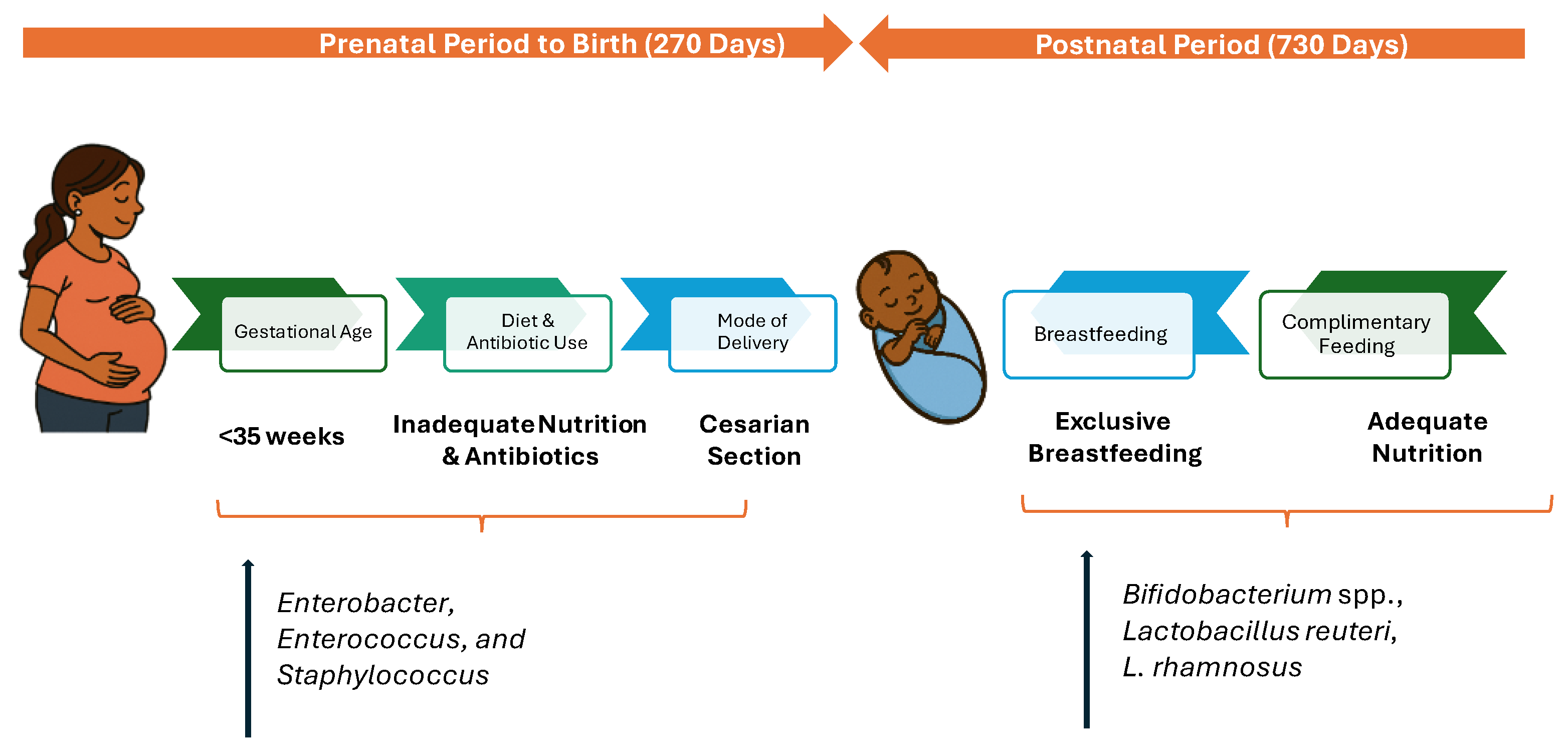
Figure 2.
Bidirectional Gut–Brain Communication Pathways.
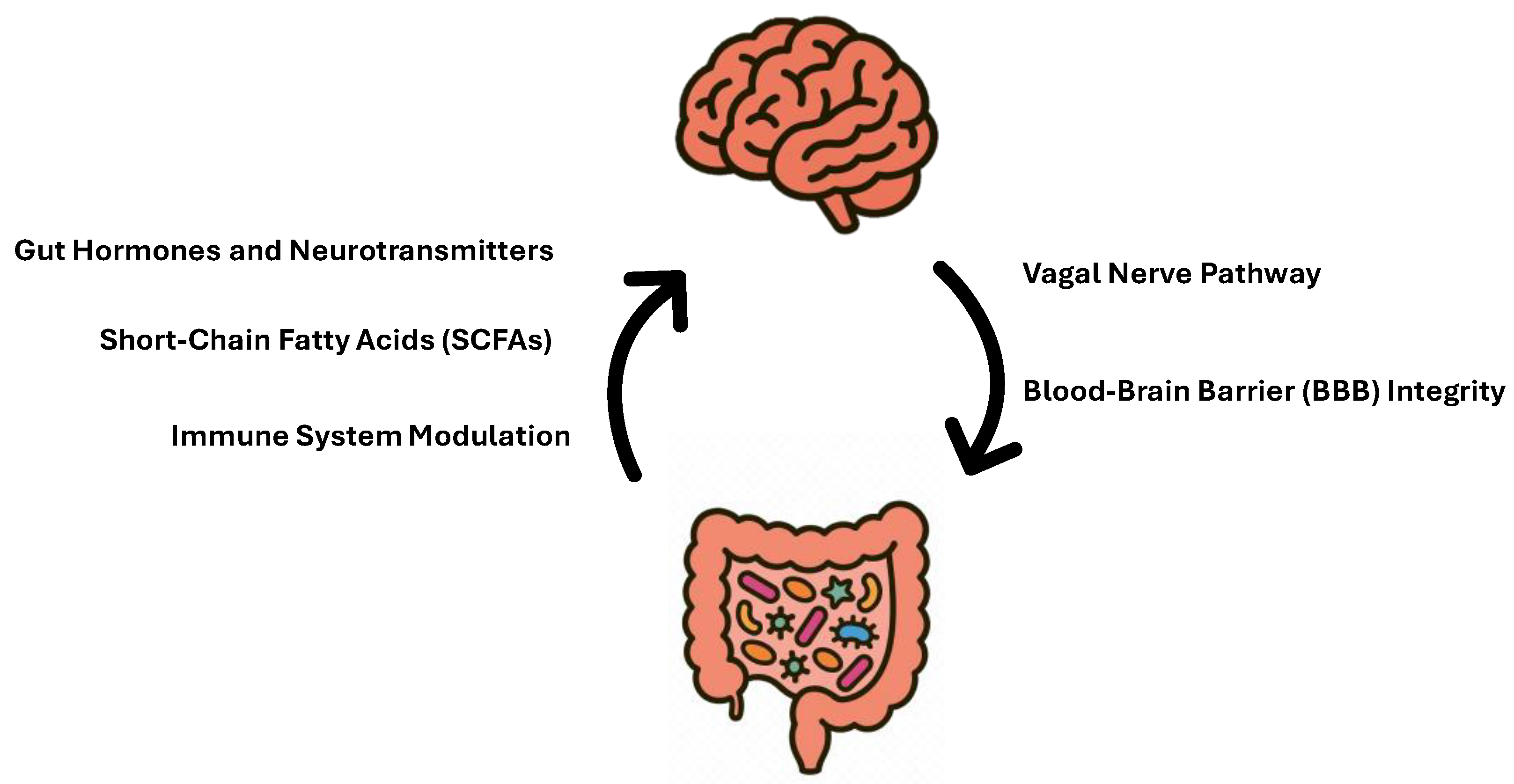
Figure 3.
Bidirectional Interplay Between Brain, Gut, and Sleep.
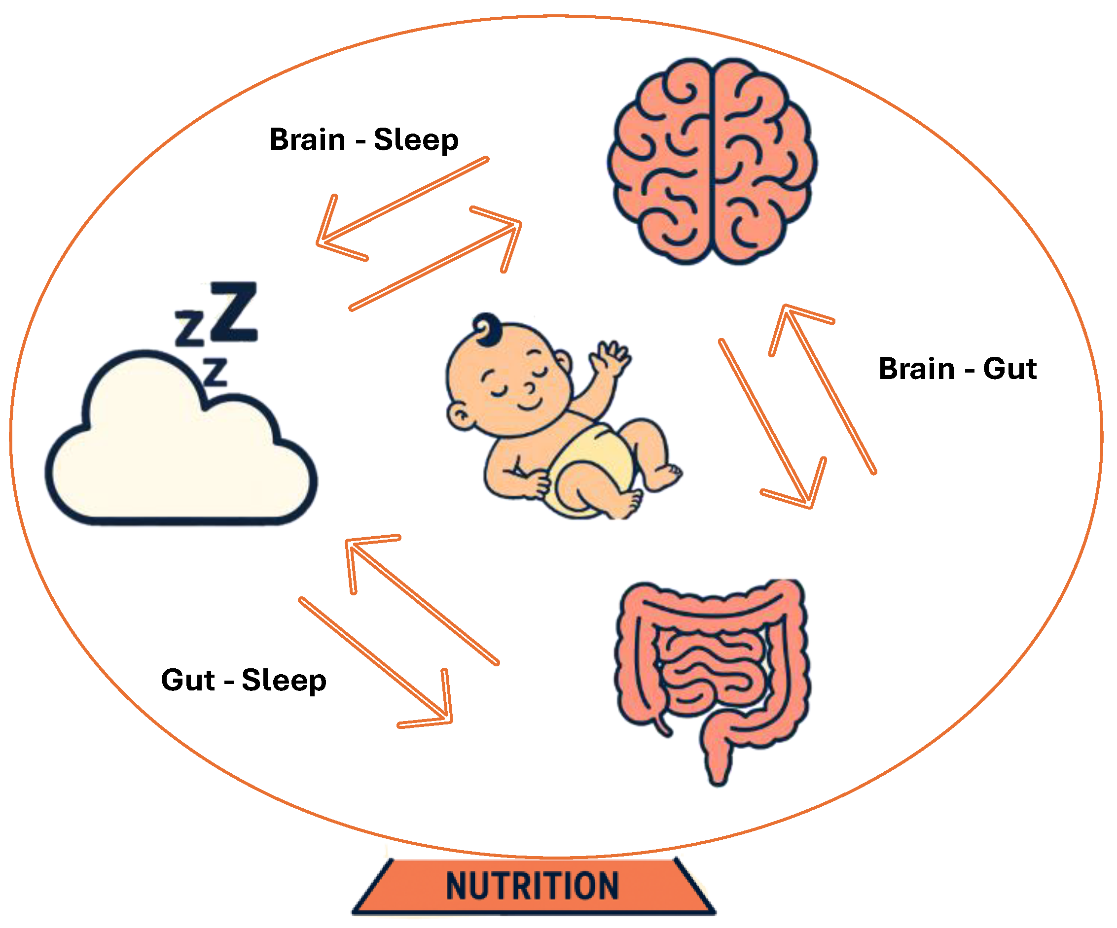

Table 1.
U.S. Guidelines on Nutrient Intake in the First 1,000 Days.
| Nutrients | Recommendations*/day | ||||
|---|---|---|---|---|---|
| Pregnancy & Lactation | Year 1 | Year 2 | |||
| 270 days | 365 days | 365 days | |||
| Pregnant Women | Breastfeeding Women | Breastfeeding Infants | Complementary Feeding | ||
| Omega-3 Fatty acids | 0-6 months | 6- 12 months | |||
| DHA+EPA (mg) | 250-375^ | 250-375^ | |||
| DHA alone (mg) | 200 -300 | 200 - 300 | Not Established | ||
| Choline (mg) | 450 b, c - 550 a | 550 a, b | Not Established | 150 a, b | 200 a, b |
| Folate (mcg DFE) | 600 a, b, c | 500 b - 600a | Not Established | 80 a,b | 150a, b |
| Iodine (mcg) | 220 b, c- 290 a | 290a, b | Not Established | 130a | 90 a |
| Vitamin B12 (mcg) | 2.6 b, c- 2.8 a | 2.8 a, b | Not Established | 0.5 a, b | 0.9 a, b |
| Iron (mg) | 27 a, b, c | 9 b-27 a | Not Established | 11 a, b | 7 a, b |
| Vitamin D (mcg) | 15 a,b, c | 15 a,b | 10 a,b | 10 a, b | 15 a, b |
a Food and Drug Administration (21 CFR Part 101). b Dietary Guidelines for Americans, 2020-2025. c American College of Obstetricians and Gynecologists. ^ Recommendation is made in terms of seafood (8 to 12 ounces (2–3 servings) of a variety of low-mercury seafood per week). * Recommendations are based on guidelines by bodies such as Food and Drug Administration (21 CFR Part 101), DGA, ACOG for healthy population only. Pregnant women with co-morbidities such as gestational diabetes, risk of preeclampsia, obesity and preterm infants and/or with health conditions are excluded.
Table 2.
Existing Gaps and Future Strategies for Supporting Outcomes in the First 1000 Days.
| Key Nutrients | Role | Evidence Strength | Research Gaps |
|---|---|---|---|
| Neurodevelopmental Outcomes | |||
| Omega-3 Fatty acids | DHA, the dominant omega-3 fatty acid in the developing brain, is vital for neuronal membrane structure and synaptic maturation. As the body cannot produce omega-3s, sufficient intake of DHA/EPA is essential for healthy brain development. | Strong evidence from RCTs and systematic reviews; heterogeneity exists for a dose dependent outcome | Absence of standardized, mandatory guidelines for omega-3 supplementation beyond 6 months of age. |
| Choline | Choline influences stem cell proliferation and apoptosis, thereby altering brain and spinal cord structure and function and influencing risk for NTDs and memory function. | Moderate evidence from observational studies; limited RCTs in humans. | Absence of reliable biomarkers to detect choline deficiency and limited number of randomized controlled trials (RCTs) specifically investigating the effects of choline supplementation alone on infant neurodevelopment. |
| Folate | Folate supports cell creation, DNA/RNA synthesis, and neurotransmitter formation, which are essential for the proper development of the nervous system and prevention of NTDs. | Strong evidence for folic acid in NTD prevention; limited evidence on other folate forms. | Absence of clinical studies directly linking different forms of folic acid such as 5-MTHF to NTD prevention |
| Iodine | Iodine is a necessary component of thyroid hormones, which are vital for the growth and maturation of the brain and central nervous system. | Strong evidence from population-level interventions; deficiency clearly linked to deficits. | Limited data on optimal supplementation levels in iodine-sufficient populations. |
| Vitamin B12 | Vitamin B12 supports DNA synthesis, myelination (the formation of protective nerve sheaths), and synaptogenesis (the creation of brain connections) | Moderate evidence: deficiency linked to adverse outcomes, but supplementation trials inconsistent. | Lack of long-term, well-designed studies to determine the efficacy and safety of vitamin B12 supplementation during pregnancy for both maternal health and fetal developmental outcomes - particularly on the various forms of B12. |
| Iron | Iron supports myelination, the synthesis of monoamine neurotransmitters, and provides energy for the rapidly growing brain. | Strong evidence that deficiency impairs cognition; supplementation helps but effect size varies. | Limited evidence on timing/dosing of supplementation in non-deficient populations. |
| Vitamin D | Research indicates that Vitamin D may contribute to preserving cognitive function through induction of neuroprotection, modulation of oxidative stress, regulation of calcium homeostasis, and inhibition of inflammatory processes | Emerging evidence for cognition; observational links strong. | Lack of long-term, dose dependent clinical trials focusing on neurodevelopmental outcomes. |
| Probiotics | Probiotics may support infant cognition through the gut-brain axis, particularly for preterm infants | Strong emerging evidence | Long-term trials in healthy infants |
| Sleep | |||
| Omega-3 Fatty acids | Omega-3 fatty acids have important roles in sleep regulation, with evidence suggesting that EPA and DHA influence serotonin activity and that DHA contributes to melatonin synthesis. | Emerging evidence especially in the first 1000 days | Long-term, well-designed trials for better sleep outcomes in mother-infants |
| Vitamin D | Vitamin D plays a multifaceted role in sleep, from influencing melatonin production to activating vitamin D receptors (VDRs) in brain regions that regulate the sleep–wake cycle. | Plausible mechanisms and some supportive studies. | High-quality, consistent RCT evidence showing clear, generalisable sleep improvements |
| Iron | Iron deficiency affects sleep by disrupting the brain’s ability to synthesize essential neurotransmitters like serotonin, dopamine, and norepinephrine, which regulate mood and sleep-wake cycles. | Strong emerging evidence | High-quality, consistent RCT evidence showing clear, generalisable sleep improvements |
| Probiotics | Probiotics may improve sleep quality by regulating the gut-brain axis through the production of neurotransmitters like serotonin and dopamine. | Strong emerging evidence | Long-term, well-designed trials for better sleep outcomes in mother-infants |
Disclaimer/Publisher’s Note: The statements, opinions and data contained in all publications are solely those of the individual author(s) and contributor(s) and not of MDPI and/or the editor(s). MDPI and/or the editor(s) disclaim responsibility for any injury to people or property resulting from any ideas, methods, instructions or products referred to in the content. |
© 2025 by the authors. Licensee MDPI, Basel, Switzerland. This article is an open access article distributed under the terms and conditions of the Creative Commons Attribution (CC BY) license (http://creativecommons.org/licenses/by/4.0/).
Copyright: This open access article is published under a Creative Commons CC BY 4.0 license, which permit the free download, distribution, and reuse, provided that the author and preprint are cited in any reuse.


